Microtubules are dynamic polymers which play a central role in a number of cellular processes, most particularly cell division, as they are key constituents of the mitotic spindle [1]. Their shape can be described as hollow tubes of about 25 nm external diameter constituted of a protein named tubulin. The functional form of this protein is a heterodimer formed in turn through non-covalent binding of two monomeric con-stituents. These are two structurally related polypeptides of about 450 amino acid residues which are called - and -tubulin [2]. For cell division to occur in a normal way, mi-crotubules must be in a constant state of formation and dis-ruption, a process named microtubule dynamics in which GTP hydrolysis into GDP plays a key role [3].
It is easy to understand that any molecule which exerts some type of action on microtubule dynamics will be able to influence the cell division process, not only of normal cells but also of tumoral ones. Since such an influence may be exerted by molecules that bind to any of the tubulin compo-nents, it is not surprising that tubulin-binding molecules (TBMs) constitute a very important class of anticancer agents [4]. TBMs are able to interfere with microtubule as-sembly and functions, either by causing disruption of the microtubules or else through their stabilization. In both cases, this results in mitotic arrest of eukaryotic cells and subsequent cell death, likely by an apoptosis-like process called mitotic catastrophe [5]. Most of the hitherto described active drugs are natural products or derivatives thereof [6].
*Address correspondence to this author at the Depart. de Q. Orgánica, Univ.
Major drugs can already be found on the market and many other promising compounds are in clinical trials [4, 6].
TBMs may be divided in two broad categories, those that bind to -tubulin and those that bind to -tubulin. The latter group is presently by far the most numerous and contains products which cause either disruption or stabilization of microtubules. Among the drugs that belong to this group, the well-known colchicine [7] exerts its effects by causing dis-ruption of microtubules. In contrast, another renowned repre-sentative of the same group, paclitaxel, was the first-described tubulin-interacting drug with the ability to stabilize microtubules [8]. Despite the fact that they exert opposite effects on the mitotic spindle, both drugs are known to bind to -tubulin, even though to different sites within that protein subunit. The mechanisms of action [9] of many of these TBMs and the molecular aspects [10] of their interactions with tubulin have been studied using a broad palette of methods [11].
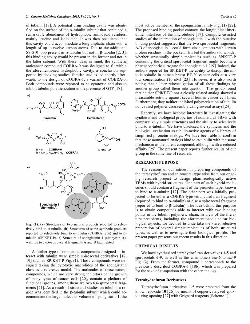
The number of products reported to bind to -tubulin is very small, the naturally occurring 5,6-dihydro- -pyrone pironetin Fig. (1) being the first example [12], followed a short time later by the peptide-like hemiasterlin family [13]. Pironetin proved a potent inhibitor of tubulin assembly and was found to arrest cell cycle progression in the G2/M phase [14]. This feature has motivated a number of groups to un-dertake total syntheses of this natural compound [15]. Some synthetic and biological studies on modified variants of pi-ronetin have previously been published [16].
The development of lipophilic tetrahydrofuran deriva-tives such as the two compounds named with the acronyms COBRA-0 or COBRA-1 Fig. (1) was the result of computer-assisted studies based on previously published structural data
2 Current Medicinal Chemistry, 2013, Vol. 20, No. 1 Carda et al.
of tubulin [17]. A potential drug binding cavity was identi-fied on the surface of the -tubulin subunit that contained a remarkable abundance of hydrophobic aminoacid residues, mainly leucine and isoleucine. It was then postulated that this cavity could accommodate a long aliphatic chain with a length of up to twelve carbon atoms. Due to the additional S9-S10 loop present in -tubulin but not in -tubulin [2, 3], this binding cavity would be present in the former and not in the latter subunit. With these ideas in mind, the synthetic anticancer compound COBRA-0 was designed to fit within the aforementioned hydrophobic cavity, a conclusion sup-ported by docking studies. Similar studies led shortly after-wards to the design of COBRA-1, a variant of COBRA-0. Both compounds were reported to be cytotoxic and also to inhibit tubulin polymerization in the presence of GTP [18].
Fig. (1). (a) Structures of two natural products reported to selec-
tively bind to -tubulin. (b) Structures of some synthetic products
reported to selectively bind to -tubulin (COBRA type) and to -
tubulin (SPIKET-P). c) Structure of spongistatin 1 (altohyrtin A),
with the two 6,6-spiroacetal fragments A and B highlighted.
A further type of nonnatural compounds designed to in-teract with tubulin were simple spiroacetal derivatives [17, 19] such as SPIKET-P Fig. (1). These compounds were de-signed taking the cytotoxic macrolides of the spongistatin class as a reference model. The molecules of these natural compounds, which are very strong inhibitors of the growth of many types of cancer cells [20], contain a plethora of functional groups, among them are two 6,6-spiroacetal frag-ments [21]. As a result of structural studies on tubulin, a re-gion was identified in the -tubulin subunit which could ac-commodate the large molecular volume of spongistatin 1, the
most active member of the spongistatin family Fig. (1) [22]. The proposed binding pocket contacts the longitudinal inter-dimer interface of the microtubule [17]. Computer-assisted studies of the interaction of spongistatin 1 with the putative binding pocket suggested that the two spiroacetal fragments A/B of spongistatin 1 could form close contacts with certain protein residues in the pocket. This led the authors to wonder whether structurally simple molecules such as SPIKET-P containing the critical spiroacetal fragment might become a pharmacophoric surrogate for spongistatin 1 [19]. Indeed, the authors reported for SPIKET-P the ability to disrupt the mi-totic spindle in human breast BT-20 cancer cells at a very low concentration (10 nM) [23]. However, it is also worth noting that a later reinvestigation of all these findings by another group called them into question. This group found that neither SPIKET-P nor a closely related analog showed a measurable activity against several human cancer cell lines. Furthermore, they neither inhibited polymerization of tubulin nor caused polymer disassembly using several assays [24].
Recently, we have become interested in investigating the synthesis and biological properties of nonnatural TBMs with comparatively simple structures and the ability to selectively bind to -tubulin. We have disclosed the synthesis and the biological evaluation as tubulin-active agents of a library of simplified pironetin analogs. We have been able to confirm that these nonnatural analogs bind to -tubulin with the same mechanism as the parent compound, although with a reduced affinity [25]. The present paper reports further results of our group in the same line of research.
RESEARCH PURPOSE
The reasons of our interest in preparing compounds of the tetrahydrofuran and spiroacetal type arise from our ongo-ing research project to design pharmacologically active TBMs with hybrid structures. One part of such hybrid mole-cules should contain a fragment of the pironetin type, known to bind to -tubulin [12]. The other part was initially pro-jected to be either a COBRA-type tetrahydrofuran fragment (reported to bind to -tubulin) or else a spiroacetal fragment (reported to bind to -tubulin). The idea behind this purpose is to obtain compounds able to interact with two binding points in the tubulin polymeric chain. In view of the litera-ture precedents, including the aforementioned unclear bio-logical aspects, we decided to undertake the stereoselective preparation of several simple molecules of both structural types, as well as to investigate their biological profile. The present paper presents our recent results in this direction.
CHEMICAL RESULTS
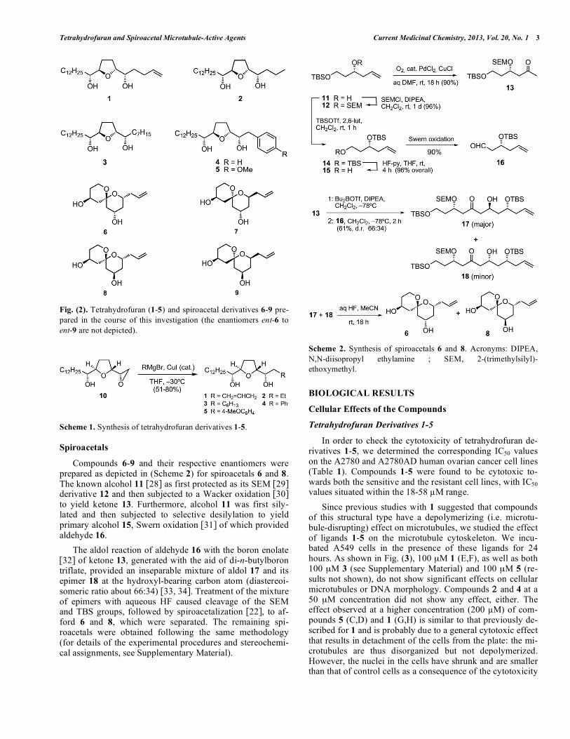
We have synthesized tetrahydrofuran derivatives 1-5 and spiroacetals 6-9, as well as the enantiomers ent-6 to ent-9 Fig. (2). From the former, compound 1 corresponds to the previously described COBRA-1 [18b], which was prepared for the sake of comparison with the other analogs.
Tetrahydrofuran Derivatives
Tetrahydrofuran derivatives 1-5 were prepared from the known epoxide 10 [26] by means of copper-catalyzed epox-ide ring opening [27] with Grignard reagents (Scheme 1).
Tetrahydrofuran and Spiroacetal Microtubule-Active Agents Current Medicinal Chemistry, 2013, Vol. 20, No. 1 3
Fig. (2). Tetrahydrofuran (1-5) and spiroacetal derivatives 6-9 pre-
pared in the course of this investigation (the enantiomers ent-6 to
ent-9 are not depicted).
Scheme 1. Synthesis of tetrahydrofuran derivatives 1-5.
Spiroacetals
Compounds 6-9 and their respective enantiomers were prepared as depicted in (Scheme 2) for spiroacetals 6 and 8. The known alcohol 11 [28] as first protected as its SEM [29] derivative 12 and then subjected to a Wacker oxidation [30] to yield ketone 13. Furthermore, alcohol 11 was first sily-lated and then subjected to selective desilylation to yield primary alcohol 15, Swern oxidation [31] of which provided aldehyde 16.
The aldol reaction of aldehyde 16 with the boron enolate [32] of ketone 13, generated with the aid of di-n-butylboron triflate, provided an inseparable mixture of aldol 17 and its epimer 18 at the hydroxyl-bearing carbon atom (diastereoi-someric ratio about 66:34) [33, 34]. Treatment of the mixture of epimers with aqueous HF caused cleavage of the SEM and TBS groups, followed by spiroacetalization [22], to af-ford 6 and 8, which were separated. The remaining spi-roacetals were obtained following the same methodology (for details of the experimental procedures and stereochemi-cal assignments, see Supplementary Material).
Scheme 2. Synthesis of spiroacetals 6 and 8. Acronyms: DIPEA,
N,N-diisopropyl ethylamine ; SEM, 2-(trimethylsilyl)-
ethoxymethyl.
BIOLOGICAL RESULTS
Cellular Effects of the Compounds
Tetrahydrofuran Derivatives 1-5
In order to check the cytotoxicity of tetrahydrofuran de-rivatives 1-5, we determined the corresponding IC50 values on the A2780 and A2780AD human ovarian cancer cell lines (Table 1). Compounds 1-5 were found to be cytotoxic to-wards both the sensitive and the resistant cell lines, with IC50 values situated within the 18-58 M range.
Since previous studies with 1 suggested that compounds of this structural type have a depolymerizing (i.e. microtu-bule-disrupting) effect on microtubules, we studied the effect of ligands 1-5 on the microtubule cytoskeleton. We incu-bated A549 cells in the presence of these ligands for 24 hours. As shown in Fig. (3), 100 μM 1 (E,F), as well as both 100 μM 3 (see Supplementary Material) and 100 μM 5 (re-sults not shown), do not show significant effects on cellular microtubules or DNA morphology. Compounds 2 and 4 at a 50 μM concentration did not show any effect, either. The effect observed at a higher concentration (200 M) of com-pounds 5 (C,D) and 1 (G,H) is similar to that previously de-scribed for 1 and is probably due to a general cytotoxic effect that results in detachment of the cells from the plate: the mi-crotubules are thus disorganized but not depolymerized. However, the nuclei in the cells have shrunk and are smaller than that of control cells as a consequence of the cytotoxicity
4 Current Medicinal Chemistry, 2013, Vol. 20, No. 1 Carda et al.
Table 1. Effect of Tetrahydrofuran Derivatives 1-5 on the Growth of A2780 and A2780AD (MDR Overexpressing P-glycoprotein)
Ovarian Carcinomas
Ligand A2780 (μM)a
A2780AD (μM)a
R/Sb
Paclitaxel 0.002 ± 0.0003 1.3 ± 0.028 650
1 28.8 ± 2.2 58.3 ± 11.5 2
2 28.6 ± 1.45 29.4 ± 1.14 1.02
3 42.3 ± 11 52 ± 6.5 1.23
4 18.5 ± 0.067 31.3 ± 1.03 1.7
5 20 ± 0.32 31.7 ± 0.208 1.58
a Values mean IC50 as the mean ± standard error of three independent experiments. b Resistance index (the relative resistance of A2780AD cell line, obtained dividing the IC50 of the resistant cell line by that of the parental A2780 cell line).
Fig. (3). Effect of the treatment with tetrahydrofuran derivatives 1 and 5 on the cytoplasmic microtubule network (A, C, E, G) and DNA (B,
D, F, H) of A549 lung carcinoma cells. The cells were incubated with DMSO (A,B), 200 M 5 (C,D), 100 M 1 (E,F), and 200 M 1 (G,H).
For the other tetrahydrofuran derivatives, see Supplementary Material. Microtubules were immunostained with -tubulin monoclonal antibod-
ies; DNA was stained with Hoechst 33342. Scale bar = 10 m.
of these compounds. This effect was also observed with 100 μM 2 and 4 (see Supplementary Material).
We also studied the effect of these compounds on the cell cycle of A549 cells (see Table 5). Even concentrations of 100 M or higher of 1-5 did not alter the cell distribution in a significant way as compared with the control cells (Table 5 only depicts results obtained with 1 and 3 but similar results were observed for 2, 4 and 5).
These observations were confirmed by the absence of in-hibition of purified tubulin microtubule assembly. The criti-cal concentration of tubulin required for assembly was de-termined in GAB in the presence of a large excess (100 μM) of compounds 1-5 (Table 2). As shown in the Table, the con-centration of tubulin required to produce assembly (critical concentration [35]) oscillate between 3.0 and 3.5 μM in the presence of these compounds, similarly as in their absence (3.3 μM). The observed in vitro effect correlates well with the immunofluorescence results shown in Fig. (3).
Table 2. Critical Concentration Values of Tubulin for
Ligand-induced Microtubule Assembly Induced by
Tetrahydrofuran Derivatives 1-5 (Ligand Concen-
trations are 25 μM for Docetaxel and 100 μM for the
Ligands)
Ligand Cr (μM)a
Control 3.3 ± 0.5
Docetaxel 1.1 ± 0.3
1 3.4 ± 0.2
2 3.5 ± 0.1
3 3.0 ± 0.3
4 3.2 ± 0.4
5 3.3 ± 0.2
a Cr values are the mean ± standard error of three independent experiments.
Tetrahydrofuran and Spiroacetal Microtubule-Active Agents Current Medicinal Chemistry, 2013, Vol. 20, No. 1 5
Table 3. Effect of Spiroketals 6-9 and ent-6/ent-9 on the Growth of A2780 and A2780AD (MDR Overexpressing P-glycoprotein)
Ovarian Carcinomas
Ligand A2780 (μM)a
A2780AD (μM)a
R/Sb
Taxol 0.002 ± 0.0003 1.3 ± 0.028 650
6 > 300c > 300c
ent-6 > 300c > 300c
7 42 ± 2.8 100
ent-7 95 ± 3.5 100
8 21.15 ± 0.9 84.7 ± 0.2 4
ent-8 > 100c > 100c
9 > 300c > 300c
ent-9 40.95 ± 0.7 44 ± 4.2 1.07
a Values mean IC50 as the mean ± standard error of three independent experiments. b Resistance index (the relative resistance of A2780AD cell line, obtained dividing the IC50 of the resistant cell line by that of the parental A2780 cell line). c Not determined.
Fig. (4). Effect of the treatment with spiroacetals on the cytoplasmic microtubule network (A, C, E, G) and on DNA (B, D, F, H) of A549
lung carcinoma cells. The cells were incubated with DMSO (A,B), 300 M 6 (C,D), 100 M 8 (E,F) and 100 M 7 (G,H). For the other spi-
roacetals, see Supplementary Material. Microtubules were immunostained with -tubulin monoclonal antibodies; DNA was stained with
Hoechst 33342. Insets are mitotic spindles from the same preparation. Scale bar = 10 m.
Spiroacetals 6-9 and their Enantiomers
As with compounds 1-5, we have determined the IC50 values for spiroacetals 6-9 and their enantiomers ent-6/ent-9 on the A2780 and A2780AD human ovarian cancer cell lines (Table 3). According to the results shown in the table, com-pounds 7, ent-7, 8 and ent-9 were found to be cytotoxic for the A2780 cancer cell line (IC50 < 100 μM), with 8 and ent-9 being also cytotoxic for the A2780AD cell line. The remain-ing spiroacetals were not cytotoxic at the concentrations tested.
When we studied the effect of the spiroacetals on the mi-crotubule cytoskeleton of A549 cells, we found that concen-
trations of compounds ent-6, ent-7, ent-8, 9 and ent-9 as high as 100-300 μM have no effect on either the microtubule net-work or the DNA of these cells (see Fig. 2 in the Supplemen-tary Material). Fig. (4) shows the results observed with ligands 6-8. Interestingly, it has been found that 100 μM 8 (E,F) and 100 μM 7 (G,H) disorganize the microtubule net-work and induce cytoplasmic microtubule bundles, mitotic asters and micronucleated cells, all in the same way as mi-crotubule-stabilizing agents do.
We studied as well the effect of these spiroacetals on the cell cycle of A549 cells. As a matter of fact, compounds 6, ent-6, ent-7, ent-8, 9 and ent-9 at concentrations up to 100
M did not alter the cell cycle of these cells, the cell distri-
6 Current Medicinal Chemistry, 2013, Vol. 20, No. 1 Carda et al.
bution being similar to that of control cells (results not shown). However, ligands 7 and 8 caused a substantial ac-cumulation of cells in the G2/M phase of the cell cycle (> 40% as compared with ca. 26% for the control), as well as the appearance of a sub G1 peak of presumably apoptotic cells (Table 5). In any case, ligands 6-9 and their enanti-omers show a measurable depolymerizing effect on the in vitro assembly of microtubules. The critical tubulin concen-tration [35] required for assembly was determined in GAB in the presence of a large excess (100 μM) of the compounds. As shown in (Table 4), the concentration of tubulin required to produce assembly increases substantially in the presence of compounds 7, 8 and ent-8, whereas 6, ent-6, ent-7, 9 and ent-9 cause only a moderate increase in the critical concen-tration observed. Ligands 7, ent-8 and particularly 8 have a marked microtubule depolymerizing effect, with an increase of about 60-130% in the critical concentration being required for assembly.
Table 4. Critical Concentration Values of Tubulin for
Ligand-induced Microtubule Assembly Induced by
Spiroketals 6-9 and ent-6/ent-9 (Ligand Concentra-
tions are 25 μM for Docetaxel and 100 μM for the
Ligands)
Ligand Cr (μM)a
Control 3.3 ± 0.39
Docetaxel 1.1 ± 0.2
6 3.5 ± 0.6
ent-6 4.1 ± 0.6
7 5.4±1.0
ent-7 4.3 ± 0.3
8 7.7 ± 0.1
ent-8 6.3 ± 0.8
9 3.8 ± 0.8
ent-9 4.0 ± 0.7
a Cr values are the mean ± standard error of three independent experiments.
DISCUSSION
The results obtained indicate that the previously reported cytotoxic effects of COBRA-0 [17] and COBRA-1 (= com-pound 1) are confirmed in the case of 1. However, and in contrast with the conclusions of the previous report, these effects are not due to interactions with the microtubule net-work. First of all, no accumulation of cells in the G2/M phase, typical of compounds which block chromatide segre-gation by affecting microtubule dynamics of the cell cycle, is observed here. Secondly, they produce no visible effect in the microtubule network. Indeed, the micrographs shown in Fig. (3) are very similar to those previously published [18] and indicate that, as commented above, the cellular effect observed is due to a general cytotoxic effect which results in detachment of the cells from the plate, i.e., the microtubules are disorganized but not depolymerized. These findings have
led us to discard these compounds for our projected prepara-tion of hybrids.
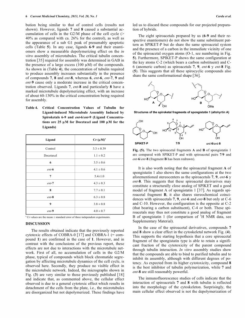
The eight spiroacetals prepared by us (6-9 and their re-spective enantiomers) do not show the same substituent pat-tern as SPIKET-P but do share the same spiroacetal system and the presence of a carbon in the immediate vicinity of one of the spiroacetal oxygen atoms (O-1, see numbering in Fig. 5). Furthermore, SPIKET-P shows the same configuration at the key atoms C-2 (which bears a carbon substituent) and C-6 (anomeric carbon) as spiroacetals 7, 9, ent-6 y ent-8 Fig. (5). This suggests that all these spirocyclic compounds also share the same conformational shape [36].
Fig. (5). The two spiroacetal fragments A and B of spongistatin 1
are compared with SPIKET-P and with spiroacetal pairs 7/9 and
ent-6/ent-8 (fragment B has been redrawn).
It is also worth noting that the spiroacetal fragment A of spongistatin 1 also shows the same configurations at the two aforementioned stereocenters as the spiroacetals 7, 9, ent-6 y ent-8. This suggests that these spiroacetal derivarives may constitute a structurally close analog of SPIKET and a good model of fragment A of spongistatin 1 [37]. As regards spi-roacetal fragment B, it also shares stereochemical coinci-dences with spiroacetals 7, 9, ent-6 and ent-8 but only at C-6 and C-10. However, the configuration is the opposite at C-2 (that bearing a carbon substituent), C-4 or both. These spi-roacetals may thus not constitute a good analog of fragment B of spongistatin 1 (for comparison of
1H NMR data, see
Supplementary Material).
In the case of the spiroacetal derivatives, compounds 7 and 8 show a clear effect in the cytoskeletal network Fig. (4). This supports the starting hypothesis that a core spiroacetal fragment of the spongistatin type is able to retain a signifi-cant fraction of the cytotoxicity of the parent compound through tubulin interaction. In vitro assembly studies show that the compounds are able to bind to purified tubulin and to inhibit its assembly, although with different degrees of po-tency. As expected from its higher cytotoxicity, compound 8 is the best inhibitor of tubulin polymerization, while 7 and ent-8 are still reasonably powerful.
The inmunofluorescence studies of cells indicate that the interaction of spiroacetals 7 and 8 with tubulin is reflected into the morphology of the cytoskeleton. Surprisingly, the main cellular effect observed is not the depolymerization of
Tetrahydrofuran and Spiroacetal Microtubule-Active Agents Current Medicinal Chemistry, 2013, Vol. 20, No. 1 7
the cytoskeleton but a bundling effect: the microtubular net-work appears disorganized and the microtubules are forming bundles. This is a typical effect of microtubule-stabilizing agents and suggests that, either the compounds are modified inside the cell and/or, at the intracellular concentrations and perhaps in the presence of other cellular proteins, the interac-tions of the two compounds with tubulin produce additional effects not observed in experiments in vitro.
In summary, we have shown that, in contrast to SPIKET-P [24], several similar spiroacetals display cytotoxicity, show interactions with tubulin and have a measurable effect on the assembly of microtubules. However, the precise mechanism of action and the possible binding sites of these spiroacetals still remain to be established.
MATERIALS AND METHODS
Chemistry. General Procedures
The general reaction conditions and the physical and spectral data of all synthetic intermediates and final com-pounds are described in detail as Supplementary Material. The samples of all compounds used for the biological studies were purified to >95% by means of preparative HPLC.
Cell Culture
Human A549 non small lung carcinoma cells were cul-tured in RPMI 1640 supplemented with 10% FCS, glu-tamine, and antibiotics as previously described [38]. Human ovarian carcinomas A2780 and A2780AD (MDR overex-
pressing P-glycoprotein) were cultured as above with the addition of 0.25 units/mL of bovine insulin.
Cytotoxicity Assays, Indirect Immunofluorescence and Cell Cycle
Cytotoxic evaluation was performed with A2780 and A2780AD cells with the MTT assay modified as previously described [39]. Indirect immunofluorescence was performed in A549 cells that had been cultured overnight in 12 mm round coverslips and incubated a further 24 hours in the ab-sence (drug vehicle DMSO) or in the presence of different ligand concentrations. Attached cells were permeabilized with Triton X100 and fixed with 3.7% formaldehyde. Micro-tubules were specifically stained with DM1A -tubulin monoclonal antibodies and DNA with Hoechst 33342 as previously described [40]. The preparations were examined using a Zeiss axioplan epifluorescence microscope and the images were recorded in a Hamamatsu 4742-95 cooled CCD camera.
Tubulin Assembly Inhibition Assay
The effect of the compounds in the assembly of purified tubulin was determined by incubating 20 μM purified tubulin at 37 ºC for 30 minutes in GAB (glycerol assembling buffer, 3.4 M glycerol, 10 mM sodium phospate, 1 mM EGTA, 1 mM GTP, 6 mM MgCl2 at pH 6.5) in the presence of 25 μM docetaxel, 100 μM of one of the analogs PTA-PF or JPP or 2 μL DMSO (vehicle). The samples were processed and the
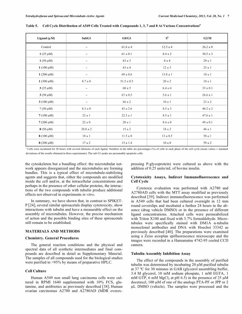
Table 5. Cell Cycle Distribution of A549 Cells Treated with Compounds 1, 3, 7 and 8 At Various Concentrationsa
Ligand (μM) SubG1
G0/G1
Sb
G2/M
Control 61.6 ± 4 12.5 ± 4 26.2 ± 0
1 (25 μM) 61 ± 0.1 8.4 ± 2 30.5 ± 3
1 (50 μM) 63 ± 3 8 ± 4 29 ± 1
1 (100 μM) 63 ± 4 12 ± 3 25 ± 1
1 (200 μM) 69 ± 0.6 13.8 ± 1 18 ± 1
1 (300 μM) 8.7 ± 0 51.5 ± 0.5 20 ± 2 18 ± 1
3 (25 μM) 60 ± 5 6.4 ± 4 33 ± 0.1
3 (50 μM) 67 ± 0.5 5.6 ± 1 26.6 ± 1
3 (100 μM) 66 ± 2 10 ± 1 23 ± 2
7 (50 μM) 8.3 ± 0 43 ± 2.6 8.5 ± 3 40.2 ± 2
7 (100 μM) 22 ± 1 22.3 ± 1 8.5 ± 1 47.6 ± 1
7 (200 μM) 22 ± 0 20 ± 1 8.4 ± 0 49 ± 0.1
8 (50 μM) 20.8 ± 2 15 ± 2 18 ± 2 46 ± 1
8 (100 μM) 18 ± 1 11.5 ± 0 13 ± 0.5 58 ± 1
8 (200 μM) 17 ± 2 13 ± 1.4 10 ± 0 59 ± 2
a Cells were incubated for 20 hours with several dilutions of each ligand. Numbers in the table are percentages (%) of cells in each phase of the cell cycle (mean values ± standard
deviations of the results obtained in three experiments). The sub G1 peaks are presumably apoptotic cells.
8 Current Medicinal Chemistry, 2013, Vol. 20, No. 1 Carda et al.
critical concentration for tubulin assembly [35] in the pres-ence of the ligands calculated as described [41].
CONFLICT OF INTEREST
The author(s) confirm that this article content has no con-flicts of interest.
ACKNOWLEDGEMENTS
Financial support has been granted by the Spanish Minis-try of Education and Science (CTQ2008-02800), by the Con-sellería d´Empresa, Universitat i Ciencia de la Generalitat Valenciana (ACOMP09/113) and by the BANCAJA-UJI Foundation (P1-1B2002-06 and P1-1B-2008-14). The bio-logical work has been supported in part by grants from the Spanish Ministry of Education and Science (BIO2007-61336) and from the Comunidad de Madrid (grants S2010/ BMD-2457 and BIPEDD2-CM) (both to J.F.D.). We further thank the Matadero Municipal Vicente de Lucas in Segovia for providing the calf brains which were the source of tubu-lin.
SUPPLEMENTARY MATERIALS
Supplementary material is available on the publisher’s web site along with the published article.
REFERENCES
[1] Fojo, T., Ed. The Role of Microtubules in Cell Biology, Neurobiol-
ogy and Oncology; Humana Press: Totowa, New Jersey, 2008.
[2] (a) Amos, L. A. Microtubule structure and its stabilization. Org.
Biomol. Chem., 2004, 2, 2153-2160. (b) Wade, R. H. On and
around microtubules. Mol. Biotechnol., 2009, 43, 177-191.
[3] (a) Nogales, E.; Wang, H.-W. Structural intermediates in microtu-
bule assembly and disassembly: how and why?. Curr. Opin. Cell