41
ESPECTROSCOPIA DE FLUORESCENCIA PARTE II PROTEINAS -FRET
ESPECTROSCOPIA DE FLUORESCENCIA
PARTE II
PROTEINAS -FRET

R. Meyer (1897) used the term “fluorophore” to describe chemical groups which tended to be associated with fluorescence; this word was analogous to “chromophore” which was first used in 1876 by O.N. Witt to describe groups associated with color.
Adolph Von Beyer (1871) a German chemist, synthesized Spiro[isobenzofuran-1(3H),9'-[9H]xanthen]-3-one, 3',6'-dihydroxy.
Gregorio Weber (1952) synthesized dansyl chloride for attachment to proteins and used polarization to study protein hydrodynamics - these studies initiated the field of quantitative biological fluorescence.
FLUORESCEIN!!!
Shimomura, Johnson and Saiga (1962) discovered Green Fluorescent Protein in the Aequorea jellyfish
Sir George Stokes (1852) created the term “Fluorescence”.
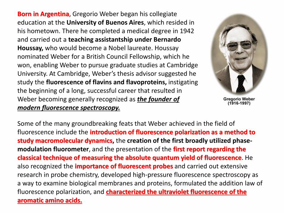
Born in Argentina, Gregorio Weber began his collegiate education at the University of Buenos Aires, which resided in his hometown. There he completed a medical degree in 1942 and carried out a teaching assistantship under Bernardo Houssay, who would become a Nobel laureate. Houssay nominated Weber for a British Council Fellowship, which he won, enabling Weber to pursue graduate studies at Cambridge University. At Cambridge, Weber’s thesis advisor suggested he study the fluorescence of flavins and flavoproteins, instigating the beginning of a long, successful career that resulted in Weber becoming generally recognized as the founder of modern fluorescence spectroscopy.
Some of the many groundbreaking feats that Weber achieved in the field of fluorescence include the introduction of fluorescence polarization as a method to study macromolecular dynamics, the creation of the first broadly utilized phase-modulation fluorometer, and the presentation of the first report regarding the classical technique of measuring the absolute quantum yield of fluorescence. He also recognized the importance of fluorescent probes and carried out extensive research in probe chemistry, developed high-pressure fluorescence spectroscopy as a way to examine biological membranes and proteins, formulated the addition law of fluorescence polarization, and characterized the ultraviolet fluorescence of the aromatic amino acids.
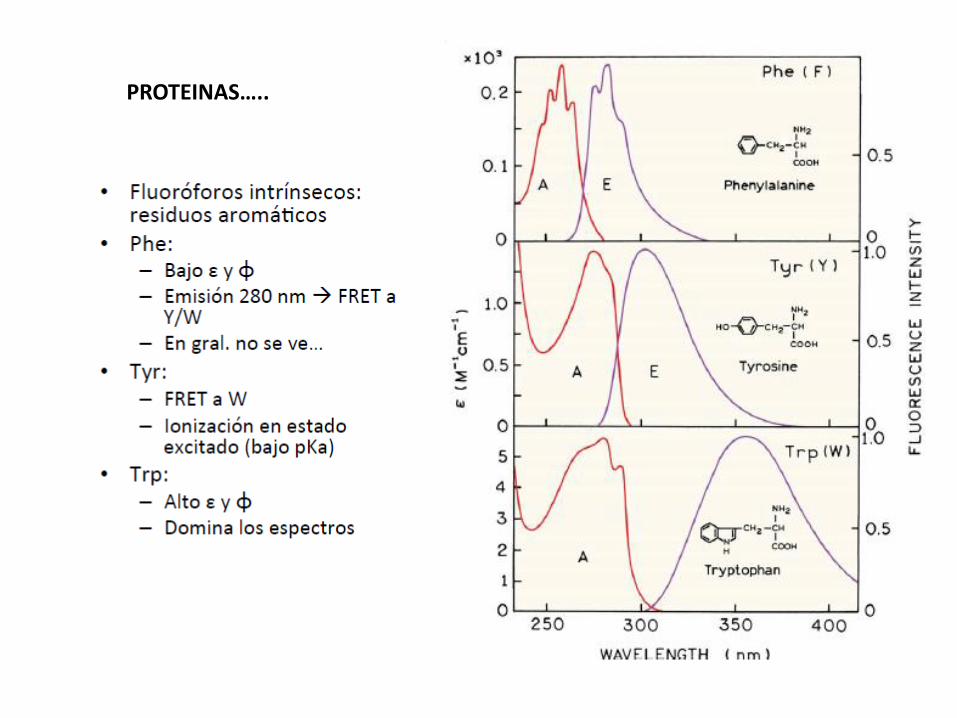
PROTEINAS…..
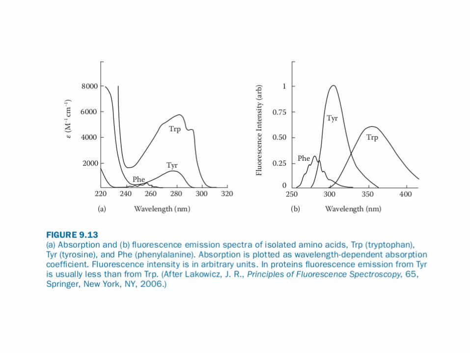
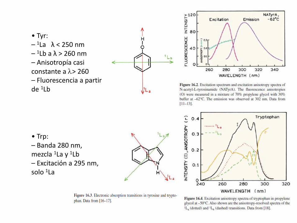
• Tyr:– 1La λ < 250 nm– 1Lb a λ > 260 nm– Anisotropía casi constante a l> 260– Fluorescencia a partir de 1Lb
• Trp:– Banda 280 nm,mezcla 1La y 1Lb– Excitación a 295 nm,solo 1La
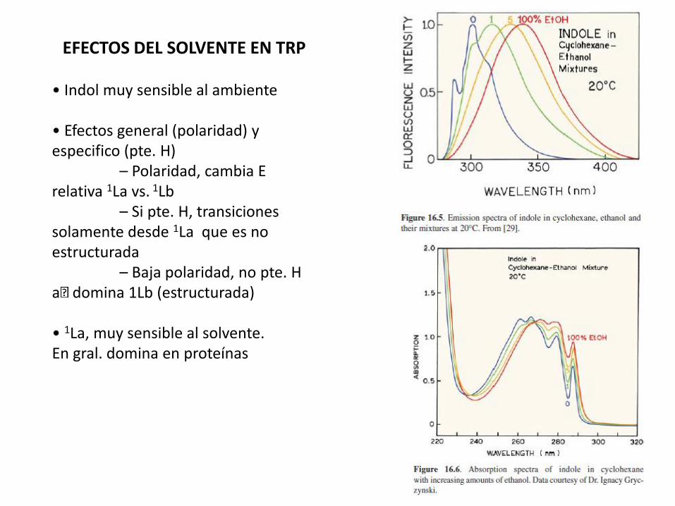
EFECTOS DEL SOLVENTE EN TRP
• Indol muy sensible al ambiente
• Efectos general (polaridad) y especifico (pte. H)
– Polaridad, cambia E relativa 1La vs. 1Lb
– Si pte. H, transiciones solamente desde 1La que es no estructurada
– Baja polaridad, no pte. Ha domina 1Lb (estructurada)
• 1La, muy sensible al solvente.En gral. domina en proteínas

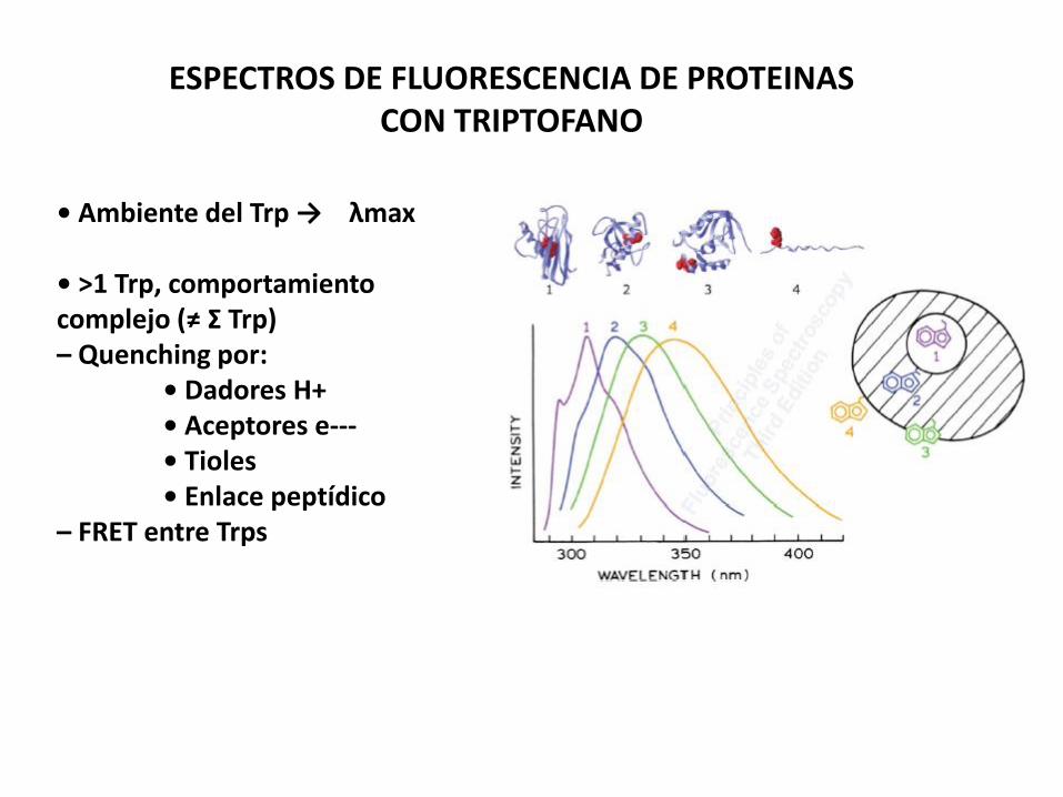
ESPECTROS DE FLUORESCENCIA DE PROTEINASCON TRIPTOFANO
• Ambiente del Trp → λmax
• >1 Trp, comportamiento complejo (≠ Σ Trp)– Quenching por:
• Dadores H+• Aceptores e--‐• Tioles• Enlace peptídico
– FRET entre Trps

AZURINA DE P. fluorescens
Dada la complejidad de múltiples trp, hay muchos estudios en proteínas con un solo Trp.Trp en un entorno hidrofóbico.No hay muchos casos en donde se veaun espectro casi similar al indol en ciclohexano.Emisión desde 1Lb
Excitación a distintas long. De onda (275 y 292 nm) combinado con agentes desnaturalizantes
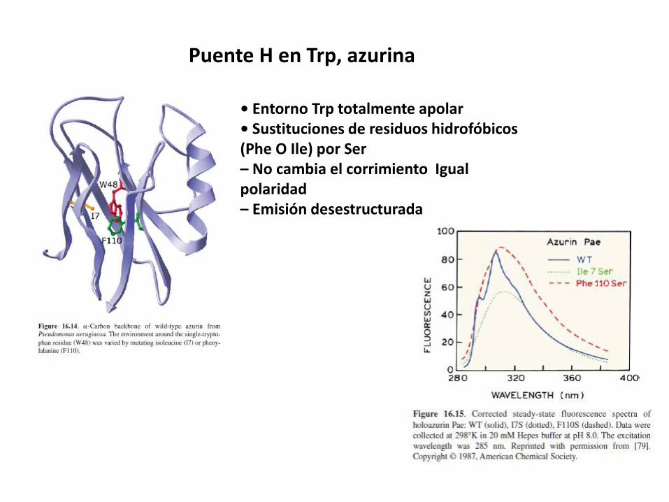
Puente H en Trp, azurina
• Entorno Trp totalmente apolar• Sustituciones de residuos hidrofóbicos(Phe O Ile) por Ser– No cambia el corrimiento Igual polaridad– Emisión desestructurada
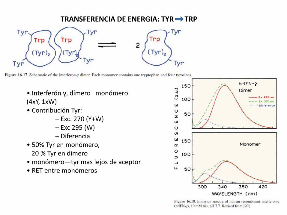
TRANSFERENCIA DE ENERGIA: TYR TRP
• Interferón γ, dímero monómero(4xY, 1xW)• Contribución Tyr:
– Exc. 270 (Y+W)– Exc 295 (W)– Diferencia
• 50% Tyr en monómero,20 % Tyr en dimero
• monómero—tyr mas lejos de aceptor• RET entre monómeros
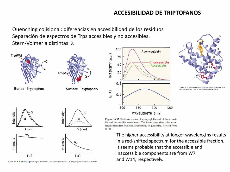
ACCESIBILIDAD DE TRIPTOFANOS
Quenching colisional: diferencias en accesibilidad de los residuosSeparación de espectros de Trps accesibles y no accesibles. Stern-Volmer a distintas l
The higher accessibility at longer wavelengths results in a red-shifted spectrum for the accessible fraction. It seems probable that the accessible and inaccessible components are from W7and W14, respectively.

DISCRIMINACION DE SITIOS EN CALMODULINA
Calmodulin has four binding sites for calcium, but littlewas known about the sequence of binding and possibilityof cooperativity between the four binding sites.No W in WT!!
These measurements revealed thesequence of calcium binding to calmodulin, and revealedinteractions between the various binding sites.
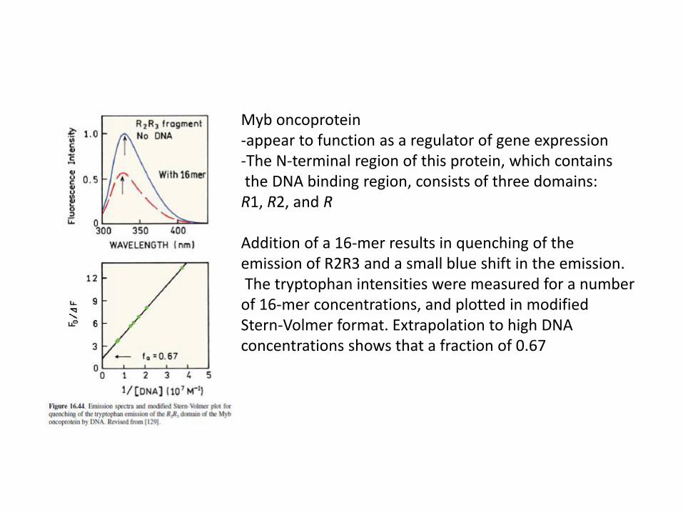
Myb oncoprotein-appear to function as a regulator of gene expression-The N-terminal region of this protein, which containsthe DNA binding region, consists of three domains:
R1, R2, and R
Addition of a 16-mer results in quenching of theemission of R2R3 and a small blue shift in the emission.The tryptophan intensities were measured for a number
of 16-mer concentrations, and plotted in modifiedStern-Volmer format. Extrapolation to high DNA concentrations shows that a fraction of 0.67
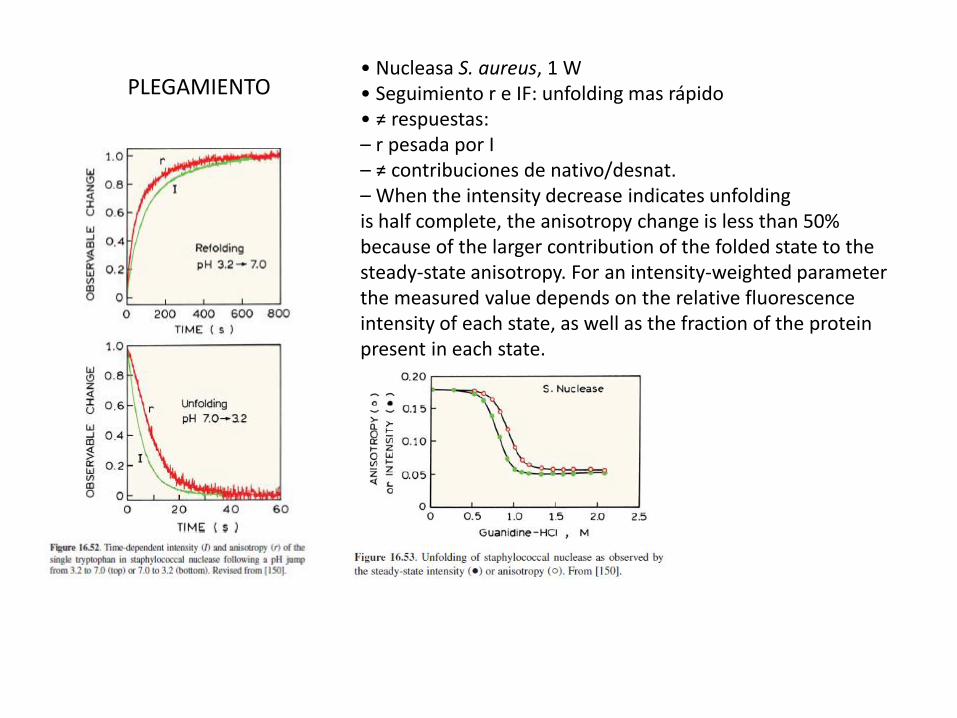
PLEGAMIENTO• Nucleasa S. aureus, 1 W• Seguimiento r e IF: unfolding mas rápido• ≠ respuestas:– r pesada por I– ≠ contribuciones de nativo/desnat.– When the intensity decrease indicates unfoldingis half complete, the anisotropy change is less than 50%because of the larger contribution of the folded state to the steady-state anisotropy. For an intensity-weighted parameterthe measured value depends on the relative fluorescenceintensity of each state, as well as the fraction of the proteinpresent in each state.

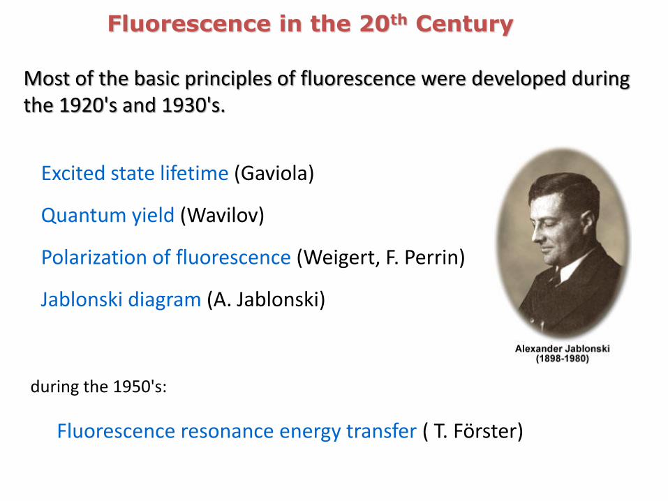
Most of the basic principles of fluorescence were developed during the 1920's and 1930's.
Fluorescence in the 20th Century
Fluorescence resonance energy transfer ( T. Förster)
Excited state lifetime (Gaviola)
Quantum yield (Wavilov)
Polarization of fluorescence (Weigert, F. Perrin)
during the 1950's:
Jablonski diagram (A. Jablonski)
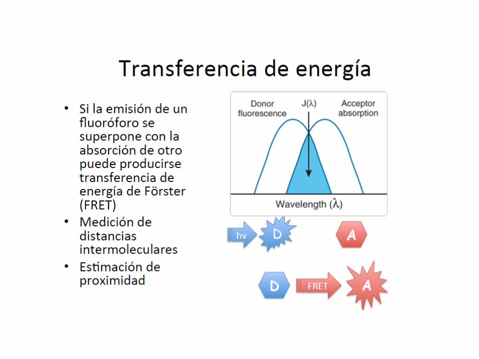

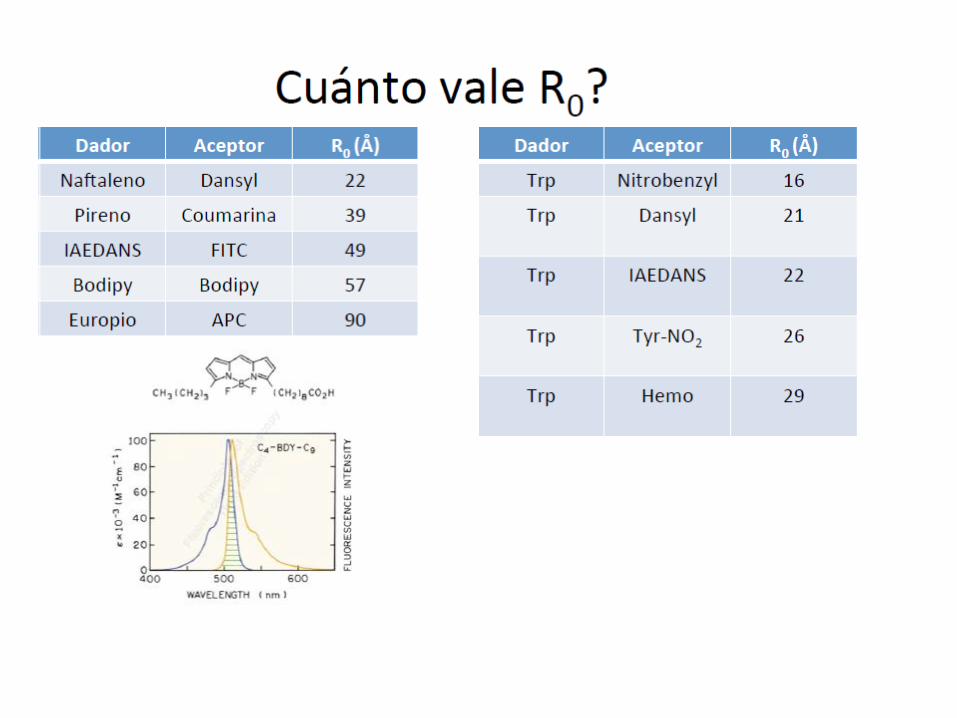
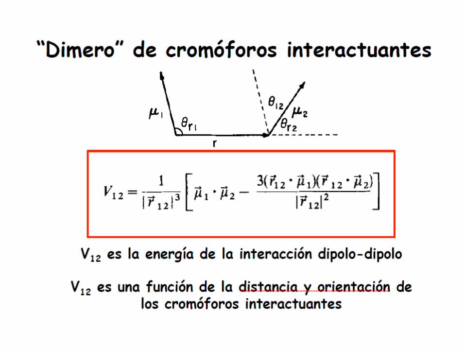
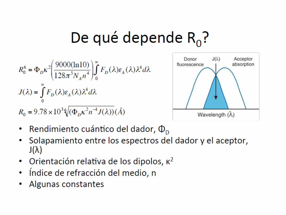
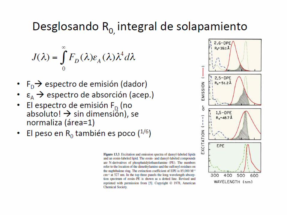
TRANSFERENCIA DE ENERGIA
The rate of energy transfer dependsi) upon the extent of spectral overlap of the emission spectrum of
the donor with the absorption spectrum of the acceptor,ii) the quantum yield of the donor, iii) the relative orientation of the donor and acceptor transition
dipoles,iv) and the distance between the donor and acceptor molecules.
La eficiencia de transferencia es la fracción de fotones absorbidos por el aceptor que se transfieren al dador
Suma de procesos de relajación
TRANSFERENCIA DE ENERGIA


It is important to remember that resonance energytransfer is a process that does not involve emission andreabsorption of photons.
Radiative transfer depends upon non-molecular optical properties of the sample, such as the size of the sample container, the path length, the optical densities of the sample at the excitation and emission wavelengths, and the geometric arrangement of the excitation and emission light paths. In contrast to these trivial factors, non-radiative energy transfer contains a wealth of structural information concerning the donor–acceptor pair

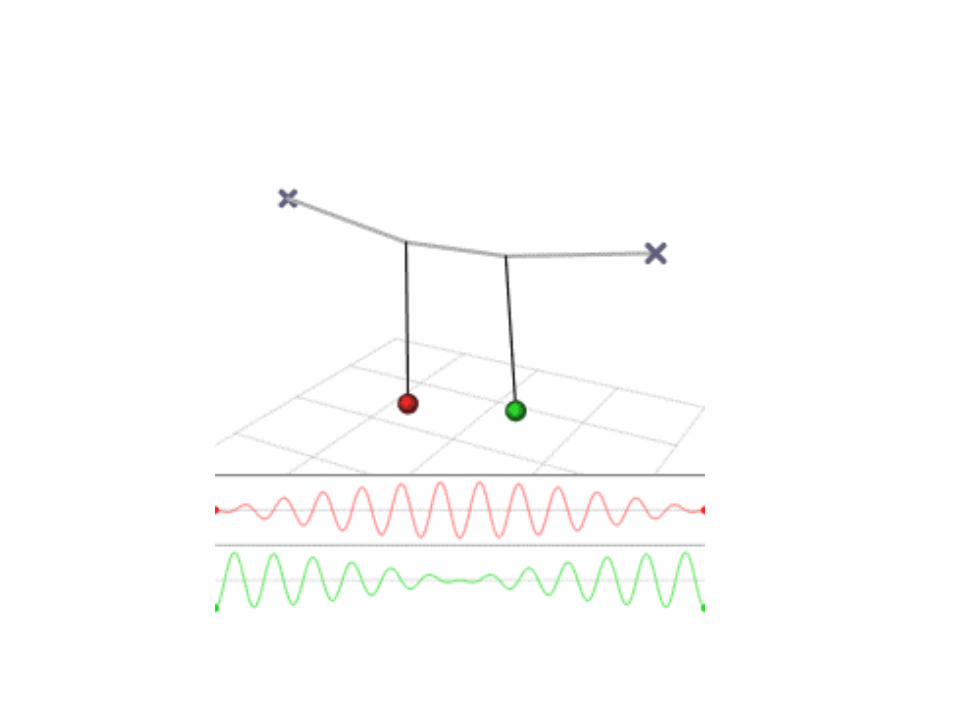
Hence RET is similarto the behavior of coupled oscillators, like two swings on a commonsupporting beam.


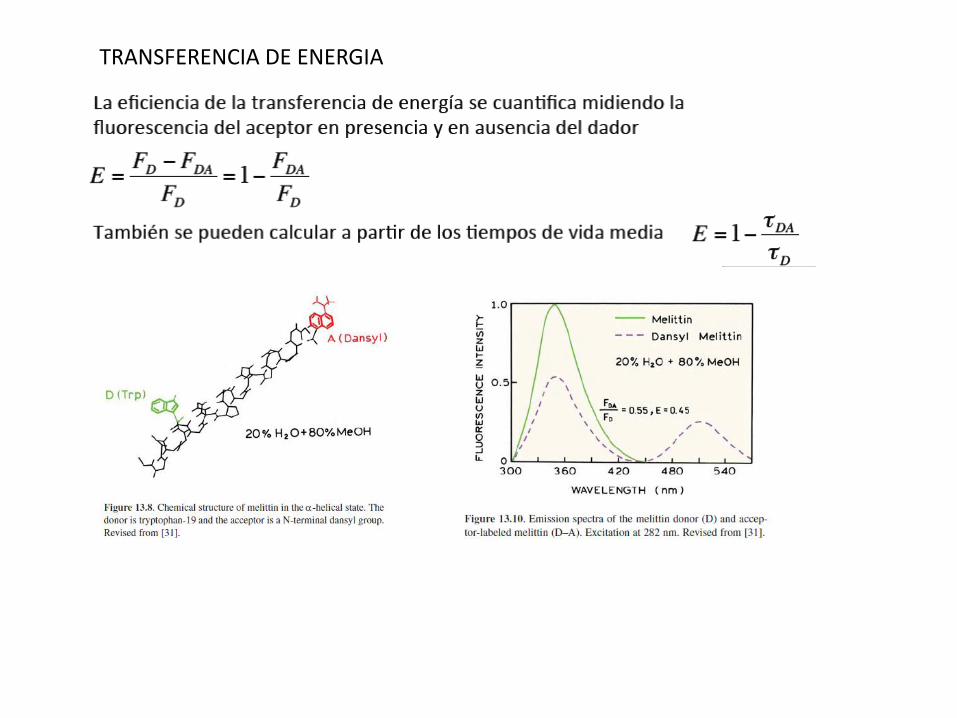
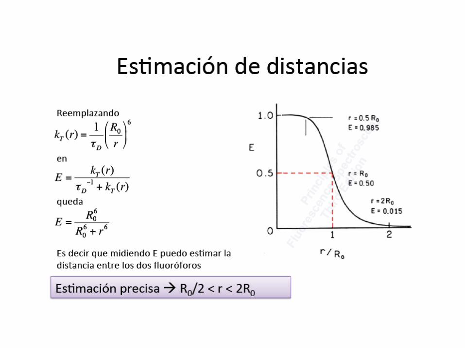
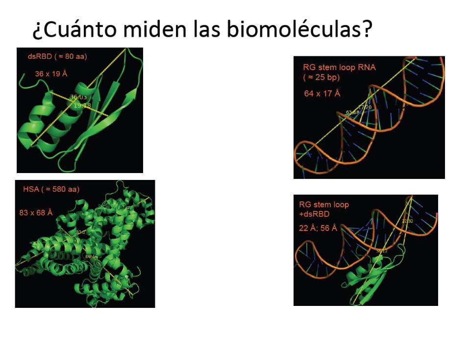
Calculo de r
*Melitina, 26 residuosGIFAVLKVLTTFLPALISWIKRKRQQMarcada con Dansyl, N-terminal
*En MeOH/H2O se comporta como una hélice rígida 1.5 Å/residuo*Ro=23.6 Å, E=0.45*r=? Compatible con hélice?*Problema: marcado parcial del aceptor: sobreestimación de distancias (FDA,med>FDA,real)
Calculando r
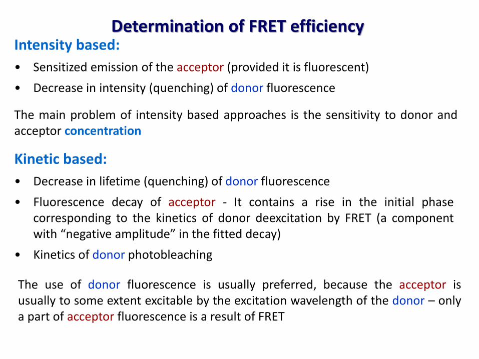
Determination of FRET efficiencyIntensity based:
• Sensitized emission of the acceptor (provided it is fluorescent)
• Decrease in intensity (quenching) of donor fluorescence
The main problem of intensity based approaches is the sensitivity to donor andacceptor concentration
Kinetic based:
• Decrease in lifetime (quenching) of donor fluorescence
• Fluorescence decay of acceptor - It contains a rise in the initial phasecorresponding to the kinetics of donor deexcitation by FRET (a componentwith “negative amplitude” in the fitted decay)
• Kinetics of donor photobleaching
The use of donor fluorescence is usually preferred, because the acceptor isusually to some extent excitable by the excitation wavelength of the donor – onlya part of acceptor fluorescence is a result of FRET
A wide variety of other factors must be considered in order to optimize FRET measurements. One of the primary issues is the relative brightness of the donor and acceptor fluorophores. In general, due to the limited dynamic range of most microscopes, fluorophores that are comparable in brightness tend to yield more satisfactory results. A significant mismatch in brightness often leads to signal from one fluorophore saturating the detector channel, while the signal from the other (dimmer) fluorophore is lost in the noise floor.
Another pitfall that often occurs is direct excitation of the acceptor at the wavelength used to excite the donor, leading to excess acceptor emission that does not result from FRET. This artifact is referred to as acceptor spectral bleed-through. Additionally, fluorescence emission from the donor can leak into the acceptor detection channel (known as donor spectral bleed-through), which also results in artificially high FRET values (Figure 2). Due to the fact that these sources of bleed-through will be present in virtually all FRET pairs, they must be inevitably addressed during FRET measurements. Choosing FRET pairs that feature a large degree of separation between emission peaks reduces bleed-through, but also might compromise the amount of spectral overlap. Too often, the result will be a large decrease in FRET signal that offsets any reduction of the spectral bleed-through problem.http://zeiss-campus.magnet.fsu.edu/articles/spectralimaging/spectralfret.html