All Wales Tissue Viability Nurse Forum Fforwm Nyrsys Hyfywedd Meinwe Cymru Gyfan All Wales Guidance This document has been created by the All Wales Tissue Viability Nurse (AWTVN) Forum and is based on the European Pressure Ulcer Advisory Panel and National Pressure Ulcer Advisory Panel. Prevention and treatment of pressure ulcers: quick reference guide. Washington DC: National Pressure Ulcer Advisory Panel; (EPUAP/NPUAP 2009) and formatted from the Getting Started Kit: How to Guide Document devised by Institute for Health Improvement. Essential Elements of Essential Elements of Pressure Ulcer Pressure Ulcer Prevention & Management Prevention & Management
Transcript
All Wales Tissue Viability Nurse Forum
Fforwm Nyrsys Hyfywedd Meinwe Cymru Gyfan
All Wales Guidance
This document has been created by the All Wales Tissue Viability Nurse (AWTVN) Forum and is based on the European Pressure
Ulcer Advisory Panel and National Pressure Ulcer Advisory Panel. Prevention and treatment of pressure ulcers: quick reference
guide. Washington DC: National Pressure Ulcer Advisory Panel; (EPUAP/NPUAP 2009) and formatted from the Getting Started
Kit: How to Guide Document devised by Institute for Health Improvement.
Essential Elements of Essential Elements of
Pressure Ulcer Pressure Ulcer
Prevention & ManagementPrevention & Management
2
Part 1Part 1
Essential Elements of
Pressure Ulcer
Prevention
All Wales Guidance
This document has been created by the All Wales Tissue Viability Nurse (AWTVN) Forum and is based on the European Pres-
sure Ulcer Advisory Panel and National Pressure Ulcer Advisory Panel. Prevention and treatment of pressure ulcers: quick
reference guide. Washington DC: National Pressure Ulcer Advisory Panel; (EPUAP/NPUAP 2009) and formatted from the Get-
ting Started Kit: How to Guide Document devised by Institute for Health Improvement.
This document should be read in conjunction
with Essential Elements of Pressure Ulcer
Management.
3
EPUAP/NPUAP Statement
The purpose of the prevention recommendations is to guide
evidence based care to prevent the development of pressure
ulcers. The guidance will apply to all vulnerable individuals of
all age groups. It is intended for the use of health care professionals
involved in the care of patients and vulnerable people who are at risk of
developing pressure ulcers. Whether, they are in a hospital, long-term care, assisted living at home
or any other setting, and regardless of their diagnosis or health care needs. It will also help guide patients
and carers on the range of prevention strategies that are available.
All Wales Tissue Viability Nurse Forum
The All Wales Tissue Viability Nurse (AWTVN) Forum has scrutinised the guidelines, taken strength of
evidence into account, considered ways of working in Wales and the needs of the Welsh population
and adapted the EPUAP/NPUAP (2009) guidelines for use within all care settings in Wales.
This adapted document is deliberately succinct, identifying the standard of care the AWTVN Forum expects
for service users in Wales. For full explanations and details of the strength of evidence for each statement
please refer to the original EPUAP/NPUAP (2009) guidelines www.epuap.org
The Case for Preventing Acquired Pressure Ulcers
Cost to patient - pain, extended hospital stay, possible death.
Estimated cost of treating pressure ulcers in the UK is estimated at between £1.4 -
£2.1 billion annually – 4% of total NHS expenditure (Bennett, 2004)
Cost of treating one Category 4 pressure ulcer is estimated at £10,551 (Bennett,
2004).
Annual spend on dressing materials by the NHS is £89 million (DoH, 2005).
It’s envisaged that the number of patients with pressure ulcers will increase (EPUAP,
Aim to position the seated individual so as to maintain his/her full range of activities whilst
also offloading the heels and providing adequate pressure relief to other vulnerable areas.
Select a posture that is acceptable for the individual and minimises the pressures and shear
exerted on the skin and soft tissues.
Use a pressure redistributing / alternating seat cushion for individuals sitting in a chair
whose mobility is reduced and who are, thus, at risk of pressure ulcer development.
Place the feet of the individual on a footstool or footrest when the feet do not reach the
floor.
Limit the time an individual spends seated in a chair.
Heels
Inspect the skin of the heels regularly.
Ensure that the heels are free of the surface
of the bed.
Heel-protection devices / pillows should ele-
vate the heel completely (offload them) in
such a way as to distribute the weight of the
leg along the calf without putting pressure on
the Achilles tendon. The knee should be in
slight flexion to prevent obstruction of the
popliteal vein.
14
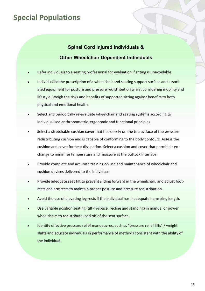
Special Populations
Spinal Cord Injured Individuals &
Other Wheelchair Dependent Individuals
Refer individuals to a seating professional for evaluation if sitting is unavoidable.
Individualise the prescription of a wheelchair and seating support surface and associ-
ated equipment for posture and pressure redistribution whilst considering mobility and
lifestyle. Weigh the risks and benefits of supported sitting against benefits to both
physical and emotional health.
Select and periodically re-evaluate wheelchair and seating systems according to
individualised anthropometric, ergonomic and functional principles.
Select a stretchable cushion cover that fits loosely on the top surface of the pressure
redistributing cushion and is capable of conforming to the body contours. Assess the
cushion and cover for heat dissipation. Select a cushion and cover that permit air ex-
change to minimise temperature and moisture at the buttock interface.
Provide complete and accurate training on use and maintenance of wheelchair and
cushion devices delivered to the individual.
Provide adequate seat tilt to prevent sliding forward in the wheelchair, and adjust foot-
rests and armrests to maintain proper posture and pressure redistribution.
Avoid the use of elevating leg rests if the individual has inadequate hamstring length.
Use variable position seating (tilt-in-space, recline and standing) in manual or power
wheelchairs to redistribute load off of the seat surface.
Identify effective pressure relief manoeuvres, such as “pressure relief lifts” / weight
shifts and educate individuals in performance of methods consistent with the ability of
the individual.
15
Patients Requiring Palliative Care
Complete a comprehensive assessment of the individual.
Reposition and turn the individual at periodic intervals, in accordance with the individ-
ual’s wishes and tolerance. Establish a flexible repositioning schedule based on individual
preferences and tolerance and the pressure-redistribution characteristics of the support
surface. Observe the individual’s choices in turning, including whether he/she has a
“position of comfort,” after explaining the rationale for turning. Comfort is of primary
importance and may supersede pressure ulcer prevention for individuals who are ac-
tively dying or have conditions causing them to have a single position of comfort.
Pre-medicate the individual 20 to 30 minutes prior to a scheduled position change for
individuals who experience significant pain on movement.
Strive to reposition an individual receiving palliative care at least every 4 hours on a high
specification pressure redistributing mattress or every 2 hours on a standard mattress.
Consider changing the support surface to improve pressure redistribution and comfort.
Individualise the turning and repositioning schedule, ensuring that it is consistent with
the individual’s goals and wishes, current clinical status and combination of co-morbid
conditions, as medically feasible. Document turning and repositioning, as well as the fac-
tors influencing these decisions (e.g. individual wishes or medical needs).
Strive to maintain adequate nutrition and hydration compatible with the individual’s
condition and wishes. Adequate nutritional support is often not attainable when the in-
dividual is unable or refuses to eat, based on certain disease states. Allow the individual
fluids and foods of choice and offer several small meals per day. Consider offering nutri-
tional protein supplements.
Maintain skin integrity to the extent possible. Apply skin emollients as per manufac-
turer’s directions to maintain adequate skin moisture and prevent dryness.
Minimise the potential adverse effects of incontinence on skin.
Special Populations
16
Critically Ill Patients
Consider the need to change support surfaces for individuals with poor local and sys-temic oxygenation and perfusion to improve pressure redistribution, shear reduction, and microclimate control and utilise additional features as needed (e.g. turn assistance, percussion).
Consider the need to change support surfaces for individuals who cannot be turned for medical reasons, such as spinal instability and haemodynamic instability. Resume rou-tine repositioning as soon as these conditions stabilise.
Consider slow, gradual turns allowing sufficient time for stabilisation of haemodynamic and oxygenation status.
Consider more frequent small shifts in position to allow some re-perfusion in individuals who cannot tolerate frequent major shifts in body position.
Prevent shear injury when lateral rotation features are used. Assess skin frequently for shear injury. Secure the individual with bolster pads (provided by the manufacturer) to prevent sacral shearing. The individual should be aligned properly in the centre of the surface.
Continue to turn the individual and assess skin for pressure and shear damage. Consider discontinuing lateral rotation at the first sign of tissue damage, and re-evaluate the indi-vidual and the support surface. Weigh the risks and benefits of continued lateral rota-tion for individuals in respiratory distress.
Change lateral rotation support surface to a support system with improved pressure redistribution, shear reduction and microclimate control, and without rotation when there is evidence of shear injury. Position the individual off the area of injury as much as possible.
Patients in the Operating Room
Refine risk assessment of individuals undergoing surgery by examining additional factors
that are likely to increase risk of pressure ulcer development, such as length of operation,
increased hypotensive episodes intra operatively, low core temperature during surgery and
reduced mobility on day one post operatively.
Use a high specification pressure redistributing mattress on the operating table for all indi-
viduals identified as being at risk of pressure ulcer development.
Position the patient in such a way as to reduce the risk of pressure ulcer development dur-
ing surgery.
Elevate the heels completely (offload them) in such a way as to distribute the weight of the
leg along the calf without putting all the pressure on the Achilles tendon. The knee should
be in slight flexion.
Pay attention to pressure redistribution prior to and after surgery.
17
Bariatric Patients
Match the individual to the bed from the time of admission, using a bed that supports
the weight and check that the mattress doesn’t “bottom out”. Ensure that the bed sur-
face is sufficiently wide to allow turning of the individual and confirm that the width of
the bariatric individual does not reach the side rails of the bed when the individual is
turned side-to-side.
Consider using features that provide air flow over the surface of the skin to facilitate
fluid evaporation if the skin is excessively moist.
Use a wheelchair and chair wide enough to accommodate the individual’s girth and pro-
vide bariatric walkers, overhead trapezes on beds and other devices to support contin-
ued mobility and independence.
Get adequate assistance to fully inspect all skin folds as pressure ulcers may develop be-
neath folds of skin, in locations where tubes have been compressed or where compres-
sion has occurred by tissue with a high adiposity (fat).
Use pillows or other positioning devices to offload pannus (apron) or other large skin
folds and prevent skin-on-skin pressure.
18
Part 2Part 2
Essential Elements of
Pressure Ulcer
Management
All Wales Guidance
This document has been created by the All Wales Tissue Viability Nurse (AWTVN) Forum and is based on the European Pres-
sure Ulcer Advisory Panel and National Pressure Ulcer Advisory Panel. Prevention and treatment of pressure ulcers: quick
reference guide. Washington DC: National Pressure Ulcer Advisory Panel; (EPUAP/NPUAP 2009) and formatted from the Get-
ting Started Kit: How to Guide Document devised by Institute for Health Improvement.
This document should be read in conjunction
with Essential Elements of Pressure Ulcer
Prevention.
19
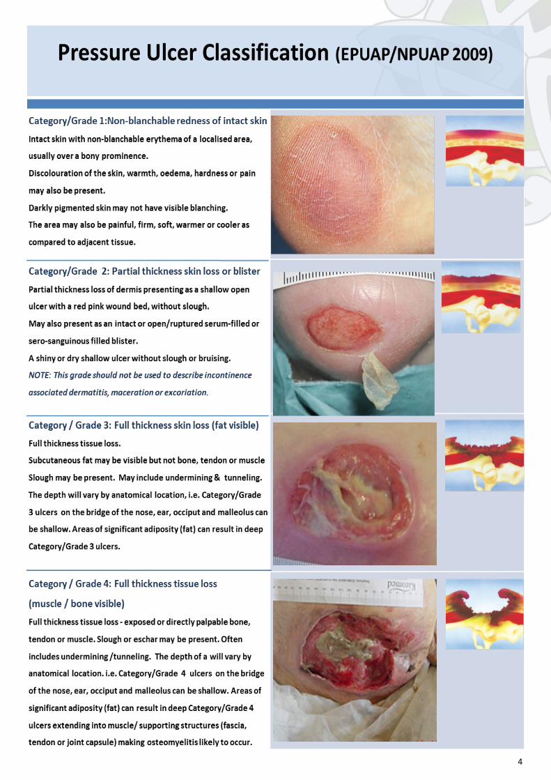
Do not use a pressure ulcer classification system to
describe tissue loss in wounds other than pressure
ulcers.
Do not classify pressure ulcers on mucous membranes.
Complete an initial assessment of the individual with a pressure ulcer, to include:
A complete health / medical and social history.
The individual’s and family’s goals of care (If the individual is unable to participate, consult with
family and/or significant others).
A focused physical examination that includes:
Factors that may affect healing (e.g. impaired perfusion, impaired sensation, systemic infection)
Vascular assessment in the case of extremity ulcers (e.g. physical examination, history of claudication and ankle-
brachial index or toe pressure)
Laboratory tests and x-rays as needed
Nutritional assessment.
Pain related to pressure ulcers.
What recommendations can be put in place to ensure the
Classification of Pressure Ulcers is correctly performed?
Use EPUAP/NPUAP pressure ulcer classification system to document the level of tissue loss. Confirm
the reliability of classifications among the professionals responsible for classifying pressure ulcers.
Educate professionals about special assessment techniques to be used in darkly pigmented individu-
als.
Educate professionals on differentiating pressure ulcers from other types of wounds (e.g. venous ul-
Educate professionals about the appropriate use of the classification system and the appearance of
different tissue types at common pressure ulcer sites.
Classification of Pressure Ulcers – see page 4
Assessment
20
Risk for developing additional pressure ulcers.
Psychological health, behaviour and cognition.
Social and financial support systems.
Functional capacity, particularly in regard to
positioning, posture, the need for assistive equip -
ment and personnel.
The employment of and adherence to pressure relieving manoeuvres.
Functional capacity, particularly in regard to positioning, posture, the need for assistive equip-
ment and personnel.
The employment of and adherence to pressure relieving manoeuvres.
Integrity of seating and bed surfaces (wear and tear).
The individual’s / family member’s knowledge and belief about developing and healing pressure
ulcers.
Teach the individual and his/her family about the normal healing process and keep them in-
formed about progress (or lack of progress) toward healing, including signs and symptoms that
should be brought to the professional’s attention.
Reassess the individual if the ulcer does not show signs of healing as expected despite adequate
local wound care, pressure redistribution and nutrition.
Expect some signs of healing in most individuals within 2 weeks.
Adjust expectations in the presence of multiple factors (particularly un-modifiable factors) that
impair wound healing (e.g. persistent under nutrition, poor perfusion and co-morbidities known
to impair wound healing).
21
Screen and assess nutritional status for each individual with a pressure ulcer at admission, with each condi-
tion change and when progress toward pressure ulcer closure is not observed.
Refer all individuals with a pressure ulcer to the dietician for early assessment of and intervention for nutri-
tional problems.
What recommendations can be put in place to improve pressure ul-
cer
assessment?
Assess the pressure ulcer initially, document and re-assess at least weekly.
With each dressing change, observe the pressure ulcer for developments that may in-
dicate the need for a change in treatment (e.g. wound improvement, wound deteriora-
tion, more or less exudate, signs of infection, or other complications).
Assess and accurately document physical characteristics such as location, category,
size, tissue type(s), wound bed and periwound condition, wound edges, sinus tracts,
undermining, tunneling, exudate, necrotic tissue, odour, presence/absence of granula-
tion tissue and epithelialisation.
Position the individual in a consistent (documented) neutral position for wound meas-
urement. Select a uniform, consistent method for measuring wound length, width and
depth to facilitate meaningful comparisons of wound measurements across time. Care
should be taken to avoid causing injury when probing the depth of a wound bed or de-
termining the extent of undermining or tunneling.
Use weekly pressure ulcer assessment findings to plan interventions that will best pro-
mote healing for the current status of the ulcer.
Assess progress toward healing:
Nutrition
22
What recommendations can be put in place to optimise nutrition and
hydration?
Assess weight status for each individual to determine weight history and significant
weight loss from usual body weight (> 5% change in 30 days or > 10% in 180
days).
Assess the individual’s ability to eat independently.
Assess the adequacy of total nutrient intake (food, fluid, oral supplements, enteral /
parenteral feeds).
Provide sufficient calories, 30-35 kcalories/kg body weight for individuals under
stress with a pressure ulcer. Adjust formula based on weight loss, weight gain, or
level of obesity. Individuals who are underweight or who have had significant unin-
tentional weight loss may need additional kcalories to cease weight loss and/or re-
gain lost weight
Revise and modify (liberalise) dietary restrictions when limitations result in de-
creased food and fluid intake. These adjustments are to be managed by a dietician
or medical professional.
Provide enhanced foods and/or oral supplements between meals if needed.
Consider nutritional support (enteral or parenteral nutrition) when oral intake is in-
adequate. This must be consistent with the individual’s goals.
23
Assess all individuals, including neonates and children, for pain related
to a pressure ulcer or its treatment using an appropriate
validated scale. An assessment of pain should include an
assessment of body language and non-verbal cues (e.g. change
in activity, loss of appetite, guarding, grimacing and moaning).
Pain assessment and management
What recommendations can ensure effective management of pain?
Use a hoist or transfer sheet to minimise friction and/or shear when repositioning an
individual, keeping bed linens smooth and unwrinkled.
Position the individual off the pressure ulcer whenever possible. Avoid postures that
increase pressure and discomfort.
Minimise pressure ulcer pain by handling all wounds gently.
Organise care delivery to ensure that it is coordinated with pain medication admini-
stration and that minimal interruptions follow. Set priorities for treatment.
Encourage individuals to request a “time out” during any procedure that causes pain.
Reduce pressure ulcer pain by keeping the wound bed covered and moist, and using
a non-adherent dressing. (Note: stable dry eschar is usually not moistened).
Use dressings less likely to cause pain and/or those likely to require less frequent
dressing changes.
For an individual with pain from a pressure ulcer, music, meditation, distraction, con-
versations and guided imagery are sometimes beneficial.
24
This section addresses support surface recommendations:
For individuals with existing pressure ulcers.
Refer to the Support Surfaces section in the Prevention
Guidelines for information on prevention of additional pressure ulcers
and general guidance on positioning.
Support surfaces alone neither prevent nor heal pressure ulcers. They are to be used as part of a total
program of prevention and treatment.
When pressure ulcers deteriorate, or fail to heal, the professional should consider replacing the existing
support surface with one that will improve pressure redistribution and microclimate (heat and moisture
control) for the individual. Changing the support surface is only one of several strategies to consider. The
individual and his/her pressure ulcer should be re-evaluated. Preventive interventions and local wound
care should also be intensified as needed. A significant increase in risk status may also prompt such re-
evaluation of the individual and the support surface.
Support surfaces and Positioning
What recommendations can be put in place to optimise
pressure relief?
Provide a support surface that is properly matched to the individual’s needs for pressure re-
distribution, shear reduction, comfort and microclimate (heat and moisture) control.
Do not position an individual directly on a pressure ulcer. If pressure over the area cannot be
relieved by repositioning, or if there are pressure ulcers on multiple turning surfaces, evalu-
ate the individual and provide a support surface properly matched to his/her needs, consid-
ering the following factors:
Number, severity, and location of the pressure ulcer(s).
Risk for additional pressure ulcers.
Need for additional features, such as ability to control moisture, temperature and fric-
tion / shear.
Aim to keep the individual off the pressure ulcer(s) as much as possible.
25
Consider higher specification foam or similar non-powered pressure redistribution mattresses and
cushions for individuals with Category / Grade 1 and 2 pressure ulcers.
Replace the existing mattress with a support surface that provides better pressure redistribution,
shear reduction and microclimate control for the individual if he/she:
Cannot be positioned off of the ulcer or has pressure ulcers on 2 or more turning surfaces
(e.g. the sacrum and trochanter), limiting turning options.
Fails to heal or demonstrates ulcer deterioration despite appropriate comprehensive care.
Is at high risk for additional ulcers.
‘Bottoms out” on the existing support surface.
Before replacing the existing mattress and/or cushion, evaluate the effectiveness of previous and
current prevention and treatment plans, and set treatment goals consistent with the individual’s
goals, values and lifestyle.
Choose a support surface that is compatible with the care setting.
Verify that the support surface is still functioning to its original intended specifications before using
it for an individual with an existing pressure ulcer.
Identify and prevent potential complications of support surface use, such as power failure, fatigue
and ineffective functioning when bed angles are too acute. Daily manual check on all positioning.
Evaluate the appropriateness and functionality of the support surface on every encounter.
Choose positioning devices and incontinence pads that are compatible with the support surface.
Limit the amount of linen and pads placed on the bed and/or chair.
Continue to reposition the individual regardless of the support surface in use. Establish repositioning
frequency based on the characteristics of the support surface and the individual’s response.
Inspect the skin for additional damage each time the individual is turned or repositioned. Allow 20-
30 minutes for redness to disperse.
Do not turn the individual onto a body surface that is damaged or still reddened from a previous
episode of pressure loading, especially if the area of redness does not blanch (i.e. Category/Grade I).
Limit head-of-bed elevation to 30° unless contraindicated by medical condition.
Encourage individuals to sleep in a 30° to 40° side-lying position or flat in bed if not contraindicated.
Avoid prolonged head-of-bed elevation and a slouched position that places pressure and shear on
the sacrum and coccyx.
Use transfer aids to reduce friction and shear. Do not drag the individual while repositioning.
Do not leave moving and handling equipment under the individual after use.
Do not leave the individual on a bedpan longer than necessary.
Do not use ring or doughnut shaped devices.
Do not apply heating devices (e.g. hot water bottles, heating pads, built-in bed warmers) directly on
pressure ulcers. Heat increases the metabolic rate, induces sweating and decreases the tolerance of
the tissue for pressure.
Give training to individuals where appropriate on the importance of small regular repositioning that
they can perform themselves.
26
If sitting in a chair is necessary for individuals with Category / Grade III and IV pressure ulcers
on the sacrum / coccyx or ischia (buttock), limit sitting to three times a day in periods of 60
minutes or less.
Consult a seating specialist to prescribe an appropriate seating surface and/or positioning tech-
niques to avoid or minimise pressure on the ulcer. Re-evaluate the seating surface and the indi-
vidual's posture if pressure ulcers worsen or fail to improve on the seating surface selected.
Ensure that the feet are properly supported either directly on the floor, on a footstool, or on
footrests when sitting (upright) in a bedside chair or wheelchair.
Avoid seating an individual with an ischial (buttock) ulcer in a fully erect posture.
Consider periods of bed rest to promote ischial and sacral ulcer healing.
Develop a schedule for progressive sitting according to the individual’s tolerance and pressure
ulcer response.
Select a cushion that effectively redistributes the pressure away from pressure ulcer.
Determine the effects of posture and deformity on pressure distribution.
Consider mobility and lifestyle needs in selecting support surfaces.
Seating
Relieve pressure under the heel(s) by placing legs on a pillow to “float the heels” off the bed or by
placing the leg in a device with heel suspension that completely offloads the pressure ulcer.
Apply any devices according to the manufacturer’s instructions.
Ensure that the device is not too tight and does not create additional pressure damage.
Check device placement more frequently in individuals with neuropathy, peripheral arterial dis-
ease, lower-extremity oedema and those who are likely to develop oedema.
Remove the device periodically to assess skin integrity.
Heels
27
What recommendations can be put in place:
To optimise wound management?
Pressure Ulcer Cleansing
Consider using cleansing solutions with surfactants to clean pressure ulcers with
debris, confirmed infection, suspected infection, or suspected high levels of bacterial colonisation.
Debridement of Pressure Ulcers
DO NOT DEBRIDE STABLE, HARD, DRY ESCHAR IN ISCHAEMIC LIMBS
Heels
Stable (dry, adherent, intact without erythema or fluctuance) eschar on the heels serves as “the
body’s natural (biological) cover” and should not be removed.
Perform a thorough vascular assessment prior to debridement of lower extremity pressure ulcers
(e.g. rule out arterial insufficiency).
Assess wound daily for signs of erythema, tenderness, oedema, purulence, fluctuance, crepitance,
and/or malodour (i.e. signs of infection) and consult an appropriate medical professional urgently in
the presence of any of these symptoms. Urgent debridement may be considered in the presence of
the above symptoms if consistent with the individual’s wishes and overall goals of care.
General Recommendations
Debride devitalised tissue within the wound bed or edge of pressure ulcers when appropriate to the
individual’s condition and consistent with overall goals of care.
Select the debridement method(s) most appropriate to: the individual’s condition; goals of care; ul-
cer/periulcer status; type, quantity, and location of necrotic tissue; care setting; and professional ac-
cessibility/capability.
Use autolytic and/or biosurgical methods of debridement when there is no urgent clinical need for
drainage or removal of necrotic tissue.
Consider the need for surgical debridement in the presence of advancing cellulitis, crepitus, fluctu-
ance and/or sepsis secondary to ulcer-related infection.
Wound Management
28
Assess pressure ulcers at every dressing change and confirm the appropriateness of the cur-
rent dressing regime.
Select a dressing from the local formulary which is suitable for the type of tissue present in
the ulcer wound bed and follow manufacturer recommendations for use.
The plan of care should guide usual dressing wear times and contain provisionary plans for
dressing changes as needed (for family, the individual, and staff) due to soilage and loosen-
ing, etc.
Negative Pressure Wound Therapy (NPWT) for Pressure Ulcers
Do not use NPWT in individuals with untreated osteomyelitis
Consider NPWT Therapy as an early adjuvant for the treatment of deep, Category / Grade III and IV
pressure ulcers.
Debride the pressure ulcer of necrotic tissue prior to the use of NPWT Therapy.
Follow a safe regime in applying and removing the NPWT system.
Evaluate the pressure ulcer with each dressing change.
If pain is anticipated or reported, consider placing a non-adherent interface dressing on the wound
bed, lowering the level of pressure and/or changing the type of pressure (continuous or intermit-
tent).
Educate the individual and his/her family about NPWT when used in the home setting.
Offload ulcer site and observe for new pressure damage when bridging or tubing is used.
Dressings for Pressure Ulcers
The selection of the wound dressing should be based on the tissue
in the ulcer bed, the condition of the skin around the ulcer
bed and the goals of the person with the ulcer. Generally
maintaining a moist ulcer bed is the ideal when the ulcer bed is
clean and granulating to promote healing or closure. The type of dressing
may change over time as the ulcer heals or deteriorates. Refer to the Local
Clinical Practice Guideline for a more complete description of all dressing types as well as discussion
of indications and contraindications for their use.
29
Infection
Follow Local Wound Management Guidelines on the assessment,
diagnosis and treatment of infection.
Follow local infection-control policies to prevent
self-contamination and cross-contamination in individuals
with pressure ulcers
Infection is not common in Category / Grade I or II ulcers, assessment of infection should focus on Category / Grade III and IV ulcers.
Infection may spread beyond the pressure ulcer, resulting in serious systemic infections such as cellulitis, fasciitis, osteomyelitis, systemic inflammatory response syndrome (SIRS), or sepsis. To avoid these serious consequences, professional should focus on identification of high-risk indi-viduals, prevention, early detection and prompt, effective treatment of pressure ulcer infection.
Surgery for Pressure Ulcers
These recommendations focus on the care of the individual and not on specific surgical techniques, deci-
sions are better left to an experienced surgeon who has an understanding of the unique needs of the pa-
tient.
What recommendations can be put in place?
Evaluate the need for surgical consultation for operative repair in individuals with Category /
Grade III or IV pressure ulcers that are not closing with conservative treatment, or for indi-
viduals who desire more rapid closure of the ulcer.
Confirm the individual’s end-of-life preferences if anticipating surgery.
Obtain a surgical consultation for possible urgent drainage and/or debridement if the pres-
sure ulcer has advancing cellulitis or is a suspected source of sepsis.
Assess for osteomyelitis. If present, infected bone must be resected prior to or during surgical
closure.
Prior to surgery, optimize physical and psychosocial factors that may impair surgical wound
healing.
Confirm the presence of a positive social network at home.
Initiate a program of progressive sitting according to the surgeon’s orders. When weight bear-
ing on the operative site is allowed, weight bearing should be graduated and progressive. Sit-
ting should increase in time if no erythema is noted over weight-bearing areas.
Skin tolerance to pressure over the wound site should be assessed after each sitting period.
Position the individual only on a pressure-redistributing chair cushion when he/she is sitting
in a chair
30
These recommendations address the unique needs of
critically ill, spinal-cord-injured, and bariatric individuals,
in relation to pressure redistribution, shear reduction and
microclimate control.
Critically Ill Individuals
Continued use of lateral rotation may be necessary for individuals in respiratory distress. In all cases, the
risks and benefits of continued lateral rotation should be weighed in individuals with existing pressure ul-
cers.
Spinal-Cord-Injured Individuals and Other Wheelchair Dependant Individuals
Ideally, ischial ulcers should heal in an environment where the ulcers are free of pressure and other me-
chanical stress. Total bedrest may be prescribed to create a pressure free wound environment. However,
this approach comes with potential physical complications (e.g. muscle wasting, deconditioning, respira-
tory complications), psychological harm, social isolation and financial challenges for the individual and his/
her family.
Balancing physical, social, and psychological needs against the need for total offloading (i.e. total bedrest)
creates a challenging dilemma for the individual and the professional. Use of a wheelchair is imperative for
spinal-cord-injured individuals. Sitting time may need to be restricted when ulcers are present on sitting
surfaces. Seating cushions must be high immersion, uniform loading distribution cushions.
What recommendations can be put in place?
Consider alternative methods of pressure redistribution (or avoid lateral rotation beds) in indi-
viduals with sacral or buttock pressure ulcers.
Offload the pressure ulcer(s) in individuals undergoing lateral rotation therapy.
Inspect the pressure ulcer and the periulcer skin for shear injury with every dressing change.
Shear injury may appear as deterioration of the ulcer edge, undermining and/or as increasing
inflammation of periulcer skin or the ulcer.
Special Populations
31
What recommendations can be put in place?
Refer individuals to a seating professional for evaluation if sitting is unavoidable.
Seat spinal-cord-injured individuals with ischial (buttock) ulcers on a seating support surface
that provides contour, uniform pressure distribution and high immersion or offloading.
Consider periods of bed rest to promote ischial and sacral ulcer healing.
Limit sitting time for spinal-cord-injured individuals with ischial ulcers according to skin toler-
ance and pressure ulcer response.
Develop a schedule for progressive sitting according to the individual’s tolerance and pressure
ulcer response.
Maintain proper positioning and postural control.
Bariatric (Obese) Individuals
Get adequate assistance to fully inspect all skin folds. Pressure ulcers may develop in unique locations,
such as beneath folds of skin and in locations where tubes and other devices have been compressed be-
tween skin folds but may also result from tissue pressure across the buttocks and other areas of high adi-
pose (fat) tissue concentration.
What recommendations can be put in place?
Assess pressure ulcers carefully for signs of infection and delays in healing, which are more
common in bariatric individuals.
Provide adequate nutrition to support healing. Obese individuals may lack adequate nutrients
to support healing of pressure ulcers. Goals of weight loss may need to be postponed or modi-
fied to ensure that adequate nutrients are provided for healing (see Nutritional Section).
32
Individuals Receiving Palliative Care
Pain management, odour control and exudate control are
the aspects of pressure ulcer care that tend to be
most closely related to supporting the individual’s
comfort.
What recommendations can be put in place?
Set treatment goals consistent with the values and goals of the individual, while consider-
ing family input.
Assess the impact of the pressure ulcer on quality of life for the individual and his/her
family.
Set a goal to enhance quality of life, even if the pressure ulcer cannot be healed or treat-
ment does not lead to closure/healing.
Assess the individual initially and with any significant change in condition, re-evaluate the
plan of care.
Assess the pressure ulcer initially and with each dressing change, at least weekly (unless
the individual is actively dying) and document findings.
Monitor the ulcer in order to continue to meet the goals of comfort and reduction in
wound pain, addressing wound symptoms, such as, odour and exudate.
Manage the pressure ulcer and periwound area on a regular basis as consistent with the