35

Essentials ofOral Biology
Oral Anatomy, Histology, Physiology andEmbryology
Second Edition
Essentials ofOral Biology
Oral Anatomy, Histology, Physiology andEmbryology
Second Edition
Maji Jose MDS, PhD
Professor and Head Department of Oral Pathology Yenepoya Dental College and
Hospital Yenepoya University
Deralakatte, Mangalore 575018
Karnataka, IndiaEmail: [email protected]

CBS Publishers & Distributors Pvt LtdNew Delhi • Bengaluru • Chennai • Kochi • Mumbai •
Kolkata Hyderabad • Pune • Nagpur • Manipal • Vijayawada • Patna
Disclaimer
Science and technology are constantly changing fields. New research andexperience broaden the scope of information and knowledge. The authorshave tried their best in giving information available to them while preparingthe material for this book. Although, all efforts have been made to ensureoptimum accuracy of the material, yet it is quite possible some errors mighthave been left uncorrected. The publisher, the printer and the authors will notbe held responsible for any inadvertent errors, omissions or inaccuracies.
eISBN: 978-93-877-4261-1
Copyright © Authors and Publisher
First eBook Edition: 2017
All rights reserved. No part of this eBook may be reproduced or transmittedin any form or by any means, electronic or mechanical, includingphotocopying, recording, or any information storage and retrieval systemwithout permission, in writing, from the authors and the publisher.
Published by Satish Kumar Jain and produced by Varun Jain forCBS Publishers & Distributors Pvt. Ltd.
Corporate Office: 204 FIE, Industrial Area, Patparganj, New Delhi-110092
Ph: +91-11-49344934; Fax: +91-11-49344935; Website: www.cbspd.com;www.eduport-global.com; E-mail: [email protected];[email protected]
Head Office: CBS PLAZA, 4819/XI Prahlad Street, 24 Ansari Road,Daryaganj, New Delhi-110002, India.

Ph: +91-11-23289259, 23266861, 23266867; Fax: 011-23243014; Website:www.cbspd.com; E-mail: [email protected];[email protected].
Branches
Bengaluru: Seema House 2975, 17th Cross, K.R. Road, Banasankari 2nd
Stage, Bengaluru - 560070, Karnataka
Ph: +91-80-26771678/79; Fax: +91-80-26771680; E-mail:[email protected]
Chennai: No.7, Subbaraya Street Shenoy Nagar Chennai - 600030, TamilNadu
Ph: +91-44-26680620, 26681266; E-mail: [email protected]
Kochi: 36/14 Kalluvilakam, Lissie Hospital Road, Kochi - 682018, Kerala
Ph: +91-484-4059061-65; Fax: +91-484-4059065; E-mail: [email protected]
Mumbai: 83-C, 1st floor, Dr. E. Moses Road, Worli, Mumbai - 400018,Maharashtra
Ph: +91-22-24902340 - 41; Fax: +91-22-24902342; E-mail:[email protected]
Kolkata: No. 6/B, Ground Floor, Rameswar Shaw Road, Kolkata - 700014
Ph: +91-33-22891126 - 28; E-mail: [email protected]
Representatives
Hyderabad
Pune
Nagpur
Manipal
Vijayawada
Patna
O
Foreword
ral biology, which includes oral anatomy, histology, physiology andembryology is one of the most important and useful subjects among thevarious basic science subjects, in the dental curriculum. Understanding
of this subject widens the mental comprehension and strengthens the basicconcepts of different dental science specialties. A thorough knowledge of thissubject is sure to mould a dental student into an effective and efficientclinician.
I am happy that Dr Maji Jose is bringing out a textbook Essentials ofOral Biology (Oral Anatomy, Histology, Physiology and Embryology)for students pursuing dentistry. Visualizing the integrated perspective of thesubject, she has been successful in gathering together the diverse elements oforal biological sciences, which in the past had been scattered throughoutmany textbooks. I am sure that this book will be useful for dental students toimprove their knowledge in oral biological sciences as well as to help them toconfidently face the exam.
I hope that this work will receive the deserved attention andencouragement from both dental students and teachers.
I wish the author and this book all success.
BH Sripathi Rao MDS
Principal Yenepoya Dental College and Hospital
Former Executive Committee Member; Dental Council of India

▪
▪
▪
▪
▪
Contributors
Dr Heera RProfessor, Department of Oral PathologyGovernment Dental CollegeThiruvananthapuram
Dr Girish KLProfessor, Department of Oral PathologySri Mookambika Institute of Dental SciencesKulasekaram, KanyakumariTamil Nadu
Dr Rajeesh Mohammed PKProfessor, Department of Oral PathologyKMCT Dental College Mukkam, KozhikodeKerala
Dr Usha BalanAssistant ProfessorCollege of DentistryKing Khalid UniversityAbah, KSA
Dr Ajeesha FerozDepartment of Oral Pathology
Mahe Institute of Dental SciencesMahe, UT of Puducherry

T
Preface to Second Edition
he textbook Essentials of Oral Biology presents all subsections of OralBiology in single book, described in five sections: Oral Embryology,Oral Histology, Oral and Dental Anatomy, Oral Physiology and Allied
Topics. This text provides a comprehensive coverage of all the topicsincluded in the curriculum specified by Dental Council of India and variousIndian universities. Different topics are dealt in detail in 50 chapters withflowcharts, tables and color diagrams to make learning more simple andpleasant. This book also includes an additional section on expected questionsfrom each chapter, commonly asked in examinations of various Indianuniversities, to assist the students in examination preparations.
This book has been designed in a way to keep the characteristics of astandard textbook for undergraduate students. The topics are explained insimple and lucid language. Concepts are presented in a simple and clearmanner to help an undergraduate student develop a comprehensiveknowledge in this basic science subject which makes a sound base forlearning pathologic basis of diseases.
I am gratified that original edition has received a good response. A positivefeedback on the first edition of the book and various encouraging commentsreceived from students and teachers, who have used the book, has encouragedme to come out with second edition. The second edition is a revised andupdated version with flowcharts, more tables and color diagrams to furtherease the learning process. A discussion on clinical considerations is added toeach chapter in order to guide the students to clinical application of oralbiology. While preparing the second edition, I have followed the originalpolicy “simple presentation and lucid language” which enables a self-study.
I offer this book to the dental students, hoping that this will ensure anenjoyable and rewarding study of oral biology.

Maji Jose
I
Acknowledgements
thank God Almighty for all the blessings He has showered on me in thisventure. The preparation of this textbook was possible only with the helpand cooperation of a number of people.I would like to express my gratitude to Dr Sripathi Rao, Principal,
Yenepoya Dental College, for his kind words of encouragement and moralsupport, received at every stage of the preparation of this book. I also thankhim for writing the Foreword to the book.
I would like to express my heartfelt thanks to Dr Heera R, faculty of OralPathology, Government Dental College, Thiruvananthapuram, my teacherand friend, for giving me all guidance, moral support, and for sharing herknowledge at different stages of my work, and also for contribution in thebook.
I gratefully acknowledge the constant support of Dr Rajeesh MohammedPK, Dr Girish KL, Dr Usha Balan and Dr Ajeesha Firoz who have alsocontributed chapters to this book. I am indebted to Head of the Departmentand all my colleagues of Department of Oral Pathology, Yenepoya DentalCollege, Mangalore, especially Dr Joshy, Dr Meera and Dr Haziel DianaJenifer, for their constructive suggestions and timely support.
The talented staff of CBS Publishers & Distributors deserve praise for theirrole in shaping this book.
I owe a great deal of regard and gratitude to my parents, teachers andbeloved students who have played a major role in making me what I amtoday. I thank my husband Mr Ajoy S. Joseph, and my children, Joe and Jiya,who stood by me at all the stages and exhibited patience and affection whichenabled me to carry on with the work smoothly.
We would like to thank Mr S.K. Jain (CMD), Mr. Varun Jain (Director),Mr. YN Arjuna (Senior Vice President – Publishing and Editorial), and Mr.Ashish Dixit (Business Head – Digital Publishing, Marketing & Sales) and

his team at CBS Publishers & Distributors Pvt. Ltd. for their skill,enthusiasm, support, patience and excellent professional approach inproducing and publishing this eBook.
Finally, I thank each and everyone whose contribution, direct or indirect,has made the preparation of this book a pleasant task.
Maji Jose

1.
2.
3.
4.
1.
2.
Syllabus
Oral Biology course includes instructions in the subject of DentalMorphology, Oral Embryology, Oral Histology and Oral Physiology.
I. TOOTH MORPHOLOGY
Introduction to tooth morphology: Human dentition, types ofteeth, function, Palmer’s and binomial notation systems, toothsurfaces, their junctions—line angles and point angles, definition ofterms used in dental morphology, geometric concepts in toothmorphology, contact areas and embrasures—clinical significance.Morphology of permanent teeth: Description of individual teeth,including a note on their chronology of development, differencesbetween similar class of teeth and identification of individual teeth.Morphology of deciduous teeth: Generalized differences betweendeciduous and permanent teeth. Description of individual deciduousteeth, including their chronology of development.Occlusion
II. ORAL EMBRYOLOGY
Brief review of development of face, jaws, lip, palate, andtongue, with applied aspects.Development of teeth: Detailed study of different stages ofdevelopment of crown, root and supporting tissues of tooth anddetailed study of formation of calcified tissues. Applied aspects of

3.
4.
1.
2.
3.
4.
5.
6.
7.
1.
2.
disorders in development of teeth.Eruption of deciduous and permanent teeth: Mechanisms in tootheruption, different theories and histology of eruption, formation ofdentogingival junction, role of gubernacular cord in eruption ofpermanent teeth.Shedding of teeth: Mechanisms of shedding of deciduous teeth.Complications of shedding.
III. ORAL HISTOLOGY
Detailed microscopic study of enamel, dentin, cementum andpulp tissue: Age changes.Detailed microscopic study of periodontal ligament and alveolarbone: Age changes, histological changes in periodontal ligament.Detailed microscopic study of oral mucosa: Variation in structurein relation to functional requirements, mechanisms of keratinizationclinical parts of gingiva, dentogingival and mucocutaneous junctionsand lingual papillae and age changes.Salivary glands: Detailed microscopic study of acini and ductalsystem.TM joint: Review of basic anatomical aspects and microscopicstudy.Maxillary sinus: Microscopic study, functions and clinicalrelevance of maxillary sinus in dental practice.Processing of hard and soft tissues for microscopic study:Ground sections, decalcified sections and routine stainingprocedures.
IV. ORAL PHYSIOLOGY
Saliva: Composition of saliva—formation of saliva and mechanismsof secretion, functions of saliva.Mastication: Masticatory force, need for mastication, peculiarities

3.
4.
5.
6.
7.
of masticatory muscles, masticatory cycle, masticatory reflexes andneural control of mastication.Deglutition: Review of the steps in deglutition, swallowing ininfants, neural control of deglutition.Calcium and phosphorus metabolism: Source, requirements,absorption, distribution, functions and excretion, clinicalconsiderations.Theories of mineralization: Definition, mechanisms, theories ofmineralization.Physiology of taste: Innervation of taste buds and taste pathway,physiologic basis of taste sensation, age changes.Physiology of speech
About the Book
Essentials ofOral BiologyOral Anatomy, Histology, Physiology and
Embroyology
is the completely rewritten, thoroughly revised, fairly enlarged andprudently updated edition of the popular book. All topics of oral biologyare described in five sections: • Oral Embryology, • Oral Histology, • Oraland Dental Anatomy, • Oral Physiology and • Allied Topics.
This text provides a comprehensive coverage of all the topics includedin the curriculum specified by Dental Council of India and various Indianuniversities.
Various topics covered in 50 different chapters, revised and updatedwith flowcharts, tables and colour diagrams to make learning more simpleand pleasant. A discussion on clinical considerations is added to guide thestudents to clinical application of oral biology. An additional section onexpected questions of each chapter, commonly asked in examinations ofvarious Indian universities, will assist the students on examinationpreparations.
This book is based on more than two decades of experience of the authoras a teacher and examiner of oral biology. This edition maintains thehallmark of the earlier edition: Lucid language and simple presentation.

About the Author
Maji Jose MDS, PhD is currently Professor and Head,Department of Oral Pathology, Yenepoya Dental College andHospital, Yenepoya University, Deralakatte, Mangalore,Karnataka. She has more than two decades of experience inteaching oral biology and oral pathology to undergraduate andpostgraduate students in various distinguished dental colleges such asManipal College of Dental Sciences, KVG Dental College, Sullia, andYenepoya Dental College. She has been examiner for both undergraduateand postgraduate students at various universities.
Dr Jose, well acknowledged as a teacher, examiner and researcher,obtained PhD in 2013 from Yenepoya University and has to her credit over50 scientific publications in Indian and international journals of repute. Herother textbook Manual of Oral Histology and Oral Pathology (CBS) iswell accepted and widely used by dental students and teachers of manyIndian and foreign universities.

1.
2.
3.
4.
5.
6.
7.
8.
9.
10.
Contents
Foreword by BH Sripathi Rao
Contributors
Preface to Second Edition
About The Book
About The Author
Section 1: Oral Embryology
General Embryology
Development of Orofacial Structures
Section 2: Oral Histology
Development of Tooth
Enamel and Amelogenesis
Dentin and Dentinogenesis
Pulp
Cementum and Cementogenesis
Periodontal Ligament
Alveolar Bone
Oral Mucosa

11.
12.
13.
14.
15.
16.
17.
18.
19.
20.
21.
22.
23.
24.
25.
26.
27.
28.
29.
30.
31.
32.
33.
Salivary Glands
Temporomandibular Joint
Maxillary Sinus
Section 3: Oral and Dental Anatomy
Introduction to Dental Anatomy
Deciduous Maxillary Anterior Teeth
Deciduous Mandibular Anterior Teeth
Deciduous Maxillary Molars
Deciduous Mandibular Molars
Comparison between Deciduous and Permanent Dentition
Permanent Maxillary Central Incisors
Permanent Maxillary Lateral Incisors
Permanent Mandibular Central Incisors
Permanent Mandibular Lateral Incisors
Permanent Maxillary Canines
Permanent Mandibular Canines
Permanent Maxillary First Premolars
Permanent Maxillary Second Premolars
Permanent Mandibular First Premolars
Permanent Mandibular Second Premolars
Permanent Maxillary First Molars
Permanent Maxillary Second Molars
Permanent Maxillary Third Molars
Permanent Mandibular First Molars

34.
35.
36.
37.
38.
39.
40.
41.
42.
43.
44.
45.
46.
47.
48.
49.
50.
Permanent Mandibular Second Molars
Permanent Mandibular Third Molars
Occlusion
Section 4: Oral Physiology
Eruption
Shedding
Saliva
Physiology of Taste and Speech
Mastication
Deglutition
Calcium Phosphorus Metabolism
Mineralization
Hormonal Influence on Orofacial Structures
Age Changes of Oral Tissues
Section 5: Allied Topics
Tissue Processing
Microscope
Muscles of Orofacial Region
Vascular and Nerve Supply of Orofacial Region
Appendix

1.
2.
Section 1
Oral Embryology
General Embryology
Development of Orofacial Structures
1
E
General Embryology
Formation of blastocyst
Germ layers
Neural crest cells
Pharyngeal arches and pouches
mbryology is the study of growth and differentiation which an organismundergo during its development from a single fertilized cell to a complexindependent living being.
Every animal starts life in the form of a simple cell, i.e. the fertilized egg orzygote. Zygote is formed by two cells, namely the germ cells of parents.Fertilization occurs when male and female gamates (spermatozoon andovum) unite to form zygote.
The intrauterine life of human beings can be devided into embryonicperiod which lasts for 8 weeks after fertilization which will be followed byfetal period which continues throughout pregnancy that ends in birthapproximately after 280 days.
After fertilization, rapid proliferation of cells takes place leading toformation of a cell mass called morula. This morula is a “golf ball” like alittle mass of cells and consists of a group of centrally placed cells termed asinner cell mass, surrounded by a peripheral layer of cells (Fig. 1.1). Oncemorula enters into the uterine cavity by 7 to 8 days, it turns into a fluid filledstructure due to seepage of fluid, which separates the inner cell mass fromperipheral layer of cells. The resultant structure is called blastocyst (Fig. 1.2).
This blastocyst is lined by a layer of cells called trophoblasts. Thetrophoblasts are derived from the outer layer of morula, which later gives rise

to placenta and is also involved in implantation of the embryo. Within theblastocyst, the inner cell mass can be seen attached to one side of the inneraspect. This inner cell mass or embryoblasts forms the embryonic stem cellsthat gives rise to embryo.
Fig. 1.1: Morula
Fig. 1.2: Blastocyst
At this stage, the blastocyst has two different types of cells. The inner cellmass that occupies the center portion and an outer layer that surrounds thiscell mass. As the blastocyst develops further, some cells of the inner cellmass differentiate into flattened cells and line the free surface while the othercells change into columnar cells. The flattened cells constitute the endodermwhile the columnar cells forms the ectoderm. Thus, by 8th day of gestationthe embryo appears like a ‘bilaminar circular disc’.
As the development proceeds, in a localized area close to the futurecephalic end of the disc, flattened cells of endoderm changes into columnarcells. This circular area where the changes takes place is called ‘prochordalplate’. The region where the prochordal plate is formed is the head end andopposing end is tail end of the embryo. Prochordal plate provides the disc anantero-posterior axis and a bilateral symmetry.
After the formation of the prochordal plate, the cells of ectodermproliferate near the tail end, forming another structure called the primitivestreak. These proliferating cells initially form a thickening and later spread

•••
sideways between ectoderm and endoderm forming a third layer calledmesoderm. This mesodermal layer spreads and separates the ectoderm andendoderm throughout the disc except for the circular region of prochordalplate. So by the 16th day, the embryonic disc has three layers: Ectoderm,endoderm and mesoderm. These three primary germ layers give rise todifferent tissues and organs of our body.
Germ Layer Derivatives
Structures of Ectodermal Origin areCutaneous structures
Skin and its appendagesOral mucous membraneEnamel of teeth
Neural system-central and peripheral nerve systems
Structures of Mesodermal Origin areCardiovascular system—heart and blood vessels
Locomotor system—bones and muscles
Connective tissue
Components of teeth other than enamel
Structures of Endodermal Origin areLining epithelium of respiratory tract
Lining epithelium of alimentary tract
Secretory cells of liver and pancreas
As the development progresses, the circular disc shaped embryo becomeselongated and pear shaped. The region of prochordal plate where ectodermand endoderm remain in contact forms the ‘buccopharyngeal membrane’.
The cranial end of the primitive streak thickens to form primitive node.The cells proliferate from primitive node and extend between the ectodermand endoderm, along the central axis up till the prochordal plate. This forms

notochordal process or head process. Ectoderm over the notochorddifferentiates to form neural plate which develops an invagination and formsthe neural tube. This neural tube extends from primitive node to prochordalplate. The cranial part of neural tube forms the brain and caudal part formsthe spinal cord.
The enlarging embryonic disc develops folds at its head end (cranial fold),tail end (caudal fold) and laterally, making the embryo entirely covered byectoderm.
Neural Crest CellsNeural crest cells are a group of pleuripotent cells that develop fromectoderm along the lateral margins of neural plate. These cells migrateextensively in the developing embryo between ectoderm and endoderm andintra-mesodermally and differentiate into different types of cells that formsvarious tissues of the body.
The neural crest cells move around the sides of the developing headbeneath the surface of ectoderm as sheets of cells. They migrate and form theentire connective tissue of upper facial region; while in the lower facialregion they migrate into already existing mesenchyme. Therefore theconnective tissue beneath the developing ectoderm in this region is calledectomesenchyme.
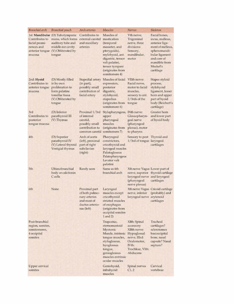
Derivatives of the branchial arches, pharyngeal pouches and cranialsomites

The Structures that Develop from the Neural Crest CellsIn the head and neck region neural crest cells differentiate to form most of theconnective tissue components including bone, cartilage, dermis and tissuesthat form tooth except enamel and also contributes to formation of musclesand arteries of this region.
Neural crest cells migrate to the trunk region giving rise to neural, endocrineand pigment producing cells. In the trunk sensory ganglions, Schwann cellsand neurons are also derived from neural crest cells.
Neural crest cells have a significant role in craniofacial development andformation of teeth. A developmental disorder called Treacher Collinsyndrome which manifest with various craniofacial developmental defects iscaused due to defective migration of neural crest cells. Defective migration ofneural crest cells can also cause defective dentition.
Branchial Arches and PouchesThe developing oral cavity, stomatodeum is situated between the developingbrain and pericardium. In the early stages, neck is not present. Later, series ofmesodermal thickenings develop in the wall of the cranial part of foregutresulting in the formation of neck between stomodeum and pericardium.These cylindrical thickenings are called branchial arches or pharyngealarches (Fig. 1.3).
Pharyngeal arches are six in number and extend from lateral wall ofpharynx, towards the medial direction, to approach its counterpart extendingfrom other side. The inner aspect of each arch is covered by endoderm andouter aspect by ectoderm. The central core is made up of mesenchyme, whichis surrounded by ectomesenchyme, which is of neural crest origin. Theendoderm extends outwards between the branchial arches in the form ofpouches called pharyngeal pouches. The pharyngeal pouches meet theectodermal clefts which are formed by invagination of ectoderm lining theouter surface of the pharyngeal arches.

Fig. 1.3: Pharyngeal arches and pouches
The mesoderm of each arch gives rise to a skeletal element (which can beeither a cartilage or bone), muscle and an arterial arch. Each pharyngeal archhas a nerve which supplies the structures that develop from that arch.
There are six pharyngeal arches. 1st arch is named as mandibular arch,which plays a very important role in craniofacial development.
2nd arch is hyoid arch and the 5th arch disappears soon after formation.The remaining 3, 4, 6 arches do not have specific names.
2
O
Development of OrofacialStructures
Formation of orofacial structure
rofacial structures develop primarily from first, second and thirdbranchial arches by fusion of various processes.
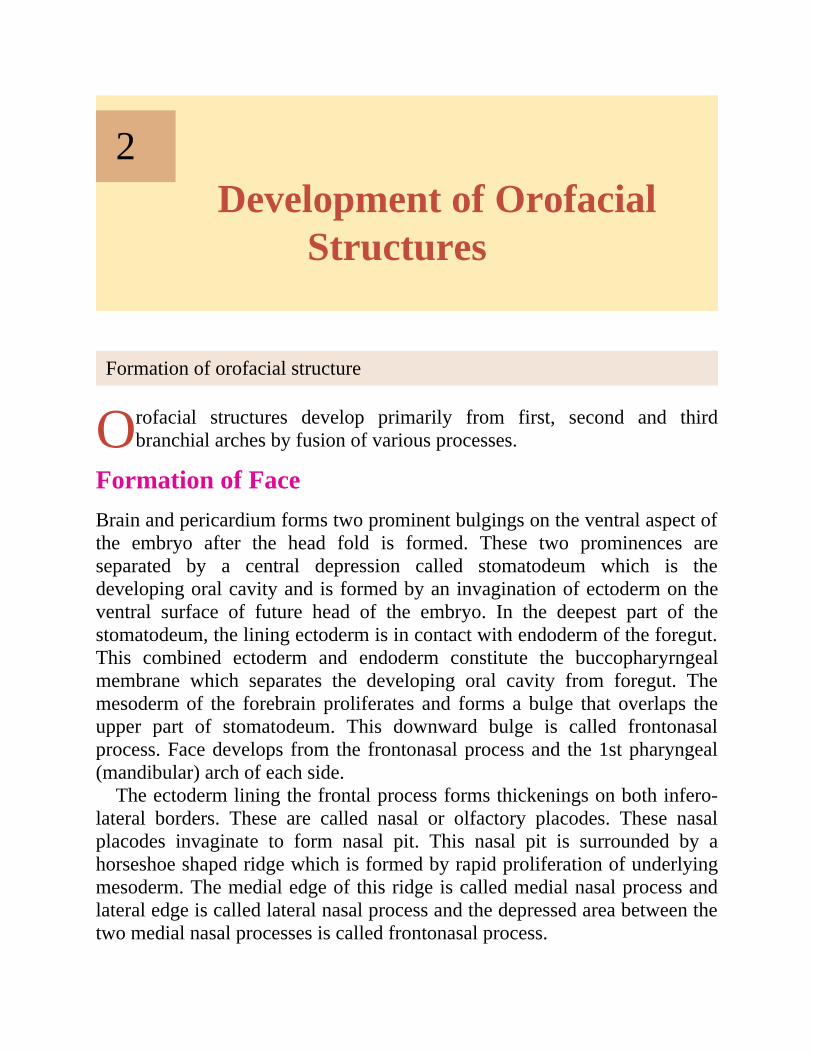
Formation of FaceBrain and pericardium forms two prominent bulgings on the ventral aspect ofthe embryo after the head fold is formed. These two prominences areseparated by a central depression called stomatodeum which is thedeveloping oral cavity and is formed by an invagination of ectoderm on theventral surface of future head of the embryo. In the deepest part of thestomatodeum, the lining ectoderm is in contact with endoderm of the foregut.This combined ectoderm and endoderm constitute the buccopharyrngealmembrane which separates the developing oral cavity from foregut. Themesoderm of the forebrain proliferates and forms a bulge that overlaps theupper part of stomatodeum. This downward bulge is called frontonasalprocess. Face develops from the frontonasal process and the 1st pharyngeal(mandibular) arch of each side.
The ectoderm lining the frontal process forms thickenings on both infero-lateral borders. These are called nasal or olfactory placodes. These nasalplacodes invaginate to form nasal pit. This nasal pit is surrounded by ahorseshoe shaped ridge which is formed by rapid proliferation of underlyingmesoderm. The medial edge of this ridge is called medial nasal process andlateral edge is called lateral nasal process and the depressed area between thetwo medial nasal processes is called frontonasal process.
At the same time the mandibular arches that form the lateral wall ofstomatodeum gives off a bud-like projection called maxillary process (oneither side). The remaining part of the mandibular arch forms the mandibularprocess.
The face is derived from the five prominences that surround thestomatodeum. These prominences are frontonasal process, pair of maxillaryprocesses and a pair of mandibular processes (Fig. 2.1).
Lower LipLower lip develops from the mandibular processes which grow mediallytowards each other and fuses at midline. This forms the lower margin ofstomatodeum. As the development continues an ectodermal proliferationoccurs which extends into the ectomesenchyme. The structure developed iscalled vestibular lamina and it gives rise to a V-shaped sulcus that separatesthe lip from the tooth bearing area.
Fig. 2.1: Development of face
Upper LipMandibular arch on either side gives rise to process called maxillaryprocesses. These processes grow forward and medially towards one anotherabove the stomatodeum. As they do so, these processes first fuse with lateralnasal process and later with medial nasal process. The frontonasal processgrows downwards at a faster rate and reaches the same level that of maxillaryprocess. The inferolateral part of the frontonasal process is now called asglobular process. As the maxillary process grows, the frontonasal processbecomes narrower and the external nares formed by the fusion of medial andlateral processes come closer. Both maxillary processes form the major partof lip except for philtrum region. In this region mesoderm is derived fromfrontonasal process. The ectoderm of the maxillary process overgrows thismesoderm to meet that of the opposite side. The upper lip is separated fromthe developing jaw in the same manner as that of lower lip.
CheekAfter formation of upper and lower lip the lateral margins of maxillary andmandibular processes fuses with each other to form cheek.
FORMATION OF PALATE
During the medial growth of maxillary processes, they not only form theupper lip but also extend backward on either side of stomatodeum. From thisbackward extension of maxillary process, two plates like shelves growmedially. These are called palatal processes (Fig. 2.2). Meanwhile theprimary palate is formed from the frontonasal process. Initially these threestructures are widely separated because of the vertical orientation of palatalprocesses (lateral shelves) on either side of the tongue. During 8th week ofintrauterine development after the descent of tongue, the palatine shelvesalter their position from vertical to horizontal direction as a preparation totheir fusion. Two palatal shelves, which grows medially towards each otherand fuse in the midline and with the posterior margin of the primary palate toform a flat and unarched roof of the mouth, separating nasal cavity from oral

cavity. Palatal shelves also fuse with nasal septum to separate two nasalcavities. The fusing palatal shelves overlap the primary anterior palate andthe junction of union of these three palatal components is marked by incisivepapilla overlying the incisive canal.
Fig. 2.2: Development of palate
Ossification of palate starts at the 8th week of intrauterine life byintramembranous ossification of mesoderm. The hard palate grows in length,breadth and height and changes into an arch shaped roof for the mouth. Theapposition growth of the alveolar process also contributes to deepening aswell as widening of the vault of palate.
Ossification does not occur in the most posterior part of the palate givingrise to the region of soft palate. Myogenic mesenchyme from the 1st, 2nd and4th arches migrate to this region giving rise to musculature of soft palate.
Development of Tongue
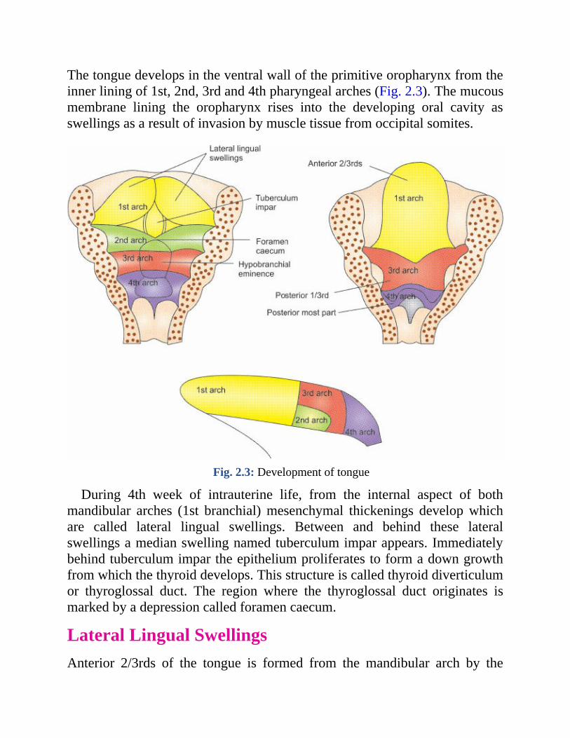
The tongue develops in the ventral wall of the primitive oropharynx from theinner lining of 1st, 2nd, 3rd and 4th pharyngeal arches (Fig. 2.3). The mucousmembrane lining the oropharynx rises into the developing oral cavity asswellings as a result of invasion by muscle tissue from occipital somites.
Fig. 2.3: Development of tongue
During 4th week of intrauterine life, from the internal aspect of bothmandibular arches (1st branchial) mesenchymal thickenings develop whichare called lateral lingual swellings. Between and behind these lateralswellings a median swelling named tuberculum impar appears. Immediatelybehind tuberculum impar the epithelium proliferates to form a down growthfrom which the thyroid develops. This structure is called thyroid diverticulumor thyroglossal duct. The region where the thyroglossal duct originates ismarked by a depression called foramen caecum.
Lateral Lingual SwellingsAnterior 2/3rds of the tongue is formed from the mandibular arch by the

fusion of two lateral lingual swellings and tuberculum impar. As the lingualswellings grow and fuse with each other, they over grow the tuberculumimpar and therefore the ectodermal lining of entire anterior 2/3rds is derivedfrom these two swellings and is of ectodermal origin. After these structuresfuses the epithelium at the periphery proliferates into the mesenchyme toform a horseshoe shaped lamina all around. The central cells of this laminadegenerate to form linguo-gingival groove which separate the body of thetongue from floor of the mouth except for the region of frenum of tongue.
The posterior 1/3rd of the tongue develops from another swelling known ashypobranchial eminence. This hypobranchial eminence is derived from 2nd,3rd and 4th arches. The epithelial lining of posterior 1/3rd is endodermal inorigin. As the development progresses the mesoderm of the 3rd branchialarch overgrow the mesoderm of 2nd arch and joins with mesoderm of 1starch. The second arch mesoderm remains buried below the surface (Fig. 2.3).A V-shaped ‘sulcus terminalis’ demarcate the anterior 2/3rds and posterior1/3rd of tongue. The posterior most part of the tongue is derived from the 4tharch.
The epithelium of the tongue is derived partly from both ectoderm andendoderm and is single layered initially which later turns to stratifiedsquamous epithelium. Circumvallate papillae develop by 2nd to 5th monthsof intrauterine life. Fungiform papillae develop at an earlier stage by 11thweek of intrauterine life while filiform papillae develop later anddevelopment is completed only postnatally. The taste buds develop by theinductive interaction between epithelial cells and invading gustatory nervecells from chorda tympani, glossopharyngeal and vagus nerves. The mucosalining the posterior part of the tongue becomes pitted by deep crypts thatdevelop into lingual tonsil.
The muscles of the tongue have a dual origin. The intrinsic musclesprobably arise in situ in the pharyngeal arch mesenchyme while the extrinsicmuscles arise in the occipital somite region opposite to origin of hypoglossalnerve. The muscle mass migrates forward beneath the mucosal layers of thetongue which also carries the hypoglossal nerve.
In the initial stages of development, tongue enlarges rapidly and occupiesthe whole of stomatodeum. Later as the stomatodeum increases in size thetongue descends down allowing the palatal shelves to become horizontal. Theentire tongue is in the mouth at birth and by the 4th year posterior 1/3rddescends down to pharynx. The size of the tongue doubles in length, width


