Tao et al. Cell Death and Disease (2018) 9:309 DOI 10.1038/s41419-018-0372-9 Cell Death & Disease
ART ICLE Open Ac ce s s
Estradiol signaling mediates genderdifference in visceral adiposity viaautophagyZhipeng Tao1, Louise D. Zheng1, Cayleen Smith1, Jing Luo1, Alex Robinson1, Fabio A. Almeida2, Zongwei Wang3,Aria F. Olumi3, Dongmin Liu1 and Zhiyong Cheng 1
AbstractExcessive adiposity (particularly visceral fat mass) increases the risks of developing metabolic syndrome. Women havelower deposit of visceral fat than men, and this pattern becomes diminished postmenopausally, but the underlyingmechanism remains largely unknown. Here, we show that the gender difference in visceral fat distribution iscontrolled by an estradiol–autophagy axis. In C57BL/6J and wild-type control mice, a higher visceral fat mass wasdetected in the males than in the females, which was associated with lower expression of estrogen receptor α (ERα)and more active autophagy in males vs. females. However, deletion of ERα normalized autophagy activity andabolished the gender difference in visceral adiposity. In line with the adiposity-reducing effect of the ERα–autophagyaxis, we found that downregulation of ERα and increased autophagy activity were required for adipogenesis, whileinduction of estradiol signaling dampened autophagy and drastically prevented adipogenesis. Mechanistically, theestradiol-ERα signaling activated mTOR, which phosphorylated and inhibited ULK1, thereby suppressing autophagyand adipogenesis. Together, our study suggests that the lower visceral adiposity in the females (vs. the males) arisesfrom a more active estradiol-ERα signaling, which tunes down autophagy and adipogenesis.
IntroductionWhite adipose (or fat) tissues (WAT) play a central role in
metabolic homeostasis through energy storage and endo-crine functions1,2. It has been shown that fat depots at dis-tinct anatomical (e.g., subcutaneous vs. visceral) locationshave intrinsic differences in hormone response, geneexpression, remodeling, and metabolism3–9. Excessive visc-eral fat is associated with metabolic syndrome development(e.g., insulin resistance) in animal models and humans,whereas subcutaneous fat is benign or protective9–13.
Compared with women, men have more visceral fat14,15.Intriguingly, visceral fat mass increases in post-menopausalwomen (characteristic of reduced estrogen secretion), whichcan be prevented by estrogen replacement therapy16,17.These findings underscored an important role of estrogensignaling in the regulation of fat development and distribu-tion, yet the molecular mechanism remains largely elusive.WAT mass development and maintenance are dependent
on adipocyte turnover. It was estimated that the rate ofadipocyte turnover was 10% per year in humans and 1–5%per day in mice18,19. The overall adipocyte number in WATis balanced by adipogenesis (i.e., the differentiation of pre-adipocyte into adipocytes) and adipocyte apoptosis18–20.Emerging evidence suggests that autophagy, the majorintracellular degradation and remodeling system, regulatesboth adipocyte differentiation and apoptosis21–23. Blockage ofautophagy through ablation of Atg5 or Atg7 substantiallyprevents adipocyte differentiation and promotes adipocyte
changesweremade. The images or other third partymaterial in this article are included in the article’s Creative Commons license, unless indicated otherwise in a credit line to thematerial. Ifmaterial is not included in the article’s Creative Commons license and your intended use is not permitted by statutory regulation or exceeds the permitted use, you will need to obtainpermission directly from the copyright holder. To view a copy of this license, visit http://creativecommons.org/licenses/by/4.0/.
Correspondence: Dongmin Liu ([email protected]) or Zhiyong Cheng ([email protected])1Department of Human Nutrition, Foods, and Exercise, Fralin Life ScienceInstitute, College of Agriculture and Life Science, Virginia Tech, Blacksburg, VA24061, USA2Department of Health Promotion, Social & Behavioral Health, College of PublicHealth, University of Nebraska Medical Center, Omaha, NE, USAFull list of author information is available at the end of the articleEdited by B Zhivotovsky
Official journal of the Cell Death Differentiation Association
apoptosis, which significantly reduces fat mass in mice21–23.We and others have showed that autophagy is required tomaintain PPARγ and FSP27, the key regulators of adipocytedifferentiation and lipid droplet formation in fat cells24,25.Suppression of autophagy downregulates PPARγ and FSP27,concomitant with dampened adipocyte differentiation andmarginal lipid accumulation in the cells24,25. Therefore,autophagy acts as a critical regulator of WAT remodelingand maintenance.Given the above-mentioned evidence that implies
estrogen and autophagy in WAT regulation, we asked thequestion whether autophagy might interact with estrogensignaling, and how it might link to the gender difference invisceral adiposity. Here, we show that male mice hadhigher visceral fat mass than the females, which wasassociated with lower expression of estrogen receptors(ERα) in the visceral adipose tissue. Activation of estradiol(E2) signaling suppressed autophagy via an mTOR-ULKpathway, which inhibited adipogenesis and was associatedwith a lower visceral adiposity in the mice. However,deletion of ERα normalized autophagy activity and gender-dependent difference in visceral adiposity. Our data revealsfor the first time an E2–ERα–autophagy axis that con-tributes to the gender difference in visceral fat distribution.
ResultsFemale mice had lower visceral WAT (vWAT) mass thanmale miceA higher vWAT volume in men than women has been
observed across races14,15. To determine if mice have asimilar pattern of fat distribution, we examined C57BL/6Jat the age of 6–7 weeks (Fig. 1). As expected, the femalemice had lower body weight than males (average 15.4 g vs.17.5 g, p < 0.05; Fig. 1a). The net weights of gonadal WAT(the largest visceral fat depots in mice) and subcutaneousWAT (inguinal fat depots, sWAT) were both lower infemale mice than in the males (Fig. 1b, c). However, afternormalization against the body weight only the vWATmass remained lower in females than in males (average0.87 vs. 1.21%, p < 0.05; Fig. 1d), while the differences insWAT mass became indiscernible between the males andfemales (average 0.45 vs. 0.49%, p= 0.14; Fig. 1e).Therefore, the ratios of visceral fat to body weight (orvWAT percentage) reveal a gender-dependent phenotypein mice as observed previously in human subjects.
Autophagy activity in vWAT was lower in female than malemiceAutophagosome formation is characterized by lipida-
tion of LC3 to form LC3-phospholipid conjugate (LC3-II),which can be degraded by lysosomal hydrolase in auto-lysosome26,27. In sWAT, steady-state LC3-II levels did notdiffer between female and male mice (Fig. 2a, b). However,a significantly lower (50%, p < 0.01) steady-state LC3-II
was detected in vWAT from male mice vs. that fromfemale mice (Fig. 2c, d), suggesting that autophagicdegradation of LC3-II in vWAT was more active in themales than in the females. To test this, we detected LC3-IIturnover (or autophagy flux)25,27,28, by detecting theaccumulation of LC3-II after treating WAT explant cul-tures with autophagy inhibitors bafilomycin A1 and leu-peptin (BL) for 4 h. In line with the steady-state levels ofLC3-II in sWAT being indiscernible between the malesand females (Fig. 2a, b), autophagy flux in sWAT did notshow significant difference between the males and females(Fig. 2e, f). However, the autophagy flux in vWAT wassignificantly higher in the males (1.6-fold upregulated,p < 0.05) than in the females (Fig. 2e, f). The turnover ofp62, which is selectively degraded by autophagy, furthervalidated the higher autophagy activity in vWAT frommale mice than that from female mice (Fig. 2g, h). Thesedata support the notion that increased LC3-IIturnover results in a reduced steady-state level of LC3-II(Fig. 2c, d)26,29. Together, female mice had lower autop-hagy activity in vWAT than the males.
Estrogen receptors were upregulated in vWAT fromfemales vs. malesE2 signaling is primarily funneled through ERα and
ERβ30. Compared with the males, the female mice hadsimilar expression of ERα and ERβ in sWAT (Fig. 3a, b).However, vWAT had significant lower protein levels ofERα and ERβ in male mice than in female mice (Fig. 3c,d), being 40% for ERα (p < 0.01) and 54% for ERβ (p <0.05). Interestingly, the overall protein levels of ERβ invWAT appeared to be much lower than that of ERα forboth genders (Fig. 3c, d), suggesting that ERαmight play adominant role in mediating E2 actions in vWAT.
Adipogenesis was associated with downregulation ofestrogen receptors but upregulation of autophagy activityThe observation of lower expression of ER but higher
autophagy activity in the males vs. females (Figs. 2, 3)prompted us to examine whether this reciprocal relationexists in adipogenesis, the process that is critical for adi-pose tissue development and maintenance31,32. As shownin Fig. 4a and b, ERα was downregulated by 80% (p < 0.01)and ERβ by 54% (p < 0.05) during adipogenesis, which wascharacterized by drastic accumulation of lipid in the cells(Fig. 4c). In addition, the downregulation of ERs wasassociated with 81% (p < 0.0001) reduction in the steady-state level of LC3-II (Fig. 4d, e). Autophagy flux assays byLC3-II turnover suggested that the differentiated adipo-cytes had an autophagy activity 2.6-fold (p < 0.0001)higher than the preadipocytes, which was further verifiedby the turnover of p62 (Fig. 4f, g). These results recapi-tulated the pattern observed in vWAT, where steady-statelevel of LC3-II was reduced due to increased autophagy
Tao et al. Cell Death and Disease (2018) 9:309 Page 2 of 13
Official journal of the Cell Death Differentiation Association
activity (Fig. 2)26,29. Importantly, the in vitro and in vivodata work in concert to reveal a reciprocal relationshipbetween autophagy activity and ER levels.
Estradiol signaling suppressed autophagy andadipogenesisTo examine whether E2 signaling per se regulate adi-
pocyte autophagy and adipogenesis, we treated 3T3L1
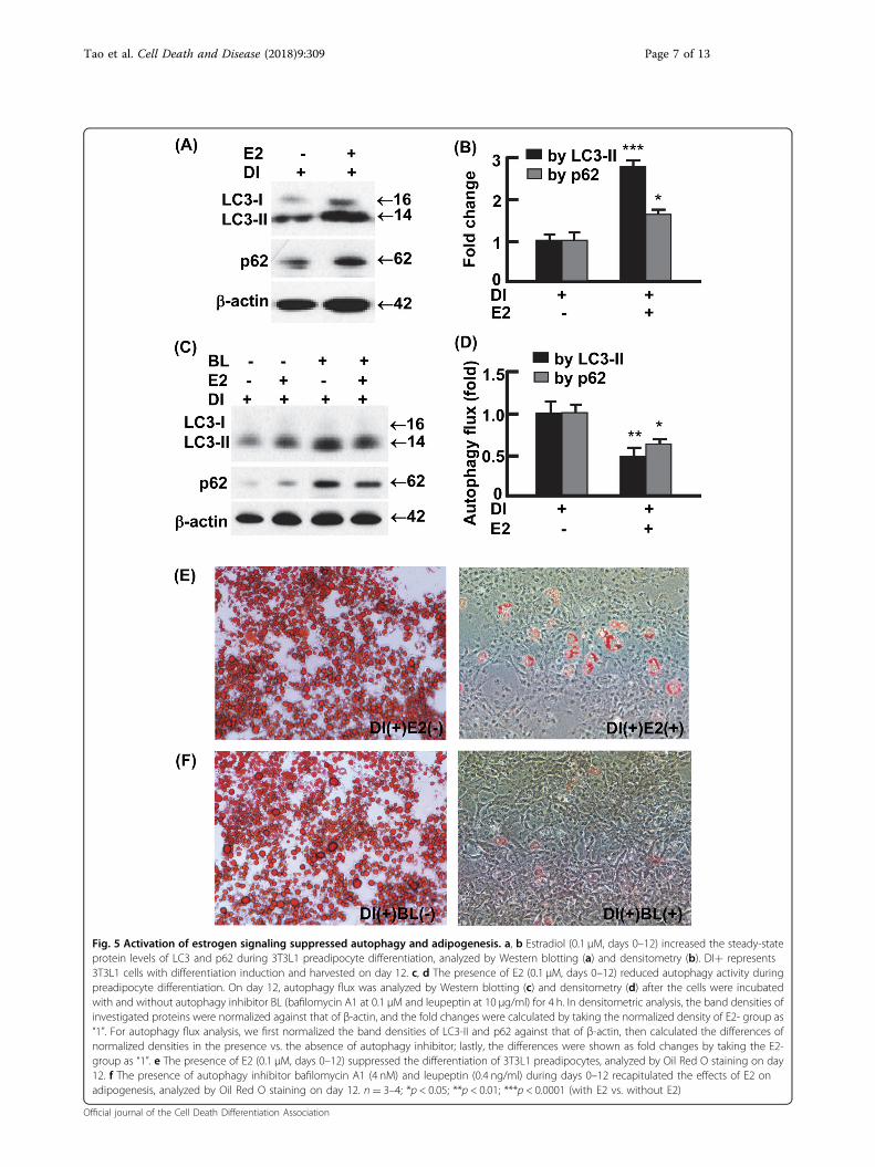
preadipocytes with or without E2 (0.1 μM) on day 0through day 12, during which adipogenesis was inducedaccording to an established protocol (see Materials andMethods)25,33–35. E2 treatment substantially increased theaccumulation of LC3-II (2.8-folds, p < 0.0001), as well asp62 (1.6-fold, p < 0.05), the selective substrate of autop-hagy for degradation (Fig. 5a, b)25,33,36. The E2-enhancedaccumulation of LC3-II and p62 was associated with
Fig. 1 Gender difference existed in the percentage of visceral but not subcutaneous fat. a The body weights of male and female mice at theage of 6–7 weeks. b The net weights of visceral WAT from male and female mice. c The net weights of subcutaneous WAT from male and femalemice. d The percentage of visceral WAT from male and female mice, normalized against the body weights. e The percentage of subcutaneous WATfrom male and female mice, normalized by body weights. *p < 0.05; **p < 0.01; n.s. not significant; n= 4–6
Tao et al. Cell Death and Disease (2018) 9:309 Page 3 of 13
Official journal of the Cell Death Differentiation Association
reduced autophagy flux (Fig. 5c, d), suggesting that E2dampens autophagy activity. In addition, the E2-treatedcells were barely differentiated into mature adipocytes andshowed marginal lipid accumulation compared withvehicle-treated cells (Fig. 5e). Likewise, treatment of3T3L1 preadipocytes with the established autophagyinhibitor BL almost completely prevented adipogenesis(Fig. 5f). In addition, the presence of E2 or autophagy
inhibitor BL similarly inhibited autophagy and adipogen-esis in primary stromal vascular cells isolated from vWAT(Fig. 1s). Thus, E2 signaling suppresses adipogenesis atleast in part via autophagy inhibition.
Estradiol signaling suppressed autophagy via mTOR-ULK1To explore the mechanism of E2 regulating autophagy,
we analyzed the interactions of E2 signaling and proteins
Fig. 2 Autophagy activities showed gender difference in visceral but not subcutaneous fat. a, b The steady-state protein levels of LC3 insubcutaneous WAT, analyzed by Western blotting (a) and densitometry (b). c, d The steady-state protein levels of LC3 in visceral WAT, analyzed byWestern blotting (c) and densitometry (d). e–h Measurement of autophagy flux in subcutaneous and visceral WAT. The WAT explant cultures wereincubated with and without autophagy inhibitor bafilomycin A1 (0.1 μM) and leupeptin (10 μg/ml) for 4 h, and the turnovers of LC3-II and p62 wereexamined by Western blotting (e, g) and densitometry (f, h). In densitometric analyses, the band densities of investigated proteins were normalizedagainst that of GAPDH or β-actin, and the fold changes were calculated by taking the normalized density of female group as “1”. For autophagy flux,we first normalized the band densities of LC3-II and p62 against that of GAPDH, then calculated the differences of normalized densities in thepresence vs. the absence of autophagy inhibitor; lastly, the differences were shown as fold changes by taking the female group as “1”. BL bafilomycinA1 and leupeptin, M male, F female; *p < 0.05; **p < 0.01; n.s. not significant; n= 3–4
Tao et al. Cell Death and Disease (2018) 9:309 Page 4 of 13
Official journal of the Cell Death Differentiation Association
that are known to control autophagy, including ULK1,beclin 1, Atg5, Atg7, and Atg1237–39. We found thatbeclin 1 was upregulated during adipocyte differentiation,but E2 treatment had marginal effect on beclin 1 level(Fig. 2s, A–B). ULK1 was activated during adipogenesis,because the mTOR-mediated inhibitory phosphorylationof ULK1 at Ser 757 (p-ULK1Ser757) was significantlyreduced37–39. However, E2 treatment suppressed ULK1by increasing p-ULK1Ser757, concomitant with the acti-vation of mTOR indicated by phosphorylation at Ser2448(p-mTORSer2448) (Fig. 6a, b)37–41. These data suggest thatE2 signaling acts on ULK1 but not beclin 1, although bothproteins participate in autophagy initiation (i.e., formationof the isolation membrane)37–39. Moreover, no discerniblechange was detected in Atg5, Atg7, and Atg12-Atg5conjugate, the proteins or components that regulatemembrane elongation37,39, during adipocyte differentia-tion or during E2 treatment (Fig. 2s, A–B). These results,along with the above observation that E2 suppressedautophagy in adipocytes (Figs. 4–5), suggest that E2 mayregulate autophagy via the mTOR-ULK1 cascade.To validate the E2/ER signaling-mTOR-ULK1 pathway
in vivo, we examined adipose tissues from male andfemale C57BL/6J mice (Fig. 6c, d). In line with the femaleshaving higher ER levels in vWAT than the males (Fig. 3),the activating phosphorylation of mTOR (p-mTORSer2448)
was enhanced by 4.8-fold (p < 0.0001), and the mTOR-mediated inhibition of ULK1 (p-ULK1Ser757) was 2.1-foldstronger (p < 0.05; Fig. 6e, f). In sWAT, however, no sta-tistically significant difference was detected (Fig. 6c, d),consistent with the male and the females showing com-parable levels of ER in sWAT (Fig. 3). Notably, we did notdetect significant difference in beclin 1, Atg5, Atg7, orAtg12-Atg5 conjugate, between the male and the females(Fig. 2s, C–D). Together, our in vitro and in vivo resultssupport the hypothesis that E2/ER signaling regulatesadipose autophagy via the mTOR-ULK1 pathway.
Ablation of ERα normalized autophagy activity andabolished gender difference in visceral adiposityERα and ERβ have been shown to suppress or enhance
autophagy in different cancer cells42–46. To determine theprimary role player in the regulation of autophagy andadipocyte differentiation, we treated 3T3L1 cells withselective agonists of ERα (PPT) and ERβ (DPN)47,48. PPTreduced autophagy activity and suppressed adipogenesisbut DPN had marginal effect, suggesting that ERα playedthe dominant role (Fig. 3s). To validate this, we examinedautophagy and visceral adiposity in ERα knockout (KO)mice (Fig. 7)49. As observed in the C57BL/6J mice, thecontrol (or wild-type, WT) females had higher expressionof ERα and stronger inhibition of ULK1 (p-ULK1Ser757) in
Fig. 3 The expression of estrogen receptors (ER) showed gender difference in visceral but not subcutaneous fat. a–b The protein levels ofERα and ERβ in subcutaneous WAT, analyzed by Western blotting (a) and densitometry (b). c–d The protein levels of ERα and ERβ in visceral WAT,analyzed by Western blotting (c) and densitometry (d). In densitometric analysis, the band densities of investigated proteins were normalized againstthat of β-actin, and the fold changes were calculated by taking the normalized density of female group as “1”. *p < 0.05; **p < 0.01; n.s. not significant;n= 3–4
Tao et al. Cell Death and Disease (2018) 9:309 Page 5 of 13
Official journal of the Cell Death Differentiation Association
Fig. 4 Adipogenesis was associated with downregulation of ER but upregulation of autophagy activity. a–b The protein levels of ERα andERβ in preadipocytes and mature (or differentiated) adipocytes, analyzed by Western blotting (a) and densitometry (b). DI differentiation induction. DI+ represents differentiated 3T3L1 cells (mature adipocytes) harvested on day 12; DI− represents 3T3L1 cells without differentiation induction (i.e.,preadipocytes) harvested on day 12. c Oil Red O staining to detect the differentiation of preadipocytes into mature adipocytes. On day 12, massivelipid accumulation was detected in mature adipocytes but not in preadipocytes. d, e The steady-state protein levels of LC3 in preadipocytes andmature adipocytes, analyzed by Western blotting (d) and densitometry (e) on day 12. f, g Measurement of autophagy flux in preadipocytes andmature adipocytes. On day 12, the cells were incubated in the presence or absence of autophagy inhibitor BL (bafilomycin A1 at 0.1 μM andleupeptin at 10 μg/ml) for 4 h, and the turnovers of LC3-II and p62 were examined by Western blotting (f) and densitometry (g). In densitometricanalysis, the band densities of investigated proteins were normalized against that of GAPDH or β-actin, and the fold changes were calculated bytaking the normalized density of DI− group as “1”. For autophagy flux analysis, we first normalized the band densities of LC3-II and p62 against thatof β-actin, then calculated the differences of normalized densities in the presence vs. the absence of autophagy inhibitor; lastly, the differences wereshown as fold changes by taking the DI− group as “1”. BL bafilomycin A1 and leupeptin. *p < 0.05; **p < 0.01; ***p < 0.0001; n= 3–4
Tao et al. Cell Death and Disease (2018) 9:309 Page 6 of 13
Official journal of the Cell Death Differentiation Association
Fig. 5 Activation of estrogen signaling suppressed autophagy and adipogenesis. a, b Estradiol (0.1 μM, days 0–12) increased the steady-stateprotein levels of LC3 and p62 during 3T3L1 preadipocyte differentiation, analyzed by Western blotting (a) and densitometry (b). DI+ represents3T3L1 cells with differentiation induction and harvested on day 12. c, d The presence of E2 (0.1 μM, days 0–12) reduced autophagy activity duringpreadipocyte differentiation. On day 12, autophagy flux was analyzed by Western blotting (c) and densitometry (d) after the cells were incubatedwith and without autophagy inhibitor BL (bafilomycin A1 at 0.1 μM and leupeptin at 10 μg/ml) for 4 h. In densitometric analysis, the band densities ofinvestigated proteins were normalized against that of β-actin, and the fold changes were calculated by taking the normalized density of E2- group as“1”. For autophagy flux analysis, we first normalized the band densities of LC3-II and p62 against that of β-actin, then calculated the differences ofnormalized densities in the presence vs. the absence of autophagy inhibitor; lastly, the differences were shown as fold changes by taking the E2-group as “1”. e The presence of E2 (0.1 μM, days 0–12) suppressed the differentiation of 3T3L1 preadipocytes, analyzed by Oil Red O staining on day12. f The presence of autophagy inhibitor bafilomycin A1 (4 nM) and leupeptin (0.4 ng/ml) during days 0–12 recapitulated the effects of E2 onadipogenesis, analyzed by Oil Red O staining on day 12. n= 3–4; *p < 0.05; **p < 0.01; ***p < 0.0001 (with E2 vs. without E2)
Tao et al. Cell Death and Disease (2018) 9:309 Page 7 of 13
Official journal of the Cell Death Differentiation Association
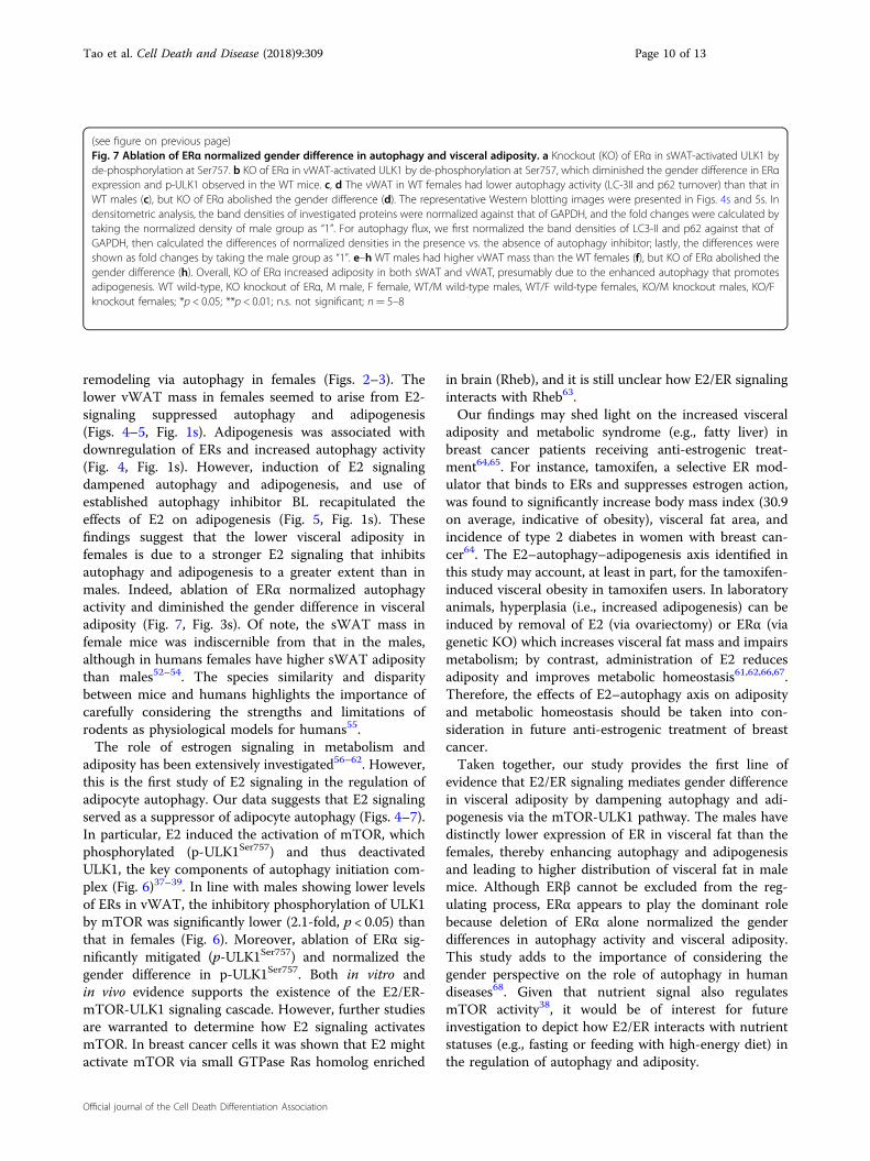
vWAT than the WT males, although sWAT showed nogender difference in ERα and p-ULK1Ser757 (Fig. 7a, b).However, KO of ERα reduced the inhibitory phosphor-ylation of ULK1 (p-ULK1Ser757) in both sWAT andvWAT, and, most importantly, it abolished the genderdifference in vWAT (Fig. 7a, b). Consistently, the gender-dependent difference in autophagy flux was diminished bythe KO of ERα in vWAT (Fig. 7c–d; Figs. 4s–5s). In linewith the enhanced autophagy (Fig. 7a–d; Figs. 4s–5s),which was found to promote adipogenesis (Figs. 4–5;Fig. 1s), the loss of ERα increased both sWAT and vWATmasses in the ERα KO mice compared with the WT mice(Fig. 7e–h). Furthermore, the gender difference in vWATmass was abolished by the ablation of ERα (Fig. 7f, h).Therefore, the E2-ERα signaling cascade plays the central
role in the gender difference in visceral adiposity viaregulating autophagy.
DiscussionIncreased visceral adiposity has been strongly associated
with higher risks of developing metabolic disorders9–13.Females have lower visceral fat mass than males, but thisgender difference is diminished in older age groupsbecause post-menopausal women have increased visceraladiposity; the age-related accumulation of visceral fat inpost-menopausal women is likely due to drasticallyreduced estrogen levels14–17,50,51. Indeed, estrogen repla-cement therapy prevents post-menopausal women fromexcessive visceral adiposity, underlining estrogen signal-ing as an important regulator of visceral distribution16,17.
Fig. 6 Estradiol-ER signaling regulated autophagy via mTOR-ULK1 pathway. a, b E2 (0.1 μM, days 0–12) induced the activating phosphorylationof mTOR (Ser2448) and the mTOR-catalyzed inhibitory phosphorylation of ULK1 (Ser757), analyzed by Western blotting (a) and densitometry (b). DI+represents 3T3L1 cells with differentiation induction and harvested on day 12, and DI− represents 3T3L1 cells without differentiation and harvestedon day 12. c, d The females and males showed comparable phosphorylation of mTOR (Ser2448) and ULK1 (Ser757), analyzed by Western blotting (c)and densitometry (d). e, f The females showed significantly stronger phosphorylation of mTOR (Ser2448) and ULK1 (Ser757) than the males, analyzedby Western blotting (e) and densitometry (f). In densitometric analysis, the band densities of investigated proteins were normalized against that ofGAPDH or β-actin, and the fold changes were calculated by taking the normalized density of DI-E2- group (a and b) or female group (c–f) as “1”. *p <0.05; **p < 0.01; ***p < 0.0001; n.s. not significant; n= 3–4
Tao et al. Cell Death and Disease (2018) 9:309 Page 8 of 13
Official journal of the Cell Death Differentiation Association
To understand the underlying mechanism, we investi-gated the interaction between estrogen signaling andautophagy, and its relation with adipogenesis and visceral
adiposity in mice. We found that female mice had lowervWAT mass than males (Fig. 1), which was associatedwith higher expression of ERs but lower activity of cell
Fig. 7 (See legend on next page.)
Tao et al. Cell Death and Disease (2018) 9:309 Page 9 of 13
Official journal of the Cell Death Differentiation Association
remodeling via autophagy in females (Figs. 2–3). Thelower vWAT mass in females seemed to arise from E2-signaling suppressed autophagy and adipogenesis(Figs. 4–5, Fig. 1s). Adipogenesis was associated withdownregulation of ERs and increased autophagy activity(Fig. 4, Fig. 1s). However, induction of E2 signalingdampened autophagy and adipogenesis, and use ofestablished autophagy inhibitor BL recapitulated theeffects of E2 on adipogenesis (Fig. 5, Fig. 1s). Thesefindings suggest that the lower visceral adiposity infemales is due to a stronger E2 signaling that inhibitsautophagy and adipogenesis to a greater extent than inmales. Indeed, ablation of ERα normalized autophagyactivity and diminished the gender difference in visceraladiposity (Fig. 7, Fig. 3s). Of note, the sWAT mass infemale mice was indiscernible from that in the males,although in humans females have higher sWAT adipositythan males52–54. The species similarity and disparitybetween mice and humans highlights the importance ofcarefully considering the strengths and limitations ofrodents as physiological models for humans55.The role of estrogen signaling in metabolism and
adiposity has been extensively investigated56–62. However,this is the first study of E2 signaling in the regulation ofadipocyte autophagy. Our data suggests that E2 signalingserved as a suppressor of adipocyte autophagy (Figs. 4–7).In particular, E2 induced the activation of mTOR, whichphosphorylated (p-ULK1Ser757) and thus deactivatedULK1, the key components of autophagy initiation com-plex (Fig. 6)37–39. In line with males showing lower levelsof ERs in vWAT, the inhibitory phosphorylation of ULK1by mTOR was significantly lower (2.1-fold, p < 0.05) thanthat in females (Fig. 6). Moreover, ablation of ERα sig-nificantly mitigated (p-ULK1Ser757) and normalized thegender difference in p-ULK1Ser757. Both in vitro andin vivo evidence supports the existence of the E2/ER-mTOR-ULK1 signaling cascade. However, further studiesare warranted to determine how E2 signaling activatesmTOR. In breast cancer cells it was shown that E2 mightactivate mTOR via small GTPase Ras homolog enriched
in brain (Rheb), and it is still unclear how E2/ER signalinginteracts with Rheb63.Our findings may shed light on the increased visceral
adiposity and metabolic syndrome (e.g., fatty liver) inbreast cancer patients receiving anti-estrogenic treat-ment64,65. For instance, tamoxifen, a selective ER mod-ulator that binds to ERs and suppresses estrogen action,was found to significantly increase body mass index (30.9on average, indicative of obesity), visceral fat area, andincidence of type 2 diabetes in women with breast can-cer64. The E2–autophagy–adipogenesis axis identified inthis study may account, at least in part, for the tamoxifen-induced visceral obesity in tamoxifen users. In laboratoryanimals, hyperplasia (i.e., increased adipogenesis) can beinduced by removal of E2 (via ovariectomy) or ERα (viagenetic KO) which increases visceral fat mass and impairsmetabolism; by contrast, administration of E2 reducesadiposity and improves metabolic homeostasis61,62,66,67.Therefore, the effects of E2–autophagy axis on adiposityand metabolic homeostasis should be taken into con-sideration in future anti-estrogenic treatment of breastcancer.Taken together, our study provides the first line of
evidence that E2/ER signaling mediates gender differencein visceral adiposity by dampening autophagy and adi-pogenesis via the mTOR-ULK1 pathway. The males havedistinctly lower expression of ER in visceral fat than thefemales, thereby enhancing autophagy and adipogenesisand leading to higher distribution of visceral fat in malemice. Although ERβ cannot be excluded from the reg-ulating process, ERα appears to play the dominant rolebecause deletion of ERα alone normalized the genderdifferences in autophagy activity and visceral adiposity.This study adds to the importance of considering thegender perspective on the role of autophagy in humandiseases68. Given that nutrient signal also regulatesmTOR activity38, it would be of interest for futureinvestigation to depict how E2/ER interacts with nutrientstatuses (e.g., fasting or feeding with high-energy diet) inthe regulation of autophagy and adiposity.
(see figure on previous page)Fig. 7 Ablation of ERα normalized gender difference in autophagy and visceral adiposity. a Knockout (KO) of ERα in sWAT-activated ULK1 byde-phosphorylation at Ser757. b KO of ERα in vWAT-activated ULK1 by de-phosphorylation at Ser757, which diminished the gender difference in ERαexpression and p-ULK1 observed in the WT mice. c, d The vWAT in WT females had lower autophagy activity (LC-3II and p62 turnover) than that inWT males (c), but KO of ERα abolished the gender difference (d). The representative Western blotting images were presented in Figs. 4s and 5s. Indensitometric analysis, the band densities of investigated proteins were normalized against that of GAPDH, and the fold changes were calculated bytaking the normalized density of male group as “1”. For autophagy flux, we first normalized the band densities of LC3-II and p62 against that ofGAPDH, then calculated the differences of normalized densities in the presence vs. the absence of autophagy inhibitor; lastly, the differences wereshown as fold changes by taking the male group as “1”. e–h WT males had higher vWAT mass than the WT females (f), but KO of ERα abolished thegender difference (h). Overall, KO of ERα increased adiposity in both sWAT and vWAT, presumably due to the enhanced autophagy that promotesadipogenesis. WT wild-type, KO knockout of ERα, M male, F female, WT/M wild-type males, WT/F wild-type females, KO/M knockout males, KO/Fknockout females; *p < 0.05; **p < 0.01; n.s. not significant; n= 5–8
Tao et al. Cell Death and Disease (2018) 9:309 Page 10 of 13
Official journal of the Cell Death Differentiation Association
Materials and methodsMiceC57BL/6J mice were housed in plastic cages on a 12-h
light–dark photocycle and with free access to water andregular chow diet as described previously25,34; at the ageof 6–7 week old, the mice were weighed and sacrificed fortissue collection. The WT and global ERα KO mice wereobtained by breeding heterozygous males to females asdescribed previously49; at the age of 12–16 week old, theWT and ERα KO mice were weighed and sacrificed fortissue collection. The WAT pads were collected andweighed quickly before SVF isolation, explant culture forautophagy flux analysis, or snap freezing in liquid nitro-gen. Animal use procedures followed the National Insti-tutes of Health guidelines and were approved by theVirginia Tech Institutional Animal Care and UseCommittee.
3T3L1 cell culture, differentiation, and treatment3T3L1 preadipocytes (ATCC CL-173, Manassas, VA,
USA) were cultured in basal media (DMEM media con-taining 10% FBS, 100 units/ml penicillin, and 100 μg/mlstreptomycin (1× P/S)), at 37 °C in a humidified atmo-sphere of 5% CO2
33–35. The media were replaced every2 days until the cells became confluent (day 0), and after 2more days (day 2) the medium was changed to differ-entiation medium I (DMEM with 10% FBS, P/S (1×),IBMX (0.5 mM), dexamethasone (1 μM), insulin (1 μg/ml), and rosiglitazone (2 μM)). At the end of day 4, themedium was changed to differentiation medium II(DMEM with 10% FBS, P/S (1×), and insulin (1 μg/ml)).At the end of day 6, the medium was changed to basalmedia and the cells were maintained until day 12. Pre-adipocytes without differentiation induction were main-tained in basal media and supplied with fresh mediumevery 2 days till day 12. E2 at the concentrations of 1nM–10 μM has been used to treat adipocytes69–76. Ourpreliminary tests indicated that E2 at 1 nM, 10 nM, 0.1μM, and 0.2 μM imposed similar effects, but E2 at 0.1 μM(likewise 0.2 μM) was the most potent (data not shown).As such, we used E2 of 0.1 μM for the treatments startingon day 0 through day 12. Other chemicals were used atthe concentrations established previously, including PPT(0.1 μM), DPN (0.1 μM), and bafilomycin A1 (4 nM), andleupeptin (0.4 ng/ml)25,77,78, to treat the cells duringdifferentiation.
Primary stromal vascular cell culture, differentiation, andtreatmentFresh gonadal WAT from C57BL/6J mice were dis-
sected, minced, and digested as previously described79.Cells were suspended in basal media (DMEM/F12 con-taining 10% FBS and 100 units/ml penicillin and 100 μg/ml streptomycin (1× P/S)), and centrifuged at 500 × g for
5 min. The pellet was resuspended in basal media andfiltered through a 40-micron cell strainer. After cen-trifugation (500 × g) for 5 min, the pellet was resuspendedin basal media and plated on 10-cm dishes. After cellsubculture to a 95% confluence (day 0) on 6-well plates,differentiation was induced in differentiation medium(DMEM/F12 medium with 10% FBS, 1× Pen/Strep, dex-amethasone (5 μM), insulin (0.5 μg/ml), IBMX (0.5 mM),and rosiglitazone (1 μM)) for 4 days (day 4). Then the cellswere maintained in maintenance medium (DMEM/F12medium containing 10% FBS, 1× Pen/Strep, and insulin(0.5 μg/ml)) for 6 days (day 10). The treatments withchemicals (E2 at 0.1 μM, bafilomycin A1 at 4 nM, andleupeptin at 0.4 ng/ml) started on day 0 through day 10 toexamine their effects on autophagy and adipogenesis.
Oil Red O stainingThe Oil Red O working solution was freshly prepared by
mixing 0.35% stock solution with dH2O (6:4) and filtered,and the staining was conducted as described25,33,35. Afterthe media were removed, the cells were washed once withcold phosphate buffered saline, and fixed in 4% for-maldehyde at room temperature for 10min. The cellswere then washed with dH2O and air dried. Oil Red Oworking solution was added to start the staining at roomtemperature for 1 h. The stained cells were washed withdH2O for four times before the images were captured witha Nikon ECLIPSE Ti Inverted Microscope (Melville, NY,USA).
Autophagy flux assayTo measure autophagy flux in cultured cells, we treated
3T3L1 preadipocytes, stromal vascular cells, and matureadipocytes (day 10) with bafilomycin A1 (inhibitor ofautophagosome acidification, at 0.1 μM) plus leupeptin(the inhibitor of lysosomal proteases, at 10 μg/ml) for 4 h.The cells were then harvested to prepare cell lysates aspreviously described25,33,35. To measure autophagy flux inWAT explants, freshly collected adipose tissues wereminced into small tissue fragments (2–3mm3) and cul-tured for 4 h with DMEM medium supplemented with 2mM glutamine, 1% (vol/vol) antibiotic solution, and 10%(vol/vol) FBS in a CO2 incubator (37 °C, 5% CO2). TheWAT explant cultures in the presence or absence ofbafilomycin-A1 (0.1 μM) and leupeptin (10 μg/ml) werethen harvested and lysed as described previously25. Theturnover of LC3-II or p62 protein, i.e., the substrates ofautophagy for degradation, was measured by Westernblotting and image analysis to assess autophagyflux25,27,28.
Western blottingTissue and cell lysates were prepared with PLC lysis
buffer (30 mM Hepes, pH 7.5, 150mM NaCl, 10%
Tao et al. Cell Death and Disease (2018) 9:309 Page 11 of 13
Official journal of the Cell Death Differentiation Association
glycerol, 1% Triton X-100, 1.5 mM MgCl2, 1 mM EGTA,10mM NaPPi, 100mM NaF, 1 mM Na3VO4) supple-mented with protease inhibitor cocktail (Roche), and 1mM PMSF80. Total protein concentrations of the lysateswere determined using a DC protein assay kits (Bio-Rad).Antibody (catalog number) information: GAPDH (MA5-15738) and β-actin (MA5-15739) antibodies were pur-chased from Pierce (Rockford, IL, USA); Atg5 (12994s),Atg7 (8558s), Atg12 (2011s), p62 (5114s), p-mTOR(Ser2448) antibody (5536s), and p-ULK1 (Ser757) anti-body (14202s) from Cell Signaling Technology (Beverly,MA, USA); Beclin 1 (MABN16), ERα (04–820), and ERβ(GR39) antibodies from EMD Millipore (Billerica, MA,USA); and LC3B antibody (L7543) from Sigma.
Statistical analysisData are presented as mean ± SD. Differences between
the groups were validated by one-way ANOVA with theleast significant difference post hoc test to detect statis-tical differences between groups and treatments (DI+ vs.DI−, E2+ vs. E2−, and BL+ vs. BL−). Differences inautophagy and adipose parameters between males andfemales were validated by a t-test. A value of p < 0.05 wasconsidered statistically significant.
AcknowledgementsFunding for this work was provided, in part, by USDA National Institute of Foodand Agriculture Hatch Project 1007334 (Z.C.), NIH grant R18DK091811 (F.A.A.),and NIH grant 1R01AT007077 (D.L.).
Author details1Department of Human Nutrition, Foods, and Exercise, Fralin Life ScienceInstitute, College of Agriculture and Life Science, Virginia Tech, Blacksburg, VA24061, USA. 2Department of Health Promotion, Social & Behavioral Health,College of Public Health, University of Nebraska Medical Center, Omaha, NE,USA. 3Department of Urology, Massachusetts General Hospital, HarvardMedical School, Boston, MA, USA
Conflict of interestThe authors declare that they have no conflict of interest.
Publisher's noteSpringer Nature remains neutral with regard to jurisdictional claims inpublished maps and institutional affiliations.
Supplementary Information accompanies this paper at https://doi.org/10.1038/s41419-018-0372-9.
Received: 9 October 2017 Revised: 30 January 2018 Accepted: 1 February2018
References1. Kershaw, E. E. & Flier, J. S. Adipose tissue as an endocrine organ. J. Clin.
Endocrinol. Metab. 89, 2548–2556 (2004).2. Galic, S., Oakhill, J. S. & Steinberg, G. R. Adipose tissue as an endocrine organ.
Mol. Cell. Endocrinol. 316, 129–139 (2010).3. Kim, S. N. et al. Sex differences in sympathetic innervation and browning of
white adipose tissue of mice. Biol. Sex Differ. 7, 67 (2016).
4. Jeffery, E., Church, C. D., Holtrup, B., Colman, L. & Rodeheffer, M. S. Rapid depot-specific activation of adipocyte precursor cells at the onset of obesity. Nat. CellBiol. 17, 376–385 (2015).
5. Tchoukalova, Y. D. et al. Regional differences in cellular mechanisms of adi-pose tissue gain with overfeeding. Proc. Natl Acad. Sci. USA 107, 18226–18231(2010).
6. Gesta, S. et al. Evidence for a role of developmental genes in the origin ofobesity and body fat distribution. Proc. Natl Acad. Sci. USA 103, 6676–6681(2006).
7. Macotela, Y. et al. Intrinsic differences in adipocyte precursor cells from dif-ferent white fat depots. Diabetes 61, 1691–1699 (2012).
8. Macotela, Y., Boucher, J., Tran, T. T. & Kahn, C. R. Sex and depot differences inadipocyte insulin sensitivity and glucose metabolism. Diabetes 58, 803–812(2009).
9. Shi, H., Strader, A. D., Woods, S. C. & Seeley, R. J. The effect of fat removal onglucose tolerance is depot specific in male and female mice. Am. J. Physiol.Endocrin. Metab. 293, E1012–E1020 (2007).
10. Cohen, P. et al. Ablation of PRDM16 and beige adipose causes metabolicdysfunction and a subcutaneous to visceral fat switch. Cell 156, 304–316(2014).
11. Veilleux, A., Caron-Jobin, M., Noel, S., Laberge, P. Y. & Tchernof, A. Visceraladipocyte hypertrophy is associated with dyslipidemia independent of bodycomposition and fat distribution in women. Diabetes 60, 1504–1511 (2011).
12. Manolopoulos, K. N., Karpe, F. & Frayn, K. N. Gluteofemoral body fat as adeterminant of metabolic health. Int. J. Obes. 34, 949–959 (2010).
13. Pischon, T. et al. General and abdominal adiposity and risk of death in Europe.N. Engl. J. Med. 359, 2105–2120 (2008).
14. Demerath, E. W. et al. Anatomical patterning of visceral adipose tissue: race,sex, and age variation. Obesity15, 2984–2993 (2007).
15. Despres, J. P. et al. Race, visceral adipose tissue, plasma lipids, and lipoproteinlipase activity in men and women: the Health, Risk Factors, Exercise Training,and Genetics (HERITAGE) family study. Arterioscler. Thromb. Vasc. Biol. 20,1932–1938 (2000).
16. Haarbo, J., Marslew, U., Gotfredsen, A. & Christiansen, C. Postmenopausalhormone replacement therapy prevents central distribution of body fat aftermenopause. Metabolism 40, 1323–1326 (1991).
17. Gambacciani, M. et al. Body weight, body fat distribution, and hormonalreplacement therapy in early postmenopausal women. J. Clin. Endocrinol.Metab. 82, 414–417 (1997).
18. Rigamonti, A., Brennand, K., Lau, F. & Cowan, C. A. Rapid cellular turnover inadipose tissue. PLoS ONE 6, e17637 (2011).
19. Spalding, K. L. et al. Dynamics of fat cell turnover in humans. Nature 453,783–787 (2008).
20. Prins, J. B. & O’Rahilly, S. Regulation of adipose cell number in man. Clin. Sci. 92,3–11 (1997).
21. Singh, R. et al. Autophagy regulates adipose mass and differentiation in mice.J. Clin. Invest. 119, 3329–3339 (2009).
22. Zhang, Y. et al. Adipose-specific deletion of autophagy-related gene 7 (atg7)in mice reveals a role in adipogenesis. Proc. Natl Acad. Sci. USA 106,19860–19865 (2009).
23. Baerga, R., Zhang, Y., Chen, P. H., Goldman, S. & Jin, S. Targeted deletion ofautophagy-related 5 (atg5) impairs adipogenesis in a cellular model and inmice. Autophagy 5, 1118–1130 (2009).
24. Zhang, C. et al. Autophagy is involved in adipogenic differentiation byrepressesing proteasome-dependent PPARgamma2 degradation. Am. J. Phy-siol. Endocrin. Metab. 305, E530–E539 (2013).
25. Liu, L. et al. FoxO1 antagonist suppresses autophagy and lipid droplet growthin adipocytes. Cell Cycle 15, 2033–2041 (2016).
26. Zhang, J. Teaching the basics of autophagy and mitophagy to redox biolo-gists—mechanisms and experimental approaches. Redox Bio. 4, 242–259(2015).
27. Mizushima, N., Yoshimori, T. & Levine, B. Methods in mammalian autophagyresearch. Cell 140, 313–326 (2010).
28. Yamada, E. & Singh, R. Mapping autophagy on to your metabolic radar.Diabetes 61, 272–280 (2012).
29. Tao, Z., Liu, L., Zheng, L. D. & Cheng, Z. Autophagy in adipocyte differentiation.Methods Mol. Biol. (2017), https://doi.org/10.1007/7651_2017_65.
30. Heldring, N. et al. Structural insights into corepressor recognition byantagonist-bound estrogen receptors. J. Biol. Chem. 282, 10449–10455 (2007).
31. Berry, D. C., Stenesen, D., Zeve, D. & Graff, J. M. The developmental origins ofadipose tissue. Development 140, 3939–3949 (2013).
Tao et al. Cell Death and Disease (2018) 9:309 Page 12 of 13
Official journal of the Cell Death Differentiation Association
32. Rosen, E. D. & Spiegelman, B. M. What we talk about when we talk about fat.Cell 156, 20–44 (2014).
33. Liu, L. et al. FoxO1 interacts with transcription factor EB and differentiallyregulates mitochondrial uncoupling proteins via autophagy in adipocytes. CellDeath Discov. 2, 16066 (2016).
34. Liu, L. et al. Tamoxifen reduces fat mass by boosting reactive oxygen species.Cell Death Dis. 6, e1586 (2015).
35. Zou., P. et al. Targeting FoxO1 with AS1842856 suppresses adipogenesis. CellCycle 13, 3759–3767 (2014).
36. Komatsu, M. et al. The selective autophagy substrate p62 activates the stressresponsive transcription factor Nrf2 through inactivation of Keap1. Nat. CellBiol. 12, 213–223 (2010).
37. Green, D. R. & Levine, B. To be or not to be? How selective autophagy and celldeath govern cell fate. Cell 157, 65–75 (2014).
38. Kim, J., Kundu, M., Viollet, B. & Guan, K. L. AMPK and mTOR regulate autophagythrough direct phosphorylation of Ulk1. Nat. Cell Biol. 13, 132–141 (2011).
39. Fougeray, S. & Pallet, N. Mechanisms and biological functions of autophagy indiseased and ageing kidneys. Nat. Rev. Nephrol. 11, 34–45 (2015).
40. Chiang, G. G. & Abraham, R. T. Phosphorylation of mammalian target ofrapamycin (mTOR) at Ser-2448 is mediated by p70S6 kinase. J. Biol. Chem. 280,25485–25490 (2005).
41. Nave, B. T., Ouwens, M., Withers, D. J., Alessi, D. R. & Shepherd, P. R. Mammaliantarget of rapamycin is a direct target for protein kinase B: identification of aconvergence point for opposing effects of insulin and amino-acid deficiencyon protein translation. Biochem. J. 344, 427–431 (1999).
42. Pierdominici, M. M. et al. Estrogen receptor beta ligation inhibits Hodgkinlymphoma growth by inducing autophagy. Oncotarget 8, 8522–8535 (2017).
43. Ruddy, S. C. et al. Preferential estrogen receptor beta ligands reduce Bcl-2expression in hormone-resistant breast cancer cells to increase autophagy.Mol. Cancer Ther. 13, 1882–1893 (2014).
44. Guido, C. et al. Estrogen receptor beta (ERbeta) produces autophagy andnecroptosis in human seminoma cell line through the binding of the Sp1 onthe phosphatase and tensin homolog deleted from chromosome 10 (PTEN)promoter gene. Cell Cycle 11, 2911–2921 (2012).
45. Pons, D. G. et al. The presence of estrogen receptor beta modulates theresponse of breast cancer cells to therapeutic agents. Int. J. Biochem. Cell Biol.66, 85–94 (2015).
46. Hsieh, D. J. et al. 17beta-estradiol and/or estrogen receptor beta attenuate theautophagic and apoptotic effects induced by prolonged hypoxia throughHIF-1alpha-mediated BNIP3 and IGFBP-3 signaling blockage. Cell. Physiol. Bio-chem. 36, 274–284 (2015).
47. Stauffer, S. R. et al. Pyrazole ligands: structure-affinity/activity relationships andestrogen receptor-alpha-selective agonists. J. Med. Chem. 43, 4934–4947(2000).
48. Meyers, M. J. et al. Estrogen receptor-beta potency-selective ligands: structure-activity relationship studies of diarylpropionitriles and their acetylene and polaranalogues. J. Med. Chem. 44, 4230–4251 (2001).
49. Lubahn, D. B. et al. Alteration of reproductive function but not prenatal sexualdevelopment after insertional disruption of the mouse estrogen receptorgene. Proc. Natl Acad. Sci. USA 90, 11162–11166 (1993).
50. Goodman-Gruen, D. & Barrett-Connor, E. Sex differences in measures of bodyfat and body fat distribution in the elderly. Am. J. Epidemiol. 143, 898–906(1996).
51. Camhi, S. M. et al. The relationship of waist circumference and BMI to visceral,subcutaneous, and total body fat: sex and race differences. Obesity 19,402–408 (2011).
52. White, U. A. & Tchoukalova, Y. D. Sex dimorphism and depot differences inadipose tissue function. Biochim. Biophys. Acta 1842, 377–392 (2014).
53. Karastergiou, K., Smith, S. R., Greenberg, A. S. & Fried, S. K. Sex differences inhuman adipose tissues—the biology of pear shape. Biol. Sex Differ. 3, 13 (2012).
54. Fuente-Martin, E., Argente-Arizon, P., Ros, P., Argente, J. & Chowen, J. A. Sexdifferences in adipose tissue: it is not only a question of quantity and dis-tribution. Adipocyte 2, 128–134 (2013).
55. Chusyd, D. E., Wang, D., Huffman, D. M. & Nagy, T. R. Relationships betweenrodent white adipose fat pads and human white adipose fat depots. Front.Nutr. 3, 10 (2016).
56. Barros, R. P. & Gustafsson, J. A. Estrogen receptors and the metabolic network.Cell Metab. 14, 289–299 (2011).
57. Clegg, D. J. Minireview: the year in review of estrogen regulation of meta-bolism. Mol. Endocrinol. 26, 1957–1960 (2012).
58. Kim, J. H., Cho, H. T. & Kim, Y. J. The role of estrogen in adipose tissuemetabolism: insights into glucose homeostasis regulation. Endocr. J. 61,1055–1067 (2014).
59. Bluher, M. Importance of estrogen receptors in adipose tissue function. Mol.Metab. 2, 130–132 (2013).
60. Wang, A. et al. GPR30 regulates diet-induced adiposity in female mice andadipogenesis in vitro. Sci. Rep. 6, 34302 (2016).
61. D’Eon, T. M. et al. Estrogen regulation of adiposity and fuel partitioning. Evi-dence of genomic and non-genomic regulation of lipogenic and oxidativepathways. J. Biol. Chem. 280, 35983–35991 (2005).
62. Davis, K. E. et al. The sexually dimorphic role of adipose and adipocyteestrogen receptors in modulating adipose tissue expansion, inflammation,and fibrosis. Mol. Metab. 2, 227–242 (2013).
63. Yu, J. & Henske, E. P. Estrogen-induced activation of mammalian target ofrapamycin is mediated via tuberin and the small GTPase Ras homologueenriched in brain. Cancer Res. 66, 9461–9466 (2006).
64. Nguyen, M. C., Stewart, R. B., Banerji, M. A., Gordon, D. H. & Kral, J. G. Rela-tionships between tamoxifen use, liver fat and body fat distribution in womenwith breast cancer. Int. J. Obes. Relat. Metab. Disord. 25, 296–298 (2001).
65. Sheean, P. M., Hoskins, K. & Stolley, M. Body composition changes in femalestreated for breast cancer: a review of the evidence. Breast Cancer Res. Treat.135, 663–680 (2012).
66. Clegg, D. J., Brown, L. M., Woods, S. C. & Benoit, S. C. Gonadal hormonesdetermine sensitivity to central leptin and insulin. Diabetes 55, 978–987 (2006).
67. Heine, P. A., Taylor, J. A., Iwamoto, G. A., Lubahn, D. B. & Cooke, P. S. Increasedadipose tissue in male and female estrogen receptor-alpha knockout mice.Proc. Natl Acad. Sci. USA 97, 12729–12734 (2000).
68. Lista, P., Straface, E., Brunelleschi, S., Franconi, F. & Malorni, W. On the role ofautophagy in human diseases: a gender perspective. J. Cell. Mol. Med. 15,1443–1457 (2011).
69. Pektas, M., Kurt, A. H., Un, I., Tiftik, R. N. & Buyukafsar, K. Effects of 17beta-estradiol and progesterone on the production of adipokines in differentiating3T3-L1 adipocytes: role of Rho-kinase. Cytokine 72, 130–134 (2015).
70. Capllonch-Amer, G., Llado, I., Proenza, A. M., Garcia-Palmer, F. J. & Gianotti, M.Opposite effects of 17-beta estradiol and testosterone on mitochondrialbiogenesis and adiponectin synthesis in white adipocytes. J. Mol. Endocrinol.52, 203–214 (2014).
71. Chen, Y. H., Lee, M. J., Chang, H. H., Hung, P. F. & Kao, Y. H. 17 beta-estradiolstimulates resistin gene expression in 3T3-L1 adipocytes via the estrogenreceptor, extracellularly regulated kinase, and CCAAT/enhancer bindingprotein-alpha pathways. Endocrinology 147, 4496–4504 (2006).
72. Collison, M. et al. Sex hormones induce insulin resistance in 3T3-L1 adipocytesby reducing cellular content of IRS proteins. Diabetologia 43, 1374–1380(2000).
73. Luo, F. et al. 17beta-estradiol lowers triglycerides in adipocytes via estrogenreceptor alpha and it may be attenuated by inflammation. Lipids Health Dis.16, 182 (2017).
74. Jeong, S. & Yoon, M. 17beta-estradiol inhibition of PPARgamma-inducedadipogenesis and adipocyte-specific gene expression. Acta Pharmacol. Sin. 32,230–238 (2011).
75. Jenks, M. Z., Fairfield, H. E., Johnson, E. C., Morrison, R. F. & Muday, G. K. Sexsteroid hormones regulate leptin transcript accumulation and protein secre-tion in 3T3-L1 cells. Sci. Rep. 7, 8232 (2017).
76. Fatima, L. A. et al. Estrogen receptor 1 (ESR1) regulates VEGFA in adiposetissue. Sci. Rep. 7, 16716 (2017).
77. Harrington, W. R. et al. Activities of estrogen receptor alpha- and beta-selectiveligands at diverse estrogen responsive gene sites mediating transactivation ortransrepression. Mol. Cell. Endocrinol. 206, 13–22 (2003).
78. Harris, H. A., Katzenellenbogen, J. A. & Katzenellenbogen, B. S. Characterizationof the biological roles of the estrogen receptors, ERalpha and ERbeta, inestrogen target tissues in vivo through the use of an ERalpha-selective ligand.Endocrinology 143, 4172–4177 (2002).
79. Liu, L. et al. Isolation of mouse stromal vascular cells for monolayer culture.Methods Mol. Biol. 1566, 9–16 (2017).
80. Cheng, Z. et al. Foxo1 integrates insulin signaling with mitochondrial functionin the liver. Nat. Med. 15, 1307–1311 (2009).
Tao et al. Cell Death and Disease (2018) 9:309 Page 13 of 13
Official journal of the Cell Death Differentiation Association