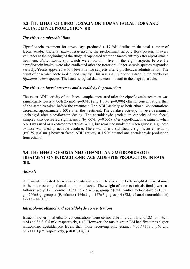
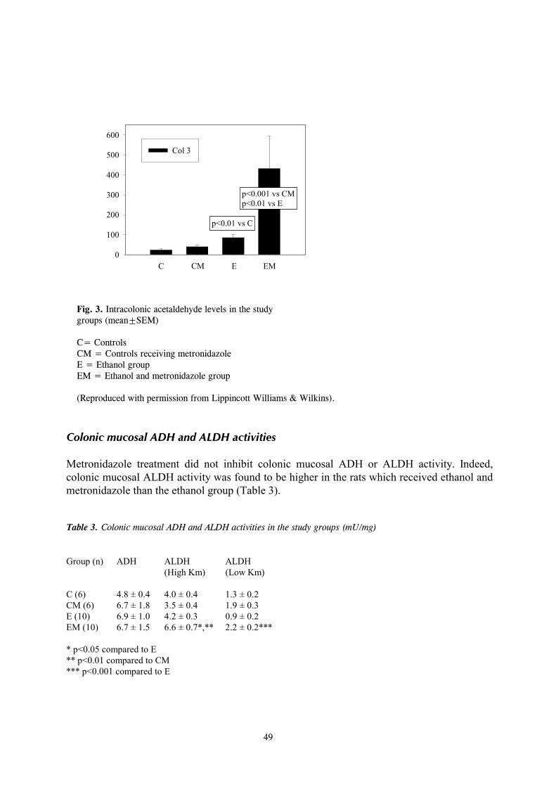
ETHANOL, ACETALDEHYDE AND GASTROINTESTINAL FLORA Regulatory factors and pathophysiological consequences of microbial ethanol oxidation and acetaldehyde production in the digestive tract Jyrki Tillonen Research Unit of Alcohol Diseases University of Helsinki Finland ACADEMIC DISSERTATION HELSINKI 2000 ISBN 952-91-2603-4 PDF
Transcript
ETHANOL, ACETALDEHYDE AND GASTROINTESTINAL FLORA
Regulatory factors and pathophysiological consequences of microbial ethanol oxidation and acetaldehyde production in the digestive tract
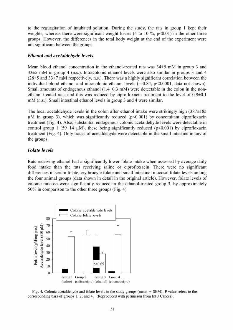
Jyrki Tillonen
Research Unit of Alcohol Diseases University of Helsinki
Finland
ACADEMIC DISSERTATION
HELSINKI 2000
ISBN 952-91-2603-4 PDF
2
Supervised by
Professor Mikko Salaspuro, M.D.
Research Unit Of Alcohol Diseases, Department of Medicine University of Helsinki
Reviewed by
Docent Onni Niemelä, M.D.
University of Oulu, Department of Medical Biochemistry
and
Docent Risto Roine, M.D.
Finnish Office for Health Care Technology Assessment
Opponent
Professor Eero Kivilaakso, M.D.
Helsinki University Central Hospital,
Department of Gastroenterological and General surgery
3
To my family
4
CONTENTS
ABBREVIATIONS 6 ORIGINAL PUBLICATIONS 7 1. INTRODUCTION 8 2. REVIEW OF THE LITERATURE 10 2.1. Biochemical characteristics of ethanol and acetaldehyde 10 2.2. Human oral and gastrointestinal microflora - an overview 11 2.3. Distribution of ethanol in the body 18 2.4. Hepatic ethanol and acetaldehyde metabolism 19 2.5. Metabolism of ethanol and acetaldehyde in the digestive tract 2.6. Microbial ethanol fermentation and oxidation 23 2.7. Alcohol and the alimentary tract 26 2.8. Alcohol and digestive tract cancers 28 2.9. Organ toxicity of acetaldehyde 32 3. AIMS OF THE STUDY 36 4. MATERIALS AND METHODS 37 4.1. Ethical considerations 37 4.2. Acetaldehyde production by human colonic contents in vitro (I) 37 4.3. The effect of ciprofloxacin on ethanol elimination in humans (II) 38 4.4. The effect of ciprofloxacin on human faecal flora and acetaldehyde production (II) 39 4.5. Sustained ethanol and metronidazole treatment in rats (III) 39 4.6. The effect of acetaldehyde on intestinal folate levels in rats (IV) 41 4.7. Human saliva studies (V, VI) 42 4.8. Gas chromatographic measurements of ethanol and acetaldehyde 43 4.9. Statistical analysis 43 5. RESULTS 44 5.1. Enzymatic production of acetaldehyde by human colonic contents in vitro (I) 44 5.2. The effect of ciprofloxacin on ethanol elimination in humans (II) 46 5.3. The effect of ciprofloxacin on human faecal flora and acetaldehyde production (II) 47 5.4. The effect of sustained ethanol and metronidazole treatment on intracolonic acetaldehyde production in rats (III) 47 5.5. The effect of ethanol and metronidazole treatment on blood ethanol and acetaldehyde levels in rats (III) 49 5.6. The effect of acetaldehyde on intestinal folate levels in rats (IV) 50 5.7. Factors influencing salivary acetaldehyde production in humans (V) 51 5.8. Microbes associated with acetaldehyde production in the human oral cavity (V, VI) 52
5
6. DISCUSSION 54 6.1. Role of catalase in acetaldehyde production by colonic contents 54 6.2. Role of colonic bacteria in extrahepatic ethanol elimination in humans 55 6.3. The effect of long-term ethanol and metronidazole treatment on intracolonic acetaldehyde levels 56 6.4. The effect of ethanol and metronidazole treatment on hepatic ethanol and acetaldehyde metabolism 57 6.5. The effect of acetaldehyde on intestinal folate levels in rats – a possible carcinogenic action of acetaldehyde 58 6.6. Acetaldehyde in saliva: influencing factors 58 6.7. Microbes associated with acetaldehyde production in the human oral cavity 59 7. SUMMARY AND CONCLUSION 61 REFERENCES 63
6
ABBREVIATIONS
ADH alcohol dehydrogenase ALDH aldehyde dehydrogenase ANOVA analysis of variance 3-AT 3-amino-1,2,3-triazole AUC area under the curve BMI body mass index CFU colony forming units CIPRO ciprofloxacin CYP cytochrome P450 DMH 1,2-dimethylhydrazine DNA deoxyribonucleic acid EER ethanol elimination rate EGF epidermal growth factor FPM first pass metabolism GI gastrointestinal GOX glucose oxidase H2O2 hydrogen peroxide IARC International Agency for Research on Cancer Ig intragastric Ip intraperitoneal kDa kiloDalton Km Michaelis constant MAF the mucosa-associated flora MEOS microsomal ethanol oxidizing system Mol wt molecular weight 4-MP 4-methylpyrazole NAD nicotinamide adenine dinucleotide NADH reduced nicotinamide adenine dinucleotide NDEA N-nitrosodiethylamine NDMA nitrosodimethylamine NDPA N-nitrodi-n-propylamine N2-Et-dg N2-ethyldeoxyguanosine O6-MeGT O6 methylguanine transferase PCA perchloric acid RER rough endoplasmic reticulum RR relative risk SA sodium azide SCFA short-chain fatty acids SEM standard error of the mean SER smooth endoplasmic reticulum Sp species Ssp subspecies Vd volume of distribution
7
ORIGINAL PUBLICATIONS
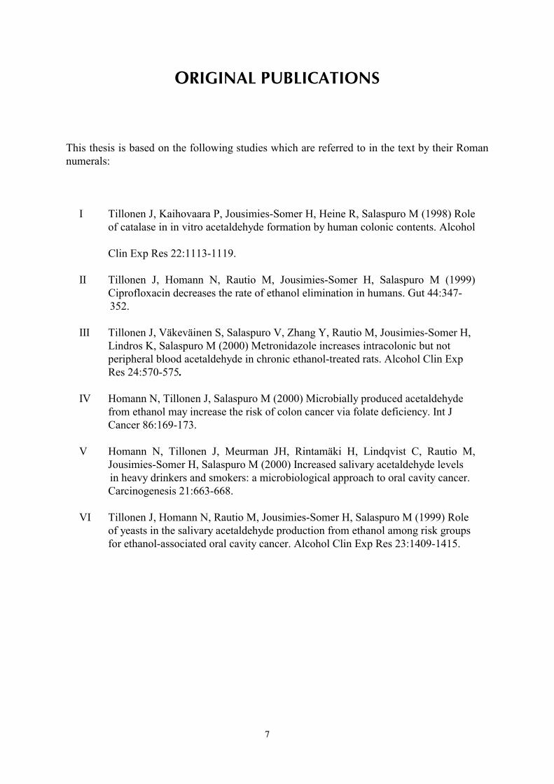
This thesis is based on the following studies which are referred to in the text by their Roman numerals:
I Tillonen J, Kaihovaara P, Jousimies-Somer H, Heine R, Salaspuro M (1998) Role of catalase in in vitro acetaldehyde formation by human colonic contents. Alcohol Clin Exp Res 22:1113-1119. II Tillonen J, Homann N, Rautio M, Jousimies-Somer H, Salaspuro M (1999) Ciprofloxacin decreases the rate of ethanol elimination in humans. Gut 44:347- 352. III Tillonen J, Väkeväinen S, Salaspuro V, Zhang Y, Rautio M, Jousimies-Somer H, Lindros K, Salaspuro M (2000) Metronidazole increases intracolonic but not peripheral blood acetaldehyde in chronic ethanol-treated rats. Alcohol Clin Exp Res 24:570-575. IV Homann N, Tillonen J, Salaspuro M (2000) Microbially produced acetaldehyde from ethanol may increase the risk of colon cancer via folate deficiency. Int J Cancer 86:169-173. V Homann N, Tillonen J, Meurman JH, Rintamäki H, Lindqvist C, Rautio M, Jousimies-Somer H, Salaspuro M (2000) Increased salivary acetaldehyde levels in heavy drinkers and smokers: a microbiological approach to oral cavity cancer. Carcinogenesis 21:663-668. VI Tillonen J, Homann N, Rautio M, Jousimies-Somer H, Salaspuro M (1999) Role of yeasts in the salivary acetaldehyde production from ethanol among risk groups for ethanol-associated oral cavity cancer. Alcohol Clin Exp Res 23:1409-1415.
8
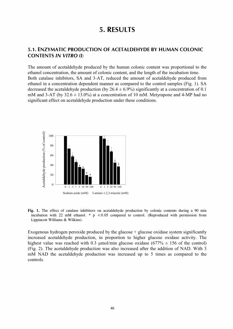
1. INTRODUCTION For millenia the consumption of alcoholic beverages has contributed to the pleasure of eating and drinking in many cultures of the world. In addition to livening up the social atmosphere, light alcohol drinking may also have beneficial effects on human health. An example of this is the “J-shaped” relation between the risk of coronary heart disease and alcohol intake. Overall morbidity is lower among those who drink lightly than those who drink more heavily or who do not drink at all (Klatsky, 1994). While the optimal or non-injurious levels of alcohol intake are difficult to estimate, they have been thought to be quite low, approximately 10-19 g/day for men and less than 10 g/day for women (Kalant and Poikolainen, 1999). When taken in excess, alcohol has devastating effects on human health by leading to breakdown of bodily functions and damaging virtually every organ of the body. Alcohol use may also lead to alcoholism, which can be defined as “the extreme dependence on excessive amounts of alcohol associated with a cumulative pattern of deviant behaviours”. Excessive alcohol consumption is known to increase the risk of developing several diseases of the liver, which are principally “fatty liver”, hepatitis, and cirrhosis. Also well-known are cases of alcohol-induced acute or chronic pancreatitis. Ethanol itself has been thought to be the hepatotoxic agent, but since only a relatively small proportion of heavy drinkers develop the most severe forms of liver damage, it is probable that other factors are also involved (Lindros, 1995). The pathogenesis of alcohol-induced pancreatic injury is still obscure, although major hypotheses so far have emphasized ethanol-induced changes in the pancreatic ductal system or the toxic effects of ethanol on pancreatic exocrine metabolism (Singh and Simsek, 1990). Less is known about other multiple gastrointestinal symptoms and organ toxicities associated with heavy alcohol use. There is evidence of small intestinal dysfunction after chronic alcohol consumption, including increased mucosal permeability, promotion of bacterial overgrowth, altered gut motility, and impaired salt and water absorption. This can lead to diarrhea, dyspepsia, nausea, and finally to malnutrition, which are common findings among actively drinking alcoholics (Persson, 1991). The association between alcohol consumption and certain digestive tract neoplasia has also been well established. Epidemiological studies have shown that cancers of the mouth, esophagus, and larynx are associated with alcohol consumption and that the risk increases in a dose-dependent manner (Doll et al., 1999). Likewise, high alcohol intake is a suspected risk factor for colorectal cancer. Although this subject has been debated, two different meta-analyses both reach the conclusion that alcohol leads to a small but significantly increased cancer risk, especially for the left colon and the rectum (Kune and Vitetta, 1992; Longnecker et al., 1990). The mechanism for the increased cancer risk associated with alcohol consumption is not clear, but has been believed to be at least in part due to the carcinogenic action of the first metabolite of ethanol, acetaldehyde (IARC, 1985, 1999). This notion is strongly supported by recent epidemiological studies which show that GI-tract cancer risk is markedly increased among heavy drinking Asian individuals with a genetically deficient ability to remove acetaldehyde (Yokoyama et al., 1998). The reactivity of acetaldehyde may also involve it in promoting organ toxicity other than malignant transformation.
9
During recent years it has become evident that the colonic microbes take part in ethanol metabolism not only by fermenting sugars to ethanol, but also by oxidizing exogenous ethanol to acetaldehyde (Jokelainen, 1997; Salaspuro, 1996, 1997). Similarly, oral microflora have been shown to produce high concentrations of acetaldehyde from ethanol (Homann et al., 1997a). Ethanol oxidation and consequent acetaldehyde production by gut microbes occurs at ethanol concentrations that are relevant to those after moderate alcohol drinking (Homann et al., 1997a; Jokelainen et al., 1994). Furthermore, since the capacity of intestinal mucosa and flora to metabolise acetaldehyde further is limited, acetaldehyde accumulates locally in the areas of the digestive tract covered by microbes (Koivisto and Salaspuro, 1996; Nosova et al., 1998). Due to its high reactivity, toxicity and carcinogenicity, acetaldehyde can be expected to cause organ damage wherever it exists at high concentrations. Therefore, microbial ethanol oxidation and consequent acetaldehyde production may have important implications for the pathogenesis of symptoms and organ toxicity associated with excessive alcohol use. Understanding the mechanisms behind alcohol-induced gastrointestinal morbidity is helpful in their management and a prerequisite for their prevention. The present study thus investigates the enzymes involved in microbial ethanol oxidation, its contribution to total ethanol elimination, and possible regulatory factors, and is intended to obtain evidence about possible organ toxicity related to microbial acetaldehyde production.
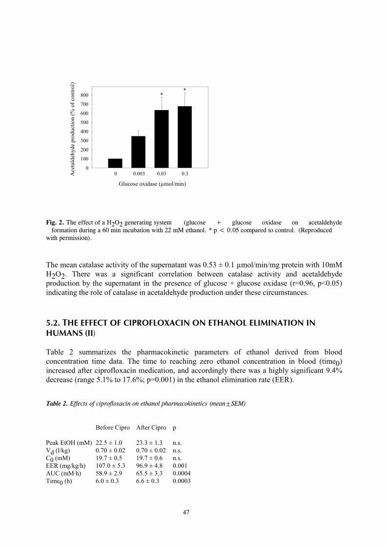
10
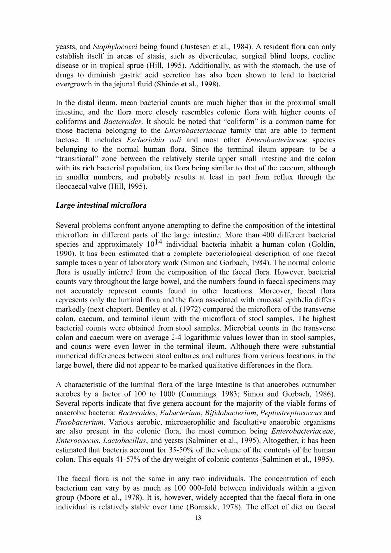
2. REVIEW OF THE LITERATURE 2.1. BIOCHEMICAL CHARACTERISTICS OF ETHANOL AND ACETALDEHYDE Ethyl alcohol (CH3CH2OH; Mol wt 46.1) is the accurate term for ethanol. The synonyms ”alcohol and alcoholic beverages” are also commonly used in the literature and in colloquial language. The term “alcohol” is, however, slightly misleading, since several other alcohols, like methanol, also exist. In the context of this thesis the terms ethanol and alcohol are used as synonyms, and alcoholic beverages means any products that contain ethanol. Ethanol is obtained by fermentation of carbohydrates contained in a variety of natural products. Preparation of absolute anhydrous ethanol (percentage by weight approximately 99.5%) for research purposes needs special distillation procedures. The density of absolute ethanol at 20 C compared with water at 4 C is 0.789. The melting point of absolute ethanol is -114.1 C and the boiling point is 78.5 C. Important characteristics of ethanol are its small molecular size and miscibility in water in any proportion. However, ethanol is only slightly soluble in fat; tissue fat takes up about 4% of the amount of ethanol dissolved in an equal volume of water (IARC, 1988; Wallgren and Barry III, 1970). A number of different systems are used to indicate the ethanol content, dosage, solutions for administration and concentration in body fluids. To indicate ethanol content or concentration of ethanol in alcoholic beverages, percentage by weight (% w/w) or volume (% v/v) are used. The dosage for scientific reasons should preferably be expressed as weight of ethanol given per unit of body weight of the test organism (g/kg body weight). For solutions to be administered, when the basis is pure ethanol, the most convenient way is to prepare solutions that contain a known weight of ethanol in a given volume of the final solution. The clearest expression is thus percentage weight by volume or % w/v. Percentage by volume (%v/v) can also be employed. There are several ways of expressing ethanol concentrations in body fluids and in in vitro incubations. Concentrations are normally stated in millimoles per litre (mM) or as a mille scale o/oo. One per mille equals one gram ethanol per litre, or 0.1 % w/v, or 21.7 mM. In some scientific publications, blood alcohol is also given as “mg %”, indicating milligrams ethanol per 100 ml. Table 1 shows the equivalence of units used in measuring concentrations of ethanol in body fluids. Units used in this book are as follows: blood and body fluid ethanol concentrations are generally given as mM. Dosages given are expressed as g/kg body weight, and solutions for that purpose either in %w/v or %v/v.
11
Table 1. Equivalence of units used in measuring concentration of ethanol in body fluids (Modified from Wallgren and Barry III, 1970) Per cent (%) Per mille (‰) mg/100 ml mM g/100 ml mg/ml or g/l (“mg%) 0.001 0.01 1 0.217 0.01 0.1 10 2.17 0.1 1.0 100 21.7 0.2 2.0 200 43.4 0.3 3.0 300 65.1 0.4 4.0 400 86.8 0.5 5.0 500 108.5 0.6 6.0 600 130.2 0.7 7.0 700 151.9 0.8 8.0 800 173.6 0.9 9.0 900 195.3 1.0 10.0 1,000 217.0 Acetaldehyde (CH3CHO; Mol wt 44.1) is a byproduct of the organic chemicals industry. It also occurs in vehicle exhaust and the smoke of tobacco cigarettes. For our research purposes the main source of acetaldehyde formation is intracellular oxidation of ethanol. The density of pure acetaldehyde at 20 C is 0.778, and its melting point is -123.5 C and the boiling point 20.1 C. Like ethanol, acetaldehyde is miscible with water and most common organic solvents (IARC, 1985, 1999). Since the concentrations of acetaldehyde in this thesis are given either as µM or µmol/l, which are equivalent, an acetaldehyde concentration of 100 µM equals 4.4 mg/l. 2.2 HUMAN ORAL AND GASTROINTESTINAL MICROFLORA - AN OVERVIEW Salivary microbial flora The mouth cannot be regarded as a single, uniform environment. The majority of investigations on the flora of the mouth have been concerned either with saliva or dental plaque. For the aims and purposes of this thesis, the most important flora in the mouth is that of the saliva. The reason for this is that ethanol is present in saliva in concentrations comparable to those of blood ethanol (Jones, 1979), and saliva is in close contact with the mucosa of the oropharynx and esophagus. The salivary flora is derived from the dislodgement of microorganisms from various locations in the oral cavity i.e. the teeth, tongue, cheek and pharyngeal mucous membranes (Herrera et al., 1988; Nolte, 1977). Adult human saliva contains approximately 6 x 10⁹ microorganisms per millilitre (Nolte, 1977). Streptococci, the facultative anaerobic Gram-positive organisms, have been isolated from all sites in the mouth and comprise a large proportion of the normal oral flora. On average, Streptococci represent about 45% of the total cultivable flora from saliva and the term Streptococcus viridans group is often used to generalize these bacteria. It includes, however, at least 5 different species, of which Streptococcus salivarius appears to comprise a significant proportion. Anaerobic streptococcus forms part of the anaerobic
12
flora of the oral cavity (Marsh, 1980; Nolte, 1977). Aerobic Gram-positive Staphylococci, Stomatococcus, and Micrococci have also been isolated from the oral cavity and saliva, but not in large quantities. Corynebacterium, Lactobacillus and Actinomyces are Gram-positive rods frequently found in human oral flora, and consisting of aerobic, facultative anaerobic and strictly anaerobic species. Aerobic Neisseria and strictly anaerobic Veillonella, which are Gram-negative cocci, have been isolated in low numbers from most sites in the oral cavity and saliva. The majority of aerobic or facultatively anaerobic Gram-negative rods fall into the genus Haemophilus (Marsh, 1980). Yeasts are aerobic microorganisms that can be isolated from approximately 40% of clinically healthy mouths, and Candida albicans is the most dominant species (Stenderup, 1990). Most anaerobic oral Gram-negative rods belong to the genus Bacteroides (Marsh, 1980). Flora of the stomach and small intestine All bacteria able to live as commensals in the human body are killed by incubation at pH values below 3. The pH of the normal resting gastric juice is below 3 and so the normal resting gastric juice is bacteria free. However, even in young normochlorhydric persons, the lumen is not bacteria-free for the whole day. During a meal the gastric acid is buffered, allowing swallowed salivary bacteria to survive or even to proliferate. However, when the pH returns to less than 3 these swallowed organisms are killed (Hill, 1995). As a consequence, a resident bacterial flora in the stomach can only occur when gastric acid secretion is impaired to the point that the pH does not fall below 3-4, even in the resting stomach. Thus, most of the organisms found in the stomach very likely represent the most acid-resistant components of the oral flora; Lactobacillus, Streptococcus viridans, Neisseria, Staphylococcus, Bacteroides and Peptostreptococcus are the genera best represented (Gustafsson, 1982; Hill, 1985). Impairing gastric acid secretion leads, however, to bacterial overgrowth in the stomach (Drasar et al., 1969). Conditions resulting in achlorhydria or hypochlorhydria include for example gastric surgery that includes vagotomy, pernicious anaemia or chronic atrophic gastritis, or the prolonged use of histamine 2 receptor antagonists or proton pump inhibitors. It has been shown that treatment with antacids or cimetidine raises bacterial counts 10- to 100-fold (Snepar et al. 1982). Moreover, it has been shown that gastric and duodenal bacterial overgrowth is considerably higher in patients treated with omeprazole compared to cimetidine. This was explained by the more pronounced inhibition of gastric acid secretion (Thorens et al., 1996). An apparent exception is infection with Helicobacter pylori. This organism colonizes the mucosa below the mucin barrier and is able to resist local acid secretion via its urease activity. Since the mucosal barrier protects the organisms from luminal acid they are able to proliferate in a locally pH-controlled environment (Marshall et al., 1990). When the gastric contents enter the small bowel they are mixed with large volumes of biliary and pancreatic secretions, many of which are bactericidal and help to sterilize this material. Furthermore, there is extensive fluid secretion from the bowel mucosa, which serves to flush the crypts and prevent colonization of the mucosal layer. Small bowel transit time is only two to four hours, an additional barrier to small bowel colonization. For these reasons the normal small bowel is in general almost sterile or contains a very sparse flora of transient organisms (Hill, 1985, 1995). Anaerobes only slightly outnumber facultative organisms, Streptococci, Lactobacillus, Veillonella,
13
yeasts, and Staphylococci being found (Justesen et al., 1984). A resident flora can only establish itself in areas of stasis, such as diverticulae, surgical blind loops, coeliac disease or in tropical sprue (Hill, 1995). Additionally, as with the stomach, the use of drugs to diminish gastric acid secretion has also been shown to lead to bacterial overgrowth in the jejunal fluid (Shindo et al., 1998). In the distal ileum, mean bacterial counts are much higher than in the proximal small intestine, and the flora more closely resembles colonic flora with higher counts of coliforms and Bacteroides. It should be noted that “coliform” is a common name for those bacteria belonging to the Enterobacteriaceae family that are able to ferment lactose. It includes Escherichia coli and most other Enterobacteriaceae species belonging to the normal human flora. Since the terminal ileum appears to be a “transitional” zone between the relatively sterile upper small intestine and the colon with its rich bacterial population, its flora being similar to that of the caecum, although in smaller numbers, and probably results at least in part from reflux through the ileocaecal valve (Hill, 1995). Large intestinal microflora Several problems confront anyone attempting to define the composition of the intestinal microflora in different parts of the large intestine. More than 400 different bacterial species and approximately 1014 individual bacteria inhabit a human colon (Goldin, 1990). It has been estimated that a complete bacteriological description of one faecal sample takes a year of laboratory work (Simon and Gorbach, 1984). The normal colonic flora is usually inferred from the composition of the faecal flora. However, bacterial counts vary throughout the large bowel, and the numbers found in faecal specimens may not accurately represent counts found in other locations. Moreover, faecal flora represents only the luminal flora and the flora associated with mucosal epithelia differs markedly (next chapter). Bentley et al. (1972) compared the microflora of the transverse colon, caecum, and terminal ileum with the microflora of stool samples. The highest bacterial counts were obtained from stool samples. Microbial counts in the transverse colon and caecum were on average 2-4 logarithmic values lower than in stool samples, and counts were even lower in the terminal ileum. Although there were substantial numerical differences between stool cultures and cultures from various locations in the large bowel, there did not appear to be marked qualitative differences in the flora. A characteristic of the luminal flora of the large intestine is that anaerobes outnumber aerobes by a factor of 100 to 1000 (Cummings, 1983; Simon and Gorbach, 1986). Several reports indicate that five genera account for the majority of the viable forms of anaerobic bacteria: Bacteroides, Eubacterium, Bifidobacterium, Peptostreptococcus and Fusobacterium. Various aerobic, microaerophilic and facultative anaerobic organisms are also present in the colonic flora, the most common being Enterobacteriaceae, Enterococcus, Lactobacillus, and yeasts (Salminen et al., 1995). Altogether, it has been estimated that bacteria account for 35-50% of the volume of the contents of the human colon. This equals 41-57% of the dry weight of colonic contents (Salminen et al., 1995). The faecal flora is not the same in any two individuals. The concentration of each bacterium can vary by as much as 100 000-fold between individuals within a given group (Moore et al., 1978). It is, however, widely accepted that the faecal flora in one individual is relatively stable over time (Bornside, 1978). The effect of diet on faecal
14
flora is controversial. It has been shown that there are differences in the faecal flora between people consuming quite different diets (Aries et al., 1969; Finegold et al., 1974), but the effect of dietary alterations has turned out to be extremely difficult to demonstrate (Hill and Drasar, 1975). There is a consensus that dietary alterations change the composition of the faecal flora only slightly and very slowly or not at all (Bornside, 1978; Hill and Drasar, 1975). Studies of the metabolic activity of the flora based on measurements of bacterial enzymes have, however, revealed changes in the colonic flora as a function of diet (Simon and Gorbach, 1984). Although faecal flora in general is quite stable over prolonged periods of time, hospitalisation, for example, has been shown to lead to rapid colonization by the specific E. coli serogroups associated with a particular institute (Simon and Gorbach, 1984). The use of certain antimicrobial agents is also a potent way to alter colonic microflora. The colonic mucosa-associated flora (MAF) Studies on animals have suggested the existence of a specific mucosal-dependent flora in the colon (Savage, 1970). In contrast to the enormous amount of literature on the bacteria of faeces, there is little data on the bacteriology of the mucosa-associated flora (MAF) in humans. Nevertheless, analysis of biopsy material and specimens of surgically excised tissue have shown that human colon has a flora associated with the mucosa which is distinct from that of the gut lumen. It is reproducible, stable, and responds to antibiotic treatment differently from that of the lumen (Bleday et al., 1993; Hill, 1995; Nelson and Mata, 1970; Peach et al., 1975). One of the most important features of MAF is that it has approximately equal representation of aerobic and anaerobic organisms compared with the luminal flora (Marks et al., 1979; Peach et al., 1975). Facultative anaerobes belonging to the Enterobacteriaceae family are well represented in the MAF (Marks et al., 1979; Peach et al., 1978), whereas anaerobes have been shown to be mainly Bacteroides species (Poxton et al., 1997). The mechanisms by which microbes adhere to the mucosal membrane depend on a variety of factors. Dietary fibres may influence the composition of the bacterial flora by providing nutrients or altering the environmental conditions including peristaltic rate or mucous composition (Savage, 1978). The normal function of the absorptive epithelium presumably depends upon a suitable oxygen tension, which in turn depends upon blood flow, arterial oxygen content, and oxygen utilization. One of the earliest studies measuring oxygen tension in the gut lumen was done by using domestic duck. Oxygen tension (PO2) was found to be about 25 mm Hg close to the mucosal villi area, while it was 50 times lower in the centre of the lumen (Crompton et al., 1965). Studies with dogs have shown that the intestinal mucosa has an oxygen tension of the order of 40 mm Hg, which is between a quarter and a third of that in air and approximately similar to that of the venous blood (Hamilton et al., 1968). One study with humans showed about the same magnitude of oxygen tension in mucosa of the gut (45 mm Hg) (Dawson et al., 1965). In contrast, flatus usually has a PO2 of less than 15 mm Hg (Askevold, 1956). Utilization of O2 by colonic bacteria is thought to lower the intraluminal PO2 to the level present in flatus. Moreover, it has been shown that conventional rats have a lower intraluminal PO2 and higher pCO2 than germ-free rats. This is probably because of the consumption of O2 and the production of CO2 by bacterial metabolism (Bornside et al., 1973). Taken together, the amount of oxygen diffused to the colonic mucosa may be the predominant factor explaining the proportionally higher counts of aerobic and facultative organisms in the MAF than in the luminal flora.
15
It has been suggested that the importance of the microbes colonizing the mucosa is that they modulate the function of the mucosal barrier. Disturbing the balance of MAF has been shown to lead to alterations in paracellular pathways at the level of the tight junctions, thereby increasing mucosal permeability (Spitz et al., 1994). Moreover, intestinal microorganisms are able to oxidize and reduce many types of organic compound. Consequently, it has been shown that the MAF is of importance in regulating enzyme levels in the intestinal mucosa of the rat (Hietanen and Hänninen, 1971). This is of great importance, since aerobic and facultative anaerobic microbes are able to oxidize ethanol to acetaldehyde. Because the ratio of aerobes to anaerobes is approximately 1:1 in the vicinity of the mucosa, this could be an important site for bacterial ethanol metabolism in the gut. It has, indeed, been shown that conventional rats have significantly higher acetaldehyde levels in the mucosa of the rectum and caecum than germ-free rats after ethanol administration (Seitz et al., 1990). The effect of antimicrobial agents on human faecal flora with special reference to ciprofloxacin and metronidazole The administration of an antibiotic is undoubtedly the most potent way of altering the markedly stable microflora of the human body. Since many antimicrobial agents cause changes in the colonic flora, the severity of which depends largely upon the concentration of the agent in the luminal contents, factors other than the width of the antibacterial spectrum may be of importance for the ecological consequences of antibiotic treatment in the colon (Norrby, 1986). Accordingly, the faecal microflora can be influenced by orally taken antimicrobial agents because of incomplete absorption, secretion into the bile, or secretion from the intestinal mucosa. Parenteral antimicrobial agents which are secreted into the bile or from the intestinal mucosa can also cause significant disturbances in the large intestinal microflora (Nord et al., 1986). Ciprofloxacin is a broad spectrum fluoroquinolone antimicrobial agent. The primary mechanism of the action of ciprofloxacin is the inhibition of bacterial DNA gyrase, which disrupts bacterial DNA replication. After oral administration ciprofloxacin has an approximate bioavailability of 70% and maximum plasma concentrations are achieved in 1 to 2 hours. The drug has a large apparent volume of distribution (2.1 to 5 L/kg after oral or intravenous administration) and becomes concentrated in many body tissues and fluids, including bile, kidney, liver, gallbladder, prostate and lung tissue. Ciprofloxacin is excreted largely unmetabolised in the urine and faeces, although small amounts of metabolites have also been detected (Davis et al., 1996). Furthermore, ciprofloxacin is partly eliminated through the intestinal wall (Rohwedder et al., 1990), and the concentrations of ciprofloxacin in the faeces and intestinal mucosa are higher than the corresponding serum levels (Brismar et al., 1990). This transintestinal elimination pattern may explain the particular ability of this drug to modify the colonic flora. The effect of ciprofloxacin in vivo on the composition of faecal flora has been studied extensively in healthy volunteers. The usual dosage regimens have been from 600 to 1000 mg/day for five or more days. These studies clearly demonstrate the marked reduction or complete eradication of Enterobacteriaceae. This occurs rapidly, usually within 3 days of commencing therapy. Following discontinuation of the therapy, these bacteria return to pretreatment concentrations within 3 to 4 weeks. The effects of ciprofloxacin on Staphylococci and Enterococci are not as dramatic or consistent as on
16
the Enterobacteriaceae, although many studies report significant reduction in one or both of these groups. Generally, ciprofloxacin does not affect the levels of total anaerobic flora. However, where anaerobic rods, Fusobacterium and Bacteroides species have been analysed separately, some studies have demonstrated decreases in faecal levels (Campoli-Richards et al., 1988). Metronidazole was originally introduced to treat Trichomonas vaginalis, but is now used for the treatment of anaerobic and protozoal infections. Metronidazole is bactericidal through toxic metabolites which cause DNA strand breakage. Metronidazole given orally is absorbed almost completely, with bioavailability of >90%. Metronidazole is distributed widely and has low protein binding (<20%). The volume of distribution at steady state in adults is 0.5 to 1.1 L/kg. Metronidazole reaches 60 to 100% of plasma concentrations in most tissues studied, and is extensively metabolised by the liver into 5 metabolites. The majority of metronidazole and its metabolites are excreted into urine and faeces, with less than 12% excreted unchanged into urine (Lamp et al., 1999). Metronidazole is most active in vitro against gram-negative obligately anaerobic bacilli such as Bacteroides, including the B. fragilis group and Fusobacterium. (Bergan, 1985). Although metronidazole and its active metabolites are found in the faeces and also in colonic mucosa, there is normally little suppression of indigenous colonic flora with metronidazole therapy. This has been thought at least partly because of the degradative reduction of the drug by bowel flora under the anaerobic conditions in the colon (Finegold, 1980). However, studies done with mice have shown that high doses of metronidazole decreases obligate anaerobes in vivo in the large intestinal flora and this leads to a consequent increase in certain aerobic species. Brook and Ledney (1994) found that the mean number of facultative anaerobes rose significantly from day 6 (p<0.05), whereas strict anaerobes fell significantly (p<0.05) during the treatment with metronidazole compared to controls. In another study metronidazole treatment selectively eliminated strictly anaerobic bacteria with a concomitant 100-fold increase in aerobic and facultative bacteria (Wells et al., 1987). Similarly, in the human studies, the number of Bacteroides species has been shown to decrease and the number of E. coli and faecal Streptococci to increase in the faeces of patients with Crohn’s disease during treatment with metronidazole (Krook et al., 1981a). Among healthy human volunteers, however, the count of Bacteroides species was unchanged at the end of metronidazole treatment, but there was a significant increase in the faecal Streptococci count (p=0.03) and an almost significant increase in E. coli (p=0.06) (Krook, 1981). These dissimilarities in metronidazole’s capacity to reduce human anaerobic faecal flora was speculated to arise from the higher concentrations of the drug in the large intestine of the Crohn’s disease patients (Krook et al., 1981b). Taken together, it can be concluded that metronidazole may dose-dependently increase the number of aerobes in the colonic flora at the expense of the number of strict anaerobes. The physiological role and metabolic capacity of the intestinal flora The number of intestinal bacteria equals (Luckey, 1977) or exceeds (Cummings, 1983) the number of the cells in their human host. Because of the sparseness of the flora in the proximal GI tract, its metabolic activity is insignificant compared with those of the colonic flora. The accepted functions of the colon include the conservation of water and electrolytes and the controlled evacuation of faeces (Moran and Jackson, 1992). The
17
colon is, however, an important organ of its own with an influence on overall metabolism, and the effect may in large part be attributed to the activity of colonic microflora (Phillips, 1984). Its metabolic capacity has been estimated to be at least as great as that of the liver (Bingham, 1988) or even to exceed that of the rest of the human body (Luckey, 1977). Intestinal bacteria also have a short generation time and can undergo enzyme induction when exposed to high levels of substrate. This allows the microflora to adjust itself rapidly to any change in the environment (Gorbach and Goldin, 1990). One of the most important features is its protective function against pathogenic microbes. Antibiotic treatment may select resistant species or strains in the intestinal flora and lead to superinfections such as colitis caused by Clostridium difficile. Colonisation resistance by the normal intestinal flora is thus an important host defence mechanism. Bacteria are also the main source of antigenic materials and the intestinal flora is the most important stimulant of the body’s defence mechanisms (Gustafsson, 1982). As stated earlier, the colonic microflora is predominantly anaerobic, and able to ferment carbohydrates. The main end-products of this bacterial fermentation are short-chain fatty acids (SCFAs). The carbohydrate available for fermentation in the colon comes from endogenous sources such as mucus, and exogenous dietary sources that escape digestion in the small intestine. The main SCFAs are acetate, butyrate and propionate, all of which have been shown to stimulate the growth and well-being of the colonic mucosa. Removal of fibre from the diet results in atrophy of the mucosa. This can be reversed by the infusion of SCFAs into the colon. Production of SCFAs lowers colonic pH and increases colonic motility. Moreover, since SCFAs stimulate colonic mucosal blood flow and oxygen uptake, there is evidence suggesting that bacterial fermentation is directly involved in colonic mucosal function and also more generally in mucosal metabolism (Cummings and Macfarlane, 1991). Bile acids are produced in the liver as end-products of cholesterol metabolism and excreted into the bile as conjugates. In the intestine the primary bile acids are attacked by microbial enzymes and transformed into a variety of metabolites, which may be absorbed and further transformed by liver enzymes prior to their re-excretion into the bile, forming the enterohepatic circulation of bile acids (Cummings, 1975). Intestinal microbes may also participate to a lesser extent in heme metabolism, the end product of which, bilirubin, is hydrolysed/deconjugated in the intestine by microbial and mucosal enzymes. The deconjugated bilirubin is reduced by microbial enzymes into a complex mixture of urobilinogens, which are excreted with the faeces. Some of these are also absorbed from the large intestine and reexcreted into the bile and urine (Gustafsson, 1982). The intestinal microflora also metabolise sterols and steroid hormones. The steroid hormone metabolites reaching the intestine via the bile are usually conjugated with sulphuric acid or glucuronic acid. These conjugates are split by the intestinal microflora and the resulting free steroids are further degraded by the gut bacteria (Gustafsson, 1982). Bacterial enzymes play important roles in the metabolism of many drugs, often determining their bioavailability. For example, in 10% of patients given digoxin the drug is converted to inactive moieties by the gut flora (Lindenbaum et al., 1981). Bacterial metabolism may also be relevant to the biological effects of some drugs. An
18
example of this is a salicylazosulphapyridine, which is a complex drug containing an azo link between a sulphonamide and a salicylate. The two moieties, linked by the azo bond which is resistant to mammalian enzymes, constitute a large molecule which is not absorbed in the small bowel. This allows the drug to reach the colon, where bacterial enzymes hydrolyse the azo bond, releasing sulphapyridine and salicylic acid. Since the components are thought to act therapeutically on the colon and then to be absorbed (Phillips, 1984), a symbiosis between colonic bacterial enzymes and a therapeutic effect is clear. The above examples show that the colonic flora has several physiological functions. Because any compound taken orally or entering the intestine via the biliary tract or blood stream is a potential substrate for bacterial transformation, bacterial flora with its enzymes is also very likely to be involved in many metabolic processes of foreign compounds and also exogenous and even endogenous ethanol (Goldin, 1990; Salaspuro, 1996, 1997; Simon and Gorbach, 1984). Species differences in the intestinal flora Since conventional laboratory rats are widely used for studies on the metabolism of intestinal flora, in extrapolating the results obtained from the animal experiments to human subjects, it is important to know possible differences between the human and rat intestinal flora. The most notable differences lie in the upper regions of the gut. In man the stomach and duodenum normally harbour only transient flora, but these areas in rats are colonised by a mixed bacterial populations of about 107 - 108 organisms/g. The explanation for this is probably the bactericidal action of the strongly acidic gastric juice of the humans, whereas the pH of the rat stomach is more moderate (ph 4-5). The large intestine of rats is colonized by 103 to 10 5times higher concentrations of bacteria than the small intestine, and the colonic concentration of bacteria is equal to humans. Despite this, some differences have also been found in the activities of the reductive enzymes associated with the caecal and faecal floras of rats and humans. For example certain nitro compounds such as nitrobenzenes and dinitrotoluenes which depend on reduction by the gut flora for their toxic effects, can be much more potent in rats than in humans, who have much lower bacterial nitroreductase activity than the laboratory animals (Rowland, 1986). Taken together, large intestinal microflora in humans and rats are quantitatively comparable. Because much less is known about enzymatic similarities of the flora, caution must be taken in extrapolating results in this field. However, it may be possible to increase the degree of similarity in gut flora metabolism between laboratory rats and man by modifying the animal diet (Rumney and Rowland, 1992).
2.3. DISTRIBUTION OF ETHANOL IN THE BODY Ethanol is absorbed by simple diffusion from the gastrointestinal tract because of its small size, good water solubility, and low solubility in lipids (Wallgren and Barry III, 1970). No transport processes exist for ethanol (Crabb et al., 1987). Most of the ingested ethanol, 70-80%, is absorbed by the proximal small intestine, duodenum and upper jejunum. Slow diffusion from the stomach means that only about 20% of the oral dose is absorbed from the ventricle. After absorption, ethanol reaches the liver via the portal
19
vein (Riveros-Rosas et al., 1997). The rate of absorption is decreased by delayed gastric emptying (Oneta et al., 1998). Because gastric emptying is slow and prolonged with food in the stomach, drinking ethanol after eating a meal, regardless of the nutritional composition, delays its absorption. This produces a slower rise and lower peak value of the blood alcohol in fed than in fasting subjects (Jones et al., 1997). The concentration of the ingested ethanol also influences its absorption, at least when taken after a meal. It has been shown that postprandially taken high concentrations of ethanol result in lower blood alcohol levels than do dilute solutions, probably because of delayed gastric emptying (Roine et al., 1991,1993; Roine 2000). Once ethanol reaches the blood, it is distributed rapidly throughout the body fluids. In organs with dense vascularization and rich blood supply, such as brain, lungs, and liver, alcohol rapidly equilibrates with the blood. In contrast, the distribution of alcohol to the resting skeletal muscle is particularly slow because of the low number of functioning capillaries (Agarwal and Goedde, 1990; Dundee et al., 1971). Poor lipid solubility allows tissue lipids to take up only 4% of the amount of alcohol that can be dissolved in a corresponding volume of water. Women, who have a smaller total water volume in the body than men, thus reach higher blood ethanol levels even if both sexes ingest identical quantities of ethanol (Riveros-Rosas et al., 1997). Distribution of alcohol is mainly related to the water content of various organs and tissues, so that, for instance, ethanol concentrations in the terminal ileum are approximately equal to those of the blood (Halsted et al., 1973). Their high water content makes the ethanol concentration in saliva (Jones, 1979) and urine (Bendtsen et al., 1999) slightly higher than that in the blood. Most of the ethanol (90-95%) is metabolised by oxidation and excreted as CO2 and water. Other routes for elimination are urine, sweat, and breath. Since alcohol is not concentrated in the urine or sweat, only negligible amounts of ethanol are excreted via urine (0.3%), and sweat (0.1%). In humans under normal conditions 0.7% can be eliminated through the lungs (Holford, 1987). 2.4. HEPATIC ETHANOL AND ACETALDEHYDE METABOLISM It is generally agreed that the liver is the main organ responsible for the oxidation of ethanol. Estimations of the contribution of the liver to ethanol elimination under normal conditions vary from 75-90% (Agarwal and Goedde, 1990). However, in severe hepatic cirrhosis extrahepatic elimination of ethanol has been shown to account for up to 40% (Utne and Winkler, 1980). There are three metabolic systems capable of carrying out ethanol oxidation in the liver: cytosolic alcohol dehydrogenase, the microsomal ethanol oxidizing system located in microsomes, and catalase, located on peroxisomes. All these hepatic enzymes yield acetaldehyde as an end-product. Acetaldehyde is further converted to acetate, mainly in the mitochondria catalysed by aldehyde dehydrogenase. Alcohol dehydrogenase (ADH) ADH catalyses the reversible oxidation of many alcohols to corresponding aldehydes. In case of ethanol, the reaction is as follows:
CH3CH2OH + NAD+ ⇔ CH3CHO + NADH + H+
20
Alcohol dehydrogenase is abundant in the liver and its physiological role has been postulated to be the degradation of the low levels of alcohol produced by microbial fermentation in the gut. Another possible explanation is its role in the dehydrogenation of endogenous steroids (Krebs and Perkins, 1970). ADH is the main enzyme responsible for the oxidation of ingested ethanol in the liver. It is a dimer composed of approximately 40 kDa subunits and contains 2 zinc atoms per subunit. Six different classes of alcohol dehydrogenase have been described for mammalians; for humans, classes I-V and for rats, classes I-IV and class VI have been described (Jörnvall and Höög, 1995). In humans class I isoenzymes are coded by three genes (ADH1 to 3), and are explained by the presence of three protein subunits. In rats, class I ADH is encoded by one gene. Just recently a recommendation for a new nomenclature for expressing ADHs has been made. Five human ADH classes should be expressed by an Arabic number as follows: ADH1, ADH2, ADH3, ADH4, and ADH5. For genes, the italicized root symbol “ADH” for human and “Adh” for rat, followed by the appropriate Arabic number for the class; i.e. ADH1 or Adh1 for class I ADH genes has been recommended. Where multiple isoenzymes exist within a class, adding a capital letter after the Arabic number; i.e. ADH1A, ADH1B, and ADH1C for human class I ADHs was also suggested (Duester et al., 1999). In the literature the nomenclature has been confusing, and misconceptions about class distinction have frequently occurred. Since most papers have used the “older” nomenclature which codes classes with Roman numbers, this will be followed here. Class I ADHs, the classic liver alcohol dehydrogenases, are the most important enzymes in hepatic elimination of ethanol. These enzymes have a low Km ( 1 mM) and high Vmax for ethanol, and are responsible for the bulk of ethanol oxidation. This means that ethanol is effectively eliminated from the blood at a constant rate to very low concentrations, provided that acetaldehyde is also effectively removed. As the Km of ADH for acetaldehyde is 0.6 mM it could act as a substrate in the reverse reaction (Blair and Vallee, 1966). However, the rapid transformation of acetaldehyde to acetate keeps the reaction in the forward direction. When alcohol is oxidized to acetaldehyde via ADH, nicotinamide adenine dinucleotide (NAD) is reduced to NADH. Normally the rate of NADH production exceeds its rate of reoxidation, resulting in an increase in the liver NADH/NAD ratio. This means that the redox state of the liver is markedly reduced. Most of the acute metabolic effects of ethanol, such as the inhibition of hepatic gluconeogenesis, the decrease in citric acid cycle activity and the impairment of fatty acid oxidation, arise from this major effect of ethanol on the intermediary metabolism of the liver (Lieber, 1994). The microsomal ethanol oxidizing system (MEOS) The first indication of a possible interaction between ethanol and the endoplasmic reticulum or microsomal fraction of the hepatocyte was provided by the observations that ethanol feeding results in a proliferation of the smooth endoplasmic reticulum (SER) in rats and human (Iseri et al., 1966; Lane and Lieber, 1966). This led Lieber and DeCarli (1968) to find the cytochrome P450-dependent system, which oxidizes ethanol to acetaldehyde as follows:
In humans, the cytochrome fraction responsible for ethanol oxidation has been designated as CYP2E1. This isoenzyme is the major contributor to the MEOS in humans, although later studies have suggested that other CYP forms may also play a role (Asai et al., 1996; Niemelä et al., 1999). The term MEOS should thus be maintained in referring to the overall capacity of the microsomes to oxidize ethanol rather than to that fraction of the activity specifically catalysed by 2E1 (Lieber, 1997). Since the Km of MEOS for ethanol is 7-10 mM, it contributes to ethanol elimination at high blood ethanol levels. This explains the fact that ethanol metabolism increases with rising ethanol concentrations above the level needed to fully saturate the low Km hepatic ADH (Lieber, 1997). The contribution of the MEOS to total ethanol elimination has not yet been fully clarified. Because of the slow turnover it may be limited, and it has been estimated that only a minor part (1-5%) of all ethanol metabolism in vivo is carried out by the MEOS (Ingelman-Sundberg, 1997). The most significant role of CYP2E1 is, however, its adaptive response to constantly high blood ethanol levels. This accounts for the metabolic adaptation to high concentrations of ethanol and the acceleration of ethanol metabolism resulting from chronic alcohol consumption. This metabolic adaptation has to be distinguished from the central nervous system adaptation to ethanol which results from chronic ethanol consumption, characterized by the progressive resistance of the brain to the effects of ethanol (Lieber, 1999). In addition to acetaldehyde production during ethanol oxidation, the MEOS has been shown to produce reactive oxygen intermediates, such as superoxide radicals. This may lead to enhanced lipid peroxidation, so that MEOS may contribute to alcoholic liver disease. Moreover, CYP2E1 has a capacity to activate over 80 toxicologically important xenobiotics to potentially hepatotoxic or carcinogenic products (Lieber, 1997). Catalase Catalase, which is located in peroxisomes, can oxidize ethanol to acetaldehyde when hydrogen peroxide is available as follows:
CH3CH2OH + H2O2 ⇒ CH3CHO + 2H2O However, studies using the catalase inhibitor aminotriazole have shown that this compound does not slow ethanol metabolism in vivo (Teschke et al., 1976), and examination of the enzymatic reaction has suggested that the activity of catalase is limited in vivo by the bioavailability of hydrogen peroxide. Since the rate of hydrogen peroxide production in the liver is rather low (Boveris et al, 1972), there is some notion that catalase plays only a minor role in hepatic ethanol metabolism (less than 2%). Aldehyde dehydrogenase (ALDH) Regardless of the pathway by which ethanol is oxidised, acetaldehyde is the first metabolic product. Acetaldehyde is far more toxic than its parent compound ethanol. Fortunately, it is usually quickly further metabolised to acetate in the oxidative reaction catalysed by aldehyde dehydrogenase. The liver is the key organ for ethanol oxidation and the bulk of the ALDHs exist there. Human hepatic aldehyde dehydrogenase activity can be found in the mitochondria and cytosol (Agarwal, 1997). The main isoenzyme
22
responsible for the oxidation of acetaldehyde, both in humans and rats, has been shown to be the mitochondrial class 2 ALDH (ALDH2). This has a low Km (3 µM or less) and a high affinity for acetaldehyde (Lands, 1998). At high acetaldehyde concentrations, the increase in acetaldehyde oxidation is due to the activity of extramitochondrial ALDH, mainly cytosolic class 1 ALDH (ALDH1), which has a relatively high Km for acetaldehyde (100 µM) (Crabb et al., 1987). The low steady-state acetaldehyde concentration in the liver during alcohol metabolism (<10 µM) suggests that the mitochondrial isoenzyme is the main form responsible for the oxidation of acetaldehyde (Crabb et al., 1987). This is evidenced by experiments which show that NADH generated by the ALDH reaction appears almost exclusively in the mitochondria (Forsander, 1970). The central role of the low Km mitochondrial ALDH in acetaldehyde metabolism is strongly indicated by the finding that a mutation in the ALDH2 enzyme in humans results in impairment of the ability to dispose of acetaldehyde after ethanol ingestion. ALDH2 isoenzyme has two allelic forms; the active ALDH2*1 and the relatively inactive ALDH2*2. Patients homozygous for the ALDH2*2 allele lack ALDH2 activity, while patients heterozygous for this allele exhibit approximately half the activity of ALDH2*1 homozygotes (Crabb et al., 1989). Deficient ALDH2 isoenzyme has been found in about 50% of Japanese (Goedde et al., 1979). The homozygous form of inactive ALDH2 offers full protection against alcoholism. This has been proposed to be due to the accumulation of acetaldehyde in the blood during alcohol metabolism, which causes aversive sensations (Peng et al., 1999). Heterozygotic subjects with about half the normal ALDH2 activity can, however, drink alcohol or develop even alcohol dependency (Wall et al., 1992). After ethanol intake the heterozygotic subjects show flushing, palpitations and nausea, which are caused by elevated blood acetaldehyde levels (Wall et al., 1992). Blood acetaldehyde levels in heterozygotic subjects have been shown to be between 8 and 24 µM even after a very low dose (0.1 g/kg of body weight) of ethanol (Enomoto et al., 1991). In contrast, normal healthy subjects have very low levels (< 0.5 µM) of acetaldehyde in the peripheral blood during ethanol oxidation (Eriksson and Fukunaga, 1993). This indicates that in normal healthy individuals almost all the acetaldehyde formed is effectively oxidised in the liver. Heavy drinkers with the heterozygous ALDH2*2 genotype (ALDH2*1/2*2) can be considered as human “knock-out models” for deficient acetaldehyde removal. Consequently, the toxicity of acetaldehyde is strongly corroborated by the fact that individuals with the heterozygous ALDH2*2 genotype are at higher risk of developing alcohol abuse-related GI-tract cancer as compared to those with the normal ALDH2 genotype (Yokoyama et al., 1998). The end-product of hepatic ethanol oxidation, acetate, leaves the liver via hepatic venous blood, and is almost completely converted to CO2 and H2O in the peripheral tissues, mainly in the muscles. 2.5. METABOLISM OF ETHANOL AND ACETALDEHYDE IN THE DIGESTIVE TRACT Ethanol metabolism Although the liver is the major organ responsible for ethanol metabolism, such metabolism also occurs in the digestive tract. Intestinal metabolism of ethanol is of
23
considerable importance, since it may affect the systemic availability of alcohol and lead to local production of acetaldehyde, possibly resulting in tissue injury. Immunohistochemical studies have revealed that alcohol dehydrogenase can be detected in the mucosa of all parts of the gastrointestinal tract. Furthermore, the amount of ADH observed was higher in epithelial cells exposed to the lumen than in cells at the bottom of crypts (Pestalozzi et al., 1983). In addition to their localization, the contribution of the various ADH isoenzymes to ethanol metabolism depends on their kinetic parameters, particularly their Km values. Class IV ADH is characteristic of the upper GI tract, including the mouth and the esophagus (Dong et al., 1996; Yin et al., 1993). The Km values of the gingival ADH was estimated to be 27 mM (Dong et al., 1996), and of esophageal class IV 12 mM (Yin et al., 1993) These high Km values suggest that ethanol may be significantly metabolised in these tissues. Moreover, the esophagus is the organ of highest ADH activity in the GI tract, with a rate per mg of protein similar to that of the liver and approximately 4 times that of the stomach enzyme (Parés and Farrés, 1996). ADH was detected in human gastric mucosa decades ago (Hempel and Pietruszko, 1979; Smith et al., 1972) and has been shown to exhibit multiple isoenzymes. Class IV ADH and class I ADH coexist in the stomach, with Km values of 40 and approximately 1 mM respectively (Parés et al., 1992; Seitz and Oneta, 1998; Yin et al., 1997). Since the stomach contains both high Km class IV ADH and low Km class I ADH, this organ is a transition site for switching expression of class IV ADH to class I ADH, which is predominant in the rest of the intestinal tract (Yin et al., 1997). The significance of gastric ADH is its suggested role in the first pass metabolism of ethanol (FPM). The gastric FPM of ethanol has been used to explain the differences in the areas under the ethanol concentration-time curves (AUC) obtained after oral and intravenous alcohol application. However, the relative contribution of gastric and hepatic metabolism to FPM is still a subject of debate. Some studies suggest that the differences in AUCs may be due to the differences in ethanol absorption, and therefore speculate that gastric ethanol metabolism in rats is negligible and that there is no evidence for this phenomenon in humans (Smith et al., 1992). By contrast, there are studies indicating a significant role for gastric ethanol metabolism in the FPM (Caballeria et al., 1987; Lim et al., 1993). The estimations of FPM of ethanol to ethanol metabolism range between 1% and 20% (Seitz and Pöschl, 1997). The small and large intestinal ADH is mainly composed of class I ADH, with a Km of 1-2 mM for ethanol (Seitz and Oneta, 1998). The activity of rectal ADH was comparable to gastric ADH activity and, compared to ADH activities in other colonic segments, was found to be significantly higher (Seitz et al., 1996). This suggests that ethanol may be effectively metabolised to acetaldehyde in the colonic mucosa and especially in the rectal mucosa. In addition to ADH, rat gastric mucosa have been shown to possess catalase activity (Salmela et al., 1996), but its presence in the rest of the alimentary tract is unknown. Moreover, immunohistochemistry has revealed that rat duodenal and jejunal villous cells exhibit CYP2E1 activity, but it is not expressed or induced in the stomach, ileum, colon and rectum (Shimizu et al., 1990).
24
Acetaldehyde metabolism Cytosolic high Km ALDH3 is the only ALDH isoenzyme identified in the human mouth thus far. Both gingival and tongue tissue ALDH exhibit significant amounts of enzyme activity at 20 mM acetaldehyde (Dong et al., 1996). Regarding human gastric mucosa, ALDH3 has been estimated to account for more than 80% of the ALDH activity. Although ALDH3 has a high Km value for acetaldehyde (approximately 88 mM), it has been suggested that acetaldehyde generated by gastric ADH could be oxidized in the same tissue (Parés and Farrés, 1996; Yin et al., 1997). A different picture is seen in the esophagus, where the ALDH3 activity is 5 times below that of the gastric mucosa, while the rate of acetaldehyde production is higher than in the stomach (Yin et al., 1993). Accumulation of acetaldehyde could thus occur in esophageal tissue, contributing to the alcohol-related end-organ damage. ALDHs classes 1 and 2 can be found in the human duodenum (Liao et al., 1991), but much less is known about the ALDH activity of the rest of the small intestine. ALDHs 1, 2, and 3 have been observed in human and rat colonic mucosa, but the expression of low-Km mitochondrial ALDH2 seems to be particularly low (Koivisto and Salaspuro, 1996; Yin et al., 1994). The activity ALDH in the colonic mucosa was only slightly lower in ALDH2-deficient subjects than in normal phenotype carriers, suggesting that mucosal ALDH2 plays only a minor role in the oxidation of acetaldehyde in the colon (Yin et al., 1994). 2.6. MICROBIAL ETHANOL FERMENTATION AND OXIDATION Endogenous ethanol Measurable amounts of ethanol are normally formed in the gastrointestinal tract. In the caecum of normally fed rats, the mean ethanol concentration has been shown to be 0.9 mM. This endogenous alcohol, which derives from anaerobic degradation of glucose to ethanol by some microorganisms, is absorbed into the portal circulation and almost quantitatively removed by the liver (Krebs and Perkins, 1970). Conditions associated with bacterial overgrowth producing markedly elevated endogenous ethanol levels detected even in the blood indicate intestinal microbial-derived ethanol production in humans. For example, in some patients after a jejunoileal bypass operation, detectable fasting serum concentrations of ethanol up to 1 mM have been noted. In the past this kind of operation resulted in blind-loop formation and consequent colonisation and bacterial overgrowth of that segment (Mezey et al., 1975). Additionally, midjejunal aspirates from the patients with tropical sprue show not only an overgrowth of coliformic bacteria (Klebsiella pneumoniae, Enterobacter cloacae and Escherichia coli) but also large quantities of their fermentation end-product ethanol with concentrations from 2 mM to 31 mM (Klipstein et al., 1973). Small amounts of endogenous ethanol (1 to 27 mM) have also been found in the gastric juice of patients receiving cimetidine or antacids. Bode et al. (1984a) speculated that this resulted from an increase in the yeast and/or bacterial population in the stomach due to the reduction of gastric acid induced by the medication. The last step in bacterial alcoholic fermentation is the reduction of acetaldehyde to ethanol, catalysed by bacterial ADH (Reid and Fewson, 1994; Tamm, 1974). This has been described in full detail in the case of Escherichia coli (Clark, 1989; Dawes and Foster, 1956; Still, 1940; Wong and Barrett, 1983), group N Streptococci (Lees and
25
Jago, 1976) as well as within the Enterobacteriaceae in general (Salveson and Bergan, 1981). As already stated, this reaction is also reversible in microorganisms (Maconi et al., 1988). This means that if oxygen is present and given an excess of ethanol, the ADH mediated reaction can also run in the opposite direction and ethanol may be oxidized to acetaldehyde in a reaction in which nicotinamide adenine dinucleotide acts as an electron acceptor. Microbial acetaldehyde production in vitro As mentioned earlier, E. coli was shown to possess ADH activity in 1940 by Still. Jokelainen et al. (1996a) showed that different strains of faecal E. coli and other Gram-negative rods, mainly belonging to the Enterobacteriaceae family, were able to produce significant amounts of acetaldehyde when incubated aerobically in vitro with ethanol in reaction catalysed by NAD-dependent ADH. Later studies have shown that E. coli is also able to oxidize ethanol in microaerobic conditions, i.e. when 6% O2 is present (Salaspuro et al., 1999). This is important, since the PO2 in the colonic mucosa is known to be approximately equal to that of venous blood, and hence the conditions in the colon are more or less microaerobic (Hamilton et al., 1968). It was shown earlier that human colonic contents, i.e. the mixture of colonic bacteria, can produce acetaldehyde from ethanol in a dose-dependent manner in vitro (Jokelainen et al., 1994). The significant acetaldehyde production occurred at the ethanol concentrations that are relevant to in vivo conditions. These findings have opened up a new microbiological approach to researching acetaldehyde production and its pathological consequences in the gastrointestinal tract. Regarding the upper digestive tract, in vitro production of acetaldehyde has been reported when human mouth- and bronchopulmonary washings were incubated aerobically with ethanol. Since acetaldehyde production could be abolished by pretreatment with antibiotics, acetaldehyde formation from ethanol was thought to be of bacterial origin (Jauhonen et al., 1982; Miyakawa et al., 1986; Pikkarainen et al., 1981). Interestingly, in vitro acetaldehyde production by mouthwashings from oropharyngeal cancer patients has been shown to be significantly higher than that of the control patients (Jokelainen et al., 1996b). This suggested that microbially derived acetaldehyde production could be involved in ethanol-associated organ toxicity. Helicobacter pylori also exhibits significant cytosolic ADH activity. Consequently, H. pylori has been shown to produce acetaldehyde when intact cells are incubated with ethanol, and this has been speculated to contribute to H. pylori-associated gastroduodenal injury (Roine et al., 1992, 1995; Salmela et al., 1993, 1994). Microbial acetaldehyde production in vivo Regarding the human oral cavity, the production of marked amounts of acetaldehyde up to 140 µM in saliva after the ingestion of a moderate amount of ethanol has been demonstrated. This acetaldehyde production was significantly reduced by the use of antiseptic chlorhexidine mouthwash, indicating its microbial origin (Homann et al., 1997a). Another important finding in the same study was that in vivo salivary acetaldehyde levels correlated highly significantly with the acetaldehyde levels produced in vitro. This offers an opportunity to use the in vitro salivary test to reliably differentiate high acetaldehyde producers from low producers.
26
Intestinal in vivo microbially derived ethanol oxidation and acetaldehyde production have been shown to occur in rats with a jejunal self-filling diverticulum and concomitant bacterial overgrowth (Baraona et al., 1986). Moreover, after an acute dose of ethanol to rats, the rectal mucosal acetaldehyde concentration was significantly higher in conventional rats than germ-free rats, indicating the role of microbes in acetaldehyde production (Seitz et al., 1990). Ethanol oxidation to acetaldehyde has also been detected in the caecum of anaesthetized pigs after intragastric or intravenous ethanol administration (Jokelainen et al., 1996c). Furthermore, high intracolonic acetaldehyde levels have been detected in the caecal samples of rats after intraperitoneal ethanol administration, and these levels were effectively suppressed by pre-treatment with ciprofloxacin, which reduces the amount of aerobic bacteria in the large intestine (Visapää et al., 1998). These findings strongly suggest that microbes of the large intestine are able to oxidize ethanol by a bacteriocolonic pathway for ethanol oxidation. In a very recent human study, we also showed ethanol-derived acetaldehyde production in the stomach after treatment with lansoprazole. Medication led to marked bacterial overgrowth in the neutral stomach, and this was considered to be the most probable explanation for the enhanced acetaldehyde production detected (Väkeväinen et al., 2000). Microbial acetaldehyde metabolism Aldehyde dehydrogenase activity has been identified in the cytosol of yeasts (Steinman and Jakoby, 1968) and in anaerobic bacteria (Burdette and Zeikus, 1994). ALDH activity also exists in facultative anaerobic bacteria, which are able to produce acetaldehyde via ADH. Escherichia coli has been shown to exhibit aldehyde dehydrogenase activity (Dawes and Foster, 1956; Wong and Barrett, 1983). Nosova et al. (1996) studied 27 different bacterial species, mainly belonging to the family Enterobacteriaceae, and showed them to be capable of cytosolic NAD or NADP-linked ALDH activity. However, based on kinetic studies, the ability of bacterial ALDHs to oxidise higher concentrations of acetaldehyde was rather low (Nosova et al., 1998). Taken together with low ALDH activity in the colonic mucosa (Koivisto and Salaspuro, 1996), this offers an additional explanation for the mechanism of the accumulation of ethanol-derived acetaldehyde in the large intestine. 2.7. ALCOHOL AND THE ALIMENTARY TRACT Symptoms and functional changes associated with alcohol use. Excessive alcohol consumption is frequently associated with gastrointestinal symptoms. Although most symptoms after both acute and chronic use of alcohol are reversible and disappear during abstinence, chronic use of alcohol may cause structural or functional changes that can lead to permanent gastrointestinal diseases. An example of this is the increased risk of colorectal adenomatous polyps, which are generally regarded as premalignant lesions (Cope et al., 1991). Alcohol consumption and the risk of colorectal cancer will be discussed in the next chapter. Only the upper digestive tract, mouth, esophagus, stomach and upper small intestine are
27
directly affected by the ingested ethanol. However, since ethanol is transported by blood circulation to other organs ethanol concentrations in the terminal ileum (Halsted et al., 1973) and colon (Levitt et al., 1982) are approximately equal to those of the blood. A questionnaire showed that actively drinking alcoholics more frequently report heartburn, nausea, vomiting, diarrhea, and flatulence than the matched controls. These symptoms were transient and resolved after two weeks of sobriety. An important note of the study was that these symptoms were associated with active alcohol use and were not withdrawal symptoms (Fields et al., 1994). The etiology for the increased incidence of GI symptoms in alcoholics is probably multifactorial. Moreover, prolonged use of alcohol may have effects which differ from the acute effects of a single dose. Some studies suggest that acute alcohol ingestion may impair the function of the lower esophageal spinchter and reduce the incidence of primary peristalsis in the distal esophagus (Hogan et al., 1972; Mayer et al., 1978), although some others have reported the reverse (Keshavarzian et al., 1987; Silver et al., 1986). However, there is evidence that acute alcohol intake increases esophageal reflux episodes and impaires the acid clearance of the esophagus in the supine position (Kaufman and Kaye, 1978; Vitale et al., 1987). This may explain the increased incidence of heartburn in heavy alcohol users. Acute ingestion of alcohol has been shown to produce inflammatory changes in the stomach and duodenum of humans. The most prominent findings included haemorrhagic erosions, subepithelial blebs and infiltration of inflammatory cells into the lamina propria (Gottfried at al., 1978). Rubin et al. (1972) showed that after sustained use of ethanol, the histology of the human small intestine was normal when examined by normal light microscopy, but morphometric and electron microscopic examinations demonstrated alterations in the cell organelles of the mucosa. Others have, however, shown reduction in the villus height and a reduced mucosal surface area of villi in the small intestine of actively-drinking alcoholics (Bode et al., 1982a; Persson, 1991; Seitz et al., 1985). Several enzymes known to be located in the absorptive cells may also be affected by ethanol. Reduced activity in disaccharidases has been noted after sustained high ethanol administration (Bode et al., 1982b). Decreased lactase activity can contribute to milk intolerance with diarrhea commonly observed in alcoholics after a period of heavy drinking (Keshavarzian et al., 1986; Persson, 1991). Moreover, oral-caecal time has been shown to be decreased in recently drinking alcoholics, suggesting that the increasing transit of intestinal contents may contribute to the diarrhea commonly seen during and after an episode of heavy drinking (Keshavarzian et al., 1986). Chronic alcohol use has also been shown to induce both histological and ultrastructural changes in the human rectal mucosa. These reversible changes include inflammatory changes, a decreased number of goblet cells, and alterations in the cell organelles (Brozinsky et al., 1978). The nutritional status of advanced alcoholics is often poor, at least among lower-income and homeless alcoholic populations. The etiology of malnutrition has been generally thought to be multifactorial (Salaspuro, 1993). On average, ethanol accounts for about a half of an alcoholic’s caloric intake. It therefore displaces normal nutrients, causing malnutrition (Lieber, 1995). Secondary malnutrition also occurs through malabsorption due to the structural and functional changes in the small intestine, but pancreatic exocrine insufficiency, decreased biliary secretion and impaired hepatic metabolism of nutrients may also be involved (Lieber, 1995; Salaspuro, 1993).
28
Both acute and chronic exposure to ethanol has been shown to increase intestinal permeability to macromolecules in experimental animals. Increased small intestinal permeability has also been observed in human alcoholic patients without liver cirrhosis. This “leaky gut” phenomenon was shown to persist up to 2 weeks after cessation of drinking (Bjarnason et al., 1984), and seems to result from a temporary destabilization of intracellular junctions (Draper et al., 1983). Interestingly, high acetaldehyde levels have been shown to reversibly increase the paracellular permeability of the Caco-2 cell monolayer (Rao, 1998). Acetaldehyde production by the intestinal bacteria may thus be one mechanism by which prolonged alcohol use increases intestinal permeability. The disruption of the mucosal barrier may promote absorption of normally nonabsorbable compounds like endotoxins into the portal circulation (Bjarnason et al, 1984; Persson, 1991). Moreover, leaky gut has recently been suggested as a possible mechanism for alcohol-induced liver damage (Keshavarzian et al., 1999). Alterations in the oral and intestinal flora caused by chronic alcohol intake There are no studies available to show whether heavy alcohol consumption directly alters the oral flora. However, although the overall dental health in subjects misusing alcohol, investigated by the decayed, missing and filled teeth (DMFT) index, has been shown to be only slightly poorer than national averages in the United Kingdom, a high incidence of tooth wear and trauma to the dentition was noticed (Harris et al., 1996, 1997). Tooth wear was presumed to be caused by the regurgitation of gastric acid. This study suggests that the dental hygiene habits of alcohol misusers may be poorer than that of abstainers or moderate drinkers. Poor dental hygiene habits may favour the overgrowth of some microbes in the oral flora. Moreover, poor dietary factors and the suppressive effects of alcohol intake on various arms of the host immune defence may predispose to colonization by some bacteria and yeasts in the oral cavity (MacGregor, 1986; Oksala, 1990). Additionally, most heavy alcohol drinkers are also heavy cigarette smokers (Harris et al., 1996). Heavy smoking may be the strongest factor predisposing to microbial colonisation in the oral cavity since it has been shown to increase the presence of yeasts and Gram-positive aerobic bacteria there (Colman et al., 1976; Macgregor 1988; Rindum et al., 1994; Sakki and Knuuttila, 1996). Moreover, heavy alcohol consumption in humans has been associated with a marked (70%) reversible reduction in the flow rate of stimulated parotid saliva. Beside this, chronic alcohol abuse also reduces protein secretion, like amylase and epidermal growth factor (EGF), in parotid saliva (Dutta et al., 1992). Ohmura and coworkers (1987) suggested that low EGF levels in saliva may decrease the resistance of the mucosal barrier to chemical stress. Therefore, low salivary levels of EGF may be one possible mechanism by which ethanol or acetaldehyde may influence the development of oral lesions associated with alcohol use. Marked qualitative and quantitative changes have been observed in the flora of the small intestine of chronic alcoholics. The most significant finding was the increase in the number of bacteria per unit volume of jejunal juice, especially in the number of aerobic and facultative anaerobic Gram-negative rods and obligate anaerobic bacteria (Bode et al., 1984b). Evidence for an increased incidence of bacterial overgrowth in alcoholics also stems from studies using the hydrogen breath test after ingestion of lactulose (Bode et al., 1989). Mucosal bacterial overgrowth has also been detected more often and with higher microbial counts in the biopsy samples of the stomach and duodenum among
29
alcoholics (Hauge et al., 1997). This upper GI tract bacterial overgrowth has been speculated to be due to the increased pH of the gastric juice. Small intestinal bacterial overgrowth may lead to malabsorption or morphological changes in the small intestinal mucosa (Persson, 1991). Moreover, bacterial overgrowth might contribute to the increased release of endotoxin and lead to endotoxemia, which is often detected in patients with alcoholic liver disease (Bode, 1987). Endotoxin, a cell-wall constituent of Gram-negative bacteria, may play a role in alcoholic liver injury by activating macrophages to release cytokines (McClain et al., 1999). The small intestinal bacterial overgrowth may thus also contribute to the pathogenesis of extraintestinal disorders associated with alcohol use. Bacterial overgrowth probably also leads to increased microbially-derived acetaldehyde production from ethanol in the small intestine. 2.8. ALCOHOL AND DIGESTIVE TRACT CANCERS Cancer of the oropharynx and esophagus There is extensive epidemiological evidence, mainly from case-control studies although also from cohort studies, showing that tobacco smoking and heavy alcohol consumption increase the risk of cancers of the mouth, pharynx, esophagus and larynx (Doll et al., 1999; Franceschi et al., 1990; Grønbæk et al., 1998; Mashberg et al., 1993; Merletti et al., 1989). In Europe these factors have been estimated to account for about three-quarters of all cases (La Vecchia et al., 1997). Both smoking and alcohol consumption have been shown to be independent risk factors for upper digestive tract cancers. The cancer risk increases proportionally with the quantity of cigarettes smoked or alcoholic beverages drunk. When combined, there is epidemiological evidence indicating that alcohol and tobacco act together in a multiplicative rather than in an additive manner, having synergistic tumour-promoting effects (Blot et al., 1988; Brugere et al., 1986; La Vecchia et al., 1997). This means that the combined effect of both of these agents is greater than simply adding the effects of each together. Many attempts have been made to separate the effects of different types of alcoholic beverages in their potential for carcinogenicity. The consensus, however, is that the main component of alcoholic beverages determining the risk of cancer is ethanol (Doll et al., 1999). This is further supported by the fact that the regular use of mouthwashes with a high ethanol content increases the risk of oropharyngeal cancer (Winn et al., 1991). Other possible risk factors for upper gastrointestinal tract cancer are poor nutritional status and intake of micronutrients, hereditary factors, certain papilloma viruses, and occupational hazards (Bundgaard et al., 1995; Graham et al., 1977; La Vecchia et al., 1997; Marshall et al., 1992; Marshall and Boyle, 1996). Moreover, dentition, tooth loss, poor dental status and oral hygiene habits are associated with higher risks, especially for oral cavity cancer (Bundgaard et al., 1995; Graham et al., 1977; Maier et al., 1993; Marshall et al., 1992). It is generally agreed that the influence of poor oral hygiene as a risk factor is much less compelling than alcohol and smoking, but there is some experimental evidence that its influence might become clinically more important among alcoholics, whose poor dental status and hygiene may be a common problem (Maier et al.,1993). The reason for this finding is unclear.
30
Although alcohol and tobacco smoke are well-known independent and strong risk factors for upper gastrointestinal tract cancer, the exact mechanism by which they exert an influence upon the oral mucosa is poorly understood. Consequently, the only things known to reduce the risk of upper digestive tract cancers is to limit alcohol consumption and to stop or never start using tobacco and, perhaps, to have a regular diet rich in fruit and green vegetables (Harris, 1997). Many compounds in tobacco smoke are hazardous to health and some are undoubtedly carcinogenic (IARC, 1986). In contrast, the tumour-promoting effects of alcohol consumption are less well defined. Moreover, ethanol is not a carcinogen by standard laboratory tests. As alcohol is involved synergistically in the attributable risk of cigarette smoking, it has been suggested that unifying pathogenetic mechanism may underlie these epidemiological findings. The possible mechanisms by which alcohol may influence the development of cancers are discussed separately, with special reference to acetaldehyde in chapter 2.9. Cancer of the stomach Most epidemiological retrospective cohort studies show no increase in the stomach cancer rate among alcoholics or heavy drinkers. In these studies cancer incidence in groups with high alcohol intake was compared with that of the general population. In follow-up studies of populations with known consumption most studies showed no elevated risk, with RR from 0.9 to 1.2 (Doll et al., 1999). However, two studies (Gordon and Kannel, 1984; Kato et al., 1992) reported a significant increase with heavy drinking, with a relative risk of 3.05 in the study by Kato et al. In that study stomach cancer mortality was prospectively studied among 9753 Japanese, who frequently show the presence of partially inactive ALDH 2*2 isoenzyme (Goedde et al., 1979). It has since been shown that the mutant allele is associated with higher frequency of GI malignancies, including stomach cancer, if alcohol is consumed regularly (Yokoyama et al., 1998). This limits the extrapolation of this study to Caucasians with no such enzyme defect, but indicates the role of acetaldehyde in carcinogenesis associated with alcohol use. Some case-control studies have shown a significant positive relationship between alcohol consumption and stomach cancer (RR 1.5 - 1.7), whereas others showed no significant relationship (Doll et al., 1999). Taken together, epidemiological evidence between alcohol consumption and cancer of the stomach is far from clear. Moreover, there has been a dramatic worldwide decline in the incidence of stomach cancer, a finding in contrast to the general increase in alcohol consumption and alcohol-related diseases such as cirrhosis of the liver. IARC stated in 1988 that there is little aggregate data to suggest a causal link between drinking alcoholic beverages and stomach cancer. However, Doll et al. (1999) later concluded that alcohol may have an etiological role in the stomach cancer, albeit minor and unproven. An exception to this may be the cancer of the gastric cardia, the incidence rates of which have been recently increasing. The cardia region of the stomach is the uppermost area where the stomach adjoins the esophagus. It has been suggested that this cancer resembles a specific type of cancer of the lower oesophagus and may share common risk factors, notably tobacco and alcohol exposure (Vaughan et al., 1995). Cancer of the large intestine As with gastric cancer, there is considerable epidemiological data concerning the possible association between cancer of the large bowel and consumption of alcoholic
31
beverages. Some studies showed no evidence of such an association, while others showed a statistically significant association. The rest of the studies were somewhere “in-between” showing no consistent and significant overall association, but the RRs were elevated in some subgroups (Doll et al., 1999). However, two different meta-analyses of more than 60 studies between 1951 and 1991 both conclude that alcohol leads to a small but significantly increased cancer risk, especially for the left colon and the rectum with an estimated overall RR of 1.1 (95% CI 1.05-1.14) (Kune and Vitetta, 1992; Longnecker et al., 1990). Moreover, just recently a panel of European experts at a World Health Organization (WHO) Consensus Conference on Nutrition and Colorectal Cancer declare that alcohol had a causal effect in colorectal carcinogenesis (Scheppach et al., 1999). Possible mechanisms of ethanol-induced carcinogenesis It has been concluded that there is sufficient evidence for the carcinogenicity of alcoholic beverages in humans (IARC, 1988), but there is no experimental evidence to indicate that ethanol itself is a carcinogen (Doll et al., 1999). This means that pure ethanol has not been shown to be carcinogenic in laboratory experiments. In in vitro studies with human and other mammalian cells it has been shown that ethanol does not induce DNA damage, sister chromatid exchanges or chromosomal aberration. Most of the in vivo studies with mice or rats described in the literature cannot be used to evaluate the carcinogenicity of alcohol due to limitations in experimental design (IARC, 1988). Animal experiments suggest, however, that ethanol may act as a co-carcinogen in the production of cancers. This means that it modifies or enhances the carcinogenic potential of known carcinogens. Grici t et al. (1982, 1984) exposed C57B mice by gastric intubation to N-nitrosodiethylamine (NDEA) or N-nitrodi-n-propylamine (NDPA), either in tap water or in a 40% ethanol solution, twice a week for 50 weeks. A significant increase in the incidence of squamous cell carcinomas of the esophagus/forestomach was observed in the group given the carcinogens in ethanol as compared to those given the nitrosamines in the tap water. Seitz et al. (1984) studied the effect of chronic ethanol administration on 1,2-dimethylhydrazine (DMH) induced rectal carcinogenesis in male rats fed a nutritionally adequate liquid diet containing 36% of total energy as ethanol or isocaloric carbohydrate. Sustained ethanol ingestion increased the number of rectal tumours significantly (17 vs. 6; p<0.02). Although many hypotheses exist, experimental work has so far failed to elucidate the underlying mechanisms by which excessive consumption of alcoholic beverages may act as a co-carcinogen under certain conditions. Some of these hypotheses are listed below. Alcohol may contain congeners and other contaminants that may be carcinogenic. Several substances known or thought to cause cancer in humans have been detected in alcoholic beverages. Special attention has been paid to N-nitroso compounds, which have been related to colorectal cancer in humans (Knekt et al., 1999). Several N-nitroso compounds, e.g. nitrosodimethylamine (NDMA), have been found in higher concentrations in some beers than in other beverages (Walker et al., 1979). This is a possible mechanism for a specific carcinogenic effect of beer drinking in relation to rectal cancer. While some studies have found an elevated risk (Riboli et al., 1991), these were not confirmed by others (Potter and McMichael, 1986). The consensus is that there
32
is no appreciable and consistent difference in the risk among different types of alcoholic beverages (Doll et al., 1999). Alcohol intake generates metabolites which may be carcinogenic to humans, or ethanol itself may act as a solvent, increasing penetration of other carcinogens into the target tissue (Wight and Ogden, 1998). The major metabolite of ethanol is acetaldehyde. There is sufficient evidence for the carcinogenicity of acetaldehyde to experimental animals (IARC 1985, 1999), and indirect strong epidemiological evidence in humans (Yokoyama et al., 1998). Because of the central role of acetaldehyde in the studies of this thesis, this topic will be discussed separately in the next chapter. Either alcohol intake may reduce the intake and/or bioavailability of nutrients which could inhibit cancer, or alcohol consumption may enhance nutritional deficiencies that increase the risk of cancer. In some cases, nutritional deprivation may lead to nutritional deficiencies that may alter epithelial cell chemistry and function, increasing susceptibility to carcinogens (Blot, 1992), an example being folate deficiency. In epidemiological studies, decreased folate status has been associated with an increased risk of neoplastic transformation in the colon (Giovannucci et al., 1995). The pathogenetic mechanism of the increased cancer risk in folate deficiency has been partly elucidated. Folate is a crucial methyl group donor for many transmethylation reactions in the human body. Diminished folate leads to hypomethylation of the DNA, which has been observed in several experimental cancer models and in human cancers (Goelz et al., 1985; Kim et al., 1997). Since high levels of acetaldehyde have been shown to be able to catabolize folate in vitro (Shaw et al., 1989), there is conjecture that alcohol intake and low folate together could play a role in colorectal carcinogenesis (Anonymous, 1994; Boutron-Ruault et al., 1996; Collins et al., 1992; Giovannucci et al., 1995). Prolonged alcohol use may also inhibit the detoxification of carcinogenic compounds. Experimental studies have suggested that the effects of alcohol on the liver may block hepatic inactivation of carcinogens, thus increasing exposure to these compounds (Blot, 1992). Moreover, heavy alcohol use may catalyse the metabolic activation of some compounds into carcinogens. For example, alcohol intake induces hepatic CYP2E1, which also has a unique capacity to activate over 80 toxicologically important xenobiotics to potentially carcinogenic products (Lieber, 1997). Since oxygen free radicals are generated during alcohol metabolism, target cells may be exposed to oxidants and this may lead to activation of carcinogens at the cellular level. Increased oxidative stress may also increase the risk of DNA damage and malignant transformation. However, the tumour promotion associated with alcohol and with the generation of free radicals is in general not very clear and the role played by ethanol in this process is even less so (Mufti et al., 1993). Alcohol is also known to cause immunosuppression, a possible contributing factor in the increased cancer rate among alcoholics. The role of immunosuppression in ethanol-associated cancers is, however, questionable, since increased incidences of lymphoma, the tumour most closely associated with depressed immune function, have not been reported among heavy drinkers (Blot, 1992).
33
2.9. ORGAN TOXICITY OF ACETALDEHYDE Nearly every organ system in the human body can be affected by heavy and prolonged use of alcohol. In addition to GI-tract cancers, there is no doubt that the consumption of alcoholic beverages increases the risk of alcoholic liver diseases. As already mentioned, ethanol itself is not carcinogenic. Similarly, the fact that only a minor proportion of alcohol abusers develop the most severe forms of liver damage suggests that other mechanisms than the direct effect of ethanol in creating tissue toxicity have to exist. Acetaldehyde, the first metabolite of ethanol, possesses toxic properties that markedly exceed those of ethanol, and there is increasing evidence suggesting its part in the detrimental action of alcohol on the digestive tract. Acetaldehyde-protein adducts Acetaldehyde has properties that make it very suitable for potential nucleophilic attacks. Since many nucleophilic groups are present in the proteins, they are natural targets in various tissues for reactive acetaldehyde. The binding of acetaldehyde with proteins results in the formation of two major types of product, which have been classified as unstable and stable acetaldehyde-protein adducts. The unstable adducts may serve as intermediates in stable adduct formation and can be stabilized by reducing agents such as NADH, which is found in large amounts in the liver during ethanol metabolism. The stable adducts appear to be the most likely causes of toxic effects (Nicholls et al., 1992). In vitro, acetaldehyde has been shown to bind covalently to many cellular and extracellular proteins (Nicholls et al., 1992). In vivo, it is well established that acetaldehyde adduct formation occurs in the liver during ethanol oxidation in both experimental animals and humans (Lin et al., 1988; Niemelä et al., 1991). To localize acetaldehyde-protein adducts, immunohistochemical studies have been performed with specific antibodies. Adducts have been demonstrated mainly in the cytoplasm of the perivenular hepatocytes, where acetaldehyde production is believed to be the highest (Niemelä et al., 1991, 1994). Moreover, acetaldehyde adducts have been detected in the areas of active fibrogenesis in the liver biopsy specimens from alcoholic patients (Holstege et al., 1994). More recently, acetaldehyde adducts have also been demonstrated in the rough endoplasmic reticulum (RER), and in some peroxisomes of hepatocytes, as well as in myofibroblasts and Ito cells (Paradis et al., 1996). Although the ability of acetaldehyde to bind to hepatic proteins during ethanol metabolism has been well established, the precise role of acetaldehyde-protein adducts in the pathogenesis of alcoholic liver disease has not been clarified. One mechanism through which such adducts may be involved in alcoholic liver disease is that acetaldehyde-protein adducts may be recognized as neoantigens by the immune system. This may trigger potentially harmful immune responses directed against liver cells (Nicholls et al., 1992). The presence of circulating antibodies against acetaldehyde-protein adducts have indeed been described in humans (Israel et al., 1986; Niemelä et al., 1987). As several observations indicate that immunological mechanisms are involved in alcoholic liver disease, the appearance of such antibodies against acetaldehyde-protein adducts may be involved in the development and progression of liver injury (Tuma and Klassen, 1992). Acetaldehyde-protein adducts may also alter the biological properties of the modified
34
proteins. In the case of hepatic enzymes, the covalent binding of acetaldehyde has been shown to lead to the inhibition of the activities of enzymes which contain lysine at the catalytic site (Mauch et al., 1986). Moreover, long-term ethanol consumption has been shown to produce a decrease in O6 -methylguanine transferase (O6 -MeGT) activity in rats in vivo (Garro et al., 1986). This has been shown to be due to the acetaldehyde-cysteine adduct in the active site of the enzyme, and it occurs even at nanomolar acetaldehyde concentrations (Espina et al., 1988). O6 -MeGT is a DNA repair enzyme, which removes alkyl groups from the O6 position of guanine. Since alkylation at this position is associated with both mutagenesis and carcinogenesis (Kleihues et al., 1979; Lewis and Swenberg, 1980; Newbold et al., 1980), the inhibition of O6 -MeGT offers one explanation of how ethanol may act as a cocarcinogen, enhancing the carcinogenic potential of other agents. There is not much evidence about acetaldehyde-protein adducts in the mucosa of the gastrointestinal tract. One study shows that both exogenous acetaldehyde and that produced locally from ethanol binds to gastric mucosa. This adduct formation has been suggested to be a pathogenetic factor behind ethanol-associated gastric injury (Salmela et al., 1997). No evidence indicates whether acetaldehyde adducts are formed in the mucosal proteins in the rest of the digestive tract or not. Since microbes produce high levels of acetaldehyde from ethanol in the oral cavity and the colon, similar adduct formation at these anatomical sites as in the gastric mucosa might also occur. Acetaldehyde as a carcinogenic agent Acetaldehyde has been shown to be a highly mutagenic agent. Specifically, it may induce chromosomal aberrations, and micronuclei and/or sister chromatid exchanges in cultured mammalian cells (Dellarco, 1988; IARC, 1985). Moreover, it has been shown to induce gene mutations in human lymphocytes (He and Lambert, 1990). The induction of cytogenetic effects has been postulated to be related to the ability of acetaldehyde to form DNA-DNA and/or DNA-protein cross-links. The mechanisms of these DNA cross-links caused by acetaldehyde may involve direct attack by acetaldehyde on DNA. A series of studies has shown that acetaldehyde can react with DNA bases to produce specific types of base adduct, which is a critical initiating event in the multistage process of chemical carcinogenesis (Hemminki and Suni, 1984; Vaca et al., 1995). Under biologically relevant conditions, the most prevalent of these is N2 -ethyl-deoxyguanosine (N2 -Et-dG) (Fang and Vaca, 1995; Vaca et al., 1995). Fang and Vaca demonstrated that N2 -Et-dG becomes detectable in the liver DNA of mice treated with 10% ethanol in their drinking water for 5 weeks. This adduct was undetectable in control mice not given alcohol (Fang and Vaca, 1995). In humans such adducts have been detected in peripheral white blood cells of alcohol abusers (Fang and Vaca, 1997). DNA adducts have also been identified in nontumoral colon mucosa of human patients with colorectal cancer (Pfohl-Leszkowicz et al., 1995). Although these DNA adducts were unspecified and not necessarily related to acetaldehyde, this study indicates that covalent modification of DNA by xenobiotics may be involved in chemical carcinogenesis. Moreover, acetaldehyde-DNA adducts have also been detected in human buccal cells exposed to acetaldehyde in vitro (Vaca et al., 1998). There is sufficient evidence for the carcinogenicity of acetaldehyde to experimental animals (IARC, 1985). Acetaldehyde has been tested for carcinogenicity in rats and
35
hamsters by inhalation exposure. In such experiments, an increased incidence of carcinomas was observed in the nasal mucosa of rats (Woutersen et al., 1984), and laryngeal carcinomas were induced in hamsters (Feron et al., 1982). Moreover, in rats given either tap water or water containing acetaldehyde at a concentration of 120 mM, marked histopathological hyperplastic and hyperproliferative changes in the tongue, epiglottis, and forestomach were noted in the animals receiving acetaldehyde. These changes mimic those known to occur after treatment with alcohol (Homann et al., 1997b). Lipid peroxidation A free radical is generally defined as a molecule that contains one or more unpaired electrons. As the presence of unpaired electrons usually confers a large degree of chemical reactivity on the molecule, most free radicals (superoxide and hydroxyl radicals) may lead to cell injury by abstracting a hydrogen atom from a polyunsaturated fatty acid, and thus initiating the degradative process known as lipid peroxidation. Since lipids are major components of biological membranes, peroxidative loss of membrane integrity has been thought to lead to tissue injury (Mufti et al., 1993). Glutathione is present in all animal cells in high concentrations, and one of its functions is the protection of cells against free radicals. A severe reduction in the levels of glutathione has been shown to increase lipid peroxidation in vivo (Lieber, 1988; Wendel et at., 1979). Enhanced lipid peroxidation has been suggested as a mechanism for ethanol-associated liver injury (Situnayake et al., 1990). During ethanol oxidation reactive oxygen intermediates are produced by the MEOS. Acetaldehyde can also induce lipid peroxidation, as demonstrated in isolated perfused livers (Müller and Sies, 1982). The mechanism underlying this may be acetaldehyde’s capacity to reduce hepatic glutathione levels (Shaw et al., 1981). In addition, the incubation of rat liver supernatant with acetaldehyde results in the conversion of xanthine dehydrogenase to xanthine oxidase, an enzyme known to be able to generate superoxide radicals (Sultatos, 1988). Whether ethanol administration enhances in vivo lipid peroxidation has long been debated, but studies done on laboratory animals (Niemelä et al., 1995; Shaw et al., 1981) and humans (Niemelä et al., 1994; Shaw et al., 1983) point to this ability. Moreover, high acetaldehyde concentrations administered to rats have been shown to result in the formation of free radical reactions in vivo (Reinke et al., 1987). Taken together, ethanol-derived acetaldehyde may lead to a severe reduction in glutathione, which favours lipid peroxidation, and the damage is possibly further enhanced by the increased generation of active radicals through induced MEOS following sustained ethanol consumption. Lipid peroxidation products, which result from the attack of reactive oxygen species on lipids, also react with DNA, forming ethenobases and adducts with known carcinogenicity and miscoding potential. Thus, during chronic alcohol abuse, where levels of reactive oxygen species and lipid peroxidation products are elevated and antioxidant levels are reduced, there is potential for significant damage to DNA and the production of DNA adducts that can compromise cellular function and may lead to oncogenic transformation (Brooks, 1997).
36
Evidence in humans Acetaldehyde exposition studies with humans are nowadays naturally forbidden. In the past, however, these have been done. Human volunteers exposed for 15 min to acetaldehyde vapour (90 mg/m3) experienced mild eye irritation. Men exposed to a higher concentration of acetaldehyde (360 mg/m3) for the same time developed transient conjunctivitis. Moreover, all 14 men exposed to 241 mg/m3 of acetaldehyde for 30 min developed mild upper-respiratory tract irritation (IARC, 1985). Indirect but stronger evidence for acetaldehyde as the major factor behind ethanol-associated carcinogenesis is derived from recent studies linking the genotypes of ethanol- and acetaldehyde-metabolizing enzymes with enhanced tumour risk. Rapid metabolizing alcohol dehydrogenases (ADH3), leading to higher and quicker production of cellular acetaldehyde (Harty et al., 1997), and the lack of a low-km aldehyde dehydrogenase (ALDH2), leading to a longer and delayed exposure to acetaldehyde (Yokoyama et al., 1996a, b, c), have both been shown to be associated with an increased cancer risk in the oropharynx and esophagus. In a recent study among Orientals, the association between ALDH2 genotype mutation and cancer risk in alcoholics has been expanded to all possible alcohol-related cancers. In this study, the frequencies of the mutant ALDH2*2 allele were significantly higher in alcoholics with oropharyngeal, laryngeal, esophageal, stomach, colon and lung cancer, but not with liver or other cancers (Yokoyama et al., 1998). The relationship between alcohol consumption, ALDH2 heterozygocity, and the risk of colon cancer has also been confirmed very recently by others (Murata et al., 1999) as well as the lack of risk of alcohol-associated hepatocellular carcinoma in ALDH2 heterozygotes (Takeshita et al., 2000).
So far this phenomenon has been thought to arise from systemic effects of elevated blood acetaldehyde (Yokoyama et al., 1996a, b, c). Interestingly, however, all the organs with enhanced cancer risk are covered by microbes. It is therefore possible that the impaired detoxification of acetaldehyde in ALDH2 deficient subjects might become clinically relevant only in cases of marked acetaldehyde production by microbes. This hypothesis is strongly supported by our very recent finding demonstrating that Asians with mutant ALDH2*2 allele had 2-3 times higher in vivo salivary acetaldehyde levels after a moderate dose of ethanol than those Asians with a normal ALDH 2*1 genotype throughout the whole follow-up period of 240 minutes. The in vitro capacity of saliva to produce acetaldehyde from ethanol was equal in these two groups, which suggests that there were no obvious difference in the capacity of the oral flora to produce acetaldehyde from ethanol between the groups. The subjects with the mutant ALDH2*2 allele appeared to be able to produce higher in vivo salivary levels of acetaldehyde because their parotid glands also contributed to acetaldehyde production, a phenomenon that did not occur in subjects with the normal genotype. Possible differences in the capacity of the oral mucosa to metabolise acetaldehyde further to acetate might also explain this (Väkeväinen et al., 2000). This study, together with earlier epidemiological findings, provides strong evidence for the local carcinogenic potential of acetaldehyde in humans
37
3. AIMS OF THE STUDY
Alcohol consumption has increased in Finland from the late 1960's. Alcohol-related gastrointestinal disorders and organ injuries are consequently an increasing health problem. Relatively little is, however, known about their pathogenesis. Ethanol per se appears to be unable to cause most of them. During recent years it has become evident that microbes representing the normal flora of the alimentary tract participate in the metabolism of exogenous ethanol. Consequently, high levels of acetaldehyde are produced in those parts of the GI tract that hold the largest number of microbes - the oral cavity and colon. Because of its high toxicity and carcinogenic potential, acetaldehyde can be expected to cause organ damage wherever it exists at high concentrations. Understanding of the mechanisms behind alcohol-associated gastrointestinal morbidity could be helpful in their management and a prerequisite for their prevention. Research aimed at exploring those mechanisms is therefore justified. The specific aims of the study were: 1. To study the enzymatic mechanisms underlying acetaldehyde production from ethanol by human colonic contents. 2. To evaluate the role of microbial ethanol oxidation in ethanol metabolism in humans.
3. To explore the effect of antibiotic treatment on the formation of acetaldehyde in the
gastrointestinal tract and to clarify the bacterial species responsible for the intracolonic acetaldehyde production from ethanol. 4. To study the effect of chronic ethanol treatment on intracolonic acetaldehyde levels
in rats. 5. To investigate the effect of folate depletion on the carcinogenic action of acetaldehyde. 6. To elucidate the factors and microbial species which regulate the microbial production of acetaldehyde from ethanol in the oral cavity of humans.
38
4. MATERIALS AND METHODS 4.1. ETHICAL CONSIDERATIONS In the studies with humans volunteers (I, II, V, VI) informed consent was given, after approval by the Ethical Committee at the Helsinki University Central Hospital. The rat studies were approved either by the ethical committee of the Helsinki University Central Hospital (III) or by the Committee on Animal Experimentation of the County Council (IV). Animal studies were performed according to the institutional guidelines and principles of the Animal Care Unit of the University of Helsinki. 4.2. ACETALDEHYDE PRODUCTION BY HUMAN COLONIC CONTENTS IN VITRO (I) Collection of colonic contents Colonic contents were collected from 14 Finnish patients undergoing colonoscopy for lower GI symptoms. The age of the patients ranged from 29 to 74 years (mean 51 years). The exclusion criterion was the use of antibiotics during the period of 4 weeks preceding the colonoscopy. During the colonoscopy, approximately 10 ml of colonic content was aspirated through a fiberoscope from the caecum and transverse colon. The samples were frozen immediately at -80 C pending analysis. Determination of acetaldehyde production in vitro Samples were first lyophilized for 24 hr, whereafter the dry mass was dissolved in 100 mM of potassium phosphate buffer (pH 7.4) at a concentration of 10 mg/ml, except when studying the effect of different quantities of the colonic contents. 400 µl of resuspended colonic content was transferred into a gas chromatographic vial, and 50 µl of potassium phosphate buffer (final concentration 100 mM, pH 7.4) containing ethanol (final concentration 22 mM) was added and the vials were immediately tightly closed. The vials were incubated for 60 min at 37 C. The effect of different incubation times, increasing ethanol concentrations and different amounts of colonic contents on acetaldehyde production were also tested. To study the effect of different enzyme inhibitors, Sodium azide (SA, a catalase inhibitor), 3-amino-1,2,3-triazole (3-AT, a catalase inhibitor), 4-methylpyrazole (4-MP, an ADH inhibitor), or metyrapone (a MEOS inhibitor) were added at increasing concentrations 15 min prior to ethanol/buffer mixture to the vials containing colonic contents. To confirm catalase and ADH as the enzymes responsible for acetaldehyde production, increasing concentrations of the hydrogen peroxide generating system (final glucose concentration 10 mM, glucose oxidase 0.003 - 0.3 µmol/min) or exogenous NAD (final concentration 1 - 10 mM) were added to the incubation mixture. After incubations reactions were stopped by injecting 50 µl of 6 mol/l perchloric acid (PCA) through the rubber septum of the vial. Acetaldehyde was analysed by head space gas chromatography as described in chapter 4.8.
39
Analyses of catalase activity To prepare supernatant for measurement of enzyme activity, the colonic contents were dissolved in 100 mM potassium phosphate buffer and first sonicated. This was followed by centrifugation of the sonicate at 100,000 g at 5 C for 60 minutes. Catalase activity was determined spectrophotometrically at 240 nm after the addition of 10 mM of H2O2 at 37 C. Catalase activity was related to the protein concentration of the supernatant (Lowry et al., 1951). 4.3. THE EFFECT OF CIPROFLOXACIN ON ETHANOL ELIMINATION IN HUMANS (II) Volunteers Eight healthy Caucasian males (age range of 21-31 years, mean BMI 23.8 ± 0.4 kg/m2) participated in the study. None of the subjects had received any antibiotics for four weeks preceding the study. The volunteers’ weekly average consumption of alcohol was about 70 grams of ethanol. All participants were told to refrain from ethanol for at least 36 hours before the study. Study design The design was open, non-randomized, and non-placebo controlled, each subject serving as his own control. The two study days were separated by a 1-week interval, and the protocol was exactly the same on both occasions. Two intravenous lines were placed in the antecubital veins, one for the administration of ethanol and one for obtaining blood samples. Ethanol (0.63 g/kg body weight) was mixed in 5% glucose solution at 7% v/v concentration and was administered intravenously at a constant rate over a 30-min period. Repeated blood samples (3 ml) were taken into vacutainer tubes for measurements of blood alcohol level by head space gas chromatography. Baseline samples were taken before ethanol administration had started (time 0) and at five minute intervals during the first hour, at 15 minute intervals during the second hour, and at 20 minute intervals until the breath ethanol analyser showed no detectable blood ethanol levels. For seven days between the experiments, the volunteers received 750 mg ciprofloxacin orally twice a day. Pharmacokinetic calculations of ethanol in blood The concentration-time profiles of ethanol were evaluated according to zero-order kinetics. The y-intercept of the regression line (C0) is the concentration of ethanol in blood if the dose of 0.63 g/kg was distributed into total body water immediately after the infusion started. The ratio of the dose of ethanol (g/kg) divided by the parameter C0 is the apparent volume of distribution of ethanol (Vd). The ethanol elimination rate (EER) from the body was obtained by dividing the dose given by the estimated time of reaching zero concentration of ethanol in blood (time0). The time0 parameter corresponds to the x-intercept of the concentration-time regression equation. The areas under the concentration-time profiles (AUCs) were determined by the trapezoidal method (Rangno et al., 1981).
40
The effect of ciprofloxacin on human hepatic ADH in vitro A piece of human liver tissue was obtained from a patient undergoing surgery. The tissue was first homogenised 1:4 with 100 mM potassium phosphate buffer, pH 7.4. The homogenate was centrifuged at 1000 g at 4 C for 10 min, followed by centrifugation at 100,000 g at 5 C for 60 min to obtain the cytosolic fraction. Hepatic ADH activity was determined spectrophotometrically by monitoring the formation of NADH at 340 nm at 37 C. The ADH activity of cytosolic fraction was measured in 100 mM potassium phosphate buffer containing 3 mM of NAD and 25 mM of ethanol. The effect of ciprofloxacin on ADH activity was tested by adding increasing drug concentrations to the buffer (final concentrations 0, 1, 10, 20 µg/L).
4.4. THE EFFECT OF CIPROFLOXACIN ON HUMAN FAECAL FLORA AND ACETALDEHYDE PRODUCTION (II) Microbial analysis The faecal samples were thawed, and 1 gram of each specimen was suspended and serially diluted (10-fold) in peptone yeast extract broth. The undiluted sample and a 10 µl aliquot of the appropriate dilutions were inoculated and spread on several selective and non-selective agar media for the enumeration and isolation of total counts and main groups of aerobic and anaerobic bacteria and yeasts. The aerobic plates were incubated at 35 C in an atmosphere containing 5% CO2 for up to 5 days; anaerobic plates were incubated in anaerobic jars filled by the evacuation replacement method with mixed gas (90% N2, 5% CO2, 5% H 2) up to 14 days for the final inspection. The bacteria were enumerated and identified by established methods (Murray et al., 1999; Summanen et al., 1993). Determination of faecal ADH and catalase activity Faecal samples were collected before and after the ciprofloxacin treatment from the volunteers described in 4.3. Samples were frozen at -80 for further analysis. Supernatant from lyophilized faecal samples was prepared, and the ADH activity of the supernatant was determined as described in 4.3. Catalase activity was determined as described in 4.2. Enzyme activities were related to the protein concentrations of the supernatant (Lowry et al. 1951). Faecal acetaldehyde production in vitro 250 µl of supernatant was transferred into a gas chromatography vial, and 200 µl of potassium phosphate buffer (final concentration 100 mM, pH 7.4) containing ethanol (final concentration 22 mM) and different coenzymes (NAD or glucose + glucose oxidase or both) was added and the vials were closed. The vials were incubated for 60 minutes and the reaction was stopped by PCA. Acetaldehyde production was related to the protein concentration of the supernatant (Lowry et al., 1951). 4.5. SUSTAINED ETHANOL AND METRONIDAZOLE TREATMENT IN RATS (III) Animals and study protocol
41
Male Wistar rats (strain Hsd/wi barrier) were used and kept under conventional conditions. To study the effect of long-term ethanol administration and metronidazole treatment on ethanol and acetaldehyde metabolism in rats, 32 rats were housed individually in stainless-steel wire-bottomed cages. When the rats reached an average weight of 190 ± 1 g they were switched to a liquid diet (Lindros and Järveläinen, 1998) This was the control diet. After two days adaptation, the rats were divided into 4 groups as follows. Group 1 (C, control, n=6) Control diet, pair-fed to 3 Group 2 (CM, n=6) Control diet + metronidazole 50 mg/kg b.w./day, pair-fed to 4 Group 3 (E, ethanol, n= 10) Ethanol diet ad libitum Group 4 (EM, ethanol metronidazole, n=10) Ethanol diet ad libitum + metronidazole 50 mg/kg b.w./day The ethanol content of the diet was increased gradually up to the final 5% w/v. The diets were renewed every 24 h at 9.00. Diet intake was recorded daily and weight gain twice a week. The rats were maintained on pair-feeding for 6 weeks. Blood ethanol and acetaldehyde analysis To measure diurnal blood ethanol concentrations, tail-vein blood samples (50µl) were taken from rats receiving ethanol on four different days at 6.00, 12.00, 18.00, and 24.00 and haemolysed in 450 µl of ice-cold water. Terminal blood was taken by heart puncture. Blood ethanol and acetaldehyde levels were determined by head-space gas chromatography. Colonic ethanol and acetaldehyde levels Caecal samples were diluted 1:6 with distilled ice-cold water and mixed carefully. An aliquot of 450 µl was pipetted into gas chromatographic vials and immediately mixed with 50 µl of 6 mol/l PCA. Intestinal ethanol and acetaldehyde levels were analysed by head space gas chromatography. Preparation of the tissues for enzyme determination Before enzyme analyses, livers were perfused in situ with saline, and samples were transferred to - 80 C pending analysis. Ice-cold medium containing 0.25 M of sucrose, 5 mM of Tris, and 0.5 mM of EDTA (pH 7.2) was then added to the tissues in an amount making the tissue constitute 15 to 20% of the total. This was followed by homogenization and sonication for 9 x 5 sec at +4 C. The homogenate was centrifuged at 700 g for 15 min to remove unbroken cells and nuclei and the supernatant was used for all enzyme assays. The colons were washed with cold saline and the mucosal layers collected by gentle scraping. To analyse enzyme activity, mucosa of the large intestine were handled as described above. Determination of ALDH and ADH activity Hepatic and colonic mucosal ALDH and ADH activities were determined spectrophotometrically by measuring the formation of NADH at 340 nm at 25 C. The ALDH activity of the supernatant was assayed in 60 mM of sodium pyrophosphate buffer (pH 8.8) containing 0.5 mM of NAD, 0.1 mM of 4-MP, 2 µM of rotenone, and either 100 µM (low Km activity) or 5 mM (total activity) of acetaldehyde. The ADH activity of cytosolic fractions was
42
measured in 100 mM glycine buffer (pH 9.6) containing 1 mM of NAD, 2 µM of rotenone, and 25 mM of ethanol. The protein concentration was determined by the Bio-Rad method (Bio-Rad protein assay, Hercules, USA). Microbial analysis Caecal content microbial analysis was carried out as described in chapter 4.4. 4.6. THE EFFECT OF ACETALDEHYDE ON INTESTINAL FOLATE LEVELS IN RATS (IV) Animals and study protocol The animals were male Wistar rats (strain Hsd/wi barrier). To study the effect of ethanol-derived acetaldehyde on intestinal folate levels, 40 rats were randomly divided into 4 groups as follows. Group 1 (saline, n=10) Intragastric (ig incubation 2x/day with saline Group 2 (Saline/Cipro, n=10) Ig incubation 2 x/day with saline and ciprofloxacin 50 mg/kg Group 3 (Ethanol, n=10) Ig incubation 2 x/day with ethanol 3g/kg b.w. as 38% v/v Group 4 (Ethanol/Cipro, n=10) Ig incubation 2 x/day with ethanol and ciprofloxacin as above Intubations were carried out with the same volume in every group for 14 days. Rats had free access to tap water and standard chow (Altromin Nr 1324 Pellets, Lage, Germany) with a folate content generally accepted as meeting the basal dietary requirement for a rat (2mg folate/ kg diet) (Reeves et al., 1993). The general condition, body weight, and food intake were recorded daily. Rats were killed after anasthesia with 1 mg/kg phenobarbital on day 14 of the study, 45 minutes after the last intubation. Blood ethanol analysis Terminal blood ethanol levels were determined by head-space gas as described in 4.5. Colonic and small intestinal ethanol and acetaldehyde levels The small intestinal content, obtained 40 cm caudal from the stomach, and caecal contents (about 1.5 ml) were transferred into Eppendorf tubes containing 50 µl of PCA. Thereafter contents were spun down by centrifugation at 2400 g for 20 sec. An aliquot of 475 µl of the supernatant was immediately transferred into a gas chromatographic vial containing 25 µl of PCA. Intestinal ethanol and acetaldehyde levels were analysed by head space gas chromatography. Preparation of the tissues for folate analysis For folate measurements small intestinal and colonic mucosa were transferred to pre-weighed Eppendorf tubes. Thereafter, the wet weight of the scraped mucosa was overlaid with ten volumes of folate-lysis buffer (Kim et al., 1996). Folate glutamates were cleaved and
43
transferred by incubation with folate-free chicken pancreas conjugase, then stored at –70 C until analysed (Kim et al., 1996). Folate analysis Serum and erythrocyte folate levels were measured by conventional enzymatic methods following the manufacturer’s instructions (Simultrac Radioassay Kit, ICN Pharmaceuticals, NY, USA), and taking the current hematocrit value into account. The folate content of the gut mucosa was measured by enzymatic methods using the same kit as for the serum folate. The folate levels of the intestine was related to the mucosal protein content (Bio-Rad protein assay, Hercules, USA). 4.7. HUMAN SALIVA STUDIES (V, VI) Subjects A total of 326 Caucasian volunteers participated in the studies. This cohort consisted of 114 healthy volunteers, 122 patients seeking dental examination or treatment, 26 patients with a malignant tumour of the oral cavity (11 were untreated and 15 were in the follow-up stages), and 64 alcoholics recruited from a municipal alcohol detoxification clinic. Questionnaire A questionnaire was answered by each volunteer. Information concerning age, gender, tobacco use, alcohol use, diet, oral health status, oral hygiene habits and other characteristics were elicited. Tobacco use indicators included the average number of cigarettes smoked per day within the past 30 days, duration of smoking in years, and the date when a possible smoking cessation may have occurred. The daily tobacco consumption was calculated as cigarettes smoked per day. Alcohol consumption was estimated as the average number of drinks ingested (about 12g of pure alcohol) for every drinking day during the past 30 days and as the frequency of alcohol intake per week. The average amount of alcohol consumed as grams of pure ethanol per day was calculated from this data. Volunteers were ranked as non-drinkers (less than 1 gr/day), moderate drinkers (1-30 gr/day for females, 1-40 gr/day for males) or heavy drinkers (>30 gr/day for females, >40 gr/day for males). Salivary samples Stimulated whole saliva was collected between 9 and 12 a.m. after one minute’s use of a paraffin chewing gum (Orion Diagnostics, Espoo, Finland), and was immediately frozen at -70 C. Exclusion criteria were as follows: treatment with oral antiseptic or antibiotics in the past month, food or fluid intake, smoking or toothbrushing in the previous 90 minutes, recent alcohol intake or a measurable amount of alcohol in the saliva by head space gas chromatography. Determination of salivary acetaldehyde production capacity Saliva was thawed and preheated to 37 C before analysis. 400 µl of saliva was transferred into a gas chromatography vial, and 50 µl of potassium phosphate buffer (final concentration 100 mM, pH 7.4) containing ethanol (final concentration 22mM) was added and the vials were
44
immediately tightly closed. The vials were incubated for 90 minutes and the reaction was stopped by injecting 50 µl of PCA, whereafter acetaldehyde was analysed using head space gas chromatography. Salivary microbiological analysis Among all 326 volunteers, the 10 saliva samples with the lowest and the highest acetaldehyde production were chosen for microbial analysis (V). Since this analysis revealed that yeasts were found in higher concentrations and more frequently among the subjects with higher acetaldehyde production, all the saliva samples with acetaldehyde production of more than 250 µM (23 samples) or less than 40 µM (32 samples) were used to assess the prevalence of yeasts (VI). The saliva samples were thawed, and serially diluted in peptone yeast extract broth. A 10 µl quantity of undiluted sample and the appropriate dilutions were inoculated on several selective and nonselective agar media for the enumeration and isolation of aerobic and anaerobic bacteria and yeasts. The aerobic plates were incubated at 36 C in an atmosphere containing 5% CO2 for a total of 5-7 days, and anaerobic plates in anaerobic jars filled with the evacuation replacement method with mixed gas (85% N2, 10% CO 2, 5% H 2) were incubated up to 14 days for the final inspection. Bacterial counts were determined by multiplying the number of colonies by the dilution factor, adjusted for inoculation volume (V). Yeast colonies (VI) were enumerated and identified by germ tube test and by API ID 32 C (bioMérieux, Marcy l’Etoile, France). The yeast cultures were then harvested three times before yeast suspensions were prepared. The actual number of viable yeasts, expressed as colony forming units (CFU/ml), in the vials was determined by quantitative viable count (VI). Acetaldehyde production by isolated yeast strains Acetaldehyde analysis was carried out as with the saliva samples, except the incubations were carried out for 60 minutes. 4.8. GAS CHROMATOGRAPHIC MEASUREMENTS OF ETHANOL AND ACETALDEHYDE Acetaldehyde production from ethanol by colonic contents in vitro (I), faecal samples in vitro (II), caecal samples in vivo (III, IV), salivary samples in vitro (V), and isolated oral yeasts (VI) was analysed using head space gas chromatography by heating the vials to a temperature of 37 C as reported earlier (Pikkarainen et al., 1979). The conditions for analysis were: Column 60/80 Carbopack B/5% Carbowax 20M, 2 m x 1/8" (Supelco Inc, Bellefonte, PA, USA); oven temperature, 85 C; transfer line and detector temperature, 200 C; carrier gas flow rate (N2), 20 ml/min. Acetaldehyde production after certain incubation times in vitro (I, II, V, VI) or in in vivo studies immediately after collection was stopped with PCA. An artifactual formation of acetaldehyde from ethanol prior to the headspace analysis (Eriksson and Fukunaga, 1993) is a problem associated with analysis of acetaldehyde in biological fluids. Precipitation of proteins results in the non-enzymatic production of artifactual acetaldehyde, an effect that cannot be completely eliminated, even by centrifugation and removal of the protein precipitates (Sippel, 1972; Stowell et al., 1977). Several analytical modifications have been described in order to minimize artifactual acetaldehyde formation;
45
nevertheless, parallel analyses with control samples using corresponding ethanol concentrations should be carried out (Eriksson and Fukunaga, 1993). To control for non-enzymatic artifactual acetaldehyde formation from ethanol during the protein precipitation, perchloric acid was added simultaneously with ethanol into additional incubation vials (incubation time 0). The acetaldehyde concentrations of these control samples were subtracted from acetaldehyde values obtained after longer incubation periods (I-VI). 4.9. STATISTICAL ANALYSIS All results are expressed as means ± SEM (I-VI). To test the effect of drugs or conditions on acetaldehyde production ANOVA for repeated measures, followed by Bonferroni’s t test was used (I). The possible statistical significance of the differences before and after the ciprofloxacin intake was analysed by Student’s t test (II). In the animal studies the differences between the groups were analysed by ordinary ANOVA, followed by the Tukey-Kramer Multiple Comparison Test. Logarithmic transformation was performed when appropriate (III, IV). To analyse the effect of various factors on salivary acetaldehyde production, a Spearman correlation matrix was computed for the entire study population as a preliminary analysis. This was followed by multivariate regression analyses. As co-linearity was obvious for smoking and heavy alcohol intake, the multivariate analyses were re-run for non-smokers and for moderate and non-drinkers in order to adjust for this confounding factor. A multiple linear regression analysis, a forward stepwise regression analysis (r2 as the best criterion) was run with the best descriptor for all variables, setting acetaldehyde production as the dependent variable (V). When the statistical significance of the differences between “high” and “low” acetaldehyde producers was compared, the nonparametric unpaired Mann Whitney U test was used (V, VI). Fisher’s exact test was used to identify possible differences in the presence of yeasts between two groups (VI).
46
5. RESULTS 5.1. ENZYMATIC PRODUCTION OF ACETALDEHYDE BY HUMAN COLONIC CONTENTS IN VITRO (I) The amount of acetaldehyde produced by the human colonic content was proportional to the ethanol concentration, the amount of colonic content, and the length of the incubation time. Both catalase inhibitors, SA and 3-AT, reduced the amount of acetaldehyde produced from ethanol in a concentration dependent manner as compared to the control samples (Fig. 1). SA decreased the acetaldehyde production (by 26.4 ± 6.9%) significantly at a concentration of 0.1 mM and 3-AT (by 32.6 ± 13.0%) at a concentration of 10 mM. Metyrapone and 4-MP had no significant effect on acetaldehyde production under these conditions.
Col 1
Ace
tald
ehyd
e pr
oduc
tion
(% o
f con
trol)
0
20
40
60
80
100
*
*
*
***
*
*
*
*
Sodium azide (mM) 3-amino-1,2,3-triazole (mM)
0.1 .5 1 5 10 501000 1 5 10 10050
Fig. 1. The effect of catalase inhibitors on acetaldehyde production by colonic contents during a 90 min incubation with 22 mM ethanol. * p <0.05 compared to control. (Reproduced with permission from Lippincott Williams & Wilkins). Exogenous hydrogen peroxide produced by the glucose + glucose oxidase system significantly increased acetaldehyde production, in proportion to higher glucose oxidase activity. The highest value was reached with 0.3 µmol/min glucose oxidase (677% ± 156 of the control) (Fig. 2). The acetaldehyde production was also increased after the addition of NAD. With 3 mM NAD the acetaldehyde production was increased up to 5 times as compared to the controls.
47
Glucose oxidase (µmol/min)
Ace
tald
ehyd
e pr
oduc
tion
(% o
f con
trol)
0
100
200
300
400
500
600
700
800*
*
0 0.003 0.03 0.3
Fig. 2. The effect of a H2O2 generating system (glucose + glucose oxidase on acetaldehyde formation during a 60 min incubation with 22 mM ethanol. * p < 0.05 compared to control. (Reproduced with permission). The mean catalase activity of the supernatant was 0.53 ± 0.1 µmol/min/mg protein with 10mM H2O2. There was a significant correlation between catalase activity and acetaldehyde production by the supernatant in the presence of glucose + glucose oxidase (r=0.96, p<0.05) indicating the role of catalase in acetaldehyde production under these circumstances. 5.2. THE EFFECT OF CIPROFLOXACIN ON ETHANOL ELIMINATION IN HUMANS (II) Table 2 summarizes the pharmacokinetic parameters of ethanol derived from blood concentration time data. The time to reaching zero ethanol concentration in blood (time0) increased after ciprofloxacin medication, and accordingly there was a highly significant 9.4% decrease (range 5.1% to 17.6%; p=0.001) in the ethanol elimination rate (EER). Table 2. Effects of ciprofloxacin on ethanol pharmacokinetics (mean±SEM) Before Cipro After Cipro p Peak EtOH (mM) 22.5 ± 1.0 23.3 ± 1.3 n.s. Vd (l/kg) 0.70 ± 0.02 0.70 ± 0.02 n.s. C0 (mM) 19.7 ± 0.5 19.7 ± 0.6 n.s. EER (mg/kg/h) 107.0 ± 5.3 96.9 ± 4.8 0.001 AUC (mM·h) 58.9 ± 2.9 65.5 ± 3.3 0.0004 Time0 (h) 6.0 ± 0.3 6.6 ± 0.3 0.0003
48
5.3. THE EFFECT OF CIPROFLOXACIN ON HUMAN FAECAL FLORA AND ACETALDEHYDE PRODUCTION (II) The effect on microbial flora Ciprofloxacin treatment for seven days produced a 17-fold decline in the total number of faecal aerobic bacteria. Enterobacteriaceae, the predominant aerobic flora present in every volunteer at the beginning of the study, disappeared from the faeces entirely after ciprofloxacin treatment. Enterococcus sp., which were found in five of the eight subjects before the ciprofloxacin intake, were also eradicated after the treatment. Other aerobic species responded variably. Yeasts appeared at low levels in two subjects after ciprofloxacin administration. The count of anaerobic bacteria declined slightly. This was mainly due to a drop in the number of Bifidobacterium species. The bacteriological data is seen in detail in the original article. The effect on faecal enzymes and acetaldehyde production The mean ADH activity of the faecal samples measured after the ciprofloxacin treatment was significantly lower at both 25 mM (p=0.013) and 1.5 M (p=0.006) ethanol concentrations than of the samples taken before the treatment. The ADH activity at both ethanol concentrations decreased approximately 60% after the treatment. The catalase activity, however, remained unchanged after ciprofloxacin dosing. The acetaldehyde production capacity of the faecal samples also decreased significantly (by 60%, p=0.007) after ciprofloxacin treatment when NAD was used as a cofactor to activate ADH, but remained unaltered when glucose + glucose oxidase was used to activate catalase. There was also a statistically significant correlation (r=0.75, p<0.001) between faecal ADH activity at 1.5 M ethanol and acetaldehyde production from ethanol. 5.4. THE EFFECT OF SUSTAINED ETHANOL AND METRONIDAZOLE TREATMENT ON INTRACOLONIC ACETALDEHYDE PRODUCTION IN RATS (III). Animals All animals tolerated the six-week treatment period. However, the body weight decreased most in the rats receiving ethanol and metronidazole. The weight of the rats (initials-finals) were as follows: group 1 (C, control) 185±3 g - 214±3 g, group 2 (CM, control metronidazole) 188±3 g - 206±3 g, group 3 (E, ethanol) 194±2 g - 177±7 g, group 4 (EM, ethanol metronidazole) 192±3 - 146±5 g. Intracolonic ethanol and acetaldehyde concentrations Intracolonic terminal ethanol concentrations were comparable in groups E and EM (34.0±2.0 mM and 36.8±0.6 mM respectively, n.s.). However, the rats in group EM had five times higher intracolonic acetaldehyde levels than those receiving only ethanol (431.4±163.5 µM and 84.7±14.4 µM respectively, p<0.01, Fig. 3).
49
0
100
200
300
400
500
600
Col 3
C CM E EM
p<0.001 vs CMp<0.01 vs E
p<0.01 vs C
Fig. 3. Intracolonic acetaldehyde levels in the study groups (mean±SEM) C= Controls CM = Controls receiving metronidazole E = Ethanol group EM = Ethanol and metronidazole group (Reproduced with permission from Lippincott Williams & Wilkins). Colonic mucosal ADH and ALDH activities Metronidazole treatment did not inhibit colonic mucosal ADH or ALDH activity. Indeed, colonic mucosal ALDH activity was found to be higher in the rats which received ethanol and metronidazole than the ethanol group (Table 3). Table 3. Colonic mucosal ADH and ALDH activities in the study groups (mU/mg) Group (n) ADH ALDH ALDH (High Km) (Low Km) C (6) 4.8 ± 0.4 4.0 ± 0.4 1.3 ± 0.2 CM (6) 6.7 ± 1.8 3.5 ± 0.4 1.9 ± 0.3 E (10) 6.9 ± 1.0 4.2 ± 0.3 0.9 ± 0.2 EM (10) 6.7 ± 1.5 6.6 ± 0.7*,** 2.2 ± 0.2*** * p<0.05 compared to E ** p<0.01 compared to CM *** p<0.001 compared to E
50
Bacteriological analysis of caecal contents Ethanol and metronidazole treatment led to 3-fold reduction in the total anaerobic caecum flora compared to the rats receiving only ethanol (p<0.05). Particularly the number of Bacteroides fragilis were reduced (p<0.01). By comparison, ethanol and metronidazole treatment produced a 18-fold increase in the number of total aerobes (p<0.01) compared to the ethanol group, and especially the number of Enterobacteriaceae (p<0.05) and -haemolytical Streptococci (p<0.001) increased. It should also be noted that ethanol treatment alone increased significantly the number of total aerobes and Enterobacteriaceae as compared to the controls. The exact bacteriological counts are seen in the original article. 5.5. THE EFFECT OF ETHANOL AND METRONIDAZOLE TREATMENT ON BLOOD ETHANOL AND ACETALDEHYDE LEVELS IN RATS (III) Ethanol intake, blood ethanol and acetaldehyde concentrations Ethanol intakes were equal in groups E and EM: 11.8±0.2 vs. 11.4±0.2 g/kg body wt. per day respectively (n.s.). Animals in groups E and EM were found to be ethanol-intoxicated throughout the 24 h period, and no significant differences in the diurnal blood ethanol levels between these groups could be detected during the trial. The terminal blood ethanol levels in groups E and EM were 38.0±1.8 mM and 42.1±2.0 mM respectively (n.s.). Terminal blood acetaldehyde levels were 7.0±0.3 µM in group E and 7.4±0.2 µM in group EM (n.s.). Liver ADH and ALDH activity Metronidazole treatment did not inhibit hepatic ADH or ALDH enzymes in a statistically significant way (Table 4.). Table 4. Hepatic ADH and ALDH activities in the study groups (mU/mg) Group (n) ADH ALDH ALDH (High Km) (Low Km) C (6) 17.7 ± 1.3 9.2 ± 1.7 8.5 ± 1.3 CM (6) 16.7 ± 1.6 7.6 ± 1.0 7.6 ± 0.7 E (10) 16.2 ± 0.8 11.5 ± 1.0 6.5 ± 0.8 EM (10) 15.5 ± 0.5 13.2 ± 0.9* 6.3 ± 0.6 * p<0.05 compared to CM 5.6. THE EFFECT OF ACETALDEHYDE ON INTESTINAL FOLATE LEVELS IN RATS (IV) Animals All animals tolerated the two weeks’ experiment, except of one rat from group 4 that died due
51
to the regurgitation of intubated solution. During the study, the rats in group 1 kept their weights, whereas there were significant weight losses (4 to 10 %, p<0.01) in the other three groups. However, the differences in the total body weight at the end of the experiment were not significant between the groups. Ethanol and acetaldehyde levels Mean blood ethanol concentration in the ethanol-treated rats was 34±5 mM in group 3 and 33±5 mM in group 4 (n.s.). Intracolonic ethanol levels were also similar in groups 3 and 4 (28±5 and 33±7 mM respectively, n.s.). There was a highly significant correlation between the individual blood ethanol and intracolonic ethanol levels (r=0.84, p<0.0001, data not shown). Small amounts of endogenous ethanol (1.4±0.3 mM) were detectable in the colon in the non-ethanol-treated rats, and this was reduced by ciprofloxacin treatment to the level of 0.9±0.1 mM (n.s.). Small intestinal ethanol levels in group 3 and 4 were similar. The local acetaldehyde levels in the colon after ethanol intake were strikingly high (387±185 µM in group 3), which was significantly reduced (p<0.001) by concomitant ciprofloxacin treatment (Fig. 4). Also, substantial endogenous colonic acetaldehyde levels were detectable in control group 1 (59±14 µM), these being significantly reduced (p<0.001) by ciprofloxacin treatment (Fig. 4). Only traces of acetaldehyde were detectable in the small intestine in any of the groups. Folate levels Rats receiving ethanol had a significantly lower folate intake when assessed by average daily food intake than the rats receiving saline or ciprofloxacin. There were no significant differences in serum folate, erythrocyte folate and small intestinal mucosal folate levels among the four animal groups (data shown in detail in the original article). However, folate levels of colonic mucosa were significantly reduced in the ethanol-treated group 3, by approximately 50% in comparison to the other three groups (Fig. 4).
0
10
20
30
40
50
60
70
80
Colonic acetaldehyde levelsColonic folate levels
Group 1(saline)
Group 2(saline/cipro)
Group 3(ethanol)
Group 4(ethanol/cipro)
p<0.05Fola
te le
vel (
pM/m
g pr
ot)
Ace
tald
ehyd
e le
vel (
x10
µM)
Fig. 4. Colonic acetaldehyde and folate levels in the study groups (mean ± SEM). P value refers to the corresponding bars of groups 1, 2, and 4. (Reproduced with permisson from Int J Cancer).
52
5.7. FACTORS INFLUENCING SALIVARY ACETALDEHYDE PRODUCTION IN HUMANS (V) The various regression analyses clearly showed that smoking and heavy alcohol intake are powerful predictors of microbial acetaldehyde production. As these factors themselves showed colinearity, analysis was repeated for all non-smokers (n=189) to estimate the influence attributable to alcohol intake alone, and for subjects with only moderate or no alcohol consumption to estimate the influence of smoking alone (n=259). This analysis method demonstrated that both were independent risk factors of higher salivary acetaldehyde production. Smoking and heavy alcohol consumption increased the salivary acetaldehyde production in comparison to the control groups by 60-75%, and combined misuse further increased it (Fig. 5).
Ace
tald
ehyd
e pr
oduc
tion
(µm
ol/l)
020406080
100120140160180200220
Nonsmoker,moderate alcohol(n=173)
Smoker,moderatealcohol(n=82)
Nonsmoker,heavydrinker(n=14)
Smoker,heavydrinker(n=53)
* * *
Fig. 5. Salivary acetaldehyde production in correlation with smoking and drinking. * p < 0.05 versus non-smokers, moderate alcohol consumption. (Reproduced with permission from Carcinogenesis). Age (inverse correlation, r=-0.13, p=0.02) and the reported frequency of dry mouth (positive correlation, r=0.1, p=0.06) were other, less compelling factors possibly associated with increased acetaldehyde production. Dry mouth also showed co-linearity with smoking and alcohol intake. After adjustment for confounders, the reported frequency of a dry mouth showed a slight but significant contribution to salivary acetaldehyde production, at least in non-smokers. It can be estimated that a subject with very frequent complaints of a dry mouth has approximately 20% higher salivary acetaldehyde levels than subjects without such symptoms. None of the other variables contributed significantly to salivary acetaldehyde production. Patients with cancer of the oral cavity did not have salivary acetaldehyde production which differed significantly from the rest of the cohort. This was true both for patients with fresh, untreated tumours as for patients in follow-up after surgical operation. Patient characteristics and statistical analysis are shown in detail in the original article.
53
5.8. MICROBES ASSOCIATED WITH ACETALDEHYDE PRODUCTION IN THE HUMAN ORAL CAVITY (V, VI) Microbial analysis of the ten saliva samples with the highest and ten with the lowest acetaldehyde production capacity showed a clear increase in the total count of aerobes among "high" producers. Aerobic species that were significantly associated with higher acetaldehyde production were Streptococcus salivarius, the hemolytic viridans group Streptococci, Corynebacterium sp., Stomatococcus sp. and yeasts. No bacterial species were found to be significantly more frequent in the saliva of "low" acetaldehyde producers. Yeasts were not only found at higher concentrations but also more frequently among the ten subjects with high acetaldehyde production (8 of 10 versus 2 of 10, p=0.02). Since this analyses revealed that the presence of yeasts could be an important microbiological factor that may determine an individual’s capacity to produce acetaldehyde from ethanol in saliva, yeasts were isolated from all the saliva samples that produced acetaldehyde of more than 250 µM and less than 40 µM. Yeasts colonization was found in 78% of the high acetaldehyde-producing salivas, compared with 47% in the low acetaldehyde-producing salivas (p = 0.026). Among the yeast carriers, the number of isolated strains per subject were similar in both groups. However, the density of yeasts was higher in high than in low producers (p = 0.025). Of the 55 subjects, 8 were heavy users of both tobacco (15 to 40 cigarettes/day, mean 23) and alcohol (78 to 150 g/day, mean 112 g). All of these saliva samples belonged to the high acetaldehyde producing group, and the presence of yeasts in this group was statistically significant compared to the group of non-smokers and moderate (<30 grams per day) or non-drinkers (n=20) (100% in smokers/drinkers vs 45% in controls, p=0.0097). Moreover, the yeast counts were higher in the group of smokers/drinkers than the controls (1.8 ± 1.3 x10⁵ CFU/ml vs. 3.1 ± 2.6 x 10⁴ CFU/ml, p=0.0081). Each of the yeast species isolated were able to produce acetaldehyde from ethanol in vitro, and the acetaldehyde production capacity was in proportion to the length of the incubation, the quantity of yeasts, and the concentration of ethanol. However, there was a 180-fold difference in the acetaldehyde production capacity between the strains over 60 min of incubation, ranging from 1.3 nmol ach/106 CFU to 236.4 nmol ach/106 CFU (Strain dependent data shown in detail in the original article). Candida albicans was the dominating yeast species in both groups, constituting 88% of all oral isolates (87% and 90% among the high and low producing salivas respectively). Moreover, C. albicans strains isolated from the high acetaldehyde-producing salivas produced significantly higher acetaldehyde levels from ethanol than C. albicans strains from low acetaldehyde producing salivas (73.1 nmol ach/106 CFU vs. 43.2 nmol ach/106 CFU, p = 0.035).
54
6. DISCUSSION 6.1. ROLE OF CATALASE IN ACETALDEHYDE PRODUCTION BY COLONIC CONTENTS Under microaerophilic or aerobic conditions and in the presence of excess ethanol, some microorganisms can oxidize ethanol to acetaldehyde. In the past this bacteriocolonic pathway for ethanol oxidation was primarily thought to be mediated via reversed microbial ADH reaction (Jokelainen et al., 1996a). However, some members of the Enterobacteriaceae family appear to produce considerable amounts of acetaldehyde from ethanol with minimal ADH activity (Jokelainen et al., 1996a). This raised the question of whether enzymes other than ADH also participate in bacterial acetaldehyde production. A good explanation of this may be the existence of catalase in these bacteria. The enzyme catalase is, indeed, present in most cytochrome-containing aerobic and facultative anaerobic bacteria, and only strict anaerobes lack this enzyme (Deisseroth and Dounce, 1970). For example, members of the Enterobacteriaceae family like Escherichia coli, Klebsiella pneumoniae, Klebsiella oxytoca, Proteus mirabilis, Hafnia alvei (Chester and Moskowits, 1987), as well as Helicobacter pylori (Hazell et al., 1991) and a number of yeasts (Fukui et al., 1975; Tosado-Acevedo et al., 1992) have been shown to possess catalase activity. The physiological role of catalase in bacteria is to protect aerobes and aerotolerant anaerobes from the toxicity of oxygen, resulting from its reduction to hydrogen peroxide (H2O2). Catalase protects bacteria by catalysing the reaction: 2H2O2 2H2O + O2 (Fukui et al., 1975). In addition to catalysing the breakdown of H2O2 (catalatic activity) the enzyme also has another function. In the presence of low concentrations of H2O2, it is able to oxidize electron donors (peroxidatic activity) (Percy, 1984). In the case of ethanol, this reaction yields acetaldehyde (Oshino et al., 1973). Because catalase has two enzymatic functions it is possible that catalase may react either catalatically or peroxidatically depending on the microenvironment of the bacterial cells. In this study acetaldehyde production by human colonic contents was inhibited by catalase inhibitors, sodium azide and 3 -amino-1,2,4-triazole, but not with cytochrome P-450 inhibitor, metyrapone, or the alcohol dehydrogenase inhibitor, 4-methylpyrazole. The poor inhibitory effect of 4-MP can be explained by its low efficacy on microbial ADHs. Another possibility is that the amount of NAD in lyophilizised contents or inside colonic bacteria is so slight that the microbial ADHs are not able to take part in the bacterial acetaldehyde production from ethanol under these in vitro conditions. The fact that acetaldehyde production was markedly increased after the addition of exogenous NAD is in favour of the latter possibility. The amount of acetaldehyde produced increased in the presence of both exogenous NAD or the H2O2 producing system. Thus in colonic contents there are at least two enzyme systems that can produce acetaldehyde from ethanol. The aspirated colonic contents are a mixture of electrolyte solution, sloughed mucosal cells, and mainly colonic bacteria. Moreover, since lyophilization has been used as a gentle method for the preservation of biological material, particularly proteins, the enzymes that participate in acetaldehyde production are probably of bacterial origin. Our findings indicate that in addition to microbial alcohol dehydrogenase, microbial catalase may also participate in bacterial ethanol metabolism and acetaldehyde production in the colon.
55
6.2. ROLE OF COLONIC BACTERIA IN EXTRAHEPATIC ETHANOL ELIMINATION IN HUMANS The most important route of ethanol elimination is its metabolism in the liver, and only about one percent is excreted unmetabolised (Holford, 1987). However, extrahepatic elimination of ethanol occurs as well. In rats, the extrahepatic gastrointestinal metabolism of circulating ethanol has been shown to be up to 30% of that in the liver (Huang et al., 1993) and in patients with cirrhosis of the liver, extrahepatic elimination has been estimated to constitute about 40% of the total ethanol elimination (Utne and Winkler, 1980). Our previous studies show that bacterial ethanol metabolism in the large intestine may be one of the major extrahepatic pathways for ethanol oxidation (Jokelainen et al., 1997). This study showed that one week’s treatment with ciprofloxacin reduces the ethanol elimination rate in humans by 9.4%. No change in peak ethanol concentration or volume of distribution was detected. The intravenous administration of ethanol excludes the possible effects of altered gastric emptying and gastric first-pass metabolism on ethanol pharmacokinetics (Fraser, 1997). Ciprofloxacin did not inhibit hepatic ADH activity in vitro in concentrations known to exist in the liver tissue during treatment (Dan et al., 1987). It has, however, been reported to reduce the hepatic metabolism of coadministered xanthines, such as theophylline and caffeine, leading to increased serum concentrations and reduced elimination of these substances (Radandt et al., 1992). The mechanism behind this effect is the inhibition of CYP1A2 activity (Mizuki et al., 1996). CYP1A2 has been shown to be able to metabolise ethanol in MEOS, although the major contributor to the MEOS in humans is the isoenzyme CYP2E1 (Asai et al., 1996). Thus the inhibitory effect of ciprofloxacin on the EER could at least partly be the consequence of the drug’s interference with cytochrome mediated ethanol oxidation. Since the contribution of the MEOS to ethanol metabolism, however, is at most 5% (Ingelman-Sundberg, 1997), the interactions between ciprofloxacin and ethanol oxidizing enzymes in the liver probably explain only a small part of the reduction in the ethanol elimination rate. Hepatic metabolism of ethanol may also be reduced because of the changes in hepatic blood flow. Ciprofloxacin, however, has no effect on the clearance of indocyanine green, a dye largely extracted by the liver, which indicates the lack of any effect on hepatic blood flow (Nix et al., 1987). Thus the decrease in ethanol elimination found in this study is unlikely to be the result of decreased hepatic blood flow. This study is also in line with our previous findings with rats. Jokelainen et al. (1997) reported a 9% (p<0.02) reduction in ethanol elimination in rats after four days’ high dose ciprofloxacin treatment, with a concomitant decrease in faecal aerobic bacteria and ADH activity. The results of this study suggest that the decrease in the ethanol elimination produced by ciprofloxacin is at least partly due to the reduction of gut aerobic flora and the consequent inhibition of ethanol oxidation via colonic bacteria. The findings from the faecal analysis support this hypothesis. In this study ciprofloxacin treatment showed a tendency to decrease aerobic flora, with only small changes in the anaerobic bacteria. Anaerobes that were only slightly affected were Bifidobacterium sp. The marked change was the complete suppression of the Enterobacteriaceae, which was the predomint species before the medication. Enterococcus sp., initially present in 63% of volunteers, also disappeared. The other aerobic species responded variably, and slight overgrowth of yeasts occurred only in two volunteers of eight. The bacteriological changes in faecal flora induced by ciprofloxacin are well in line with the previous studies (Campoli-Richards et al., 1988).
56
This is the first study to show that human stool samples possess ADH and catalase activity and to produce acetaldehyde from ethanol in vitro. The faecal ADH activity and acetaldehyde production capacity decreased significantly after one week’s ciprofloxacin treatment, whereas catalase activity remained unaltered. Furthermore, there was a significant correlation between faecal ADH activity and acetaldehyde production. Since Enterobacteriaceae has been shown to produce acetaldehyde from ethanol in vitro (Jokelainen et al., 1996a), these findings suggest that ethanol oxidation by colonic bacteria in man is probably mediated by Enterobacteriaceae and ADH-associated reactions.
6.3. THE EFFECT OF LONG-TERM ETHANOL AND METRONIDAZOLE TREATMENT ON INTRACOLONIC ACETALDEHYDE LEVELS In the present study, alcohol treatment for 6 weeks led to elevated intracolonic acetaldehyde levels. Acetaldehyde production was further increased 5-fold after metronidazole treatment. Since metronidazole has been shown to increase the number of aerobic bacteria in the large intestine at the expense of the number of strict anaerobes both in experimental animals and humans (Brook and Ledney, 1994; Krook, 1981), it is an effective drug against anaerobic bacteria. This was also confirmed by our study, the total count of aerobes and especially that of Enterobacteriaceae and Staphylococcus species being significantly higher in the caecal contents of the metronidazole-treated rats than among their corresponding controls. Since metronidazole treatment did not inhibit the ALDH activity of the colonic mucosa, the increase in intracolonic acetaldehyde production from ethanol can be explained by the replacement of intestinal anaerobes by ADH-containing facultative Enterobacteriaceae. These results indicate the significance of these Gram-negative bacteria in ethanol oxidation and acetaldehyde production in the large intestine. We have previously shown marked intracolonic acetaldehyde production from ethanol in experimental animals after a single dose of ethanol (Jokelainen et al., 1996c; Visapää et al., 1998). This is the first study to show that intracolonic acetaldehyde levels also remain high during long-term ethanol intake, indicating that colonic mucosa or bacteria do not adapt, at least not completely, to removing toxic acetaldehyde during chronic ethanol intoxication. However, adaptation might occur to a certain extent since colonic mucosal ALDH activity was significantly higher in the rats receiving ethanol and metronidazole than in the ethanol treated rats. On the other hand, the quantitative and qualitative changes in the intestinal microflora after long-term ethanol intake, i.e. the increase in the number of Enterobacteriaceae as shown in this study, may increase microbial acetaldehyde production. Acetaldehyde accumulation inside the large intestine has been related to the low ALDH activity of the colonic mucosa and intestinal bacteria (Koivisto and Salaspuro, 1996; Nosova et al., 1996). In this study colonic mucosal ALDH activities were four times lower than in the liver, supporting the previous concept. Acetaldehyde concentrations in the colon are much higher than in the liver, but the pathogenetic role of intracolonic acetaldehyde remains still, at least partly, uncertain. Since ciprofloxacin treatment has been shown to decrease intracolonic microbially-derived acetaldehyde production from ethanol (Visapää et al., 1998) and metronidazole increases it, these treatment models can be used as a tool to investigate gastrointestinal morbidity associated with alcohol and high intracolonic acetaldehyde production.
57
6.4. THE EFFECT OF ETHANOL AND METRONIDAZOLE TREATMENT ON HEPATIC ETHANOL AND ACETALDEHYDE METABOLISM Metronidazole did not inhibit hepatic ALDH or ADH activity. Furthermore, blood ethanol and acetaldehyde levels were similar in ethanol and ethanol and metronidazole receiving rat groups. These findings suggest that metronidazole treatment has no significant effect on ethanol metabolism in the rats. This is interesting, since some case-reports and uncontrolled studies with metronidazole identify disulfiram-like symptoms when taken with ethanol (Alexander, 1985; Goodwin and Reinhard, 1972; Lehmann et al., 1966; Strassman et al., 1970). Disulfiram itself blocks hepatic low-Km aldehyde dehydrogenase, which leads to increased blood acetaldehyde levels if used with ethanol. This causes unpleasant symptoms, flushing, palpitations, headache, nausea, and sometimes vomiting (Peachey and Sellers, 1981). The mechanism behind metronidazole related disulfiram-like reaction has been thought to be similar to that of disulfiram (Lau et al., 1992). However, others have shown that metronidazole does not increase blood acetaldehyde levels or inhibit ALDH in rats (Kalant et al., 1972; Vasiliou et al., 1986). Since these findings were confirmed in the present study, there is no proper explanation for the reported disulfiram-like effects of metronidazole after alcohol intake. The existence of metronidazole and alcohol interaction is also called into question in two very recent review articles (Garey and Rodvold, 1999; Williams and Woodcock, 2000). Despite this, patients should be advised not to consume these two agents together. Other antimicrobial agents which may have disulfiram-like effects include beta-lactams, e.g. cephamandole, cefoperazone and moxalactam, with N-methyltetrazolylthiomethyl groups at the 3-position of the cephalosporin nucleus (Buening et al., 1981; Matsubara et al., 1987; Uri and Parks, 1983). These drugs have been shown to inhibit liver ALDH activity and to increase the levels of acetaldehyde in the blood after ethanol intake, leading to a real disulfiram-like reaction. Our study implies that reported disulfiram-like effects caused by metronidazole and alcohol intake might develop in a different way. One explanation could be the high intracolonic acetaldehyde formation from ethanol after metronidazole treatment. At the periphery, acetaldehyde levels comparable to those found in this study have been shown to induce histamine release from purified Mast cells (Koivisto et al., 1999) and to depress histamine elimination, resulting in elevated histamine levels in tissues (Zimatkin and Anichtchik, 1999). Thus, it is possible that acetaldehyde-induced histamine release may contribute to various hypersensitivity reactions caused by alcohol ingestion. This is supported by findings that histamine can mediate the acetaldehyde-induced flushing reaction. Elevated plasma histamine levels following the administration of histamine correlate well with clinical symptoms resembling the alcohol-induced flushing in orientals (Zimatkin and Anichtchik, 1999). Thus, it may hypothetically be that the mechanism producing the metronidazole related disulfiram-like effects associated with alcohol intake are rather located in the large intestine microflora’s capacity to produce acetaldehyde than in the liver. There is no data suggesting whether metronidazole inhibits human hepatic ALDH activity. In humans and rats the isoenzyme mainly responsible for the oxidation of ethanol-derived acetaldehyde is hepatic mitochondrial ALDH2. Noteworthy, however, is nitrefazole’s ability to cause a disulfiram-like reaction with elevated blood acetaldehyde level, flushing, tachycardia, and hypotension after ethanol ingestion (Suokas et al., 1985). Nitrefazole is structurally related to metronidazole, except that the nitro group in nitrefazole is in position 4 of the imidazole ring, whereas metronidazole is mainly substituted in position 5 (Klink et al., 1985). It has been shown to cause a strong and long-lasting inhibition of hepatic mitochondrial ALDH (Klink et al., 1985). The structural differences between metronidazole and nitrefazole may generate
58
dissimilarities in their ALDH inhibitory potential as with various beta-lactams. There is evidence that hepatic ALDH2 in rats differs in its sensitivity to disulfiram and nitrefazole compared to the human one, although both drugs inhibit it in both species (Zorzano and Herrera, 1990); thus it is possible that human and rat liver ALDHs differ in their sensitivity to the inhibitory effect of metronidazole as well. One must therefore be cautious in extrapolating the past and present results obtained with rats to human subjects. 6.5. THE EFFECT OF ACETALDEHYDE ON INTESTINAL FOLATE LEVELS IN RATS - A POSSIBLE CARCINOGENIC ACTION OF ACETALDEHYDE High alcohol and low folate intake are independent risk factors for colorectal carcinogenesis (Giovannucci et al., 1995; Kune and Vitetta, 1992; Longnecker et al., 1990). It has been suggested that folate deficiency enhances colorectal neoplasia by depleting labile methyl groups and by inducing DNA hypomethylation (Cravo et al., 1992). Acetaldehyde, a known carcinogen, has been postulated to be a factor possibly responsible for ethanol-associated carcinogenesis, since high levels accumulate in the large intestine through the microbial oxidation of alcohol (Salaspuro, 1996, 1997). Moreover, if high alcohol intake and low-folate diet are observed, the attributable relative risk of colon cancer increases synergistically in a multiplicative manner, while in individuals with supraphysiological folate repletion, no elevated cancer risk from alcohol is observed (Boutron-Ruault et al., 1996; Giovannucci et al., 1995). Increased alcohol intake and a low-folate diet thus exert a synergistic effect on colorectal carcinogenesis, and are influenced by each other (Anonymous, 1994). One explanation for this could be the high intracolonic acetaldehyde levels formed after ethanol administration, since it has been shown that high acetaldehyde levels can cleave folate to inactive forms via acetaldehyde/xanthine oxidase-generated superoxide (Shaw et al., 1989). This interesting in vitro mechanism, by which alcohol leads to folate deficiency, has often been considered insignificant in vivo as the concentrations of acetaldehyde have been thought to be too low. However, as shown in this study, the local intracolonic acetaldehyde levels may reach the required level. Low vapour pressure and high water solubility (Matysiak-Budnik et al., 1996) enable most acetaldehyde in the colonic lumen to reach the colonic mucosa (Seitz et al., 1990). The observed decreased folate levels of colonic mucosa in ethanol-treated rats are thus probably caused by the cleavage of folate through high intracolonic levels of acetaldehyde produced from ethanol by gut microbes. The involvement of high colonic acetaldehyde in local folate decrease is also supported by the fact that the effect of alcohol on mucosal folate levels was effectively attenuated by a concomitant treatment with ciprofloxacin, which reduced the intracolonic acetaldehyde levels to 5% of that found after treatment with alcohol alone. Moreover, acetaldehyde levels in the small intestine were very low, and only the folate level of the colonic mucosa, not small intestinal mucosa, was decreased by alcohol. Low acetaldehyde in the small intestine can be explained by the fact that small intestine is colonized by fewer bacteria than the large intestine (Rowland, 1986). As indicated by normal serum folate levels and by the sufficient folate uptake of the rats treated with ethanol decreased nutritional intake cannot explain our findings. Mechanisms other than this antimicrobial effect of ciprofloxacin on acetaldehyde production from ethanol are unlikely to play a significant role, as there were no differences in the colonic folate levels between the control-saline group and the control-ciprofloxacin group.
59
6.6. ACETALDEHYDE IN SALIVA: INFLUENCING FACTORS Although alcohol and tobacco smoke are well-known independent risk factors for upper gastrointestinal tract cancer, their combined action on these epithelia has remained poorly understood. There is epidemiological evidence indicating that alcohol and tobacco act together in a multiplicative rather than additive manner and, accordingly, seem to have synergistic tumor-promoting effects (La Vecchia et al., 1997). As alcohol is involved synergistically in the attributable risk of both smoking and poor oral hygiene, it is conceivable that there may be a unifying pathogenetic mechanism behind these epidemiological findings. This may be the local production of carcinogenic acetaldehyde from ethanol by oral microbes. Salivary acetaldehyde may reach all target tissues of the upper aerodigestive tract, including the larynx, pharynx, oral cavity, and esophagus, via normal distribution and evaporation. In the present study, we were able to demonstrate that smoking and heavy alcohol consumption significantly increase salivary acetaldehyde production. Smoking showed a positive linear correlation and it can be estimated that a smoker with a daily consumption of approximately twenty cigarettes has an increased salivary acetaldehyde production of about 50-60%. This implies that smokers, even after moderate alcohol intake, produce much higher levels of carcinogenic acetaldehyde in the oral cavity than non-smokers. The evidence for increased microbial salivary acetaldehyde production in smokers, together with the epidemiological description of the multiplicative carcinogenic action of alcohol and smoking, suggests that the salivary acetaldehyde production mediated by microbes could be the biologically plausible pathogenetic mechanism for these findings. Alcohol seems to interact and increase salivary acetaldehyde production only if consumed heavily; when an increase is observed it is dose dependent. Smoking and alcohol together increase the salivary acetaldehyde production by about 100% as compared to non-smokers and moderate alcohol consumers.
6.7. MICROBES ASSOCIATED WITH ACETALDEHYDE PRODUCTION IN THE HUMAN ORAL CAVITY As stated above, one possible pathogenetic explanation underlying the joint effect of tobacco smoking and alcohol drinking on oropharyngeal carcinogenesis could be the increased local production of acetaldehyde from ethanol by oral microbes. This however could also be due to an altered oral microflora producing more acetaldehyde. This theory is supported by the literature and our results. Smoking has been shown to increase the number of yeasts in the oral flora (Sakki and Knuuttila, 1996). In general, a microbial "switch" with a significant increase in the proportion of Gram-positive versus Gram-negative bacteria has been described in smokers (Colman et al., 1976; Macgregor, 1988), whereas Neisseriae spp. have been reported to occur less frequently in the oral cavity of smokers (Colman et al., 1976). Although, microbial changes in the oral microflora of alcoholics have not been described, slight immunodeficiency associated with alcoholism together with poor oral hygiene and nutritional defects may favour the growth of bacteria and Candida albicans in the oral cavity (MacGregor, 1986; Oksala, 1990). These are well in line with our observation that almost all aerobic Gram-positive bacteria were significantly increased in "high" acetaldehyde producers (the facultative commensals Staphylococcus sp. and Streptococcus mutans being the only exceptions), whereas the Gram-negative aerobic bacteria, Haemophilus
60
sp. and the already-mentioned Neisseria sp. were not associated with higher acetaldehyde production. Thus, there is in general a good link between our microbial observations that some species are associated with higher acetaldehyde production and the well-known effect of smoking on the oral microflora. Moreover, our microbial analyses revealed that yeasts (and possibly Corynebacterium sp.) were found in higher loads and more frequently in the high acetaldehyde-producing saliva group than the low group. Since Candida albicans was the dominating yeast species isolated from the saliva and C. albicans produced on average higher acetaldehyde levels than other yeast species detected, this can be regarded as the most common and important yeasts species with respect to acetaldehyde production capacity from ethanol. This is further supported by the finding that C. albicans strains isolated from the high acetaldehyde-producing salivas formed significantly higher acetaldehyde levels than those isolated from the low acetaldehyde-producing salivas. C. albicans is an aerobic microorganism exhibiting ADH activity and it is closely associated with mucosal membranes. C. albicans colonization is also often present in the case of oral cancer, but whether these yeasts are causally involved in the development of this cancer is still not clear (Krogh et al., 1987; Sciubba, 1995). Oral leukoplakia is a premalignant transformation often invaded by yeasts, and if untreated 5-10% of the cases will develop into carcinoma. C. albicans infection together with simultaneous existence of several etiological factors seem to play a role in the malignant transformation (Banoczy, 1977; Krogh et al., 1987). It has been hypothesized that certain Candida types from oral leukoplakia have higher nitrosation potential than others, which might indicate the involvement of specific yeast types in the transformation of leukoplakia into carcinoma (Krogh et al., 1987). In the light of our hypothesis, an additional plausible etiological explanation could be alcohol drinking and consequent high acetaldehyde production via reversed ADH-mediated reaction by certain Candida albicans strains.
61
7. SUMMARY AND CONCLUSION The key findings of the present study were: 1. These in vitro results demonstrate that in addition to alcohol dehydrogenase (ADH), colonic contents and faeces exhibit catalase activity, which probably is of bacterial origin. This indicates that part of the acetaldehyde produced in the large intestine via the bacteriocolonic pathway for ethanol oxidation may be catalase dependent. However, it is probable that the role of catalase-dependent acetaldehyde production by large intestinal microflora is limited, and the main source of acetaldehyde is via the reversed ADH reaction. 2. Ciprofloxacin treatment decreases the ethanol elimination rate by a mean of 9.4% in man, with a concomitant decrease in faecal ADH activity and acetaldehyde production in vitro. Since there is no evidence that ciprofloxacin interferes with hepatic ethanol metabolism, our findings can be explained by the reduction of aerobic and facultative anaerobic bacteria in the lumen and mucosal surfaces of the human large intestine. These findings support the evidence for colonic bacteria having a significant, and at least approximately 10% role in the extrahepatic ethanol elimination in humans. 3. Ciprofloxacin treatment effectively reduced the intracolonic acetaldehyde production from ethanol in rats to 5% of that found after treatment with ethanol alone. In the human study, ciprofloxacin effectively eradicated Enterobacteriaceae from the stool and reduced faecal ADH activity. Before medication these bacteria were the main aerobic species in the faeces, and it has previously been shown that isolated Enterobacteriaceae are able to produce acetaldehyde from ethanol in a reaction catalysed by bacterial ADH in vitro. Moreover, metronidazole and ethanol treatment of rats increased acetaldehyde production in the large intestine to 5 times that of rats treated with ethanol only. Since this was associated with an increase in caecal aerobic flora, especially Enterobacteriaceae and Streptococcus species, it can be concluded that facultative Gram-negative Enterobacteriaceae are probably responsible for acetaldehyde production in the large intestine. 4. We have already demonstrated intracolonic acetaldehyde production from ethanol in experimental animals after a single dose of ethanol. Our previous results show that intracolonic acetaldehyde levels also remain high during long-term ethanol intake. This indicates that colonic mucosa or bacteria do not adapt to remove acetaldehyde during long-term ethanol intoxication. Since acetaldehyde is toxic and carcinogenic, the knowledge that acetaldehyde levels remain high even after long-term alcohol use implies that microbially produced acetaldehyde may play a role in the gastrointestinal symptoms and morbidity associated with chronic alcohol use. 5. High alcohol intake leads to local folate deficiency in rat colonic mucosa, probably via the high levels of microbially produced acetaldehyde. Our preliminary results suggest that microbial production of acetaldehyde from ethanol also occurs in vivo in the human colon. Accordingly, these results suggest a possible mechanism by which the unique and synergistic effects of high alcohol and low folate intake on colorectal carcinogenesis might be explained. 6. Tobacco smoking and alcohol consumption are the most potent external risk factors for upper digestive tract cancer. In this study we demonstrated an increased microbial salivary acetaldehyde production associated with these conditions. Moreover, our results demonstrate
62
that almost all aerobic Gram-positive bacteria and Candida albicans were found in significantly higher numbers in the saliva among high acetaldehyde producers. Numerous studies support the hypothesis that acetaldehyde is the substance behind the tumor-promoting effect of alcohol on the mucosa of the oral cavity. Our findings thus provide a biologically plausible mechanism for the synergistic and multiplicative manner in which the attributable cancer risks of alcohol and smoking act.
63
REFERENCES Agarwal DP (1997) Molecular genetic aspects of alcohol metabolism and alcoholism. Pharmacopsychiatry 30:79-84. Agarwal DP, Goedde HW (1990) Pharmacogenetics of alcohol dehydrogenase (ADH). Pharmacol Ther 45:69-83. Alexander I (1985) ‘Alcohol-antabuse’ syndrome in patients receiving metronidazole during gynaecological treatment. Br J Clin Pract 39:292-293. Anonymous (1994) Folate, alcohol, methionine, and colon cancer risk: is there a unifying theme? Nutr Rev 52:18-20. Aries V, Crowther JS, Drasar BS, Hill MJ, Williams REO (1969) Bacteria and the aetiology of cancer of the large bowel. Gut 10:334-335. Asai H, Imaoka S, Kuroki T, Monna T, Funae Y (1996) Microsomal ethanol oxidizing system activity by human hepatic cytochrome P450s. J Pharmacol Exp Ther 277:1004-1009. Askevold F (1956) Investigations on the influence of diet on the quantitity and composition of intestinal gas in humans. Scand J Clin Lab Invest 8:87-94. Banoczy J (1977) Follow-up studies in oral leukoplakia. J Maxillofac Surg 5:69-75. Baraona E, Julkunen R, Tannenbaum L, Lieber CS (1986) Role of intestinal bacterial overgrowth in ethanol production and metabolism in rats. Gastroenterology 90:103-110. Bendtsen P, Hultberg J, Carlsson M, Jones AW (1999) Monitoring ethanol exposure in a clinical setting by analysis of blood, breath, saliva, and urine. Alcohol Clin Exp Res 23:1446-1451. Bentley DW, Nichols RL, Condon RE, Gorbach SL (1972) The microflora of the human ileum and intraabdominal colon: Results of direct needle aspiration at surgery and evaluation of the technique. Lab Clin Med 79:421-429. Bergan T (1985) Antibacterial activity and pharmacokinetics of nitroimidazoles. A review. Scand J Infect Dis (Suppl.) 46:64-71. Bingham SA (1988) Meat, starch, and nonstarch polysaccharides and large bowel cancer. Am J Clin Nutr 48:762-767. Bjarnason I, Ward K, Peters TJ (1984) The leaky gut of alcoholism: possible route of entry for toxic compounds. Lancet i:179-182. Blair AH, Vallee BL (1966) Some catalytic properties of human liver alcohol dehydrogenase. Biochem 5:2026-2034. Bleday R, Braidt J, Ruoff K, Shellito PC, Ackroyd FW (1993) Quantitative cultures of the mucosal- associated bacteria in the mechanically prepared colon and rectum. Dis Colon Rectum 36:844-849. Blot WJ (1992) Alcohol and cancer. Cancer Res (Suppl.) 52:2119-2123. Blot WJ, McLaughlin JK, Winn DM, Austin DF, Greenberg RS, Preston-Martin S, Bernstein L, Schoenberg JB,
64
Stemhagen A, Freumeni Jr. JF (1988) Smoking and drinking in relation to oral and pharyngeal cancer. Cancer Res 48:3282-3287. Bode JC, Bode C, Heidelbach R, Dürr H-K, Martini GA (1984b) Jejunal microflora in patients with chronic alcohol abuse. Hepatogastroenterology 31:30-34. Bode JC, Knüppel H, Schwerk W, Bode C (1982b) Activities of cytoplasmic, mitochodrial and brush border enzymes in jejunal mucosa of chronic alcoholics. Z Gastroenterologie 20:228-233. Bode JC, Knüppel H, Schwerk W, Lorenz-Meyer H, Dürr HK (1982a) Quantitative histomorphometric study of the jejunal mucosa in chronic alcoholics. Digestion 23:265-270. Bode C, Kolepke K, Schäfer K, Bode JC (1989) Hydrogen breath test in alcoholics: evidence for bacterial overgrowth in the small intestine (abstract). Alcohol Alcohol 24:368. Bode C, Kugler V, Bode JC (1987) Endotoxemia in patients with alcoholic and non-alcoholic cirrhosis and in subjects with no evidence of chronic liver disease following acute alcohol excess. J Hepatol 4:8-14. Bode JC, Rust S, Bode C (1984a) The effect of cimetidine treatment on ethanol formation in the human stomach. Scand J Gastroenterol 19:853-856. Bornside GH (1978) Stability of human fecal flora. Am J Clin Nutr 31:141-144. Bornside GH, Cherry GW, Myers MB (1973) Intracolonic oxygen tension and in vivo bactericidal effect of hyperbaric oxygen on rat colonic flora. Aerosp Med 44:1282-1286. Boutron-Ruault M-C, Senesse P, Faivre J, Couillault C, Belghiti C (1996) Folate and alcohol intakes: related or independent roles in the adenoma-carcinoma sequence? Nutr Cancer 26:337-346. Boveris A, Oshino N, Chance B (1972) The cellular production of hydrogen peroxide. Biochem J 128:617-630. Brismar B, Edlund C, Malmborg A-S, Nord CE (1990) Ciprofloxacin concentrations and impact of the colon microflora in patients undergoing colorectal surgery. Antimicrob Agents Chemother 34:481-483. Brook I, Ledney GD (1994) The treatment of irradiated mice with polymicrobial infection caused by Bacteroides fragilis and Escherichia coli. J Antimicrob Chemother 33:243-252. Brooks PJ (1997) DNA damage, DNA repair, and alcohol toxicity - a review. Alcohol Clin Exp Res 21:1073-1082. Brozinsky S, Fani K, Grosberg SJ, Wapnick S (1978) Alcohol ingestion-induced changes in the human rectal mucosa: light and electron microscopic studies. Dis Colon Rectum 21:329-335. Brugere J, Guenel P, Leclerc A, Rodriguez J (1986) Differential effects of tobacco and alcohol in cancer of the larynx, pharynx and mouth. Cancer 57:391-395. Buening MK, Wold JS, Israel KS, Kammer RB (1981) Disulfiram-like reaction to -lactams. JAMA 245:2027. Bundgaard T, Wildt J, Frydenberg M, Elbrond O, Nielsen JE (1995) Case-control study of squamous cell cancer of the oral cavity in Denmark. Cancer Causes Control 6:57-67. Burdette D, Zeikus JG (1994) Purification of acetaldehyde dehydrogenase and alcohol dehydrogenases from Thermoanaerobacter ethanolicus 39E and characterization of the secondary-alcohol dehydrogenase (2 Adh) as a bifunctional alcohol dehydrogenase-acetyl-CoA reductive thioesterase. Biochem J 302:163-170.
65
Caballeria J, Baraona E, Lieber CS (1987) The contribution of the stomach to ethanol oxidation in the rat. Life Sci. 41:1021-1027. Campoli-Richards DM, Monk JP, Price A, Benfield P, Todd PA, Ward A (1988) Ciprofloxacin. A review of its antibacterial activity, pharmacokinetic properties and therapeutic use. Drugs 35:373-447. Chester B, Moskowits LB (1987) Rapid catalase supplemental test for identification of members of the family Enterobacteriaceae. J Clin Microbiol 25:439-441. Clark DP (1989) The fermentation pathways of Escherichia coli. FEMS Microbiol Rev. 63:223-234. Collins TD, Eisenga BH, Bhandari SD, McMartin KE (1992) Effects of ethanol on tissue folate incorporation and recovery from folate deficiency in rats. Alcohol Clin Exp Res 16:757-763. Colman G, Beighton D, Chalk AJ, Wake S (1976) Cigarette smoking and the microbial flora of the mouth. Aust Dent J 21:111-118. Cope GF, Wyatt JI, Pinder IF, Lee PN, Heatley RV, Kelleher J (1991) Alcohol consumption in patients with colorectal adenomatous polyps. Gut 32:70-72. Crabb DW, Bosron WF, Li T-K (1987) Ethanol metabolism. Pharmacol Ther 34:59-73. Crabb DW, Edenberg HJ, Bosron WF, Li T-K (1989) Genotypes for aldehyde dehydrogenase deficiency and alcohol sensitivity. The inactive ALDH2*2 allele is dominant. J Clin Invest 83:314-316. Cravo ML, Mason JB, Dayal Y, Hutchinson M, Smith D, Selhub J, Rosenberg IH (1992) Folate deficiency enhances the development of colonic neoplasia in dimethylhydrazine-treated rats. Cancer Res 52:5002-5006. Crompton DWT, Shrimpton DH, Silver IA (1965) Measurements of the oxygen tension in the lumen of the small intestine of the domestic duck. J Exp Biol 43:473-478. Cummings JH (1975) The colon: absorptive, secretory and metabolic functions. Digestion 13:232-240. Cummings JH (1983) Fermentation in the human large intestine: evidence and implications for health. Lancet i:1206-1208. Cummings JH, Macfarlane GT (1991) The control and consequences of bacterial fermentation in the human colon. J Appl Bacteriol 70:443-459. Dan M, Verbin N, Gorea A, Nagar H, Berger SA (1987) Concentrations of ciprofloxacin in human liver, gallbladder, and bile after oral administration. Eur J Clin Pharmacol 32:217-218. Davis R, Markham A, Balfour JA (1996) Ciprofloxacin. An updated review of its pharmacology, therapeutic efficacy and tolerability. Drugs 51:1019-1074. Dawes EA, Foster SM. (1956) The formation of ethanol in Escherichia coli. Biochim Biophys Acta 22:253-265. Dawson AM, Trenchard D, Guz A (1965) Small bowel tonometry: assessment of small gut mucosal oxygen tension in dog and man. Nature 206:943-944. Deisseroth A, Dounce AL (1970) Catalase: physical and chemical properties, mechanism of catalysis, and physiological role. Physiol Rev 50:319-375.
66
Dellarco VL (1988) A mutagenicity assessment of acetaldehyde. Mutat Res 195:1-20. Doll R, Forman D, La Vecchia C, Woutersen R (1999) Alcoholic beverages and cancers of the digestive tract and larynx, in Health Issues Related to Alcohol Consumption (Macdonald I ed) pp 351-393, MPG Books Limited, Bodmin, Cornwall. Dong Y-J, Peng T-K, Yin S-J (1996) Expression and activities of class IV alcohol dehydrogenase and class III aldehyde dehydrogenase in human mouth. Alcohol 13:257-262. Draper LR, Gyure LA, Hall JG, Robertson D (1983) Effect of alcohol on the integrity of the intestinal epithelium. Gut 24:399-404. Drasar BS, Shiner M, McLeod GM (1969) Studies on the intestinal flora. I. The bacterial flora of the gastrointestinal tract in healthy and achlorhydric persons. Gastroenterology 56:71-79. Duester G, Farr s J, Felder MR, Holmes RS, Höög J-O, Par s X, Plapp BV, Yin S-J, Jörnvall H (1999) Recommended nomenclature for the vertebrate alcohol dehydrogenase gene family. Biochem Pharmacol 58:389-395. Dundee JW, Isaac M, Taggart J (1971) Blood ethanol levels following rapid intravenous infusion. Quart J Stud Alcohol 32:741-747. Dutta SK, Orestes M, Vengulekur S, Kwo P (1992) Ethanol and human saliva: effect of chronic alcoholism on flow rate, composition, and epidermal growth factor. Am J Gastroenterol 87:350-354. Enomoto N, Takase S, Yasuhara M, Takada A (1991) Acetaldehyde metabolism in different aldehyde dehydrogenase-2 genotypes. Alcohol Clin Exp Res 15:141-144. Eriksson CJP, Fukunaga T (1993) Human blood acetaldehyde (update 1992). Alcohol Alcohol (Suppl. 2) 9-25. Espina N, Lima V, Lieber CS, Garro AJ (1988) In vitro and in vivo inhibitory effect of ethanol and acetaldehyde on O6-methylguanine transferase. Carcinogenesis 9:761-766. Fang J-L, Vaca CE (1995) Development of a 32P-postlabelling method for the analysis of adducts arising through the reaction of acetaldehyde with 2'-deoxyguanosine-3' -monophosphate and DNA. Carcinogenesis 16:2177-2185. Fang J-L, Vaca CE (1997) Detection of DNA adducts of acetaldehyde in peripheral white blood cells of alcohol abusers. Carcinogenesis 18:627-632. Feron VJ, Kruysse A, Woutersen RA (1982) Respiratory tract tumours in hamsters exposed to acetaldehyde vapour alone or simultaneously to benzo(a)pyrene or diethylnitrosamine. Eur J Cancer Clin Oncol 18:13-31. Fields JZ, Turk A, Durkin M, Ravi NV, Keshavarzian A (1994) Increased gastrointestinal symptoms in chronic alcoholics. Am J Gastroenterol 89:382-386. Finegold SM (1980) Metronidazole. Ann Intern Med 93:585-587. Finegold SM, Attebery HR, Sutter VL (1974) Effect of diet on human fecal flora: comparison of Japanese and American diets. Am J Clin Nutr 27:1456-1469 Forsander OA (1970) Influence of ethanol on the redox state of the liver. Quart J Stud Alcohol 31:550-570. Franceschi S, Talamini R, Barra S, Barón AE, Negri E, Bidoli E, Serraino D, La Vecchia C (1990) Smoking and drinking in relation to cancers of the oral cavity, pharynx, larynx, and esophagus in Northern Italy. Cancer Res 50:6502-6507.
67
Fraser AG (1997) Pharmacokinetic interactions between alcohol and other drugs. Clin Pharmacokinet 33:79-90. Fukui S, Tanaka A, Kawamoto S, Yasuhara S, Teranishi Y, Osumi M (1975) Ultrastructure of methanol-utilizing yeast cells: appearance of microbodies in relation to high catalase activity. J Bacteriol 123:317-328. Garey KW, Rodvold KA (1999) Disulfiram reactions and anti-infective agents. Infect Med 16:741-744. Garro AJ, Espina N, Farinati F, Salvagnini M (1986) The effects of chronic ethanol consumption on carcinogen metabolism and on O6 -methylguanine transferase-mediated repair of alkylated DNA. Alcohol Clin Exp Res (Suppl.) 10:73-77. Giovannucci E, Rimm EB, Ascherio A, Stampfer MJ, Colditz GA, Willett WC (1995) Alcohol, low-methionine-low-folate diets, and risk of colon cancer in men. J Natl Cancer Inst 87:265-273. Goedde HW, Harada S, Agarwal DP (1979) Racial differences in alcohol sensitivity: a new hypothesis. Hum Genet 51:331-334. Goelz SE, Vogelstein B, Hamilton SR, Feinberg AP (1985) Hypomethylation of DNA from benign and malignant human colon neoplasms. Science 228:187-190. Goldin BR (1990) Intestinal microflora: metabolism of drugs and carcinogens. Ann Med 22:43-48. Goodwin DW, Reinhard J (1972) Disulfiramlike effects of trichomonacidal drugs. A review and double-blind study. Quart J Stud Alcohol 33:734-740. Gorbach SL, Goldin BR (1990) The intestinal microflora and the colon cancer connection. Rev Infect Dis (Suppl. 2) 12:252-261. Gordon T, Kannel WB (1984) Drinking and mortality. The Framingham study. Am J Epidemiol 120:97-107. Gottfried EB, Korsten MA, Lieber CS (1978) Alcohol-induced gastric and duodenal lesions in man. Am J Gastroenterol 70:587-592. Graham S, Dayal H, Rohrer T, Swanson M, Sultz H, Shedd D, Fischman S (1977) Dentition, diet, tobacco, and alcohol in the epidemiology of oral cancer. J Natl Cancer Inst 59:1611-1618. Grici t L, Castegnaro M, Béréziat J-C (1982) Influence of ethyl alcohol on the carcinogenic activity of N-nitrosodi-n-propylamine, in N-nitroso compounds:occurrence and biological effects (Bartsch H, O’Neill IK, Castegnaro M, Okada M eds) pp 643-648, IARC Scientific Publication No. 41. International Agency for Research on Cancer, Lyon. Grici t L, Castegnaro M, Béréziat J-C (1984) Influence of ethyl alcohol on carcinogenesis induced with N-Nitrosodiethylamine, in Models, mechanisms and etiology of tumour promotion (Börzsönyi M, Day NE, Lapis K, Yamasaki H eds) pp 413-417, IARC Scientific Publication No. 56. International Agency for Research on Cancer, Lyon. Grønbæk M, Becker U, Johansen D, Tønnesen H, Jensen G, Sørensen TIA (1998) Population based cohort study of the association between alcohol intake and cancer of the upper digestive tract. BMJ 317:844-847. Gustafsson BE (1982) The physiological importance of the colonic microflora. Scand J Gastroenterol (suppl 77):117-131. Halsted CH, Robles EA, Mezey E (1973) Distribution of ethanol in the human gastrointestinal tract. Am J Clin Nutr 26:831-834.
68
Hamilton JD, Dawson AM, Webb JPW (1968) Observations upon small gut “mucosal” pO2 and pCO2 in anesthetized dogs. Gastroenterology 55:52-60. Harris EL (1997) Association of oral cancers with alcohol consumption: exploring mechanisms. J Natl Cancer Inst 89:1656-1657. Harris C, Warnakulasuriya KAAS, Gelbier S, Johnson NW, Peters TJ (1997) Oral and dental health in alcohol misusing patients. Alcohol Clin Exp Res 21:1707-1709. Harris CK, Warnakulasuriya KAAS, Johnson NW, Gelbier S, Peters TJ (1996) Oral health in alcohol misusers. Community Dent Health 13:199-203. Harty LC, Caporaso NE, Hayes RB, Winn DM, Bravo-Otero E, Blot WJ, Kleinman DV, Brown LM, Armenian HK, Fraumeni Jr. JF, Shields PG (1997) Alcohol dehydrogenase 3 genotype and risk of oral cavity and pharyngeal cancers. J Natl Cancer Inst 89:1698-1705. Hauge T, Persson J, Danielsson D (1997) Mucosal bacterial growth in the upper gastrointestinal tract in alcoholics (heavy drinkers). Digestion 58:591-595. Hazell SL, Evans Jr. DJ, Graham DY (1991) Helicobacter pylori catalase. J Gen Microbiol 137:57-61. He SM, Lambert B (1990) Acetaldehyde-induced mutation at the hprt locus in human lymphocytes in vitro. Environ Mol Mutagen 16:57-63. Hemminki K, Suni R (1984) Sites of reaction of glutaraldehyde and acetaldehyde with nucleosides. Arch Toxicol 55:186-190. Hempel JD, Pietruszko R (1979) Human stomach alcohol dehydrogenase: isoenzyme composition and catalytic properties. Alcohol Clin Exp Res 3:95-98. Herrera JL, Lyons II MF, Johnson LF (1988) Saliva: its role in health and disease. J Clin Gastroenterol 10:569-578. Hietanen E, Hänninen O (1971) Comparison of mucosal metabolism in the small intestine of specific pathogen-free and conventional rats. Scand J Gastroenterol 6:127-131. Hill MJ, Drasar BS (1975) The normal colonic bacterial flora. Gut 16:318-323. Hill M (1985) Normal and pathological microbial flora of the upper gastrointestinal tract. Scand J Gastroenterol (Suppl. 111) 20:1-5. Hill MJ (1995) The normal gut bacterial flora, in Role of gut bacteria in human toxicology and pharmacology (Hill MJ ed) pp 3-17, Burgess Science Press, Basingstoke. Hogan WJ, Viegas de Andrade SR, Winship DH (1972) Ethanol-induced acute esophageal motor dysfunction. J Appl Physiol 32:755-760. Holford NHG (1987) Clinical pharmacokinetics of ethanol. Clin Pharmacokinet 13:273-292. Holstege A, Bedossa P, Poynard T, Kollinger M, Chaput JC, Houglum K, Chojkier M (1994) Acetaldehyde-modified epitopes in liver biopsy specimens of alcoholic and nonalcoholic patients: Localization and association with progression of liver fibrosis. Hepatology 19:367-374.
69
Homann N, Jousimies-Somer H, Jokelainen K, Heine R, Salaspuro M (1997a) High acetaldehyde levels in saliva after ethanol consumption: methodological aspects and pathogenetic implications. Carcinogenesis 18:1739-1743. Homann N, Kärkkäinen P, Koivisto T, Nosova T, Jokelainen K, Salaspuro M (1997b) Effects of acetaldehyde on cell regeneration and differentiation of the upper gastrointestinal tract mucosa. J Natl Cancer Inst 89:1692-1697. Huang M-t, Huang C-c, Chen M-y (1993) In vivo uptake of ethanol and release of acetate in rat liver and GI. Life Sci 53:165-170. IARC (1985) Acetaldehyde, in IARC monographs on the evaluation of the carcinogenic risk of chemicals to humans. Allyl compounds, aldehydes, epoxides and peroxides. Vol 36, pp 101-132. International Agency for Research on Cancer, Lyon. IARC (1986) IARC monographs on the evaluation of the carcinogenic risk of chemicals to humans. Tobacco smoking. Vol 38. International Agency for Research on Cancer, Lyon. IARC (1988) IARC monographs on the evaluation of carcinogenic risks to humans. Alcohol drinking. Vol 44. International Agency for Research on Cancer, Lyon. IARC (1999) Acetaldehyde, in IARC monographs on the evaluation of carcinogenic risks to humans. Re-evaluation of some organic chemicals, hydrazine and hydrogen peroxide. Vol 71 part two, pp 319-335. International Agency for Research on Cancer, Lyon. Ingelman-Sundberg M (1997) Cytochrome P450 in alcohol metabolism (abstract). Alcohol Alcohol 32:317. Iseri OA, Lieber CS, Gottlieb LS (1966) The ultrastructure of fatty liver induced by prolonged ethanol ingestion. Am J Pathol 48:535-555. Israel Y, Hurwitz E, Niemelä O, Arnon R (1986) Monoclonal and polyclonal antibodies against acetaldehyde-containing epitopes in acetaldehyde-protein adducts. Proc Natl Acad Sci USA 83:7923-7927. Jauhonen P, Baraona E, Miyakawa H, Lieber CS (1982) Origin of breath acetaldehyde during ethanol oxidation. Effect of long-term cigarette smoking. J Lab Clin Med 100:908-916. Jokelainen K (1997) Acetaldehyde production by gastrointestinal bacteria: a new approach to the pathogenesis of alcohol-related gastrointestinal diseases. Alcohol Res 2:197-199. Jokelainen K, Heikkonen E, Roine R, Lehtonen H, Salaspuro M (1996b) Increased acetaldehyde production by mouthwashings from patients with oral cavity, laryngeal, or pharyngeal cancer. Alcohol Clin Exp Res 20:1206-1210. Jokelainen K, Matysiak-Budnik T, Mäkisalo H, Höckerstedt K, Salaspuro M (1996c) High intracolonic acetaldehyde values produced by a bacteriocolonic pathway for ethanol oxidation in piglets. Gut 39:100-104. Jokelainen K, Nosova T, Koivisto T, Väkeväinen S, Jousimies-Somer H, Heine R, Salaspuro M (1997) Inhibition of bacteriocolonic pathway for ethanol oxidation by ciprofloxacin in rats. Life Sci 61:1755-1762. Jokelainen K, Roine RP, Väänänen H, Färkkilä M, Salaspuro M (1994) In vitro acetaldehyde formation by human colonic bacteria. Gut 35:1271-1274. Jokelainen K, Siitonen A, Jousimies-Somer H, Nosova T, Heine R, Salaspuro M (1996a) In vitro alcohol dehydrogenase-mediated acetaldehyde production by aerobic bacteria representing the normal colonic flora in man. Alcohol Clin Exp Res 20:967-972.
70
Jones AW (1979) Distribution of ethanol between saliva and blood in man. Clin Exp Pharmacol Physiol 6:53-59. Jones AW, Jonsson KA, Kechagias S (1997) Effect of high-fat, high-protein, and high-carbohydrate meals on the pharmacokinetics of a small dose of ethanol. Br J Clin Pharmacol 44:521-526. Justesen T, Nielsen OH, Jacobsen IE, Lave J, Rasmussen SN (1984) The normal cultivable microflora in upper jejunal fluid in healthy adults. Scand J Gastroenterol 19:279-282. Jörnvall H, Höög J-O (1995) Nomenclature of alcohol dehydrogenases. Alcohol Alcohol 30:153-161. Kalant H, LeBlanc AE, Guttman M, Khanna JM (1972) Metabolic and pharmacologic interaction of ethanol and metronidazole in the rat. Can J Physiol Pharmacol 50:476-484. Kalant H, Poikolainen K (1999) Moderate drinking: concepts, definitions and public health significance, in Health Issues Related to Alcohol Consumption (Macdonald I ed) pp 1-26, MPG Books Limited, Bodmin, Cornwall. Kato I, Tominaga S, Matsumoto K (1992) A prospective study of stomach cancer among a rural Japanese population: a 6-year survey. Jpn J Cancer Res 83:568-575. Kaufman SE, Kaye MD (1978) Induction of gastro-oesophageal reflux by alcohol. Gut 19:336-338. Keshavarzian A, Holmes EW, Patel M, Iber F, Fields JZ, Pethkar S (1999) Leaky gut in alcoholic cirrhosis: a possible mechanism for alcohol-induced liver damage. Am J Gastroenterol 94:200-207. Keshavarzian A, Iber FL, Dangleis MD, Cornish R (1986) Intestinal-transit and lactose intolerance in chronic alcoholics. Am J Clin Nutr 44:70-76. Keshavarzian A, Iber FL, Ferguson Y (1987) Esophageal manometry and radionuclide emptying in chronic alcoholics. Gastroenterology 92:651-657. Kim Y-I, Pogribny IP, Basnakian AG, Miller JW, Selhub J, James SJ, Mason JB (1997) Folate deficiency in rats induces DNA strand breaks and hypomethylation within the p53 tumor suppressor gene. Am J Clin Nutr 65:46-52. Kim Y-I, Salomon RN, Graeme-Cook F, Choi S-W, Smith DE, Dallal GE, Mason JB (1996) Dietary folate protects against the development of macroscopic colonic neoplasia in a dose responsive manner in rats. Gut 39:732-740. Klatsky AL (1994) Epidemiology of coronary heart disease - influence of alcohol. Alcohol Clin Exp Res 18:88-96. Kleihues P, Doerjer G, Keefer LK, Rice JM, Roller PP, Hodgson RM (1979) Correlation of DNA methylation by methyl(acetoxymethyl)nitrosoamine with organ-specific carcinogenicity in rats. Cancer Res 39:5136-5140. Klink R, Pachler KGR, Gottschlich R (1985) Nitrefazole, a 4-nitroimidazole derivate with a marked inhibitory effect on aldehyde dehydrogenase. Synthesis and structural assignment. Arzneimittelforschung 35:1220-1222. Klipstein FA, Holdeman LV, Corcino JJ, Moore WEC (1973) Enterotoxigenic intestinal bacteria in tropical sprue. Ann Intern Med 79:632-641. Knekt P, Järvinen R, Dich J, Hakulinen T (1999) Risk of colorectal and other gastro-intestinal cancers after exposure to nitrate, nitrite and N-nitroso compounds: a follow-up study. Int J Cancer 80:852-856.
71
Koivisto T, Kaihovaara P, Salaspuro M (1999) Acetaldehyde induces histamine release from purified rat peritoneal mast cells. Life Sci 64:183-190. Koivisto T, Salaspuro M (1996) Aldehyde dehydrogenases of the rat colon: comparison with other tissues of the alimentary tract and the liver. Alcohol Clin Exp Res 20:551-555. Krebs HA, Perkins JR (1970) The physiological role of liver alcohol dehydrogenase. Biochem J 118:635-644. Krogh P, Hald B, Holmstrup P (1987) Possible mycological etiology of oral mucosal cancer: catalytic potential of infecting Candida albicans and other yeasts in production of N-nitrosobenzylmethylamine. Carcinogenesis 8:1543-1548. Krook A (1981) Effect of metronidazole and sulfasalazine on the normal human faecal flora. Scand J Gastroenterol 16:587-592. Krook A, Danielsson D, Kjellander J, Järnerot G (1981a) The effect of metronidazole and sulfasalazine on the fecal flora in patients with Crohn’s disease. Scand J Gastroenterol 16:183-192. Krook A, Lindström B, Kjellander J, Järnerot G, Bodin L (1981b) Relation between concentrations of metronidazole and Bacteroides spp in faeces of patients with Crohn’s disease and healthy individuals. J Clin Pathol 34:645-650. Kune GA, Vitetta L (1992) Alcohol consumption and the etiology of colorectal cancer: a review of the scientific evidence from 1957 to 1991. Nutr Cancer 18:97-111. Lamp KC, Freeman CD, Klutman NE, Lacy MK (1999) Pharmacokinetics and pharmacodynamics of the nitroimidazole antimicrobials. Clin Pharmacokinet 36:353-373. Lands WEM (1998) A review of alcohol clearance in humans. Alcohol 15:147-160. Lane BP, Lieber CS (1966) Ultrastructural alterations in human hepatocytes following ingestion of ethanol with adequate diets. Am J Pathol 49:593-603. Lau AH, Lam NP, Piscitelli SC, Wilkes L, Danziger LH (1992) Clinical pharmacokinetics of metronidazole and other nitroimidazole anti-infectives. Clin Pharmacokinet 23:328-364. La Vecchia C, Tavani A, Franceschi S, Levi F, Corrao G, Negri E (1997) Epidemiology and prevention of oral cancer. Oral Oncol 33:302-312. Lees GJ, Jago GR (1976) Acetaldehyde: an intermediate in the formation of ethanol from glucose by lactic acid bacteria. J Dairy Res 43:63-73. Lehmann HE, Ban TA, Naltchayan E (1966) Metronidazole in the treatment of the alcoholic. Psychiatr Neurol 152:395-401. Levitt MD, Doizaki W, Levine AS (1982) Hypothesis: metabolic activity of the colonic bacteria influences organ injury from ethanol. Hepatology 2:598-600. Lewis JG, Swenberg JA (1980) Differential repair of O6 -methylguanine in DNA of rat hepatocytes and nonparenchymal cells. Nature 288:185-187. Liao C-S, Lin J-S, Chang C-P, Chao T-J, Chao Y-C, Cheng T-C, Wu C-W, Yin S-J (1991) Stomach and duodenal alcohol and aldehyde dehydrogenase isozymes in Chinese. Proc Natl Sci Counc B ROC 15:92-96.
72
Lieber CS (1988) Biochemical and molecular basis of alcohol-induced injury to liver and other tissues. N Engl J Med 319:1639-1650. Lieber CS (1994) Hepatic and metabolic effects of ethanol: Pathogenesis and prevention. Ann Med 26:325-330. Lieber CS (1995) Medical disorders of alcoholism. N Engl J Med 333:1058-1065. Lieber CS (1997) Cytochrome P-4502E1: Its physiological and pathological role. Physiol Rev 77:517-544. Lieber CS (1999) Microsomal ethanol-oxidizing system (MEOS): the first 30 years (1968-1998) - a review. Alcohol Clin Exp Res 23:991-1007. Lieber CS, DeCarli LM (1968) Ethanol oxidation by hepatic microsomes: Adaptive increase after ethanol feeding. Science 162:917-918. Lim Jr. RT, Gentry RT, Ito D, Yokoyama H, Baraona E, Lieber CS (1993) First-pass metabolism of ethanol is predominantly gastric. Alcohol Clin Exp Res 17:1337-1344. Lin RC, Smith RS, Lumeng L (1988) Detection of protein-acetaldehyde adduct in the liver of rats fed alcohol chronically. J Clin Invest 81:615-619. Lindenbaum J, Rund DG, Butler Jr. VP, Tse-Eng D, Saha JR (1981) Inactivation of digoxin by the gut flora: reversal by antibiotic therapy. N Engl J Med 305:789-794. Lindros KO (1995) Alcoholic liver disease: pathobiological aspects. J Hepatol (suppl. 1) 23:7-15. Lindros KO, Järveläinen HA (1998) A new oral low-carbohydrate alcohol liquid diet producing liver lesions: a preliminary account. Alcohol Alcohol 33:347-353. Longnecker MP, Orza MJ, Adams ME, Vioque J, Chalmers TC (1990) A meta-analysis of alcoholic beverage consumption in relation to risk of colorectal cancer. Cancer Causes Control 1:59-68. Lowry OH, Rosebrough NJ, Farr AL, Randall RJ (1951) Protein mesurement with the folic phenol reagent. J Biol Chem 193:265-275. Luckey TD (1977) Bicentennial overview of intestinal microecology. Am J Clin Nutr 30:1753-1761. Macgregor IDM (1988) Smoking, saliva and salivation. J Dent 16:14-17. MacGregor RR (1986) Alcohol and immune defence. JAMA 256:1474-1479. Maconi E, Griffini A, Cavazzoni V, Aragozzini F (1988) Reduction of acetaldehyde to ethanol by some micro-organisms and its stereospecificity. Biochem J 250:929-932. Maier H, Zöller J, Herrmann A, Kreiss M, Heller WD (1993) Dental status and oral hygiene in patients with head and neck cancer. Otolaryngol Head Neck Surg 108:655-661. Marks CG, Hawley PR, Peach SL, Drasar BS, Hill MJ (1979) The effects of phthalylsulphathiazole on the bacteria of the colonic mucosa and intestinal contents as revealed by the examination of surgical samples. Scand J Gastroenterol 14:891-896. Marsh P (1980) The normal oral flora, in Oral Microbiology (Marsh P ed) pp 11-24, Nelson, London. Marshall BJ, Barrett LJ, Prakash C, McCallum RW, Guerrant RL (1990) Urea protects Helicobacter
73
(Campylobacter) pylori from the bactericidal effect of acid. Gastroenterology 99:697-702. Marshall JR, Boyle P (1996) Nutrition and oral cancer. Cancer Causes Control 7:101-111. Marshall JR, Graham S, Haughey BP, Shedd D, O’Shea R, Brasure J, Wilkinson GS, West D (1992) Smoking, alcohol, dentition and diet in the epidemiology of oral cancer. Eur J Cancer B Oral Oncol 28:9-15. Mashberg A, Boffetta P, Winkelman R, Garfinkel L (1993) Tobacco smoking, alcohol drinking, and cancer of the oral cavity and oropharynx among U.S. veterans. Cancer 72:1369-1375. Matsubara T, Otsubo S, Ogawa A, Nakao H (1987) Effects of beta-lactam antibiotics on the acetaldehyde-metabolizing system in germ-free rats. Jpn J Pharmacol 45:115-119. Matysiak-Budnik T, Jokelainen K, Kärkkäinen P, Mäkisalo H, Ohisalo J, Salaspuro M (1996) Hepatotoxicity and absorption of extrahepatic acetaldehyde in rats. J Pathol 178:469-474. Mauch TJ, Donohue Jr. TM, Zetterman RK, Sorrell MF, Tuma DJ (1986) Covalent binding of acetaldehyde selectively inhibits the catalytic activity of lysine-dependent enzymes. Hepatology 6:263-269. Mayer EM, Grabowski CJ, Fisher RS (1978) Effects of graded doses of alcohol upon esophageal motor function. Gastroenterology 75:1133-1136. McClain CJ, Barve S, Deaciuc I, Kugelmas M, Hill D (1999) Cytokines in alcoholic liver disease. Semin Liver Dis 19:205-219. Merletti F, Boffetta P, Ciccone G, Mashberg A, Terracini B (1989) Role of tobacco and alcoholic beverages in the etiology of cancer of the oral cavity/oropharynx in Torino, Italy. Cancer Res 49:4919-4924. Mezey E, Imbembo AL, Potter JJ, Rent KC, Lombardo R, Holt PR (1975) Endogenous ethanol production and hepatic disease following jejunoileal bypass for morbid obesity. Am J Clin Nutr 28:1277-1283. Miyakawa H, Baraona E, Chang JC, Lesser MD, Lieber CS (1986) Oxidation of ethanol to acetaldehyde by bronchopulmonary washings: role of bacteria. Alcohol Clin Exp Res 10:517-520. Mizuki Y, Fujiwara I, Yamaguchi T (1996) Pharmacokinetic interactions related to the chemical structures of fluoroquinolones. J Antimicrob Chemother (Suppl. A) 37:41-55. Moore WEC, Cato EP, Holdeman LV (1978) Some current concepts in intestinal bacteriology. Am J Clin Nutr 31:33-42. Moran BJ, Jackson AA (1992) Function of the human colon. Br J Surg 79:1132-1137. Mufti SI, Eskelson CD, Odeleye OE, Nachiappan V (1993) Alcohol-associated generation of oxygen free radicals and tumor promotion. Alcohol Alcohol 28:621-638. Murata M, Tagawa M, Watanabe S, Kimura H, Takeshita T, Morimoto K (1999) Genotype difference of aldehyde dehydrogenase 2 gene in alcohol drinkers influences the incidence on Japanese colorectal cancer. Jpn J Cancer Res 90:711-719. Murray PR, Baron EJ, Pfaller MA, Tenover FC, Yolken RH (1999) Manual of clinical microbiology, 7th edn. ASM Press, Washington DC. Müller A, Sies H (1982) Role of alcohol dehydrogenase activity and of acetaldehyde in ethanol-induced ethane and pentane production by isolated perfused rat liver. Biochem J 206:153-156.
74
Nelson DP, Mata LJ (1970) Bacterial flora associated with the human gastrointestinal mucosa. Gastroenterology 58:56-61. Newbold RF, Warren W, Medcalf ASC, Amos J (1980) Mutagenicity of carcinogenic methylating agents is associated with a spesific DNA modification. Nature 283:596-599. Nicholls R, de Jersey J, Worrall S, Wilce P (1992) Modification of proteins and other biological molecules by acetaldehyde: adduct structure and functional significance. Int J Biochem 24:1899-1906. Niemelä O, Juvonen T, Parkkila S (1991) Immunohistochemical demonstration of acetaldehyde-modified epitopes in human liver after alcohol consumption. J Clin Invest 87:1367-1374. Niemelä O, Klajner F, Orrego H, Vidins E, Blendis L, Israel Y (1987) Antibodies against acetaldehyde-modified protein epitopes in human alcoholics. Hepatology 7:1210-1214. Niemelä O, Parkkila S, Pasanen M, Viitala K, Villanueva JA, Halsted CH (1999) Induction of cytochrome P450 enzymes and generation of protein-aldehyde adducts are associated with sex-dependent sensitivity to alcohol-induced liver disease in micropigs. Hepatology 30:1011-1017. Niemelä O, Parkkila S, Ylä-Herttuala S, Halsted C, Witztum JL, Lanca A, Israel Y (1994) Covalent protein adducts in the liver as a result of ethanol metabolism and lipid peroxidation. Lab Invest 70:537-546. Niemelä O, Parkkila S, Ylä-Herttuala S, Villanueva J, Ruebner B, Halsted CH (1995) Sequential acetaldehyde production, lipid peroxidation, and fibrogenesis in micropig model of alcohol-induced liver disease. Hepatology 22:1208-1214. Nix DE, DeVito JM, Whitbread MA, Schentag JJ (1987) Effect of multiple dose oral ciprofloxacin on the pharmacokinetics of theophylline and indocyanine green. J Antimicrob Chemother 19:263-269. Nolte WA (1977) The oral microflora, in Oral microbiology with basic microbiology and immunology (Nolte WA ed) pp 197-233, the C.V. Mosby Company, St. Louis, Missouri. Nord CE, Heimdahl A, Kager L (1986) Antimicrobial induced alterations of the human oropharyngeal and intestinal microflora. Scand J Infect Dis (Suppl.) 49:64-72. Norrby SR (1986) Ecological consequences of broad spectrum versus narrow spectrum antibacterial therapy. Scand J Infect Dis (Suppl.) 49:189-195. Nosova T, Jokelainen K, Kaihovaara P, Heine R, Jousimies-Somer H, Salaspuro M (1998) Characteristics of aldehyde dehydrogenases of certain aerobic bacteria representing human colonic flora. Alcohol Alcohol 33:273-280. Nosova T, Jokelainen K, Kaihovaara P, Jousimies-Somer H, Siitonen A, Heine R, Salaspuro M (1996) Aldehyde dehydrogenase activity and acetate production by aerobic bacteria representing the normal flora of human large intestine. Alcohol Alcohol 31:555-564. Ohmura E, Emoto N, Tsushima T Watanabe S, Takeuchi T, Kawamura M, Shigemoto M, Shizume K (1987) Salivary immunoreactive human epidermal growth factor (IR-hEGF) in patients with peptic ulcer disease. Hepatogastroenterology 34:160-163. Oksala E (1990) Factors predisposing to oral yeast infections. Acta Odontol Scand 48:71-74. Oneta CM, Simanowski UA, Martinez M, Allali-Hassani A, Parés X, Homann N, Conradt C, Waldherr R, Fiehn W, Coutelle C, Seitz HK (1998) First pass metabolism of ethanol is strikingly influenced by the speed of gastric emptying. Gut 43:612-619.
75
Oshino N, Oshino R, Chance B (1973) The characteristics of the ‘peroxidatic’ reaction of catalase in ethanol oxidation. Biochem J 131:555-567. Paradis V, Scoazec J-Y, Köllinger M, Holstege A, Moreau A, Feldmann G, Bedossa P (1996) Cellular and subcellular localization of acetaldehyde-protein adducts in liver biopsies from alcoholic patients. J Histochem Cytochem 44:1051-1057. Parés X, Cederlund E, Moreno A, Saubi N, Höög J-O, Jörnvall H (1992) Class IV alcohol dehydrogenase (the gastric enzyme). Structural analysis of human -ADH reveals class IV to be variable and confirms the presence of a fifth mammalian alcohol dehydrogenase class. FEBS 303:69-72. Parés X, Farrés J (1996) Alcohol and aldehyde dehydrogenases in the gastrointestinal tract, in Alcohol and the gastrointestinal tract (Preedy VR, Watson RR eds) pp 41-56, CRC Press, New York. Peach SL, Drasar BS, Hawley PR, Hill MJ, Marks CG (1975) Mucosal flora of the human colon. Gut 16:824. Peach S, Lock MR, Katz D, Todd IP, Tabaqchali S (1978) Mucosal-associated bacterial flora of the intestine in patients with Crohn’s disease and in a control group. Gut 19:1034-1042. Peachey JE, Sellers EM (1981). The disulfiram and calcium carbimide acetaldehyde-mediated ethanol reactions. Pharmacol Ther 15:89-97. Peng G-S, Wang M-F, Chen C-Y, Luu S-U, Chou H-C, Li T-K, Yin S-J (1999) Involvement of acetaldehyde for full protection against alcoholism by homozygosity of the variant allele of mitochondrial aldehyde dehydrogenase gene in Asians. Pharmacogenet 9:463-476. Percy ME (1984) Catalase: an old enzyme with a new role? Can J Biochem Cell Biol 62:1006-1014. Persson J (1991) Alcohol and the small intestine. Scand J Gastroenterol 26:3-15. Pestalozzi DM, Bühler R, von Wartburg JP, Hess M (1983) Immunohistochemical localization of alcohol dehydrogenase in the human gastrointestinal tract. Gastroenterology 85:1011-1016. Pfohl-Leszkowicz A, Grosse Y, Carrière V, Cugnenc P-H, Berger A, Carnot F, Beaune P, de Waziers I (1995) High levels of DNA adducts in human colon are associated with colorectal cancer. Cancer Res 55:5611-5616. Phillips SF (1984) Functions of the large bowel: An overview. Scand J Gastroenterol 93:1-12. Pikkarainen PH, Baraona E, Jauhonen P, Seitz HK, Lieber CS (1981) Contribution of oropharynx microflora and of lung microsomes to acetaldehyde in expired air after alcohol ingestion. J Lab Clin Med 97:631-636. Pikkarainen PH, Salaspuro MP, Lieber CS (1979) A method for determination of “free” acetaldehyde in plasma. Alcohol Clin Exp Res 3:259-261. Potter JD, McMichael AJ (1986) Diet and cancer of the colon and rectum: a case-control study. J Natl Cancer Inst 76:557-569. Poxton IR, Brown R, Sawyerr A, Ferguson A (1997) Mucosa-associated bacterial flora of the human colon. J Med Microbiol 46:85-91. Radandt JM, Marchbanks CR, Dudley MN (1992) Interactions of fluoroquinolones with other drugs: mechanisms, variability, clinical significance, and management. Clin Infect Dis 14:272-284.
76
Rangno RE, Kreeft JH, Sitar DS (1981) Ethanol “dose dependent” elimination: Michaelis-Menten v classical kinetic analysis. Br J Clin Pharmacol 12:667-673. Rao RK (1998) Acetaldehyde-induced increase in paracellular permeability in Caco-2 cell monolayer. Alcohol Clin Exp Res 22:1724-1730. Reeves PG, Nielsen FH, Fahey Jr. GC (1993) AIN-93 purified diets for laboratory rodents: Final report of the American Institute of Nutrition ad hoc writing commitee on the reformulation of the AIN-76A rodent diet. J Nutr 123:1939-1951. Reid MF, Fewson CA (1994) Molecular characterization of microbial alcohol dehydrogenases. Crit Rev Microbiol 20:13-56. Reinke LA, Lai EK, DuBose CM, McCay PB (1987) Reactive free radical generation in vivo in heart and liver of ethanol-fed rats: correlation with radical formation in vitro. Proc Natl Acad Sci USA 84:9223-9227. Riboli E, Cornée J, Macquart-Moulin G, Kaaks R, Casagrande C, Guyader M (1991) Cancer and polyps on the colorectum and lifetime consumption of beer and other alcoholic beverages. Am J Epidemiol 134:157-166. Rindum JL, Stenderup A, Holmstrup P (1994) Identification of Candida albicans types related to healthy and pathological oral mucosa. J Oral Pathol Med 23:406-412. Riveros-Rosas H, Julian-Sanchez A, Pi a E (1997) Enzymology of ethanol and acetaldehyde metabolism in mammals. Arch Med Res 28:453-471. Rohwedder RW, Bergan T, Thorsteinsson SB, Scholl H (1990) Transintestinal elimination of ciprofloxacin. Diagn Microbiol Infect Dis 13:127-133. Roine RP (2000) Interaction of prandial state and beverage concentration on alcohol absorption. Alcohol Clin Exp Res 24:411-412. Roine RP, Gentry RT, Lim Jr. RT, Baraona E, Lieber CS (1991) Effect of concentration of ingested ethanol on blood alcohol levels. Alcohol Clin Exp Res 15:734-738. Roine RP, Gentry RT, Lim Jr. RT, Heikkonen E, Salaspuro M, Lieber CS (1993) Comparison of blood alcohol concentrations after beer and whiskey. Alcohol Clin Exp Res 17:709-711. Roine RP, Salmela KS, Höök-Nikanne J, Kosunen TU, Salaspuro M (1992) Alcohol dehydrogenase mediated acetaldehyde production by Helicobacter pylori - a possible mechanism behind gastric injury. Life Sci 51:1333-1337. Roine RP, Salmela KS, Salaspuro M (1995) Alcohol metabolism in Helicobacter pylori-infected stomach. Ann Med 27:583-588. Rowland IR (1986) Reduction by the gut microflora of animals and man. Biochem Pharmacol 35:27-32. Rubin E, Rybak BJ, Lindenbaum J, Gerson CD, Walker G, Lieber CS (1972) Ultrastructural changes in the small intestine induced by ethanol. Gastroenterology 63:801-814. Rumney CJ, Rowland IR (1992) In vivo and in vitro models of the human colonic flora. Crit Rev Food Sci Nutr 31:299-331. Sakki T, Knuuttila M (1996) Controlled study of the association of smoking with lactobacilli, mutans streptococci
77
and yeasts in saliva. Eur J Oral Sci 104:619-622. Salaspuro M (1993) Nutrient intake and nutritional status in alcoholics. Alcohol Alcohol 28:85-88. Salaspuro M (1996) Bacteriocolonic pathway for ethanol oxidation: characteristics and implications. Ann Med 28:195-200. Salaspuro M (1997) Microbial metabolism of ethanol and acetaldehyde and clinical consequences. Addict Biol 2:35-46. Salaspuro V, Nyfors S, Heine R, Siitonen A, Salaspuro M, Jousimies-Somer H (1999) Ethanol oxidation and acetaldehyde production in vitro by human intestinal strains of Escherichia coli under aerobic, microaerobic, and anaerobic conditions. Scand J Gastroenterol 34:967-973. Salmela KS, Kaihovaara P, Salaspuro M, Roine RP (1996) Role of catalase in rat gastric mucosal ethanol metabolism in vitro. Alcohol Clin Exp Res 20:1011-1015. Salmela KS, Roine RP, Höök-Nikanne J, Kosunen TU, Salaspuro M (1994) Acetaldehyde and ethanol production by Helicobacter pylori. Scand J Gastroenterol 29:309-312. Salmela KS, Roine RP, Koivisto T, Höök-Nikanne J, Kosunen TU, Salaspuro M (1993) Characteristics of Helicobacter pylori alcohol dehydrogenase. Gastroenterology 105:325-330. Salmela KS, Sillanaukee P, Itälä L, Väkeväinen S, Salaspuro M, Roine RP (1997) Binding of acetaldehyde to rat gastric mucosa during ethanol oxidation. J Lab Clin Med 129:627-633. Salminen S, Isolauri E, Onnela T (1995) Gut flora in normal and disordered states. Chemotherapy (suppl. 1) 41:5-15. Salveson A, Bergan T (1981) Enterobacteria differentiated by gas-liquid chromatography of metabolites. Int J Microbiol Hyg 250:104-112. Savage DC (1970) Associations of indigenous microorganisms with gastrointestinal mucosal epithelia. Am J Clin Nutr 23:1495-1501. Savage DC (1978) Factors involved in colonization of the gut epithelial surface. Am J Clin Nutr 31:131-135. Scheppach W, Bingham S, Boutron-Ruault M-C, Gerhardsson de Verdier M, Moreno V, Nagengast FM, Reifen R, Riboli E, Seitz HK, Wahrendorf J (1999) WHO Consensus statement on the role of nutrition in colorectal cancer. Eur J Cancer Prev 8:57-62. Sciubba JJ (1995) Oral leukoplakia. Crit Rev Oral Biol Med 6:147-160. Seitz HK, Czygan P, Waldherr R, Veith S, Raedsch R, Kässmodel H, Kommerell B (1984) Enhancement of 1,2-dimethylhydrazine-induced rectal carcinogenesis following chronic ethanol consumption in the rat. Gastroenterology 86:886-891. Seitz HK, Egerer G, Oneta C, Krämer S, Sieg A, Klee F, Simanowski UA (1996) Alcohol dehydrogenase in the human colon and rectum. Digestion 57:105-108. Seitz HK, Oneta CM (1998) Gastrointestinal alcohol dehydrogenase. Nutr Rev 56:52-60. Seitz HK, Pöschl G (1997) The role of gastrointestinal factors in alcohol metabolism. Alcohol Alcohol 32:543-549.
78
Seitz HK, Simanowski UA, Garzon FT, Rideout JM, Peters TJ, Koch A, Berger MR, Einecke H, Maiwald M (1990) Possible role of acetaldehyde in ethanol-related rectal cocarcinogenesis in the rat. Gastroenterology 98:406-413. Seitz HK, Velasquez D, Waldherr R, Veith S, Czygan P, Weber E, Deutsch-Diescher OG, Kommerell B (1985) Duodenal gamma-glutamyltransferase activity in human biopsies: effect of chronic ethanol consumption and duodenal morphology. Eur J Clin Invest 15:192-196. Shaw S, Jayatilleke E, Herbert V, Colman N (1989) Cleavage of folates during ethanol metabolism. Role of acetaldehyde/xanthine oxidase-generated superoxide. Biochem J 257:277-280. Shaw S, Jayatilleke E, Ross WA, Gordon ER, Lieber CS (1981) Ethanol-induced lipid peroxidation: potentiation by long-term alcohol feeding and attenuation by methionine. J Lab Clin Med 98:417-424. Shaw S, Rubin KP, Lieber CS (1983) Depressed hepatic glutathione and increased diene conjugates in alcoholic liver disease. Evidence of lipid peroxidation. Dig Dis Sci 28:585-589. Shimizu M, Lasker JM, Tsutsumi M, Lieber CS (1990) Immunohistochemical localization of ethanol-inducible P450IIE1 in the rat alimentary tract. Gastroenterology 99:1044-1053. Shindo K, Machida M, Fukumura M, Koide K, Yamazaki R (1998) Omeprazole induces altered bile acid metabolism. Gut 42:266-271. Silver LS, Worner TM, Korsten MA (1986) Esophageal function in chronic alcoholics. Am J Gastroenterol 81:423-427. Simon GL, Gorbach SL (1984) Intestinal flora in health and disease. Gastroenterology 86:174-193. Simon GL, Gorbach SL (1986) The human intestinal microflora. Dig Dis Sci (Suppl 9) 31:147-162. Singh M, Simsek H (1990) Ethanol and the pancreas. Current status. Gastroenterology 98:1051-1062. Sippel HW (1972) Thiourea, an effective inhibitor of the non-enzymatic ethanol oxidation in biological extracts. Acta Chem Scand 26:3398-3400. Situnayake RD, Crump BJ, Thurnham DI, Davies JA, Gearty J, Davis M (1990) Lipid peroxidation and hepatic antioxidants in alcoholic liver disease. Gut 31:1311-1317. Smith T, DeMaster EG, Furne JK, Springfield J, Levitt MD (1992) First-pass gastric mucosal metabolism of ethanol is negligible in the rat. J Clin Invest 89:1801-1806. Smith M, Hopkinson DA, Harris H (1972) Alcohol dehydrogenase isozymes in adult human stomach and liver: evidence for activity of the ADH3 locus. Ann Hum Genet 35:243-253. Snepar R, Poporad GA, Romano JM, Kobasa WD, Kaye D (1982) Effect of cimetidine and antacid on gastric microbial flora. Infect Immun 36:518-524. Spitz J, Hecht G, Taveras M, Aoys E, Alverdy J (1994) The effect of dexamethasone administration on rat intestinal permeability: the role of bacterial adherence. Gastroenterology 106:35-41. Steinman CR, Jakoby WB (1968) Yeast aldehyde dehydrogenase. II. Properties of the homogeneous enzyme preparations. J Biol Chem 243:730-734. Stenderup A (1990) Oral mycology. Acta Odontol Scand 48:3-10.
79
Still JL (1940) Alcohol enzyme of Bact. coli. Biochem J 34:1177-1182. Stowell AR, Greenway RM, Batt RD (1977) Acetaldehyde formation during deproteinization of human blood samples containing ethanol. Biochem Med 18:392-401. Strassman HD, Adams B, Pearson AW (1970) Metronidazole effect on social drinkers. Quart J Stud Alcohol 31:394-398. Sultatos LG (1988) Effects of acute ethanol administration on the hepatic xanthine dehydrogenase/oxidase system in the rat. J Pharmacol Exp Ther 246:946-949. Summanen P, Baron EJ, Citron DM, Strong CA, Wexler HM, Finegold SM (1993) Wadsworth anaerobic bacteriology manual. 5th edn. Star Publishing, California. Suokas A, Kupari M, Pettersson J, Lindros K (1985) The nitrefazole-ethanol interaction in man: cardiovascular responses and the accumulation of acetaldehyde and catecholamines. Alcohol Clin Exp Res 9:221-227. Takeshita T, Yang X, Inoue Y, Sato S, Morimoto K (2000) Relationship between alcohol drinking, ADH2 and ALDH2 genotypes, and risk for hepatocellular carcinoma in Japanese. Cancer Lett 149:69-76. Tamm C (1974) Transformation of organic compounds by microbial enzymes. FEBS Lett 48:7-21. Teschke R, Hasumura Y, Lieber CS (1976) Hepatic ethanol metabolism: respective roles of alcohol dehydrogenase, the microsomal ethanol oxidizing system, and catalase. Arch Biochem Biophys 175:635-643. Thorens J, Froehlich F, Schwizer W, Saraga E, Bille J, Gyr K, Duroux P, Nicolet M, Pignatelli B, Blum AL, Gonvers JJ, Fried M (1996) Bacterial overgrowth during treatment with omeprazole compared with cimetidine: a prospective randomised double blind study. Gut 39:54-59. Tosado-Acevedo R, Toranzos GA, Alsina A (1992) Extraction and purification of a catalase from Candida albicans. P R Health Sci 11:77-80. Tuma DJ, Klassen LW (1992) Immune responses to acetaldehyde-protein adducts: role in alcoholic liver disease. Gastroenterology 103:1969-1973. Uri JV, Parks DB (1983) Disulfiram-like reaction to certain cephalosporins. Ther Drug Monit 5:219-224. Utne HE, Winkler K (1980) Hepatic and extrahepatic elimination of ethanol in cirrhosis. With estimates of intrahepatic shunts and Km for ethanol elimination. Scand J Gastroenterol 15:297-304. Vaca CE, Fang J-L, Schweda A, Elke KH (1995) Studies of the reaction of acetaldehyde with deoxynucleosides. Chem Biol Interact 98:51-67. Vaca CE, Nilsson JA, Fang J-L, Grafström RC (1998) Formation of DNA adducts in human buccal epithelial cells exposed to acetaldehyde and methylglyoxal in vitro. Chem Biol Interact 108:197-208. Vasiliou V, Malamas M, Marselos M (1986) The mechanism of alcohol intolerance produced by various therapeutic agents. Acta Pharmacol Toxicol 58:305-310. Vaughan TL, Davis S, Kristal A, Thomas DB (1995) Obesity, alcohol, and tobacco as risk factors for cancers of the esophagus and gastric cardia: adenocarcinoma versus squamous cell carcinoma. Cancer Epidemiol Biomarkers Prev 4:85-92. Visapää J-P, Jokelainen K, Nosova T, Salaspuro M (1998) Inhibition of intracolonic acetaldehyde production and
80
alcoholic fermentation in rats by ciprofloxacin. Alcohol Clin Exp Res 22:1161-1164. Vitale GC, Cheadle WG, Patel B, Sadek SA, Michel ME, Cuschieri A (1987) The effect of alcohol on nocturnal gastroesophageal reflux. JAMA 258:2077-2079. Väkeväinen S, Tillonen J, Agarwal DP, Srivastava N, Salaspuro M (2000) High salivary acetaldehyde after a moderate dose of alcohol in ALDH2-deficient subjects: strong evidence for the local carcinogenic action of acetaldehyde. Alcohol Clin Exp Res 24:873-877. Väkeväinen S, Tillonen J, Salaspuro M, Jousimies-Somer H, Nuutinen H, Färkkilä M (2000) Hypochlorhydria induced by a proton pump inhibitor leads to intragastric microbial production of carcinogenic acetaldehyde from ethanol. Alimentary Pharmacol Ther, in press. Walker EA, Castegnaro M, Garren L, Toussaint G, Kowalski B (1979) Intake of volatile nitrosamines from consumption of alcohols. J Natl Cancer Inst 63:947-951. Wall TL, Thomasson HR, Schuckit MA, Ehlers CL (1992) Subjective feelings of alcohol intoxication in Asians with genetic variations of ALDH2 alleles. Alcohol Clin Exp Res 16:991-995. Wallgren H, Barry III H (1970) Some basic data on the chemistry and pharmacology of ethyl alcohol, in Actions of alchol, Volume 1, Biochemical, Physiological and Psychological Aspects (Wallgren H and Barry III H eds) pp 17-73, Elsevier Publishing Company, Amsterdam. Wells CL, Maddaus MA, Reynolds CM, Jechorek RP, Simmons RL (1987) Role of anaerobic flora in the translocation of aerobic and facultatively anaerobic intestinal bacteria. Infect Immun 55:2689-2694. Wendel A, Feuerstein S, Konz K-H (1979) Acute paracetamol intoxication of starved mice leads to lipid peroxidation in vivo. Biochem Pharmacol 28:2051-2055. Wight AJ, Ogden GR (1998) Possible mechanisms by which alcohol may influence the development of oral cancer - a review. Oral Oncology 34:441-447. Williams CS, Woodcock KR (2000) Do ethanol and metronidazole interact to produce a disulfiram-like reaction? Ann Pharmacother 34:255-257. Winn DM, Blot WJ, McLaughlin JK, Austin DF, Greenberg RS, Preston-Martin S, Schoenberg JB, Fraumeni Jr. JF (1991) Mouthwash use and oral conditions in the risk of oral and pharyngeal cancer. Cancer Res 51:3044-3047. Wong P-k, Barrett EL (1983) Aerobic and anaerobic alcohol dehydrogenases in Escherichia coli. FEMS Microbiol Lett 22:143-148. Woutersen RA, Appelman LM, Feron VJ, van der Heijden CA (1984) Inhalation toxicity of acetaldehyde in rats. II. Carcinogenicity study: Interim results after 15 months. Toxicology 31:123-133. Yin S-J, Chou F-J, Chao S-F, Tsai S-F, Liao C-S, Wang S-L, Wu C-W, Lee S-C (1993) Alcohol and aldehyde dehydrogenases in human esophagus: comparison with the stomach enzyme activities. Alcohol Clin Exp Res 17:376-381. Yin S-J, Liao C-S, Lee Y-C, Wu C-W, Jao S-W (1994) Genetic polymorphism and activities of human colon alcohol and aldehyde dehydrogenases: no gender and age differences. Alcohol Clin Exp Res 18:1256-1260. Yin S-J, Liao C-S, Wu C-W, Li T-T, Chen L-L, Lai C-L, Tsao T-Y (1997) Human stomach alcohol and aldehyde dehydrogenases: comparison of expression pattern and activities in alimentary tract. Gastroenterology 112:766-
81
775. Yokoyama A, Muramatsu T, Ohmori T, Higuchi S, Hayashida M, Ishii H (1996a) Esophageal cancer and aldehyde dehydrogenase-2 genotypes in Japanese males. Cancer Epidemiol Biomarkers Prev 5: 99-102. Yokoyama A, Muramatsu T, Ohmori T, Makuuchi H, Higuchi S, Matsushita S, Yoshino K, Maruyama K, Nakano M, Ishii H (1996b) Multiple primary esophageal and concurrent upper aerodigestive tract cancer and the aldehyde dehydrogenase-2 genotype of Japanese alcoholics. Cancer 77:1986-1990. Yokoyama A, Muramatsu T, Ohmori T, Yokoyama T, Okuyama K, Takahashi H, Hasegawa Y, Higuchi S, Maruyama K, Shirakura K, Ishii H (1998) Alcohol-related cancers and aldehyde dehydrogenase-2 in Japanese alcoholics. Carcinogenesis 19:1383-1387. Yokoyama A, Ohmori T, Muramatsu T, Higuchi S, Yokoyama T, Matsushita S, Matsumoto M, Maruyama K, Hayshida M, Ishii H (1996c) Cancer screening of upper aerodigestive tract in Japanese alcoholics with reference to drinking and smoking habits and aldehyde dehydrogenase-2 genotype. Int J Cancer 68:313-316. Zimatkin SM, Anichtchik OV (1999) Alcohol-histamine interactions. Alcohol Alcohol 34:141-147. Zorzano A, Herrera E (1990) Differences in kinetic characteristics and in sensitivity to inhibitors between human and rat liver alcohol dehydrogenase and aldehyde dehydrogenase. Gen Pharmac 21:697-702.