21
Etiology of COPD and In Vitro Models 1 Holger P. Behrsing, Ph.D. Principal Scientist Inhalation Toxicology Program
| Date post: | 01-May-2019 |
| Category: |
Documents |
| Upload: | nguyentuyen |
| View: | 222 times |
| Download: | 0 times |

Etiology of COPD and In Vitro Models
1
Holger P. Behrsing, Ph.D.
Principal Scientist
Inhalation Toxicology Program
Outline: Etiology of COPD
Part 1. Overview of COPD
1. Definitions of COPD
2. Medical manifestations/disease states encompassed
3. Risk factors, exacerbations, & comorbities
4. Responses to tobacco smoke inhalation
5. Examples of bronchitis & emphysema
6. Summary of COPD etiology

COPD: Historical Definition
Patients afflicted with COPD can have one or more symptoms of chronic bronchitis, emphysema, or both. These individuals have increased susceptibility to infection and air pollution.
• Chronic bronchitis – Excessive mucous production
– Airway wall thickening
– Epithelial squamous metaplasia
– Leukocyte recruitment
• Emphysema – Airspace enlargement
– Parenchymal destruction
• Small airways disease (Prof. Dr. Dirkje S. Postma)
– A collection of a wide variety of diseases affecting small airways
COPD: Current Definitions
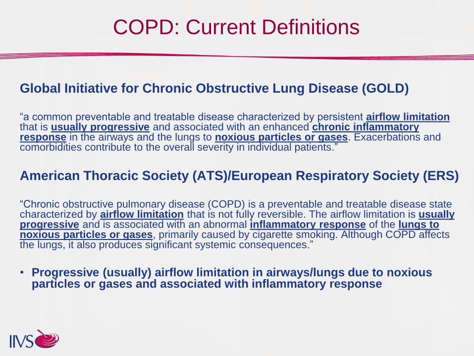
Global Initiative for Chronic Obstructive Lung Disease (GOLD) “a common preventable and treatable disease characterized by persistent airflow limitation that is usually progressive and associated with an enhanced chronic inflammatory response in the airways and the lungs to noxious particles or gases. Exacerbations and comorbidities contribute to the overall severity in individual patients.”
American Thoracic Society (ATS)/European Respiratory Society (ERS)
“Chronic obstructive pulmonary disease (COPD) is a preventable and treatable disease state characterized by airflow limitation that is not fully reversible. The airflow limitation is usually progressive and is associated with an abnormal inflammatory response of the lungs to noxious particles or gases, primarily caused by cigarette smoking. Although COPD affects the lungs, it also produces significant systemic consequences.”
• Progressive (usually) airflow limitation in airways/lungs due to noxious particles or gases and associated with inflammatory response
COPD: Risk Factors, Exacerbations, & Comorbidities
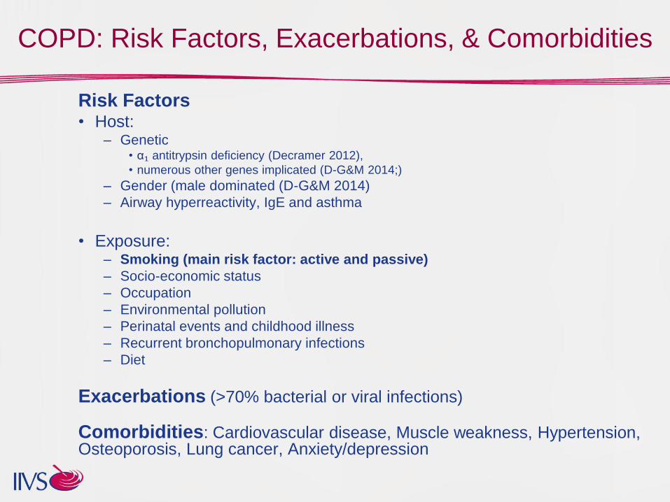
Risk Factors • Host:
– Genetic • α₁ antitrypsin deficiency (Decramer 2012),
• numerous other genes implicated (D-G&M 2014;)
– Gender (male dominated (D-G&M 2014)
– Airway hyperreactivity, IgE and asthma
• Exposure: – Smoking (main risk factor: active and passive)
– Socio-economic status
– Occupation
– Environmental pollution
– Perinatal events and childhood illness
– Recurrent bronchopulmonary infections
– Diet
Exacerbations (>70% bacterial or viral infections)
Comorbidities: Cardiovascular disease, Muscle weakness, Hypertension, Osteoporosis, Lung cancer, Anxiety/depression

http://www.nature.com/nri/journal/v8/n3/full/nri2254
Schematic of Tobacco-COPD events
Nature Reviews Immunology 8, 183-192
(March 2008) Peter J. Barnes

Tobacco Smoke Exposure: Changes in Lining of the bronchus
H: Cilia – sweep mucous & particulates
I: Columnar cells – yield cilia
J: Goblet cells – produce mucous
L: Basal cells – comprise bottom layer
Illustration & Excerpt Source: WhyQuit.com
• columnar cells are starting to be crowded out and displaced by additional layers of basal cells
• fewer cilia are present and are functioning at a much lower level of efficiency
• chemicals in tobacco smoke are toxic to cilia, first slowing them down, soon paralyzing them all together and then destroying them.
Illustration & Excerpt Source: WhyQuit.com
• ciliated columnar cells are totally displaced.
• smoker is more prone to infection from the
loss of the cleansing mechanism of the cilia
• O: abnormal cells are cancerous squamous
cells. These cells will eventually break
through the basement membrane wall and
invade into underlying lung tissue
• Illustration & Excerpt Source: WhyQuit.com
Normal lining of the bronchus Early changes Later changes
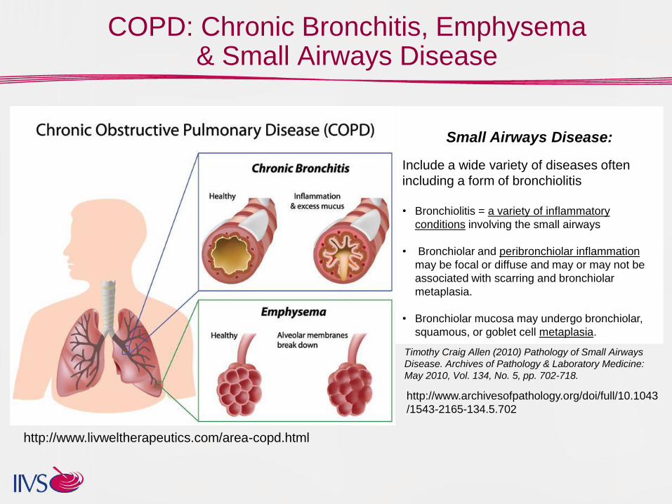
COPD: Chronic Bronchitis, Emphysema & Small Airways Disease
http://www.livweltherapeutics.com/area-copd.html
Small Airways Disease:
Include a wide variety of diseases often
including a form of bronchiolitis
• Bronchiolitis = a variety of inflammatory
conditions involving the small airways
• Bronchiolar and peribronchiolar inflammation
may be focal or diffuse and may or may not be
associated with scarring and bronchiolar
metaplasia.
• Bronchiolar mucosa may undergo bronchiolar,
squamous, or goblet cell metaplasia.
Timothy Craig Allen (2010) Pathology of Small Airways
Disease. Archives of Pathology & Laboratory Medicine:
May 2010, Vol. 134, No. 5, pp. 702-718.
http://www.archivesofpathology.org/doi/full/10.1043
/1543-2165-134.5.702

Chronic Bronchitis
Comparison of airway features in a healthy individual and
in a patient with chronic obstructive pulmonary disease
A: Normal airway.
B: In COPD, airways are narrowed by infiltration of inflammatory cells,
mucosal hyperplasia, and deposition of connective tissue in the
peribronchiolar space.
Hogg JC, Timens W. The pathology of chronic obstructive pulmonary
disease. Annu Rev Pathol 2009; 4: 435–59. http://www.livweltherapeutics.com/area-copd
A B

Emphysema
http://www.livweltherapeutics.com/area-copd http://www.headingfortheexits.com/emphsema-can-kill-you-because-planes-fly/
Comparison of airway features in a healthy individual and
in a patient with chronic obstructive pulmonary disease
A: Normal airway.
B: Emphysema: Septal collapse is evident. These changes are permanent
and cause a decrease in number of alveoli, an increase in size of alveoli,
and most importantly, a net decrease in the surface area available for gas
exchange.
A B A B

Chronic Obstructive Pulmonary Disease (COPD)
Tissue Response:
1. Cytokines/chemokines
2. Increased integrin and
adhesion molecule
expression
3. Monocyte recruitment
(persistent influx of
neutrophils)
4. Protease/antiprotease
imbalance
5. Adverse cellular ion
homeostasis-dehydration
6. Oxidative stress
7. Inflammation
Tissue Effects:
1. Ciliary dysfunction
2. Increased mucous
secretion
3. Fibroblast activation
4. Goblet cell hyperplasia
5. Bronchial epithelial
squamous metaplasia
6. Narrowing of airways
7. Collagen deposition
8. Parenchyma/tissue
destruction
9. Injury/repair cycling
Pulmonary Effects:
1. Reduced lung elasticity
2. Reduced airflow
3. Airspace enlargement
4. Small airway
remodeling
5. Vascular remodeling
6. Hyperinflation
7. Chronic inflammation
8. Fibrosis
Clinical
manifestations:
1. Chronic bronchitis
2. Emphysema
3. Small Airways Disease
4. Increased susceptibility to
infection and air pollutants
COPD:
Progressive (usually)
airflow limitation in
airways/lungs due to
noxious particles or gases
and associated with
inflammatory response
Initiating event:
Tobacco exposure or
other toxic insult to lung
epithelium
1. Ligand-receptor
interactions
2. Intracellular response
3. Oxidative stress
4. Initiation of autocrine,
paracrine, and endocrine
signaling
5. Cellular damage
Initiation, Progression, & Manifestation of COPD

Outline: In Vitro Models
Part 2. Overview of In Vitro Pulmonary Models
1. Introduction to In Vitro/Ex Vivo models
2. Types of models currently used in mainstream research
– Cell lines
– Primary cells
– 3D airway cultures
– Ex vivo tissue
3. Important considerations in choice of model
4. Upcoming Technologies

In Vitro/ex vivo Models
• A host of in vitro/ex vivo pulmonary models are available
• Used for a multiplicity of applications including:
– Drug development
• Efficacy
• Adverse effects
– Assessment of environmental toxicants
– Personal care & cosmetics product development
– Etc.
• For this workshop, a focus on models and assays that have
demonstrated fit for purpose
– Suitability in detecting one or more components in COPD etiology
– Commercially available
13

In Vitro Models for COPD
1. Cell lines: immortalized cells – Immature, transformed or cancer cells that have the capacity to expand
and (possibly) mature to some degree
2. Primary cells – Derived from normal or diseased tissues but may not have capacity to
expand greatly in number (e.g. limited supply)
3. 3D cultures/tissues – Reconstructed airway epithelium
4. Ex vivo tissues – Precision cut lung slices (PCLS)
5. New technologies: – Lung on a Chip
• Wyss Institute: Dr. Donald Ingber
• RPI & UNC
14

1. Cell Lines
Advantages: – Economical means to generate cell based data
– Typically very reproducible
– Usually straightforward to use and easy to culture
– Can be easily expanded, cryostored, and banked
for later use
Disadvantages: – Not considered as physiological as primary or 3D models
– Are transformed or derived from cancerous tissue
– Passage “drift” can occur
E.g. H292: mucoepidermoid carcinoma origin
BEAS-2B: adenovirus transformed bronchial epithelial cell line
NCI-H441: lung adenocarcinoma epithelial cell line
A549: adenocarcinoma of alveolar origin (lack of tight junctions)
The Cell Line NCl-H441 Is a Useful in Vitro Model for Transport Studies of Human Distal Lung Epithelial Barrier
Johanna J. Salomon,† Viktoria E. Muchitsch,† Julia C. Gausterer,† Elena Schwagerus,† Hanno Huwer,‡ Nicole Daum,§ Claus-Michael
Lehr,§ and Carsten Ehrhardt†,*
15
A549 cell line http://www.invitro.de/bildergalerie.html

2. Primary Cells
Advantages: – Not immortalized
– More representative of individuals in population
– Can be expanded, cryo-stored, and banked for later use, but donor variability impacts the
quality of tissue
Disadvantages: – Not immortalized and can be expensive
– Reproducibility is variable across different donors
– Can be difficult to culture and utilize
Cell origins: • Tracheobronchial Epithelia
• Alveolar Epithelia
• Cells of disease states available
E.g.
• Normal human bronchial epithelial cells (NHBE)
• NHBE + fibroblast co-cultures
16
Human bronchial-tracheal
epithelial cells (Lonza)
http://www.lonza.com/products-services/bio-
research/primary-cells/human-cells-and-
media/airway-cells-and-media/nhbe-normal-
human-bronchial-tracheal-epithelial-cells.aspx
3. 3D Epithelial Cultures
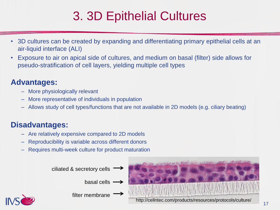
• 3D cultures can be created by expanding and differentiating primary epithelial cells at an
air-liquid interface (ALI)
• Exposure to air on apical side of cultures, and medium on basal (filter) side allows for
pseudo-stratification of cell layers, yielding multiple cell types
Advantages: – More physiologically relevant
– More representative of individuals in population
– Allows study of cell types/functions that are not available in 2D models (e.g. ciliary beating)
Disadvantages: – Are relatively expensive compared to 2D models
– Reproducibility is variable across different donors
– Requires multi-week culture for product maturation
17
ciliated & secretory cells
basal cells
filter membrane http://cellntec.com/products/resources/protocols/culture/

4. Ex Vivo Lung Tissues: PCLS
• Lung slices are created from whole lungs by inflating with agarose
solution, coring gelled tissues, and slicing cores in a precision slicer
• PCLS (typically ~300-1000 μm thickness) sliced from cores can be
cultured using ALI insert, roller drum method, or rocking platform
• PCLS can be cultured for days or weeks and are used for acute or
chronic exposures and/or evaluation
18
Advantages: – Most (?) physiologically relevant of non-whole organ ex vivo models
– More representative of individuals in population
– Allows study of cell types/functions that are not available in other models
• E.g. macrophages, airway contractility, etc.
Disadvantages: – Availability of high quality tissue is infrequent, highly variable quality across donors
– Reproducibility is variable across different donors
– Labor intensive setup procedure by well trained staff required
Credit: BASF/Fraunhofer
http://www.item.fraunhofer.de/en/business_
units_new/pre-clinical_pharmacology/Ex-
vivo_methods.html
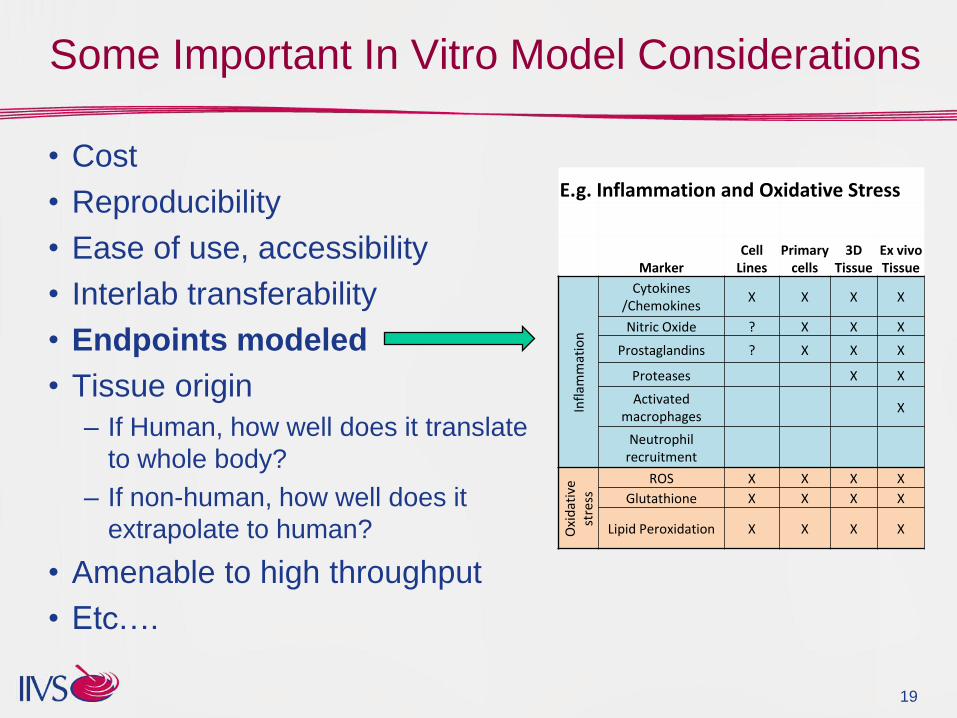
Some Important In Vitro Model Considerations
• Cost
• Reproducibility
• Ease of use, accessibility
• Interlab transferability
• Endpoints modeled
• Tissue origin
– If Human, how well does it translate
to whole body?
– If non-human, how well does it
extrapolate to human?
• Amenable to high throughput
• Etc….
19
E.g. Inflammation and Oxidative Stress
Marker
Cell Lines
Primary cells
3D Tissue
Ex vivo Tissue
Infl
amm
atio
n
Cytokines /Chemokines
X X X X
Nitric Oxide ? X X X
Prostaglandins ? X X X
Proteases X X
Activated macrophages
X
Neutrophil recruitment
Oxi
dat
ive
st
ress
ROS X X X X
Glutathione X X X X
Lipid Peroxidation X X X X
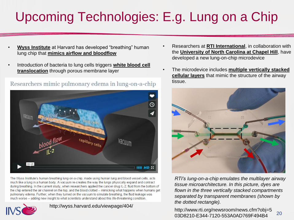
Upcoming Technologies: E.g. Lung on a Chip
20
• Researchers at RTI International, in collaboration with
the University of North Carolina at Chapel Hill, have
developed a new lung-on-chip microdevice
• The microdevice includes multiple vertically stacked
cellular layers that mimic the structure of the airway
tissue.
RTI’s lung-on-a-chip emulates the multilayer airway
tissue microarchitecture. In this picture, dyes are
flown in the three vertically stacked compartments
separated by transparent membranes (shown by
the dotted rectangle). http://wyss.harvard.edu/viewpage/404/
• Wyss Institute at Harvard has developed “breathing” human
lung chip that mimics airflow and bloodflow
• Introduction of bacteria to lung cells triggers white blood cell
translocation through porous membrane layer
http://www.rti.org/newsroom/news.cfm?obj=5
03D8210-E344-7120-553A0AD769F494B4

21
Etiology of COPD and In Vitro Models
Thank you!
Helpful references:
COPD • Jeffery PK (2000) Comparison of the structural and inflammatory features of COPD and asthma.
Giles F Filley Lecture Chest 117: 251S–260S.
• Barnes PJ (2000) Chronic obstructive pulmonary disease. N Engl J Med 343:269–280.
• van den Berge, M. et al. (2011) Small Airway Disease in Asthma and COPD, Clinical Implications
Chest 2011;139(2): 412–423
In Vitro Pulmonary Models • BeruBe, K. et al. (2009) In Vitro Models of Inhalation Toxicity and Disease The Report of a FRAME
Workshop ATLA — Alternatives to Laboratory Animals, Vol. 37, No. 1, 02.2009, p. 89-141.
• Jason Adamson, Linsey E Haswell, Gary Phillips and Marianna D Gaca (2011). In Vitro Models of
Chronic Obstructive Pulmonary Disease (COPD), Bronchitis, Dr. Ignacio MartÃn-Loeches (Ed.),
ISBN: 978-953-307-889-2, InTech, Available from: http://www.intechopen.com/books/bronchitis/in-
vitro-models-of-chronicobstructive-pulmonary-disease-copd


