176
Etiology of soybean-induced enteritis in fish paula uran.indd 1 paula uran.indd 1 27-3-2008 10:27:32 27-3-2008 10:27:32

Etiology of soybean-induced enteritis in fish
paula uran.indd 1paula uran.indd 1 27-3-2008 10:27:3227-3-2008 10:27:32

Promotor
Prof. Dr. J.A.J. Verreth Hoogleraar Aquacultuur en Visserij Wageningen Universiteit
Co-promotoren
Dr. Ir. J.W. Schrama Universitair hoofddocent Leerstoelgroep Aquacultuur en Visserij Wageningen Universiteit Dr. J.H.M.W. Rombout Universitair hoofddocent Leerstoelgroep Celbiologie en Immunologie Wageningen Universiteit
Promotiecommissie
Prof. Dr. Ir. M.W.A. Verstegen (Wageningen Universiteit) Prof. Dr. J.M. Wells (Wageningen Universiteit) Prof. Dr. Å. Krogdahl (Norwegian School of Veterinary Science, Oslo) Dr. Ir. G.T. Rijkers (Universitair Medisch Centrum, Utrecht)
Dit onderzoek is uitgevoerd binnen de onderzoekschool Wageningen Institute of Animal Sciences (WIAS)
paula uran.indd 2paula uran.indd 2 27-3-2008 10:27:5127-3-2008 10:27:51

Etiology of soybean-induced enteritis in fish
Paula A. Urán Carmona
Proefschrift ter verkrijging van de graad van doctor
op gezag van de rector magnificus van Wageningen Universiteit,
Prof. Dr. M. J. Kropff in het openbaar te verdedigen
op dinsdag 22 april 2008 des namiddags te vier uur in de Aula
paula uran.indd 3paula uran.indd 3 27-3-2008 10:27:5127-3-2008 10:27:51

Urán, P.A., 2008. Etiology of soybean-induced enteritis in fish. PhD thesis, Wageningen University, The Netherlands. ISBN: 978-90-8504-909-8
paula uran.indd 4paula uran.indd 4 27-3-2008 10:27:5227-3-2008 10:27:52
Abstract
The inclusion of soybean meal (SBM), especially in the diet of Atlantic salmon,
induces an inflammatory response of the distal intestinal mucosa, known as SBM-induced
enteritis. A semi-quantitative scoring system was developed to assess the extent of the
morphological changes observed in this study. The influence of SBM feeding has been
investigated taking into account several dietary and non-dietary factors possibly involved in
the induction of the disorder. It has been found that the severity of enteritis and its kinetics are
dose-dependent. Electron microscopy studies indicated a block of the endocytosis process and
a strong decrease of the microvilli length. Comparative studies were carried out in an
omnivorous species and for the first time ever reported, the results suggested that the
symptoms of enteritis also occur in common carp. Contrary to the observations in studies with
Atlantic salmon, the common carp started to recover from week four onwards. Several
cytokines were presumed to influence this process and they were correlated to the modulation
of the inflammatory process triggered by the SBM-containing diet. The influence of different
factors was measured according to the degree of enteritis developed. Low temperature (8 °C
vs. 12 °C) seem to delay the onset of the symptoms. On the other hand, it was suggested that
SBM-induced enteritis was not strongly influenced by either salinity or age. The extent of
enteritis in Atlantic salmon depends on the origin and/or the processing of the soybeans. The
morphological changes observed were induced when soyasaponins were fed to Atlantic
salmon alone or in combination with other soybean components suggestion their possible role
on the induction of enteritis. The actual causative components and its mechanisms of action
need further research. It is concluded that the etiology and further development of SBM-
induced enteritis is related to dietary factors rather than non-dietary factors. SBM inclusion
levels and the commercial source used for the diet formulation have a great impact on the
severity of the disorder, mainly affecting the endocytosis process. This thesis evidenced that
the endocytosis block is directly related to the disappearance of the supranuclear vacuoles,
which can be considered as the most striking feature in the onset of enteritis.
paula uran.indd 5paula uran.indd 5 27-3-2008 10:27:5327-3-2008 10:27:53

Abbreviations AMP antimicrobial peptides ANFs anti-nutritional factors BG basophilic granulocytes Ct cycle threshold E efficiency EG eosinophilic granulocytes EM electron microscopy FM fishmeal FO fish oil GAR-HRP goat-anti-rabbit-HRP GC goblet cells GOI gene of interest HKG house-keeping gene HSP heat shock proteins IBD inflammatory bowel diseases IEL intra-epithelial lymphocytes LM light microscopy LP lamina propria MF mucosal folds MFAA methanol, formalin and acetic acid Mv microvilli NMR nuclear magnetic resonance PBS phosphate buffered saline PBS-t phosphate buffered saline-tween RQ-PCR real time quantitative-polymerase chain reaction R relative expression ratio SB soybean SBM soybean meal SM sub-epithelial mucosa SNV supranuclear vacuoles
paula uran.indd 6paula uran.indd 6 27-3-2008 10:27:5327-3-2008 10:27:53

Contents Chapter 1 General Introduction
9
Chapter 2 Soybean meal-induced enteritis in Atlantic salmon (Salmo salar L.) at
different temperatures
21
Chapter 3 Time-related changes of the intestinal morphology of Atlantic salmon
(Salmo salar L.) at two different soybean meal inclusion levels
39
Chapter 4 Soybean meal-induced enteritis in common carp (Cyprinus carpio L.)
and the gene expression of inflammatory mediators in intestinal
leukocytes
61
Chapter 5 Soybean meal-induced uptake block in the distal enterocytes of Atlantic
salmon (Salmo salar L.)
85
Chapter 6 Variation in commercial sources of soybean meal influences the
severity of enteritis in Atlantic salmon (Salmo salar L.)
101
Chapter 7 Saponin-containing subfractions of soybean molasses induce enteritis
in the distal intestine of Atlantic salmon
117
Chapter 8 General Discussion 137
Semi-quantitative scoring system 149 Summary 157 Samenvatting 161 Resumen 165 List of publications 170 Acknowledgments 172 Training and Supervision Program 174
paula uran.indd 7paula uran.indd 7 27-3-2008 10:27:5427-3-2008 10:27:54

chapter hoofdstukken.indd 1 26-3-2008 12:36:20

GENERAL INTRODUCTION
Chapter 1
chapter hoofdstukken.indd 2 26-3-2008 12:36:21

Chapter 1
10
Fishmeal and fishmeal replacement
Fishmeal (FM) and fish oil (FO) are important commodities for the production of
animal feeds. FM is an important source of protein for fish and is often the only source that
complies with most of their nutritional requirements. In addition, FO is a rich source of
polyunsaturated fatty acids which constitutes the main source of lipids, particularly in fish.
FM and FO are derived from a natural resource composed of wild-caught pelagic fish from
the sea. Since 1985, world production of FM has stabilized at six to seven million tonnes and
for FO at one million tonnes. Aquaculture presently accounts for 35 percent of the world's FM
consumption. In recent decades aquaculture has been growing at a higher rate than all other
animal food-producing sectors, with an average annual growth rate of 8.8 percent per year
(FAO 2006). At this strong growth, pressures on the fish stocks supporting the production of
FM and FO will continue or even increase while they are already being utilized at their
maximum level of exploitation. Therefore, a strong competition on the market can be
expected for FM and FO resources, possibly leading to high prices and low availability.
Knowing that feed often comprises more than 50% of the total production costs (El-Sayed
1999; Fagbenro 1999), for both economic and sustainability reasons, a cheap and reliable
source of protein is needed to ensure a cost-effective and sustainable aquaculture. Therefore
the replacement of FM for fish diets is a high priority. Among the alternatives, plant-based
formulations are the cheapest, and many have a suitable protein profile and will be available
in the long term (Carter & Hauler 2000; Francis et al. 2001; Glencross et al. 2004; Gatlin et
al. 2007). These alternative protein sources have to ensure an excellent growth performance
and health status of the cultured species but also have to meet requirements for taste, odour,
and the consumer’s acceptability.
Soybean as Fishmeal replacement
Oilseeds, in particular, soybean (Glycine max L.), and grain products have great
potential as protein and/or oil sources for fish feeds (Alexis & Nengas 2001). Nonetheless, SB
contains anti-nutritional factors (ANF’s) which may inhibit nutrient utilization and
digestibility. Oligosaccharides, non-starch polysaccharides, saponins, protease inhibitors,
antigenic compounds, lectins, phytic acid, tannins, phytoestrogens alkaloids, gossypols are
well known ANF’s (Alexis & Nengas 2001; Francis et al. 2001). Diverse feed processing
paula uran.indd 10paula uran.indd 10 27-3-2008 10:27:5627-3-2008 10:27:56

General Introduction
11
techniques like dry or wet heating, aqueous-extraction and the addition of supplements can
reduce the final content of ANF’s (Rumsey et al. 1994; Buttle et al. 2001; Refstie et al. 2005)
and reduce their negative impacts (Francis et al. 2001).
The negative effects of SB products inclusion also depend on the source and type of
this product and the level of replacement. According to literature, commercial diets for
salmonids may contain about 34% to 47% protein and 28% to 40% lipid (Refstie et al. 2001).
Different SB products could fulfil this high protein demand, however, the problem remains
that the more refined the formulations are, the more expensive the feed becomes. Many
studies have searched for the optimum inclusion level with the lowest amount of noxious
factors. Different treatments of SB and inclusion levels have been tested. Some of the results
indicate that for salmonids, diets containing SB protein concentrate shows a growth
performance as good as the high quality FM (Olli et al. 1995; Storebakken et al. 1998). This
is followed in performance by full-fat, dehulled solvent-extracted and solvent-extracted SB
meal. The latter seems to reduce growth with increasing levels of inclusion (Krogdahl et al.
2003). In African catfish (Clarias gariepinus Burchell) diets, replacement of FM by dehulled
solvent-extracted SB meal was possible up to the level of 50% (even 75% when methionine
supplementation was used) without compromising growth and feed utilization efficiency
(Fagbenro & Davies 2001). For Atlantic halibut (Hippoglossus hippoglossus) 36% full-fat SB
meal may be added to the diets without negative effects on growth, feed efficiency or
intestinal morphology (Grisdale-Helland et al. 2002). Egyptian sole (Solea aegyptiaca) can be
fed with a diet containing up to 30% SB meal without any reduction in the growth rate or
induction of histopathology of the gut (Bonaldo et al. 2007).
SB-induced enteritis in Atlantic salmon
In Atlantic salmon (Salmo salar L.), replacing 20% fish meal protein by dehulled
solvent-extracted SB meal does not impair growth (Olli et al. 1994, 1995). This seems to
corroborate with the results of several other authors, who all demonstrated that replacing
small amounts of FM (e.g. 20% or less) by SB products does not lead to significant growth
depression (Bjerkeng et al. 1997; Refstie et al. 2001; Opstvedt et al. 2003). However,
Krogdahl et al. (2003) showed that at low inclusion levels, dietary SB meal could induce
intestinal disorders, which on their turn could lead to growth depression later on. These
paula uran.indd 11paula uran.indd 11 27-3-2008 10:27:5827-3-2008 10:27:58

Chapter 1
12
disorders were first described by van den Ingh et al. (1991, 1996) and named by Baeverfjord
& Krogdahl (1996) as “non- infectious sub-acute enteritis”. The typical signs of this intestinal
disorder are: a shortening of the mucosal folds, loss of the normal supranuclear vacuolisation;
a thickening of both lamina propria and sub-epithelial mucosa with a severe infiltration of
inflammatory cells (particularly macrophages and eosinophilic granulocytes) and increased
numbers of goblet cells in the epithelium (van den Ingh et al. 1991, 1996; Baeverfjord &
Krogdahl 1996; Krogdahl et al. 2000; Refstie et al. 2000; Buttle et al. 2001).
Up to now, the degree of enteritis and its impact on the epithelial mucosa was mainly
described as either slight, moderate or severe, usually based on qualitative analyses only
(Refstie et al. 2000, 2001; Sanden et al. 2005; Bakke-Mckellep et al. 2007). The majority of
studies (Krogdahl et al. 2000, 2003; Refstie et al. 2005; Lilleeng et al. 2007) used an end
point approach, with a response analysis not earlier than 20 days after SB meal feeding. Such
an approach does not provide information on the development process of the disorder, while
this is crucial information for the comparison of species and for different husbandry and
environmental parameters.
It is still unclear how SB products really cause enteritis in Atlantic salmon. The
development of enteritis may be related to the risk for secondary diseases, which could be
facilitated by the SB-induced morphological changes in the intestine. Krogdahl et al. (2000)
studied the disease resistance and local immune response in Atlantic salmon fed different SB
products during a cohabitation challenge using Aeromonas salmonicida ssp. salmonicida. In
animals with clinical signs of enteritis, gut permeability increased. This could have facilitated
the colonization of the epithelium by pathogens, which in combination with other factors such
as diarrhoea, reduced nutrient digestibility and reduced growth, make fish more vulnerable to
disease outbreaks.
The main structure affected seems to be the proximal part of the distal intestine, also
called the second gut segment. This gut segment is considered as more sensitive to food-borne
enteropathies because it is the major site of endocytosis of intact proteins (Stroband et al.
1979, Stroband & van der Veen 1981; Rombout et al. 1985; Sire & Vernier 1992; Bakke-
McKellep et al. 2000). Endocytosis could well play an essential role in the development of
the intestinal disorder, but until now, data are not available to support this hypothesis. This
could also be related to the fact that most of the observations on SB-induced enteritis are
paula uran.indd 12paula uran.indd 12 27-3-2008 10:27:5927-3-2008 10:27:59

General Introduction
13
restricted to light rather than to electron microscopy. It is clear that, if the etiology of the SB-
induced enteritis process needs to be understood, more information on the function of the
second gut segment, particularly on its role in absorption and immunity, has to be gathered.
More knowledge on the early development of enteritis will contribute to the elucidation of the
mechanisms behind this disorder. The identification of the early symptoms of enteritis can be
used not only as indicators of the severity and/or speed at which the disorder develops but
also as a tool to assess the development of the inflammatory process. Since most research on
the impact of SB products, and in particular, of SB meal, focused on salmonids, it is of high
importance to investigate whether enteritis also occurs in other species. In this regard,
omnivorous fish species may be highly interesting since they are usually exposed to SB
products. Common carp (Cyprinus carpio L.) is an example of an omnivorous fish. In
addition, it is a representative of species lacking the stomach (Fig. 1). Therefore, it may not
only provide information of why and how some species respond stronger than others to SB
products, but also common carp can be an interesting model to compare with stomach
containing species like Atlantic salmon.
Figure 1 Scheme of different intestinal tract a stomachless fish (left) and a more complex stomach
containing fish (right). (Adapted from Stroband et al. 1980).
paula uran.indd 13paula uran.indd 13 27-3-2008 10:28:0027-3-2008 10:28:00

Chapter 1
14
Up to now only qualitative methods were used to assess enteritis. These methods are
insufficient to study the etiology of the disorder and to compare it between species,
environmental factors and different SB products. The mentioned ambitions can only be
realized if a quantitative assessment method is developed and used as an instrument to
compare the effects of SB-induced enteritis.
Aim and scope of this Thesis
The overall objective of the present study was to elucidate the mechanism behind the
inflammatory process induced by SB products. Based on previous observations, it is
hypothesised that the altered endocytosis process is the driving mechanism behind the SB
meal-induced enteritis. Since the development of enteritis has been mainly studied in relation
to the type of SB diet, more attention will be paid to non-diet related factors like i.e.
husbandry conditions and animal-intrinsic factors like the endocytosis process. It is
reasonable to consider that the hampered endocytosis can also occur in other fish species.
Therefore, the etiology of the disorder will be investigated both in Atlantic salmon and in
common carp.
In this study the following objectives/aspects are addressed:
Development of a scoring system which can be used as a tool to further study and
compare the kinetics of the disorder under different conditions.
The etiology of the SB meal-induced enteritis, focusing on the early development of
the disorder.
Evaluation of the effects of non-diet related factors on the development of the disorder
such as water temperature.
Evaluation of the effects of diet related factors on the development of the disorder
such as SB inclusion levels, commercial sources, soyasaponins content.
Investigation of possible enteritis in an omnivorous fish species (common carp) and a
better identification of the mechanism responsible for the possible induction of the
disorder.
paula uran.indd 14paula uran.indd 14 27-3-2008 10:28:0127-3-2008 10:28:01

General Introduction
15
Figure 2. Summary of the thesis setup.
In Chapter 2, the impact of water temperature on the development of enteritis is assessed by
means of a semi-quantitative scoring system. In Chapter 3, the onset of the enteritis
development is described and the kinetics of the enteritis process is analysed at two different
SB meal inclusion levels. This chapter gives also information on the changes at the
ultrastructural level in the epithelium of the distal intestine. In Chapter 4, the effect of dietary
SB meal is compared in omnivorous common carp and carnivorous Atlantic salmon. In this
study common carp was continuously fed on animal protein before being transferred to the SB
meal diets (20% inclusion level). At this inclusion level, Atlantic salmon usually develops
severe clinical signs of enteritis. The kinetics of several cytokines are included to illustrate the
regulation of the inflammatory process. In Chapter 5, an attempt is made to link the
morphological changes observed at the light microscopical level and the changes observed at
ultrastructural level (endocytosis). Young salmon were used to establish whether age and
freshwater conditions can have any influence on the severity of SB meal-induced enteritis
described previously for older fish kept in seawater. Chapter 6 aims to clarify if different
commercial sources of SB meal can result in dissimilar severity degrees of the enteritis
Etiology of enteritis
Scoring system (Chapter 2)
Atlantic salmon Common carp
Kinetics (Chapter 3)
Mechanisms (Chapter 5)
Non-dietary factors (Chapter 2)
Dietary factors (Chapters 6 and 7)
Kinetics and Mechanism (Chapter 4)
paula uran.indd 15paula uran.indd 15 27-3-2008 10:28:0227-3-2008 10:28:02
Chapter 1
16
process in Atlantic salmon using the mentioned semi-quantitative scoring system. In Chapter
7, attention is paid to possible causative components of the SB-induced enteritis, by means of
phase separation of the SB molasses and the biochemical composition of the saponin-
containing sub-fractions. Finally in Chapter 8, the overall results obtained from this study
will be summarized and discussed, together with possible explanations and indications for the
underlying mechanisms involved in the development of the SB-induced inflammatory
response.
References
Alexis, M.N. & Nengas, I. (2001) Current state of knowledge concerning the use of soy products in
diets for feeding sea bass and sea bream needs for future research. National centre for marine
research, Athens-Greece. American Soybean Association. 5, 1-32.
Baeverfjord, G. & Krogdahl, Å. (1996) Development and regression of soybean meal induced enteritis
in Atlantic salmon, Salmo salar L., distal intestine: a comparison with the intestines of fasted fish.
J. Fish Dis., 19, 375-387.
Bakke-Mckellep, A.M., Press, C.Mcl., Baeverfjord, G., Krogdahl, Å. & Landsverk, T. (2000) Changes
in immune and enzyme histochemical phenotypes of cells in the intestinal mucosa of Atlantic
salmon, Salmo salar L., with soybean meal-induced enteritis. J. Fish Dis., 23, 115-127.
Bakke-McKellep, A.M., Koppang, E.O., Gunnes, G., Sanden, M., Hemre, G.I., Landsverk, T. &
Krogdahl, A. (2007) Histological, digestive, metabolic, hormonal and some immune factor
responses in Atlantic salmon, Salmo salar L., fed genetically modified soybeans. J. Fish Dis., 30,
65-79.
Bjerkeng, B., Refstie, S., Fjalestad, K.T. Storebakken, T., Rødbotten, M.& Roem, A.J. (1997) Quality
parameters of the flesh of Atlantic salmon (Salmo salar) as affected by dietary fat content and full-
fat soybean meal as a partial substitute for fish meal in the diet. Aquaculture, 157, 297-309.
Bonaldo, A., Roem, A.J., Mariani, L., Fagioli, P., Pecchini, A., Parma, L. & Gatta, P.P. (2007) The
influence of different levels of soybean meal in diets for ongrowing gilthead sea bream (Sparus
aurata) and European sea bass (Dicentrarchus labrax). Ital. J. Anim. Sci., 6, 790-790.
paula uran.indd 16paula uran.indd 16 27-3-2008 10:28:0327-3-2008 10:28:03

General Introduction
17
Buttle, L.G., Burrells, A.C., Good, J.E., Williams, P.D., Southgate, P.J. & Burrells C. (2001) The
binding of soybean agglutinin (SBA) to the intestinal epithelium of Atlantic salmon Salmo salar
and Rainbow trout, Oncorhynchus mykiss, fed high levels of soybean meal. Vet. Immunol.
Immunopathol., 80, 237-244.
Carter, C.G. & Hauler, R.C. (2000) Fishmeal replacement by plant meals in extruded feeds for
Atlantic salmon, Salmo salar L. Aquaculture, 185, 299-311.
El-Sayed, A.F.M. (1999) Alternative dietary protein sources for farmed tilapia, Oreochromis spp.
Aquaculture, 179, 149-168.
Fagbenro, O.A. (1999) Comparative evaluation of heat-processed winged bean (Psophocarpus
tetragonolobus) meals as partial replacement for fish meal in diets for the African catfish (Clarias
gariepinus). Aquaculture, 170, 297-305.
Fagbenro, O.A. & Davies, S. J. (2001) Use of soybean flour (dehulled, solvent-extracted soybean) as a
fish meal substitute in practical diets for African catfish, Clarias gariepinus (Burchell 1822):
growth, feed utilization and digestibility. J. Appl. Ichthyol., 17, 64-69.
FAO (2006) Fisheries Technical Paper. No. 500. Rome, 134p.
Francis, G., Makkar, H. P.S. & Becker, K. (2001) Antinutritional factors present in plan-derived
alternate fish feed ingredients and their effects in fish. Review Article. Aquaculture 199, 197-227.
Gatlin, D.M., Barrows, F.T., Brown, P., Dabrowski, K., Gaylord, T.G., Hardy, R.W., Herman, E., Hu,
G.S., Krogdahl, A., Nelson, R., Overturf, K., Rust, M., Sealey, W., Skonberg, D., Souza, E.J.,
Stone, D., Wilson, R. & Wurtele, E. (2007) Expanding the utilization of sustainable plant products
in aquafeeds: a review. Aquac. Res., 38, 551-579.
Glencross, B.D., Carter, C.G., Duijster, N., Evans, D.R., Dods, K., McCafferty, P., Hawkins, W.E.,
Maas, R. & Sipsas, S. (2004) A comparison of the digestibility of a range of lupin and soybean
protein products when fed to either Atlantic salmon (Salmo salar) or rainbow trout (Oncorhynchus
mykiss). Aquaculture, 237, 333-346.
Grisdale-Helland, B., Helland, S.J. Baeverfjord, G. & Berge, G.M. (2002) Full-fat soybean meal in
diets for Atlantic halibut: growth, metabolism and intestinal histology. Aquacult. Nutr., 8, 265-
270.
van den Ingh, T.S.G.A.M., Krogdahl, Å., Olli, J.J., Hendriks, H.G.C.J.M. & Koninkx, J.G.J.F. (1991)
Effects of soybean-containing diets on the proximal and distal intestine in Atlantic salmon (Salmo
salar): a morphological study. Aquaculture, 94, 297-305.
van den Ingh, T.S.G.A.M., Olli, J.J. & Krogdahl, Å. (1996) Alcohol-soluble components in soybeans
cause morphological changes in the distal intestine of Atlantic salmon, Salmo salar L. J. Fish Dis.,
19, 47-53.
paula uran.indd 17paula uran.indd 17 27-3-2008 10:28:0427-3-2008 10:28:04
Chapter 1
18
Krogdahl, Å., Bakke-McKellep, A.M., Røed, K.H. & Baeverfjord, G. (2000) Feeding Atlantic salmon
Salmo salar L. soybean products: effects on disease resistance (furunculosis), and lysozyme and
IgM levels in the intestinal mucosa. Aquacult. Nutr., 6, 77-84.
Krogdahl, Å., Bakke-McKellep, A.M. & Baeverfjord, G. (2003) Effects of graded levels of standard
soybean meal on intestinal structure, mucosal enzyme activities,
and pancreatic response in Atlantic salmon (Salmo salar L.). Aquacult. Nutr., 9, 361-371.Lilleeng, E.,
Froystad, M.K., Ostby, G.C., Valen, E.C. & Krogdahl, A. (2007) Effects of diets containing
soybean meal on trypsin mRNA expression and activity in Atlantic salmon (Salmo salar L).
Comp. Biochem. Phys. A, 147, 25-36.
Olli, J.J., Krogdahl, Å. & Våbenø, A. (1995) Dehulled solvent-extracted soybean meal as a protein
source in diets for Atlantic salmon, Salmo salar L. Aquac. Res., 26,167-174.
Olli, J.J., Krogdahl, Å., Van Den Ingh T.S.G.A. & Brattas, L.E. (1994) Nutritive value of four soybean
products in diets for Atlantic salmon, Salmo salar L. Acta Agric. Scand., Sect. A, Animal Sci.,
44:50-60.
Opstvedt, J.A., A. Hope, B. & Pike I.H. (2003) Efficiency of feed utilization in Atlantic salmon
(Salmo salar L.) fed diets with increasing substitution of fish meal with vegetable proteins.
Aquaculture, 212, 365-379.
Refstie, S., Korsøen, Ø.J., Storebakken, T., Baeverfjord, G., Lein, I. & Roem, A.J. (2000) Differing
nutritional responses to dietary soybean meal in rainbow trout (Oncorhynchus mykiss) and
Atlantic salmon (Salmo salar). Aquaculture, 190, 49-63.
Refstie, S., Strorebakken, T., Baeverfjord G. & Roem A.J. (2001) Long-term protein and lipid growth
of Atlantic salmon (Salmon salar) fed diets with partial replacement of fishmeal by soy protein
products at medium or high lipid level. Aquaculture, 193, 91-106.
Refstie, S., Sahlstrom, S., Brathen, E., Baeverfjord, G. & Krogdahl, P. (2005) Lactic acid fermentation
eliminates indigestible carbohydrates and antinutritional factors in soybean meal for Atlantic
salmon (Salmo salar). Aquaculture, 246, 331-345.
Rombout, J.H.W.M., Lamers, C.H.J., Helfrich, M.H., Dekker, A. & Taverne-Thiele, J.J. (1985)
Uptake and transport of intact macromolecules in the intestinal epithelium of carp (Cyprinus
carpio L.) and the possible immunological implications. Cell Tissue Res., 239, 519-530.
Rumsey, G.L., Siwicki, A.K., Anderson D.P. & Bowser, P.R. (1994) Effect of soybean protein on
serological response, non-specific defense mechanisms, growth, and protein utilization in rainbow
trout. Vet. Immunol. Immunopath., 41, 323-339.
paula uran.indd 18paula uran.indd 18 27-3-2008 10:28:0527-3-2008 10:28:05
General Introduction
19
Sanden, M., Berntssen, M.H.G., Krogdahl, A., Hemre, G.I. & Bakke-McKellep, A.M. (2005) An
examination of the intestinal tract of Atlantic salmon, Salmo salar L., parr fed different varieties
of soy and maize. J. Fish Dis., 28, 317-330.
Sire, M.F. & Vernier, J.M. (1992) Intestinal absorption of protein in teleost fish. Comp. Biochem. Phys
A, 103, 771-781.
Storebakken, T., Shearer, K.D. & Roem, A.J. (1998) Availability of protein, phosphorus and other
elements in fish meal, soy-protein concentrate and phytase-treated soy-protein-concentrate-based
diets to Atlantic salmon, Salmo salar. Aquaculture, 161, 365-379.
Stroband, H.W.J., Van Der Meer, H. & Timmermans, L.P.M. (1979) Regional function
differenctiation in the gut of the grass carp, Ctenopharyngodon idella (Val.). Histochemistry, 64:
235-249.
Stroband, H.W.J; Rombout, J.H.W.M. & Davina, J.H.M. (1980) Maagloze Vissen. Bouw en functie
van het darmkanaal. Natuur en techniek. 48e jaargang, nr. 1, Cat. nr. 578, p.56
Stroband, H.W.J. & van der Veen, F.H. (1981) Localization of protein absorption during transport of
food in the intestine of the grasscarp, Ctenopharyngodon idella (Val.). J. Exp. Zool., 218, 149-156.
paula uran.indd 19paula uran.indd 19 27-3-2008 10:28:0627-3-2008 10:28:06

chapter hoofdstukken.indd 3 26-3-2008 12:36:50

Chapter 2
In press:Aquaculture Nutrition, (2008)
DOI:10.1111/j.1365-2095.2007.00534.x
Soybean meal-induced enteritis in Atlantic salmon (Salmo salar L.) at different temperatures
P.A. Urán1,2, J.W.Schrama1, J.H.W.M. Rombout2,A. Obach3, L. Jensen3, W. Koppe3 & J.A.J. Verreth1
1Aquaculture and Fisheries Group and 2Cell Biology and Immunology Group, Wageningen Institute of Animal Sciences, Wageningen University, The Netherlands 3Skretting, Aqua-culture Research Centre, Stavanger, Norway
chapter hoofdstukken.indd 4 26-3-2008 12:36:50

Chapter 2
22
Abstract
This study evaluates the effect of temperature on the development of intestinal disorders when
Atlantic salmon are fed soybean meal (SBM). In this study 20% of the dietary fishmeal (FM)
was replaced by solvent-extracted Hipro SBM. Atlantic salmon reared at two different water
temperatures (8 ºC and 12 ºC), were fed a control diet and an experimental diet for 20 days.
Samples were taken at days 7 and 20. The extent of the morphological changes was assessed
using a semi-quantitative scoring system developed for this purpose. The study demonstrates
that enteritis is affected by temperature. The intestinal disorders were more severe in fish
reared at 12 °C compared to those reared at 8 °C. It can be concluded from this study that
temperature changes the speed but not the type of SBM-induced enteritis expressed as a delay
on the response when Atlantic salmon are kept at lower temperatures.
paula uran.indd 22paula uran.indd 22 27-3-2008 10:28:0927-3-2008 10:28:09

Temperature and SBM-induced enteritis in Atlantic salmon
23
Introduction
Soybean meal (SBM) has been suggested as one of the best alternatives to replace
fishmeal (FM) for salmonid diets. However, the inclusion of SBM induces enteritis in
Atlantic salmon (Salmo salar L.). Baeverfjord & Krogdahl (1996) described this condition as
“a non- infectious sub-acute inflammation of the distal intestine”. These pathological changes
seem to be particularly present on the distal intestinal segment rather than on the proximal as
reported in several studies on salmonids (van den Ingh et al. 1991; Burrells et al. 1999;
Nordrum et al. 2000; Buttle et al. 2001).
The symptoms that define the condition are: a shortening of the mucosal folds; a loss
of the normal supranuclear vacuolisation of the absorptive cells in the intestinal epithelium; a
widening of the central stroma within the mucosal folding, with increased amounts of
connective tissue; a profound infiltration of inflammatory cells in the lamina propria (van den
Ingh et al. 1991; van den Ingh et al. 1996; Baeverfjord & Krogdahl 1996; Krogdahl et al.
2000; Refstie et al. 2000; Buttle et al. 2001); an increased presence of IgM (Bakke-McKellep
et al. 2000), an increased amount of goblet cells in the epithelium, as well as a decreased
height of the microvilli together with increased microvillar vesicle formation (van den Ingh et
al. 1991).
The inclusion levels, varieties, origins and processing techniques of the different
soybean (SB) products along with husbandry conditions influence the occurrence of these
symptoms. Previous studies have primarily focused on the impact of diet formulation on fish
performance. An exception is the study of Nordrum et al. (2000) who investigated the effect
of salinity on the development of enteritis in salmonids. Several studies have demonstrated
that the absorptive capacity in salmonids is negatively influenced by high inclusion levels of
SBM in the diet. Rumsey et al. (1994) suggested that antigenic soya protein affects non-
specific defence mechanisms, growth performance and protein utilization in rainbow trout.
Nordrum et al. (2000) found that the effects of SBM on the intestinal morphology of rainbow
trout were of less magnitude than for salmon. Atlantic salmon seem to develop a more severe
enteritis condition than other fish species. What more, despite the importance of this species,
little information is available on the impact of different management-related factors on the
occurrence of enteritis. In the present study the effects of temperature were evaluated.
paula uran.indd 23paula uran.indd 23 27-3-2008 10:28:1027-3-2008 10:28:10

Chapter 2
24
Preliminary studies (unpubl. observ.) suggested that enteritis becomes less severe at higher
temperatures. The current study will investigate whether temperature can indeed affect the
enteritis process either by influencing the metabolic rate or by having a direct impact on the
normal nutrient absorption process. For this purpose a semi-quantitative scoring system is
introduced. This scoring system has great potential as a diagnostic tool for the histological
evaluation of an inflamed intestine. The possible effects of temperature on the aggravation of
the mentioned symptoms will be investigated.
Material and Methods
Fish rearing conditions
The experiment was carried out at the Skretting fish trials station, Lerang, Jørpeland,
Norway. For this experiment, 40 Atlantic salmon (AquaGen strain) were sampled for gut
histology measurements. The Atlantic salmon used originated from a stock of fish present at
the research station. The experiment consisted on a 14-day adaptation period and a 20-day
experimental period. At the start of the adaptation period the fish weighed approximately 300
g. Four indoor tanks with a diameter of 1 m each were used. The water volume in the tanks
was 400 L. The stocking density was 50 fish per tank. Each tank was kept at flow rate of 12 to
15 L min-1. Seawater pumped from 90 m depth in the fjord, with a salinity of 34 ‰ and an
oxygen concentration above 9 ppm, was used as the inlet water. The temperature of the inlet
water was 8 °C or 12 °C depending on the experimental treatment (2 tanks per water
temperature). Prior to the adaptation period fish were kept at 8 °C. The applied photoperiod
was 18L : 6D.
Diets and feeding
The diets were produced at Skretting Feed Technology Plant (Stavanger, Norway).
Two diets were formulated: a control diet (0SBM) and an experimental diet (20SBM) (Table
1). The major ingredients in the control diet, 0SBM, were: FM (protein content above 70%),
fish oil (FO) and wheat. This control diet did not contain SBM. The experimental diet,
20SBM, contained 20% solvent-extracted Hipro SBM (Cargill, The Netherlands). In the
paula uran.indd 24paula uran.indd 24 27-3-2008 10:28:1127-3-2008 10:28:11

Temperature and SBM-induced enteritis in Atlantic salmon
25
20SBM diet, FM, FO as well as wheat were exchanged for 20% SBM compared to the control
diet. Diets were formulated to be iso-nitrogenous and iso-energetic on a crude protein and a
crude lipid basis. Diets were supplemented with a standard vitamin and mineral premix. Feed
was produced as extruded 4 mm sinking pellets.
Prior to the experiment the fish were fed a commercial salmon diet (Skretting,
Stavanger, Norway), without any SB products. During the adaptation period all fish were fed
with the control diet (0SBM). At the start of the experimental period (day 1), fish of one of
the tanks at each water temperature were fed the experimental diet (20SBM) while fish in the
two others continued to be fed with 0SBM. Fish were fed 20% in excess. Feed was divided
into two meals per day and it was provided by automatic feeders.
Table 1 Ingredients and chemical composition of the experimental diets.
Diets1 0SBM 20SBM Ingredients (g kg-1)
Fishmeal2 564.3 475.3 Extracted soybean meal3 0 200 Wheat 210.6 70 Fish Oil4 222.6 252.1 Vitamin premix 1.3 1.3 Mineral premix 1.3 1.3
Pigment premix Yttrium oxide 0.1 0.1 Carophyll Pink 0.6 0.6
Chemical composition (by analysis) Crude Protein (g kg-1) 429.8 450.6 Crude Lipid (g kg-1) 277.1 301.4 Target dry matter (%) 95 95 Fat NMR (%) 30.5 32.8 Protein (%) 43.1 45.2 Moisture (%) 5.1 4.7 Ash (%) 7.2 7.3
1 Amount of fish meal replaced by Soybean meal (SBM) in percent.
2 LT North Atlantic, from Egersund, Norway.
3 Cargill, The Netherlands.
4 Northern hemisphere.
paula uran.indd 25paula uran.indd 25 27-3-2008 10:28:1227-3-2008 10:28:12

Chapter 2
26
Chemical analysis of diets
The nutrient composition of the experimental diets was determined using standard
techniques for proximate analyses. Crude protein content was determined by the Kjeldahl
nitrogen measurement according to the Nordic Committee on Food Analysis, Method No. 6,
4th edition 2003. Crude fat content was measured by low field nuclear magnetic resonance.
Moisture content in samples was measured by drying to constant weight at 102-105 °C for 16-
18 h. Ash content was measured by combustion at 540 °C for 16-18 h, after which the
remaining residues were weighed, both according to the Nordic Committee on Food Analysis
Method No. 23, 3rd edition 1991. The preceding analyses were carried out at the Skretting
Aquaculture Research centre, Stavanger, Norway. (See Table 1 for chemical composition).
Sampling for intestinal morphology
During the experimental period, fish gut was sampled for a histological assessment of
enteritis developed at 7 and 20 days after SBM feeding. At each sampling moment, five fish
per treatment group were sampled (per water temperature two experimental diets). The
samples were taken from alternate tanks to avoid drops in feed intake due to sampling stress.
Directly after the morning meal, the fish were anaesthetized using 0,05 g L-1 metacaine
(Argent chemical laboratories, USA) and thereafter killed by a sharp blow to the head. The
distal intestine was dissected from the point were the intestinal diameter increases, the mucosa
becomes darker and annular rings are clearly noticeable. A two-centimeter section of
dissected distal intestine of each fish was taken and gently rinsed with cold (4ºC) saline water.
Samples were fixed in a 4% phosphate-buffered formalin with a pH of 7.2 and stored at room
temperature. After dehydration by standard procedures, samples were embedded in paraffin.
Transverse sections of 5 μm thickness were cut using a Microm HM 350 rotary microtome
(Heidelberg, Germany) and thereafter mounted on glass slides. Each slide contained from 3 to
4 sections of a complete cut of an annular ring of distal intestine where all layers were visible
and well represented. After de-paraffination, sections were stained using a mix of
Haematoxylin/Eosin and Alcian blue pH 2.5. Alcian blue staining enhances the contrast
between goblet cells and the supranuclear vacuoles. Slides were blindly evaluated after
randomization.
paula uran.indd 26paula uran.indd 26 27-3-2008 10:28:1327-3-2008 10:28:13

Temperature and SBM-induced enteritis in Atlantic salmon
27
Scoring system
For this study a semi-quantitative scoring system was used. In this scoring system six
separate parameters of soybean-induced enteritis were quantified independently, according to:
1) the appearance and length of the mucosal folds (MF); 2) the presence and size of
supranuclear vacuoles (SNV); 3) the abundance of goblet cells (GC); 4) the degree of
infiltration abundance and of eosinophilic granulocytes into the lamina propria and into the
sub-epithelial mucosa (EG); 5) the degree of widening of the lamina propria (LP); and 6) the
degree of thickening of the sub-epithelial mucosa (SM). Each of these parameters was scored
on a scale from 1 to 5, including half values between categories. An increasing scoring value
represents a more severe enteritis condition. Sections were photographed with an Olympus
DP 50 digital camera connected to a Nikon Microphot-FXA light microscope (Badhoevedorp,
The Netherlands). The pictures were processed and analysed using the AnalySiS Extended
Pro 3.1 software (Soft Imaging System GmbH, Münster, Germany). A detailed description of
the morphological/histological appearance per characteristic for the different scoring values
from 1 to 5 is given in Table 2. Different degrees of enteritis are shown in Figure 1. An
overall value of the degree of enteritis was calculated by averaging the scores of the six
separate parameters (MF, SNV, GC, EG, LP and SM). (For illustrations of the different scores
see annex or check at http://www.afi.wur.nl/UK/Publications/).
Statistics
Preliminary analysis of the slides showed that SBM-induced enteritis was not present
in those fish fed the control diet (0SBM). Therefore, the effect of water temperature and
sampling moment (i.e., days after changing to the SBM diet) on scorings of the separate
enteritis parameters (MF, SNV, GC, EG, LP and SM) as well as the overall mean enteritis
scoring were analysed by a 2-way ANOVA of the fish at the experimental diet (20SBM).
Furthermore, it was assessed as to how development of enteritis was related to the combined
effect of days and water temperature, after exposing the fish to the 20SBM diet by using
degree-days. This was done by a linear regression of degree-day on the mean enteritis score.
These analyses were done using the general linear model procedure of SAS (1999). Error
term analysis using the univariate procedure of SAS (1999) showed that scoring values of all
paula uran.indd 27paula uran.indd 27 27-3-2008 10:28:1427-3-2008 10:28:14

Chapter 2
28
separate parameters and the overall mean score were normally distributed. The level of
significance was established at P < 0.05.
Table 2 Description of the semi-quantitative scoring system using different parameters to assess the degree of
enteritis developed by Atlantic salmon fed a soybean meal-containing diet
Score Parameter Score Parameter
Mucosal folds (MF) Supranuclear vacuoles (SNV)
1 Basal length 1 Basal SNV size
2 Some shrinkage and bloating 2 Some size reduction
3 Diffused shrinkage and onset of tissue disruption 3 Diffused size reduction
4 Diffused tissue disruption 4 Onset of extinction
5 Total tissue disruption 5 No SNV
Goblet cells (GC) Eosinophilic granulocytes (EG)
1 Scattered cells 1 Few in SM basal small quantity
2 Increased number and sparsely distributed 2 Increased number in SM and some migration into LP
3 Diffused number widely spread 3 Increased migration into LP
4 Densely grouped cells 4 Diffused number in LP and SM
5 Highly abundant and tightly-packed cells 5 Dense EG in LP and SM
Lamina propria (LP) Sub-epithelial mucosa (SM)1
1 Normal size LP 1 Normal SM
2 Increased size of LP 2 Increased size SM
3 Medium size LP 3 Medium size SM
4 Large LP 4 Large SM
5 Largest LP 5 Largest SM 1Other common names used by different authors to describe the intestinal sub-epithelial mucosa in fish:
Submucosa: Rumsey et al. 1994; Baeverfjord & Krogdahl 1996; Burrells et al. 1999; Olsen et al. 2000; Sitjà-
Bobadilla et al. 2005.
Connective tissue: van den Ingh et al. 1991, 1996; Reite 1997.
Underlying connective tissue: Reite & Evensen 2006.
paula uran.indd 28paula uran.indd 28 27-3-2008 10:28:1527-3-2008 10:28:15

Temperature and SBM-induced enteritis in Atlantic salmon
29
Figure 1 Distal intestine of Atlantic salmon during the enteritis process (for more details see annex or check at
http://www.afi.wur.nl/UK/Publications/). Supranuclear vacuoles SNV, goblet cells GC, lamina propria LP,
eosinophilic granulocytes EG, mucosal folds MF and sub-epithelial mucosa SM (not shown). A) normal
epithelium with tall finger-like MF; SNV are normally aligned. Some scattered GC in normal amount; LP is a
thin and delicate core of cells. Scores are considered as basal values. B) SNV are present as small vesicles, GC
and EG population is increased. C) completely disturbed epithelium, showing infiltration of inflammatory cells
especially EG into the LP; SNV are not longer present, GC are highly abundant; mucosal folds MF have a
stubby appearance. (H & E, Alcian blue staining). Bar is 20 μm.
Results
Qualitative description of morphological changes
Figure 2 shows the intestinal morphology of salmon fed the SB diet (20SBM) at day 7
and day 20 for both water temperatures (8 °C and 12 °C) in comparison to salmon fed the
control diet at 12 °C at day 7 and day 20 of the experiment. The control diet (0SBM),
formulated to contain 100% FM as sole protein source did not induce any sign of enteritis.
Water temperature did not affect the morphology of the distal intestine of those fish fed the
0SBM diet.
All fish fed the SBM-based diet developed enteritis. It was observed after 7 days of
feeding that even a 20% inclusion level was enough to induce enteritis at both temperatures.
A B C
GC
SNV
LP
SNV
LP
LP
EG
EG
no SNV
paula uran.indd 29paula uran.indd 29 27-3-2008 10:28:1727-3-2008 10:28:17

Chapter 2
30
The degree of enteritis increased over time at both temperatures, expressing a
progressive condition after 20 days of feeding but the reaction was stronger in those fish
reared at 12 °C. The observed changes were related to the loss of the regular alignment of the
SNV, the increased infiltration of inflammatory cells in the SM and LP, and the increased
number of GC among the enterocytes. After 20 days of SBM feeding, a more progressive
response at both temperatures was observed. The width of the SM had increased steadily and
the vacuolization had been completely disturbed. At the lower temperature, these parameters
had been less affected, indicated by the presence of less shortened MF, less infiltrated SM and
LP and less increased EG and GC. Indeed, supranuclear vacuolization is somehow less
disrupted compared to that found in fish reared at 12 °C.
During the trial, no mortality was observed for any of the treatments.
Semi-quantitative scoring results
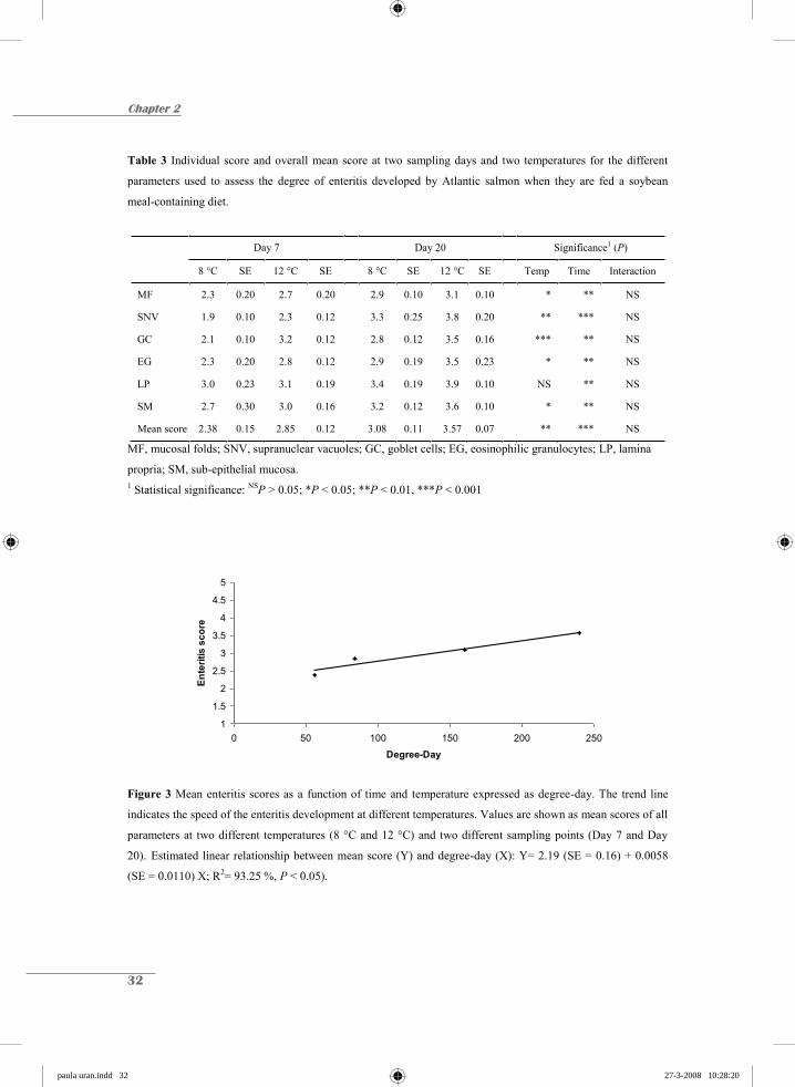
The scoring of MF, SNV, GC, EG, LP, SM, and the mean score were all significantly
different between sampling moments (P < 0.01) and for all parameters except for LP they
were all significantly different between temperatures, (P < 0.05) (Table 3).
Furthermore, the scoring of enteritis parameters like SNV and GC were the most
affected by the water temperature (P < 0.01). For all parameters a higher value was scored at
a water temperature of 12 °C compared to 8 °C. The interaction effect between sampling
moment and water temperature was not present for any of the parameters scored. Plotting the
mean score value against degree-day shows that the SBM-induced enteritis is delayed when
fish are kept at lower temperatures. The mean score was linearly related to the degree-day
(R2= 93.25%; P < 0.05, Fig.3).
paula uran.indd 30paula uran.indd 30 27-3-2008 10:28:1827-3-2008 10:28:18

Temperature and SBM-induced enteritis in Atlantic salmon
31
Figure 2 Morphological appearance of the distal intestine of fish reared at two different temperatures and
fed either a fishmeal-based diet (0SBM) as control or a soybean meal-based (20SBM) diet. Distal Intestinal
epithelium of fish fed the control diet that were kept at A) 8 °C and B) at 12 °C. Distal intestinal appearance
after 7 days of SBM feeding of C) fish kept at 8 °C and D) fish kept at 12 °C. Distal intestinal appearance
after 20 days of SBM feeding, E) fish kept at 8 °C and F) fish kept at 12 °C. (H & E, Alcian blue staining).
Bar is 50 μm.
A B
C D
E F
paula uran.indd 31paula uran.indd 31 27-3-2008 10:28:1927-3-2008 10:28:19
Chapter 2
32
Table 3 Individual score and overall mean score at two sampling days and two temperatures for the different
parameters used to assess the degree of enteritis developed by Atlantic salmon when they are fed a soybean
meal-containing diet.
Day 7 Day 20 Significance1 (P)
8 °C SE 12 °C SE 8 °C SE 12 °C SE Temp Time Interaction
MF 2.3 0.20 2.7 0.20 2.9 0.10 3.1 0.10 * ** NS
SNV 1.9 0.10 2.3 0.12 3.3 0.25 3.8 0.20 ** *** NS
GC 2.1 0.10 3.2 0.12 2.8 0.12 3.5 0.16 *** ** NS
EG 2.3 0.20 2.8 0.12 2.9 0.19 3.5 0.23 * ** NS
LP 3.0 0.23 3.1 0.19 3.4 0.19 3.9 0.10 NS ** NS
SM 2.7 0.30 3.0 0.16 3.2 0.12 3.6 0.10 * ** NS
Mean score 2.38 0.15 2.85 0.12 3.08 0.11 3.57 0.07 ** *** NS
MF, mucosal folds; SNV, supranuclear vacuoles; GC, goblet cells; EG, eosinophilic granulocytes; LP, lamina
propria; SM, sub-epithelial mucosa. 1 Statistical significance: NSP > 0.05; *P < 0.05; **P < 0.01, ***P < 0.001
1
1.5
2
2.5
3
3.5
4
4.5
5
0 50 100 150 200 250
Degree-Day
Ente
ritis
sco
re
Figure 3 Mean enteritis scores as a function of time and temperature expressed as degree-day. The trend line
indicates the speed of the enteritis development at different temperatures. Values are shown as mean scores of all
parameters at two different temperatures (8 °C and 12 °C) and two different sampling points (Day 7 and Day
20). Estimated linear relationship between mean score (Y) and degree-day (X): Y= 2.19 (SE = 0.16) + 0.0058
(SE = 0.0110) X; R2= 93.25 %, P < 0.05).
paula uran.indd 32paula uran.indd 32 27-3-2008 10:28:2027-3-2008 10:28:20

Temperature and SBM-induced enteritis in Atlantic salmon
33
Discussion and Conclusions
The intestinal epithelium is an important site for the absorption of nutrients, immunity,
osmotic balance, recycling of enzymes and macronutrients. Several authors have stated that
the distal intestine of teleost fish is the principal site for the endocytosis of intact proteins,
assuring its absorption and intracellular digestion (Stroband & van der Veen 1981; Rombout
et al. 1985). This high endocytotic capacity possibly makes the distal intestine more sensitive
to food-borne enteropathies. It is well known that quality and quantity of food are important
factors in the development of the intestinal mass and the mucosal architecture (Buddington et
al. 1997), but environmental conditions may also have a strong influence. Temperature is
considered one of the most influential environmental factors on the development and growth
of fish. In salmonids, it may affect physiological functions, feeding behaviour, stress
responses and susceptibility to pathogenic organisms by affecting the innate immune system
(Alcorn et al. 2002; Magnadóttir 2006).
The current study has shown that the severity of enteritis increased with water
temperature, but the mechanism behind this increase remains unclear. Houpe et al. (1996)
reported that environmental temperature influenced the functional demands of the intestine by
altering the metabolism and the nutrient uptake. They suggested that fish exposed to different
temperatures may adjust their absorptive capacities by influencing the activity of transporters
when the absorptive tissue surface area is increased, the density of transporters is adapted or
the physical and chemical characteristics of the apical membrane are adjusted, or perhaps, the
combination of all processes. Due to the relatively higher metabolic activity of fish reared at
higher temperatures and the uninterrupted exposure to noxious agents, the reaction to SBM
feeding might have been stronger. Although there were no visual differences on feed intake
between the two groups, a possible effect of feed intake in the observed different response
cannot be neglected. The influence of feed intake on the development of enteritis needs to be
further investigated.
Temperature could have affected the exposure time to noxious agents present in SBM
by influencing the time digesta remains in contact with the intestinal epithelium. However,
when the digesta passage rate increases at a higher temperature, the exposure time decreases.
Therefore, the results of this study indicate that this is not the case since the highest degree of
paula uran.indd 33paula uran.indd 33 27-3-2008 10:28:2227-3-2008 10:28:22
Chapter 2
34
enteritis was observed in fish reared at the higher temperature. Therefore, the stronger degree
of enteritis seems to be more correlated to higher metabolism rather than to a lower digesta
passage rate.
The influence of a higher metabolism could be reflected on the efficiency of the
intestinal enzymatic activity. Any disruption on the normal activity of the enzymes linked to
the brush-border membrane could have a severe impact on the uptake process and the loss of
the regular supranuclear vacuolization. Krogdahl et al. (2003) showed a reduced enzymatic
activity in the distal intestine with increasing SBM inclusion levels. The effect of temperature
in the disappearance of the SNV due to reduced enzymatic activity is still an open question.
The current findings on time related changes of enteritis are in line with the study of
Baeverfjord & Krogdahl (1996) in which the presence of all signs of the condition after day 7
of SBM feeding is described. After 20 days of SBM feeding, the condition was fully
developed and the outline of the intestinal tissue was transformed. Furthermore, the effect of
temperature is not equal for all measured parameters. SNV and GC seemed to be more
severely affected by temperature constituting the fast responders during the development of
the enteritis, whereas the structural parameters were less affected, especially in the case of LP
where the difference among the two groups was not significant. Once the immune system is
triggered, the cellular components activate the immune cascade, and consequently, the
appearance of the intestinal epithelium starts to adapt and respond to those changes. Salmon
recurrently exposed to the noxious agent contained in SBM showed no signs of recovery
during the experimental period. On the contrary, an aggravation of the symptoms was noticed.
Indeed, proof that the structural parameters are affected to a lesser extent after exposure to
SBM at the two different temperatures, supports the fact that the time digesta is in contact
with the intestinal epithelium, which, nonetheless, is not a main factor in explaining the
higher degree of enteritis in fish kept at higher temperature.
From the present study, it can be concluded that temperature influences the enteritis
process more concretely at higher temperatures, suggesting that the enteritis developed at a
lower temperature seems to be a delay rather than other type or mechanism by which the
aggravation of the symptoms is generally explained. Transit time is not the main factor
causing the stronger reaction when Atlantic salmon are reared at higher temperatures. Instead
an increase in the metabolic rate may well be the most suitable explanation of this
paula uran.indd 34paula uran.indd 34 27-3-2008 10:28:2327-3-2008 10:28:23
Temperature and SBM-induced enteritis in Atlantic salmon
35
phenomenon. Membrane fluidity and membrane composition, altered over time, might
partially explain the progressive condition developed by fish reared at the higher temperature.
However, strong indications that the endocytosis process might be altered by changes in water
temperature were outlined in this study. This fact constitutes an important feature to consider
when studying the impact of SBM-feeding in the development of the enteritis condition in
Atlantic salmon.
Acknowledgments
This research was supported by “Instituto Colombiano para el Desarrollo de la Ciencia y la
Tecnología”, Colciencias, and Skretting ARC, Stavanger, Norway. We would like to acknowledge the
staff at both the Skretting fish trails station and Skretting ARC for their technical assistance, for their
help during the collection of the samples and for the lab analyses. The first author would like to thank
Jasper van Houcke for his contribution to the development of the semi-quantitative scoring system.
References
Alcorn, S.W., Murray, A.L. & Pascho, R.J. (2002) Effects of rearing temperature on immune functions
in sockeye salmon (Oncorhynchus nerka). Fish Shellfish Immun., 12, 303-334.
Baeverfjord, G. & Krogdahl, Å. (1996) Development and regression of soybean meal induced enteritis
in Atlantic salmon, Salmo salar L., distal intestine: a comparison with the intestines of fasted fish.
J. Fish Dis., 19, 375-387.
Bakke-Mckellep, A.M., Press, C.Mcl., Baeverfjord, G., Krogdahl, Å. & Landsverk, T. (2000) Changes
in immune and enzyme histochemical phenotypes of cells in the intestinal mucosa of Atlantic
salmon, Salmo salar L., with soybean meal-induced enteritis. J. Fish Dis., 23, 115-127.
Buddington, R.K., Krogdahl, Å. & Bakke-McKellep, A.M. (1997) The intestines of carnivorous fish:
structure and functions and the relations with diet. Acta Physiol. Scand., 161, 67-80.
Burrells, C., Williams, P.D., Southgate P.J. & Crampton, V.O. (1999) Immunological, physiological
and pathological responses of rainbow trout (Oncorhynchus mykiss) to increasing dietary
concentrations of soybean proteins. Vet. Immunol. Immunopathol., 72, 277-288.
paula uran.indd 35paula uran.indd 35 27-3-2008 10:28:2427-3-2008 10:28:24

Chapter 2
36
Buttle, L.G., Burrells, A.C., Good, J.E., Williams, P.D., Southgate, P.J. & Burrells, C. (2001) The
binding of soybean agglutinin (SBA) to the intestinal epithelium of Atlantic salmon Salmo salar
and Rainbow trout, Oncorhynchus mykiss, fed high levels of soybean meal. Vet. Immunol.
Immunopathol., 80, 237-244.
Houpe, K.L., Malo, C., Oldham, P.B. & Buddington, R.K. (1996) Thermal modulation of channel
catfish intestinal dimensions, BBM fluidity, and glucose transport. Am. J. Physiol. 270
(Regulatory Integrative Comp. Physiol. 39), R1037-R1043.
van den Ingh, T.S.G.A.M., Krogdahl, Å., Olli, J.J., Hendriks, H.G.C.J.M. & Koninkx, J.G.J.F. (1991)
Effects of soybean-containing diets on the proximal and distal intestine in Atlantic salmon (Salmo
salar): a morphological study. Aquaculture, 94, 297-305.
van den Ingh, T.S.G.A.M., Olli, J.J. & Krogdahl, Å. (1996) Alcohol-soluble components in soybeans
cause morphological changes in the distal intestine of Atlantic salmon, Salmo salar L. J. Fish Dis.,
19, 47-53.
Krogdahl, Å., Bakke-McKellep, A.M., Røed, K.H. & Baeverfjord, G. (2000) Feeding Atlantic salmon
Salmo salar L. soybean products: effects on disease resistance (furunculosis), and lysozyme and
IgM levels in the intestinal mucosa. Aquacult. Nutr., 6, 77-84.
Krogdahl, Å., Bakke-McKellep, A.M. & Baeverfjord, G. (2003) Effects of graded levels of standard
soybean meal on intestinal structure, mucosal enzyme activities, and pancreatic response in
Atlantic salmon (Salmo salar L.). Aquacult. Nutr., 9, 361-371.
Magnadóttir, B. (2006) Innate immunity of fish (overview). Fish Shellfish Immun., 20, 137-151.
Nordrum, S., Bakke-McKellep, A.M., Krogdahl, Å. & Buddington, R.K. (2000) Effects of soybean
meal and salinity on intestinal transport of nutrients in Atlantic salmon (Salmo salar L.) and
rainbow trout (Oncorhynchus mykiss). Comp. Biochem. Phys. B, 125, 317-335.
Olsen, R. E., Myklebust, R., Ringø, E. & Mayhew, T. M. (2000) The influences of dietary linseed oil
and saturated fatty acids on caecal enterocytes in Arctic char (Salvelinus alpinus L.): a quantitative
ultrastructural study. Fish Physiol. Biochem., 22, 207-216.
Refstie, S., Korsøen, Ø.J., Storebakken, T., Baeverfjord, G., Lein, I. & Roem, A.J. (2000) Differing
nutritional responses to dietary soybean meal in rainbow trout (Oncorhynchus mykiss) and
Atlantic salmon (Salmo salar). Aquaculture, 190, 49-63.
Reite, O.B. (1997) Mast cells/eosinophilic granule cells of salmonids: staining properties and
responses to noxious agents. Fish Shellfish Immun., 7, 567-584.
Reite, O.B. & Evensen, Ø. (2006) Inflammatory cells of teleostean fish: A review focusing on mast
cells/eosinophilic granule cells and rodlet cells. Fish Shellfish Immun., 20, 192-208.
paula uran.indd 36paula uran.indd 36 27-3-2008 10:28:2527-3-2008 10:28:25

Temperature and SBM-induced enteritis in Atlantic salmon
37
Rombout, J.H.W.M., Lamers, C.H.J., Helfrich, M.H., Dekker, A. & Taverne-Thiele, J.J. (1985)
Uptake and transport of intact macromolecules in the intestinal epithelium of carp (Cyprinus
carpio L.) and the possible immunological implications. Cell Tissue Res., 239, 519-530.
Rumsey, G.L., Siwicki, A.K., Anderson, D.P. & Bowser P.R. (1994) Effect of soybean protein on
serological response, non-specific defense mechanisms, growth, and protein utilization in rainbow
trout. Vet. Immunol. Immunopathol., 41, 323-339.
SAS (1999) SAS/STAT® User’s guide, V8. SAS Institute Inc., Cary, NC, USA.
Sitjà-Bobadilla, A., Peña-Llopis, S., Gómez-Requeni, P., Médale, F., Kaushik, S. & Pérez-Sánchez, J.
(2005) Effect of fish meal replacement by plant protein sources on non-specific defence
mechanisms and oxidative stress in gilthead sea bream (Sparus aurata). Aquaculture, 249, 387-
400.
Stroband, H.W.J. & van der Veen, F.H. (1981) Localization of protein absorption during transport of
food in the intestine of the grass carp, Ctenopharyngodon idella (Val.). J. Exp. Zool., 218, 149-
156.
paula uran.indd 37paula uran.indd 37 27-3-2008 10:28:2627-3-2008 10:28:26

chapter hoofdstukken.indd 5 26-3-2008 12:37:16
Chapter 3
Time-related changes of the intestinal morphology of Atlantic salmon (Salmo salar L.) at two different
soybean meal inclusion levels
P.A. Urán1,2, J.W. Schrama1, J.H.W.M. Rombout2, J.J. Taverne-Thiele2, A. Obach3, W. Koppe3, J.A.J. Verreth1
1Aquaculture and Fisheries Group and 2Cell Biology and Immunology Group, Wageningen Institute of Animal Sciences, Wageningen University, The Netherlands 3Skretting, Aqua-culture Research Centre, Stavanger, Norway
Submitted for publication
chapter hoofdstukken.indd 6 26-3-2008 12:37:17

40
Chapter 3
Abstract
Soybean meal (SBM) induces enteritis in the distal intestine of Atlantic salmon. The
present study assesses the effects of SBM doses on the kinetics of the enteritis process. Fish
of 300g, kept at 12°C, were fed diets with different SBM inclusions: 0%, 10% and 20 % SBM
for 57 days. Samples of the distal intestine of 5 fish per treatment were taken for histological
and electron microscopic analysis. A semi-quantitative scoring system was used to assess the
degree of the morphological changes induced by SBM feeding in the distal intestine
epithelium. The first signs of enteritis appeared earlier in the salmon fed the 20SBM diet than
those salmon fed the 10SBM diet. Thereafter, it increased steadily with time, displaying no
signs of recovery. Furthermore, at the lower dose, the process marking the onset of enteritis
began more gradually than at the higher dose and it displayed a tendency to level off after 13
to 20 days of continuous feeding. Electron microscopy indicated that the endocytosis process
was hampered at day 3 of 20SBM and at 7 days of 10SBM. Furthermore, a strong reduction
of microvilli was already evident after 7 days of 20SBM feeding, thus indicating a decreased
uptake capacity of the distal enterocytes. In addition, transformation and migration of
eosinophilic granulocytes was observed, which, in combination with the lysozyme C-
immunoreactivity supports their protective role during the inflammatory process in the distal
gut of Atlantic salmon. It can be concluded that the severity of enteritis and its kinetics are
dose-dependent, showing no signs of recovery during feeding with diets containing SBM,
which conversely, gives clear indications of an increased innate immunity.
paula uran.indd 40paula uran.indd 40 27-3-2008 10:28:2927-3-2008 10:28:29

SBM inclusion levels and the kinetics of enteritis
41
Introduction
Soybean (SB) is widely known to contain adverse anti-nutritional compounds that may
induce intestinal disorders in salmonids being especially harmful to Atlantic salmon (Salmo
salar L.). Rainbow trout Oncorhynchus mykiss (Walbaum) seems to be less affected by the
SB noxious factors that induce enteritis as was documented in previous studies (Nordrum et
al. 2000; Refstie et al. 2000; Buttle et al. 2001). When Atlantic salmon is fed on soybean
meal (SBM)-based diets, the morphology of the distal intestine is disturbed, which has been
described by Baeverfjord & Krogdahl (1996) as “non- infectious sub-acute enteritis”. The
changes in the distal intestinal mucosa are described as: a deep shortening of the mucosal
folds, a decreasing number of the supranuclear vacuoles in absorptive cells, a widening of the
central stroma with a correspondingly high amount of connective tissue and an increased
infiltration of inflammatory cells in the lamina propria (van den Ingh et al. 1991, 1996,
Baeverfjord & Krogdahl, 1996), an increased number of goblet cells, and a shortening of the
microvilli (van den Ingh et al. 1991). These pathological changes seem to be particularly
present in the distal intestinal segment rather than on the proximal segment, as has already
been reported in several studies on salmonids (van den Ingh et al. 1991; Burrells et al. 1999,
Nordrum et al. 2000, Buttle et al. 2001).
Baeverfjord & Krogdahl (1996) established that after 3 weeks of SBM-feeding, the
development of enteritis became critical for those Atlantic salmon, being fed a relatively high
SBM-based diet (c. 33%) which induced all the above-mentioned characteristic signs of
enteritis within the first week of experimental feeding. A more recent study (Krogdahl et al.
2003), using different solvent-extracted SBM inclusion levels, indicated that the degree of
enteritis in the distal intestine, being measured at 60 days of SBM feeding, augmented with
increasing SBM. Furthermore, even at lower SBM levels (15 to 25%), growth performance,
feed conversion, apparent digestibility/utilization of macronutrients and energy were affected.
Almost all studies on enteritis focused on the more advanced stages of the inflammation
process (> 20 d after SBM feeding); with the exception of the kinetic study of Baeverfjord &
Krogdahl (1996) which used a 33% dietary inclusion solvent extracted SBM. They found the
initial signs of morphological changes at day 2, after feeding the SBM diet, at day 7 all typical
characteristics of enteritis were present but the severity increased up until the last sampling
paula uran.indd 41paula uran.indd 41 27-3-2008 10:28:3027-3-2008 10:28:30
42
Chapter 3
point at 21 days. Thus, morphological information on the onset of enteritis is limited as well
as on the influence of the SBM level on the kinetics of enteritis. Information on the
ultrastructural changes of the distal epithelium is lacking, both regarding the kinetics and the
influence of SBM doses.
In this study the kinetics of morphological changes is assessed in fish fed diets
containing 10% and 20% of SBM as part of the protein fraction. The main objective of this
study is to describe the progressive morphological changes in the distal intestine of salmon
that are fed diets containing different inclusion levels of a SBM variety positively selected to
give a strong reaction. For this purpose the two mentioned diets and a control diet containing
fishmeal as the sole protein source were fed to salmon. Morphological parameters
characteristic for inflamed distal intestinal mucosa were assessed at seven different time
points for the duration of the experimental period. A previously introduced semi-quantitative
scoring system (Urán et al. 2008) was used in order to elucidate the impact on the intestinal
morphology. In addition, as well as paying attention to ultrastructural changes during the
enteritis process, a preliminary qualitative study on the role and contribution of the
eosinophilic granulocytes to the inflammatory process is also presented.
Materials and Methods
Fish and rearing conditions
The experiment was carried out at Skretting Fish Trials Station, Lerang (Jørpeland,
Norway). For this experiment, 300 Atlantic salmon (AquaGen strain) were sampled for gut
histology and electron microscopy. The Atlantic salmon originated from a stock of fish
already present at the research station. The experiment consisted of a two-week adaptation
period and an eight-week experimental period. At the start of the adaptation period the fish
weighed approximately 300 g.
For the experiment six indoor tanks with a diameter of 1 m each were used. The water
volume in the tanks was 400 l. The stocking density was 50 fish per tank. Each tank was kept
at flow through, at 12-15 l min-1 system. Seawater was pumped from a depth of 90m in the
fjord, with a salinity of 34 ‰ and an oxygen concentration above 9 ppm. Prior the adaptation
paula uran.indd 42paula uran.indd 42 27-3-2008 10:28:3127-3-2008 10:28:31

SBM inclusion levels and the kinetics of enteritis
43
period fish were kept at 8 °C. During the adaptation and the experimental period, water
temperature was kept at 12 °C. The applied photoperiod was 18L : 6D.
Diets and feeding
Feed was produced at Skretting Feed Technology Plant, (Stavanger, Norway). Three
diets were formulated: a control diet (0SBM) and two experimental diets (10SBM and
20SBM) (Table 1). The major ingredients in the 0SBM diets were: fishmeal (protein content
above 70%), fish oil, and wheat. This control diet did not contain any SBM. For the
experimental diets, fishmeal and wheat were exchanged, in the case of the 10SBM diet for
10% SBM, and in the case of the 20SBM diet for 20% SBM (solvent-extracted Hipro SBM)
(Table 1). All diets were produced to be iso-energetic and iso-nitrogenic on a crude protein
and a crude lipid basis. Diets were supplemented with a standard vitamin and mineral premix.
Feed was produced as extruded 4 mm sinking pellets.
Prior to the experiment the fish were fed a commercial salmon diet (Skretting,
Stavanger, Norway), which did not contain any SB products. During the adaptation period all
tanks were fed with the control diet (0SBM). At the start of the experimental period (day 1),
for each diet group, two tanks were changed to either a 10SBM or a 20SBM experimental
diet. Two tanks reminded at the control diet. Fish were fed 20% in excess. Feed was divided
into two meals per day and provided by automatic feeders.
Chemical analysis of diets
The nutrient composition of the experimental diets was determined using standard
techniques for proximate analyses. Crude protein content was determined by the Kjeldahl
Nitrogen measurement in accordance with the Nordic Committee on Food Analysis, Method
No.6, 4th edition, 2003. Crude fat content was measured by low field nuclear magnetic
resonance. Moisture content in the samples was measured by drying to constant weight at
102-105 °C for 16-18 h. Ash content was measured by combustion at 540 °C for 16-18 h,
after which the remaining residues were weighed, both in accordance with the Nordic
Committee on Food Analysis Method No.23, 3rd edition, 1991. The preceding analyses were
carried out at the Skretting Aquaculture research centre (Stavanger, Norway). For chemical
composition see Table 1.
paula uran.indd 43paula uran.indd 43 27-3-2008 10:28:3227-3-2008 10:28:32

44
Chapter 3
Table 1 Formulation and chemical composition of the experimental diets
Amount of protein replaced by soybean meal in percentage 0 10 20
Ingredients (g kg-1) Fishmeal1 564.3 519.8 475.3 Extracted soybean meal2 0 100 200 Wheat 210.6 140 70 Fish Oil 3 222.6 237.3 252.1 Vitamin premix 1.3 1.3 1.3 Mineral premix 1.3 1.3 1.3
Pigment premix Yttrium oxide 0.1 0.1 0.1 Carophyll Pink 0.6 0.6 0.6 Total 1000.8 1000.4 1000.7
Chemical composition by analysis
Crude Protein (g kg-1) 429.8 440.2 450.6 Crude Lipid (g kg-1) 277.1 289.2 301.4 Target dry matter (%) 95 95 95 Fat NMR (%) 30.5 31.5 32.8 Protein (%) 43.1 43.8 45.2 Moisture (%) 5.1 6.2 4.7 Ash (%) 7.2 7.0 7.3
1 LT North Atlantic, Egersund, Norway.
2 HiPro solvent-extracted soybean meal, protein content above 70%.
3 Northern Hemisphere.
Sampling for intestinal morphology
During the experimental period, fish gut was sampled for histological measurements at
seven different time points: day 3, 5, 7, 13, 20, 36, 57. At each sampling moment, five fish per
treatment group were sampled (5 fish from the control group and 5 from each of the two
experimental diets). The samples were taken from alternate tanks to avoid any drops in feed
intake due to sampling stress. Directly after the morning meal, the fish were anaesthetized
paula uran.indd 44paula uran.indd 44 27-3-2008 10:28:3427-3-2008 10:28:34
SBM inclusion levels and the kinetics of enteritis
45
using 0,05 g L-1 metacaine (Argent chemical laboratories, USA), and thereafter killed by a
sharp blow to the head. The distal intestine was dissected from the place were the intestinal
diameter increases, the mucosa becomes darker and annular rings are clearly noticeable.
Light Microscopy (LM)
For LM analysis a two-centimeter section of distal intestine of each fish was taken and
gently rinsed with cold (4 ºC) saline. Samples were fixed in 4% phosphate-buffered formalin
pH of 7.2 and stored at room temperature. After dehydration, in accordance with standard
procedures, samples were embedded in paraffin. Transverse sections of 5 μm thickness were
stained using a mixture of Haematoxylin & Eosin and Alcian blue pH 2.5. Alcian blue
staining enhances the contrast between goblet cells and the supranuclear vacuoles. Slides
were blindly evaluated after randomization.
Semi-Quantitative Scoring system
The LM sections were evaluated according to the semi-quantitative method developed
at Wageningen University (Urán et al. 2008), which assesses the degree of SBM-induced
enteritis on the Atlantic salmon distal intestine considering the following criteria: 1. the
morphology of the mucosal folds (MF); 2. the presence and size of supranuclear vacuoles
(SNV); 3. the abundance of goblet cells (GC); 4. the infiltration of eosinophilic granulocytes
(EG) into the lamina propria and sub-epithelial mucosa; 5. the degree of widening of the
lamina propria (LP); and 6. the degree of thickening of the sub-epithelial mucosa (SM).
Sections were photographed with an Olympus DP 50 digital camera connected to a Nikon
Microphot-FXA light microscope (Badhoevedorp, the Netherlands). The pictures were
processed and analyzed using the AnalySiS Extended Pro 3.1 software (Soft Imaging System
GmbH, Münster, Germany).
Each of these parameters was scored on a scale from 1 to 5 (Table 2). An increasing
scored value represents a more severe enteritis condition. (For illustrations of the different
scores, see annex or check list of special publication at
http://www.afi.wur.nl/UK/Publications/).
paula uran.indd 45paula uran.indd 45 27-3-2008 10:28:3527-3-2008 10:28:35
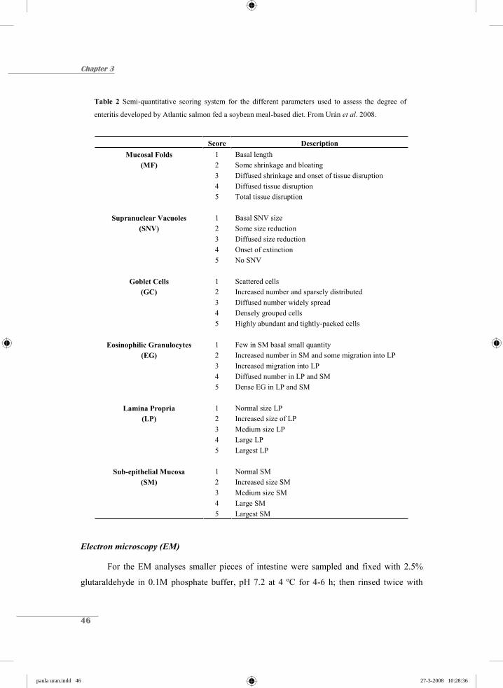
46
Chapter 3
Table 2 Semi-quantitative scoring system for the different parameters used to assess the degree of
enteritis developed by Atlantic salmon fed a soybean meal-based diet. From Urán et al. 2008.
Score Description Mucosal Folds 1 Basal length
(MF) 2 Some shrinkage and bloating 3 Diffused shrinkage and onset of tissue disruption 4 Diffused tissue disruption 5 Total tissue disruption
Supranuclear Vacuoles 1 Basal SNV size (SNV) 2 Some size reduction
3 Diffused size reduction 4 Onset of extinction 5 No SNV
Goblet Cells 1 Scattered cells (GC) 2 Increased number and sparsely distributed
3 Diffused number widely spread 4 Densely grouped cells 5 Highly abundant and tightly-packed cells
Eosinophilic Granulocytes 1 Few in SM basal small quantity (EG) 2 Increased number in SM and some migration into LP
3 Increased migration into LP 4 Diffused number in LP and SM 5 Dense EG in LP and SM
Lamina Propria 1 Normal size LP (LP) 2 Increased size of LP
3 Medium size LP 4 Large LP 5 Largest LP
Sub-epithelial Mucosa 1 Normal SM (SM) 2 Increased size SM
3 Medium size SM 4 Large SM 5 Largest SM
Electron microscopy (EM)
For the EM analyses smaller pieces of intestine were sampled and fixed with 2.5%
glutaraldehyde in 0.1M phosphate buffer, pH 7.2 at 4 ºC for 4-6 h; then rinsed twice with
paula uran.indd 46paula uran.indd 46 27-3-2008 10:28:3627-3-2008 10:28:36

SBM inclusion levels and the kinetics of enteritis
47
buffer 0.1 M phosphate buffer pH 7.2 plus 0.2 M sucrose and kept in the same buffer at 4 ºC
for transport. Samples were post-fixed on 1% OsO4 for 1-2 h and thereafter, embedded in
Epon 812 (Electron microscopy Sciences, Fort Washington, USA). Semi-thin sections (1 μm)
were mounted on glass slides and stained with toluidine blue. Ultra-thin sections were cut
using a diamond knife installed on a Reichert Ultracut (Leica, Rijswijk, the Netherlands) and
mounted on copper grids. Sections were counter-stained using uranyl acetate followed by lead
citrate and examined in a Philips EM 208 electron microscope (Philips, Eindhoven, the
Netherlands). Due to the limitations of this technique, two animals per treatment and one for
the control were embedded in epon and further processed. In order to exclude local
differences, two different parts of the same sample were analyzed. Pictures were made using a
SIS Megaview III digital camera (Soft Imaging System GmbH, Münster, Germany)
connected to the Philips EM 208. The pictures were processed and analyzed using the
AnalySiS Extended Pro 3.1 software.
Immunohistochemistry (Lysozyme C)
Transverse section of distal intestine from control and SBM fed fish were
deparaffinised and hydrated, in accordance with standard procedure. Sections were treated
with Tris EDTA buffer pH 9 and kept at 95 °C for 20 min. Sections were then washed in PBS
with 0.1 % Triton X-100 (PBS-t) and kept at room temperature. Afterwards, sections were
incubated with 5% normal goat serum for 30 min, and then incubated with anti-human
lysozyme C (1:50, Dako, Glostrup, Denmark) for 1 h. After washing twice with PBS-t,
sections were incubated with horseradish peroxidase-labelled Goat-anti-rabbit-HRP (GAR-
HRP, Dako, Glostrup, Denmark) for 1h. Slides were washed twice with PBS and kept on 0,05
M, pH 5 sodium acetate buffer for 10 min. Slides were incubated with 3-Amino-9-ethyl-
carbazole substrate (AEC, Sigma-Aldrich, St-Louis, USA) until a reaction was observed and
then they were counter-stained with Haematoxylin, rinsed in running tap water, and
embedded in Kaiser’s glycerine gelatine (Merck, Darmstadt, Germany). Sections were
analyzed and photographed with an Olympus DP 50 digital camera connected to a Nikon
Microphot-FXA light microscope. The pictures were processed and analysed using the
AnalySiS Extended Pro 3.1 software.
paula uran.indd 47paula uran.indd 47 27-3-2008 10:28:3727-3-2008 10:28:37
48
Chapter 3
Statistical analysis
All calculations were made using the SAS System (SAS 1999). The parameters used
to score the degree of enteritis were analysed for the effect of diet using PROC GLM for each
sampling point separately. The error terms of these analyses were tested for homogeneity of
variances and normal distribution, using, respectively, the Levene’s test and the Shapiro-Wilk
test. Due to significant non-normality, data were analysed using a Kruskal-Wallis test. Pair-
wise comparison was performed using the Mann-Whitney U-test. Herein, results are
considered statistically significant when P-values are below 0.05.
Results
Morphological changes of the distal intestine
The control diet (0SBM) did not induce any sign of enteritis. The MF appeared as long
finger-like structures, composed of enterocytes which were perfectly aligned along the
mucosal epithelium. The nucleus of the enterocytes was located between the mid to basal part
of the cell. The enterocytes presented normal round and SNV. The GC were present in a basal
amount and they were scattered among the enterocytes. The LP was a delicate and single thin
layer of cells underlying the epithelium. The SM found between the basal part of the folds and
the stratum compactum was composed of a basal EG population forming the stratum
granulosum. All control samples for the entire duration of the experimental period followed
the same pattern described above. This is reflected in Fig.1 by the fact that all the semi-
quantitative scores remained at the basal level of 1 for all the measured parameters, at all
sampling points.
Fish fed diets containing SBM showed no visible morphological changes up until day
3. The first visible changes occurred on day 5 for the 20SBM diet. At this point, the SNV
started to shrink and their regular alignment was disturbed. An increasing cell infiltration into
the SM was also noticed. At day 5, for the 10SBM diet, there were no noticeably changes. At
day 7 the above-mentioned morphological changes were detectable in both SBM diets but to a
lesser degree for 10SBM diet (Fig. 1).
paula uran.indd 48paula uran.indd 48 27-3-2008 10:28:3927-3-2008 10:28:39

SBM inclusion levels and the kinetics of enteritis
49
Figure 1 Enteritis parameters scored using the Wageningen semi-quantitative scoring system (Urán et al. 2008),
where 1 indicates normal intestinal morphology and 5 indicates the highest degree of soybean meal (SBM) -
induced enteritis in the distal intestine of Atlantic salmon. The changes on the morphology of the mucosal folds
MF, the presence of the supranuclear vacuoles SNV, the abundance of goblet cells GC, the degree of infiltration
of eosinophilic granulocytes EG, the widening of the lamina propria LP, and the thickening of the sub-epithelial
mucosa SM, are shown. Three different diets, a control diet containing fishmeal as protein source and 0% of
soybean meal (0SBM), a diet containing 10% and 20% of Soybean meal (10SBM and 20SBM respectively),
both as replacements of fishmeal in the diet were fed for a period of 57 days.
The MF started to shrink with subsequent tissue disruption. The LP and SM had
increased in size at both SBM inclusions. The SNV displayed a significant reduction and the
number of GC increased steadily, particularly in the 20SBM diet. At day 13 major changes
were distinctive between the two SBM diets. The fish fed the 20SBM diet presented a heavily
changed intestinal mucosa, while the fish fed 10SBM showed only minor changes on MF, LP
SNV
0
1
2
3
4
5
0 7 14 21 28 35 42 49 56 63
Ent
eriti
s sc
ore
0SBM 10SBM 20SBMEG
0
1
2
3
4
5
0 7 14 21 28 35 42 49 56 63
Ent
eriti
s sc
ore
0SBM 10SBM 20SBM
MF
0
1
2
3
4
5
0 7 14 21 28 35 42 49 56 63
Ent
eriti
s sc
ore
0SBM 10SBM 20SBMGC
0
1
2
3
4
5
0 7 14 21 28 35 42 49 56 63
Ent
eriti
s sc
ore
0SBM 10SBM 20SBMLP
0
1
2
3
4
5
0 7 14 21 28 35 42 49 56 63
Days of feeding
Ent
eriti
s sc
ore
0SBM 10SBM 20SBM
SM
0
1
2
3
4
5
0 7 14 21 28 35 42 49 56 63
Days of feeding
Ent
eriti
s sc
ore
0SBM 10SBM 20SBM
paula uran.indd 49paula uran.indd 49 27-3-2008 10:28:4027-3-2008 10:28:40
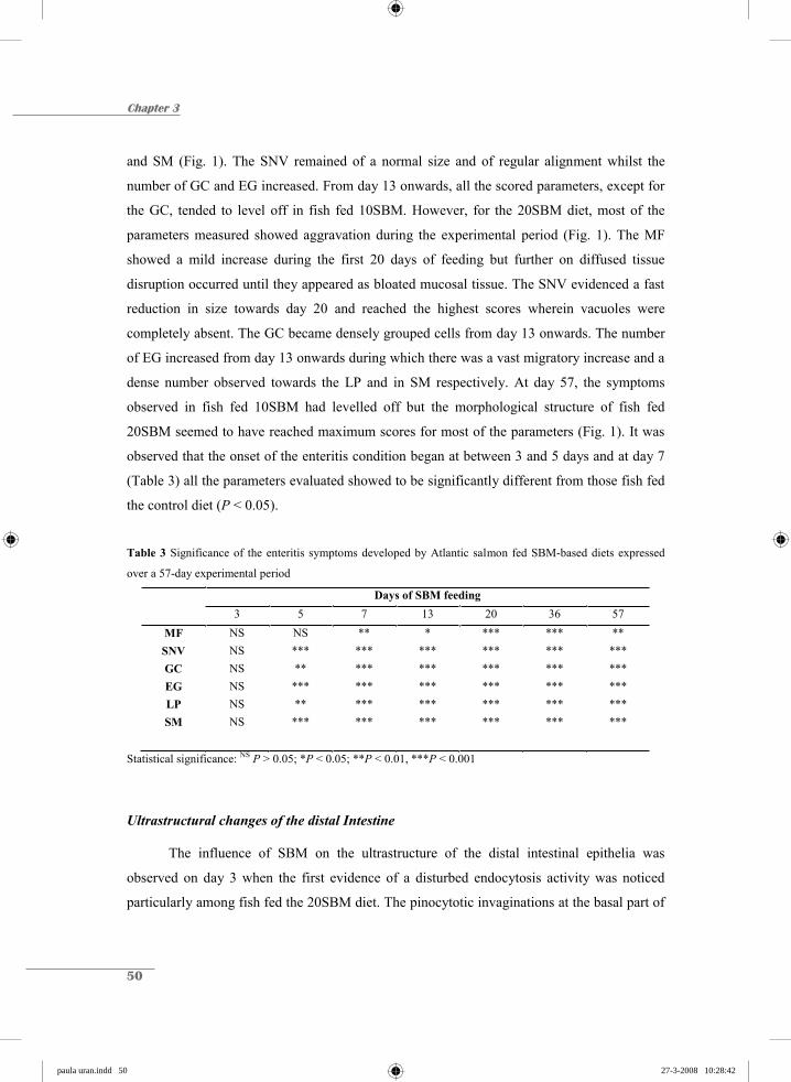
50
Chapter 3
and SM (Fig. 1). The SNV remained of a normal size and of regular alignment whilst the
number of GC and EG increased. From day 13 onwards, all the scored parameters, except for
the GC, tended to level off in fish fed 10SBM. However, for the 20SBM diet, most of the
parameters measured showed aggravation during the experimental period (Fig. 1). The MF
showed a mild increase during the first 20 days of feeding but further on diffused tissue
disruption occurred until they appeared as bloated mucosal tissue. The SNV evidenced a fast
reduction in size towards day 20 and reached the highest scores wherein vacuoles were
completely absent. The GC became densely grouped cells from day 13 onwards. The number
of EG increased from day 13 onwards during which there was a vast migratory increase and a
dense number observed towards the LP and in SM respectively. At day 57, the symptoms
observed in fish fed 10SBM had levelled off but the morphological structure of fish fed
20SBM seemed to have reached maximum scores for most of the parameters (Fig. 1). It was
observed that the onset of the enteritis condition began at between 3 and 5 days and at day 7
(Table 3) all the parameters evaluated showed to be significantly different from those fish fed
the control diet (P < 0.05).
Table 3 Significance of the enteritis symptoms developed by Atlantic salmon fed SBM-based diets expressed
over a 57-day experimental period
Days of SBM feeding 3 5 7 13 20 36 57
MF NS NS ** * *** *** ** SNV NS *** *** *** *** *** *** GC NS ** *** *** *** *** *** EG NS *** *** *** *** *** *** LP NS ** *** *** *** *** *** SM NS *** *** *** *** *** ***
Statistical significance: NS P > 0.05; *P < 0.05; **P < 0.01, ***P < 0.001
Ultrastructural changes of the distal Intestine
The influence of SBM on the ultrastructure of the distal intestinal epithelia was
observed on day 3 when the first evidence of a disturbed endocytosis activity was noticed
particularly among fish fed the 20SBM diet. The pinocytotic invaginations at the basal part of
paula uran.indd 50paula uran.indd 50 27-3-2008 10:28:4227-3-2008 10:28:42

SBM inclusion levels and the kinetics of enteritis
51
the Mv were less frequent than in controls. After 7 days of 20SBM feeding, these membrane-
bound vesicles were not visible and there was no observation of any new engulfed material at
the level of the terminal web, whereas in controls it still seemed to be filled with small
pinocytotic vesicles pinching off from the apical membrane and moving towards the
endosomes to finally fuse into the SNV (Fig. 2).
The disappearance of these structures was exacerbated towards the end of the
experiment, specifically, on day 57 where virtually no SNV were observed. At the 10SBM
diet, these changes all occurred at a lower velocity. The first evidence of the disappearance of
the pinocytotic vesicles was noticed from day 7 onwards. After 57 days of continuous
feeding, the disappearance of SNV did not reach the same levels as observed in the fish fed
20SBM. In addition, the microvilli (Mv) of fish fed 20SBM had dramatically decreased in
size, measuring roughly one third of their normal length at day 7 when compared to the
controls and this situation remained constant until the end of the experiment with no signs of
recovery (Fig. 3). The brush border membrane of fish fed 10SBM also showed a decrease but
to a much lesser extent. At day 7, less invaginations were observed but Mv were of normal
size as compared to the controls and the SNV were reduced but still present until the end of
the feeding trial.
Figure 2 Ultrastructure of the distal intestine epithelium of Atlantic salmon fed the control diet. A) Normal
absorptive cell shown as normally vacuolated enterocyte with high formation of pinocytotic vesicles (v) fusing
into supranuclear vacuoles (SNV). The microvilli (Mv) are normal in size. Bar is 2μm. B) Detail of cytoplasmic
invaginations (arrow) pinching off the apical membrane to fuse into bigger vesicles (v). Bar is 0.5μm.
A
B
SNV v
Mv B
paula uran.indd 51paula uran.indd 51 27-3-2008 10:28:4327-3-2008 10:28:43

52
Chapter 3
Figure 3 Ultrastructure of the distal intestine epithelium of Atlantic salmon fed soybean meal (SBM) based diet.
A) As yet, no changes can be observed after 3 days of feeding a diet containing 10% SBM (10SBM). (The
pinocytotic vesicles are indicated by the arrows), whereas B) shows the changes after 3 days of feeding a diet
containing 20% SBM (20SBM). The pinocytotic vesicles started to diminish (arrow). C) After day 7 the first
signs of a disrupted uptake process are observed in the 10SBM diet, indicated by the decreased formation of
membrane-bound vesicles, however, microvilli (Mv) are still of normal size. D) After 7 days at the 20SBM diet,
these invaginations disappeared and new engulfed material was not observed at the apical cytoplasm, Mv have
reduced their size to nearly a third of their normal length. E) After day 57 at the 10% SBM feeding the changes
at the distal intestine epithelium were not as severe but there still remains some size reduction of the brush
border membrane and the absence of newly formed vesicles at the terminal web were observed. F) After 57 days
at the 20% SBM feeding, a severe reduction of the Mv size and the total absence of the new vesicles and SNV
were noticed. Bar is 2 μm.
D
F
B
C Mv
A
Mv
E
paula uran.indd 52paula uran.indd 52 27-3-2008 10:28:4427-3-2008 10:28:44

SBM inclusion levels and the kinetics of enteritis
53
There was a clear difference in the appearance of the granular content of the EG
observed under the electron microscope. During the first 7 days of 20SBM feeding, the
granules began changing their density and this process continued until day 20 with some of
them appearing as electron lucent in comparison to the electron dense granules observed in
the controls (Fig. 4). After 20 days, the amount increased steadily with a strong presence of
intermediate electron dense granules. In fish fed 10SBM the transformation was milder and
electron dense or intermediate dense granules were observed less frequently.
Figure 4 Eosinophilic granulocytes (EG) of the distal intestine epithelium of Atlantic salmon. A) Appearance of
intestinal EG of fish fed a control diet (no inclusion of soybean meal); note the predominant electron dense
granules. B) Predominant type of EG with intermediate and electron lucent granules, present in lamina propria
and sub-epithelial mucosa after 7 days of feeding a diet containing 10% SBM (10SBM). C) Day 7 after feeding a
diet containing 20% SBM (20SBM), note the tendency of more intermediate and electron lucent granules. D)
Appearance of EG after 57 days of feeding a 10SBM diet, with electron lucent granules as the most abundant
type. E) EG after 57 days of feeding a 20SBM diet, showing predominantly electron lucent granules. Bar is 2
μm.
A
B C
D E
paula uran.indd 53paula uran.indd 53 27-3-2008 10:28:4527-3-2008 10:28:45
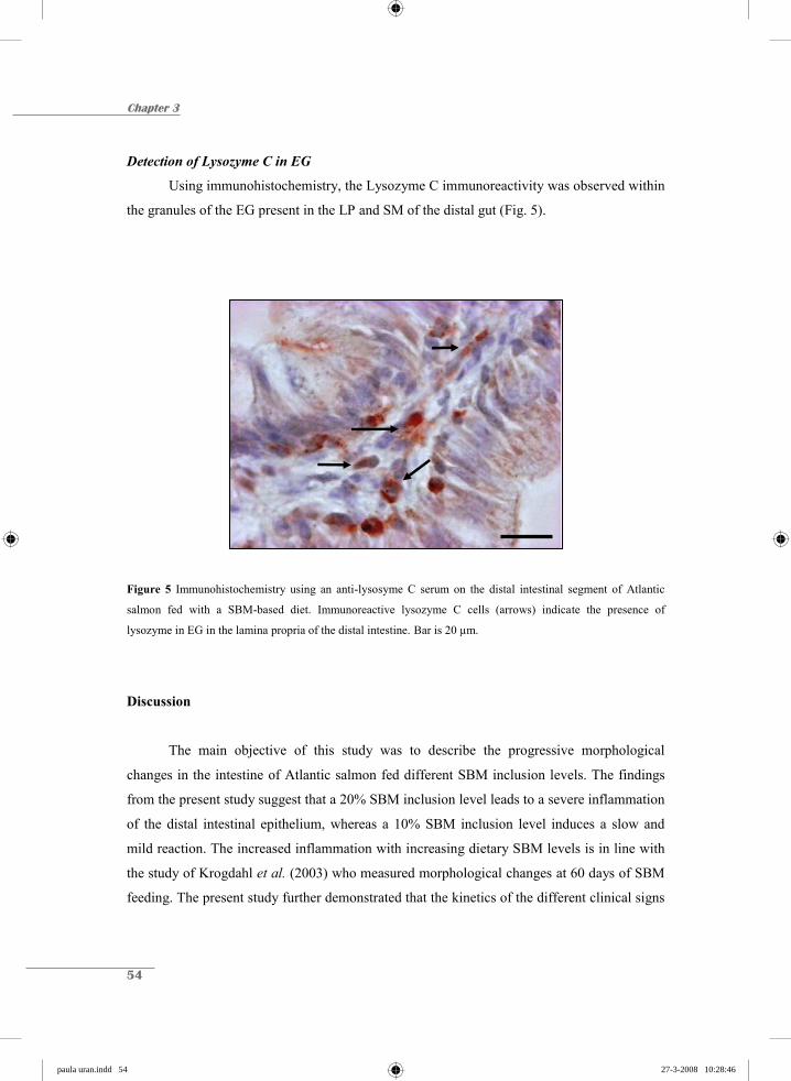
54
Chapter 3
Detection of Lysozyme C in EG
Using immunohistochemistry, the Lysozyme C immunoreactivity was observed within
the granules of the EG present in the LP and SM of the distal gut (Fig. 5).
Figure 5 Immunohistochemistry using an anti-lysosyme C serum on the distal intestinal segment of Atlantic
salmon fed with a SBM-based diet. Immunoreactive lysozyme C cells (arrows) indicate the presence of
lysozyme in EG in the lamina propria of the distal intestine. Bar is 20 μm.
Discussion
The main objective of this study was to describe the progressive morphological
changes in the intestine of Atlantic salmon fed different SBM inclusion levels. The findings
from the present study suggest that a 20% SBM inclusion level leads to a severe inflammation
of the distal intestinal epithelium, whereas a 10% SBM inclusion level induces a slow and
mild reaction. The increased inflammation with increasing dietary SBM levels is in line with
the study of Krogdahl et al. (2003) who measured morphological changes at 60 days of SBM
feeding. The present study further demonstrated that the kinetics of the different clinical signs
paula uran.indd 54paula uran.indd 54 27-3-2008 10:28:4627-3-2008 10:28:46

SBM inclusion levels and the kinetics of enteritis
55
differ depending on the inclusion level. The 20SBM inclusion level boosts the inflammatory
process, leading to a faster and steadily increasing reaction. This dose gives a more severe
response that progressively worsens for most of the parameters assessed. At the lower dose
(10SBM), the same symptoms appear to be quite stable with the tendency to level off after 13
to 20 days of continuous SBM feeding. Therefore, the results obtained from this study showed
that SBM-induced enteritis is a dose-dependent process with no clear signs of recovery.
It was only after 7 days of SBM feeding that all morphological signs of enteritis
became visible, an observation that is conducive with the light microscopy observations of
Baeverfjord & Krogdahl (1996). Nonetheless, in the current study, electron microscopy
revealed the first signs of distal epithelial disturbances after 3 days of 20SBM-feeding. These
findings suggest that the endocytosis process might be hampered resulting in the absence of
small vesicles that finally fuse into the SNV. Rombout et al. (1985) described this process as
the normal transport mechanism in the distal gut of common carp. King et al. (1982) observed
changes of the intestinal Mv, disruption of the terminal web and swelling of the apical
cytoplasm possibly explained by the disturbances in the membrane-associated transport
processes in rats fed kidney bean lectins. It remains to be investigated as to whether the
reduction in microvillous surface area at the brush border membrane and the decreased
pinocytotic uptake in this study are correlated to the disappearance of the SNV. Krogdahl &
Bakke-McKellep (2005) mentioned that changes in enzyme activities may also be the result
of changes in tissue mass, cell numbers, morphology and enzyme synthesis in the absorptive
cells. So, it may also be speculated that the atrophy of the Mv and probably the hampered
uptake process observed in this study could be the consequence of an unbalanced enzymatic
profile caused by contact with noxious factors present in SB fractions. Knudsen et al. (2007)
postulated soyasaponins or a combination with factors like antigenic SB proteins or even with
opportunistic intestinal microflora to be the causal factors that trigger the inflammatory
reaction. Some components were suggested to hamper vacuolization most likely by disturbing
the endocytosis process.
An interesting feature observed in this study was the ultrastructural change in the EG.
Although their real function is not completely understood, it is suggested that they are
important contributors to the inflammation process and therefore to the innate immunity
(Kodama et al. 2002). In salmonids these EG got their name due to the acidophilic nature,
paula uran.indd 55paula uran.indd 55 27-3-2008 10:28:4727-3-2008 10:28:47

56
Chapter 3
staining the granules red after the use of eosin (Ezeasor & Stokoe, 1980). These cells are
widely distributed in connective tissue, especially in the gastro-intestinal tissue and gills
(Sveinbjørnsson et al. 1996), particularly the ones located at the intestinal tract can form a
considerable basal cell population arranged as a continuous layer, the so-called stratum
granulosum (cf. Paulsen et al. 2001). According to the present observations, it is suggested
that these cells undergo an activation process, changing the granular content from electron
dense to electron lucent, in this case as a response to the SBM-induced inflammation.
According to Reite (1996), the EG are part of a protective barrier against certain substances,
in our case the SBM. Due to their similar morphology and enzymatic profile they are
suggested to be mammalian mast cell analogues (Ellis 1985), sharing the presence of basic
proteins and acidic glycosaminoglycans in the cytoplasmic granules, but differing in their
absence of histamine, leukotrienes C4 and B4 and prostaglandin D2 (cf. Powell et al. 1991) but
also IgE-receptors. Like mammalian mast cells, fish EG appear to be abundantly present at
sites of persistent inflammation (cf. Reite & Evensen, 2006). Their granules seem to contain
antimicrobial substances which are involved in the fish non-specific defence mechanism
(Sveinbjørnsson et al. 1996). This study supports the presence of lysozyme in the EG granules
and suggests, at least, an antimicrobial function for the EG. In combination with a strong
increase in GC and probably mucus production, the secretion of antimicrobial substances by
EG may be a very powerful innate immune reaction during the inflammation process. In any
case this function is supported by the infiltration of the EG inwards the LP of the MF.
However, via the use of electron microscopy it is still not clear whether the change in granular
content (from dense to lucent) is related to the secretion of the granular content. A possible
explanation to this observation could be the alteration of the physicochemical properties of
the granules or simply a difference on the emptying rates of the granules forming a mixed
population of heterogeneous maturation stages where certain types are predominant. In this
case, the more advanced the condition is the more electron-lucent granules are present.
Previous studies have suggested the presence of other antimicrobial peptides (AMP) like
Piscidin in striped bass hybrid skin, gill and gut mast cells (Silphaduang & Noga, 2001);
pleurocidin in winter flounder gill EG (Murray et al. 2007). Nevertheless, the mechanism
through which these substances are released, whether this is an activation or a degranulation
process, and the way in which it may be related to changes in the ultrastructure of the
paula uran.indd 56paula uran.indd 56 27-3-2008 10:28:4827-3-2008 10:28:48
SBM inclusion levels and the kinetics of enteritis
57
granules, remains to be clarified.
In conclusion, SBM-induced enteritis is a dose-dependent process that increases over
time with no clear signs of recovery. Nonetheless, this process does provide clear indications
of an increased innate immunity. The morphological changes observed in SNV and Mv are
indications of a hampered endocytosis process that, in the long term, might affect the
resistance against commensals and pathogens present in the hind gut. This study, related to
the kinetics of the inflammatory process, constitutes a useful tool for the diagnosis of the early
development of SBM-induced enteritis. As soon as the substances inducing the
aforementioned changes are known, optimal sources of proteins can be selected to solve the
entanglements of the fish meal replacement strategies for the optimization of plant-based feed
formulations for Atlantic salmon.
Acknowledgements
This research was supported by “Instituto Colombiano para el Desarrollo de la Ciencia y la
Tecnología” Colciencias and Skretting ARC, Stavanger, Norway. We would like to acknowledge the
staff at both the Skretting fish trails station and Skretting ARC for their technical assistance, for their
help during the feeding trial, the collection of the samples and for the lab analyses.
References
Baeverfjord, G. & Krogdahl, Å. (1996) Development and regression of soybean meal induced enteritis
in Atlantic salmon, Salmo salar L., distal intestine: a comparison with the intestines of fasted fish.
J. Fish Dis., 19, 375-387.
Burrells, C., Williams, P.D., Southgate P.J. & Crampton, V.O. (1999) Immunological, physiological
and pathological responses of rainbow trout (Oncorhynchus mykiss) to increasing dietary
concentrations of soybean proteins. Vet. Immunol Immunopathol., 72, 277-288.
Buttle, L.G., Burrells, A.C., Good, J.E., Williams, P.D., Southgate, P.J. & Burrells C. (2001) The
binding of soybean agglutinin (SBA) to the intestinal epithelium of Atlantic salmon Salmo salar
paula uran.indd 57paula uran.indd 57 27-3-2008 10:28:4927-3-2008 10:28:49
58
Chapter 3
and Rainbow trout, Oncorhynchus mykiss, fed high levels of soybean meal. Vet. Immunol.
Immunopathol., 80, 237-244.
Ellis, A.E. (1985) Eosinophilic Granular Cells (EGC) and histamine Responses to Aeromonas
salmonicida toxins in Rainbow trout. Dev. Comp. Immunol., 9, 251-260.
Ezeasor, D.N. & Stokoe, W.M. (1980) A cytochemical, light and electron microscopic study of the
Eosinophilic granule cells in the gut of the rainbow trout, Salmo gairdneri Richardson. J. Fish
Biol., 17, 619-634.
van den Ingh T.S.G.A.M., Krogdahl Å., Olli J.J., Hendriks H.G.C.J.M. & Koninkx J.G.J.F. (1991)
Effects of soybean-containing diets on the proximal and distal intestine in Atlantic salmon (Salmo
salar): a morphological study. Aquaculture, 94, 297-305.
van den Ingh T.S.G.A.M., Olli J.J. & Krogdahl Å. (1996) Alcohol-soluble components in soybeans
cause morphological changes in the distal intestine of Atlantic salmon, Salmo salar L. J. Fish Dis.,
19, 47-53.
King, T.P., Pusztai, A. & Clarke, E.M.W. (1982) Kidney bean (Phaseolus vulgaris) lectin-induced
lesions in rat small intestine. J. Comp. Path., 92, 357-373.
Knudsen, D., Urán, P., Arnous, A., Koppe, W. & Frøkiær, H. (2007) Saponin-containing subfractions
of soybean molasses induce enteritis in the distal intestine of Atlantic salmon. J. Agric. Food
Chem., 55, 2261-2267.
Kodama, H., Tijiwa, K., Moritomo, T. & Nakanishi, T. (2002) Granulocyte responses to experimental
injection of live and formalin-killed bacteria in carp (Cyprinus carpio). Vet. Immunol.
Immunopathol., 90, 101-105.
Krogdahl, Å., Bakke-McKellep, A.M. & Baeverfjord, G. (2003) Effects of graded levels of standard
soybean meal on intestinal structure, mucosal enzyme activities, and pancreatic response in
Atlantic salmon Salmo salar L. Aquac. Nutr., 9, 361-371.
Krogdahl, Å. &Bakke-McKellep, A.M. (2005) Fasting and refeeding cause rapid changes in intestinal
tissue mass and digestive enzyme capacities of Atlantic salmon (Salmo salar L.). Comp. Biochem.
Phis. A, 141, 450-460.
Murray, H.M., Leggiadro, C.T. & Douglas, S.E. (2007) Immunocytochemical localization of
pleurocidin to the cytoplasmic granules of eosinophilic granular cells from the winter flounder
gill. J. Fish Biol. 70, 336-345.
Nordrum, S., Bakke-Mckellep, A.M., Krogdahl, Å. & Buddington, R.K. (2000) Effects of soybean
meal and salinity on intestinal transport of nutrients in Atlantic salmon (Salmo salar L.) and
rainbow trout (Oncorhynchus mykiss). Comp. Biochem. Physiol., 125B, 317-335.
paula uran.indd 58paula uran.indd 58 27-3-2008 10:28:5027-3-2008 10:28:50
SBM inclusion levels and the kinetics of enteritis
59
Paulsen, S., Sveinbjørnsson, B. & Robertsen, B. (2001) Selective staining and disintegration of
intestinal eosinophilic granule cells in Atlantic salmon after intraperitoneal injection of the zinc
chelator dithizone. J. Fish Biol., 58, 768-775.
Powell, M.D., Wright, G.M. & Burka, J.F. (1991) Degranulation of eosinophilic granule cells induced
by capsaicin and substance-P in the intestine of the rainbow trout (Oncorhynchus-Mykiss
Walbaum). Cell Tissue Res., 266, 469-474.
Reite, O.B. (1996) The mast cell nature of granule cells in the digestive tract of the pike, Esox lucius:
similarity to mammalian mucosal mast cells and globule leucocytes. Fish Shellfish Immun., 6,
363-369.
Reite, O.B. & Evensen Ø. (2006) Inflammatory cells of teleostean fish: A review focusing on mast cell
eosinophilic granule cells and rodlet cells. Fish Shellfish Immun., 20, 192-208.
Refstie, S., Korsøen, Ø.J., Strorebakken, T., Baeverfjord G., Lein, I. & Roem A.J. (2000) Differing
nutritional responses to dietary soybean meal in rainbow trout (Oncorhynchus mykiss) and
Atlantic salmon (Salmo salar). Aquaculture, 190, 49-63.
Rombout J.H.W.M., Lamers, C.H.J., Helfrich, M.H., Dekker, A. & Taverne-Thiele, J.J. (1985) Uptake
and transport of intact macromolecules in the intestinal epithelium of carp (Cyprinus carpio L.)
and the possible immunological implications. Cell tissue Res., 239, 519-530.
SAS (1999) SAS/STAT® User’s guide, V8. SAS institute Inc., Cary, NC, USA.
Silphaduang, U. & Noga, E. J. (2001) Peptide antibiotics in mast cells of fish. Nature, 414, 268-269.
Sveinbjørnsson, B., Olsen, R. & Paulsen, S. (1996) Immunocytochemical localization of lysozyme in
intestinal eosinophilic granule cells (EGCs) of Atlantic salmon, Salmo salar L. J. Fish dis., 19,
349-355.
Urán, P.A., Schrama, J.W., Rombout, J.H.W.M., Obach, A., Jensen, L., Koppe, W. & Verreth, J.A.J.
(2008) Soybean -induced enteritis in Atlantic salmon (Salmo salar L.) at different temperatures.
Aquacult. Nutr., in press.
paula uran.indd 59paula uran.indd 59 27-3-2008 10:28:5227-3-2008 10:28:52

chapter hoofdstukken.indd 7 26-3-2008 12:37:44

Chapter 4
Accepted in revised form as:Soybean meal induces intestinal inflammation in common carp (Cyprinus carpio L.)
Fish and Shellfish Immunology (2008)DOI: 10.1016/j.fsi.2008.02.013
Soybean meal-induced enteritis in common carp (Cyprinus carpio L.) and the gene expression of inflammatory mediators in intestinal leukocytes
P.A. Urán1,2, A. Gonçalves2, J.J. Taverne-Thiele2,J.W. Schrama1, J.A.J. Verreth1 & J.H.W.M. Rombout2
1Aquaculture and Fisheries Group and 2Cell Biology and Immunology Group, Wageningen Institute of Animal Sciences, Wageningen University, The Netherlands
chapter hoofdstukken.indd 8 26-3-2008 12:37:45

Chapter 4
62
Abstract
The development of soybean meal (SBM) induced enteritis, in the hindgut of the
omnivorous common carp (Cyprinus carpio L.). The developed condition was assessed when
carp, continuously fed on animal protein, were transferred to a diet in which 20% of the
protein was replaced by SBM. After week one most of the inflammation parameters were
already present, but at week three, a strong aggravation of the condition was observed which
included: a shortening of the mucosal folds, the disappearance of the supranuclear vacuoles,
an increased number of goblet cells, a thickened lamina propria and sub-epithelial mucosa
with increased numbers of basophilic granulocytes as well as a decreased uptake capacity of
enterocytes (impaired endocytosis and microvilli). Contrary to previous observations made
with respect to Atlantic salmon, common carp start to recover from the forth to the fifth week
after switching to SBM feeding. At this stage, the supranuclear vacuoles refill and most of the
parameters revert to basal levels. During the enteritis process, real time quantitative PCR
analyses were conducted to measure the expression of inflammatory and anti-inflammatory
cytokine genes in the isolated intestinal epithelial leukocytes. The pro-inflammatory
interleukin 1β (IL-1β) and tumour necrosis factor α1 (TNF-α1) genes were up-regulated
during the inflammation process while the anti-inflammatory interleukin 10 (IL-10) was
down-regulated after an initial up-regulation at week one. Transforming growth factor
(TGF ) expression showed an up-regulation from week three onwards despite the high Ct
value and the low primer efficiency shown. This study confirms the contribution of
intraepithelial lymphocytes IEL (mainly T-like cells) and basophils in the enteritis process. In
addition, the results show a clear involvement of up- and down-regulated cytokine genes in
both the onset and recovery of the SBM-induced enteritis in the hindgut of carp.
paula uran.indd 62paula uran.indd 62 27-3-2008 10:28:5427-3-2008 10:28:54

SBM induces enteritis in common carp
63
Introduction
Soybean meal (SBM)-containing diets are known to induce an inflammatory response
in the distal intestinal epithelium of certain carnivorous species. This condition is well
described for salmonid fish (Baeverfjord & Krogdahl, 1996) when fish are transferred to a
SBM-containing diet. Buddington et al. (1997) stated that carnivorous fish are less adapted to
diets containing ingredients of plant origin. The typical signs of SBM-induced enteritis in the
distal intestinal mucosa of Atlantic salmon are: a shortening of the mucosal folds with
reduced absorptive capacity of the enterocytes lining the epithelium (Urán 2008) with
subsequent loss of the normal supranuclear vacuolisation; a thickening of both lamina propria
and sub-epithelial mucosa with a severe infiltration of inflammatory cells, particularly of
macrophages and eosinophilic granulocytes; and, increased numbers of goblet cells in the
epithelium (van den Ingh et al. 1991, 1996; Baeverfjord & Krogdahl 1996; Krogdahl et al.
2000; Refstie et al. 2000; Buttle et al. 2001).
The severity of SBM-induced enteritis differs between species. The reaction is less
strong in rainbow trout (Oncorhynchus mykiss) than that found in Atlantic salmon (Salmo
salar L.; Nordrum et al. 2000). Only limited information on the occurrence of SBM-induced
enteritis is available for non-salmonid fish species. In Atlantic halibut (Hippoglossus
hippoglossus) (Grisdalle-Helland et al. 2002), Atlantic cod (Gadus morhua) (Refstie et al.
2006; Hansen et al. 2006), channel catfish (Ictalurus punctatus) fingerlings (Evans et al.
2005), and Egyptian sole (Solea aegyptiaca) (Bonaldo et al. 2006), no histopathological
differences were observed in the intestine after feeding a SBM-based diet, however, sampling
in all these experiments on non-salmonids was conducted only from 9 weeks onwards.
Therefore, it cannot be ruled out that these species did react but were able to adapt to the
SBM-diet.
Soyasaponins are probably the trigger to initiate the inflammation response observed
in Atlantic salmon (Knudsen et al. 2007). Therefore, more attention needs to be paid to the
earlier stages of the enteritis process and the mechanisms behind it. In order to answer the
question as to whether SBM-feeding has an impact on omnivorous fish, common carp
(Cyprinus carpio L.) has been used in this study to describe the time-related changes in
intestinal morphology. Due to the fact that a recent study in Atlantic salmon (Bakke-
paula uran.indd 63paula uran.indd 63 27-3-2008 10:28:5627-3-2008 10:28:56
Chapter 4
64
McKellep et al. 2007) demonstrated an infiltration and up-regulation of T cell specific genes
during SBM-enteropathy, special attention has been paid to the expression of inflammatory
and anti-inflammatory cytokine genes in intestinal T-like cells.
Carp were kept on an animal protein-based diet (fishmeal) from the time of hatching
and for 3, 6 or 9 months, when they were switched to a SBM-diet for a 5-week period. This
diet showed to induce severe enteritis in Atlantic salmon (Urán et al. 2008).. Fish were part of
3 different experimental set-up and only the results from the 9 month old fish are presented in
this current study. Samples for light and electron microscopy were collected together with
samples for real-time quantitative Polymerase Chain Reaction, which was used to study the
gene expression of interleukin 1β (IL-1 ), tumour necrosis factor α1 (TNF- 1) , interleukin
10 (IL-10), and transforming growth factor (TGF ).
Animals, materials and methods
Fish and rearing conditions
Common carp (Cyprinus carpio L.) of the R8R3 strain were bred and kept at “De
Haar-Vissen” facilities of the Animal Sciences Group of Wageningen University (The
Netherlands). Carp larvae were kept in recirculating, filtered, UV-sterilised water at 25 ± 1ºC.
From 4 dpf until 4 weeks of age, the larvae were fed Artemia salina nauplii. Thereafter, the
carp were transferred to another system and kept in recirculating, filtered, UV-sterilised water
at 23 ºC. Prior to the experimental period, carp were fed a fishmeal-based diet (0SBM; Table
1). At 9 months of age, 60 carps weighing 40.0 ± 2 g were randomly distributed over two
tanks (30 fish per tank). During a 2 week adaptation period all fish were fed the control diet
(0SBM). After the adaptation period, the fish in one tank were fed the experimental diet
(20SBM) for 5 weeks, while the other fish remained on the control diet. In the 20SBM diet
(Table 1), fishmeal, fish oil, and wheat were exchanged for 20% SBM compared to the
control diet. Diets were formulated to be iso-nitrogenous and iso-energetic on a crude protein
and a crude lipid basis. Diets were supplemented with a standard vitamin and mineral premix.
Feed was produced as extruded sinking pellets (Skretting, Aquaculture Research Centre,
paula uran.indd 64paula uran.indd 64 27-3-2008 10:28:5727-3-2008 10:28:57

SBM induces enteritis in common carp
65
Stavanger, Norway). The fish were fed 4% of their body weight per day, which was divided
into two equal servings and given by hand.
Table 1 Ingredients and chemical composition of the experimental diets.
Diets1 0SBM 20SBM Ingredients (g kg-1)
Fishmeal2 564.3 475.3 Soybean meal3 - 200.0 Wheat 210.6 70.0 Fish Oil4 222.6 252.1 Vitamin premix 1.3 1.3 Mineral premix 1.3 1.3 Pigment premix
Yttrium oxide 0.1 0.1 Carophyll Pink 0.6 0.6
Chemical composition (by analysis) (g kg-1)
Crude Protein 429.0 450.0 Crude Lipid 277.0 301.0 Moisture 51.0 47.0 Ash 72.0 73.0
1 Amount of fish meal replaced by soybean meal (SBM) expressed in percentage (0 and 20 % respectively). 2 LT North Atlantic, from Egersund, Norway. 3 HiPro solvent-extracted soybean meal, protein content above 70%. 4 Northern hemisphere.
Chemical analysis of diets
The nutrient composition of the experimental diets was determined using standard
techniques for proximate analyses. Crude protein content was determined by the Kjeldahl
Nitrogen measurement in accordance with the Nordic Committee on Food Analysis, Method
No.6, 4th edition, 2003. Crude fat content was measured by low field nuclear magnetic
resonance. Moisture content in the samples was measured by drying to constant weight at
102-105 °C for 16-18 hours. Ash content was measured by combustion at 540 °C for 16-18
paula uran.indd 65paula uran.indd 65 27-3-2008 10:28:5827-3-2008 10:28:58
Chapter 4
66
hours, after which the remaining residues were weighed, both in accordance with the Nordic
Committee on Food Analysis Method No.23, 3rd edition, 1991. The preceding analyses were
carried out at the Skretting aquaculture research centre (Stavanger, Norway). For chemical
composition see Table 1.
Fish sampling
Fish fed the control diet were sampled at the start of the experiment at week 0, (all
controls are expected to have the same trend according to previous observations) and fish fed
the 20SBM diet were sampled at weeks 1, 2, 3, 4, and 5. At each sampling moment 5 fish
were sampled, 2 fish were used for light and electron microscopy and 3 fish for cytokine gene
expression analysis. Fish were sacrificed with a 0.03 % of tricaine methane sulphonate (TMS;
Crescent Research Chemicals, Phoenix, Arizona, USA), buffered with 0.06 % sodium
bicarbonate to a pH of 7.2.
Light microscopy (LM)
The second gut segment from 2 fish per time point sampled at each treatment was
dissected. A two-centimeter section of the distal intestine was taken and fixed in a mix of
methanol, formalin and acetic acid (MFAA) at a ratio of 85:10:5. After dehydration, in
accordance with standard procedures, samples were embedded in paraffin. Three to four
cross-sections of 5 μm thickness were stained using Alcian blue pH 2.5 followed by
Crossman staining to enhance both the contrast between goblet cells and the supranuclear
vacuoles and the presence of basophilic granulocytes, respectively. The intestinal morphology
was evaluated semi-quantitatively according to the severity of the main characteristics of the
SBM-induced enteritis: the morphology of the mucosal folds (MF); the presence and size of
supranuclear vacuoles (SNV); the abundance of goblet cells (GC); the infiltration of
basophilic granulocytes (BG) into the lamina propria (LP) and sub-epithelial mucosa; the
degree of widening of the LP; and the degree of thickening of the sub-epithelial mucosa (SM).
Sections were photographed with an Olympus DP 50 digital camera (Olympus, Japan)
connected to a Nikon Microphot-FXA light microscope (Nikon, Badhoevedorp, the
Netherlands). The pictures were processed and analyzed using the AnalySiS Extended Pro 3.1
software (Soft Imaging System GmbH, Münster, Germany).
paula uran.indd 66paula uran.indd 66 27-3-2008 10:29:0027-3-2008 10:29:00
SBM induces enteritis in common carp
67
Electron microscopy (EM)
For the EM analyses, smaller pieces of the second gut segment were collected from
fish fed the 20SBM diet. The samples were fixed in 1% OsO4, 2% glutaraldehyde, 1%
K2Cr2O7 in 0.1 M sodium cacodylate buffer, pH 7.2 at 4 ºC for 1 hour and were then rinsed
twice in aqua dest. Samples were dehydrated in ethanol and propylene oxide and thereafter,
embedded in Epon 812 (Electron Microscopy Sciences, Fort Washington, USA). Ultra-thin
sections were cut using a diamond knife installed on a Reichert Ultracut (Leica, Rijswijk, the
Netherlands) and mounted on copper grids. Sections were counter-stained using uranyl
acetate followed by lead citrate and examined in a Philips EM 208 electron microscope
(Philips, Eindhoven, the Netherlands). Pictures were taken using a Mega View III digital
camera connected to the Philips EM 208. The pictures were processed and analyzed using the
AnalySiS Extended Pro 3.1 software (Soft Imaging System GmbH, Münster, Germany).
Immunohistochemical detection of T-like cells
The second gut segment of 3 fish was collected from 0SBM and 20SBM fed fish.
Samples were fixed and dehydrated and embedded in paraffin, as was previously described
for LM. Transverse section of 5 μm thickness were mounted on poly-L-lysine coated slides
(Menzel Gläser, Braunschweig, Germany), then sections were deparaffinised using xylene at
40 °C for 5 minutes and hydrated in accordance with standard procedure. Sections were
heated for 30 minutes at 95 ºC in a 0.01 M citrate buffer (pH 6). To inhibit endogenous
protein digestion, sections were treated with 5 μg/ml Proteinase K at 37 °C for 10 minutes and
then incubated with 5 % Normal Goat serum (NGS) at room temperature for 30 minutes.
Slides were incubated for 1 hour with Rabbit-anti-human CD3 serum (N° A0452, Dako
Glostrup, Denmark; dilution 1:50). After being washed twice with PBS-tween (PBS-t),
sections were incubated with polyclonal horseradish peroxidase-labelled Goat-anti-rabbit-
HRP (GAR-HRP, N° P0448, Dako, Glostrup, Denmark) as a secondary antibody, for 1 hour.
Slides were washed twice with PBS-t and kept on 0,05 M, pH 5 sodium acetate buffer for 10
minutes. Slides were incubated with 3-Amino-9-ethyl-carbazole substrate (AEC, Sigma-
Aldrich, St-Louis, USA) until a reaction was observed and then they were counter-stained
with Haematoxylin, rinsed in running tap water, and embedded in Kaiser’s glycerine gelatine
paula uran.indd 67paula uran.indd 67 27-3-2008 10:29:0127-3-2008 10:29:01

Chapter 4
68
(Merck, Darmstadt, Germany). All necessary controls, such as omission of the first and/or
second antibody and reaction with a non-immune serum showed negative results.
Sections were analyzed and photographed with an Olympus DP 50 digital camera
connected to a Nikon Microphot-FXA light microscope (Badhoevedorp, The Netherlands).
The pictures were processed and analysed using the AnalySiS Extended Pro 3.1 software
(Soft Imaging System GmbH, Münster, Germany).
RNA isolation and cDNA synthesis of second gut segment leukocytes
Cell suspensions were prepared from the second gut segment of 3 fish at all sampling
point. The epithelium was scraped off through a 50 μm mesh in c-RPMI containing 0.1%
sodium azide, and then washed for 10 minutes at 370 x g at 4 °C in c-RPMI and resuspended
in 2 ml of c-RPMI. The cell suspensions were centrifuged in a discontinuous Percoll gradient
(Amersham Pharmacia Biotech AB, Uppsala, Sweden), and then diluted in c-RPMI to yield
densities of 1.02, 1.07 and 1.08 g ml-1. After centrifugation for 30 minutes at 450 x g at 4 °C,
the cells layered at the 1.02/1.07 and 1.07/1.08 interfaces were collected, mixed in the same
collection tube and washed in cRPMI for 10 minutes at 450 x g and washed in 1 ml PBS
buffer and centrifuged again. Thereafter, the cells were lysed by adding 350 μl of 0.1 M β-
mercaptoethanol, RLT buffer (1:100) mix. Total RNA extraction was carried out using the
RNeasy mini kit (Qiagen), strictly in accordance with the manufacturer’s instructions. RNA
concentrations were measured by spectrophotometry (Genequant, Pharmacia Biotech,
Uppsala, Sweden) at a 260 nm wavelength. RNA was stored at -80°C until further use. An
aliquot of 2 μg RNA of each sample was treated with 2 μl 10x DNase I reaction buffer and 2
μl DNase amplification grade (Invitrogen, Carlsbad, USA) adding nuclease free water to a
total volume of 16 μl, and incubated for 15 minutes at room temperature. DNase was
inactivated by adding 2 μl 25 mM EDTA and incubated for 10 minutes at 65 °C. cDNA was
synthesised using reverse transcriptase superscriptTM (Invitrogen), strictly in accordance with
the manufacturer’s instructions. Aliquots of RNA from each sample were treated without RT
superscriptTM, hence, they were considered as non-RT controls.
paula uran.indd 68paula uran.indd 68 27-3-2008 10:29:0227-3-2008 10:29:02

SBM induces enteritis in common carp
69
Real time quantitative-polymerase chain reaction (RQ-PCR)
cDNA samples were first diluted 1:10 in RNase-free water. To aliquots of 5 μl of diluted
cDNA, 9 μl of master mix (7 μl 2x QuantiTec SYBR Green Master Mix (Qiagen), 0.84 μL 5
μM forward and reverse primers (300 nM each) and RNase-free water to complete the
volume) were added. Quantitative PCR was performed in a 72-well Rotor-Gene™ (Corbett
Research, Sydney, Australia). The primers corresponding to the genes of interest including
their efficiency (E) are listed in Table 2. Cycling conditions were: denaturation at 95ºC for 15
minutes, 40 cycles of RQ-PCR with a three-step: amplification denaturation at 95ºC for 15
seconds; annealing at 60ºC for 30 seconds; elongation at 72ºC for 30 seconds; and a final hold
at 60ºC for 1 minute. Melting was performed with continuous fluorescence acquisition from
slow heating starting at 60ºC and reaching up to 90ºC with a rate of 1ºC increase for every 5
seconds. For each gene, the threshold, E and melting temperature were determined.
Fluorescence values were obtained using a Rotor-gene analysis software V6.0.2 The
fluorescence emitted by SYBR-green attached to the double stranded DNA defined the cycle
threshold (Ct) or take-off value of the reaction when the threshold of the most efficient
amplification for the primer combination in question was reached (Huttenhuis et al., 2006).
Data were analyzed using the Pfaffl method (Pfaffl, 2001) to obtain the relative expression
ratio (R) of a target gene given by:
R = (EGOI^ (Ctreference- Ctsample) )/(EHKG^ (Ctreference-Ctsample) ) where E is the efficiency of primer
combination, GOI is the gene of interest, reference is the control fish at the start of the
experiment (week 0) of SBM and HKG is the house-keeping gene. The average E value per
run was calculated for each of the primers and for the counterpart sample. The relative gene
expression ratio R was calculated for a target gene relative to the HKG. It was further
calculated based on the E and the Ct value of the sample against week 0, which is considered
the control sample. For both the target and the house-keeping gene an average of the E CT
CONTROL was considered for the calculations. 40S and -actin were used as the HKG, both with
comparable results. Only 40S was used as the final HKG and the results are presented based
on its expression.
paula uran.indd 69paula uran.indd 69 27-3-2008 10:29:0427-3-2008 10:29:04
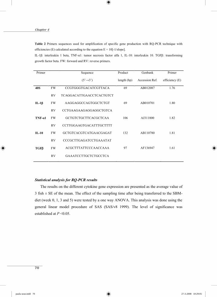
Chapter 4
70
Table 2 Primers sequences used for amplification of specific gene production with RQ-PCR technique with
efficiencies (E) calculated according to the equation E = 10[-1/slope].
IL-1β: interleukin 1 beta, TNF-α1: tumor necrosis factor alfa 1, IL-10: interleukin 10. TGF : transforming
growth factor beta. FW: forward and RV: reverse primers.
Primer Sequence
(5’→3’)
Product
length (bp)
Genbank
Accession Ref.
Primer
efficiency (E)
40S FW CCGTGGGTGACATCGTTACA 69 AB012087 1.76
RV TCAGGACATTGAACCTCACTGTCT
IL-1β FW AAGGAGGCCAGTGGCTCTGT 69 AB010701 1.80
RV CCTGAAGAAGAGGAGGCTGTCA
TNF-α1 FW GCTGTCTGCTTCACGCTCAA 106 AJ311800 1.82
RV CCTTGGAAGTGACATTTGCTTTT
IL-10 FW GCTGTCACGTCATGAACGAGAT 132 AB110780 1.81
RV CCCGCTTGAGATCCTGAAATAT
TGF FW ACGCTTTATTCCCAACCAAA 97 AF136947 1.61
RV GAAATCCTTGCTCTGCCTCA
Statistical analysis for RQ-PCR results
The results on the different cytokine gene expression are presented as the average value of
3 fish ± SE of the mean. The effect of the sampling time after being transferred to the SBM-
diet (week 0, 1, 3 and 5) were tested by a one way ANOVA. This analysis was done using the
general linear model procedure of SAS (SAS/v8 1999). The level of significance was
established at P <0.05.
paula uran.indd 70paula uran.indd 70 27-3-2008 10:29:0527-3-2008 10:29:05
SBM induces enteritis in common carp
71
Results
The results from 3 different experiments conducted in 3, 6 and 9 month old common
carp respectively, were closely observed for a 5 week period with LM, EM and RQ-PCR. Due
to the fact that all 3 experiments produced similar results in all the techniques used, only the
data obtained from 9 month old carp are described in this study.
Morphological changes of the distal gut segment
Fish fed 0SBM did not show any sign of enteritis (Fig. 1A). The MF were of normal
length and shape, and were composed of absorptive cells with SNV aligned lengthwise. The
nucleus of the enterocytes was located between the mid to basal regions of the cell. A basal
amount of GC was observed scattered among the enterocytes. The LP was a thin layer of
cells. The SM, found between the basal part of the folds and muscularis mucosa, contained
numerous BG.
The distal intestine of fish fed continuously on 20SBM for 7 days revealed the first
evidence of enteritis (Fig. 1B). The SNV started to shrink and were less regularly aligned. An
increasing cell infiltration, particularly BG and lymphocytes into the LP and SM, was also
noticed (not shown). The MF did not show any shrinkage compared to the control. Both the
LP and SM had increased in size. After 14 days of 20SBM feeding (Fig. 1C), the SNV are
considerably reduced and the number of GC has increased. The MF remained of the same
length but with a stumpy appearance and a heavily infiltrated LP. After 3 weeks (Fig. 1D),
there was a strong reduction in the size of SNV and the GC was more abundant. The number
of BG had increased and they appeared to have invaded the LP. At week 4 (Fig. 1E), the
reduced vacuolization is even stronger and the LP remained infiltrated with BG and were not
showing any further increase in thickness. The number of GC appeared to have declined. At
week 5 (Fig. 1F), most of the assessed parameters appeared to be in the process of recovery to
the normal situation but the SNV do not yet have the size and the appearance of SNV in
control samples and the LP is still thicker compared with control.
paula uran.indd 71paula uran.indd 71 27-3-2008 10:29:0627-3-2008 10:29:06

Chapter 4
72
Figure 1 Intestinal morphology of common carp. A) Control fish fed the control diet (0SBM). Appearance of the
distal intestinal morphology after feeding a diet containing 20% soybean meal (20SBM) B) 1 week. C) 2 weeks,
D) 3 weeks, E) 4 weeks and F) 5 weeks after continuous feeding. SNV: supranuclear vacuoles, GC: goblet cells,
BG: basophilic granulocytes, LP: lamina propria. Alcian Blue and Crossman staining. Bar is 50μm.
SNV LP
(A)
(E)
(C) (D)
(F)
(B)
GC
BG
SNV LP
(A)
(E)
(C) (D)
(F)
(B)
GC
BG
paula uran.indd 72paula uran.indd 72 27-3-2008 10:29:0827-3-2008 10:29:08
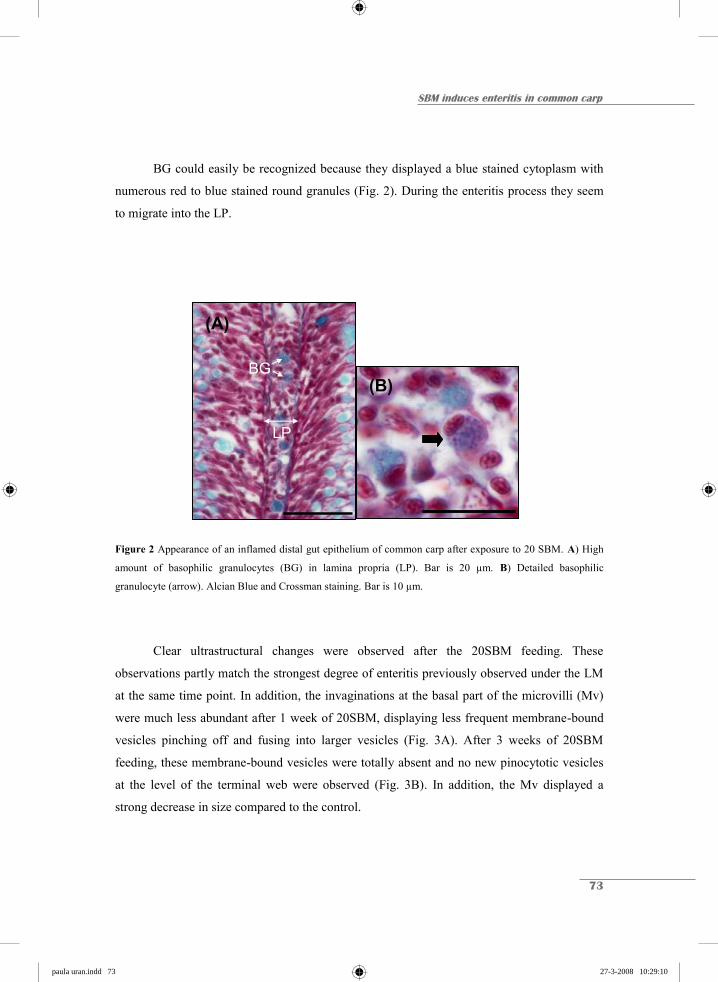
SBM induces enteritis in common carp
73
BG could easily be recognized because they displayed a blue stained cytoplasm with
numerous red to blue stained round granules (Fig. 2). During the enteritis process they seem
to migrate into the LP.
Figure 2 Appearance of an inflamed distal gut epithelium of common carp after exposure to 20 SBM. A) High
amount of basophilic granulocytes (BG) in lamina propria (LP). Bar is 20 μm. B) Detailed basophilic
granulocyte (arrow). Alcian Blue and Crossman staining. Bar is 10 μm.
Clear ultrastructural changes were observed after the 20SBM feeding. These
observations partly match the strongest degree of enteritis previously observed under the LM
at the same time point. In addition, the invaginations at the basal part of the microvilli (Mv)
were much less abundant after 1 week of 20SBM, displaying less frequent membrane-bound
vesicles pinching off and fusing into larger vesicles (Fig. 3A). After 3 weeks of 20SBM
feeding, these membrane-bound vesicles were totally absent and no new pinocytotic vesicles
at the level of the terminal web were observed (Fig. 3B). In addition, the Mv displayed a
strong decrease in size compared to the control.
LP
BG
(A)
(B)
LP
BG
LP
BG
(A)
(B)
paula uran.indd 73paula uran.indd 73 27-3-2008 10:29:1027-3-2008 10:29:10

Chapter 4
74
LP
lumen
(A)
lumen
LP
(B)
Figure 3 Ultrastructure of the distal intestinal enterocytes of common carp A) after 1 week of 20SBM feeding.
Arrows indicate the membrane-bound vesicles fusing into larger vesicles (v). Microvilli (Mv) are seen as large,
tightly packed finger-like structures. B) distal intestinal enterocyte after 3 weeks of 20SBM. A vesicle-free
terminal web (arrow). Note the shorter and more disperse Mv. Bar is 2 μm.
Immnohistochemical detection of T-like cells
The described methodology using anti-CD3 antibody to detect T-like cells in the
second gut segment of common carp gave clear indications of the presence of CD3 +
intraepithelial lymphocytes (IEL) located at the basal part of the epithelium lining the LP at
week 0 and 1 (Fig. 4A). After 3 and 5 weeks of continuous 20SBM feeding, a migration of
these positive cells was observed towards the apical part of the epithelial cells (Fig. 4B).
Figure 4 Migration of CD3 +T-like cells in distal intestinal epithelium of common carp fed 20SBM. A) CD3 +
cells in the basal part of the epithelial lining in contact with lamina propria (LP) at 1 week after feeding (arrow).
B) CD3 + cells migrating towards the apical part of the epithelium (arrows) at 3 weeks after feeding. Bar is
50 m.
MvMv
(A) (B)v
MvMv
(A) (B)v
paula uran.indd 74paula uran.indd 74 27-3-2008 10:29:1127-3-2008 10:29:11
SBM induces enteritis in common carp
75
Gene expression during the SBM-induced enteritis
To understand the possible implications of the IEL during the enteritis process, the
expression of pro-inflammatory IL-1β, TNF-α1, and anti-inflammatory IL-10 and TGF
genes were monitored at weeks 0, 1, 3 and 5 (Fig. 5). Although only 3 animals per sampling
point were measured, the results presented here are similar to two other trials, and hence, the
results can be considered as representative. The different cytokine genes measured at these
time points and their expression relative to the house-keeping gene 40S (which except for
TGF , all values are above the HKG), are described as follows (average expression levels are
given in Fig. 5):
Pro-inflammatory cytokines (IL-1β and TNF- α1)
Numerical differences between sampling points on the pro-inflammatory cytokines IL-
1 β and TNF-α1 expression were found. In comparison with control fish fed 0SBM and
sampled at week 0, expression of IL-1β showed a peak at week 1. The expression of IL-1β
was at all time points above the control levels. TNF-α1 gene expression was up-regulated at
all time points relative to control fish. Its expression increased from week 1 until week 5.
Anti-inflammatory cytokines (IL-10 and TGFβ)
IL-10 gene expression was different between sampling points (P<0.03) Strong up-
regulation of IL-10 gene expression was observed after week 1 of SBM feeding but at week 3
and 5, the expression level was down-regulated again to values either lower (week 3 ) or
similar (week 5) than the control level. TGF gene expression was up-regulated at all time
points relative to control fish. However, at week 3 and 5 the levels were higher although there
was a high variation among the triplicates and the difference between weeks was not
significant.
paula uran.indd 75paula uran.indd 75 27-3-2008 10:29:1327-3-2008 10:29:13

Chapter 4
76
Figure 5 Kinetics of cytokine gene expression measured with real time quantitative PCR in IEL of the distal gut
of carp fed on 20SBM compared with the control (0SBM). Average Ct value (number of cycles to pass the
threshold) at week 0: IL-1β: 26.6, TNF-α1: 25.7, IL-10 24.0, TGF : 29.2. A) IL-1β, B) TNF-α1, C) IL-10 and
D) TGFβ. Amplification is related to the house keeping gene 40S. Bars represent average values of 3 fish per
sampling point ± SE.
Discussion
Soybean meal (SBM) fed to carp, Cyprinus carpio L., has been shown to cause an
inflammatory response in the second gut segment of this omnivorous species similar to that
earlier described SBM-induced enteritis in carnivorous Atlantic salmon (Urán et al. 2008),
feeding exactly the same formulation as used in this study. This SBM-induced inflammation
is a well described phenomenon in Atlantic salmon and has been reported by Baeverfjord &
Krogdahl (1996) as “non-infectious sub-acute enteritis of the distal epithelial mucosa”. The
typical signs of the distal intestinal mucosa are described as: a shortening of the MF; a loss of
the normal supranuclear vacuolisation of the enterocytes; a widening of the lamina propria
(LP); a profound infiltration of inflammatory cells into the LP (van den Ingh et al. 1991,
1996; Baeverfjord & Krogdahl 1996; Krogdahl et al. 2000; Refstie et al. 2000; Buttle et al.
(B)
0123456
0 1 3 5
(A)
0123456
0 1 3 5
(C)
23456
(D)
23456
Expr
essi
on ra
tio (R
)
(B)
0123456
0 1 3 5
(A)
0123456
0 1 3 5
(C)
23456
(D)
23456
Expr
essi
on ra
tio (R
)
paula uran.indd 76paula uran.indd 76 27-3-2008 10:29:1527-3-2008 10:29:15

SBM induces enteritis in common carp
77
2001); an increased presence of eosinophils (Urán et al. 2008) and monocytes/macrophages
and neutrophils in the LP; an increased amount of GC in the epithelium (Bakke-McKellep et
al. 2000); as well as a shortening of the Mv and a strongly decreased endocytotic activity of
the enterocytes (van den Ingh et al. 1991; Urán 2008). In addition, more recently increased T-
cell like reactivity was shown in the inflamed intestine of Atlantic salmon (Bakke-McKellep
et al. 2007).
These morphological and cellular indicators of enteritis reported for Atlantic salmon
were also observed in common carp, including the migration of basophils (morphological
homologues of salmon eosinophils) into the LP and a slight migration of CD3ε+ T-like cells
towards the apical part of the epithelium. The main difference observed between both species
was a slower start of the enteritis process in carp and after 4 weeks a recovery of the enteritis
is observed in common carp, while in Atlantic salmon the condition tends to worsen unless
SBM is withdrawn from its dietary regime. Baeverfjord & Krogdahl (1996) reported a full
recovery of the distal intestinal epithelium of Atlantic salmon after only 21 days of the switch
to a soybean-free diet.
A reduced endocytotic capacity was observed in common carp as it was previously
reported for Atlantic salmon (Urán 2008). After 3 weeks of SBM feeding, the endocytotic
capacity of the enterocytes is reduced coinciding with the disappearance of SNV as was
observed in their salmon counterparts. The recovery of common carp along with the re-
appearance of SNV could be attributed to the omnivorous nature of species which have a
higher ability than carnivorous species to modulate digestive functions to digest plant protein
(Buddington et al. 1997).
As far as we know, this is the first time that such a temporary enteritis effect has been
reported for common carp, a species that usually receives a plant-based diet. Even though
such feeds are effective for raising omnivorous and herbivorous species, they are less
successful for carnivores, because of the assumed digestive limitations (Buddington et al.
1997). Despite the fact that in the present study a SB variety was selected which is known to
induce enteritis, these phenomena may occur as well in aquaculture when young fish start to
ingest food pellets containing SBM. Even though carp seems to adapt to SBM and completely
recovers from the enteritis, more attention should be paid to the vulnerability of these animals
during this inflammation process. Our studies on Atlantic salmon showed a diminished
paula uran.indd 77paula uran.indd 77 27-3-2008 10:29:1627-3-2008 10:29:16

Chapter 4
78
endocytotic capacity of the second segment, and reduced Mv and a strongly reduced uptake of
anally intubated ferritin in 20SBM fed Atlantic salmon (Urán 2008).
Knudsen et al. (2007) suggested that SBM molasses, mainly soyasaponins may be the
inducers of inflammation. Saponins are well studied as adjuvants on animal immune
responses and enhance cell mediated and antibody responses (Rajput et al. 2007). In addition,
they are also effective via oral use, although saponins do not enter the intestinal epithelial
barrier (Pickering et al. 2006). In trout fed SBM, saponins can enhance the specific plasma
antibody responses after an intraperioneal injection with formalin fixed Aeromonas
salmonicida bacterin (Penn 2005). In addition, saponins appear to also be involved in non-
specific immune reactions, such as inflammation and monocyte proliferation (cf. Rajput et al.
2007). Finally, saponins also induce the production of cytokines, such as interleukins and
interferons, which may explain their immunostimulation effects (Rajput et al. 2007). In mice
an orally given purified Quillaja saponaria saponin derivative (QS-21) combined with a
protein tetanus toxoid vaccine, could give Th-1 responses after high doses and Th-2 responses
after low doses of QS-21 (Boyaka et al. 2001), again strongly indicating the interference of
saponins with cytokines. Savan & Sakai 2006, described cytokines as important mediators in
the immune response through the regulation of local and systemic immune inflammatory and
regulatory events. In addition, the involvement of T-cell like reactivity has been suggested in
the intestinal inflammatory response of Atlantic salmon because of the up-regulation of gene
expression of T cell-specific molecules: CD3, CD4, CD8α and CD8β (Bakke-Mckellep et al.
2007). As far as we know, this study has, for the first time, reported the expression of
cytokine genes in a fish enteritis process, in particular the expression of IL-1β and TNF-α1 as
pro-inflammatory and IL-10 and TGFβ as anti-inflammatory cytokine. The basal expressions
of all the four genes studied are given in Fig. 5 in which the order of expression is IL-
10>>TNF-α1 IL-1β >>TGFβ. As far as we know, the expression of these cytokines
(excluding IL-10) is only described for the trout gut (Mulder et al. 2007), however, it has
been measured with RT-PCR and whole extracts of the gut, instead of the IEL which has been
used in this current study. The main difference observed is the low expression of TGFβ in
carp IEL compared to a relatively high expression of TGFβ in the distal gut of trout. Whether
this difference is related to the other isolation method used, or to species differences, or even
due to the different methodology used (semi-qualitative vs. quantitative) remains to be seen.
paula uran.indd 78paula uran.indd 78 27-3-2008 10:29:1727-3-2008 10:29:17

SBM induces enteritis in common carp
79
The expression of the IL-1β and TNF-α1 genes are both above the control levels at
week 1, 3 and 5 after the start of 20SBM feeding, strongly suggesting that these genes are
both involved in the enteritis process. However, it is remarkable that the expression of both
genes seem to peak, showing a trend to be up-regulated at different weeks: IL-1β at week 1
and TNF-α1 at week 5, when fish had already started to recover. On the other hand, it has to
be mentioned that the IL-1β was reproducible in the three comparable experiments while the
peak of TNF-α1 was only found in this experiment. The expression of the IL-10 gene shows a
tendency to be up-regulated with values above the control level at week 1, while this elevation
was more expected before or during the recovery phase of enteritis. In mice it has been
demonstrated that saponin can induce IL-10 production after a subcutaneous injection
combined with antigen (Tadokoro et al. 1996). The relation between IL-10 and enteritis can
be better visualized through the well studied inflammatory bowel diseases (IBD) in humans,
although these intestinal disorders, such as ulcerative colitis and Crohn’s disease, are more
related to the intolerance to the resident microbial population of the gut. (Frogsberg 2006; cf.
Bakke-McKellep et al. 2007). Up-regulation of IL-10 mRNA levels but also IL-10 production
was measured in the gut tissue of active ulcerative colitis and Crohn’s disease, as well as
elevated levels of IL-10 in serum (cf. Autschbach et al. 1998). These authors describe that IL-
10 is not generally down-regulated in active IBD and is even more strongly expressed by the
mononuclear cells in the submucosa. Their results suggest that the elevated production of IL-
10 in the submucosa is insufficient to down-regulate the pro-inflammatory action of
inflammatory cytokines as IL-1β in the LP. It has to be mentioned that the present cytokine
expression results are obtained using isolated cell fractions that are supposed to contain
mainly intraepithelial leukocytes (Rombout et al. 1993). During recovery, IL-10 may be up-
regulated elsewhere in the 2nd segment and hence not visible in this study. Nevertheless, no
explanation can yet be given for the short term, up-regulation of IL-10 around the 1st week of
inflammation when the inflammation process is still far from clear.
The up-regulation trend shown by TGF at weeks 3 and 5, also corresponds with the
recovery of the inflamed carp gut. Hence, TGF can be considered as a general down-
regulator of cell-mediated immune responses (Mulder et al. 2007) and as such can be seen as
the inducer for the recovery phase.
paula uran.indd 79paula uran.indd 79 27-3-2008 10:29:1927-3-2008 10:29:19
Chapter 4
80
Due to such factors, one could possibly conclude that the down-regulation of IL-10
after week 1 and up-regulation of IL-1β and TNF-α1 and TGF from week 1 onwards may
play an inducing role in the SBM-induced enteritis process but these observations need more
attention and additional measurements in other intestinal compartments to explain their
significance.
The migration of basophils into the LP of inflamed carp gut is similar to the eosinophil
reaction in the second segment of Atlantic salmon (Urán 2008). The latter study and others
(Sveinbjørnsson et al. 1996; Silphaduang & Noga 2001; Murray et al. 2007) have shown the
production of lysozyme by these granulocytes and hence an anti-microbial function for these
cells. Although the used anti-lysozyme antibody did not react with carp basophils there is
enough evidence (unpublished RQ-PCR results on isolated basophils) to state that they
produce lysozyme C. Therefore, the reaction of the basophils may be dedicated as one of the
defence mechanisms to protect the inflamed gut against microbial invasion.
In conclusion, in the present study we presented the kinetics of SBM-induced enteritis
for common carp along with the cytokine regulation profile after a continuous exposure to
antigens present in the diet. The onset of the condition was set at week 1 with the initiation of
a profound alteration of the distal intestinal architecture. This study also confirms the
contribution of IEL, basophils, T-like cells, GC and enterocytes to the onset and regulation of
the inflammatory process. In contrast to Atlantic salmon, common carp starts to recover its
endocytotic capacity towards the end of the experiment but a persistently higher expression
value of pro-inflammatory cytokine genes is still observed. It suggests a unique adaptation of
the intestinal tissue of common carp to poor quality protein sources while a constant
surveillance of the innate immune system ensures the integrity and health of the intestinal
epithelium.
Acknowledgments
This study was supported by “Instituto Colombiano para el Desarrollo de la Ciencia y la
Tecnología”, Colciencias, and Skretting ARC, Stavanger, Norway. We would like to acknowledge A.
Cánovas for his contribution to the detection of T-like cells and to M. Forlenza for her support with
the RQ-PCR technique. The results presented are part of the PhD thesis of P.A. Urán.
paula uran.indd 80paula uran.indd 80 27-3-2008 10:29:2027-3-2008 10:29:20
SBM induces enteritis in common carp
81
References
Autschbach, F., Braunstein, J., Helmke, B., Zuna, I., Schurmann, G., Niemir, Z. I., Wallich, R., Otto,
H. F. & Meuer, S. C. (1998) In situ expression of interleukin-10 in non-inflamed human gut and in
inflammatory bowel disease. Am. J. Pathol., 153, 121-130.
Baeverfjord, G. & Krogdahl, Å. (1996) Development and regression of soybean meal induced enteritis
in Atlantic salmon, Salmo salar L., distal intestine: a comparison with the intestines of fasted fish.
J. Fish Dis., 19, 375-387.
Bakke-Mckellep, A. M., Press, C. Mcl., Baeverfjord, G., Krogdahl, Å. & Landsverk, T. (2000)
Changes in immune and enzyme histochemical phenotypes of cells in the intestinal mucosa of
Atlantic salmon, Salmo salar L., with soybean meal-induced enteritis. J. Fish Dis., 23, 115-127.
Bakke-McKellep, A.M., Froystad, M.K., Lilleeng, E., Dapra, F., Refstie, S., Krogdahl, A. &
Landsverk, T. (2007) Response to soy: T-cell-like reactivity in the intestine of Atlantic salmon,
Salmo salar L. J. Fish Dis., 30, 13-25.
Bonaldo, A., Roem, A.J., Mariani, L., Fagioli, P., Pecchini, A., Parma, L. & Gatta, P. P. (2006) The
influence of different levels of soybean meal in diets for ongrowing gilthead sea bream (Sparus
aurata) and European sea bass (Dicentrarchus labrax). Ital. J. Anim. Sci., 6, 790-790.
Boyaka, P.N., Marinaro, M., Jackson, R.J., van Ginkel F.W., Cormet-Boyaka, E., Kirk, K.L., Kensil,
D.K. & McGhee, G.R. (2001) Oral QS-21 requires early IL-4 help for induction of mucosal and
systemic immunity. J. Immunol., 166, 2283 - 2290.
Buddington, R.K., Krogdahl, Å. & Bakke-McKellep, A.M. (1997) The intestines of carnivorous fish:
structure and functions and the relations with diet. Acta Physiol. Scand., 161, 67-80.
Buttle, L.G., Burrells, A.C., Good, J.E., Williams, P.D., Southgate, P.J. & Burrells, C. (2001) The
binding of soybean agglutinin (SBA) to the intestinal epithelium of Atlantic salmon Salmo salar
and Rainbow trout, Oncorhynchus mykiss, fed high levels of soybean meal. Vet. Immunol.
Immunopathol., 80, 237-244.
Evans, J.J., Pasnik, D.J., Peres, H., Lim, C. & Klesius, P.H. (2005) No apparent differences in
intestinal histology of channel catfish (Ictalurus punctatus) fed heat-treated and non-heat-treated
raw soybean meal. Aquacult. Nutr., 11, 123-129.
Frogsberg, G. (2006) Innate and adaptive immunity in childhood celiac disease. Umeå University
Medical Dissertations. New series No. 1054, 69 pp. ISBN: 91-7264-162-2.
Grisdalle-Helland B., Helland, S.J. Baeverfjord, G. & Berge, G.M. (2002) Full-fat soybean meal in
diets for Atlantic halibut: growth, metabolism and intestinal histology. Aquacult. Nutr., 8, 265-
270.
paula uran.indd 81paula uran.indd 81 27-3-2008 10:29:2127-3-2008 10:29:21

Chapter 4
82
Hansen, A.C., Rosenlund, G., Karlsen, R., Olsvik, P.A. & hemre, G.I. (2006) The inclusion of plant
protein in cod diets, its effects on macronutrient digestibility, gut and liver histology ad heat shock
protein transcription. Aquac. Res., 37, 773-784.
Huttenhuis H.B.T., Taverne-Thiele, A.J., Grou, C.P.O., Bergsma, J., Saeij, J.P.J., Nakayasu, C. &
Rombout, J.H.W.M. (2006) Ontogeny of the common carp (Cyprinus carpio L.) innate immune
system. Dev. Comp. Immnunol., 30, 557-574.
van den Ingh, T.S.G.A.M., Krogdahl, Å., Olli, J.J., Hendriks, H.G.C.J.M. & Koninkx, J.G.J.F. (1991)
Effects of soybean-containing diets on the proximal and distal intestine in Atlantic salmon (Salmo
salar): a morphological study. Aquaculture, 94, 297-305.
van den Ingh, T.S.G.A.M., Olli, J.J. & Krogdahl, Å. (1996) Alcohol-soluble components in soybeans
cause morphological changes in the distal intestine of Atlantic salmon, Salmo salar L. J. Fish Dis.,
19, 47-53.
Knudsen, D., Urán, P., Arnous, A., Koppe, W. & Frøkiær, H. (2007). Saponin-containing subfractions
of soybean molasses induce enteritis in the distal intestine of Atlantic salmon. J. Agric. Food
Chem., 55, 2261-2267.
Krogdahl, Å., Bakke-McKellep, A.M., Røed, K.H. & Baeverfjord, G. (2000) Feeding Atlantic salmon
Salmo salar L. soybean products: effects on disease resistance (furunculosis), and lysozyme and
IgM levels in the intestinal mucosa. Aquacult. Nutr., 6, 77-84.
Nordrum, S., Bakke-McKellep, A.M., Krogdahl, Å. & Buddington, R.K. (2000) Effects of soybean
meal and salinity on intestinal transport of nutrients in Atlantic salmon (Salmo salar L.) and
rainbow trout (Oncorhynchus mykiss). Comp. Biochem. Phys. B, 125, 317-335.
Mulder, I. E., Wadsworth, S. & Secombes, C. J. (2007) Cytokine expression in the intestine of
rainbow trout (Oncorhynchus mykiss) during infection with Aeromonas salmonicida. Fish
Shellfish Immunol., 23, 747-759
Murray, H.M., Leggiadro, C.T. & Douglas, S.E., (2007) Immunocytochemical localization of
pleurocidin to the cytoplasmic granules of eosinophilic granular cells from the winter flounder
gill. J. Fish Biol., 70, 336-345.
Penn, M.H. (2005) The effects of dietary soybean saponins on growth and performance, intestinal
histology and immune response of first feeding rainbow trout Oncorhynchus mykiss. Thesis
Dissertation University of Ohio. 121 p. http://www.ohiolink.edu/etd/send-
pdf.cgi/Penn%2C%20Michael%20H..pdf?osu1119027556
Pickering, R.J. Smith, S.D., Strugnell, R.A., Wesselingh, S.L. & Webster, D.E. (2006) Crude saponins
improve the immune response to an oral plant-made measles vaccine. Vaccine, 24, 144-150.
paula uran.indd 82paula uran.indd 82 27-3-2008 10:29:2327-3-2008 10:29:23
SBM induces enteritis in common carp
83
Pfaffl, M.W. (2001) A new mathematical model for relative quantification in real-time RT-PCR.
Nucleic Acids Res, 29: 2002-2007.
Rajput, Z.I., Hu, S., Xiao, Ch. & Arijo, A.,G. (2007) Adjuvant effect of saponins on animal immune
responses. J. Zhejiang Univ. Sci. B, 8, 153-161.
Refstie, S., Korsøen, Ø.J., Storebakken, T., Baeverfjord, G., Lein, I. & Roem, A.J. (2000) Differing
nutritional responses to dietary soybean meal in rainbow trout (Oncorhynchus mykiss) and
Atlantic salmon (Salmo salar). Aquaculture, 190, 49-63.
Refstie, S., Landsverk, T., Bakke-McKellep, A.M., Ringo, E., Sundby, A., Shearer, K.D., Krogdahl,
A. (2006) Digestive capacity, intestinal morphology, and microflora of 1-year and 2-year old
Atlantic cod (Gadus morhua) fed standard or bioprocessed soybean meal. Aquaculture, 261, 269-
284.
Rombout, J.H.W.M. Taverne-thiele, A.J. & Villena, M.I. (1993) The Gut-Associated Lymphoid-
Tissue (GALT) of carp (Cyprinus-Carpio L) - an immunocytochemical analysis. Dev. Comp.
immunol., 17, 55-66.
SAS (1999) SAS/STAT® User’s guide, version 8. SAS Institute, Carey NC, USA.
Savan, R. & Sakai, M. (2006) Genomics of fish cytokines: review. Comp. Biochem. Phys. D., 1, 89-
101.
Silphaduang, U. & Noga, E. J., 2001. Peptide antibiotics in mast cells of fish. Nature, 414, 268-269.
Sveinbjørnsson, B., Olsen, R. & Paulsen, S. (1996). Immunocytochemical localization of lysozyme in
intestinal eosinophilic granule cells (EGCs) of Atlantic salmon, Salmo salar L. J. Fish dis., 19,
349-355.
Tadokoro, C.E., Macedo, M.S. & Abrahamsohn, I.A. (1996) Saponin adjuvant primes for a dominant
interleukin-10 production to ovalbumin and to Trypanosoma cruzi antigen. Immunology, 89, 368-
374.
Urán, P.A. (2008) Etiology of soybean-induced enteritis in fish. PhD thesis dissertation, Animal
Sciences group, Wageningen University, The Netherlands.
Urán, P.A., Schrama, J.W., Rombout, J.H.W.M., Obach, A., Jensen, L., Koppe, W. & Verreth, J.A.J.
(2008) Soybean meal-induced enteritis in Atlantic salmon (Salmo salar L.) at different
temperatures. Aquacult. Nutr. in press
paula uran.indd 83paula uran.indd 83 27-3-2008 10:29:2427-3-2008 10:29:24

chapter hoofdstukken.indd 9 26-3-2008 12:38:11

Chapter 5
Soybean meal-induced uptake block in the distal enterocytes of Atlantic salmon (Salmo salar L.)
P.A. Urán1,2, R. Aydin1, J. W. Schrama1, J.A.J. Verreth1 & J.H.W.M. Rombout2
1Aquaculture and Fisheries Group and 2Cell Biology and Immunology Group, Wageningen Institute of Animal Sciences, Wageningen University, The Netherlands
Submitted for publication
chapter hoofdstukken.indd 10 26-3-2008 12:38:11
Chapter 5
86
Abstract
The replacement of fishmeal by soybean meal (SBM) induces enteritis in the distal
intestine of Atlantic salmon. The enterocytes at the distal intestinal mucosa are heavily
hampered and the disappearance of supranuclear vacuoles (SNV) seems to be the most
suitable indicator for the onset of enteritis. In this study ferritin was used to evaluate the
changes in uptake capacity of the distal enterocytes under the influence of a SBM-based diet.
After one week of SBM-feeding, ferritin uptake was rather diminished and the SNV
decreased in size compared to the control. At three weeks ferritin uptake could not be detected
anymore and the SNV were invisible in the enterocytes. The linkage between the
disappearance of SNV and endocytosis block will be discussed.
paula uran.indd 86paula uran.indd 86 27-3-2008 10:29:2627-3-2008 10:29:26

SBM impairs uptake in salmon enterocytes
87
Introduction
Soybean meal (SBM) is a potential alternative among the plant materials to reduce the
use of fishmeal (FM) for aquafeeds. However, the presence of some anti-nutritional factors
hampers its utilization for salmonid diets. It is known that SBM induces a “sub-acute
inflammation of the distal intestine” in Atlantic salmon (Baeverfjord & Krogdahl 1996),
characterised by the loss of the normal supranuclear vacuolisation, a shortening of the
mucosal folds (MF), a thickening of both lamina propria (LP) and sub-epithelial mucosa (SM)
with a severe infiltration of macrophages and eosinophilic granulocytes (EG), an increased
numbers of goblet cells (GC) in the epithelium (van den Ingh et al. 1991; van den Ingh et al.
1996; Baeverfjord & Krogdahl 1996; Krogdahl et al. 2000; Refstie et al. 2000; Buttle et al.
2001; Urán et al. 2008).
The impact of SBM on the distal intestine of Atlantic salmon is striking but not yet
well understood, despite the interest and the commercial importance of the species (Bakke-
McKellep et al. 2000). It has been generally accepted that the second gut segment (main part
of the distal intestine), is the principle site of endocytosis of intact proteins in teleost fish
(Stroband et al. 1979, Stroband & van der Veen 1981; Rombout et al. 1985; Sire & Vernier
1992). Although the high endocytotic capacity of the distal enterocytes can have absorptive as
well as immunological impact, it possibly makes the distal intestine also more sensitive to
food-borne enteropathies (Bakke-McKellep et al., 2000). Indeed enterocytes of the distal
intestine are the first cells affected when Atlantic salmon is fed SBM diets (Urán et al. 2008).
Previous ultrastructural studies conducted on old Atlantic salmon and young common carp,
have shown a severe decrease in endocytosis by the distal enterocytes, although carp showed
a slower impairment and a subsequent recovery after 5 weeks (Urán 2008).
The aim of this study was to evaluate the impact of SBM-feeding on the enterocyte
uptake capacity of Atlantic salmon. Young salmon were used to establish whether age and
freshwater conditions had any influence on the severity of SBM-induced enteritis described
for old fish kept in seawater. Therefore the same experimental diet was used as in previous
experiments on old fish (Urán et al. 2008). Special attention is paid to the correlation between
impaired uptake and the disappearance of the supranuclear vacuoles (SNV). Therefore ferritin
is used as macromolecule because it can be detected easily at the light microscopical level.
paula uran.indd 87paula uran.indd 87 27-3-2008 10:29:2727-3-2008 10:29:27

Chapter 5
88
Materials and Methods
Fish rearing conditions
The experiment was carried out at ‘De Haar Vissen’ facilities, Animal Science group,
Wageningen University, The Netherlands. The Atlantic salmon (Irish strain) originated from
the fish farm Forellenzucht Hischquellen, schloβ Holte-Stukenbrock, Germany. The
experiment consisted of a two-week adaptation period and three-week experimental period.
At the start of the adaptation period the fish weighted approximately 30 g.
For the experiment four circular indoor tanks with a diameter of 1 m each were used.
The water volume in the tanks was 600 L. Two tanks were stocked with 28 fish each and the
other two tanks with 36 fish each, according to the treatment. Each tank was kept at a flow
through system. Freshwater was pumped to the tanks at a temperature of 13 ± 2°C and an
oxygen concentration above 9 ppm (saturated), pH of 8.2 and NH4+, NO2
-, NO3- levels kept at
0. The water conductivity was 180 μs. The photoperiod was approximately 10L : 14D (natural
light regime during winter time).
Diets and feeding
Feed was produced at Skretting Feed Technology Plant, (Stavanger, Norway). Two
diets were formulated: a control diet (0SBM) and a experimental diets (20SBM) (Table 1).
The major ingredients in the 0SBM diets were: FM, fish oil (FO), and wheat. This control diet
did not contain any SBM. For the experimental diet, FM and wheat were exchanged for 20%
SBM (solvent-extracted Hipro SBM). The diets were produced to be iso-energetic and iso-
nitrogenic on a crude protein and a crude lipid basis. Diets were supplemented with a standard
vitamin and mineral premix. Feed was produced as extruded 3 mm sinking pellets.
Prior to the experiment and during the adaptation period all tanks were fed with the
control diet (0SBM). At the start of the experimental period, two of the fours tanks were
changed to the 20SBM experimental diet. The other two tanks remained at the control diet.
Fish were fed 20% in excess. Feed was divided into two meals per day and provided by
automatic belt feeders.
paula uran.indd 88paula uran.indd 88 27-3-2008 10:29:2827-3-2008 10:29:28

SBM impairs uptake in salmon enterocytes
89
Table 1 Chemical composition of the experimental diets.
Diets1
0 SBM 20 SBM
Ingredients (g kg-1)
Fishmeal2 562.4 470.3
Soybean meal3 0.0 200.0
Wheat 201.1 84.7
Fish Oil4 234.5 243.0
Vitamin premix 1.0 1.0
Mineral premix 1.0 1.0
Analyzed chemical composition (g kg-1)
Dry matter 914 941
Crude Protein 415 446
Crude fat 268 287
Ash 88 89 1 Amount of protein replaced by Soybean meal in percent. 2 LT north Atlantic, Egersund, Norway. 3 Extracted solvent HiPro Soybean Meal. 4 Northern hemisphere.
Chemical analysis of diets
Feed samples were ground using a 1 mm screen before analysing dry matter, ash,
crude protein and crude fat content. Dry matter content was determined by drying samples for
4 hours at 103°C until constant weight (ISO 6496, 1983) and ash content by incineration in a
muffle furnace for 4 hours at 550°C (ISO 5984, 1978). Crude fat content was determined by
petroleum ether extraction in a Soxhlet apparatus (ISO/DIS, 1996). Crude protein (N x 6.25)
was measured by the Kjeldahl method after acid digestion (ISO 5983, 1979). The analysed
chemical composition of the diets is given in Table 1.
Administration of ferritin
A solution containing 1% horse spleen ferritin (Sigma-Aldrich, Germany) in
phosphate-buffered saline (PBS), was anally intubated directly into the second gut segment.
paula uran.indd 89paula uran.indd 89 27-3-2008 10:29:3027-3-2008 10:29:30

Chapter 5
90
Fish were starved 24 hours before the intubation procedure to ensure an empty intestine for
the better delivery of the markers. Before the procedure, fish were anaesthetised using 0.05%
tricaine methane sulphonate (Crescent Research Chemicals, Phoenix, AZ) TMS buffered with
0.1 % sodium bicarbonate, in tank water.
Sampling scheme
Sampling for distal intestine was performed at various time points after ferritin
intubation. At the week 1, samples were taken at 90min, 6 and 12 h. and at week 3 at 30, 60,
90 min and 6 h after intubation for both dietary groups (n=8 per time point/diet). To check for
background ferritin staining and to discard the induction of inflammation due to intubation,
PBS intubated fish (n=4) and no intubated fish (n=2) were sampled after 90 min, each at week
1 and 3 from both 0SBM and 20SBM diet groups. After intubation fish were kept in smaller
tanks (120 L), under the same conditions as in the original ones, and were individually tagged
to control the exact time after intubation.
Light Microscopy (LM)
At each time point after intubation, fish were sacrificed with an overdose of
anaesthesia. For LM analysis a two-centimetre section of distal intestine (thicker region of the
intestinal tract characterized by more prominent and darker annular rings) of each fish was
dissected out and fixed in a mix of methanol, formalin, acetic acid (85:10:5). After 24 hours
the samples were transferred to 70% ethanol and stored until further processing. The samples
were dehydrated using standard procedures and embedded in paraffin. Transverse sections of
5 μm thickness were made and stained with Haematoxylin/Eosin and Alcian blue (pH 2.5), to
enhance the contrast between GC and SNV.
Scoring enteritis
The LM sections were evaluated according to the semi-quantitative method developed
at Wageningen University (Urán et al. 2008), which assesses the degree of SBM-induced
enteritis on the Atlantic salmon distal intestine considering the criteria fully described in
Chapter 2. (For illustrations of the different scores, see annex or check the list of special
paula uran.indd 90paula uran.indd 90 27-3-2008 10:29:3127-3-2008 10:29:31
SBM impairs uptake in salmon enterocytes
91
publication attachments at http://www.afi.wur.nl/UK/Publications/). Three out of the eight
sampled fish per time point and diet, were scored and included in the analysis.
Assessment of the presence of ferritin in tissue
For the detection of ferritin in tissue, slides were stained with Perls’ staining procedure
described by Romeis (1968), to detect the presence of iron components in tissue. All sampling
points after intubation were analyzed at week 1 and week 3 after SBM-feeding. Sections were
photographed with an Olympus DP 50 digital camera (Olympus, Japan) connected to a Nikon
Microphot-FXA light microscope (Badhoeverdorp, the Netherlands). The pictures were
processed and analyzed using the AnalySiS Extended Pro 3.1 software (Soft Imaging System
GmbH, Münster, Germany).
Statistical analysis
All calculations were made using the SAS System (SAS 1999). The parameters used
to score the degree of enteritis were analysed for the effect of diet using PROC GLM for each
sampling point (week 1 or week 3) separately. The results are considered statistically
significant when P-values are below 0.05.
Results
Morphological changes of the distal intestine
The control diet (0SBM) did not induce any sign of enteritis. The MF appeared as long
finger-like structures, composed of enterocytes which were perfectly aligned along the
mucosal epithelium. The nucleus of the enterocytes was located between the mid to basal part
of the cell. The enterocytes presented normal round to oval SNV. The GC were present in a
basal number and were scattered among the enterocytes. The LP was a delicate and single thin
layer of cells underlying the epithelium. In the SM a basal number of EG was present as
reflected in Fig.1. All semi-quantitative scores remained at the basal level for the measured
parameters at all sampling points, indicating the absence of any sign of enteritis.
paula uran.indd 91paula uran.indd 91 27-3-2008 10:29:3327-3-2008 10:29:33

Chapter 5
92
Fish fed 20SBM showed morphological changes at week 1. At this sampling moment, fish fed
the 20SBM diet compared to fish fed the control diet had different scores for MF, SNV, LP
and SM (P < 0.05) but not for GC (P = 0.14) and EG (P = 0.06). The MF become shorter with
subsequent tissue disruption. The SNV start to shrink and their regular alignment was
disturbed. A tendency for an increased infiltration of EG into the LP and SM was noticed. The
amount of GC is numerically higher and the LP and SM had increased in size. At week 3 after
20SBM-feeding, heavily changed intestinal mucosal architecture could be detected. Most of
the parameters measured were more affected worsen along the experimental period (Fig. 1)
and at this sampling moment all score parameters were different between fish fed the control
and 20SBM diet (P < 0.001). Diffused tissue disruption occurred at the level of the MF,
appearing as bloated mucosal tissue. The SNV were completely absent. The GC appeared
densely packed especially at the tip of the folds. The number of EG increased in SM, also
with abundant presence along the LP.
In Table 2, the inflammatory response observed in young animals of an initial average
weight of 30 g kept in freshwater was compared to the response observed in a previous
experiment using old fish of an average weight of 300 g kept in seawater during the
experimental period (Urán 2008). The results of this comparison suggest that both young and
old fish experience a similar SBM-induced enteritis with an onset of the condition at week 1
and an aggravation at week 3. Some of the parameters tended to be higher in younger fish but
for GC and EG the effect was slightly lower than in their old counterparts. In summary, the
inflammation process was more or less similar at both ages.
paula uran.indd 92paula uran.indd 92 27-3-2008 10:29:3427-3-2008 10:29:34

SBM impairs uptake in salmon enterocytes
93
11.5
22.5
33.5
44.5
5
Week 1 Week 3
0SBM
E
11.5
22.5
33.5
44.5
5
Week 1 Week 3
20SBM
F
a
b
a
b
a
b
a
b
11.5
22.5
33.5
44.5
5
Week 1 Week 3
D
a
b
a
a
11.5
22.5
33.5
44.5
5
Week 1 Week 3
A
a
b
a
b
11.5
22.5
33.5
44.5
5
Week 1 Week 3
B
aa
b
b
11.5
22.5
33.5
44.5
5
Week 1 Week 3
C
Entie
ritis
sco
re
a
a
a
b
Figure 1 Parameters used to assess the degree of enteritis of the distal intestinal mucosa of Atlantic salmon after
feeding a soybean meal-containing diet (20SBM) compared to a control diet (0SBM) at 1 and 3 weeks of
continuous feeding, results are presented as mean values ± SE. The degree of enteritis has been assessed using
the semi-quantitative scoring system (Urán et al., 2008). A. Mucosa folds (MF) , B. Supranuclear vacuoles
(SNV), C. Goblet cells (GC), D. Eosinophilic granulocytes (EG), E. Lamina propria (LP), F. Sub-epithelial
mucosa (SM). Means at each sampling point (week) having a different letter (ab) differ significantly (P < 0.05).
paula uran.indd 93paula uran.indd 93 27-3-2008 10:29:3527-3-2008 10:29:35
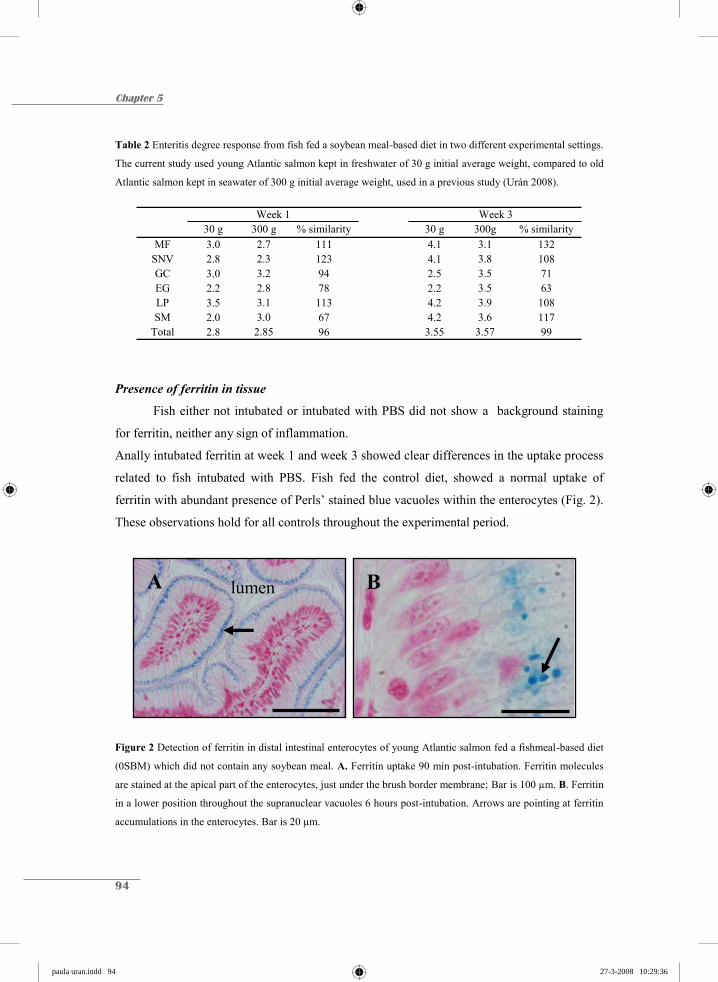
Chapter 5
94
Table 2 Enteritis degree response from fish fed a soybean meal-based diet in two different experimental settings.
The current study used young Atlantic salmon kept in freshwater of 30 g initial average weight, compared to old
Atlantic salmon kept in seawater of 300 g initial average weight, used in a previous study (Urán 2008).
30 g 300 g % similarity 30 g 300g % similarityMF 3.0 2.7 111 4.1 3.1 132
SNV 2.8 2.3 123 4.1 3.8 108GC 3.0 3.2 94 2.5 3.5 71EG 2.2 2.8 78 2.2 3.5 63LP 3.5 3.1 113 4.2 3.9 108SM 2.0 3.0 67 4.2 3.6 117
Total 2.8 2.85 96 3.55 3.57 99
Week 1 Week 3
Presence of ferritin in tissue
Fish either not intubated or intubated with PBS did not show a background staining
for ferritin, neither any sign of inflammation.
Anally intubated ferritin at week 1 and week 3 showed clear differences in the uptake process
related to fish intubated with PBS. Fish fed the control diet, showed a normal uptake of
ferritin with abundant presence of Perls’ stained blue vacuoles within the enterocytes (Fig. 2).
These observations hold for all controls throughout the experimental period.
Figure 2 Detection of ferritin in distal intestinal enterocytes of young Atlantic salmon fed a fishmeal-based diet
(0SBM) which did not contain any soybean meal. A. Ferritin uptake 90 min post-intubation. Ferritin molecules
are stained at the apical part of the enterocytes, just under the brush border membrane; Bar is 100 μm. B. Ferritin
in a lower position throughout the supranuclear vacuoles 6 hours post-intubation. Arrows are pointing at ferritin
accumulations in the enterocytes. Bar is 20 μm.
A lumen B
paula uran.indd 94paula uran.indd 94 27-3-2008 10:29:3627-3-2008 10:29:36

SBM impairs uptake in salmon enterocytes
95
In 20SBM fed fish, the tendency on the observed ferritin uptake at both week 1 and
week 3 was impaired. At week 1, Perls’ stained ferritin was detected within the enterocytes
but the amount is substantially decreased compared to the control diet 0SBM (Fig. 3A). There
were no clear differences between intubation points however after 6 hours Perls’ stained
ferritin, was also detected basally towards the LP (Fig. 3B). At week 3, all fish analysed did
not show any ferritin uptake, although, ferritin was clearly present in the lumen at all time
points (Fig. 3C and 3D).
Figure 3 Ferritin uptake after 1 and 3 weeks of SBM feeding. A) Ferritin uptake at week 1 after 90 min post-
intubation. B) Ferritin uptake at week 1 after 6 hours post-intubation. Perls’ stained ferritin in enterocytes is
indicated with arrows. C) No uptake at week 3 after 90 min post-intubation. D) No uptake at week 3 after 6
hours post-intubation. Perls’ stained ferritin is only present in the lumen (arrows). Bar is 100 μm.
B
D
lumen
A
C lumen
paula uran.indd 95paula uran.indd 95 27-3-2008 10:29:3827-3-2008 10:29:38

Chapter 5
96
Discussion and Conclusions
In the current study, young salmon kept in freshwater were used to assess the typical
morphological changes characteristic of a SBM-induced enteritis and its impact on the uptake
capacity of the inflamed distal gut. At week 1 the symptoms of the enteritis condition were
already present but at week 3 they became more severe. The symptoms were similar to
previous observations on old Atlantic salmon kept in sea water and fed the same type of SBM
variety (Urán 2008). These results at least suggest that the severity of enteritis is not related to
age or smoltification of the fish. Only the numbers of GC and EG tended to be higher in old
fish, but this may be related to development and hence an increased immunity at later ages.
Old Atlantic salmon kept in freshwater also develop SBM-induced enteritis (Bakke-McKellep
et al. 2000). More recently the influence of SBM feeding on the parr to smolt transformation
was studied (Bakke-McKellep et al. 2006) and despite the inflamed intestine and apparent
reduction in digestive functions augmented effects on osmoregulatory capacity was not found
in SBM-fed fish. In conclusion salinity seems not to have a remarkable influence on the
severity of SBM-induced enteritis.
In this study the impact of SBM on the endocytosis process has been examined, using
ferritin as a large and easy detectable macromolecule. At week 1, ferritin uptake was reduced,
however still present into a certain extent in some of the fish analysed. At week 3 ferritin
uptake in the vacuoles was not observed, coinciding with a fully developed enteritis
condition. These results are in line with earlier described disappearance of endocytosis
vesicles and invaginations at the ultrastructural level (Urán 2008). Previous LM and EM
studies using ferritin as marker molecule (Stroband & van der Veen, 1981; Rombout et al.
1985; Rombout & van der Berg 1989) have evidenced that endocytotic vesicles fuse with
each other and with lysosomes, finally resulting in an accumulation and digestion of ferritin in
the SNV. The present results also show the correlation between endocytotic uptake and SNV
formation. A complete block of endocytosis even results in the disappearance of SNV, one of
the most obvious signs of SBM-induced enteritis. In this study the complete block occurs
between 1 and 3 weeks of SBM feeding, while in earlier ultrastructural observations it was
reported at 1 week (Urán 2008). Although, electron microscopy, allowing only minor parts to
be studied, is not the most suitable technique to estimate the onset of the endocytosis block, a
paula uran.indd 96paula uran.indd 96 27-3-2008 10:29:3927-3-2008 10:29:39

SBM impairs uptake in salmon enterocytes
97
complete uptake block will probably be obtained short after the 1st week of SBM-feeding. It
has been well documented that ferritin uptake is the result of fluid phase uptake in a process
that seems to be receptor driven; in any case horseradish peroxidase (HRP; Rombout et al.
1985) and LTB (Companjen et al. 2006) have been shown to be taken up by receptors.
However, the exact function of this endocytosis process has never been elucidated. Which
substances in SBM are responsible for the endocytosis block is still unclear, although it has
been suggested that SB lectins may block receptor sites in the distal intestine (cf. Nordrum et
al. 2000). More recently it has been suggested that the disappearance of SNV might be caused
by an unbalanced enzymatic activity (Krogdahl & Bakke-McKellep 2005). However, this
study strongly suggest that endocytosis block is the first step, followed by changes in and
disappearance of the SNV. More attention has to be paid to the subsequent reactions of
enterocytes (i.e. production of danger signals like heat shock proteins) and the onset of
inflammation reactions in the connective tissue. It has been demonstrated that T cells and EG
play an important role in the SBM-induced enteritis process of the distal gut (Bakke-
McKellep et al. 2007), resulting in elevated levels of inflammatory cytokines (Urán 2008).
In conclusion, more research has to be dedicated to the SBM substances responsible
for the endocytosis block, for a better selection of suitable soybeans to be included in the diet
formulation of Atlantic salmon. Strong variations in severity of enteritis between different SB
batches are described in this thesis and may be used to find the most important causative
agent. In addition, more research is needed to translate the endocytosis block and subsequent
disappearance of SNV into the severe enteritis process observed in the distal gut.
Acknowledgements
This research was supported by “Instituto Colombiano para el Desarrollo de la Ciencia y la
Tecnología” Colciencias and Skretting ARC, Stavanger, Norway. We would like to thank the bio
technicians at De Haar Vissen for animal husbandry and technical assistance, for their help during the
feeding trial and the collection the samples.
paula uran.indd 97paula uran.indd 97 27-3-2008 10:29:4027-3-2008 10:29:40
Chapter 5
98
References
Baeverfjord, G. & Krogdahl, Å. (1996) Development and regression of soybean meal induced enteritis
in Atlantic salmon, Salmo salar L., distal intestine: a comparison with the intestines of fasted fish.
J. Fish Dis., 19, 375-387.
Bakke-Mckellep, A. M., Press, C. Mcl., Baeverfjord, G., Krogdahl, Å. & Landsverk, T. (2000)
Changes in immune and enzyme histochemical phenotypes of cells in the intestinal mucosa of
Atlantic salmon, Salmo salar L., with soybean meal-induced enteritis. J. Fish Dis., 23, 115-127.
Bakke-McKellep, A.M., Refstie, S., Stefansson, S.O., Vanthanouvong, V., Roomans, G., Hemre, G.I.
& Krogdahl, Å. (2006) Effects of dietary soybean meal and photoperiod cycle on osmoregulation
following seawater exposure in Atlantic salmon smolts. J. Fish Biol., 69, 1396-1426.
Bakke-McKellep, A. M., Froystad, M. K., Lilleeng, E., Dapra, F., Refstie, S., Krogdahl, A. &
Landsverk, T. (2007) Response to soy: T-cell-like reactivity in the intestine of Atlantic salmon,
Salmo salar L. J. Fish Dis., 30, 13-25.
Buttle, L.G., Burrells, A.C., Good, J.E., Williams, P.D., Southgate, P.J. & Burrells, C. (2001) The
binding of soybean agglutinin (SBA) to the intestinal epithelium of Atlantic salmon Salmo salar
and Rainbow trout, Oncorhynchus mykiss, fed high levels of soybean meal. Vet. Immunol.
Immunopathol., 80, 237-244.
Companjen, A.R., Florack, D.E.A., Slootweg, T., Borst, J.W. & Rombout, J.H.W.M. (2006) Improved
uptake of plant derived LTB-linked proteins in carp gut and induction of specific humoral immune
responses upon in feed delivery. Fish Shellfish Immunol., 21, 251-260.
van den Ingh, T.S.G.A.M., Krogdahl, Å., Olli, J.J., Hendriks, H.G.C.J.M. & Koninkx, J.G.J.F. (1991)
Effects of soybean-containing diets on the proximal and distal intestine in Atlantic salmon (Salmo
salar): a morphological study. Aquaculture, 94, 297-305.
van den Ingh, T.S.G.A.M., Olli, J.J. & Krogdahl, Å. (1996) Alcohol-soluble components in soybeans
cause morphological changes in the distal intestine of Atlantic salmon, Salmo salar L. J. Fish Dis.,
19, 47-53.
ISO (1978) Animal feeding stuff. Determination of crude ash. ISO 5984. International Organization
for Standardization. Geneva, Switzerland.
ISO (1979) Animal feeding stuff. Determination nitrogen content and calculation of crude protein
content. ISO 5983. International Organization for Standardization. Geneva, Switzerland.
ISO (1983) Animal feeding stuff. Determination of moisture content. ISO 6496. International
Organization for Standardization. Geneva, Switzerland.
paula uran.indd 98paula uran.indd 98 27-3-2008 10:29:4227-3-2008 10:29:42
SBM impairs uptake in salmon enterocytes
99
ISO/DIS (1996) Animal feeding stuff. Determination of fat content. Category B. 6492. Geneva,
Switzerland.
Krogdahl, Å., Bakke-McKellep, A.M., Røed, K.H. & Baeverfjord, G. (2000) Feeding Atlantic salmon
Salmo salar L. soybean products: effects on disease resistance (furunculosis), and lysozyme and
IgM levels in the intestinal mucosa. Aquacult. Nutr., 6, 77-84.
Krogdahl, Å. & Bakke-McKellep, A.M. (2005) Fasting and refeeding cause rapid changes in intestinal
tissue mass and digestive enzyme capacities of Atlantic salmon (Salmo salar L.). Comp. Biochem.
Phys. A., 41, 450-460.
Nordrum, S., Bakke-McKellep, A.M., Krogdahl, Å. & Buddington, R.K. (2000) Effects of soybean
meal and salinity on intestinal transport of nutrients in Atlantic salmon (Salmo salar L.) and
rainbow trout (Oncorhynchus mykiss). Comp. Biochem. Phys. B, 125, 317-335.
Refstie, S., Korsøen, Ø. J., Storebakken, T., Baeverfjord, G., Lein, I. & Roem, A. J. (2000) Differing
nutritional responses to dietary soybean meal in rainbow trout (Oncorhynchus mykiss) and
Atlantic salmon (Salmo salar). Aquaculture, 190, 49-63.
Rombout, J.H.W.M., Lamers, C.H.J., Helfrich, M.H., Dekker, A. & Taverne-Thiele, J.J. (1985)
Uptake and transport of intact macromolecules in the intestinal epithelium of carp (Cyprinus
carpio L.) and the possible immunological implications. Cell Tissue Res., 239, 519-530.
Rombout, J.H.W.M. & van der Berg, A.A. (1989) Immunological importance of the second gut
segment of carp. I. Uptake and processing of antigens by epithelial cells and macrophages. J. Fish
Biol,. 35, 13-22.
Romeis, B. (1968) Mikroskopische technik. 16thEd, R. Oldenbourg Verlag. München, Wien, Germany.
SAS (1999) SAS/STAT® User’s guide, V8. SAS Institute Inc., Cary, NC, USA.
Sire, M.F. & Vernier, J.M. (1992) Intestinal absorption of protein in teleost fish. Comp. Biochem.
Phys. A., 103, 771-781.
Stroband, H.W.J., van der Meer, F.H. & Timmermans, L.P.M. (1979) Regional functional
differentiation in the gut of the Grasscarp, Ctenopharyngodon idella (Val.). Histochemistry, 64,
235-249.
Stroband, H.W.J. & van der Veen, F.H. (1981) Localization of protein absorption during transport of
food in the intestine of the grasscarp, Ctenopharyngodon idella (Val.). J. Exp. Zool., 218, 149-156.
Urán, P.A. (2008) Etiology of soybean-induced enteritis in fish. PhD thesis dissertation, Animal
Sciences group, Wageningen University, The Netherlands.
Urán, P.A., Schrama, J.W., Rombout, J.H.W.M., Obach, A., Jensen, L., Koppe, W. & Verreth, J.A.J.
(2008) Soybean meal-induced enteritis in Atlantic salmon (Salmo salar L.) at different
temperatures. Aquacult. Nutr., In press.
paula uran.indd 99paula uran.indd 99 27-3-2008 10:29:4327-3-2008 10:29:43

chapter hoofdstukken.indd 11 26-3-2008 12:40:16
Chapter 6
Submitted for publication
Variation in commercial sources of soybean meal influences the severity of enteritis in
Atlantic salmon (Salmo salar L.)
P.A. Urán1,2, J.W. Schrama1, S. Jaafari1, G. Baardsen3, J.H.W.M. Rombout2, W. Koppe3 & J.A.J. Verreth1
1Aquaculture and Fisheries Group and 2Cell Biology and Immunology Group, Wageningen Institute of Animal Sciences, Wageningen University, The Netherlands 3Skretting, Aqua-culture Research Centre, Stavanger, Norway
chapter hoofdstukken.indd 12 26-3-2008 12:40:17

Chapter 6
102
Abstract
Soybean meal (SBM) is a potential alternative for the replacement of fishmeal in
aquafeeds. In Atlantic salmon, however, dietary SBM causes an inflammation of the distal
intestine, known as enteritis. The objective of the present study is to verify whether different
(geographically spread) commercial sources of SBM yield contrasting inflammatory
responses. To do so, six SBM batches, from different origins, were included in the Atlantic
salmon diets at the level of 20%. After 4 weeks of feeding the distal intestine of the salmon
was sampled and scored by a semi-quantitative scoring system, which assessed six separated
parameters, characterizing the extent of enteritis. The overall mean score as well as the score
of the separate parameters varied between the different commercial sources of SBM included
in the diet. The variation in SBM caused different degrees of disparity in the score of the
separate parameters. The parameter that was most affected by the variation in the source of
SBM was the disappearance of supranuclear vacuoles in enterocytes. In contrast, the increase
in goblet cells showed the smallest variation between the different SBM sources. This study
shows that different commercial sources of SBM can result in differences in the severity of
SBM-induced enteritis in Atlantic salmon.
paula uran.indd 102paula uran.indd 102 27-3-2008 10:29:4627-3-2008 10:29:46
SBM commercial sources and enteritis in Atlantic salmon
103
Introduction
Finding alternative protein sources to replace fishmeal in fish feed is important if the
growth of the aquaculture industry is to be sustained (Francis et al. 2001; Tacon 2003).
Soybean meal (SBM) is one such potential alternative (Gatlin et al. 2007), and consequently,
it has already been used for several fish species. However, most plant-derived nutrient sources
contain various anti-nutritional substances (Francis et al. 2001). Low-processed soybean (SB)
products (incl. SBM) induce a non-infectious intestinal inflammation in the second gut
segment (distal intestine) of Atlantic salmon (Baeverfjord & Krogdahl, 1996). This SB-
induced enteritis is characterized by: a shortening of the mucosal folds; loss of the normal
supranuclear vacuolization of the absorptive cells in the intestinal epithelium; a widening of
the central stroma within the mucosal folding, with increased amounts of connective tissue; a
profound infiltration of the inflammatory cells in the lamina propria (van den Ingh et al. 1991;
1996; Baeverfjord & Krogdahl 1996; Krogdahl et al. 2000; Refstie et al. 2000; Buttle et al.
2001); an increased number of goblet cells in the epithelium, a shortening of the microvilli
(van den Ingh et al. 1991) and finally a strongly decreased endocytotic activity of the
enterocytes (Urán 2008). Immunological mechanisms are probably involved in the
pathogenesis, but the precise causes of the inflammatory process have not, as yet, been
identified. Nonetheless, some authors suggest that alcohol-soluble components of the SBM
may induce the inflammatory process (van den Ingh et al. 1996; Krogdahl et al. 2000,
Knudsen et al. 2007).
Considerable variation can exist in the nutritional value (nutrient content, anti-nutritional
factors, etc.) among sources (batches) of the same plant ingredient (Jiang 2001), due to factors
such as: genetics, growing conditions, harvesting, processing, storage, etc. Numerous studies
have compared the severity of the enteritis induced by different types of SB products such as:
full-fat SB (raw or toasted), solvent-extracted SBM, soy protein concentrates, SB molasses
(Olli & Krogdahl 1995; Bjerkeng et al. 1997; Krogdahl et al. 2000; Refstie et al. 2000;
Grisdale-Helland et al. 2002; Knudsen et al. 2007). However, it is not known whether the
severity of SB-induced enteritis is also affected by the variation between different commercial
sources of SB products (e.g., SBM). This study wants to clarify whether different commercial
paula uran.indd 103paula uran.indd 103 27-3-2008 10:29:4727-3-2008 10:29:47
Chapter 6
104
sources of SBM can result in differences in the severity of SB-induced enteritis in Atlantic
salmon.
Materials and Methods
Fish and rearing conditions
The experiment was carried out at the Skretting Fish Trials Station, Lerang, Jørpeland,
Norway. The experiment consisted of a 3-week adaptation and a 4-week experimental period.
Seawater-adapted Atlantic salmon (Salmo salar L.; AquaGen strain), which originated from a
stock of fish present at the research station, were fed one of seven experimental diets during
the experimental period. The average fish weight was 241 g and 396 g, respectively, at the
start of the adaptation period and the end of the experimental period. Fish were randomly
assigned to one of seven circular (1 m diameter), 400 L, fibreglass tanks at a stocking density
of 25 fish per tank. Tanks were equipped with feed-waste collectors and were continuously
supplied with seawater (15 L min-1), which was pumped from a 100 m depth in the fjord. Inlet
seawater had a salinity of 34 ‰ and an oxygen concentration of about 9 ppm. The inlet water
temperature remained constant at 12°C. Tanks were stationed indoors, where the photoperiod
was 18L: 6D. At the end of the experimental period, after having been fed one of the
experimental diets for 4 weeks, nine fish per treatment (63 fish in total) were sacrificed with
an overdose of anaesthetic (Finquel MS-222, Argent Chemical Laboratories, United States)
for gut histological measurements.
Diets and feeding
The current study aims at assessing whether different commercial sources (batches) of
solvent extracted SBM cause differences in the degree of enteritis. To create a large variation
between the different SBM batches, six SBM batches (“SBM1” to “SBM6”) were purchased,
all of which originated from different commercial sources (SBM producing plants) from
around the world: three from North America and one from South America, Europe and
Australia, respectively (Table 1). The SBM1 batch had a lower crude protein content (44.9%)
than the other SBM batches, in which the crude protein content ranged from between 48.2 to
paula uran.indd 104paula uran.indd 104 27-3-2008 10:29:4827-3-2008 10:29:48
SBM commercial sources and enteritis in Atlantic salmon
105
49.2%. The amino acid profile was similar between the different SBM batches. The crude
fibre content of the different SMB batches was not correlated with the crude protein content
and ranged from between 2.9 to 4.7%.
Seven experimental diets were formulated: one control diet (FM), which contained
fishmeal and no SBM; and six SBM diets (“SBM1” to “SBM6”) each containing one of the
six SBM batches (Table 2). The control diet (FM) was a mixture of fishmeal, fish oil, wheat
starch and a standard vitamin and mineral premix. In the SBM diets, 20% SBM was included.
Diets were formulated to have similar crude protein (i.e., iso-nitrogenous) and crude fat
content (i.e., iso-lipidic). Compared to the FM diets, SBM was exchanged by both fish meal
and wheat starch in the SBM diets. Furthermore, the fish oil content was slightly increased in
the SBM diets to keep the crude fat content equal between diets (Table 2). Due to the lower
crude protein content of the SBM1 batch (Table 1), a slightly smaller amount of fishmeal was
replaced in the SBM1 diet compared to other SBM diets (Table 2). For the SBM2 to SBM6
diets, the ingredients exchanged were kept similar, ignoring the small differences in crude
protein content. The diets were produced at Skretting Feed Technology Plant (Stavanger,
Norway), in the form of extruded 4 mm sinking pellets.
The experimental diets were randomly assigned to one of the seven tanks and were fed
to the salmon during the 4-week experimental period. During the adaptation period, fish were
fed a commercial salmon diet containing a low amount of SBM (3%). During both periods,
fish were fed twice a day using automatic feeders. The experimental protocol aimed at 10%
overfeeding of the fish, which was checked by the feed-waste collection.
Chemical composition analysis
The chemical composition of the different SBM sources was determined using
standard techniques for proximate analyses. Samples were analysed for crude protein,
moisture, crude fibre and amino acid profile. Crude protein content was determined by the
Kjeldahl nitrogen measurement in accordance with the Nordic Committee on Food Analysis,
Method No.6, 4th edition, 2003. Moisture content in the samples was measured by drying to
constant weight at 102-105 °C for 16-18 hours. Crude fibre was calculated according to EEG
L344/35-37, 1992. The amino acid profile was determined according to the EU-method
“commission directive 98/64/EC, 1998 (Table 1).
paula uran.indd 105paula uran.indd 105 27-3-2008 10:29:4927-3-2008 10:29:49

Chapter 6
106
Table 1 Chemical composition and background information on the different commercial sources of solvent-
extracted soybean meal (SBM) tested
SBM1 SBM2 SBM3 SBM4 SBM5 SBM6
Background on source of SBM: Production plant A B C D E F Location of production planta NA EU SA NA NA AU
Nutrient composition g/kg Crude protein 449 492 492 492 484 482 Moisture 119 115 123 113 115 113 Crude fibre 35 29 37 28 36 47
Amino Acids % Arginine 3.3 3.6 3.6 3.7 3.5 3.6 Histidine 1.2 1.3 1.3 1.3 1.3 1.3 Isoleucine 1.9 2.1 2.1 2.1 2.0 1.9 Leucine 3.4 3.7 3.7 3.7 3.6 3.5 Lysine 2.8 2.9 2.9 3.0 3.0 2.9 Methionin 0.6 0.7 0.6 0.7 0.7 0.6 Cystin 0.7 0.7 0.7 0.8 0.8 0.7 Phenylalanine 2.2 2.5 2.5 2.5 2.4 2.3 Tyrosine 1.5 1.6 1.6 1.6 1.5 1.5 Threonine 1.8 1.9 1.9 2.0 1.9 1.9 Valine 2.0 2.1 2.2 2.2 2.1 2.1 Alanine 1.9 2.1 2.1 2.1 2.1 2.0 Aspartic acid 5.1 5.7 5.7 5.7 5.5 5.4 Glutamic acid 8.3 9.4 9.2 9.4 9.2 8.9 Glycine 1.8 2.0 2.0 2.1 2.0 2.0 Proline 2.3 2.5 2.5 2.5 2.5 2.4 Serine 2.4 2.6 2.6 2.6 2.5 2.5
a NA = North America; EU = Europe; SA = South America; AU = Australia.
Table 2 Ingredient composition of the experimental diets
FMa SBM1a SBM2 to SBM6a
Ingredients g/kg
Soybean meala 0 200 200
Fishmealb 587 464 453
Wheat starch 223 138 148
Nordic fish oil 189 197 198
Vitamin/mineral premix 2 2 2 a FM (fishmeal) is the control diet; SBM1 to SBM6 are the experimental diets containing solvent extracted
soybean meal, the number refer to the commercial SBM source described in Table 1. The SBM diets were
formulated to contain similar amounts of crude protein (42%), crude fat (25%) , ash (8%), starch (13%) and
gross energy (23 MJ/kg). b Scandinavian LT fishmeal.
paula uran.indd 106paula uran.indd 106 27-3-2008 10:29:5027-3-2008 10:29:50
SBM commercial sources and enteritis in Atlantic salmon
107
Sampling and assessment of the degree of enteritis
After the 4-week experimental period, samples of the distal intestine (considered as
the section from the distal end of the mid intestine to the anus) were collected from nine fish
per tank. The fish were killed by an overdose of anaesthetic. The intestines were removed
immediately and rinsed in cold (4 °C) saline. Samples for light microscopy (LM) were placed
in 4% neutral phosphate-buffered formalin with a pH of 7.2. After dehydration, samples were
embedded in paraffin following standard histological procedures. Transverse sections of
approximately 5 μm were cut and thereafter mounted on glass slides and stained using
haematoxylin and eosin. The LM sections were evaluated according to the semi-quantitative
method developed at Wageningen University (Urán et al. 2008), which assesses the degree of
SB-induced enteritis in the distal intestine of Atlantic salmon in accordance with the
following criteria: 1. the appearance and length of the mucosal folds (MF); 2. the presence
and size of supranuclear vacuoles (SNV); 3. the number of goblet cells (GC); 4. the degree of
infiltration abundance and of eosinophilic granulocytes (EG) into the lamina propria and into
the sub-epithelial mucosa; 5. the degree of widening of the lamina propria (LP); and, 6. the
degree of thickening of the sub-epithelial mucosa (SM). Each of these parameters was scored
on a scale from 1 to 5. A score of increasing value represents a more severe enteritis
condition. For illustration of the different scores, see annex or check list of special
publications at http://www.afi.wur.nl/UK/Publications/. Additionally, an overall enteritis
score was calculated per fish as the average score of the six parameters scored per fish (MF,
SNV, GC, EG, LP and SM).
Statistical analysis
The effect of the experimental diet on the separate, scored enteritis parameters (MF,
SNV, GC, EG, LP and SM) and the overall enteritis score were analysed by a one-way
ANOVA, using PROC GLM of SAS (SAS 1999). Error term analysis using PROC
UNIVARIATE (SAS 1999) showed that all enteritis parameters were normally distributed. A
post hoc comparison of means between diets was done using the Tukey test. The level of
significance was set at P < 0.05.
paula uran.indd 107paula uran.indd 107 27-3-2008 10:29:5227-3-2008 10:29:52
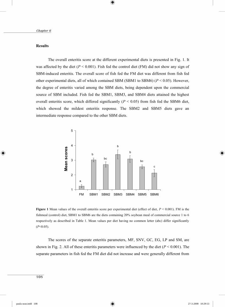
Chapter 6
108
Results
The overall enteritis score at the different experimental diets is presented in Fig. 1. It
was affected by the diet (P < 0.001). Fish fed the control diet (FM) did not show any sign of
SBM-induced enteritis. The overall score of fish fed the FM diet was different from fish fed
other experimental diets, all of which contained SBM (SBM1 to SBM6) (P < 0.05). However,
the degree of enteritis varied among the SBM diets, being dependent upon the commercial
source of SBM included. Fish fed the SBM1, SBM3, and SBM4 diets attained the highest
overall enteritis score, which differed significantly (P < 0.05) from fish fed the SBM6 diet,
which showed the mildest enteritis response. The SBM2 and SBM5 diets gave an
intermediate response compared to the other SBM diets.
1
2
3
4
5
FM SBM1 SBM2 SBM3 SBM4 SBM5 SBM6
Mea
n sc
ores
a
bbc
b
b
bc
c
Figure 1 Mean values of the overall enteritis score per experimental diet (effect of diet, P < 0.001). FM is the
fishmeal (control) diet, SBM1 to SBM6 are the diets containing 20% soybean meal of commercial source 1 to 6
respectively as described in Table 1. Mean values per diet having no common letter (abc) differ significantly
(P<0.05).
The scores of the separate enteritis parameters, MF, SNV, GC, EG, LP and SM, are
shown in Fig. 2. All of these enteritis parameters were influenced by the diet (P < 0.001). The
separate parameters in fish fed the FM diet did not increase and were generally different from
paula uran.indd 108paula uran.indd 108 27-3-2008 10:29:5327-3-2008 10:29:53

SBM commercial sources and enteritis in Atlantic salmon
109
B
1
2
3
4
5
FM SBM1 SBM2 SBM3 SBM4 SBM5 SBM6
SNV
scor
esb
ad
bcd
a
ac
bcd bcd
D
1
2
3
4
5
FM SBM1 SBM2 SBM3 SBM4 SBM5 SBM6
EG s
core
s
c
ab aba a
ab b
E
1
2
3
4
5
FM SBM1 SBM2 SBM3 SBM4 SBM5 SBM6
LP s
core
s
cd
aab
aab
ab
bc
A
1
2
3
4
5
FM SBM1 SBM2 SBM3 SBM4 SBM5 SBM6
MF
scor
es
b
acab
a aac
bc
C
1
2
3
4
5
FM SBM1 SBM2 SBM3 SBM4 SBM5 SBM6
GC
sco
res
c
a ab a a ab
b
F
1
2
3
4
5
FM SBM1 SBM2 SBM3 SBM4 SBM5 SBM6
SM s
core
s
e
bdbd
ababc
dcd
fish fed the SBM diets, except for MF, SNV and LP. Regarding the parameters MF, SNV and
LP, fish fed the FM diet did not significantly differ from fish fed the SBM diets, which gave a
mild enteritis response (especially the SBM6 diet).
Figure 2 Mean values of the enteritis parameters scored per experimental diet: A) mucosal folds (MF); B)
supranuclear vacuoles (SNV); C) goblet cells (GC); D) eosinophilic granulocytes (EG); E) lamina propria (LP);
F) sub-epithelial mucosa (SM); (diet effect, P < 0.001 for all parameters). FM is the fish meal (control) diet,
SBM1 to SBM6 are the diets containing 20% soybean meal of commercial source 1 to 6 as described in Table 1.
Mean values per diet having no common letter (abcde) differ significantly (P < 0.05).
paula uran.indd 109paula uran.indd 109 27-3-2008 10:29:5427-3-2008 10:29:54
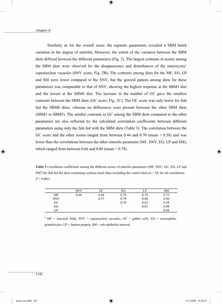
Chapter 6
110
Similarly as for the overall score, the separate parameters revealed a SBM batch
variation in the degree of enteritis. However, the extent of the variation between the SBM
diets differed between the different parameters (Fig. 2). The largest contrasts in scores among
the SBM diets were observed for the disappearance and disturbances of the enterocytes’
supranuclear vacuoles (SNV score; Fig. 2B). The contrasts among diets for the MF, EG, LP
and SM were lower compared to the SNV, but the general pattern among diets for these
parameters was comparable to that of SNV, showing the highest response at the SBM3 diet
and the lowest at the SBM6 diet. The increase in the number of GC gave the smallest
contrasts between the SBM diets (GC score, Fig. 2C). The GC score was only lower for fish
fed the SBM6 diets, whereas no differences were present between the other SBM diets
(SBM1 to SBM5). The smaller contrasts in GC among the SBM diets compared to the other
parameters are also reflected by the calculated correlation coefficients between different
parameters using only the fish fed with the SBM diets (Table 3). The correlation between the
GC score and the other scores ranged from between 0.44 and 0.70 (mean = 0.58) and was
lower than the correlations between the other enteritis parameter (MF, SNV, EG, LP and SM),
which ranged from between 0.66 and 0.88 (mean = 0.78).
Table 3 Correlation coefficients among the different scores of enteritis parameters (MF, SNV, GC, EG, LP and
SMa) for fish fed the diets containing soybean meal (thus excluding the control diet) (n = 54; for all correlations
P < 0.001)
SNV GC EG LP SM MF 0.66 0.44 0.70 0.79 0.73
SNV 0.57 0.79 0.80 0.80 GC 0.70 0.63 0.58 EG 0.81 0.88 LP 0.84
a MF = mucosal folds; SNV = supranuclear vacuoles; GC = goblet cells; EG = eosinophilic
granulocytes; LP = lamina propria; SM = sub-epithelial mucosa.
paula uran.indd 110paula uran.indd 110 27-3-2008 10:29:5627-3-2008 10:29:56

SBM commercial sources and enteritis in Atlantic salmon
111
Discussion and Conclusions
In this study, the inclusion of soybean meal (SBM) in the diet of Atlantic salmon
induced the classical signs of enteritis in the distal intestine, affecting the intestinal epithelium
at a structural and cellular level, as had been previously described in various studies (van den
Ingh et al. 1991, 1996; Baeverfjord & Krogdahl 1996; Krogdahl et al. 2000; Refstie et al.
2000; Buttle et al. 2001). In literature, several studies show that the type of soybean (SB)
product (e.g., full-fat SB, solvent-extracted SB, SB molasses), can result in differences in
enteritis response (Krogdahl et al. 2000; Refstie et al. 2000, 2001, 2005; Sanden et al. 2005;
Knudsen et al. 2007). The current study demonstrates that the severity of enteritis also varies
between different commercial sources (batches) of the same SB product, e.g., SBM. This
finding raises the question: what is the cause of this variation in enteritis’ responses between
the commercial sources of SBM? However, this is difficult to answer because the causative
agent has not yet been identified. In the current study, the differences in enteritis’ responses
between the experimental diets were not related to the crude protein or crude fibre content of
the SBM sources. The correlation coefficient between the mean enteritis score per diet and
crude protein content of the SBM batch was 0.001 (P = 0.997; n=6), and crude fibre content
was -0.562 (P = 0.246; n=6). Nonetheless, it should be made clear that the absence of
significant correlation might be due to the small number of tested SBM batches (n=6) in
combination with the small differences in composition between the SBM batches (Table 1).
The absence of a relation between the degree of enteritis with crude protein as well as crude
fibre is in line with the hypothesis that one or more alcohol-soluble components of SB might
induce an inflammatory response (Olli et al. 1995, van den Ingh et al. 1996, Krogdahl et al.
2000). The study of Knudsen et al. (2007) suggests that soyasaponins may be involved in
triggering the enteritis response.
In the current study, a semi-quantitative scoring system was used to assess the degree
of enteritis, which scores six separate indicative parameters of the inflammation response:
mucosal folds (MF); supranuclear vacuoles (SNV); goblet cells (GC); eosinophilic
granulocytes (EG); lamina propria (LM); and sub-epithelial mucosa (SM). The degree of
variation between the commercial SBM sources varied among the different parameters scored
(Fig. 2). The observation that the disappearance of SNV in enterocytes displayed the largest
paula uran.indd 111paula uran.indd 111 27-3-2008 10:29:5727-3-2008 10:29:57

Chapter 6
112
differences between SBM sources suggests that the appearance of SNV in enterocytes is the
most sensitive parameter to detect (small) differences in the causative agent(s) within SBM
sources. This is supported by the statement of Krogdahl et al. (2003), that the manifested
enteritis condition is characterized by the absence of SNV, being the first organelles affected.
The smaller variation in scoring of the other parameters might be an indication that the shifts
in these parameters occur during a more secondary stage of the enteritis cascade. GC are
known to be involved in the innate defence system through the production of mucus that gives
protection and which acts as a lubricant of the alimentary tract against chemical and
mechanical damage (Marchetti et al. 2006). The smallest differences between SBM sources
were visible in the abundance of GC in the MF. Only at one SBM batch (SBM6), which gave
the lowest enteritis response for all other parameters, the number of GC was lower compared
to the other SBM batches. These data suggest that GC respond more to a threshold value than
to a proportional dose response, while other enteritis parameters are more likely to respond
depending on dose.
The current observation between SBM source variation in the enteritis response has
both scientific and practical implications. When studies on SB-induced enteritis are
compared, it must be realized that differences among studies might be due to source (batch)
variation in the SB ingredients used. For a proper comparison of the impact of different SB
products (e.g., full-fat SB, solvent-extracted SBM, soy protein concentrates etc.), differences
in the origin of the SB might bias the contrasts displayed in the enteritis response.
Furthermore, the general applicability of results from studies on the dose response of the
dietary inclusion of SBM is hampered by the existence of inter-source (batch) variations. For
practical feed formulation, the observed presence of SBM batch variations necessitates extra
safety margins for the inclusion of SBM if no specific information is present on the type of
SBM batch. Obtaining indicative parameters (requiring additional research) for estimating the
potential of specific batches of SBM on the enteritis response might facilitate higher inclusion
levels of SBM in Atlantic salmon diets without compromising the health status of the fish.
paula uran.indd 112paula uran.indd 112 27-3-2008 10:29:5827-3-2008 10:29:58

SBM commercial sources and enteritis in Atlantic salmon
113
Acknowledgements
This research was supported by “Instituto Colombiano para el Desarrollo de la Ciencia y la
Tecnología” Colciencias and Skretting ARC, Stavanger, Norway. We would like to acknowledge the
staff at both the Skretting fish trails station and Skretting ARC for their technical assistance, for their
help during the feeding trial, the collection of the samples and for the lab analyses.
References
Baeverfjord, G. & Krogdahl, Å. (1996) Development and regression of soybean meal induced enteritis
in Atlantic salmon, Salmo salar L., distal intestine: a comparison with the intestines of fasted fish.
J. Fish Dis., 19, 375-387.
Bjerkeng, B., Refstie, S., Fjalestad, K.T. Storebakken, T., Rødbotten, M. & Roem, A.J. (1997) Quality
parameters of the flesh of Atlantic salmon (Salmo salar) as affected by dietary fat content and full-
fat soybean meal as a partial substitute for fish meal in the diet. Aquaculture, 157, 297-309.
Buttle, L.G., Burrells, A.C., Good, J.E., Williams, P.D., Southgate, P.J. & Burrells, C. (2001) The
binding of soybean agglutinin (SBA) to the intestinal epithelium of Atlantic salmon Salmo salar
and Rainbow trout, Oncorhynchus mykiss, fed high levels of soybean meal. Vet. Immunol.
Immunopathol., 80, 237-244.
Francis, G., Makkar, H. P.S. & Becker, K. (2001) Antinutritional factors present in plan-derived
alternate fish feed ingredients and their effects in fish. Review Article. Aquaculture, 199, 197-227.
Gatlin, D.M., Barrows, F.T., Brown, P., Dabrowski, K., Gaylord, T.G., Hardy, R.W., Herman, E., Hu,
G.S., Krogdahl, Å., Nelson, R., Overturf, K., Rust, M., Sealey, W., Skonberg, D., Souza, E.J.,
Stone, D., Wilson, R. & Wurtele, E. (2007) Expanding the utilization of sustainable plant products
in aquafeeds: a review. Aquac. Res., 38, 551-579.
Grisdale-Helland, B., Helland, S.J. Baeverfjord, G. & Berge, G.M. (2002) Full-fat soybean meal in
diets for Atlantic halibut: growth, metabolism and intestinal histology. Aquacult. Nutr., 8, 265-
270.
van den Ingh, T.S.G.A.M., Krogdahl, Å., Olli, J.J., Hendriks, H.G.C.J.M. & Koninkx, J.G.J.F. (1991)
Effects of soybean-containing diets on the proximal and distal intestine in Atlantic salmon (Salmo
salar): a morphological study. Aquaculture, 94, 297-305.
van den Ingh, T.S.G.A.M., Olli, J.J. & Krogdahl, Å. (1996) Alcohol-soluble components in soybeans
cause morphological changes in the distal intestine of Atlantic salmon, Salmo salar L. J. Fish Dis.,
19, 47-53.
paula uran.indd 113paula uran.indd 113 27-3-2008 10:30:0027-3-2008 10:30:00
Chapter 6
114
Jiang, Z. (2001) Ingredient variation: its impact and management. In: van der Poel, A.F.B., Váhl, J.L.
& Kwakkel, R.P. (Eds.), Advances in Nutritional Technology: Proceedings of the 1st World Feed
conference Utrecht Wageningen Press, Wageningen, The Netherlands. pp. 47-56.
Knudsen, D., Urán, P., Arnous, A., Koppe, W. & Frøkiær, H. (2007) Saponin-containing subfractions
of soybean molasses induce enteritis in the distal intestine of Atlantic salmon. J. Agric. Food
Chem., 55, 2261-2267.
Krogdahl, Å., Bakke-McKellep, A.M., Røed, K.H. & Baeverfjord, G. (2000) Feeding Atlantic salmon
Salmo salar L. soybean products: effects on disease resistance (furunculosis), and lysozyme and
IgM levels in the intestinal mucosa. Aquacult. Nutr., 6, 77-84.
Krogdahl, Å., Bakke-McKellep, A.M. & Baeverfjord, G. (2003) Effects of graded levels of standard
soybean meal on intestinal structure, mucosal enzyme activities, and pancreatic response in
Atlantic salmon (Salmo salar L.). Aquacult. Nutr., 9, 361-371.
Marchetti, L., Capacchietti, M., Sabbieti, M.G., Accili, D., Materazzi, G. & Menghi, G. (2006)
Histology and carbohydrate histochemistry of the alimentary canal in the rainbow trout
Oncorhynchus mykiss. J. Fish Biol., 68, 1808-1821.
Olli, J.J. & Krogdahl, Å. (1995) Alcohol soluble components of soybeans seem to reduce fat
digestibility in fishmeal-based diets for Atlantic salmon, Salmo salar L. Aquac. Res., 26, 831-835.
Olli, J.J., Krogdahl, Å. & Våbenø, A. (1995) Dehulled solvent-extracted soybean meal as a protein
source in diets for Atlantic salmon, Salmo salar L. Aquac. Res., 26,167-174.
Refstie, S., Korsøen, Ø. J., Storebakken, T., Baeverfjord, G., Lein, I. & Roem, A.J. (2000) Differing
nutritional responses to dietary soybean meal in rainbow trout (Oncorhynchus mykiss) and
Atlantic salmon (Salmo salar). Aquaculture, 190, 49-63.
Refstie, S., Strorebakken, T., Baeverfjord G. & Roem A.J. (2001) Long-term protein and lipid growth
of Atlantic salmon (Salmon salar) fed diets with partial replacement of fishmeal by soy protein
products at medium or high lipid level. Aquaculture, 193, 91-106.
Refstie, S., Sahlstrom, S., Brathen, E., Baeverfjord, G. & Krogedal, P. (2005) Lactic acid fermentation
eliminates indigestible carbohydrates and antinutritional factors in soybean meal for Atlantic
salmon (Salmo salar). Aquaculture, 246, 331-345.
Sanden, M., Berntssen, M.H.G., Krogdahl, Å., Hemre, G.I. & Bakke-McKellep, A.M. (2005) An
examination of the intestinal tract of Atlantic salmon, Salmo salar L., parr fed different varieties
of soy and maize. J. Fish Dis., 28, 317-330.
SAS (1999) SAS/STAT® User’s guide, V8. SAS institute Inc., Cary, NC, USA.
paula uran.indd 114paula uran.indd 114 27-3-2008 10:30:0127-3-2008 10:30:01
SBM commercial sources and enteritis in Atlantic salmon
115
Tacon, A.J. (2003) Aquaculture production trends analysis. FAO fisheries Circular. No. 886, Rev. 2.
Rome, 95 pp.
Urán, P.A. (2008) Etiology of soybean-induced enteritis in fish. PhD thesis dissertation, Animal
Sciences group, Wageningen University, The Netherlands.
Urán, P.A., Schrama, J. W., Rombout, J.H.W.M., Obach, A., Jensen, L., Koppe, W., Verreth, J.A.J.
(2008) Soybean meal-induced enteritis in Atlantic salmon (Salmo salar L.) at different
temperatures. Aquacult. Nutr. in press.
paula uran.indd 115paula uran.indd 115 27-3-2008 10:30:0227-3-2008 10:30:02

chapter hoofdstukken.indd 13 26-3-2008 19:49:43
Chapter 7
Saponin-containing subfractions of soybean molasses induce enteritis in the distal intestine
of Atlantic salmon
D. Knudsen1,4, P.A. Urán2, A. Arnous3, W. Koppe4 & H. Frøkiær1
1BioCentrum-DTU, Nutritional Immunology Group, Center of Biological Sequence Analysis, Technical University of Denmark; 2Wageningen Institute of Animal Sciences, Wageningen University, The Netherlands; 3BioProcess Engineering, Dept. of Chemical Engineering, Technical University of Denmark, 4Skretting Aquaculture Research Centre, Stavanger, Norway
Published in:Journal of Agricultural and Food Chemistry. 2007, 55, 2261-2267
© American Chemical Society (2007), reproduced with permission
chapter hoofdstukken.indd 14 26-3-2008 19:49:44
Chapter 7
118
Abstract
The current work aimed at tracing the causative components for soybean-induced
enteritis in Atlantic salmon (Salmo salar L.). Soybean molasses was subjected to phase-
separation using n-Butanol. Three sub-fractions were obtained: butanol phase, precipitate and
water phase. The biochemical composition of soybean molasses and the obtained sub-
fractions were analyzed in detail: Protein, fat and ash were quantified according to standard
methods. Sucrose, raffinose and stachyose were quantified using high-performance anion-
exchange chromatography. Soyasaponins were quantified using reverse phase high-
performance liquid chromatography. Finally, sodium dodecyl sulfate polyacrylamide gel
electrophoresis was used to evaluate the size distribution of the proteins present in each
fraction. Molasses and the different sub-fractions were thereafter fed to Atlantic salmon in
two successive fish trials. The level of intestinal inflammation was evaluated by light
microscopy using a semi-quantitative scoring system. Histological assessments revealed that
Atlantic salmon fed a combination of butanol phase and precipitate displayed significant
enteritis. Atlantic salmon fed the water phase displayed normal intestinal morphology.
Conclusions: The causative components for soybean-induced enteritis withstand butanol
treatment and prolonged heating at 70 C. Sucrose, raffinose, stachyose, nor soybean proteins
larger than 10 kDa induce enteritis alone. Soyasaponins, or components that follow the same
solubility pattern, trigger the inflammatory reaction. We therefore suggest that soybean-
induced enteritis in Atlantic salmon is induced by soyasaponins alone, or by soyasaponins in
combination with other factors e.g. antigenic soybean proteins or the intestinal microflora.
paula uran.indd 118paula uran.indd 118 27-3-2008 10:30:0527-3-2008 10:30:05
Saponing-containing subfractions of soybean molasses induce enteritis
119
Introduction
A limited supply of fishmeal could hamper future growth in the aquaculture industry
and much effort has therefore been made to find alternative protein sources that could replace
fishmeal in feed formulations for carnivorous fish (Tacon 2003; FAO 2002; Naylor et al.
2000; Francis et al. 2001). From an ecological viewpoint an ideal solution would be to find a
suitable low-cost plant-derived protein. Soybean meal (SBM) is one of the promising
candidates due to its high protein content and steady supply. Several studies have revealed,
however, that high inclusion levels of low-processed soybean (SB) products induce intestinal
inflammation in the hindgut of Atlantic salmon (van den Ingh et al. 1991,1996; Baeverfjord &
Krogdahl 1996; Bakke-McKellep et al. 2000). The inflammatory reaction is associated with
several morphological changes, including: loss of supranuclear vacuoles in the absorptive
enterocytes, widening of the lamina propria of mucosal folds, increased amounts of
connective tissue between the base of the mucosal folds and stratum compactum, shortening
of mucosal fold height, and infiltration of inflammatory cells in the lamina propria (van den
Ingh et al. 1991, 1996; Baeverfjord & Krogdahl 1996). The normal morphology of the distal
intestine in Atlantic salmon can be seen in figure 1A while figure 1B displays typical
soybean-induced enteritis. The enteritis associated with soybean meal currently limits its use
in diets for Atlantic salmon.
Despite considerable work, the causative components for the condition remain
unidentified. Important clues can, however, be found as to their identity. Van de Ingh et al.
(1991, 1996) demonstrated that they follow the protein fraction when the oil is extracted by
hexane.
Alcohol extracted SB protein concentrate did not, however, induce inflammation. In
addition, it was found that fish fed soybean molasses (the by-product of alcohol extraction)
displayed the same signs of inflammation as fish fed SBM. It can thus be concluded that the
causative components are soluble in aqueous alcohol and resist alcohol treatment at elevated
temperatures. SB molasses is a brown liquid composed of 60% dry matter, 5% protein, 5%
lipids, 5% ash and 45% nitrogen free extracts. Sucrose, raffinose and stachyose constitute for
approximately 35% of molasses. The remaining 10% of the nitrogen free extracts includes
other sugars, isoflavones and saponins (supplier data, Solae Europe, S.A., Switzerland).
paula uran.indd 119paula uran.indd 119 27-3-2008 10:30:0627-3-2008 10:30:06
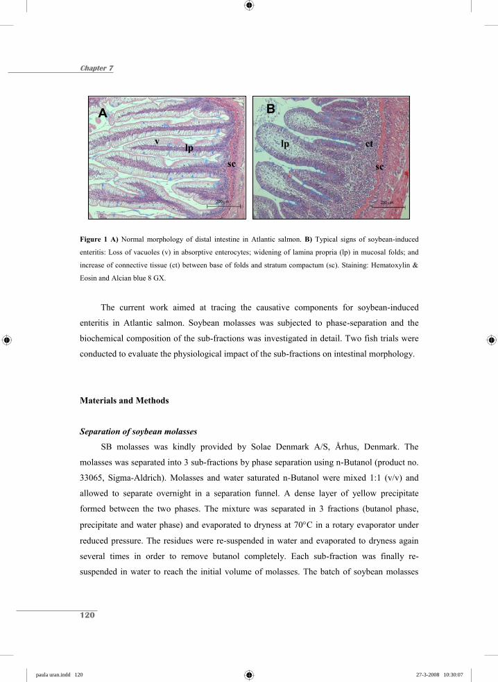
Chapter 7
120
Figure 1 A) Normal morphology of distal intestine in Atlantic salmon. B) Typical signs of soybean-induced
enteritis: Loss of vacuoles (v) in absorptive enterocytes; widening of lamina propria (lp) in mucosal folds; and
increase of connective tissue (ct) between base of folds and stratum compactum (sc). Staining: Hematoxylin &
Eosin and Alcian blue 8 GX.
The current work aimed at tracing the causative components for soybean-induced
enteritis in Atlantic salmon. Soybean molasses was subjected to phase-separation and the
biochemical composition of the sub-fractions was investigated in detail. Two fish trials were
conducted to evaluate the physiological impact of the sub-fractions on intestinal morphology.
Materials and Methods
Separation of soybean molasses
SB molasses was kindly provided by Solae Denmark A/S, Århus, Denmark. The
molasses was separated into 3 sub-fractions by phase separation using n-Butanol (product no.
33065, Sigma-Aldrich). Molasses and water saturated n-Butanol were mixed 1:1 (v/v) and
allowed to separate overnight in a separation funnel. A dense layer of yellow precipitate
formed between the two phases. The mixture was separated in 3 fractions (butanol phase,
precipitate and water phase) and evaporated to dryness at 70 C in a rotary evaporator under
reduced pressure. The residues were re-suspended in water and evaporated to dryness again
several times in order to remove butanol completely. Each sub-fraction was finally re-
suspended in water to reach the initial volume of molasses. The batch of soybean molasses
A
v lp sc
B
sc
lp ct
paula uran.indd 120paula uran.indd 120 27-3-2008 10:30:0727-3-2008 10:30:07

Saponing-containing subfractions of soybean molasses induce enteritis
121
used in the present work contained 62% dry matter (w/w). Preliminary analyses of the
obtained fractions revealed that 15% of the total dry matter was recovered in the butanol
phase, 35% in the precipitate and 50% in the water phase. Molasses and the obtained sub-
fractions were analyzed for protein, fat, ash, sucrose, raffinose, stachyose and soyasaponins.
Sodium Dodecyl Sulfate PolyAcrylamide Gel Electrophoresis (SDS-PAGE) was used to
evaluate the size distribution of the proteins present within each fraction.
Chemical analysis of diets
Dry matter measurements were done by drying to constant weight at 102-105°C. Ash
was measured by burning samples at 550 C for 16-18 hours. Crude protein was quantified as
N x 6.25 using a Kjeltec Auto Sampler-System (Tecator AB, Sweden) according to Nordic
Committee on Food Analysis, Method No. 6, 4th edition 2003. Total fat was measured by
acid hydrolysis using a Soxtec 2050 extraction system (Foss Analytical, Denmark) according
to Nordic Committee on Food Analysis, Method No. 160, 1998.
Quantification of oligosaccharides
Quantification was done using high-performance anion-exchange chromatography with
pulsed amperometric detection (HPAEC-PAD). Standards of sucrose, raffinose and stachyose
were purchased from Sigma-Aldrich, Inc., MO, USA (product # S1174, R0250 and S4001,
respectively). Separation was done on a Dionex HPAEC-PAD system using a Dionex
CarboPac PA-1 column. The injection volume was 10 l and the compounds were eluted
isocratically with 200 mM NaOH for 15 minutes at a flow rate of 1.0 mL/min. The
oligosaccharides were identified by comparing their retention times to the authentic standards.
Quantification of the oligosaccharide was accomplished by reference to standard curves made
for each of the three oligosaccharides. The molasses fractions were diluted 1:3000 in distilled
H2O, centrifuged at 14000g for 5 minutes, and filtered through a 0.22 m GHP membrane
filter prior to injection into the HPAEC-PAD system.
Quantification of soyasaponins
Separation and quantification of soyasaponins was performed using reverse phase high-
performance liquid chromatography with diode array detection (HPLC-DAD) as described
paula uran.indd 121paula uran.indd 121 27-3-2008 10:30:0927-3-2008 10:30:09

Chapter 7
122
previously (Knudsen et al. 2006). Briefly, the separation was achieved using a Hewlett-
Packard series 1050 HPLC-DAD system with a 250 mm x 4.6 mm i.d., 5 m, Supercosil ABZ
+Plus, C18 reverse phase column (SUPELCO). The mobile phases were 0.05% trifluroacetic
acid in water (solvent A) and 0.05% trifluroacetic acid in acetonitrile (solvent B). The
gradient elution was linear from 25 to 50% B, 0-65 min; linear from 50 to 60% B, 65-70 min;
linear from 60-100% B, 70-75 min; isocratic at 100% B, 75-85 min; then linear from 100-
25% B, 85-90 min and finally isocratic at 25% B, 90-100 min. The flow rate was 0.5 ml/min,
the injection volume was 50 l and the column temperature was 30 C. Identification of SB
saponins was confirmed by HPLC retention time, UV absorption spectra recorded at 200-350
nm and LC-MS using positive electrospray ionization. Molasses and molasses sub-fractions
were diluted 1:10 in 70% aqueous ethanol and centrifuged at 15000g for 5 minutes before
injection on the HPLC system. The following soyasaponins were detected and quantified; Ab,
Ac, Af, Ba, Bb, Bc, Ba-DDMP, Bb-DDMP and Bc-DDMP (see Knudsen et al. 2006 for
molecular structure).
SDS-PAGE
The size distribution of the proteins present in the different molasses fractions was
evaluated by SDS-PAGE according to (Schägger & von Jagow, 1987). The electrophoresis
was done using 10-20% tricin gradient gels (Novex, Invitrogen, Groningen, The Netherlands).
The re-suspended molasses fractions were diluted 1:10 in H2O and mixed 1:1 with sample
buffer (0.1 M Tris buffer, 8% (w/v) SDS, 24% (v/v) glycerol, 0.025% (w/v) Coomassie blue,
0.04 M 1,4-Dithiothreitol, pH 6.8). The mixtures were boiled for 5 minutes before they were
loaded on the gel (10 L per well). A standard protein mixture was included on the gel for
molecular weight estimation (Mark12TM, Product no. LC5677, Noves, Invitrogen, the
Netherlands). Electrophoresis was carried out for 1 h and 20 minutes at 125V (constant).
Finally, the gel was stained with Coomassie blue.
Production of feed for Fish Trial 1
Six diets were produced as 4 mm pellets by twin-screw extrusion cooking (TX57,
Wenger Manufacturing, Inc., United States) at Skretting Feed Technology Plant, Stavanger,
Norway. Molasses and molasses fractions were mixed with the other ingredients before
paula uran.indd 122paula uran.indd 122 27-3-2008 10:30:1127-3-2008 10:30:11

Saponing-containing subfractions of soybean molasses induce enteritis
123
extrusion. The molasses batch contained 620 g dry matter per kg and 15% of this dry matter
was recovered in the butanol phase, 35% in the precipitate and 50% in the water phase. By
knowing these ratios it was possible to calculate how much soybean molasses the added
amounts of sub-fractions corresponded to. The different sub-fractions were included at a level
that corresponded to approximately 10% (w/w, wet basis) soybean molasses. The recipes are
shown in Table 1.
Table 1 Formulation of diets for Fish Trial 1
Diet A B C D E F Fish meal (Scandinavian LT) g/kg 490 625 625 625 625 625 Wheat g/kg 108 120 120 120 120 120 Wheat Starch g/kg 0 53 9 0 0 62 Minerals, vitamins, pigment g/kg 3 3 3 3 3 3 Defatted soybean meal a g/kg 200 0 0 0 0 0 Butanol phase b g/kg 0 9 0 0 0 0 Precipitate & Water phase b g/kg 0 0 53 0 0 0 Butanol treated molasses b g/kg 0 0 0 62 0 0 Untreated molasses b g/kg 0 0 0 0 62 0 Fish Oil c g/kg 199 190 190 190 190 190 Total g/kg 1000 1000 1000 1000 1000 1000 Molasses equivalents d 10% 10% 10% 10% a Denofa, Norway b Dry matter c Northern Hemisphere d Wet matter basis
Production of feed for Fish Trial 2
Dry pellets (4 mm) were produced by twin-screw extrusion cooking (TX57, Wenger) at
Skretting Feed Technology Plant, Stavanger, Norway. The pellets had the following
composition; 80% fishmeal, 15.5% wheat, 4% wheat starch and 0.5% premix (minerals,
vitamins and pigment). These dry pellets were used as a carrier matrix to test the impact of all
different combinations of sub-fractions. Instead of adding the molasses fractions before
extrusion the molasses fractions were coated on the dry pellets using a specially designed lab-
scale vacuum coater. A known amount of dry molasses sub-fraction was re-suspended in
water and sprayed onto the dry pellets under reduced pressure. The coated pellets were then
paula uran.indd 123paula uran.indd 123 27-3-2008 10:30:1227-3-2008 10:30:12

Chapter 7
124
dried at 102 C to achieve a moisture content of 10%. Using the same lab-scale coater, pellets
were thereafter coated with sufficient fish oil to ensure they would sink. The different sub-
fractions were again included at a level that corresponded to approximately 10% (w/w, wet
basis) soybean molasses. The quantity of oil required to ensure pellets would sink differed
between diets. Consequently, the control diet (not containing any molasses), and the diet
containing the butanol phase only were relatively high in fish oil content compared to the
other diets (Table 2). In summary, all diets contained the same carrier matrix but were coated
with different sub-fractions of molasses and quantities of oil. Recipes for all diets are shown
in Table 2.
Table 2 Formulation of diets for Fish Trial 2 a
Diet
A B C D E F G H Fish meal (Scandinavian LT) g/kg 574 591 570 587 574 581 583 540 Wheat g/kg 111 114 110 113 111 112 113 104
Wheat Starch g/kg 29 29 28 29 28 29 29 27
Minerals, vitamins, pigment g/kg 3 3 3 3 3 3 3 3
Butanol phase (DM) g/kg 10 0 0 0 0 0 0 0
Precipitate (DM) g/kg 0 23 0 0 0 0 0 0
Water phase (DM) g/kg 0 0 31 0 0 0 0 0
Butanol phase & Precipitate (DM) g/kg 0 0 0 41 0 0 0 0
Butanol phase & Water phase (DM) g/kg 0 0 0 0 44 0 0 0
Precipitate & Water phase (DM) g/kg 0 0 0 0 0 57 0 0
Untreated molasses (DM) g/kg 0 0 0 0 0 0 66 0
Fish Oil (Northern Hemisphere) g/kg 273 240 258 227 240 218 213 326
Total g/kg 1000 1000 1000 1000 1000 1000 1000 1000
Molasses equivalents (wet matter basis): 11.0% 10.7% 10.1% 13.1% 10.9% 10.8% 10.6% - a DM, dry matter. All diets contained the same carrier matrix but were coated with different sub-fractions of
molasses and different quantities of fish oil
paula uran.indd 124paula uran.indd 124 27-3-2008 10:30:1327-3-2008 10:30:13
Saponing-containing subfractions of soybean molasses induce enteritis
125
Fish Trial 1
The trial was conducted at Skretting Fish Trials Station, Lerang, Jørpeland, Norway.
Seawater adapted Atlantic salmon (Salmo salar L.) with an initial average weight of 213 g,
were fed 6 different experimental diets for 62 days. Fish were randomly distributed to 18
circular 400 L fiberglass tanks at a stocking density of 30 fish per tank. The tanks were
equipped with waste feed collection and continuously supplied with seawater (15 L min-1 per
tank). Water was pumped from 90 m depth and held a constant temperature of 8.3 C during
the experiment. The 6 different diets were fed to triplicate tanks (3 tanks per treatment) twice
a day, aiming at 20% overfeeding, and waste feed was collected. At the end of the feeding
period 4 fish from each tank (12 fish in total per treatment) were sacrificed with an overdose
of anesthetic (Tricaine Methanesulfonate, Finquel MS-222, Argent Chemical Laboratories,
USA), for histological examination.
Fish Trial 2
The trial was conducted at Skretting Fish Trials Station, Lerang, Jørpeland, Norway.
Seawater adapted Atlantic salmon (Salmo salar L.) with an initial average weight of 202 g,
were fed 8 different experimental diets for 44 days. The fish were randomly distributed to 8
circular 100 L fiberglass tanks at a stocking density of 20 fish per tank. The tanks were
equipped with waste feed collection and continuously supplied with seawater (4 L min-1 per
tank). Water was pumped from 90 m depth and held a constant temperature of 9.0 C during
the experiment. The 8 different diets were fed to the 8 different tanks (1 tank per treatment)
twice a day, aiming at 20% overfeeding, and waste feed was collected. At the end of the
feeding period 10 fish from each tank were sacrificed with an overdose of anesthetic (Tricaine
Methanesulfonate, Finquel MS-222, Argent Chemical Laboratories, USA), for histological
examination.
Histological examination
A two-centimeter section of the distal intestine was carefully removed, rinsed in saline
water and fixed in phosphate buffered formaldehyde (4%, pH 7.2). Samples were then
dehydrated, embedded in paraffin and cut according to standard histological procedures.
paula uran.indd 125paula uran.indd 125 27-3-2008 10:30:1527-3-2008 10:30:15

Chapter 7
126
Slides were then stained with a combination of Hematoxylin & Eosin and Alcian blue 8 GX.
The latter was included in order to increase the contrast between goblet cells and vacuoles.
Four different morphological parameters were evaluated using light microscopy (Leica
DM 5000B) according to the scoring criteria given in Table 3. A score of “1-2” represented
normal morphology while a score of “5” was given to morphological symptoms of severe
enteritis. The semi-quantitative scoring system was adapted from Urán et al. (2004).
Histological samples were randomized and evaluated blind.
Table 3 Histological scoring system for morphological changes induced by soybeans in the distal intestine
of Atlantic salmon a.
Score Appearance
Supranuclear vacuoles 1 Large vacuoles occupy almost the entire apical part of the enterocytes. 2 Medium sized vacuoles, which occupy less than half of the enterocytes. 3 Small sized vacuoles near the apical membrane in most enterocytes. 4 Scattered small vacuoles are still present in some enterocytes. 5 No supranuclear vacuoles are present. Lamina propria of simple folds
1 Very thin and delicate core of connective tissue in all simple folds. 2 Lamina propria appears slightly more distinct and robust in some of the folds. 3 Clear increase of lamina propria in most of the simple folds. 4 Thick lamina propria in many folds. 5 Very thick lamina propria in many folds. Connective tissue (between base of folds and stratum compactum)
1 Very thin layer of connective tissue between base of folds and stratum compactum 2 Slightly increased amount of connective tissue beneath some of the mucosal folds. 3 Clear increase of connective tissue beneath most of the mucosal folds. 4 Thick layer of connective tissue beneath many folds. 5 Extreme thick layer of connective tissue beneath some folds. Mucosal folds
1 Simple and complex folds (CF) appear long and thin. Thin side-branches on CF. 2 Simple mucosal folds have medium length. CF are still long but appear thicker. 3 Simple folds have short to medium length. Side-branches on CF are stubby. 4 Thick CF are prevalent. Simple folds are short. Almost no side-branches on CF. 5 Both complex and simple folds appear very stubby.
a Adapted from Urán et al. 2004.
paula uran.indd 126paula uran.indd 126 27-3-2008 10:30:1627-3-2008 10:30:16
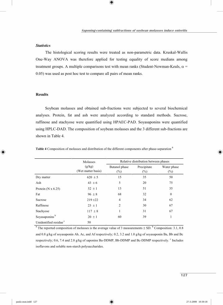
Saponing-containing subfractions of soybean molasses induce enteritis
127
Statistics
The histological scoring results were treated as non-parametric data. Kruskal-Wallis
One-Way ANOVA was therefore applied for testing equality of score medians among
treatment groups. A multiple comparisons test with mean ranks (Student-Newman-Keuls, =
0.05) was used as post hoc test to compare all pairs of mean ranks.
Results
Soybean molasses and obtained sub-fractions were subjected to several biochemical
analyses. Protein, fat and ash were analyzed according to standard methods. Sucrose,
raffinose and stachyose were quantified using HPAEC-PAD. Soyasaponins were quantified
using HPLC-DAD. The composition of soybean molasses and the 3 different sub-fractions are
shown in Table 4.
Table 4 Composition of molasses and distribution of the different components after phase-separation a
Molasses (g/kg)
(Wet matter basis)
Relative distribution between phases
Butanol phase
(%) Precipitate
(%) Water phase
(%)
Dry matter 620 5 15 35 50
Ash 43 6 5 20 75
Protein (N x 6.25) 52 1 13 51 35
Fat 96 8 68 32 0
Sucrose 219 22 4 34 62
Raffinose 23 1 2 30 67
Stachyose 117 8 1 31 67
Soyasaponins b 20 1 60 39 1
Unidentified residue c 50 a The reported composition of molasses is the average value of 3 measurements SD. b Composition: 3.1, 0.8
and 0.8 g/kg of soyasaponin Ab, Ac, and Af respectively; 0.2, 3.2 and 1.0 g/kg of soyasaponin Ba, Bb and Bc
respectively; 0.6, 7.4 and 2.8 g/kg of saponins Ba-DDMP, Bb-DDMP and Bc-DDMP respectively. c Includes
isoflavons and soluble non-starch polysaccharides.
paula uran.indd 127paula uran.indd 127 27-3-2008 10:30:1827-3-2008 10:30:18
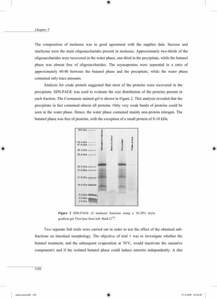
Chapter 7
128
The composition of molasses was in good agreement with the supplier data. Sucrose and
stachyose were the main oligosaccharides present in molasses. Approximately two-thirds of the
oligosaccharides were recovered in the water phase, one-third in the precipitate, while the butanol
phase was almost free of oligosaccharides. The soyasaponins were separated in a ratio of
approximately 60:40 between the butanol phase and the precipitate, while the water phase
contained only trace amounts.
Analysis for crude protein suggested that most of the proteins were recovered in the
precipitate. SDS-PAGE was used to evaluate the size distribution of the proteins present in
each fraction. The Coomassie stained gel is shown in Figure 2. This analysis revealed that the
precipitate in fact contained almost all proteins. Only very weak bands of proteins could be
seen in the water phase. Hence, the water phase contained mainly non-protein nitrogen. The
butanol phase was free of proteins, with the exception of a small protein of 8-10 kDa.
Figure 2 SDS-PAGE of molasses fractions using a 10-20% tricin
gradient gel. First lane from left, Mark12TM.
Two separate fish trials were carried out in order to test the effect of the obtained sub-
fractions on intestinal morphology. The objective of trial 1 was to investigate whether the
butanol treatment, and the subsequent evaporation at 70 C, would inactivate the causative
component/s and if the isolated butanol phase could induce enteritis independently. A diet
paula uran.indd 128paula uran.indd 128 27-3-2008 10:30:2027-3-2008 10:30:20

Saponing-containing subfractions of soybean molasses induce enteritis
129
containing regular defatted soybean meal was included for the purposes of comparison.
Results from the histological evaluation are given in Table 5. Fish fed the control diet
(without soybeans) displayed normal morphology while significant enteritis was observed in
fish fed 20% defatted soybean meal. Diets containing 10% untreated molasses and 10%
butanol treated molasses induced severe morphological changes similar to the 20% soybean
meal diet. The diet containing a combination of precipitate and water phase had only a weak
effect on all 4 morphological parameters. Interestingly, the butanol-phase containing diet had
a strong impact on vacuoles, lamina propria and connective tissue but did not provoke the
stubby appearance of mucosal folds typically associated with soybean-induced enteritis.
Table 5 Histological evaluation of distal intestine a
Fish Trial 1 Diet A:
Soybean meal Diet B: Butanol phase
Diet C: Precipitate & Water phase
Diet D: Butanol treated
molasses
Diet E: Untreated molasses
Diet F: Control
Vacuoles 2.92 0.90 bc 3.96 1.01 c 2.46 1.23 ab 3.63 0.61 bc 3.08 0.60 bc 1.50 0.60 a
Lamina Propria 3.96 0.86 c 3.04 0.84 bc 2.33 1.21 ab 3.75 0.78 c 3.63 0.57 c 1.29 0.69 a
Connective tissue 3.71 0.92 b 3.46 0.99 b 2.17 0.94 a 3.92 0.82 b 3.96 0.86 b 2.00 1.07 a
Mucosal Folds 4.13 0.68 b 2.33 0.49 a 2.42 1.06 a 3.88 0.68 b 3.71 0.69 b 1.42 0.47 a
a Intestinal cuts were scored according to the criteria listed in Table 3. A score of “1-2” represent normal
morphology while a score of “5” represent severe enteritis. Reported data are mean values from 12 fish SD.
Means followed by different letters are significantly different (Multiple comparisons test with mean ranks,
Student-Newman-Keuls, = 0.05).
Trial 2 was initiated to investigate why neither of the two sub-fractions tested in trial 1
had a strong effect on mucosal folds. Results from the histological evaluation are shown in
Table 6. Fish fed the water phase displayed normal morphology while fish fed the
combination of butanol-phase and precipitate showed the same morphological changes as fish
fed soybean molasses. Results from trial 2 were very consistent with those from trial 1,
demonstrating clearly that the trigger component was split between the butanol phase and the
precipitate whereas this component was absent from the water phase. Soyasaponins were the
only quantified components that were separated poorly between the butanol phase and
paula uran.indd 129paula uran.indd 129 27-3-2008 10:30:2127-3-2008 10:30:21

Chapter 7
130
precipitate (Table 4). Fish fed the butanol phase alone showed the same morphological
changes as observed in trial 1; vacuoles, lamina propria and connective tissue were
significantly affected but only a weak impact was observed on mucosal folds. Interestingly,
the combination of water and butanol phases had a significantly greater impact on mucosal
folds than the butanol phase alone.
Table 6 Histological evaluation of distal intestinea
Fish Trial 2
Diet A: Butanol phase
Diet B: Precipitate
Diet C: Water phase
Diet D: Butanol phase &
Precipitate
Diet E: Butanol & Water
phase
Diet F: Precipitate & Water
phase
Diet G: Untreated molasses
Diet H: Control
Vacuoles 4.20
0.86 cd 2.75
1.21 abc 1.70
0.59 a 4.40
0.94 d 3.60
0.70 bcd 2.05
1.04 ab 4.35
0.58 d 2.05
0.50 ab
Lamina Propria 3.15
0.47 b 3.00
0.82 b 1.90
0.74 ab 3.15
0.75 b 2.85
0.75 b 1.80
0.82 ab 3.10
1.13 b 1.00
0.00 a
Connective tissue
2.55
0.55 ab 2.30
0.75 ab 1.80
0.67 ab 2.95
0.93 b 2.50
0.58 ab 1.75
0.75 a 3.05
1.19 b 1.65
0.47 a
Mucosal Folds 2.25
0.54 abc 2.00
0.53 ab 1.50
0.33 a 3.50
0.91 cd 3.15
0.53 bcd 2.35
1.00 abc 3.75
0.75 d 1.25
0.26 a
a Intestinal cuts were scored according to the criteria listed in Table 3. A score of “1-2” represent normal
morphology while a score of “5” represent severe enteritis. Reported data are mean values from 10 fish SD.
Means followed by different letters are significantly different (Multiple comparisons test with mean ranks,
Student-Newman-Keuls, = 0.05).
Discussion
SB molasses has previously been shown to contain components that cause SBM-
induced enteritis in the distal intestine of Atlantic salmon (van den Ingh et al. 1996, Krogdahl
et al. 2000). In the present study soybean molasses were separated into three sub-fractions by
phase separation and both molasses and the obtained sub-fractions were subjected to
extensive biochemical analyses. All possible combinations of the three sub-fractions were fed
to Atlantic salmon and the impact on intestinal morphology was evaluated. Results revealed
that fish fed the water phase displayed normal morphology while fish fed a combination of
butanol-phase and precipitate showed the same morphological changes as fish fed soybean
paula uran.indd 130paula uran.indd 130 27-3-2008 10:30:2227-3-2008 10:30:22
Saponing-containing subfractions of soybean molasses induce enteritis
131
molasses. Soyasaponins were the only quantified components that were separated poorly
between the butanol phase and precipitate. It can thus be concluded that soyasaponins, or
components that follow the same solubility pattern, need to be present to induce the
inflammatory reaction. Bureau et al. (1998) demonstrated that Quillaja saponins cause
extensive damage to the intestinal mucosa of the hindgut in Chinook salmon and Rainbow
trout. This supports the hypothesis that soyasaponins play a key role in soybean-induced
enteritis in Atlantic salmon.
Biochemical analyses of molasses revealed that the main oligosaccharides present were
sucrose and stachyose, which is in accordance with Berg (1992). The separation of
soyasaponins was surprisingly poor and only 60% of the total amount was found in the
butanol phase, even though n-Butanol is a suitable solvent for extraction of soyasaponins
(Kitagawa et al. 1985). The remaining 40% was found in the precipitate while the water phase
contained only trace amounts of soyasaponins. It is known that soyasaponins follow the
protein fraction during production of SB protein isolates (Ireland et al. 1986). The high
amount of retained saponins in the precipitate might therefore be due to protein-saponin
interactions.
The histological evaluation in fish trial 1 confirmed that soybean molasses contains the
causative factors for soybean-induced enteritis. Moreover, the components proved to be
extremely stable since they were able to withstand both butanol treatment and evaporation to
dryness at 70°C. The butanol phase contained components that effectively disrupted
vacuolization, probably by interfering with endocytosis. The butanol phase also caused an
intermediate increase of lamina propria and connective tissue. The mean score for mucosal
folds, however, was only slightly affected. The fact that the butanol phase affected some, but
not all, of the evaluated parameters could indicate that the causative factor was poorly
separated between the two sub-fractions (1: butanol phase and 2: precipitate & water phase).
Fish trial 2 revealed that the causative component was split between butanol phase and
precipitate while the water phase was free from this component. The diet that included both
precipitate and water phase had only a weak effect on all four histological parameters in both
trials. The precipitate and water phase in combination contained more than 95% of all
sucrose, raffinose and stachyose in molasses. Hence, it can be concluded that the
oligosaccharides alone do not trigger the inflammatory reaction. The same argument holds for
paula uran.indd 131paula uran.indd 131 27-3-2008 10:30:2427-3-2008 10:30:24
Chapter 7
132
soybean proteins larger than approximately 10 kDa since the precipitate and water phase
together contained all larger proteins in molasses. It can therefore be concluded that the major
antigenic soybean proteins (including glycinin, -conglycinin, and lectins) do not induce
intestinal inflammation alone.
Gypsophila saponins have been shown to increase the transmucosal uptake of the milk
allergen -lactoglobulin in the small intestine of rats in vivo (Gee et al. 1997), and several in
vitro studies with saponins have demonstrated increased trans-epithelial uptake of
macromolecules (Alvarez & Torres-Pinedo, 1982; Onning et al. 1996; Chao et al. 1998; Sim
et al. 2005). In contrast to earlier findings in endothermic animals, soyasaponins were
recently found to resist degradation during gut passage in Atlantic salmon (Knudsen et al.
2006). The observed inflammatory reaction might therefore be a secondary effect of increased
intestinal permeability facilitated by soyasaponins. Increased intestinal permeability could
expose the underlying mucosa to antigenic soybean proteins or perhaps to intestinal
microflora. The gut microflora is known to be involved in inflammatory bowel diseases in
humans (Guarner 2006), and translocation of bacterial cells and bacterial antigens across the
mucosal barrier has also been reported in fish (Olafsen & Hansen, 1992; Ringø et al. 2001,
2003). In general, translocation of bacteria is favored by bacterial overgrowth, reduced
immunity of the host, or increased permeability of the gut lining (Ringø et al. 2003). A recent
study by Ringø et al. (2006) has demonstrated that non-digestible carbohydrates also affect
fish gut microflora. A shift in the microbial population, caused by high levels of non-
digestible carbohydrates in the feed, might therefore explain why the water phase, which was
high in carbohydrates but very low in both proteins and soyasaponins, seemed to increase the
impact of the butanol phase on the mucosal folds.
In summary, the current work demonstrates that the causative components for soybean-
induced enteritis resist butanol treatment and prolonged heating at 70°C. Sucrose, raffinose,
stachyose, nor soybean proteins larger than 10 kDa induce enteritis alone. Soyasaponins, or
components that follow the same solubility pattern, trigger the inflammatory reaction. We
therefore suggest that soybean-induced enteritis in Atlantic salmon is induced by
soyasaponins alone, or by soyasaponins in combination with antigenic soybean proteins or the
intestinal gut microflora.
paula uran.indd 132paula uran.indd 132 27-3-2008 10:30:2527-3-2008 10:30:25
Saponing-containing subfractions of soybean molasses induce enteritis
133
The present work examined the effect of crude sub-fractions of soybean molasses on
intestinal morphology in Atlantic salmon. Biochemical analyses of the sub-fractions made it
possible to rule out several of the components that could be suspected for causing soybean-
induced enteritis. However, in order to demonstrate which soybean components that are
causing enteritis in Atlantic salmon, feeding trials with purified components are required.
Further studies to investigate the effect of isolated and well-characterized soyasaponins on
intestinal morphology in Atlantic salmon are presently being done.
References
Alvarez, J.R. & Torres-Pinedo, R. (1982) Interactions of soybean lectin, soyasaponins, and glycinin
with rabbit jejunal mucosa in vitro. Pediatr. Res., 16, 728-731.
Baeverfjord, G. & Krogdahl, Å. (1996) Development and regression of soybean meal induced enteritis
in Atlantic salmon, Salmo salar L, distal intestine: A comparison with the intestines of fasted fish.
J. Fish Dis., 19, 375-387.
Bakke-McKellep, A.M., Press, C.M., Baeverfjord, G., Krogdahl, Å. & Landsverk, T. (2000) Changes
in immune and enzyme histochemical phenotypes of cells in the intestinal mucosa of Atlantic
salmon, Salmo salar L., with soybean meal-induced enteritis. J. Fish Dis., 23, 115-127.
Berg, Z. (1992) Technology of production of edible flours and protein products from soybeans.
Chapter 1.6 in FAO Agricultural Service Bulletin No. 97. ISBN 92-5-103118-5.
Bureau, D.P., Harris, A.M., & Cho, C.Y. (1998) The effects of purified alcohol extracts from soy
products on feed intake and growth of chinook salmon (Oncorhynchus tshawytscha) and rainbow
trout (Oncorhynchus mykiss). Aquaculture, 161, 27-43.
Chao, A.C., Nguyen, J.V., Broughall, M.; Recchia, J., Kensil, C.R., Daddona, P.E. & Fix, J.A. (1998)
Enhancement of intestinal model compound transport by DS-1, a modified Quillaja saponin. J.
Pharm. Sci., 87, 1395-1399.
FAO (2002) Use of fishmeal and fish oil in aquafeeds: further thoughts on the fishmeal trap, by M.B.
New & U.N. Wijkström. FAO fisheries circular No. 975. Rome, FAO, 61 pp.
Francis, G., Makkar, H.P.S. & Becker, K. (2001) Antinutritional factors present in plant-derived
alternate fish feed ingredients and their effects in fish. Aquaculture, 199, 197-227.
Gee, J.M., Wal, J.M., Miller, K., Atkinson, H., Grigoriadou, F., Wijnands, M.V., Penninks, A.H.,
Wortley, G. & Johnson, I.T. (1997) Effect of saponin on the transmucosal passage of beta-
paula uran.indd 133paula uran.indd 133 27-3-2008 10:30:2627-3-2008 10:30:26
Chapter 7
134
lactoglobulin across the proximal small intestine of normal and beta-lactoglobulin-sensitised rats.
Toxicology, 117, 219-28.
Guarner, F. (2006) Enteric flora in health and disease. Digestion, 73, 5-12.
van den Ingh, T.S.G.A.M., Krogdahl, Å., Olli, J.J., Hendriks, H.G.C.J. & Koninkx, J.G.J.F. (1991)
Effects of soybean-containing diets on the mid and distal Intestine in Atlantic Salmon (Salmo
Salar): A morphological-study. Aquaculture, 94, 297-305.
van den Ingh, T.S.G.A.M, Olli, J.J. & Krogdahl, Å. (1996) Alcohol-soluble components in soybeans
cause morphological changes in the distal intestine of Atlantic salmon, Salmo salar L. J. Fish Dis.,
19, 47-53.
Ireland, P.A., Dziedzic, S.Z. & Kearley, M.W. (1986) Saponin content of soya and some commercial
soya products by means of high performance liquid chromatography of the sapogenins. J. Sci.
Food Agric., 34, 694-698.
Kitagawa, I., Saito, M., Taniyama, T. & Yoshikawa, M. (1985) Saponin and sapogenol XXXVIII.
Structure of saponin A from soybean. Chem. Pharm. Bull., 33, 598-608.
Knudsen, D., Røn, Ø., Baardsen, G., Smedsgaard, J., Koppe, W. & Frøkiær, H. (2006) Soyasaponins
resist extrusion cooking and are not degraded during gut passage in Atlantic salmon (Salmo salar
L.). J. Agric. Food. Chem., 54, 6428-6435.
Krogdahl, A., Bakke-McKellep, A.M., Roed, K.H. & Baeverfjord, G. (2000) Feeding Atlantic salmon
Salmo salar L. soybean products: effects on disease resistance (furunculosis), and lysozyme and
IgM levels in the intestinal mucosa. Aquacult. Nutr., 6, 77-84.
Naylor, R.L., Goldburg, R.J., Primavera, J.H., Kautsky, N., Beveridge, M.C.M., Clay, J., Folke, C.,
Lubchenco, J., Mooney, H., & Troell, M. (2000) Effect of aquaculture on world fish supplies.
Nature, 405, 1017-1024.
Olafsen, J.A. & Hansen, G.H. (1992) Intact antigen uptake in intestinal epithelial cells of marine fish
larvae. J. Fish Biol., 40, 141-156.
Onning, G., Wang, Q., Westrom, B.R., Asp, N.G. & Karlsson, B.W. (1996) Influence of oat saponins
on intestinal permeability in vitro and in vivo in the rat. Br J Nutr., 76, 141-51.
Ringø, E., Lødemel, J.B., Myklebust, R., Kaino, T., Mayhew, T.M. & Olsen, R.E. (2001) Epithelium-
associated bacteria in the gastrointestinal tract of Arctic charr (salvelinus alpinus L.). An electron
microscopy study. J. Appl. Microbiol., 90, 294-300.
Ringø, E., Olsen, R. E., Mayhew, T. M. & Myklebust, R. (2003) Electron microscopy of the intestinal
microflora in fish. Aquaculture, 227, 395-415.
paula uran.indd 134paula uran.indd 134 27-3-2008 10:30:2727-3-2008 10:30:27

Saponing-containing subfractions of soybean molasses induce enteritis
135
Ringø, E., Sperstad, S., Mykleburst, R., Mayhew, T.M. & Olsen, R.E. (2006) The effect of dietary
inulin on aerobic bacteria associated with the hindgut of Arctic charr (Salvelinus, alpinus L.).
Aquac. Res., 37, 891-897.
Schägger, H. & von Jagow, G. (1987) Tricine-sodium dodecyl sulfate-polyacrylamide gel
electrophoresis for the separation of proteins in the range from 1 to 100 kDa. Anal. Biochem., 166,
368-79.
Sim, J., Zhao, H. L., Li, D.W., Cho, S.Y., Jeong, C.S., Lee, E.B. & Kim, Y.S. (2005) Effects of
saponins from the root bark of Aralia elata on the transport of chondroitin sulfate in caco-2 cell
monolayers and rats. Biol. Pharm. Bull., 28, 1043-1048.
Tacon, A.J. (2003) Aquaculture production trends analysis. FAO fisheries Circular. No. 886, Rev. 2.
Rome, FAO, 95 pp.
Urán, P.A., Rombout, J.H.W.H.; Koppe, W., Obach, A., Jensen, L., Schrama, J.W. & Verreth, J.A.J.
(2004) Effects of soybean meal on intestinal morphology of Atlantic salmon (Salmo salar L.). In
Abstracts Aquaculture Europe, Barcelona; Special Publication No. 34, 803-804.
paula uran.indd 135paula uran.indd 135 27-3-2008 10:30:2927-3-2008 10:30:29

chapter hoofdstukken.indd 15 26-3-2008 12:41:10

Chapter 8
GENERAL DISCUSSION
chapter hoofdstukken.indd 16 26-3-2008 12:41:11
Chapter 8
138
The general aim of this study was to gather more information on the mechanisms behind
soybean meal (SBM)-induced enteritis in fish. In order to do so, the following steps were
necessary:
First, a reliable scoring system to compare the kinetics of the disorder needed to be
developed (Chapter 2).
Second, a more thorough understanding of the influence of SBM on the kinetics of the
disorder with special emphasis on the early development needed to be acquired
(Chapter 3).
Third, the impact of dietary and non-dietary factors on the development of enteritis
were studied. Examples of the studied factors are inclusion levels and commercial
sources of SBM, the potential causative components in SBM, water temperature and
salinity (Chapters 2, 3, 6 and 7).
Fourth, a comparison of the etiology of enteritis in a carnivorous salmonid (Atlantic
salmon) and an omnivorous non-salmonid species (common carp) was made with
special emphasis on the changes in enterocytes, such as endocytotic activity, as well as
the presence of supranuclear vacuoles and the decrease of microvilli (Chapters 4 and
5).
Scoring Enteritis
At the start of this research project, most studies assessed the degree of SB-induced
enteritis in qualitative terms by describing histological alterations of the second gut segment
(e.g. Refstie et al. 2000, 2001; Sanden et al. 2005; Bakke-Mckellep et al. 2007a). However,
such an approach does not facilitate a comparison between studies with regard to the impact
of environmental factors or to the impact of different SB products and components.
paula uran.indd 138paula uran.indd 138 27-3-2008 10:30:3127-3-2008 10:30:31
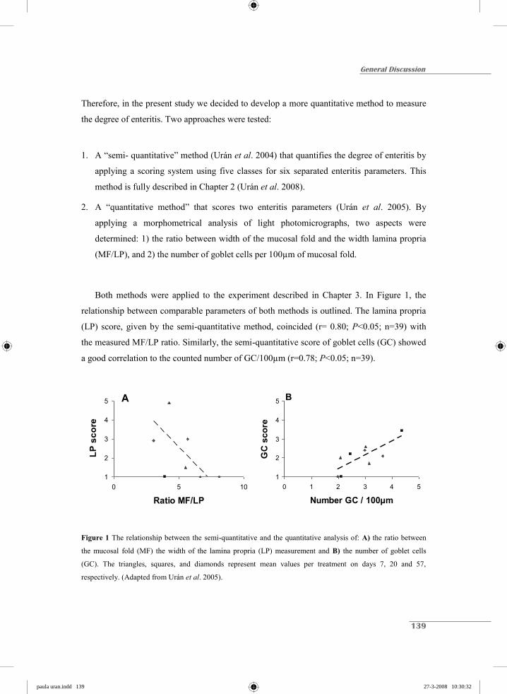
General Discussion
139
Therefore, in the present study we decided to develop a more quantitative method to measure
the degree of enteritis. Two approaches were tested:
1. A “semi- quantitative” method (Urán et al. 2004) that quantifies the degree of enteritis by
applying a scoring system using five classes for six separated enteritis parameters. This
method is fully described in Chapter 2 (Urán et al. 2008).
2. A “quantitative method” that scores two enteritis parameters (Urán et al. 2005). By
applying a morphometrical analysis of light photomicrographs, two aspects were
determined: 1) the ratio between width of the mucosal fold and the width lamina propria
(MF/LP), and 2) the number of goblet cells per 100μm of mucosal fold.
Both methods were applied to the experiment described in Chapter 3. In Figure 1, the
relationship between comparable parameters of both methods is outlined. The lamina propria
(LP) score, given by the semi-quantitative method, coincided (r= 0.80; P<0.05; n=39) with
the measured MF/LP ratio. Similarly, the semi-quantitative score of goblet cells (GC) showed
a good correlation to the counted number of GC/100μm (r=0.78; P<0.05; n=39).
Figure 1 The relationship between the semi-quantitative and the quantitative analysis of: A) the ratio between
the mucosal fold (MF) the width of the lamina propria (LP) measurement and B) the number of goblet cells
(GC). The triangles, squares, and diamonds represent mean values per treatment on days 7, 20 and 57,
respectively. (Adapted from Urán et al. 2005).
1
2
3
4
5
0 1 2 3 4 5
Number GC / 100μm
GC
sco
re
1
2
3
4
5
0 5 10
Ratio MF/LP
LP
scor
e
A B
paula uran.indd 139paula uran.indd 139 27-3-2008 10:30:3227-3-2008 10:30:32

Chapter 8
140
Despite these positive results, the quantitative method yielded a large
individual/sample variation. As a result, a large number of observations per sample are
required, making these measurements more labour intensive. Therefore, it was ultimately
decided to further develop and use the semi-quantitative method.
The accuracy of the semi-quantitative scoring method was tested by comparing the
scoring skills of two different observers, analyzing the same samples of the experiment
described in Chapter 3. This method demonstrated a strong correlation between observers. In
Figure 2, the relationship between the mean scores of two separate observers is shown
(r=0.87; P< .0001; n=77.
1
2
3
4
5
1 2 3 4 5
Observer 1
Obs
erve
r 2
Figure 2 The relationship between the semi-quantitative results from two different observers. The evaluation of
the histological slides was a blind test. The values represent the overall mean of six separated parameters per
fish.
This semi-quantitative method is an advancement on the more simplified semi-
quantitative method, presented in Morris et al. (2005), in which all the parameters were
grouped in a unique category. When their approach is used, the probability of identifying
slight changes in the enteritis response is reduced due to the low resolution of their method.
paula uran.indd 140paula uran.indd 140 27-3-2008 10:30:3427-3-2008 10:30:34

General Discussion
141
Our semi-quantitative method offers the possibility of acquiring detailed information at
different cellular and structural levels. In doing so, the etiology of the disorder, as well as the
different dietary and non-dietary factors involved in its development, can be more thoroughly
illustrated and examined.
In conclusion, our method proved to be reliable, accurate and more precise than the
hitherto known, comparable scoring methods.
The impact of different factors on the development and severity of enteritis
The present study had the objective of examining whether, and if so how, dietary and
non-dietary factors influence the inflammatory process which leads to enteritis through SBM
feeding.
With regard to the dietary factors, the occurrence of enteritis is clearly related to
feeding diets containing SBM. Several studies have attempted to replace SBM by other soy
products particularly for salmonid diets. However, most SB products contain high levels of
anti-nutritional factors, and this limits their use in aquafeeds (Alexis & Nengas 2001; Francis
et al. 2001).
Several aspects involved in the production process of different SB products may affect
the quality and the composition of the final SB product and may be responsible for variations
in the response when fed to fish. Chapter 3 illustrates an experiment in which different
inclusion levels of dehulled solvent-extracted SBM (10 and 20% of the protein fraction) were
tested in time. The SBM used was a commercial source known to induce a strong
inflammatory reaction. The lower inclusion level resulted in a mild response in contrast to the
response at the higher level which continued over time, displaying no signs of recovery. The
severity of enteritis was clearly dose-dependent. The diet containing the highest inclusion
level was also applied to the omnivorous common carp (Chapter 4).
Upon switching to SBM diets, common carp, developed similar symptoms of enteritis
as the Atlantic salmon. In contrast to the Atlantic salmon, common carp appears to recover
from this disorder after a few weeks of SBM feeding. This recovery starts with the re-
appearance of the supranuclear vacuoles (SNV) in the absorptive cells. Therefore, the
paula uran.indd 141paula uran.indd 141 27-3-2008 10:30:3527-3-2008 10:30:35

Chapter 8
142
condition of SNV might be important in revealing the mechanisms behind SBM-induced
enteritis.
Based on the fact that enteritis appears to be dose-dependent, SBM from different soy
processing plants were tested in Chapter 6. This study showed clear dissimilar enteritis
responses between the SBM from different commercial sources. Once again, the most
affected parameter was the disappearing SNV. In contrast, the goblet cells (GC) were only
slightly affected by the dietary differences. Anyway, it is suggested that most parameters
indicating enteritis react proportionally to the inclusion level of the SB product rather than to
a threshold value above which enteritis is developed and below not (yes/no response).
To date, it is not clear which component in soy is the causative agent for inducing
enteritis. The alcohol-soluble components (namely SB molasses) which result from the
extraction of SB protein concentrates are often considered as being the inducers of this
inflammatory process (Olli & Krogdahl 1995; van den Ingh et al. 1996; Krogdahl et al. 2000;
Knudsen et al. 2007). Chapter 7 demonstrates that the inflammation is induced by molecules
smaller than 10 kDa. Based on the morphological differences induced by the different
fractions used in the study we argue that glycin, -conglycinin and lectins, proteins which are
commonly presumed to be the main causative factors of enteritis, should be ruled out.
Nonetheless, when combined with soyasaponins, and perhaps with the intestinal microbiota,
they may have an influence. Saponins have been used as potent adjuvants due to their ability
to increase cell membrane permeability (Ronnberg et al. 1995; Oda et al. 2000, 2003). They
are believed to enhance small molecules and/or macromolecules to cross the cell membrane
(Alvarez & Torres-Pinedo 1982, Mick et al. 1988). However, the ferritin experiment (Chapter
5) did not show such an increased permeability when SBM was fed. As such, the
concentration and composition of soyasaponins in the SB and in the final SB product may
depend on the variety and processing conditions, as is suggested by Hu et al. (2002).
However, more research is needed to verify this hypothesis.
On the other hand, it is also possible that non-dietary factors are involved in the
etiology of the disorder. Husbandry conditions, such as temperature, may influence the
severity of enteritis by acting directly on feed intake or by affecting the metabolic rate at the
enterocyte level. In the present study (Chapter 2), water temperature influenced the speed at
which enteritis developed in Atlantic salmon. At 12 °C, enteritis developed faster and more
paula uran.indd 142paula uran.indd 142 27-3-2008 10:30:3627-3-2008 10:30:36

General Discussion
143
acute than at 8 °C. This was in contrast to the general experience in the industry (W. Koppe,
personal communication). At 8 °C a number of factors were observed to be less affected: the
disappearance of the supranuclear vacuoles (SNV), the shortening of the mucosal folds (MF);
the widening of the lamina propria (LP) and of the sub-epithelial mucosa (SM). The different
responses between the two temperatures may be explained by a difference in the metabolic
rate. Temperature could have influenced the rate at which food was ingested, or transported in
the intestinal tract, and/or absorbed by the gut. At higher temperatures, all these processes
proceed at a higher rate, increasing the contact between causative agent and animal per unit of
time.
Another non-dietary factor which was briefly addressed in this thesis was the possible
effect of salinity and age on the development of SBM-induced enteritis. In Chapter 5, young
fish, kept in freshwater, were compared with older fish (from a different experiment, Chapter
3) kept in seawater. Our results indicate that enteritis is not dependent on age or
smoltification. Usher et al. (1990) proposed an osmoregulatory role of the gut in seawater-
adapted smolts, affecting its digestive role by altering the physico-chemical conditions, the
activity of digestive enzymes, and the active transport of ions in the gut lumen. It was
expected that the changes mentioned by Usher et al. (1990) could have increased the severity
of SBM-induced enteritis of the Atlantic salmon exposed to seawater. Apparently, feeding of
SBM did not impair their osmoregulatory ability. This concurs with the previous results of
Bakke-McKellep et al. (2006) who proved that the osmoregulatory capacity of fish displaying
the typical signs of SBM-induced enteritis was maintained. These findings are in accordance
with the results of the present study. However, more detailed research is required to exclude
the influence of salinity on the enteritis process.
In conclusion, this thesis shows that dietary factors (inclusion level and different soy
sources) and temperature are probably more important than age and salinity in the severity of
enteritis in Atlantic salmon. Consequently, more attention must to be paid to the responsible
SB factors. The differences in the described plant sources may be useful to take into
consideration. A further factor derived from the present study is that in addition to Atlantic
salmon, another species (common carp), can also temporarily develop enteritis. Nonetheless,
for reasons presently unknown, common carp seems to recover from enteritis or to adapt to
the SBM.
paula uran.indd 143paula uran.indd 143 27-3-2008 10:30:3727-3-2008 10:30:37

Chapter 8
144
Mechanisms behind the development of SBM-induced enteritis
As discussed earlier, limited information is known about the SBM components that
cause enteritis and the mechanisms behind the disorder. In our study, we describe how
endocytosis in the second gut segment is blocked and that the consequent fast decrease in
SNV number and size seems to be the first symptom observed. This phenomenon occurs in
two completely different species (carp and salmon) studied in this thesis (Chapters 4 and 5
respectively). This observation suggests that the mechanism has to do with blocking a
receptor mediated uptake. Unfortunately, at present we cannot relate this to one of the SB
factors that have so far been studied. The disturbance of such an essential basic mechanism as
endocytosis must have a great impact on the functioning of enterocytes, rapidly resulting in
the disappearance of SNV, and finally, also in the decrease of the height of the microvilli. It
could be that these disturbances of the enterocytes result in the release of danger signals, such
as heat shock proteins (HSP), which may be responsible for the recruitment of leucocytes and,
as a culminating point, in the increasing inflammation of the connective tissue. The role of
HSP70 in SBM-induced enteritis has been evidenced already in Atlantic salmon by Bakke-
McKellep et al. (2007b). In addition, the up- or down-regulation of inflammatory and/or anti-
inflammatory cytokines produced by intestinal cells may also play a crucial role. In any case,
migratory reactions of intra-epithelial T cells and the up-regulation of T cell specific
responses have been described during the onset of enteritis in Atlantic salmon (Bakke-
McKellep et al. 2007c). Furthermore, there was a strong invasion of the eosinophilic
granulocytes (EG) from the sub-epithelial mucosa inwards to the lamina propria. This
observation and the change of the morphology of the EG granules indicates an activation of
these cells. EG in fish are known to produce lysozyme (Sveinbjørnsson et al. 1996;
Silphaduang & Noga 2001; Reite & Evensen 2006; Murray et al. 2007) which may have a
protective role in the inflamed intestinal tissue. In the studies conducted by Refstie et al.
(2006) and Bakke-McKellep et al (2007b), it was proposed that SBM inclusion in diets of
Atlantic salmon may influence the intestinal microbiota. An increase in the number and
diversity of the microbial community has been described. The observed reaction of EG may
be an important defence against it. Nonetheless, this is not a sufficient explanatory factor, as
shown by Krogdahl et al. (2000), who proposed a higher susceptibility to bacterial diseases
paula uran.indd 144paula uran.indd 144 27-3-2008 10:30:3827-3-2008 10:30:38

General Discussion
145
during SBM induced enteritis. Although no direct evidence is presented in this thesis, some
authors suggest that an increase in intestinal permeability facilitates the entry of microbes or
their products into the mucosal compartment (Braat et al. 2006). In line with such a
hypothesis, the resemblance of SBM-induced enteritis and inflammatory bowel disease (IBD,
such as Coeliac disease) has been suggested (Baeverfjord & Krogdahl 1996). As discussed
earlier, soyasaponins could facilitate intestinal permeability, exposing the underlying mucosa
to a variety of antigens and microbiota. This may be the reason as to why the Atlantic salmon
gut becomes seriously harmed by the SBM. However, it does not explain why common carp
seems to react similarly upon the SBM noxious substances, but appears to recover from
enteritis from week 4 onwards. It is worthwhile to investigate which mechanisms are involved
in the recovery/adaptation of common carp. This knowledge may increase the understanding
of SBM-induced enteritis also in Atlantic salmon. Moreover, the results found in common
carp also emphasize that aquaculture must be aware of the possibility of a temporary enteritis
response in many other fish species. This temporary response may have been overlooked up
until now, due to the (too) late sampling after switching to the SBM diets. Such a temporary
enteritis response may result in a higher susceptibility for diseases when fish switch to a
SBM-containing diet.
Final conclusions
The work presented in this thesis contributes to the understanding of the etiology of
the enteritis disorder. Knowing that etiology deals with the cause or origin of a disorder and
the factors which influence its development, it can be said that SBM-induced enteritis is
related more to dietary factors than non-dietary factors (i.e. temperature). Nevertheless, non-
dietary factors do have an impact on the speed at which the disorder is developed. The
inclusion level of SBM into the diet formulation has a great impact on the severity at which
all the typical symptoms of the disorder are expressed, affecting the endocytosis process and
initiating a cascade of events in the immune system. The actual causative components present
in SB are still matters for future research, especially in terms of clarifying their mechanisms
of action. It is suggested in this thesis that the decreased nutrient uptake capacity of the
paula uran.indd 145paula uran.indd 145 27-3-2008 10:30:3927-3-2008 10:30:39

Chapter 8
146
enterocytes is directly related to the disappearance of the SNV as seen in Atlantic salmon. The
reasons why the Atlantic salmon is not capable of recovering from the enteritis, while
common carp does, remain unknown.
References
Alexis, M.N. & Nengas, I. (2001) Current state of knowledge concerning the use of soy products in
diets for feeding sea bass and sea bream needs for future research. National centre for marine
research, Athens-Greece. American Soybean Association. 5, 1-32.
Alvarez, J.R. & Torres-Pinedo, R. (1982) Interactions of soybean lectin, soyasaponins, and glycinin
with rabbit jejunal mucosa in vitro. Pediatr. Res., 9: 728-731.
Baeverfjord, G. & Krogdahl, Å. (1996) Development and regression of soybean meal induced enteritis
in Atlantic salmon, Salmo salar L., distal intestine: a comparison with the intestines of fasted fish.
J. Fish Dis., 19, 375-387.
Bakke-McKellep, A. M., Refstie, S., Stefansson, S.O., Vanthanouvong, V., Roomans, G. Hemre, G.I.
& Krogdahl, Å. (2006) Effects of dietary soybean meal and photoperiod cycle on osmoregulation
following seawater exposure in Atlantic salmon smolts. J. Fish Biol., 69, 1396-1426.
Bakke-McKellep, A.M., Koppang, E.O., Gunnes, G., Sanden, M., Hemre, G.I., Landsverk, T. &
Krogdahl, A. (2007a) Histological, digestive, metabolic, hormonal and some immune factor
responses in Atlantic salmon, Salmo salar L., fed genetically modified soybeans. J. Fish Dis., 30,
65-79.
Bakke-McKellep, A.M., Penn, M. H., Salas, P.M., Refstie, S., Sperstad, S., Landsverk, T., Ringo, E. &
Krogdahl, Å. (2007b) Effects of dietary soyabean meal, inulin and oxytetracycline on intestinal
microbiota and epithelial cell stress, apoptosis and proliferation in the teleost Atlantic salmon
(Salmo salar L.). Brit. J. Nutr., 97, 699-713.
Bakke-McKellep, A.M., Frøystad, M.K., Lilleeng, E., Dapra, F., Refstie, S., Krogdahl, Å. &
Landsverk, T. (2007c) Response to soy: T-cell-like reactivity in the intestine of Atlantic salmon,
Salmo salar L. J. Fish. Dis., 30, 13-25.
Braat, H., Peppelenbosch, M.P. & Hommes, DW. (2006) Immunology of Crohn’s disease. Ann. N. Y.
Sci., 1072, 135-154.
Francis, G., Makkar, H. P.S. & Becker, K. (2001) Antinutritional factors present in plan-derived
alternate fish feed ingredients and their effects in fish. Review Article. Aquaculture, 199, 197-227.
paula uran.indd 146paula uran.indd 146 27-3-2008 10:30:4027-3-2008 10:30:40

General Discussion
147
Hu, J., Lee, S., Hendrich, S. & Murphy, P.A. (2002) Quantification of the group B soyasaponins by
high-performance liquid chromatography. J. Agr. Food Chem., 50, 2587-2594.
van den Ingh, T. S. G. A. M., Olli, J. J. & Krogdahl, Å. (1996) Alcohol-soluble components in
soybeans cause morphological changes in the distal intestine of Atlantic salmon, Salmo salar L. J.
Fish Dis., 19, 47-53.
Knudsen, D., Urán, P., Arnous, A., Koppe, W. & Frøkiær, H. (2007) Saponin-containing subfractions
of soybean molasses induce enteritis in the distal intestine of Atlantic salmon. J. Agric. Food
Chem., 55, 2261-2267.
Krogdahl, Å., Bakke-McKellep, A.M., Røed, K.H. & Baeverfjord, G. (2000) Feeding Atlantic salmon
Salmo salar L. soybean products: effects on disease resistance (furunculosis), and lysozyme and
IgM levels in the intestinal mucosa. Aquacult. Nutr., 6, 77-84.
Mick, G.J., Bonn, T., Steinberg, J. & McCormick, K. (1988) preservation of intermediary metabolism
in saponin-permeabilized rat adiocytes. J. Biol Chem., 263, 10667-10673.
Morris, P. C., Gallimore, P., Handley, J., Hide, G., Haughton, P. & Black, A. (2005) Full-fat soya for
rainbow trout (Oncorhynchus mykiss) in freshwater: Effects on performance, composition and
flesh fatty acid profile in absence of hind-gut enteritis. Aquaculture, 248, 147-161.
Murray, H.M., Leggiadro, C.T. & Douglas, S.E. (2007) Immunocytochemical localization of
pleurocidin to the cytoplasmic granules of eosinophilic granular cells from the winter flounder
gill. J. Fish Biol., 70, 336-345.
Oda, K., Matsuda, H., Murakami, t. Katayama, S., Ohgitani, T. & Yoshikawa, M. (2000) Adjuvant and
haemolytic activities of 47 saponins derived from medicinal and food plants. Biol. Chem., 381: 67-
74.
Oda, K., Matsuda, H., Murakami, T., Katayama, S., Ohgitani, T. & Yoshikawa, M. (2003)
Relationship between adjuvant activity and amphipathic structure of soyasaponins. Vaccine, 21,
2145-2151.
Olli, J.J. & Krogdahl, Å. (1995) Alcohol soluble components of soybeans seem to reduce fat
digestibility in fishmeal-based diets for Atlantic salmon, Salmo salar L. Aquac. Res., 26, 831-835.
Refstie, S., Bakke-McKellep, A.M., Penn, M.H., Sundby, A., Shearer, K. D. & Krogdahl, Å. (2006)
Capacity for digestive hydrolysis and amino acid absorption in Atlantic salmon (Salmo salar) fed
diets with soybean meal or inulin with or without addition of antibiotics. Aquaculture, 261, 392-
406.
Refstie, S., Korsøen, Ø. J., Storebakken, T., Baeverfjord, G., Lein, I. & Roem, A. J. (2000) Differing
nutritional responses to dietary soybean meal in rainbow trout (Oncorhynchus mykiss) and
Atlantic salmon (Salmo salar). Aquaculture, 190, 49-63.
paula uran.indd 147paula uran.indd 147 27-3-2008 10:30:4227-3-2008 10:30:42
Chapter 8
148
Refstie, S., Strorebakken, T., Baeverfjord G. & Roem A.J. (2001) Long-term protein and lipid growth
of Atlantic salmon (Salmon salar) fed diets with partial replacement of fishmeal by soy protein
products at medium or high lipid level. Aquaculture, 193, 91-106.
Reite, O.B. & Evensen, Ø. (2006) Inflammatory cells of teleostean fish: A review focusing on mast
cells/eosinophilic granule cells and rodlet cells. Fish Shellfish Immun., 20, 192-208.
Rönnberg, B., Fekadu, M. & Morein, B. (1995) Adjuvant activity of non-toxic Quillaja saponaria
Molina components for use in ISCOM matrix. Vaccine, 13, 1375-1382.
Sanden, M., Berntssen, M.H.G., Krogdahl, Å., Hemre, G.I. & Bakke-McKellep, A.M. (2005) An
examination of the intestinal tract of Atlantic salmon, Salmo salar L., parr fed different varieties
of soy and maize. J. Fish Dis., 28, 317-330.
Silphaduang, U. & Noga, E. J., 2001. Peptide antibiotics in mast cells of fish. Nature, 414, 268-269.
Sveinbjørnsson, B., Olsen, R. & Paulsen, S., 1996. Immunocytochemical localization of lysozyme in
intestinal eosinophilic granule cells (EGCs) of Atlantic salmon, Salmo salar L. J. Fish dis., 19,
349-355.
Urán, P.A., Rombout, J.H.W.H.; Koppe, W., Obach, A., Jensen, L., Schrama, J.W. & Verreth, J.A.J.
(2004) Effects of soybean meal on intestinal morphology of Atlantic salmon (Salmo salar L.). In
Abstracts Aquaculture Europe, Barcelona; Special Publication, 34, 803-804.
Urán, P.A., van Houcke, J., Rombout, J.H.W.M., Koppe, W., Fontanillas, R., Schrama, J.W. &
Verreth, J.A.J. (2005) Comparison of methodologies for measuring the degree of soybean induced
enteritis in Atlantic salmon (Salmo salar L.). In: Abstracts World Aquaculture, Bali Indonesia.
Special publication, CD-ROM, p. 666.
Urán, P.A., Schrama, J.W., Rombout, J.H.W.M., Obach, A., Jensen, L., Koppe, W. & Verreth, J.A.J.,
(2008) Soybean meal-induced enteritis in Atlantic salmon (Salmo salar L.) at different
temperatures. Aquacult. Nutr., in press
Usher, M.L., Talbot, C. & Eddy, F. B. (1990) Effects of transfer to seawater on digestion and gut
function in Atlantic salmon smolts (Salmo-Salar L). Aquaculture, 90, 85-96.
paula uran.indd 148paula uran.indd 148 27-3-2008 10:30:4327-3-2008 10:30:43
SEMI-QUANTITATIVE SCORING SYSTEM
Also available at:http://www.afi.wur.nl/UK/Publications/
chapter hoofdstukken.indd 17 27-3-2008 11:04:48

Semi-Quantitative Scoring System
150
Mucosal folds (MF)
5Total tissue disruption
4Diffused tissue disruption
3Diffused shrinkage and onset of tissue disruption
2Some shrinkage and bloating
1Basal length
Bar is 200 μm
paula uran.indd 150paula uran.indd 150 27-3-2008 10:30:4627-3-2008 10:30:46

Semi-Quantitative Scoring System
151
Supranuclear vacuoles (SNV)
5No SNV
4Onset of extinction
3Diffused size reduction
2Some size reduction
1Basal SNV size
Bar is 20 μm
paula uran.indd 151paula uran.indd 151 27-3-2008 10:30:4727-3-2008 10:30:47
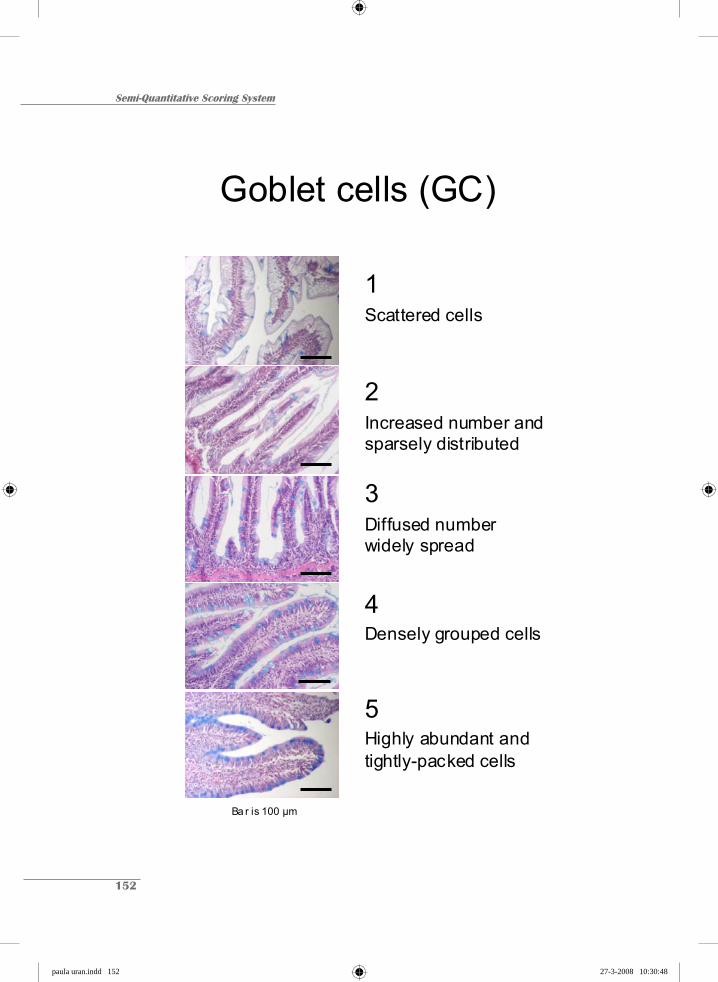
Semi-Quantitative Scoring System
152
Goblet cells (GC)
5Highly abundant and tightly-packed cells
4Densely grouped cells
3Diffused number widely spread
2Increased number and sparsely distributed
1Scattered cells
Ba r is 100 μm
paula uran.indd 152paula uran.indd 152 27-3-2008 10:30:4827-3-2008 10:30:48
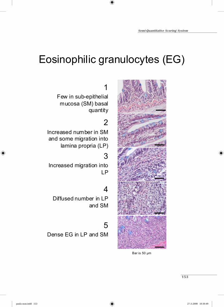
Semi-Quantitative Scoring System
153
Eosinophilic granulocytes (EG)
5Dense EG in LP and SM
4Diffused number in LP
and SM
3Increased migration into
LP
2Increased number in SM and some migration into
lamina propria (LP)
1Few in sub-epithelial mucosa (SM) basal
quantity
Bar is 50 μm
paula uran.indd 153paula uran.indd 153 27-3-2008 10:30:4927-3-2008 10:30:49

Semi-Quantitative Scoring System
154
Lamina propria (LP)
5Largest LP
4Large LP
3Medium size
2Increased size
1Normal size
Bar is 50 μm
paula uran.indd 154paula uran.indd 154 27-3-2008 10:30:5027-3-2008 10:30:50

Semi-Quantitative Scoring System
155
Sub-epithelial mucosa (SM)
5Largest SM
4Large SM
3Medium size SM
2Increased size SM
1Normal SM
Bar is 100 μm
paula uran.indd 155paula uran.indd 155 27-3-2008 10:30:5127-3-2008 10:30:51

chapter hoofdstukken.indd 18 26-3-2008 12:42:11

SUMMARY
chapter hoofdstukken.indd 19 26-3-2008 12:42:11
Summary
158
Presently, most efforts to substitute fishmeal by soybean meal (SBM) in aquafeeds are
being confronted by numerous problems and constraints. The inclusion of SBM, especially in
the diet of salmonids, induces an inflammatory response of the distal intestinal mucosa,
known as enteritis. The general aim of this study was to understand the etiology and the
underlying mechanisms of this disorder. To do so, numerous factors (both dietary and non-
dietary) were evaluated and the possible mechanisms behind the inflammatory process were
studied in both Atlantic salmon (Salmo salar L.) and common carp (Cyprinus carpio L).
In Chapter 2, water temperature was addressed as an important husbandry condition
which could influence the speed at which the disorder develops. The study demonstrated that
the onset of enteritis is delayed at a lower temperature (8 °C vs. 12 °C) A semi-quantitative
scoring system was developed to assess the extent of the morphological changes observed in
this study. This tool was further used in all the subsequent studies of this thesis.
Chapter 3 describes the influence of SBM on the kinetics of the disorder. This was
studied at two different inclusion levels over a period of 57 days. At the higher dose (20%
SBM), the signs of enteritis seemed to increase steadily whereas at the lower dose (10%
SBM) the disorder displayed a tendency to level off, a process starting from two weeks
onwards. It was concluded that the severity of enteritis and its kinetics are dose-dependent,
showing no signs of recovery during feeding SBM-containing diets. Electron microscopy
studies suggested a blocking of the endocytosis process and a strong decrease of microvilli
length. Endocytosis is an important, basic and generic process in animal physiology. As such,
if SBM or any compound in it can be shown to induce enteritis (i.e. through the possible
blocking of endocytosis), it would appear very remarkable that only salmonids develop this
disorder. Therefore, a comparative study was done in another fish species with different
(more herbivorous) feeding habits (i.e. common carp).
In Chapter 4, SBM-induced enteritis in the omnivorous common carp was studied
and compared to the SBM-induced intestinal inflammation in the carnivorous Atlantic
salmon. For the first time ever reported, the results suggested that the symptoms of enteritis
also occur in common carp. Contrary to the observations in studies with Atlantic salmon, the
common carp started to recover from week four onwards. This recovery was explicitly
noticeable by the re-appearance of the supranuclear vacuoles (SNV). The morphological
changes and the modulation of the inflammatory process were defined and correlated to the
paula uran.indd 158paula uran.indd 158 27-3-2008 10:30:5327-3-2008 10:30:53

Summary
159
up- and down-regulation of several cytokines that were presumed to influence this process.
Again in this study, electron microscopy suggested that endocytosis blocking is directly
linked to the disappearance of the SNV and the onset of enteritis symptoms.
The potential role of endocytosis in the development of the disorder was further
investigated in Chapter 5. Ferritin was used to prove the eventual blocking of endocytosis.
After one week of SBM-feeding, ferritin uptake had diminished and the SNV had decreased
in size. At week 3, ferritin uptake could not be detected and the SNV were not present in the
enterocytes. This indicates that endocytosis block is indeed the first step in the disappearance
of SNV. In addition, the results of this study on young Atlantic salmon (30 g) kept in
freshwater, facilitated a comparison with older salmon (300 g) kept in seawater, regarding the
influence of salinity on the severity of enteritis. It was suggested that SBM-induced enteritis
was not strongly influenced by either salinity or age, but further studies are needed to support
this observation.
In Chapter 6, SBM obtained from different commercial sources and production plants
world-wide were compared. The goal was to determine whether the extent of enteritis in
Atlantic salmon depends on the origin and/or the processing of the soybeans. The most
affected parameter was again the disappearance of SNV in enterocytes. In contrast, the
increase in goblet cells showed the smallest variation between the different SBM sources.
This study showed that different commercial sources of SBM can result in different severities
of SBM-induced enteritis.
Literature suggests that soyasaponins, possibly in combination with other factors, are
responsible for the initiation of the enteritis process. Therefore, Chapter 7 studies if different
sub-fractions, obtained through phase-separation of molasses with n-Butanol, had an effect on
the development of enteritis. The results indicate that enteritis was only present in fish fed
with the butonol phase and the precipitate sub-fractions. This suggests that soyasaponines,
possibly in combination with other factors (e.g. intestinal microflora) do indeed influence the
onset and development of the enteritis process.
The results of this thesis are discussed in Chapter 8. It is concluded that the etiology
and further development of SBM-induced enteritis is related to the dietary factors rather than
to the non-dietary factors. SBM inclusion levels and the commercial source used for the diet
formulation have a great impact on the severity of the disorder, mainly affecting the
paula uran.indd 159paula uran.indd 159 27-3-2008 10:30:5427-3-2008 10:30:54

Summary
160
endocytosis process. This thesis indicates that the endocytosis block and subsequent
dissapearence of the SNV seem to be crucial for the onset of the inflammation process.
Nonetheless, the actual causative components present in SB are still issues that require further
research. In addition to Atlantic salmon, another species like common carp can also
temporarily develop enteritis but after 4 weeks of SBM feeding, they start to recover. The
reasons for this recovery remain unknown.
paula uran.indd 160paula uran.indd 160 27-3-2008 10:30:5527-3-2008 10:30:55
SAMENVATTING
chapter hoofdstukken.indd 20 26-3-2008 12:42:35
Samenvatting
162
Bij het vervangen van vismeel in visvoer door sojaschroot (SBM) stuit men op
verscheidene problemen en beperkingen. Toevoeging van SBM in zalmvoer, veroorzaakt een
ontsteking (enteritis) in het distale deel van de darm. Het algemene doel van dit onderzoek
was meer inzicht te verkrijgen over de ontwikkeling, de oorzaken en de onderliggende
mechanismen betrokken bij deze ontsteking van de darm. Hiervoor zijn verschillende factoren
die deels verband houden met de voeding, deels met andere factoren, onderzocht in zowel
Atlantische zalm (Salmo salar L.) als gewone karper (Cyprinus carpio L.).
De watertemperatuur, een belangrijke houderijfactor, werd bestudeerd in hoofdstuk 2
vanwege de mogelijke invloed op de snelheid waarmee de enteritis zich ontwikkelt. Uit het
onderzoek bleek een lage (8 ºC) watertemperatuur de ontwikkeling van de enteritis te
vertragen in vergelijking tot een hoge temperatuur (12 ºC). Om dit onderzoek te kunnen
uitvoeren werd een semi-kwantitatief scoringssysteem ontwikkeld, waarmee het mogelijk was
de mate van de morfologische veranderingen in de darm vast te stellen. Deze
scoringsmethode is ook toegepast op alle verdere onderzoeken in dit proefschrift.
In hoofdstuk 3 is de invloed van SBM op het verloop van de ontsteking in de tijd (e.g.
de kinetiek) beschreven. Dit werd onderzocht door in het voer van de zalm verschillende
percentages SBM (10% versus 20%) in te brengen en de eventuele ontwikkeling van enteritis
te meten gedurende een periode van 57 dagen. Bij het dieet met 20% SBM bleef de ernst van
de enteritis toenemen in de tijd, terwijl bij het dieet met 10% SBM de enteritis na 2 weken
stabiliseerde. Er kan geconcludeerd worden dat de mate van enteritis dosis-afhankelijk is en
dat er geen herstel optreedt zolang er SBM gevoerd wordt. Elektronen microscopisch
onderzoek toonde aan dat er geen endocytose plaats vond en dat de lengte van de microvilli
sterk afnam. Endocytose is een belangrijk basaal en generiek proces in de dierlijke fysiologie.
Dit leidde tot de hypothese dat de bestudeerde enteritis waarschijnlijk niet alleen in zalm
plaats vindt.
Daarom werd in hoofdstuk 4 onderzocht wat de invloed is van het voeren van SBM
houdende diëten aan de karper, een omnivore vis. De resultaten zijn vergeleken met die van
de (carnivore) Atlantische zalm. Ook in de karper ontstaan symptomen van enteritis waarneer
SBM houdende diëten gevoerd worden. Echter, in tegenstelling tot de Atlantische zalm, trad
in de enterocyten van de karper na 4 weken herstel op, waarbij het weer verschijnen van de
supranucleaire vacuoles (SNV) het meest opvallend was. De morfologische veranderingen en
paula uran.indd 162paula uran.indd 162 27-3-2008 10:30:5727-3-2008 10:30:57

Samenvatting
163
de mate van ontsteking werden gedefiniëerd en gekoppeld aan de up- and down-regulatie van
verschillende cytokines die verondersteld worden het ontstekingsproces te reguleren.
Elektronen microscopisch onderzoek toonde aan dat ook hier eerst de endocytose geblokkeerd
werd, gevolgd door het verdwijnen van de SNV, alvorens het herstel optrad.
De mogelijke rol van de endocytose bij de ontwikkeling van de ontsteking is verder
onderzocht in hoofdstuk 5. Om aan te tonen dat er werkelijk geen endocytose meer optrad,
werd ferritine toegediend, op verschillende tijdstippen na de overschakeling op een dieet dat
SBM bevatte. Na 1 week was de opname van ferritine sterk verminderd en waren de SNV
kleiner geworden. Na 3 weken werd er geen opname van ferritine meer waargenomen en
waren de SNV verdwenen. Deze resultaten tonen aan dat de endocytose-blokkade een rol
speelt bij het verdwijnen van de SNV. Een vergelijking van de resultaten verkregen met jonge
Atlantische zalm (30 g) gehouden in zoet water en deze verkregen bij oudere dieren (300 g)
gehouden in zout water, suggereerde dat de saliniteit van het water en de leeftijd van de zalm
nauwelijks van invloed waren op de ontwikkeling van enteritis. Verder onderzoek is echter
nodig om deze waarneming te ondersteunen.
In hoofdstuk 6 werden verschillende partijen SBM, die afkomstig waren van
verschillende commerciële sojaverwerkingsbedrijven, met elkaar vergeleken om te
onderzoeken of het ontstaan van enteritis bij de Atlantische zalm afhankelijk is van herkomst
van het SBM en/of het productieproces. De meest beïnvloedde parameter was opnieuw de
verdwijning van de SNV. Uit dit onderzoek is gebleken dat de herkomst van het SBM van
belang is voor de ernst/mate van enteritis in de Atlantische zalm.
In de literatuur wordt verondersteld dat sojasaponines alleen of in combinatie met
andere factoren verantwoordelijk zijn voor de initiatie van het enteritis proces. Daarom werd
in hoofdstuk 7 onderzocht of verschillende subfracties die verkregen werden bij fasescheiding
van sojamelasse met n-Butanol, een effect hadden op de ontwikkeling van enteritis. De
resultaten tonen aan dat enteritis alleen voorkwam in vis die gevoerd werd met de subfractie
uit de butanol-fase en het precipitaat. Daarmee wordt gesuggereerd dat sojasaponines,
mogelijk in combinatie met andere factoren zoals darmflora of anderszins, inderdaad van
invloed zijn op het ontstaan en de ontwikkeling van het enteritis proces.
In hoofdstuk 8 worden de resultaten van dit proefschrift bediscussiëerd. De SBM-
geïnduceerde enteritis is duidelijk dieet afhankelijk. De herkomst van het SBM en het
paula uran.indd 163paula uran.indd 163 27-3-2008 10:30:5827-3-2008 10:30:58

Samenvatting
164
percentage SBM in het voer zijn erg belangrijk bij het ontstaan en het verloop van de enteritis.
Met name de blokkade van het endocytose proces en de daarop volgende verdwijning van de
SNV lijken cruciaal in het op gang komen van het ontstekingproces. Echter verder onderzoek
naar de bij dit proces betrokken componenten is noodzakelijk. In vergelijking met de
Atlantische zalm kunnen ook andere vissoorten tijdelijk enteritis ontwikkelen na toediening
van SBM, echter in het geval van de karper trad na 4 weken herstel op. De reden waarom
karper wel en zalm niet herstelt is nog onbekend.
paula uran.indd 164paula uran.indd 164 27-3-2008 10:30:5927-3-2008 10:30:59

RESUMEN
chapter hoofdstukken.indd 21 26-3-2008 12:45:15
Resumen
166
Actualmente, la mayoría de esfuerzos en la substitución de la harina de pescado por
harina de soya para alimento de peces, ha encontrado un gran número de obstáculos y
restricciones. Se sabe que la inclusión de esta harina de soya (SBM, por su sigla en inglés),
particularmente para dietas de salmónidos induce un proceso inflamatorio a nivel de la
mucosa del intestino posterior. Dicho proceso se conoce como enteritis. El objetivo principal
de esta investigación es entender la etiología y los mecanismos responsables de este trastorno.
Para este efecto, se evaluan numerosos factores (tanto alimentarios como no-alimentarios) y
se estudian los posibles mecanismos responsables de dicho proceso inflamatorio, tanto en
salmón del Atlántico (Salmo salar L) como en carpa común (Cyprinus carpio L.).
En el capítulo 2, se evalúa la influencia de la temperatura del agua como un factor
fundamental que puede afectar la velocidad con la que este trastorno intestinal se desarrolla.
Con este estudio se demuestra que la fase inicial de este trastorno se retrasa en los peces
mantenidos a la temperatura más baja (8 vs. 12 °C). Para poder evaluar el grado de intensidad
de los cambios ocurridos a nivel morfológico se desarrolla un sistema de clasificación semi-
cuantitativo. Esta herramienta se utiliza en este estudio y en los estudios posteriormente
mencionados en esta tesis.
El capítulo 3, describe la influencia de SBM en la cinética de dicho trastorno
intestinal. Se estudian dos niveles diferentes de inclusión a lo largo de un período de 57 días.
Los síntomas característicos de la enteritis se incrementan constantemente especialmente en
peces alimentados con la dosis más alta empleada en este estudio (20% SBM) con respecto a
la dosis más baja (10% SBM), bajo la cual los peces muestran una tendencia a estabilizar
dichos síntomas, tendencia que se acentúa a partir de la segunda semana después de iniciado
el esquema de alimentación experimental. De este estudio se concluye que la severidad de
este trastorno y su cinética, son dosis-dependientes, que no muestran ningún signo de mejoría
mientras que se continúa con este tipo de alimentación. Con base en estudios realizados
usando microscopia electrónica, se puede sugerir claramente que el proceso de endocitosis se
bloquea junto con una fuerte disminución en el tamaño de las microvellosidades que bordean
la superficie intestinal. Se sabe que la endocitosis es un importante proceso básico y genérico
en la fisiología del animal. Si se pudiera comprobar que la SBM o cualquiera de sus
componentes está induciendo la enteritis (por ejemplo, a través del bloqueo del proceso de
endocitosis), sería muy sorprendente que sólo los salmónidos fueran los únicos peces en
paula uran.indd 166paula uran.indd 166 27-3-2008 10:31:0027-3-2008 10:31:00

Resumen
167
padecer dicho trastorno. Por lo tanto, se realiza un estudio comparativo en otra especie con
diferentes hábitos (más herbívora) como por ejemplo la carpa común.
En el capítulo 4, la enteritis inducida por la SBM, se estudia en la carpa común. Dicho
trastorno en esta especie considerada omnívora, se compara con el mismo tipo de inflamación
intestinal observado en salmón del Atlántico, especie considerada como carnívora. Los
resultados indican que la carpa común también desarrolla dicha inflamación debido a la
alimentación con dietas a base de harina de soya. Este reporte constituye el primero de este
tipo en la literatura científica. Opuesto a lo observado anteriormente en el salmón del
Atlántico, la carpa común, empieza a recuperarse a partir de la cuarta semana de alimentación
a base de soya. Esta recuperación se indica explícitamente por la reaparición de las vacuolas
supra nucleares (SNV por su sigla en inglés). Todos los cambios morfológicos y la
modulación del proceso inflamatorio además de definirse, se relacionan con el incremento o
disminución de los niveles de citocinas involucradas aparentemente en la regulación del
proceso inflamatorio. Nuevamente, las observaciones realizadas en el microscopio
electrónico, revelan que el bloqueo del proceso de endocitosis está directamente ligado a la
desaparición de las vacuolas supra nucleares y a la primera aparición de los síntomas de la
enteritis.
En el capítulo 5 se investiga el papel que el proceso de endocytosis podría jugar en la
aparición de la enteritis,. Por medio del uso de la molécula ferritina, se logra demostrar el
posible bloqueo del proceso de endocitosis. Después de una semana de alimentación con
soya, rebaja la absorción de ferritina y las vacuolas supra nucleares disminuyen su tamaño. A
la tercera semana, no se puede detectar ninguna absorción de ferritina; además dichas
vacuolas ya no están presentes. Esto indica que el bloqueo de la endocitosis es efectivamente
el primer paso en la desaparición de las vacuolas. Adicionalmente, los resultados de este
estudio en juveniles de salmón del Atlántico (30 g) mantenidos en agua dulce, facilitan la
comparación con salmones más adultos (300 g) mantenidos en agua de mar, con respecto a la
severidad de la enteritis inducida por la salinidad del agua. Estos resultados sugirieren que ni
la salinidad ni la edad de los peces son factores definitivos en la acentuación de este trastorno
intestinal, lo cual requiere de mayores estudios para corroborar los resultados obtenidos.
En el capítulo 6, se comparan harinas de soya obtenidas de diferentes fuentes
comerciales y diferentes plantas de producción. El objetivo principal es determinar si el grado
paula uran.indd 167paula uran.indd 167 27-3-2008 10:31:0127-3-2008 10:31:01

Resumen
168
de enteritis desarrollado por el salmón depende del origen y/o del sistema de procesamiento
de la soya. El parámetro indicador de enteritis más afectado es nuevamente el de las vacuolas
supra nucleares a nivel de los enterocitos. Por otro lado, el incremento de las células
caliciformes presenta el grado de variación más bajo entre las diferentes fuentes de SBM. Este
estudio demuestra que las diferentes fuentes comerciales de SBM pueden presentar diferentes
grados de enteritis.
En la literatura, se sugiere que las saponinas de soya y posiblemente en combinación
con otros factores, son las responsables de la iniciación del proceso inflamatorio. Por lo tanto,
en el capítulo 7, se estudia si las diferentes fracciones de molazas de soya obtenidas por
medio de la separación de fases usando n-butanol, tienen algún efecto en el desarrollo de la
enteritis. Los resultados indican que dicho proceso se presenta solo cuando los peces son
alimentados con la combinación de la fase de butanol y el precipitado. Lo cual sugiere que las
saponinas de soya tanto solas como en combinación con otros factores como por ejemplo la
microflora intestinal entre otros, en efecto influencian la aparición y el desarrollo de la
enteritis.
En el capítulo 8, se presentan los resultados del presente trabajo de investigación. Se
concluye que la etiología y el desarrollo de la enteritis debido a la presencia de SBM en la
dieta, estan relacionados más con los factores alimentarios que con los no alimentarios. Los
niveles de inclusión en la dieta y las fuentes comerciales tienen un gran impacto en la
severidad de este trastorno, principalmente al influenciar el proceso de endocitosis. En esta
tesis se indica que el bloqueo del proceso de endocitosis relacionado con la desaparición de
las vacuolas supra nucleares parecen ser los aspectos cruciales en la aparición del proceso
inflamatorio. Sin embargo, los agentes inductores presentes el la soya, siguen aun
desconocidos y se requiere de una investigación más exhaustiva. Además se puede demostrar
que la enteritis se desarrolla temporalmente en otra especie diferente al salmón como lo es la
carpa común, que muestra indicios de recuperación a partir de la cuarta semana de
alimentación continua con SBM. Sin embargo, la razón a la que se atribuye esta recuperación
permanece sin elucidarse.
paula uran.indd 168paula uran.indd 168 27-3-2008 10:31:0227-3-2008 10:31:02

LIST OF PUBLICATIONS
ACKNOWLEDGMENTS
TRAINING AND SUPERVISION PROGRAM
chapter hoofdstukken.indd 22 26-3-2008 12:45:49

Publications
170
Peer-reviewed articles
Urán, P.A.; Gonçalves, A, Taverne-Thiele, J.J.; Schrama, J.W.; Verreth, J.A.J.; Rombout,
J.H.W.M. 2008. Soybean meal induces enteritis in common carp (Cyprinus carpio L.).
Fish and Shellfish Immunology. Available as OnlineEarly article.
DOI:10.1016/j.fsi.2008.02.013
Urán, P.A.; Schrama, J.W.; Rombout, J.H.W.M.; Koppe, W.; Obach, A.; Jensen, L.; Koppe
W.; Verreth, J.A.J. 2008. Soybean meal-induced enteritis in Atlantic salmon (Salmo
salar L.) at different temperatures. Aquaculture Nutrition. Available as OnlineEarly
article. DOI:10.1111/j.1365-2095.2007.00534.x
Knudsen, D.; Urán, P.A.; Arnous A.; Koppe W.; Frøkiær, H. 2007. Saponin containing sub-
fractions of soybean molasses induce enteritis in the distal intestine of Atlantic salmon.
Journal of Agricultural and Food Chemistry, 55, 6, 2261-2267. DOI:
10.1021/jf0626967.
Urán, P.A.; Schrama, J.W.; Rombout, J.H.W.M.; Taverne-Thiele, J.J.; Obach, A.; Koppe W.;
Verreth, J.A.J. 2008. Time-related changes of the intestinal morphology of Atlantic
salmon (Salmo salar L.) at two different soybean meal inclusion levels. (Submitted).
Urán, P.A.; Aydin, R.; Rombout, J.H.W.M.; Schrama, J.W.; Verreth, J.A.J. Alterations on
the endocytosis process when Atlantic salmon smolts (Salmo salar L.) are fed a soybean
meal-containing diet. (Submitted).
Urán, P.A.; Jaafari, S. Baardsen, G.; Schrama, J.W.; Rombout, J.H.W.M.; Verreth, J.A.J.
Soybean meal sources influence the severity of enteritis in Atlantic salmon (Salmo salar
L.). (Submitted).
paula uran.indd 170paula uran.indd 170 27-3-2008 10:31:0327-3-2008 10:31:03
Publications
171
Proceedings
Urán, P.A.; Rombout, J.H.W.M.; Schrama, J.W.; Koppe, W.; Obach, A.; Jensen, L.; Verreth,
J.A.J. 2006. Soybean meal-induced enteritis in Atlantic salmon (Salmo salar L.): an
electron microscopic study. In: Abstracts XII International symposium of fish nutrition
and feeding. Biarritz, France. INRA. pg. 134.
Urán, P.A.; Van Houcke, J.; Rombout, J.H.W.M.; Koppe, W.; Fontanillas, R.; Schrama,
J.W.; Verreth, J.A.J. 2005. Comparison of methodologies for measuring the degree of
soybean induced enteritis in Atlantic salmon (Salmo salar L.). In: Abstracts World
Aquaculture, Bali, Indonesia. Special publication. pg. 666.
Urán, P.A.; Rombout, J.H.W.M.; Koppe, W.; Obach, A.; Jensen, L.; Schrama, J.W.; Verreth,
J.A.J. 2004. Effects of soybean meal on intestinal morphology of Atlantic salmon
(Salmo salar L.) In: Abstracts Aquaculture Europe, Barcelona, Spain. European
Aquaculture Society. Special publications No. 34, 803-804.
paula uran.indd 171paula uran.indd 171 27-3-2008 10:31:0427-3-2008 10:31:04

Acknowledgments
172
Acknowledgments
The completion of this thesis has involved numerous contributions from many people
be they family, friends or colleagues. As such, I would like to take this opportunity to express
my gratitude and appreciation to all of them even when it is not possible to list each and every
one of them here.
To my Promoter, Prof. Johan Verreth, I give thanks for his support and encouragement
throughout the course of this scientific-academic “journey” of mine; for the warm words and
friendly counsel, but also for his firm line whenever I really needed to get things done! To my
co-promoters and daily supervisors, Dr. Johan Schrama and Dr. Jan Rombout, I remain
indebted to them for their endless help and immeasurable patience. I also thank them both for
their prompt comments and invaluable advice without which I may not have arrived at this
definitive stage.
On another level, this PhD research project was made possible thanks to the financial
support from several institutions. First and foremost, the “Instituto Colombiano para la
Ciencia y la Tecnología”, Colciencias, for granting me a scholarship that covered the great
bulk of the costs associated with my stay and education in the Netherlands. I also received
additional financial support from the Wageningen Institute of Animal Sciences, WIAS as well
as the Skretting Aquaculture Research Center which also permitted me the use of their
facilities in Stavanger and Lerang, Norway. Furthermore, I thank Dr. Wolfgang Koppe, my
supervisor in ARC, Skretting, for his enthusiasm and encouragement. Thanks also to Dr. Alex
Obach and Dr. Ramón Fontanillas, for their support and friendly cooperation in initially
getting my research underway. I am grateful to LASPAU, in particular to Lisa Tapiero, for
her support and timely assistance in solving any problems concerning payments and academic
reports.
I would like to acknowledge the staff and in particular the lab assistants at both
Aquaculture and Fisheries (AFI) and Cell Biology and Immunology (CBI, Nico and Henk
included) for their help and support during sampling time and lab work. Thanks to the staff at
the Skretting fish trails station and Skretting ARC and De Haar Vissen, for their help during
the collection of the samples, for the lab analyses and in particular for taking good care of the
heroes of this story.
paula uran.indd 172paula uran.indd 172 27-3-2008 10:31:0627-3-2008 10:31:06

Acknowledgments
173
To my students Jasper, Karin, Willem, Setareh, Rozelin, Adriana and Angel, your hard
work was an invaluable contribution to this thesis. A special mention must also go to David
Knudsen, a great colleague and friend in Stavanger. I thank you deeply for the relief of always
being able to count on you.
Apart from the people who directly helped me in the completion of this thesis, I want
to express my appreciation to my fellow-students at both AFI and CBI, in particular to those
that became my family and an essential part of my days. Thank you for the great fun and
unconditional support!!!
The beginning of this whole adventure was made so much easier by Pieter and his
family. Thank you for your endless affection. To all my friends here and there, then and now,
what a privilege to have met you all and have had the chance to share my path with you. To
each of you, all my love and gratitude; distance shall not ever keep us apart. All your
respective countries: Argentina, Portugal, Poland, Uruguay, Mexico, Ecuador, France, Italy,
Japan, among others, fit into this Colombian heart of mine. Thanks also to all the Colombians
I’ve shared my time with in the Netherlands and to all my friends back at home who were
showed fortitude and resilience in their friendship, both in time and distance.
Thanks Niccolò for your brave and patient love; all my gratitude to your family for
embracing me.
To my parents Resfa and Alberto, my sister Alexandra and my brother David, to
Ramiro, Dan, Cristina and the rest of my family (Foxy included), thanks for the
encouragement, thanks for keeping my life in proportions, but most of all, thanks for backing
me up in this odd idea I had of going away from home. Despite all the sacrifices, the distance
and the long years without seeing each other, the memories are still fresh, the time never
passed and the love grew stronger. Alex, lo logramos!!! Gracias nena por cuidarme y
acompañarme. Papi, que sea esta la proyección de tu pasión por la ciencia. Mami, tu presencia
siempre me rescató del agotamiento y el desinterés y mis logros siempre tomaron forma en
vos. No aflojaste ni un minuto a pesar de lo diluido de los recuerdos. Padeciste con cada uno
de estos renglones, así que estos son tan tuyos como míos.
Esta tesis esta dedicada a todos aquellos
a quienes las oportunidades les son esquivas
paula uran.indd 173paula uran.indd 173 27-3-2008 10:31:0727-3-2008 10:31:07

Training and Supervision Program
174
Training and Supervision Program Graduate School WIAS Name PhD student Paula Andrea Urán Carmona Project title Etiology of soybean-induced enteritis in fish Group Aquaculture and Fisheries Daily supervisor(s) Dr. Johan Schrama and Dr. Jan Rombout Supervisor Prof. Dr. Johan Verreth EDUCATION AND TRAINING The Basic Package year ECTS* WIAS Introduction Course (mandatory, 1.5 credits) 2003 1.5 Course on philosophy of science and/or ethics (mandatory, 1.5 credits) 2005 1.5 Subtotal Basic Package 3 Scientific Exposure International conferences Aquaculture Europe "Biotechnology for Quality", Barcelona, October 20-23 2004 1.2 World aquaculture, Bali, May 9-14 2005 1.5 XII International Symposium Fish Nutrition and Feeding, Biarritz, May 28-June 1 2006 1.5 Seminars and workshops Darmen Dag, October 31, Wageningen Centre for Food Sciences and Numico 2003 0.3 Seminar plus "Fats and Seafood for Health" 2004 0.3 WIAS Science Day 2004-7 1.2 PhD Retreat "Unity in diversity" May 13-14 2004 0.6 Seminar plus "Dietary Protein" Stable Isotopes, October 6-7 2004 0.6 Workshop "Challenges for Mediterranean Aquaculture”, Barcelona, October 19 2004 0.3 ELISA: Basic understanding and trouble shooting, October 17 and 23 2007 0.3 Presentations Oral: In Aquaculture Europe, Barcelona, October 22 2004 1.0 Poster: WIAS Science day, Wageningen, February 17 2005 1.0 Oral: In World Aquaculture, Bali, May 11 2005 1.0 Poster: WIAS Science day, Wageningen, March 9 (Prize at Best Poster) 2006 1.0 Poster: XII International Symposium Fish Nutrition and Feeding, Biarritz 2006 1.0 Subtotal International Exposure 13 In-Depth Studies Disciplinary and interdisciplinary courses Fish Immunology, Wageningen, April 10-14 2005 1.5 Ecophysiology of the gastrointestinal tract, Wageningen, February 28-March 3 2005 1.2 Fish Workshop Immunology and Vaccination, Wageningen, April 18 - 22 2006 1.5 Advanced statistics courses WIAS Course Design of Animal Experiments, September 21-23 2005 1.0 Basic and Advanced Statistics, Wageningen, June 12- 30 2006 3.0 Subtotal In-Depth Studies 8
paula uran.indd 174paula uran.indd 174 27-3-2008 10:31:0827-3-2008 10:31:08

Training and Supervision Program
175
Professional Skills Support Courses year ECTS* Use of Laboratory Animals 2004 4.0 Techniques for Scientific Writing 2003 1.2 Supervising MSc thesis work 2005 1.0 Subtotal Professional Skills Support Courses 6 Research Skills Training Preparing own PhD research proposal 2003-4 6.0 Subtotal Research Skills Training 6 Didactic Skills Training Supervising practicals and excursions Practicals: Fish Anatomy and Shrimp Defence System 2004 0.6 Practical: Gut Histology and Fishmeal Replacement 2005-8 1.2 Practical: Cell Biology and Health 2007 3.0 Supervising Theses MSc Thesis Jasper van Houcke, Wageningen University/Ghent University 2004 2.0 MSc Thesis Setareh Jaafari, Wageningen University 2005 2.0 Erasmus Thesis Adriana Gonçalves, Wageningen University/Porto University 2005 1.5 MSc Thesis Rozelin Aydin, Wageningen University/Cukurova University 2006 1.5 Erasmus Thesis Angel Cánovas, Wageningen University/Murcia University 2006 1.5 Tutorship BSc Project Karin Fieten and Willem van den Veen, Wageningen University 2004 1.5 Preparing course material FCF-30304: Fish anatomy and Shrimp defence system 2004 1.5 AFI-32306: Gut Histology and Fishmeal Replacement 2004-5 4.0 Subtotal Didactic Skills Training 20 Management Skills Training Organisation of seminars and courses Organising Committee for WIAS Science Day 2006 2005-6 1.5 Membership of boards and committees Aquarius Board 2003-4 1.0 Subtotal Management Skills Training 3 Education and Training Total 59
paula uran.indd 175paula uran.indd 175 27-3-2008 10:31:0927-3-2008 10:31:09

chapter hoofdstukken.indd 1 27-3-2008 11:07:52


