Page 1
Etiology & Pathogenesis
of Parkinson Disease Samer D. Tabbal, M.D. May 2016
Associate Professor of Neurology
Director of The Parkinson Disease & Other Movement Disorders Program
Mobile: +961 70 65 89 85 email: [email protected]
Page 2
Conflict of Interest Statement
No drug company pays
me any money
Page 3
Outline of Etiology & Pathogenesis
of Parkinson Disease
History
Definition
Pathology
Epidemiology
Pathogenesis & Etiology
Why do we care?
Page 4
Why Study Etiology &
Pathophysiology?
Page 5
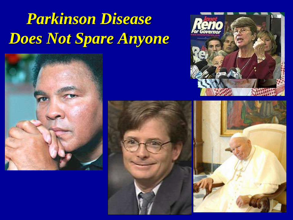
Parkinson Disease
Does Not Spare Anyone
Page 6
History of Parkinson Disease (PD) 1817: James Parkinson describes “Shaking Palsy”
1904 & 1905 : Importance of the diagnosis of paralysis of vertical movements of the eyes (Posey & Spiller )
1912: German pathologist Frederick Lewy describes neuronal cytoplasmic inclusions = Lewy bodies
1951: Apomorphine injection improved symptoms in a PD patient (Schwab)
1952: Intra-operative DBS for surgical destruction of GPi and thalamus (Spiegel)
1960: Parkinson disease is a state of dopamine deficiency (Ehringer & Hornykiewicz)
1961: First trial of IV levodopa in a PD patient (Birkmayer)
1964: Progressive supranuclear palsy: clinical and pathologic description (Steele, Richardson, Olszewski) 1968: Corticodentatonigral degeneration with neuronal achromasia (Rubeiz, Kolodny, Richardson)
1991: DBS of ventral intermediate thalamic (Vim) nucleus (Benabid)
1995: DBS for subthalamic nucleus (STN) in PD (Limousin)
Page 7
Idiopathic Parkinson Disease (PD) Definition of PD:
Parkinsonism
Degeneration of dopaminergic neurons in the substantia nigra pars compacta
Lewy bodies in degenerating neurons
Parkinsonism: 2/3 Cardinal Symptoms
Tremor at rest
Bradykinesia
Rigidity
Rule out Parkinson Plus Syndromes (atypical parkinsonism):
10% of parkinsonian patients
Rapidly disabling
Poorly treatable
Page 8
Differential Diagnosis of Parkinsonism
Parkinson Plus Syndromes Multiple system atrophy: (oligodendroglial intracytoplasmic inclusions) Shy-Drager Sd (P + autonomic Sx)
Olivo-ponto-cerebellar atrophy-OPCA (P + cerebellar Sx)
Striatonigral degeneration (P + poor response to levodopa)
Progressive supranuclear palsy (P + early imbalance + eyes movement abnormalities)
Corticobasal ganglionic degeneration-CBGD (P + unilateral dystonia + cortical sensation loss)
Parkinsonism + early dementia: Alzheimer disease + Parkinsonism
Diffuse Lewy body disease
Vascular dementia + Parkinsonism (white matter disease?)
Lower body Parkinsonism: Vascular Parkinsonism (white matter disease?)
Normal pressure hydrocephalus
IPD vs
{
Synuclei { nopathy
Synucleinopathy {
tauopathy
Page 9
Gross Pathology in Parkinson Disease
Normal midbrain Parkinson disease
SNc
Extent of injury needed to cause parkinsonian signs?
Page 10
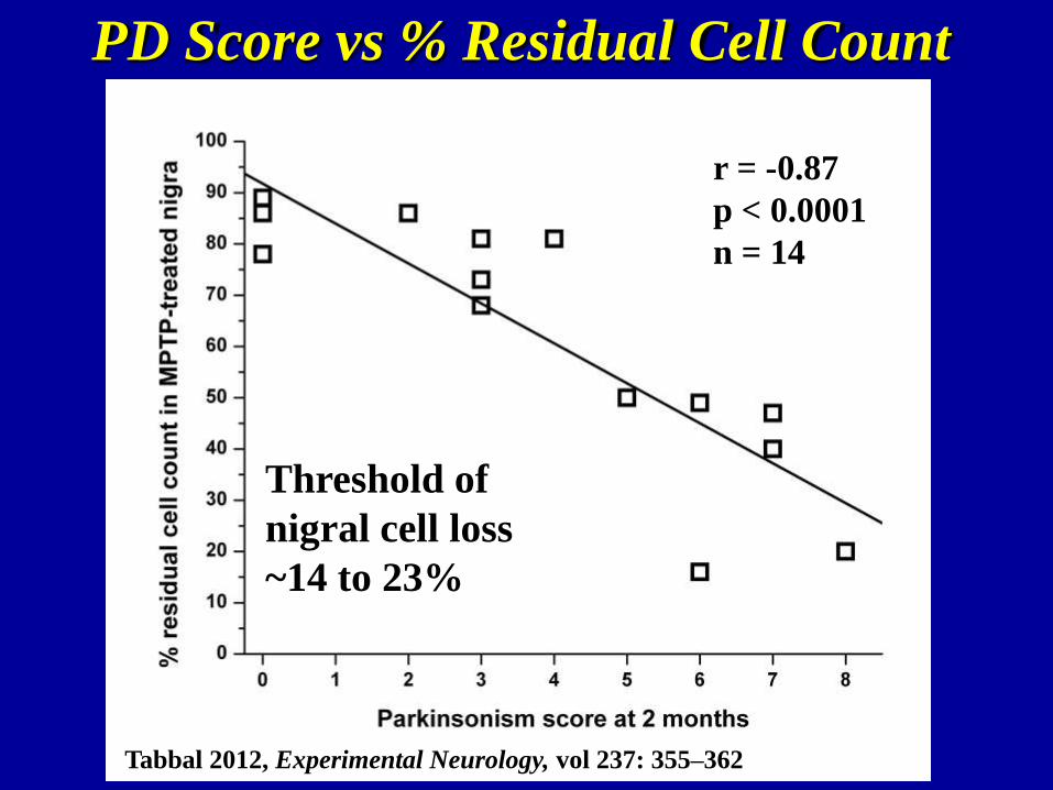
PD Score vs % Residual Cell Count
r = -0.87
p < 0.0001
n = 14
Threshold of
nigral cell loss
~14 to 23%
Tabbal 2012, Experimental Neurology, vol 237: 355–362
Page 11
PD Score vs % Residual Striatal Dopamine
r = -0.77
p = 0.016
n = 9
Threshold of
striatal dopamine
loss ~14 to 37%
Tabbal 2012, Experimental Neurology, vol 237: 355–362
Page 12
Motor symptoms develop after loosing >80% of dopaminergic neurons
Page 13
Alpha-Synuclein Normal α-synuclein:
Location: presynaptic membranes and vesicular structures
Function: synaptic vesicle recycling ?
Aggregated α-synuclein:
Major component of Lewy bodies (with ubiquitin) & Lewy
neurites
α-synuclein oligomer can transfect neurons, mediating toxicity
Adapted from Lansbury & Brice 2002
Page 14
Lewy Bodies, Neurites & Plaques Lewy Bodies’ main components
are ubiquitin & α-synuclein
α-synuclein aggregates can be:
Cytoplasmic (soma): Lewy Bodies
Axonal: Lewy neurites (d, e, f)
Extracellular: Lewy plaques (g)
● Aβ core surrounded by
α-synuclein dystrophic neurites
club-shaped filiform varicose Braak H & Del Tredici K 2009, Adv Anat Embryol Cell Biol
Page 15
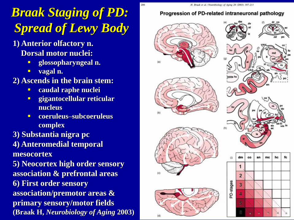
Braak Staging of PD:
Spread of Lewy Body 1) Anterior olfactory n.
Dorsal motor nuclei: glossopharyngeal n.
vagal n.
2) Ascends in the brain stem: caudal raphe nuclei
gigantocellular reticular
nucleus
coeruleus–subcoeruleus
complex
3) Substantia nigra pc
4) Anteromedial temporal
mesocortex
5) Neocortex high order sensory
association & prefrontal areas
6) First order sensory
association/premotor areas &
primary sensory/motor fields (Braak H, Neurobiology of Aging 2003)
Page 16
Behavior of Lewy Bodies Lewy bodies contains > 70 molecules, including:
calbindin, complement proteins, microfilament subunits, tubulin, microtubule associated protein 1 and 2, and Pael-R (a parkin substrate protein)
Lewy bodies formation can spread from neuron to neuron in animal models and humans (Luk KC, Science 2012)
Fetal tissue dopaminergic neurons transplanted in the striatum of humans develop Lewy bodies (Kordower, Nat Med 2008)
Lewy bodies may protect neurons from degeneration (Wakabayashi K et al, Neuropathology 2007; Bodner RA et al, Proc Natl Acad Sci 2006)
Braak Staging:
Reflects extent of spread, may not reflect sequence of spread
No correlation between Braak stage and clinical severity of PD (Burke RE, Ann Neurol 2008)
No Lewy bodies in Parkinson syndrome associated with Park 2 gene or LRRK 2 gene mutations
Page 17
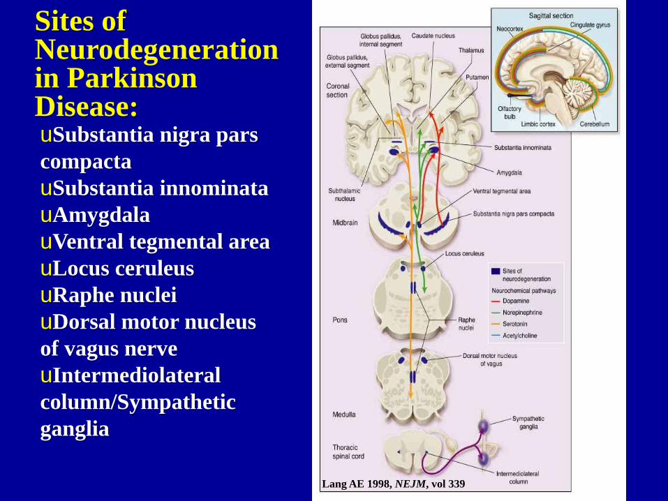
Sites of Neurodegeneration in Parkinson Disease: uSubstantia nigra pars
compacta
uSubstantia innominata
uAmygdala
uVentral tegmental area
uLocus ceruleus
uRaphe nuclei
uDorsal motor nucleus
of vagus nerve
uIntermediolateral
column/Sympathetic
ganglia
Lang AE 1998, NEJM, vol 339
Page 18
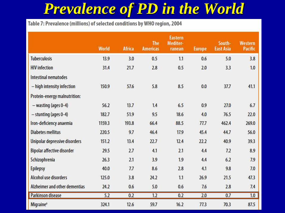
Prevalence of PD in the World
Page 19
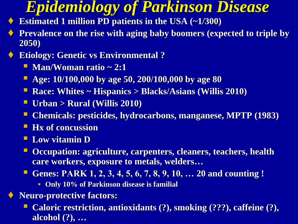
Epidemiology of Parkinson Disease ♦ Estimated 1 million PD patients in the USA (~1/300)
♦ Prevalence on the rise with aging baby boomers (expected to triple by 2050)
♦ Etiology: Genetic vs Environmental ?
Man/Woman ratio ~ 2:1
Age: 10/100,000 by age 50, 200/100,000 by age 80
Race: Whites ~ Hispanics > Blacks/Asians (Willis 2010)
Urban > Rural (Willis 2010)
Chemicals: pesticides, hydrocarbons, manganese, MPTP (1983)
Hx of concussion
Low vitamin D
Occupation: agriculture, carpenters, cleaners, teachers, health care workers, exposure to metals, welders…
Genes: PARK 1, 2, 3, 4, 5, 6, 7, 8, 9, 10, … 20 and counting !
Only 10% of Parkinson disease is familial
♦ Neuro-protective factors:
Caloric restriction, antioxidants (?), smoking (???), caffeine (?), alcohol (?), …
Page 20
Wright-Willis A et al, Neuroepidemiology 2010 ; vol 34:143–151
County level age- and race-standardized incidence (per 100,000) of Parkinson Disease
Medicare Data 2003
“Parkinson
disease
belt”
Page 21
PARK Genes, Loci & Inheritance
Klein C & Westenberger A2012, Cold Spring Harb Perspect Med 2012;2:a008888, page 3
with no Lewy bodies
with no Lewy bodies
Page 22
Genes &
Loci
Associated
with
Familial
PD
Page 23
Pathogenesis of
Parkinson Disease
Protein handling: ubiquitin & α-synuclein
Mitochondrial dysfunction
Oxidative stress: free radicals & iron
Nitrosative stress: Nitric oxide, peroxynitrite & protein nitration
Inflammation
Excito-toxicity: glutamate & calcium
Deficiency of trophic factors
Apoptosis (?)
Page 24
Ubiquitin-Proteasome System (UPS) (activating)
(conjugating)
(Ligase)
Adapted from Glickman & Ciechanover 2002
UCH-L1=Ubiquitin carboxyl-terminal hydrolase/esterase L1
Page 25
Effects of Genes on the UPS &
Protein Degradation PARK 1 (& PARK 4) code for α-synuclein: mutations cause
natively unfolded α-synuclein protein to alter its secondary
structure and self-aggregate
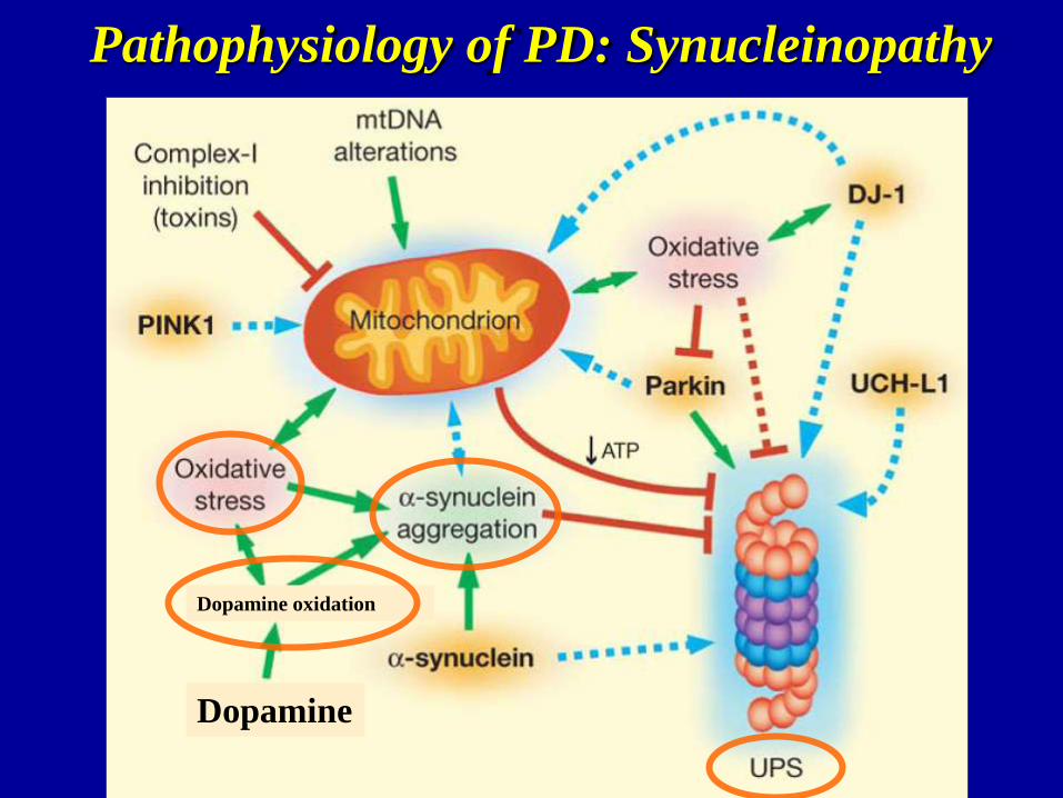
The proteins Parkin (from PARK 2), Pink1 (from PARK 6)
and DJ-1 (from PARK 7) bind to each other to form a
complex that promotes degradation of unfolded or misfolded
proteins via the UPS
PARK 9 mutations: ATP13a2 deficiency can cause lysosomal
dysfunction
Potential drug that may reduce α-synuclein in Phase I study
Nilotinib: tyrosine kinase inhibitor that interacts with α-synuclein
PRX002 vaccine: anti-α-synuclein antibodies
PD01A vaccine: induces anti-α-synuclein antibodies production
Page 26
Mitochondrial Dysfunction in PD Complex I activity is decreased by 32 to 38 % in substantia nigra
Complex I inhibition induces Lewy body-like inclusions in vivo
MPTP and rotenone inhibit complex I
Rotenone mice model: develop pathologically with fibrillary cytoplasmic inclusions staining for ubiquitin and α-synuclein
Mitochondrial dysfunction increases oxidative and nitrosative stress
Parkin, PINK1 and DJ-1 contribute to normal mitochondrial function
Mitochondrial membrane potential and intracellular ATP levels are significantly decreased in skin fibroblasts of patients with LRRK2 G2019S mutation
Mitochondrial dysfunction promotes α-synuclein aggregation
Page 27
Hypothesis: inappropriate production of reactive oxygen species (free radicals) leads to neurodegeneration
Dopamine metabolic pathways hydrogen peroxide, superoxide anions and hydroxyradicals toxic lipid peroxidation injuring cell membranes neuronal degeneration
Neuromelanin:
made of 5,6-dihydroxyindole monomers
Protects neurons: Excess dopamine and DOPA molecules are oxidized by iron catalysis quinones and semiquinones phagocytosed and stored as neuromelanin
Injures neurons: dying neurons may release neuromelanin, leading to chronic inflammation
Iron plays a critical role in oxidative metabolism
is increased by about 50% in substantia nigra of PD brains relative to controls
is a cofactor in the synthesis of neurotransmitters
Oxidative Stress
Page 28
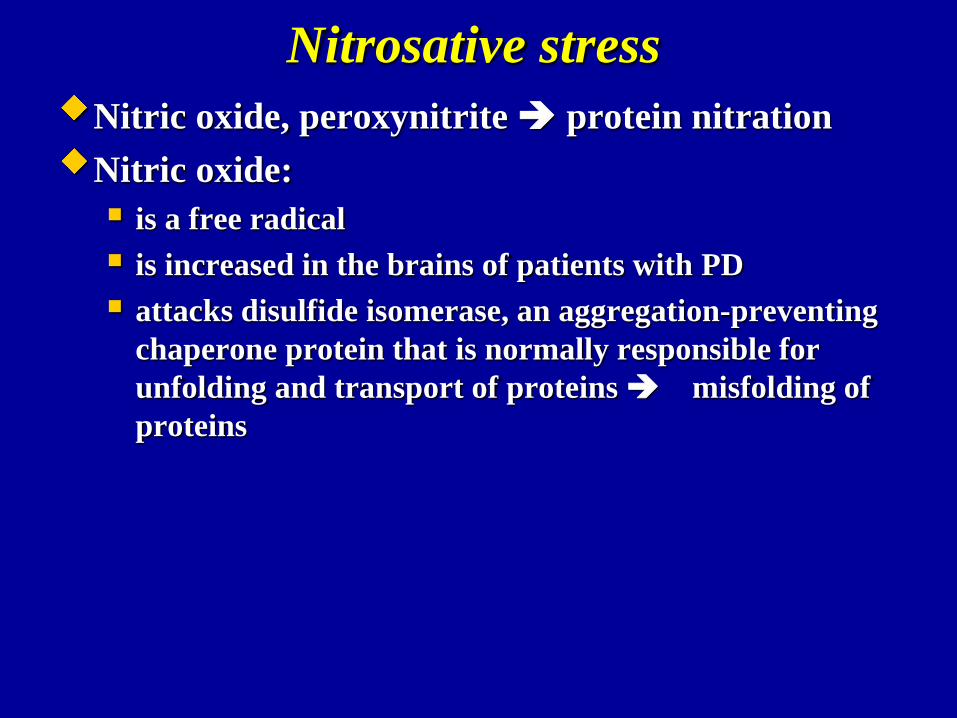
Nitrosative stress
Nitric oxide, peroxynitrite protein nitration
Nitric oxide:
is a free radical
is increased in the brains of patients with PD
attacks disulfide isomerase, an aggregation-preventing
chaperone protein that is normally responsible for
unfolding and transport of proteins misfolding of
proteins
Page 29
Pathophysiology of PD: Synucleinopathy
Dopamine
Dopamine oxidation
Page 30
The Role of Inflammation in PD
Inflammation unlikely the etiology of PD but it may contribute to neuronal injury
Cyclooxygenase-2:
the rate-limiting enzyme in prostaglandin E2 synthesis
is upregulated in patients with PD and in the MPTP mouse model of PD cyclooxygenase-2 inhibition prevents the formation of potentially toxic dopamine-quinones in MPTP mice and possibly in patients with PD
In a PET study, microglial activity in patients with PD correlated with decreased density of dopamine transporter.
Infiltration of CD4+ T-lymphocytes contributed to neuronal cell death in a mouse model of PD
Page 31
Deficiency of Trophic Factors GDNF & BDNF levels are decreased in PD patients
Is this a cause or result of neuronal degeneration?
Intraventricular GDNF: did not reach target neurons
Paliroden: failed
Exercise is very likely neuroprotective
in part through increasing neurotrophic factors?
Fasting increases neurotrophic factors in animals
Potential neuroprotective factors in on-going studies: Neurturin (CERE-120)
● Putaminal and/or nigral AAV-2 vector encoding for a neurotrophic factor
Cogane (PYM50028)
● Neurotrophic factor modulator, oral
Davunetide
● Neurotrophic protein, nasal
GM1 ganglioside
● Potentiates BDNF and NGF
Page 32
Which Pathophysiology is the Most
Significant One in PD Mitochondrial dysfunction and Oxidative stress: antioxidants have failed so far, including: MAO inhibitors
(selegiline, rasagiline), vitamin E (up to 2,000 IU/day), Co-enzyme Q10 (up to 2400 mg/day), mitoquinone ...
Uric acid (studies ongoing; risk of gout?)
Inflammation: Pioglitazone (microglia inhibitor) and minocycline (anti-inflammatory drug)
Excito-toxicity (glutamate/calcium): Riluzole failed
anti-glutamate drugs often cause hallucinations
Apoptosis: CEP-1347 and immunophilin failed
Excito-toxicity: glutamate & calcium
Deficiency of trophic factors
Page 33
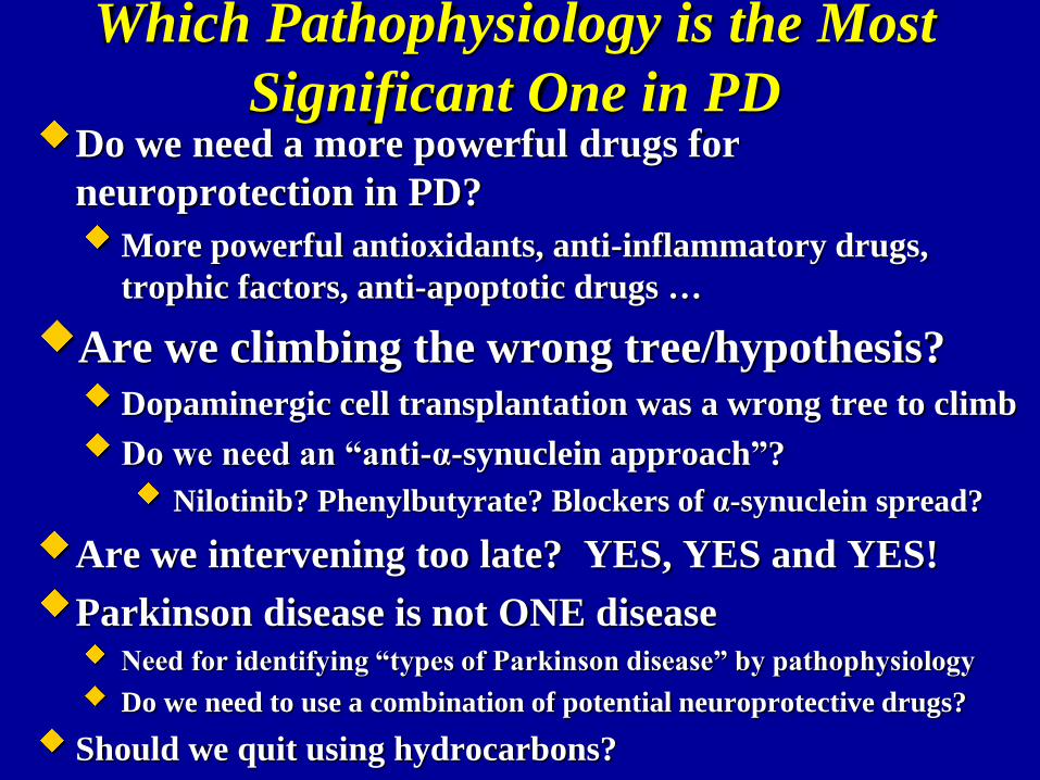
Which Pathophysiology is the Most
Significant One in PD Do we need a more powerful drugs for
neuroprotection in PD?
More powerful antioxidants, anti-inflammatory drugs,
trophic factors, anti-apoptotic drugs …
Are we climbing the wrong tree/hypothesis?
Dopaminergic cell transplantation was a wrong tree to climb
Do we need an “anti-α-synuclein approach”?
Nilotinib? Phenylbutyrate? Blockers of α-synuclein spread?
Are we intervening too late? YES, YES and YES!
Parkinson disease is not ONE disease Need for identifying “types of Parkinson disease” by pathophysiology
Do we need to use a combination of potential neuroprotective drugs?
Should we quit using hydrocarbons?
Page 34
Ann Neurol 2014
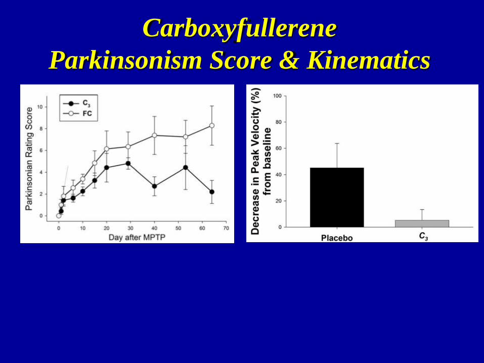
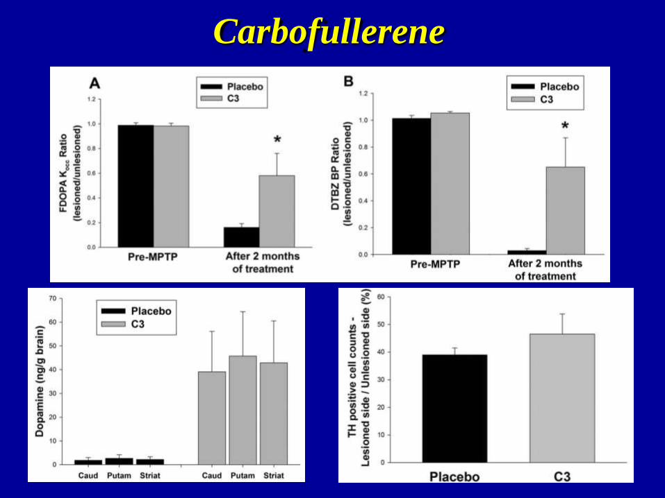
Carboxyfullerene (C3):
“The Radical Sponge” C3:
has tremendous anti-oxidant and some anti-inflammatory
effects
can be given orally
Page 35
Carboxyfullerene
Parkinsonism Score & Kinematics