26
Eugen B. Hug, MD Medical Director, ProCure Proton Therapy Centers, NY Proton Therapy for tumors of the skull base
Eugen B. Hug, MD
Medical Director,
ProCure Proton Therapy
Centers, NY
Proton Therapy for tumors of the skull base
2
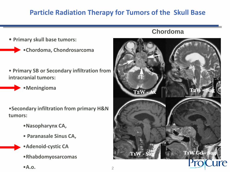
• Primary skull base tumors:
•Chordoma, Chondrosarcoma
• Primary SB or Secondary infiltration from intracranial tumors:
•Meningioma
•Secondary infiltration from primary H&N tumors:
•Nasopharynx CA,
• Paranasale Sinus CA,
•Adenoid-cystic CA
•Rhabdomyosarcomas
•A.o.
Chordoma
Particle Radiation Therapy for Tumors of the Skull Base
3
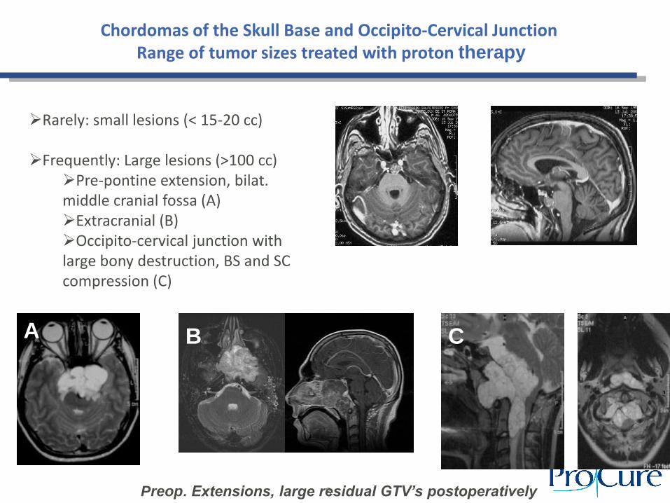
Chordomas of the Skull Base and Occipito-Cervical Junction Range of tumor sizes treated with proton therapy
Rarely: small lesions (< 15-20 cc)
Frequently: Large lesions (>100 cc) Pre-pontine extension, bilat. middle cranial fossa (A) Extracranial (B) Occipito-cervical junction with large bony destruction, BS and SC compression (C)
Preop. Extensions, large residual GTV’s postoperatively
A B C
4
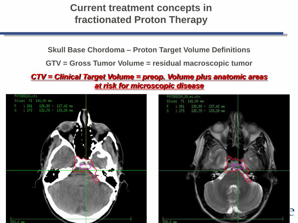
Skull Base Chordoma – Proton Target Volume Definitions
GTV = Gross Tumor Volume = residual macroscopic tumor
CTV = Clinical Target Volume = preop. Volume plus anatomic areas
at risk for microscopic disease
Current treatment concepts in
fractionated Proton Therapy
5
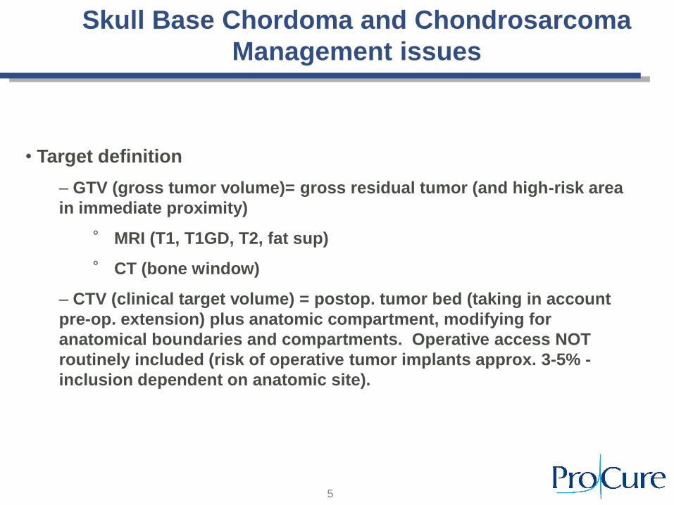
Skull Base Chordoma and Chondrosarcoma
Management issues
• Target definition
– GTV (gross tumor volume)= gross residual tumor (and high-risk area
in immediate proximity)
° MRI (T1, T1GD, T2, fat sup)
° CT (bone window)
– CTV (clinical target volume) = postop. tumor bed (taking in account
pre-op. extension) plus anatomic compartment, modifying for
anatomical boundaries and compartments. Operative access NOT
routinely included (risk of operative tumor implants approx. 3-5% -
inclusion dependent on anatomic site).
6 6
Skull Base Chordomas and Chondrosarcomas -
The importance of Oncologic Contouring
7
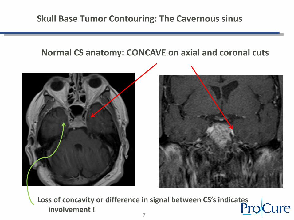
Skull Base Tumor Contouring: The Cavernous sinus
Normal CS anatomy: CONCAVE on axial and coronal cuts
Loss of concavity or difference in signal between CS’s indicates involvement !
8
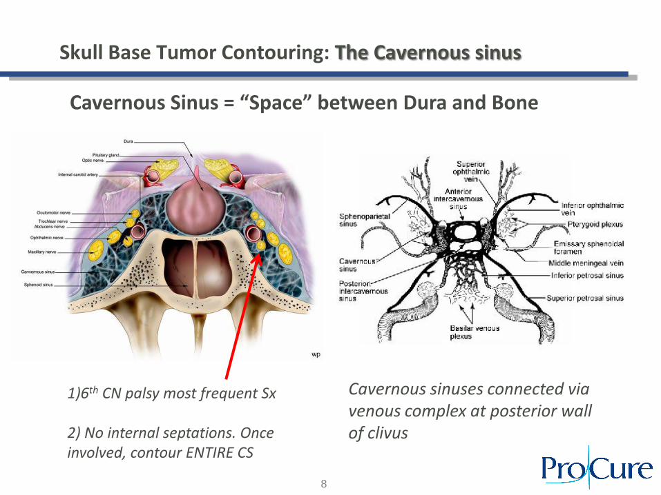
Skull Base Tumor Contouring: The Cavernous sinus
Cavernous sinuses connected via venous complex at posterior wall of clivus
1)6th CN palsy most frequent Sx 2) No internal septations. Once involved, contour ENTIRE CS
Cavernous Sinus = “Space” between Dura and Bone
9
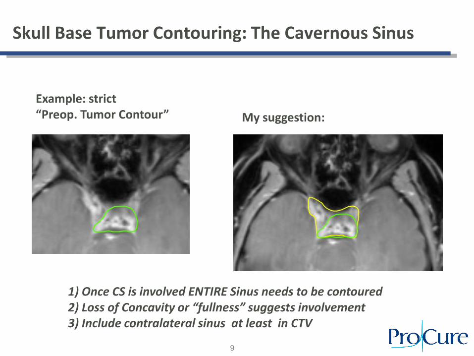
Skull Base Tumor Contouring: The Cavernous Sinus
Example: strict “Preop. Tumor Contour” My suggestion:
1) Once CS is involved ENTIRE Sinus needs to be contoured 2) Loss of Concavity or “fullness” suggests involvement 3) Include contralateral sinus at least in CTV
10
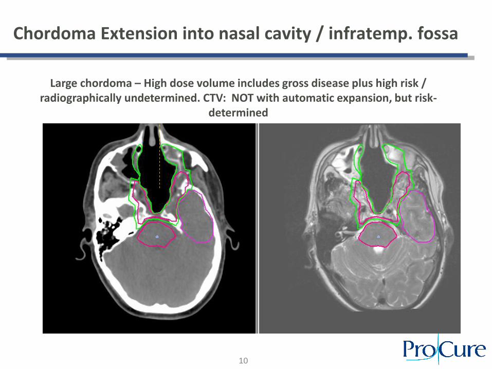
Chordoma Extension into nasal cavity / infratemp. fossa
Large chordoma – High dose volume includes gross disease plus high risk / radiographically undetermined. CTV: NOT with automatic expansion, but risk-
determined
11
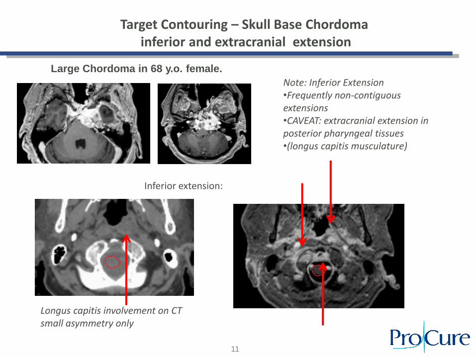
Large Chordoma in 68 y.o. female.
Note: Inferior Extension •Frequently non-contiguous extensions •CAVEAT: extracranial extension in posterior pharyngeal tissues •(longus capitis musculature)
Longus capitis involvement on CT small asymmetry only
Inferior extension:
Target Contouring – Skull Base Chordoma inferior and extracranial extension

12
Extracranial Extension: Under-contouring can be significant source of marginal failure
Involvement of posterior pharynx / longus capitis muscle requires generous target coverage – most importantly inferior: Rule: CTV extends 1 vertebral body inferior to GTV as per MRI.
Target Contouring – Skull Base Chordoma inferior and extracranial extension
13
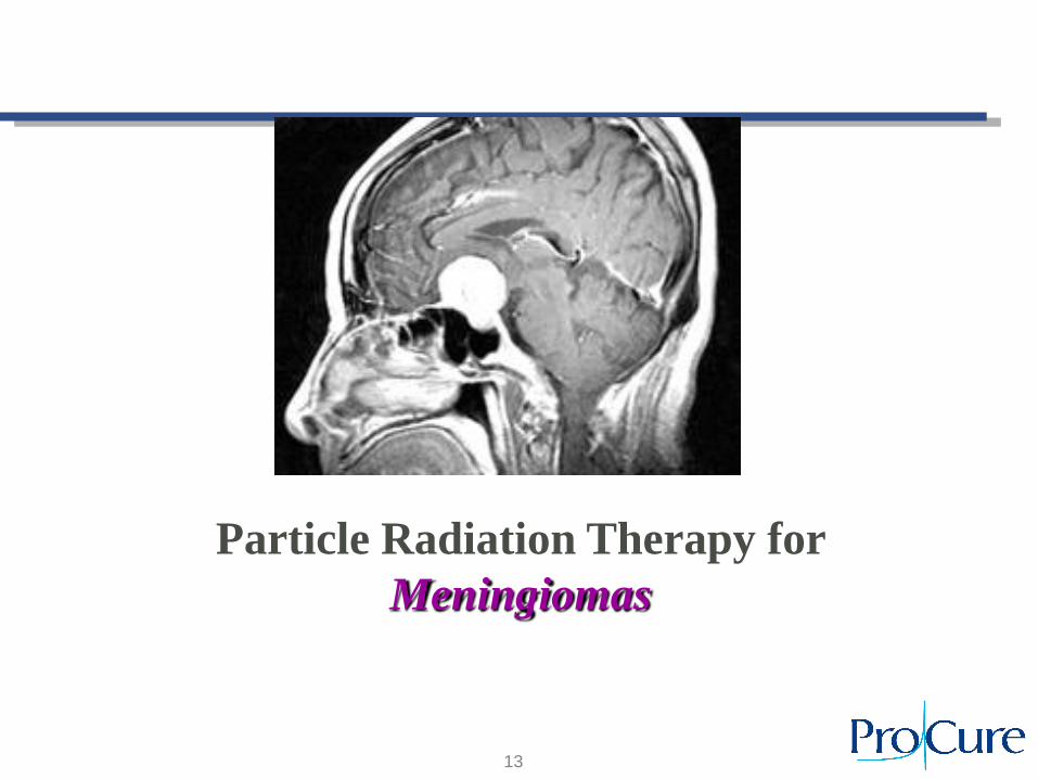
Particle Radiation Therapy for
Meningiomas
14
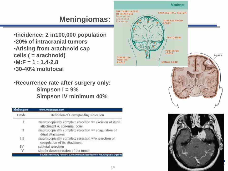
Meningiomas: •Incidence: 2 in100,000 population
•20% of intracranial tumors
•Arising from arachnoid cap
cells ( = arachnoid)
•M:F = 1 : 1.4-2.8
•30-40% multifocal
•Recurrence rate after surgery only:
Simpson I = 9%
Simpson IV minimum 40%

15
WHO re-classification 2007 (D. Louis et al, 2011)
Meningiomas: WHO Histopathologic Classification
Note: Brain invasion per se no longer Grade IV
16
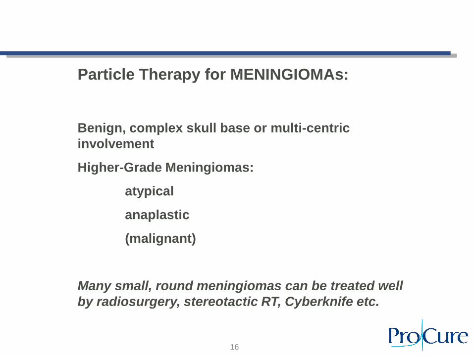
Particle Therapy for MENINGIOMAs:
Benign, complex skull base or multi-centric
involvement
Higher-Grade Meningiomas:
atypical
anaplastic
(malignant)
Many small, round meningiomas can be treated well
by radiosurgery, stereotactic RT, Cyberknife etc.
17
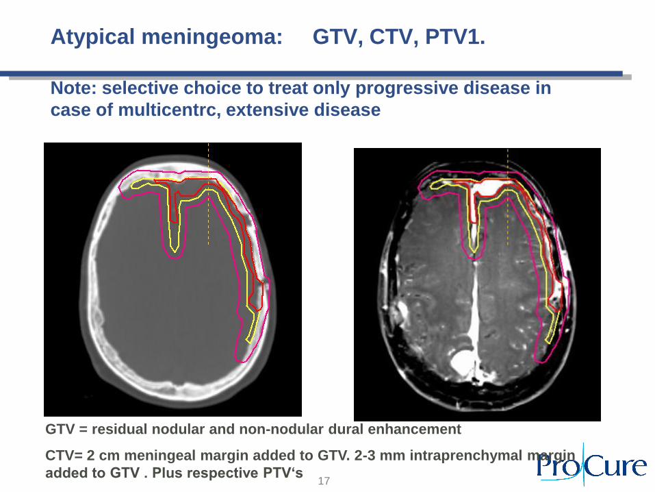
Atypical meningeoma: GTV, CTV, PTV1.
Note: selective choice to treat only progressive disease in
case of multicentrc, extensive disease
GTV = residual nodular and non-nodular dural enhancement
CTV= 2 cm meningeal margin added to GTV. 2-3 mm intraprenchymal margin
added to GTV . Plus respective PTV‘s
18
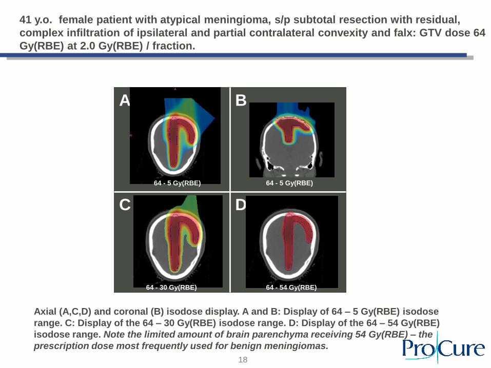
A B
C D
64 - 5 Gy(RBE) 64 - 5 Gy(RBE)
64 - 30 Gy(RBE) 64 - 54 Gy(RBE)
Axial (A,C,D) and coronal (B) isodose display. A and B: Display of 64 – 5 Gy(RBE) isodose
range. C: Display of the 64 – 30 Gy(RBE) isodose range. D: Display of the 64 – 54 Gy(RBE)
isodose range. Note the limited amount of brain parenchyma receiving 54 Gy(RBE) – the
prescription dose most frequently used for benign meningiomas.
41 y.o. female patient with atypical meningioma, s/p subtotal resection with residual,
complex infiltration of ipsilateral and partial contralateral convexity and falx: GTV dose 64
Gy(RBE) at 2.0 Gy(RBE) / fraction.
19
Particle Radiation Therapy for
Adenoid-cystic Carcinoma
of the Skull Base

20
Primary tumor :
tongue
Recurrence at 6
yrs.: skull base
Proton-Radiotherapy for skull base
tumors:
Adenoidcystic Carcinoma of the H&N
Hallmark: Perineural invasion with far proximal recurrence
Late distant metastasis

21
21
Patient: S. S, DoB 15.01.1971
Married, 2 children (2 and 7 years old)
Symptoms:
Left eyelid-weakness (ptosis)
left facial numbness
Diagnosis: (endoscopic biopsy 6/2010)
extensive sino-nasal adenoidcystic Ca.
involving left maxilla with infiltration of orbit
and skull base
pT4b cN0
Proton-Radiotherapy for skull base
tumors: Adenoidcystic Carcinoma of the
H&N
22
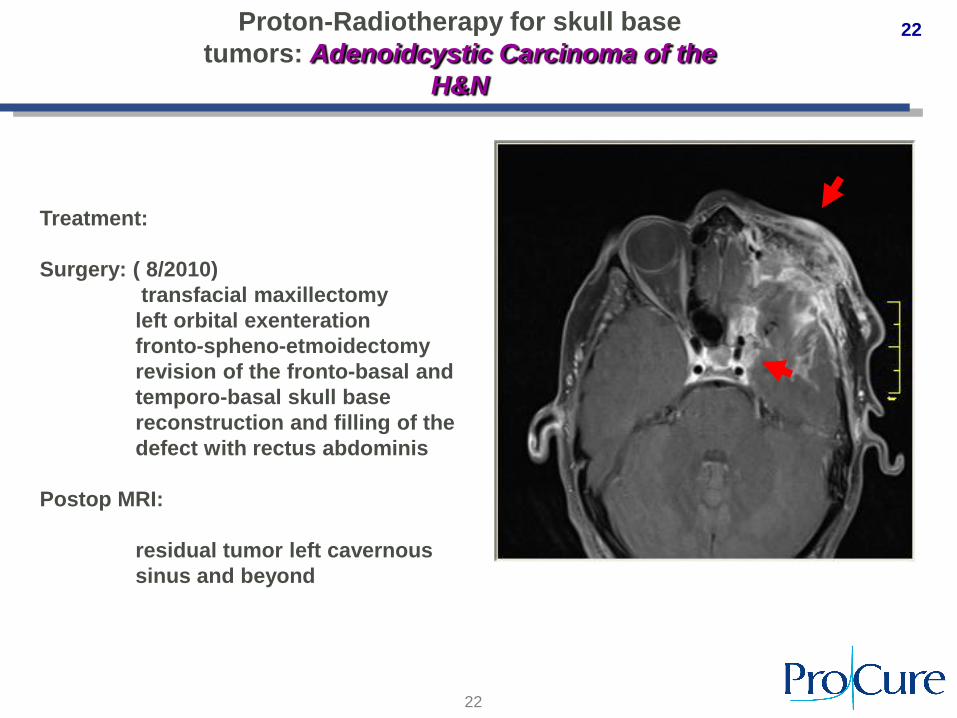
22
Treatment:
Surgery: ( 8/2010)
transfacial maxillectomy
left orbital exenteration
fronto-spheno-etmoidectomy
revision of the fronto-basal and
temporo-basal skull base
reconstruction and filling of the
defect with rectus abdominis
Postop MRI:
residual tumor left cavernous
sinus and beyond
Proton-Radiotherapy for skull base
tumors: Adenoidcystic Carcinoma of the
H&N
23
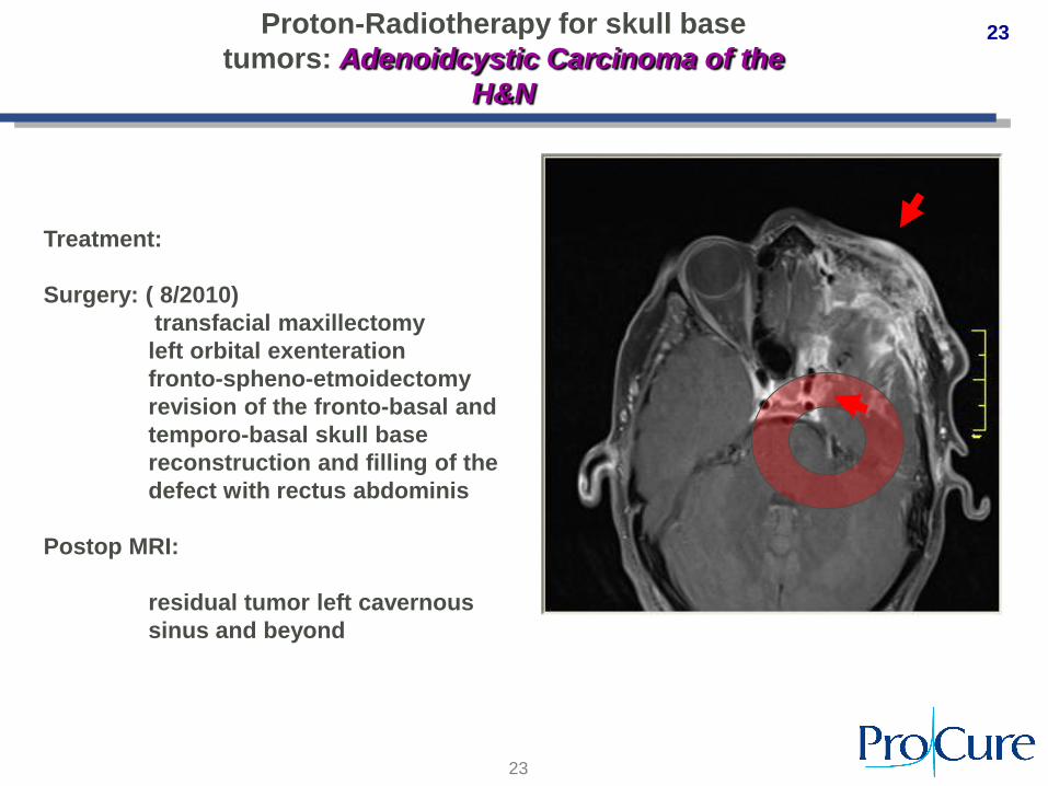
23
Treatment:
Surgery: ( 8/2010)
transfacial maxillectomy
left orbital exenteration
fronto-spheno-etmoidectomy
revision of the fronto-basal and
temporo-basal skull base
reconstruction and filling of the
defect with rectus abdominis
Postop MRI:
residual tumor left cavernous
sinus and beyond
Proton-Radiotherapy for skull base
tumors: Adenoidcystic Carcinoma of the
H&N
24
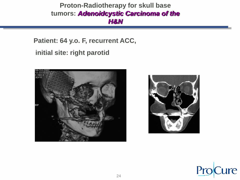
Patient: 64 y.o. F, recurrent ACC,
initial site: right parotid
Proton-Radiotherapy for skull base
tumors: Adenoidcystic Carcinoma of the
H&N
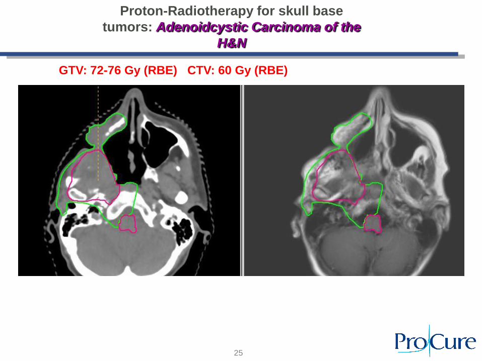
25
GTV: 72-76 Gy (RBE) CTV: 60 Gy (RBE)
Proton-Radiotherapy for skull base
tumors: Adenoidcystic Carcinoma of the
H&N
26
…. to be continued


![BEN Helps P2P [PROCURE-TO-PAY] Presents “The Procure to Pay Life Cycle” BEN Helps & P2P [PROCURE-TO-PAY] Presents “The Procure to Pay Life Cycle” Last.](https://static.documents.pub/doc/80x56/56649d6d5503460f94a4d696/ben-helps-p2p-procure-to-pay-presents-the-procure-to-pay-life-cycle.jpg)


