90


EUROPEANBRAIN
RESEARCHINSTITUTE
RITA LEVI-MONTALCINI FOUNDATION
ROME


TABLE OF CONTENTSINDICE
INTRODUCTION ................................................................................................................................................................4INTRODUZIONE
MESSAGE FROM THE GENERAL DIRECTOR ...............................................6MESSAGGIO DEL DIRETTORE GENERALE
THE INSTITUTE...........................................................................................................................................................8L’ISTITUTO
Aims/ Finalità....................................................................................................................................................................................10Structure/ Struttura......................................................................................................................................................................14Management Board/ Consiglio di Amministrazione ................................................................................................16International Scientific Council/ Consiglio Scientifico Internazionale ........................................................17
LABORATORIES, UNITS AND FACILITIES .......................................18LABORATORI, UNITÀ E INFRASTRUTTURE
Laboratories/ LaboratoriNeurotrophic Factors and Neurodegenerative Diseases Laboratory, Antonino Cattaneo...............20Nerve Growth Factor Laboratory, Pietro Calissano ................................................................................................30Neural Stem Cells and Neurogenesis Laboratory, Marco Canossa...............................................................36Pharmacology of Synaptic Plasticity Laboratory, Giuseppe Nisticò..............................................................40Neuropathic Pain Laboratory, Silvia Marinelli ............................................................................................................48Metabolism in Brain Diseases Laboratory, Michelangelo Campanella .......................................................52
Units/ UnitàMechanisms of Neuronal and Synaptic Plasticity Unit, Cristina Marchetti, Hélène Marie ..............58Cortical Microcircuits Unit, Antonio Pazienti, Alberto Bacci...............................................................................60
Facilities/ InfrastruttureGenomics Facility, Mara D’Onofrio...................................................................................................................................62Confocal Microscopy Facility, Fulvio Florenzano......................................................................................................64
ACTIVITIES........................................................................................................................................................................66ATTIVITÀ
Scientific Collaborations/ Collaborazioni scientifiche...........................................................................................68Ebri Supporting Members/ Soci sostenitori dell’Ebri.............................................................................................70Seminars and Events/ Seminari ed eventi.....................................................................................................................72Ebri Publications 2005-2012/ Pubblicazioni Ebri 2005-2012..........................................................................74The Ebri Journal/ La rivista dell’Ebri ..................................................................................................................................82Schools in the lab/ Scuole in laboratorio .......................................................................................................................83
HOW YOU CAN SUPPORT EBRI...............................................................................................................................84COME SOSTENERE LA FONDAZIONE EBRI

PAGINA 6 THE EUROPEAN BRAIN RESEARCH INSTITUTE
INTRODUCTION
As we stride into the third millennium, our attention mustfocus on a better understanding of the brain and on the ever increasing incidenceof neurodegenerative diseases. The social-sanitary costs of neurological patholo-gies are reaching astronomical proportions in western countries, due to the stea-dily-increasing average age of the populations.
THE EUROPEAN BRAIN RESEARCH INSTITUTE (EBRI) wasestablished in order to encourage a synergy between researchers coming fromdifferent scientific and humanistic backgrounds. EBRI aims to pursue studiesin basic research in the field of neuroscience and translate the research resultsinto clinical application. The Institute’s object is to bring together a critical massof researchers to share equipment and technologies to avoid the dispersion inseparate institutions of scientists of highly sophisticated and expensive instru-ments. Furthermore, EBRI intends to attract highly-qualified experts from dif-ferent countries with a particular emphasis on young researchers who havecompleted their doctoral and postdoctoral studies. This will be done by offeringthese researchers the possibility of conducting their own independent research.
The Institute proposes to involve top-level industries, ac-tive in relevant sectors, as partners in research programmes. This will createbenefits and synergies for all parties from the induced economic growth whichwill be generated. The current slowdown of economic development in Italy, re-latively greater than in other countries of a similar cultural and scientific level,can only be reversed if innovation and creativity are given priority; this will in-crease the potential of our younger generations in the most fruitful and produc-tive phase of their intellectual and creative careers.
Our Institute intends to be a centre of excellence by attrac-ting young Italian researchers with a sound scientific preparation and who aretoday obliged to seek hospitality in foreign research institutes. EBRI will also offeryoung foreign scientists research opportunities, both comparable and competitivewith other leading international centers. The research programmes will be focu-sed on the convergence and interaction of different scientific disciplines organi-zed intercommunicating and collaborative areas and not separate as before. Today,more than ever, it is important to promote the exchange of interdisciplinary andtransdisciplinary studies.
EBRI is organized on a “projected oriented” model.Such a model allows the various research groups to dynamically organize them-selves, each according to their own competences and field of interest, but withina common final objective.
At the start of the third millennium, the explorers of themind will set out, from the base camp of this Institute, to explore those fascina-ting, mysterious and still unknown realms of the galaxy: the human mind.
Rita Levi-Montalcini, PresidentPietro Calissano, Vice-President

PAGINA 7 THE EUROPEAN BRAIN RESEARCH INSTITUTE
All’inizio del terzo millennio s’impone una conoscenza sem-pre più approfondita del funzionamento del cervello e delle sue patologie. Il costo socio-sanitario delle patologie neurologiche va assumendo proporzioni im-ponenti nei Paesi del mondo occidentale a causa del prolungamento costante del-l’età media.
THE EUROPEAN BRAIN RESEARCH INSTITUTE (EBRI)Allo scopo di incentivare una sinergia di studi sul sistema nervoso che integri i ri-sultati ottenuti dai ricercatori provenienti da diversi settori scientifici, si è pro-mossa la costituzione di un Istituto di Ricerche sul Cervello. L’EBRI si prefigge dicondurre ricerche di base nel campo delle neuroscienze promuovendo, in paritempo, la ricaduta dei risultati conseguiti in quello clinico e di favorire la costi-tuzione di una massa critica di ricercatori verso obiettivi scientifici comuni, inmodo da evitare la dispersione di scienziati e di strumenti di alta complessità ecosto in istituzioni separate. Obiettivo primario dell’EBRI, inoltre, è di favorirel’affluenza di esperti altamente qualificati provenienti da diversi paesi, in parti-colare di giovani ricercatori che abbiano completato la propria formazione dot-torale e post-dottorale, offrendo loro opportunità di ricerca indipendente.
EBRI, inoltre, si propone di ottenere, attorno ai propri pro-grammi di ricerca, importanti ricadute economiche attraverso il coinvolgimentodi industrie. Infatti, il ritardo economico del nostro paese, rispetto a quelli di parilivello culturale e scientifico, si può recuperare solo puntando su innovazione ecreatività e valorizzando le potenzialità dei giovani nella loro fase di maggioreproduttività intellettuale e creativa.
Questo istituto rappresenterà un centro di eccellenza alquale potranno afferire giovani ricercatori italiani di alto livello scientifico cheancora oggi sono costretti a cercare ospitalità in laboratori stranieri, e di giovaniricercatori stranieri, che trovino nell’EBRI una opportunità di ricerca competitivacon quella dei migliori centri internazionali. I programmi di ricerca dell’EBRI sa-ranno centrati sulla confluenza ed interazione di differenti settori scientifici comu-nicanti e non più separati in compartimenti stagni, come in passato. Oggi èquanto mai importante avvalersi di continui scambi di studi interdisciplinari etransdisciplinari.
Il centro sarà organizzato su un modello “project oriented”.Tale modello consente a vari gruppi di ricerca di organizzarsi dinamicamente,ognuno secondo le proprie competenze ed interessi, all’interno di finalità ed obiet-tivi generali comuni.
All’inizio del terzo millennio gli esploratori della mente par-tiranno dalla postazione dell’EBRI, per esplorare le zone ancora incognite della piùaffascinante e misteriosa di tutte le galassie: la galassia mente.
INTRODUZIONE
Rita Levi-Montalcini, PresidentePietro Calissano, Vice-Presidente

With great pleasure I accepted in 2011 the appointmentas General Director of the European Brain Research Institute ( EBRI), after wor-king in my capacity as General Commissioner during a period of transition(January 2010 - July 2011). I also had the privilege of serving on the EBRI Boardof Directors when it was first established in 2003 by Rita Levi-Montalcini.
EBRI was established mainly to foster neurobiological re-search in the fundamental aspects (molecular, neurophysiological, neuropharma-cological and therapeutic) related to neurodegenerative diseases. Our aim is todevelop the Institute as a source of research excellence, to better understandthe molecular mechanisms underlying neurodegenerative diseases and translatethis knowledge into therapeutic strategies for these disorders.In the last two years we have consolidated certain existing activities and scienti-fic projects and we have increased our network of relations with prestigious in-ternational Institutes (Wolfson Institute, University College London; DresdenUniversity, Germany; Ceinge, University of Naples; Scuola Normale of Pisa, etc.). In addition, we have signed a three-year contract of collaboration with XiamenBioway Biotech China to further develop NGF (protein discovered by Rita Levi-Montalcni) and its therapeutic applications.Since 2010 additional laboratories (Metabolism in Brain Diseases, Neural StemCells and Neurogenesis, Pharmacology of Synaptic Plasticity) were establishedto enhance the research activity in specific fields in order to create synergy withexcellent research and academic institutions.
Since its establishment, and more recently in collabora-tion with the Italian Research Council, we have hosted a series of seminars andconferences with eminent scientists as invited speakers, who have made impor-tant contributions in Neuroscience. The seminars aim to encourage the exchangeof ideas and create awareness among scientists in topics that are closely alignedwith the research studies conducted at EBRI.
I am confident that with such a highly qualified group ofresearchers and the leadership of the new Board of Directors and the Internatio-nal Scientific Council, EBRI will continue to strive and further develop its stan-ding as a centre of excellence in brain research.I wish to thank miss Pina Moliterno, dr. Pamela Bernardo, mr. Piero Ientile, dr.Stefano Gasperini and dr. Libero Candreva for their precious collaboration bothin the Direction secretariat as well as in the various stages of the preparation ofthe present booklet.
Giuseppe Nisticò, General Director
MESSAGE FROM THE GENERAL DIRECTOR
PAGINA 8 THE EUROPEAN BRAIN RESEARCH INSTITUTE

Ho accettato con grande onore nel 2011 la proposta della professo-ressa Rita Levi-Montalcini di essere Direttore Generale dell’European Brain Research In-stitute (EBRI), dopo un periodo di transizione (Gennaio 2010-Luglio 2011) in cui ero statonominato Commissario straordinario. Avevo avuto anche il privilegio di far parte del Con-siglio di Amministrazione dell’EBRI fin dalla sua nascita nel 2003 ad opera di Rita Levi-Montalcini.
La missione fondamentale dell’EBRI rimane ancora oggi quella percui l’Istituto è nato e cioè di potenziare la ricerca neurobiologica nei suoi aspetti fondamen-tali (biologia molecolare, neurofisiologia, neurofarmacologia) alla base delle malattie neu-rodegenerative. Il nostro obiettivo è di sviluppare un Istituto che rappresenti una fonte di ricercadi eccellenza volta a comprendere meglio i meccanismi molecolari, che sottendono le malat-tie neurodegenerative e trasferire questi risultati in campo clinico con l’identificazione di nuovistrumenti terapeutici.
Negli ultimi due anni sono state consolidate alcune attività di suc-cesso ed è stata incrementata la rete di collaborazioni scientifiche con prestigiosi Istitutiinternazionali (Wolfson Institute dell’University College di Londra, Università di Dresda,l’Istituto Ceinge dell’Università di Napoli, la Scuola Normale di Pisa, ecc.). Inoltre, èstato da me firmato un accordo di collaborazione scientifica con la Xiamen Bioway Bio-tech della Repubblica Cinese, volto a sviluppare l’impiego terapeutico dell’NGF della RitaLevi-Montalcini.
Dal 2010 sono stati altresì attivati nuovi laboratori (Metabolismodelle malattie del SNC; Cellule staminali neurali e Neurogenesi; Farmacologia della pla-sticità sinaptica) al fine di potenziare ricerche integrate nelle malattie neurodegenerativeed estendere il network di collaborazione con eccellenti Centri di ricerca italiani e stranieri.
Fin dalla sua nascita l’EBRI e di recente in collaborazione con ilCNR ha organizzato una serie di seminari e conferenze di alto livello cui hanno partecipatostudiosi di fama internazionale che hanno ottenuto risultati importanti nelle Neuroscienze.Lo scopo dei seminari è di incoraggiare lo scambio di idee con i nostri giovani ricercatorie rendere partecipi gli scienziati invitati delle linee di ricerca dell’EBRI. Sono fiducioso checon lo staff di ricercatori che già lavorano nei nostri laboratori e con la guida del Consi-glio Scientifico Internazionale e del Consiglio di Amministrazione, l’EBRI saprà affermarsisempre di più e divenire un Centro di eccellenza internazionale nelle Neuroscienze.
Desidero ringraziare la sig.ra Pina Moliterno, la dott.ssa Pamela Ber-nardo, il rag. Piero Ientile, il dott. Stefano Gasperini e il dott. Libero Candreva per la loro pre-ziosa collaborazione sia nella segreteria e amministrazione della Direzione che nelle varie fasidi preparazione del presente volumetto.
Giuseppe Nisticò, Direttore Generale
PAGINA 9 THE EUROPEAN BRAIN RESEARCH INSTITUTE
MESSAGGIO DEL DIRETTORE GENERALE

THE INSTITUTEL’ISTITUTO


PAGINA 12 THE EUROPEAN BRAIN RESEARCH INSTITUTE
AIMSThe brain is the most complex organ in any living system.
The activity of neurons (the brain cells) underlies all behaviors, ranging fromrelatively simple tasks, such as walking or breathing to very complex cognitivefunctions, such as generation of theoretical and abstract concepts, appreciationof arts and literature as well as accomplishment of complex logical and memorytasks. Although a massive effort is underway to understand the mechanisms ofbrain function, Neuroscience is still an open frontier and the mysteries of thebrain remain largely unexplored.
As beautiful as the work of a healthy brain is, even subtle malfunctioning can result in deva-stating illnesses. Brain diseases include neurological and psychiatric diseasessuch as Alzheimer’s, Parkinson’s and Huntington’s diseases, ALS, epilepsy, schi-zophrenia, depression and bipolar disorders, autism, to mention a few. Pain, acommon condition underlying most human diseases, and the most frequent causewhy patients seek medical attention, also has a peripheral and central neurolo-
gical basis. Understanding how the brainworks, and how it can dysfun-ction in diseases will depend onour ability to link the differenthierarchical levels of the brainorganization into one unifiedconceptual framework, from mo-lecules to behavior to higherbrain functions. EBRI’s vision is
that the daunting tasks facingneuroscience can only be solved
through a highly interdisciplinary andintegrated effort, also involving the ex-
ploitation and development of new technolo-gies from different fields. The molecular and cellular
mechanisms for brain processes, be it developmental or adult pla-sticity processes, underlie all higher brain function, and constitute a commonplatform that integrates the biochemical, genetic, electrical processes occurringin neurons and synapses, providing the basis for higher processes such as lear-ning, memory, sensations and emotions and, most significantly, providing thebasis for a rational understanding of the deep roots of brain pathologies.
To implement this vision, research at EBRI will focus par-ticularly on the fundamental molecular and cellular mechanisms subserving thefunctions of developing and adult neurons and synapse belonging to different

PAGINA 13 THE EUROPEAN BRAIN RESEARCH INSTITUTE
brain circuits and areas, studied with a close integration of different cross-disci-plinary approaches, ranging from molecular biology and biochemistry to cell bio-logy and biophysics, electrophysiology, mouse genetics, neuropharmacology andlarge scale neurogenomics, bioinformatics, behavior, optical imaging. In light ofthis vision, the general scientific objective of EBRI will be to merge systems andcomputational neuroscience with cellular and molecular neurobiology.
EBRI aims to attract young talented investigators with dif-ferent scientific backgrounds, with a strong and passionate drive to study thebrain, particularly young investigators at their first independent positions afterpost doctoral work.
To implement these objectives via a multidisciplinary approach, collaboratingscientists with distinct backgrounds will utilizeand have access to a wide range of techniquesand experimental approaches, for the pur-pose of:
1. investigating how molecular events in-volved in synaptic plasticity lead to le-arning and memory, in well definedexperimental systems;
2. understanding the molecular basis ofneurodegenerative diseases of highsocial and medical impact, such as Al-zheimer’s Parkinson’s diseases;
3. exploring the role of Nerve Growth Fac-tor (NGF) and analogs in restoring altera-tions in LTP and synaptic plasticity inexperimental models of neurodegenerativediseases and its role in neuronal regeneration;
4. understanding the energetic requirements of neuronsin physiology and in pathology, as a basis for their well-being and activity;
5. Investigating the basis for neurogenesis in the developing and adult nervous system, and exploiting the potential of stem cell biology for the treatment of ner-vous system diseases;
6. exploiting the mechanistic studies to develop new therapeutics approaches toneurodegenerative diseases.

PAGINA 14
Il cervello è l’organo più complesso diqualsiasi sistema vivente. L’attività dei neuroni (le cellule ce-rebrali) sottende tutti i comportamenti, dai compiti più sem-plici come camminare o respirare, a funzioni cognitivecomplesse quali la produzione di teorie e concetti astratti,apprezzamento delle arti e della letteratura e realizzazione dicompiti logici e di memoria complessi. Sebbene siano incorso imponenti sforzi per comprendere i meccanismi dellefunzioni cerebrali, le Neuroscienze rappresentano ancora unafrontiera aperta e i misteri del cervello sono ancora ampia-mente inesplorati.
Le capacità di un cervello sano sono così so-fisticate che anche una minima alterazione può indurre patologieneurologiche e psichiatriche, quali Alzheimer, Parkinson, Huntington,SLA, epilessia, schizofrenia, depressione e disordini bipolari, autismo, permenzionarne soltanto alcune.
Il dolore, una sintomatologia comune alla maggior parte delle malattie dell’uomo, è una delle più fre-quenti cause per cui un paziente si rivolge al medico. Il dolore comprende due com-ponenti neurologiche, una periferica e una centrale. Comprendere come il cervellofunzioni in condizioni normali e come invece sia alterata la sua funzione in condi-zioni patologiche dipenderà dalla nostra capacità di correlare i diversi livelli dellagerarchia dell’organizzazione celebrale in una cornice concettuale unificata, a par-tire dalle molecole fino al comportamento e alle più alte funzioni cerebrali.
La visione dell’EBRI è che le sfide che ancora ci attendono nelleNeuroscienze possono essere risolte soltanto con un approccio altamente multidiscipli-nare e integrato, che coinvolge l’impiego e lo sviluppo di tecnologie avanzate prove-nienti da diversi campi. I meccanismi molecolari e cellulari alla base delle funzionicerebrali, durante lo sviluppo nei processi di plasticità neuronale, sottendono le piùalte funzioni celebrali e costituiscono una piattaforma comune in cui sono integratimeccanismi biochimici, genetici, elettrofisiologici, che si verificano a livello sinapticoe dei neuroni e sono alla base di funzioni cerebrali elevate quali apprendimento, me-moria, sensazioni ed emozioni. Tali meccanismi sono importanti al fine di comprendere
FINALITÀ
THE EUROPEAN BRAIN RESEARCH INSTITUTE

PAGINA 15 THE EUROPEAN BRAIN RESEARCH INSTITUTE
in maniera razionale le radici profonde delle patologieneurologiche e mentali.Allo scopo di perseguire questa visione, le linee di ri-cerca dell’EBRI saranno particolarmente concentratesui meccanismi fondamentali di tipo molecolare e cel-lulare alla base delle funzione dei neuroni in via disviluppo e di quelli adulti nonché delle sinapsi che co-stituiscono i diversi circuiti nelle varie aree del cervello,
studiate con una stretta integrazione di diversi approccimultidisciplinari che comprendono biologia molecolare,
biochimica, biologia cellulare, biofisica, elettrofisiologia,genetica, neurofarmacologia, neurogenomica, bioinforma-
tica, comportamento e imaging.Alla luce di queste considerazioni, l’obiettivo scientifico generale
di EBRI consiste nell’integrare le neuroscienze sistemiche e com-putazionali con il livello cellulare e molecolare.
EBRI ha come obiettivo fondamentale quello di at-trarre giovani talenti con una preparazione di base diversificata, con profonde
motivazioni per lo studio del cervello e in particolare giovani ricercatori che dopo lalaurea svolgono in maniera autonoma e indipendente i loro progetti di ricerca.
Per raggiungere gli obiettivi prefissati tramite un approccio multidisciplinare, i ricercatori di diversa for-mazione utilizzano tecniche di biologia molecolare e cellulare, genetica, biofisica,bioinformatica, gnomica e proteomica, elettrofisiologia, neurofarmacologia e na-notecnologie, al fine di:
1. studiare gli eventi molecolari coinvolti nella plasticità sinaptica alla base della me-moria e dell’apprendimento, in sistemi sperimentali ben definiti;
2. studiare le basi molecolari di malattie neurodegenerative di estrema rile-vanza sociale e medica, come Alzheimer, Parkinson;
3. studiare il ruolo del fattore di Crescita Nervoso (Nerve Growth Factor,NGF) ed i suoi analoghi nel ripristinare le alterazioni dell’LTP edella plasticità sinaptica in modelli sperimentali di malattie neu-rodegenerative e il suo ruolo nella rigenerazione neuronale;
4. comprendere i meccanismi energetici dei neuroni in condizionefisiologiche e patologiche;
5. studiare i meccanismi di base per la neurogenesi nello sviluppoe nei neuroni adulti ed esplorare l’uso potenziale di cellule sta-minali neurali nel trattamento di malattie neurologiche;
6. utilizzare i risultati conseguiti per sviluppare nuovi approcci tera-peutici per le malattie neurodegenerative.

PAGINA 16 THE EUROPEAN BRAIN RESEARCH INSTITUTE
EBRI is thuscommitted to providingyoung fellows with scientificindependence and freedom in the broad field ofNeuroscience, with a main focus on molecular and cellular mechanisms. Theresearch training of young fellows is accomplished via established collaborationswith local and international Universities (Università “La Sapienza”, Università “TorVergata”, University College of London, the Hebrew University of Jerusalem).Facilities on campus include a Conference hall, Meeting rooms, Animal House,Library with online data base with access to over 2500 journals, and a Cafeteria.
EBRI is located in a 25,000 sq meters building, organizedlike other prestigious European Research Institutes. EBRI has attracted therelocation on the same site of two other neuroscience institutes: the ResearchLaboratories of IRCSS Fondazione Santa Lucia, an Institute for Research,Hospitalization and Health Care involved in neuroscience research and neuromotorrehabilitation, and the Institute of Neurobiology and Molecular Medicine of theNational Research Council. The coexistence of the three Institutes in one locationhas led to the birth of an international Neuroscience “campus”, with a verysignificant critical mass of scientists and shared equipment.
EBRI has a core staff of about 40, including scientists,technicians, and administrative personnel, and is organized in independentlaboratories, units, as well as core facilities. Each group recruits its own personnelat international European levels, and is typically composed of one Senior or JuniorGroup-leader responsible for a staff of technicians, postdoctoral fellows, Ph.D.students and undergraduate students. EBRI also offers independent positions asJunior Group Leaders, and positions as Junior Project Leaders within an establishedLaboratory.
STRUCTURE

PAGINA 17
EBRI ha sede in un edificio di25.000 mq, con un’organizzazione analoga a quella
di altri prestigiosi Centri di ricerca europea.EBRI ha, tra l’altro, favorito l’insediamento di
altri due Istituti di Neuroscienze nella stessasede: i laboratori di Ricerca dell’IRCCS Fon-dazione Santa Lucia, un Istituto di Ricerca,Ospedalizzazione e Cura dedicato allaRiabilitazione Neuromotoria, e l’Istituto diNeurobiologia e Medicina Molecolare delCNR. La coesistenza dei tre Istituti nellastessa sede ha permesso lo sviluppo di uncampus internazionale di Neuroscienzecon una concentrazione significativa di
scienziati e attrezzature.
Il personale EBRI ècomposto da circa 40 unità tra ricercatori, tec-
nici e personale amministrativo. È strutturato inlaboratori indipendenti, unità ed infrastrutture cen-
tralizzate. Ciascun gruppo recluta il proprio personalea livello internazionale ed è tipicamente composto da un
Senior e Junior Group-leader con la responsabilità di perso-nale tecnico, laureati e laureandi, dottorandi e dottori di ricerca.
EBRI offre anche posizioni indipendenti di Junior Group Leader e posizionidi Junior Project Leader all’interno dei laboratori esistenti.
In questo modo, EBRI è impegnato nella formazione di gio-vani collaboratori attraverso la ricerca nel campo delle neuroscienze. Tale processo diformazione è favorito dall’interazione con Università locali e internazionali qualil’University College di Londra e l’Hebrew University di Gerusalemme, tramite accordidi collaborazione in corso (Università “La Sapienza” e Università “Tor Vergata”).Le infrastrutture del campus comprendono una sala conferenza, sale per le riunioni,uno stabulario, una biblioteca con data base on-line per l’accesso a più di 2500 rivi-ste scientifiche, e una caffetteria.
THE EUROPEAN BRAIN RESEARCH INSTITUTE
EBRI building at the Santa Lucia Foundation, Via del Fosso di Fiorano, Rome
Struttura dell’EBRI presso la Fondazione S. Lucia, Via del Fosso di Fiorano, Roma
STRUTTURA
PAGINA 18 THE EUROPEAN BRAIN RESEARCH INSTITUTE
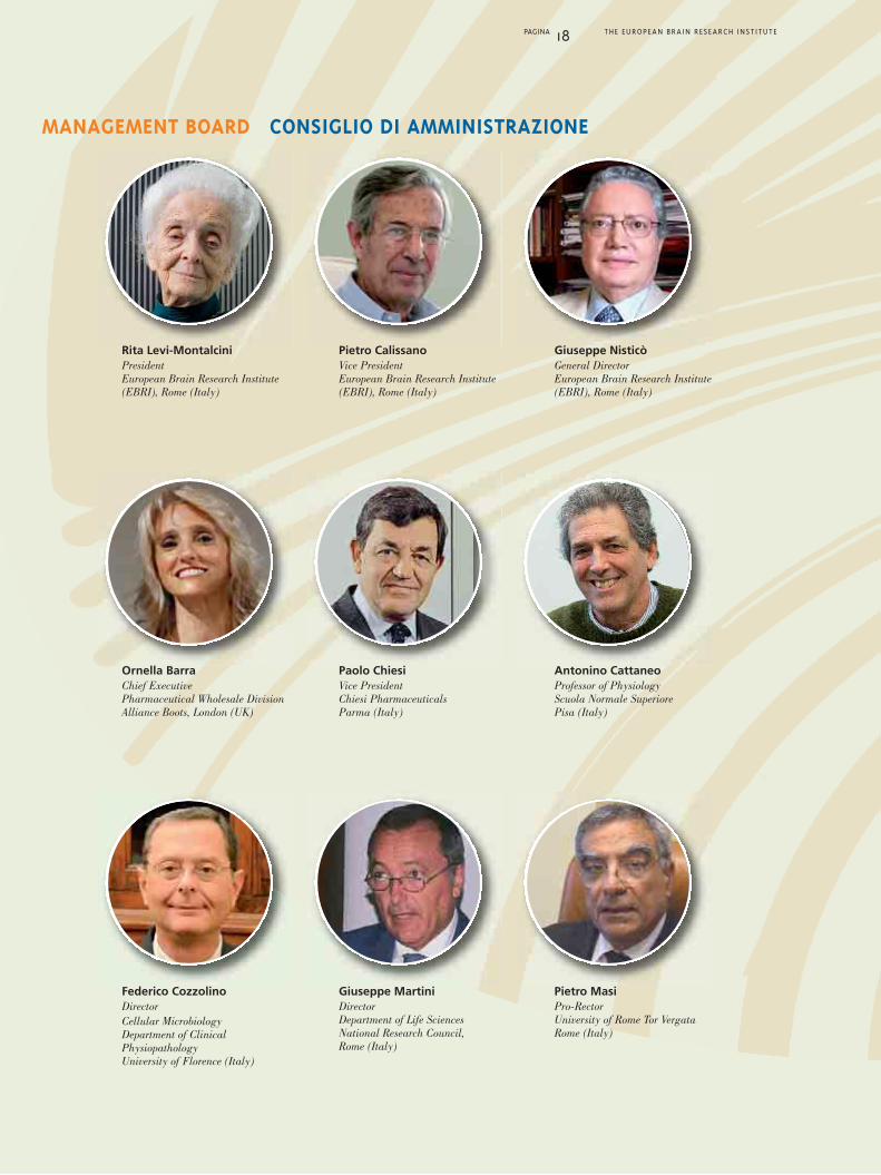
MANAGEMENT BOARD CONSIGLIO DI AMMINISTRAZIONE
Rita Levi-MontalciniPresidentEuropean Brain Research Institute(EBRI), Rome (Italy)
Pietro CalissanoVice PresidentEuropean Brain Research Institute(EBRI), Rome (Italy)
Giuseppe NisticòGeneral DirectorEuropean Brain Research Institute(EBRI), Rome (Italy)
Ornella BarraChief ExecutivePharmaceutical Wholesale DivisionAlliance Boots, London (UK)
Paolo ChiesiVice President Chiesi PharmaceuticalsParma (Italy)
Antonino CattaneoProfessor of PhysiologyScuola Normale SuperiorePisa (Italy)
Federico CozzolinoDirectorCellular MicrobiologyDepartment of ClinicalPhysiopathologyUniversity of Florence (Italy)
Giuseppe MartiniDirectorDepartment of Life SciencesNational Research Council,Rome (Italy)
Pietro MasiPro-RectorUniversity of Rome Tor VergataRome (Italy)

PAGINA 19 THE EUROPEAN BRAIN RESEARCH INSTITUTE
INTERNATIONAL SCIENTIFIC COUNCIL CONSIGLIO SCIENTIFICO INTERNAZIONALE
Moses V. ChaoChairmanProfessor of Cell Biology,Physiology and Neuroscience andPsychiatry, Skirball Institute ofBiomolecular Medicine, NYUSchool of Medicine, New York (USA)
Francesco ClementiVice-Chairman,Emeritus Professor of Pharmacology Department of Pharmacology,University of Milan (Italy)Institute of Neuroscience, CNR,Milan (Italy)
Eric AbadieScientific Advisor to theGeneral Director, Afssaps,Paris
Fabio BenfenatiDirectorDepartment of Neuroscienceand Brain TechnologiesThe Italian Institute of Technology,Genova (Italy)
Anders BjörklundProfessor of Histologyand Section ChiefWallenberg Neuroscience CenterLund University (Sweden)
Graham CollingridgeProfessor of NeuroscienceMRC Centre for SynapticPlasticityUniversity of Bristol (UK)
Richard GreenSpecial Professor ofNeuropharmacologySchool of Biomedical SciencesUniversity of Nottingham (UK)
Lamberto MaffeiEmeritus Professor of NeurobiologyScuola Normale Superiore (Pisa)President, Accademia Nazionaledei Lincei, Rome (Italy)
Gerry MelinoDirectorDepartment of ExperimentalMedicine and Biochemical SciencesMRC Toxicology UnitUniversity of Leicester (UK)
Maurizio PocchiariDirectorDepartment of Cell Biology &Neurosciences, Istituto Superioredi Sanità, Rome (Italy)
Solomon H. SnyderProfessor of NeuroscienceJohns Hopkins Medical SchoolBaltimore (USA)

LABORATORIES, UNITS LABORATORI, UNITÀ

AND FACILITIESE INFRASTRUTTURE

The activity of neurotrophic factors on their targetneurons in the adult and ageing Central NervousSystem, as well as their selective availability andtransport, represent a cross-road in themechanisms that lead to neurodegeneration. Our laboratory studies how abnormalities in thesignalling and post-translational processing ofneurotrophins in the CNS are linked to theprogressive onset of neurodegeneration. The study of the molecular causes of Alzheimer’sdisease (AD) is a major theme in the lab, with acommon focus on the early events and theupstream mechanistic drivers of AD. The final aimof research in the lab is to build on thesemechanistic insights to develop a new generationof therapies for AD and other humanneurodegenerative pathologies.Inspired by the seminal experiments byRita Levi-Montalcini on immunosympathectomy,our lab has pioneered the use of recombinantantibodies for protein knock-out in the CNS,targeting intracellular antibodies (intrabodies) toachieve protein silencing in different subcellularcompartments for mechanistic studies.By this approach, we created an antiNGF-basedtransgenic model, demonstrating that selectivelyinterfering with the function of mature NGF in theadult CNS leads to a progressiveneurodegenerative phenotype that recapitulates ina comprehensive way most ofthe major hallmarks ofAD. Studies alsoshowed that
PAGINA 22 THE EUROPEAN BRAIN RESEARCH INSTITUTE
Astrocytes and microglia from rat’s cortex
Astrociti e microglia di corteccia di ratto
❚ Antonino Cattaneo, Group Leader
❚ Corinna Giorgi, Junior Project Leader
❚ Giovanni Meli, Junior Project Leader
❚ Marcello Ceci, Postdoc
❚ Luisa Fasulo, Postdoc
❚ Agnese Lecci, Postdoc
❚ Francesca Malerba, Postdoc
❚ Annalisa Manca, Postdoc
❚ Francesca Paoletti, Postdoc
❚ Raffaella Scardigli, Postdoc
❚ Gianluca Amato, PhD Student
❚ Nina Krako, PhD Student
❚ Federico Laregina, Lab Technician
❚ Luana Pistillo, Lab Technician
Antonino Cattaneo
NEUROTROPHIC FACTORS ANDNEURODEGENERATIVE DISEASES LABORATORY

neuroinflammatory alterations constitute asignificant event very early in theneurodegeneration process in the mouse brain.Recent work by the group, has pointed to theproNGF protein, the processing precursor ofmature NGF, as a major player in theneurodegeneration process, whereby an imbalancein the levels of proNGF and NGF in the brain is anupstream driver for neurodegeneration, as part of acircular loop linking proNGF signaling to ADphenotypic endpoints. As part of this endeavour,one of our lines of research investigates, by avariety of biophysical techniques, thetridimensional structure of proNGF, in order toexplain its interaction with the receptors(p75NTR, TrkA, sortilin) and its molecular rolein neurodegeneration. In this context, the rationale for NGF as apotential therapeutic for AD is very strong. We arepursuing the development of a non invasivetherapy for AD, based on the intranasal delivery ofNGF as a non invasive, safe and effective mean toachieve pharmacologically active concentrationsof NGF in the brain. One liability of NGF as atherapeutics is, however, physiological,pronociceptive activity. This severely limit thedoses in human clinical trials. To circumventthese difficulties, we have engineered painlessNGF molecules, inspired by a human geneticmutation found in HSAN V patients, who sufferfrom a congenital insensitivity to pain and harbora point mutation in the gene coding for NGF. Wehave characterized the effect of this mutation onNGF receptor interaction and signaling propertiesand have engineered an optimized painless NGFthat we are now developing for clinical testing in
human AD patients.In collaborative work with Raffaella Scardigli(CNR), the lab is pursuing research onembryonic and adult neurogenesis. As for theformer, the aim of this study is to selectneuronal progenitors that can be committed
to a Motoneuron (MN) fate and then used intransplantation studies in animal models for MNdisorders. This knowledge would be of greatbenefit for the development of new therapeuticstrategies for the cure of MN diseases. Concerningadult neurogenesis, we are interested in defininghow neurogenesis is regulated in AD, and morespecifically in exploring thepossibility that NGF mightmodulate adultneurogenesis inphysiologicaland
pathologicalconditions.The ultimategoal of this researchis to gain new insightinto the molecularmechanisms that control adult neurogenesis inresponse to brain injury such as ADneurodegeneration, and to develop new strategiesto restore normal neurogenesis specifically inthose brain regions where it is impaired. Thisinformation will be of great help for theidentification of competent human stem cells withsimilar potential and thus for the development ofcell-replacement strategies for the cure of AD.
PAGINA 23 THE EUROPEAN BRAIN RESEARCH INSTITUTE

PAGINA 24 THE EUROPEAN BRAIN RESEARCH INSTITUTE
caratterizzare gli eventi precoci ed i meccanismidi questa patologia. L’obiettivo ultimo della ricercacondotta in laboratorio è lo sviluppo di una nuovagenerazione di terapie per la malattia di Alzheimered altre patologie neurodegenerative umane. Traendo ispirazione dai primi esperimenti diRita Levi-Montalcini sull’immunosimpatectomia,il nostro laboratorio ha sviluppato l’utilizzodi anticorpi ricombinanti per il knock-outdi proteine nel SNC, indirizzando anticorpiintracellulari (Intracorpi) per il silenziamentodi proteine in differenti compartimenti subcellulari. Attraverso questo approccio, abbiamo creatoun modello transgenico anti-NGF, dimostrandoche l’interferenza selettiva con la funzione dell’NGFmaturo nel SNC determina un fenotiponeurodegenerativo progressivo che riassume inmaniera globale la maggior parte delle principalicaratteristiche della malattia di Alzheimer. Inoltre,studi ulteriori dimostrano che alterazioni deiprocessi neuroinfiammatori costituiscono un eventoprecoce dei processi neurodegenerativi nel cervellomurino. Recenti lavori del gruppo identificanola proteina proNGF, precursore dell’NGF maturo,come attore principale di questo processo, pertantouno squilibrio dei livelli di proNGF ed NGFnel cervello sarebbe all’origine dellaneurodegenerazione, come parte di un circolovizioso che unisce il signalling del proNGFalla neurodegenerazione. Una delle linee di ricerca del gruppo indagala struttura tridimensionale del proNGF, utilizzandovarie tecniche biofisiche, con l’obiettivo di spiegarela sua interazione con i recettori (p75NTR, TrkA,sortilina) e il suo ruolo molecolare nellaneurodegenerazione.In questo contesto, il razionale per sviluppareNGF come agente terapeutico è molto forte.
L’attività dei fattori neurotrofici sui loro targetneuronali nel Sistema Nervoso Centrale (SNC)adulto e nell’invecchiamento, così come la lorodisponibilità ed il trasporto, rappresentanoun punto nodale nei meccanismi che sottendonola neurodegenerazione. Il nostro laboratorio studiacome le anomalie nel signalling e nei processipost-traslazionali delle neurotrofine nel SNCsiano correlati alla progressione dellaneurodegenerazione. Lo studio delle causemolecolari della malattia di Alzheimer è il temaprincipale del laboratorio, con l’obiettivo di
LABORATORIO FATTORI NEUROTROFICIE MALATTIE NEURODEGENERATIVE

PAGINA 25 THE EUROPEAN BRAIN RESEARCH INSTITUTE
Stiamo perseguendo lo sviluppo di una terapia noninvasiva per la MA, basata sulla somministrazioneintranasale di NGF, un metodo sicuro e noninvasivo per ottenere livelli di NGFfarmacologicamente attivi nel cervello.Un punto debole dell’NGF come agente terapeuticoè la sua ben nota attività fisiologica pro nocicettiva.Questo limita decisamente le dosi che possono esseresomministrate nei trial clinici. Per aggirare questedifficoltà, abbiamo ingegnerizzato molecole di NGF“painless” (=senza dolore), prendendo spuntoda una mutazione genetica umana scopertanei pazienti affetti da HSAN V, che soffrono di unainsensibilità al dolore congenita e sono portatoridi una mutazione puntiforme nel gene che codifical’NGF. Abbiamo caratterizzato l’effetto di questamutazione sulle proprietà di interazione e attivitàdel recettore per l’NGF ed ingegnerizzato un NGF“painless”, che stiamo attualmente sviluppandoper test clinici in pazienti.In collaborazione con Raffaella Scardigli (CNR),il laboratorio sta conducendo attività di ricercasulla neurogenesi embrionale ed adulta. Nel primocaso, con l’obiettivo di selezionare progenitorineurali che possano essere diretti aldifferenziamento in motoneuroni (MN) ed utilizzatiper studi di trapianto in modelli animali cheripropongano malattie dei motoneuroni.L’acquisizione di queste conoscenze sarà di grandeutilità per lo sviluppo di nuove strategieterapeutiche per la cura di queste malattie. Per quanto riguarda la neurogenesi adulta, siamointeressati a studiare come la neurogenesi siaregolata nella malattia di Alzheimer, e soprattuttoad esplorare la possibilità che NGF possa modularela neurogenesi adulta in condizioni fisiologichee patologiche.Queste conoscenze saranno determinantiper la identificazione di cellule staminali umanecompetenti e per lo sviluppo di strategie di“recupero cellulare” per la terapia della malattiadi Alzheimer.
Mouse NGF crystallographic structure (PBD: 1BET).Created with PyMol (www.pymol.org).
N N
CC

PAGINA 26 THE EUROPEAN BRAIN RESEARCH INSTITUTE
BIOGRAPHICAL SKETCHBIOGRAFIA
Antonino Cattaneo has worked as a PhD studentat the Scuola Normale Superiore (Pisa) withLamberto Maffei and as a postdoc and staffscientist with Rita Levi-Montalcini (Nobel Prize Laureate for discovery of NGF) at the CNRInstitute of Neurobiology in Rome, and withCesar Milstein (Nobel Prize laureate fordiscovery of monoclonal antibodies) at the MRCLaboratory of Molecular Biology (Cambridge,UK). From 1991 to 2008, he was Full Professor ofBiophysics at the International School forAdvanced Studies (SISSA) in Trieste (Italy),where he was Head of the Biophysics Departmentfrom 1991 to 1995 and the Deputy Director ofSISSA from 1996 to 2001. From 2008 to presenthe is Professor of Neurobiology at the ScuolaNormale Superiore (Pisa), where he is theDirector of the Biology Lab BioSnS.
Antonino Cattaneo is author of numerouspublications in peer-reviewed internationaljournals and is recipient of several awardsincluding Domenico Marotta Prize, ItalianAcademy of Sciences XL, the W. Jansenius Medal,Slovak Academy of Sciences and the“G. Tartufari” International Prize for Biology,Accademia Nazionale dei Lincei. He is a memberof EMBO (European Molecular BiologyOrganization) and member of the Italian Academyof Sciences XL. He is former member of theCouncil of Scientists of Human Frontier ScienceProgram Organization (HFSPO), for which he hasalso served for four years as the Chairman of theGrants Review Committee. He has been VisitingFellow of Trinity College in Cambridge (UK).He is a Team Leader of the European NeuroscienceInstitute Young Investigator Network.
❚ Antonino Cattaneo
Antonino Cattaneo è stato studente al dottoratodi ricerca alla Scuola Normale Superiore di Pisasotto la supervisione del Prof. Lamberto Maffei eha lavorato come postdoc e poi come ricercatorecon Rita Levi-Montalcini (premio Nobel per laMedicina per la scoperta del NGF) all’Istitutodi Neurobiologia di Roma del CNR, e con CesarMilstein (premio Nobel per la Medicina per lascoperta degli anticorpi monoclonali) all’IstitutoMRC di Cambridge in Inghilterra. Dal 1991 al2008 è stato professore ordinario di Biofisica allaScuola Internazionale di Studi Avanzati (SISSA)di Trieste, dove è stato direttore di Dipartimentodal 1991 al 1995 e Direttore in carica dellaSISSA dal 1996 al 2001. Dal 2008 e professoreordinario di Neurobiologia alla Scuola NormaleSuperiore (Pisa) e Direttore del laboratorio diBiologia BioSnS. Antonino Cattaneo è autore dinumerose pubblicazioni in riviste scientifiche
internazionali “peer-reviewed”, ed è stato insignitodi numerosi riconoscimenti e premi scientificiquali il “Premio Domenico Marotta”dell’Accademia Nazionale delle Scienze detta deiXL, la “Medaglia W. Jansenius” dell’AccademiaSlovacca delle Scienze ed il Premio Internazionale“G. Tartufari” per la Biologia dall’AccademiaNazionale dei Lincei. È membro dell’EMBO(European Molecular Biology Organization) emembro dell’Accademia Nazionale delle Scienzedetta dei XL. È stato membro del Consiglio degliScienziati del HFSPO (Human Frontier ScienceProgram Organization), organizzazione nellaquale ha presieduto per quattro anni laCommissione valutatrice per l’assegnazione deifinanziamenti di ricerca. È stato Visiting Fellowdel Trinity College in Cambridge (UK).È team leader del Network dei giovani ricercatoridell’Istituto Europeo di Neuroscienze.

PAGINA 27 THE EUROPEAN BRAIN RESEARCH INSTITUTE
1. Piccioli P., Di Luzio A., Amann R., Schuligoi R.,Surani M.A., Donnerer J., Cattaneo A. (1995).Neuroantibodies: ectopic expression of a recombinantanti-substance P antibody in the central nervoussystem of transgenic mice. Neuron. 15:373-384.
2. Tongiorgi E., Righi M., Cattaneo A. (1997). Activity-dependent dendritic targeting of BDNF and TrkBmRNAs in hippocampal neurons. J. Neurosci.17:9492-9505.
3. Capsoni S., Ugolini G., Comparini A., Ruberti F.,Berardi N., Cattaneo A. (2000). Alzheimer-likeneurodegeneration in aged antinerve growth factortransgenic mice. Proc. Natl. Acad. Sci. USA. 97:6826-6831.
4. De Rosa R., Garcia A.A., Braschi C., Capsoni S.,Maffei L., Berardi N., Cattaneo A. (2005). Intranasaladministration of nerve growth factor (NGF) rescuesrecognition memory deficits in AD11 anti-NGFtransgenic mice. Proc. Natl. Acad. Sci. USA. 102:3811-3816.
5. Capsoni S., Tiveron C., Amato G., Vignone D.,Cattaneo A. (2010). Dissecting the involvement ofTropomyosin kinase A and p75 neurotrophin receptorsignaling in NGF deficit-induced neurodegeneration.Proc. Natl. Acad. Sci. USA. 107:12299-12304.
6. Capsoni S., Covaceuszach S., Marinelli S., Ceci M.,Bernardo A., Minghetti L., Ugolini G., Pavone F.,Cattaneo A. (2011). Taking pain out of NGF: a“painless” NGF mutant, linked to hereditary sensoryautonomic neuropathy type V, with full neurotrophicactivity. PLoS One. 6(2):e17321.
7. Capsoni S., Carucci N.M., Cattaneo A. (2012).Pathogen free conditions slow the onset ofneurodegeneration in a mouse model of Nerve GrowthFactor deprivation. J. Alzheimers Dis., in press.
8. Ceci M., Welshhans K., Ciotti M.T., Brandi R., ParisiC., Paoletti F., Pistillo L., Bassell G.J., and Cattaneo A.(2012). RACK1 is a ribosome scaffold protein forb-actin mRNA/ ZBP1complex. PLoS One, in press.
9. Covaceuszach S., Marinelli S., Krastanova I., UgoliniG., Pavone F., Lamba D., Cattaneo A. (2012). SingleCycle Structure-based Humanization of an Anti-NerveGrowth Factor Therapeutic Antibody. PLoS One, inpress.
10. Maya-Vetencourt J.F., Baroncelli L., Viegi A.,Tiraboschi E., Castren E., Cattaneo A. and Maffei L.(2012). IGF-1 restores visual cortex plasticity in adultlife by reducing local GABA levels. Neural Plasticity,in press.
SELECTED PUBLICATIONSPUBBLICAZIONI

PAGINA 28 THE EUROPEAN BRAIN RESEARCH INSTITUTE
Arc mRNA is also emerging as a unique exampleof how neuronal activity can control every knownstep of a mRNA’s life, ranging from itstranscription, to its localization, stability andfinally translation.In my previous work, I identified a novel pathwaymodulating Arc mRNA expression, which relies onthe presence of introns in its 3’UTR. This uniquegenomic arrangement causes the mRNA to betargeted for destruction by the Nonsense MediatedDecay pathway (NMD), adding another degree ofcomplexity to the already intricate journey of ArcmRNA expression at synapses. To understand the underlying molecularmechanisms controlling Arc mRNA metabolism,I have been adopting parallel approaches aimed atthe characterization of cis and trans-acting factorsassociated with its 3’UTR. On one hand, I amutilizing biochemical techniques to isolate theribonucleoprotein particle associated with ArcmRNA 3’UTR in vivo. Mass spectrometry anddeep sequencing of this complex will allow theidentification of trans-acting factors and miRNAswhose binding is dependent on synaptic activityand splicing of the mRNA. In a parallel analysis,luciferase reporter constructs harboring deletionsand mutations of Arc 3’UTR are being adopted.This approach is aimed at understanding themolecular links between translational activation ofArc mRNA processing of its mRNA. Analyses areunderway to test whether other dendritic mRNAsundergo similar regulatory pathways as Arc.Overall, the in vivo biochemical and functionalcharacterization of both cis- and trans-actingfactors controlling the expression of dendriticmRNAs, will allow a better understanding of themechanisms controlling their expression, andlikely shed light on key pathways that linkdendritic mRNA localized expression to synapticplasticity.
The overarching focus of my research is tounderstand how post-transcriptional regulation ofgene expression, particularly at the level of mRNAmetabolism, participates in modulating neuronalfunctions. It is now clear that synaptogenesis andsynaptic plasticity rely on localized translationwithin dendrites of mRNAs encoding key synapticstructural and functional constituents. Yet, themolecular machinery underlying dendritic mRNAslocalization and regulated expression is still poorlyunderstood. A primary subject of my past and current studiesis the immediate early gene Arc, encoding aprotein whose function is critical for LTP, LTD,memory consolidation, and homeostatic plasticity.
Corinna Giorgi
❚ Corinna Giorgi, Junior Project Leader
mRNA METABOLISM IN THE NERVOUS SYSTEM
JUNIOR PROJECT LEADERS IN THE LABORATORY
PAGINA 29 THE EUROPEAN BRAIN RESEARCH INSTITUTE
La mia ricerca è finalizzata a comprendere comela regolazione post-trascrizionale dell’espressionegenica, in particolare a livello del metabolismodell’RNA, prende parte nella modulazione dellefunzioni neuronali. Da tempo è chiaro che laplasticità sinaptica e la sinaptogenesi necessitanola traduzione localizzata di mRNA codificanticostituenti strutturali e funzionali delle sinapsi.Tuttavia, molti aspetti della regolazionedell’espressione di questi mRNA dendritici sonoancora poco chiari. I miei studi si focalizzano sul mRNA dendriticocodificato dal gene Arc, la cui proteina è essenzialein molti processi alla base della memoria, inclusiLTP, LTD, consolidamento della memoria el’omeostasi sinaptica. Il messaggero di Arcrappresenta inoltre un esempio unico di comel’attività sinaptica può controllare ogni fase dellavita di un mRNA, dalla trascrizione alla sualocalizzazione, stabilità e traduzione. In passato hoidentificato un nuovo meccanismo di regolazionedell’espressione di Arc mRNA, che dipende dallapresenza di due introni nel suo 3’UTR.Questo peculiare arrangiamento genico provocal’attivazione del Nonsense Mediated Decay pathway(NMD), che degrada l’mRNA in seguito allatraduzione, aggiungendo un ulteriore livello dicomplessità alla già intricata via di espressione diArc nelle sinapsi. Per comprendere ulteriormente i meccanismimolecolari che regolano l’espressione delmessaggero di Arc, sto utilizzando diversi approccifinalizzati alla caratterizzazione degli elementiregolativi, in cis e in trans, del suo 3’UTR.Da una parte sto adottando tecniche biochimicheper la purificazione in vivo della particellaribonucleoproteica associata al 3’UTR di ArcmRNA. Analisi di spettrometria di massa e di deepsequencing di questo complesso consentirannol’identificazione di fattori regolativi e di miRNAs ilcui legame ad Arc mRNA dipende dalla attività
sinaptica e dallo splicing del messaggero.Parallelamente, utilizzo saggi di luciferasi concostrutti contenenti versioni mutate del 3’UTR diArc al fine di studiare i meccanismi che coordinanol’attivazione traduzionale di Arc al processamentodel suo messaggero. Infine, sto esaminando altrimRNA dendritici che condividono alcunepeculiarità del messaggero di Arc e che sono quindipotenzialmente regolati in maniera analoga.La caratterizzazione biochimica e funzionale deifattori molecolari coinvolti nella regolazione dimRNA dendritici dovrebbe consentire una piùapprofondita comprensione dei meccanismimolecolari che ne regolano l’espressione e che neconsentono una modulazione da parte della attivitàsinaptica.
METABOLISMO DELL’RNAm NEL SISTEMA NERVOSO

PAGINA 30 THE EUROPEAN BRAIN RESEARCH INSTITUTE
A major line of research in Cattaneo’s lab, andpursued by Giovanni Meli, as a Junior ProjectLeader, is aimed at targeting Alzheimer’Amyloid-β (Aβ) peptide oligomers (AβOs) withrecombinant intrabodies. A distinctive approach ofCattaneo’s lab has been the use of antibodies asgenes, rather than as proteins, which allowsexpressing them in different cells and in differentcompartments for silencing at the protein level(intrabodies).
We isolate antibodies from ad hoc engineeredlibraries by the “Intracellular Antibody CaptureTechnology” (IACT), an approach that allows toaddress questions that cannot be by othersilencing techniques, such as RNA interference.We have undertaken the intrabody approach todissect the cellular pathways leading to AβOsformation and actions.Amyloid-β (Aβ) peptide, derived from abnormalprocessing of its APP precursor protein, iscrucially involved in AD pathogenesis.In particular, soluble multimeric assemblies of Aβ,called Aβ oligomers (AβOs), are considered themost synaptotoxic Aβ species in the brains of ADpatients and of AD transgenic mice models.Although increasing evidence supports the role ofintracellular Aβ oligomerization and accumulation,as an early event in AD pathogenesis in humansand in transgenic mice, little is known about theintracellular processing and trafficking events ofthe different forms of AβOs. Targeting the pathological assemblies of Aβ withspecific probes, for mechanistic studies, forintracellular imaging or for therapeutic purposes,is therefore very important. Moreover, theintracellular targeting of AβOs would require theavailability of antibody domains suitable forintracellular expression.We have selected anti-AβOs recombinantantibody fragments (scFvs) that show uniqueproperties in terms of sequence, epitoperecognition, conformational selectivity,immunoreactivity towards naturally-produced Aβdeposits in AD brains, inhibition of synapticbinding of Aβ oligomers (ADDLs) andneutralization of their-induced cyto-toxicity.These novel anti-AβOs are being expressed asintrabodies to study the subcellular traffic,dynamics and functions of AβOs.
JUNIOR PROJECT LEADERS IN THE LABORATORY
Giovanni Meli
❚ Giovanni Meli, Junior Project Leader
SUBCELLULAR TARGETING OF ALZHEIMER’SAMYLOID-β (Aβ) PEPTIDE OLIGOMERSWITH INTRABODIES
PAGINA 31 THE EUROPEAN BRAIN RESEARCH INSTITUTE
Una rilevante linea di ricerca del laboratorio diCattaneo, seguita da Giovanni Meli come JuniorProject Leader, riguarda il targeting, tramiteanticorpi ricombinanti, di oligomeri del peptideAmyloid-β (Aβ).Un approccio ideato e sviluppato nel laboratorioCattaneo è l’uso degli anticorpi come geni, piuttostoche come proteine, cosa che permette l’espressionedegli stessi in diverse cellule e in distinticompartimenti subcellulari per il “silenziamento”a livello di proteina (“intrabodies”).Selezioniamo anticorpi da librerie ingegnerizzatead hoc tramite la tecnologia IACT (IntracellularAntibody Capture technology), un approccio chepermette di affrontare domande in manieraesclusiva e in modo non possibile tramite altretecniche di “silenziamento”, come l’RNAinterference.Noi stiamo utilizzando l’approccio degliintrabodies per lo studio dei pathways subcellularidi formazione e azione degli Aβ oligomeri.Il peptide Aβ deriva dal processing “anomalo” delsuo precursore APP ed è coinvolto in manieracruciale nella patogenesi di malattia di Alzheimer(MA).In particolare, complessi multimerici solubili di Aβ,chiamati Aβ oligomeri (AβOs), sono considerati lespecie più sinaptotossiche nei cervelli di pazientiAlzheimer e in modelli di topo MA. Nonostante un numero sempre maggiore di studisupporti il ruolo della oligomerizzazioneintracellulare di Aβ come evento precoce nellapatogenesi di MA nell’uomo e in topi transgenici,poco si conosce sul processamento intracellulare e iltrasporto intracellulare di diverse forme di AβOs.Colpire con sonde selettive gli aggregati patologicidi Aβ per studi meccanicistici, per imaging
intracellulare o per scopi terapeutici, è quindi moltoimportante.Inoltre, il targeting intracellulare di AβOs richiedela disponibilità di domini anticorpali utilizzabiliper l’espressione intracellulare. Abbiamo selzionato anticorpi ricombinantianti-AβOs, nel formato di single chain Fragment(scFv), che mostrano proprietà uniche in termini disequenza, riconoscimento di epitopi, selettivitàconformazionale per AβOs in vitro,immunoreattività verso depositi naturali di Aβ incervelli di malati MA, inibizione del legame allesinapsi di AβOs e neutralizzazione della lorotossicità neuronale.Adesso stiamo esprimendo gli anti-AβOs scFvscome intrabodies per studiare il processamento, ladinamica e le funzioni intracellulari degli AβOs.
TARGETING SUBCELLULARE DI OLIGOMERIDELL’AMILOIDE β NELLA M. DI ALZHEIMERCON ANTICORPI INTRACELLULARI
PAGINA 32 THE EUROPEAN BRAIN RESEARCH INSTITUTE
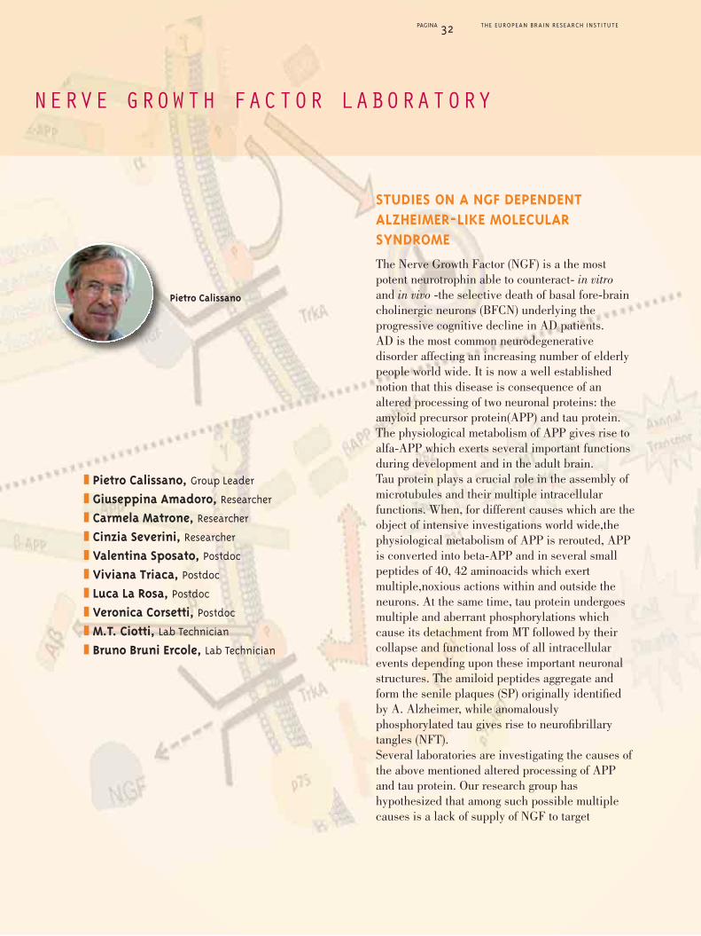
❚ Pietro Calissano, Group Leader
❚ Giuseppina Amadoro, Researcher
❚ Carmela Matrone, Researcher
❚ Cinzia Severini, Researcher
❚ Valentina Sposato, Postdoc
❚ Viviana Triaca, Postdoc
❚ Luca La Rosa, Postdoc
❚ Veronica Corsetti, Postdoc
❚ M.T. Ciotti, Lab Technician
❚ Bruno Bruni Ercole, Lab Technician
NERVE GROWTH FACTOR LABORATORY
STUDIES ON A NGF DEPENDENTALZHEIMER-LIKE MOLECULARSYNDROME
The Nerve Growth Factor (NGF) is a the mostpotent neurotrophin able to counteract- in vitroand in vivo -the selective death of basal fore-braincholinergic neurons (BFCN) underlying theprogressive cognitive decline in AD patients.AD is the most common neurodegenerativedisorder affecting an increasing number of elderlypeople world wide. It is now a well establishednotion that this disease is consequence of analtered processing of two neuronal proteins: theamyloid precursor protein(APP) and tau protein.The physiological metabolism of APP gives rise toalfa-APP which exerts several important functionsduring development and in the adult brain.Tau protein plays a crucial role in the assembly ofmicrotubules and their multiple intracellularfunctions. When, for different causes which are theobject of intensive investigations world wide,thephysiological metabolism of APP is rerouted, APPis converted into beta-APP and in several smallpeptides of 40, 42 aminoacids which exertmultiple,noxious actions within and outside theneurons. At the same time, tau protein undergoesmultiple and aberrant phosphorylations whichcause its detachment from MT followed by theircollapse and functional loss of all intracellularevents depending upon these important neuronalstructures. The amiloid peptides aggregate andform the senile plaques (SP) originally identifiedby A. Alzheimer, while anomalouslyphosphorylated tau gives rise to neurofibrillarytangles (NFT).Several laboratories are investigating the causes ofthe above mentioned altered processing of APPand tau protein. Our research group hashypothesized that among such possible multiplecauses is a lack of supply of NGF to target
Pietro Calissano

PAGINA 33 THE EUROPEAN BRAIN RESEARCH INSTITUTE
neurons, which are crucially constitutive of thecholinergic system of hippocampus. Indeed, wefound that withdrawal of NGF from target neuronsof hippocampus, cortical neurons, PC12 cells orsensory and sympathetic ganglia, is followed by aseries of molecular events in all resembling thoseoccurring in animal models of AD. We refer tothese events to as “an Alzheimer-like molecularsyndrome” Thus, following NGF withdrawal, APPundergoes an amyloid processing with productionof amyloid beta peptides and a series ofintracellular events typical of AD reported inanimal models and in specimens of AD patients.The cascade of amyloid processing, in turn, causes
tau altered phosphorylations and its anomalousbreakdown with production of a 22 KdA protein(h22Kd)which exerts a specific toxic action onsynaptic mitochondria. This truncated form of tauis also detectable in the cerebrospinal fluid of ADpatients. We presently plan to use h22Kd as adiagnostic tool for AD and possibly otherneuropathies. The crucial question that we are presentlyinvestigating is: what is the mechanism throughwhich NGF controls the physiological processingof APP, in such a way that when this neurotrophinis bound to its receptor APP metabolism follows aphysiological processing and tau protein is able toexert its multiple functions? In a preliminaryseries of studies we have already established thatthe NGF/TrkA complex and the APP protein are intight contact within the cellular membrane.When NGF is bound to TrkA, a sort of reciprocal,specific phosphorylation of both protein complexoccurs and this event somewhat channel APPmetabolism toward its physiological pathway.It is clear that understanding the specificmolecular events occurring and underlying suchinteraction is of direct relevance to one of themechanism(s) of the onset of AD. Thus, wehypothesize that other ligand/receptor complexescould operate in the same fashion as NGF/TrkA.For example, preliminary studies are pointing outseveral similarities between this NGF/TrkA andthe Insulin/receptor system.In this connection it is interesting to note thatpatients affected by diabetes type2 have an highrisk of undergoing AD.Altogether, we have established an in vitro modelof NGF-dependent cultured neurons whereby it ispossible to assess, under strictly controlledconditions, the molecular mechanisms whichcould be at the origin of AD and to test thepossible use of substances or drugs aimed atpreventing or inhibiting such events.
NGF mediated APP processing. In the upper part of the figure NGF mediated processing of APP is depicted.The lower part of the figure reports the events occurring when NGF signaling is impaired or blocked.
Ruolo dell’NGF nel metabolismo dell’APP. Nella parte superiore si vedono i dettagli interferenza dell’NGF sul processing dell’APP.Nella parte inferiore della figura si vedono gli eventi che si verificano quando il segnale dell’NGF è inibito o bloccato.

PAGINA 34 THE EUROPEAN BRAIN RESEARCH INSTITUTE
Il morbo di Alzheimer (AD) rappresenta l’affezionedegenerativa più comune degli anziani.Studi condotti in numerosi laboratori nel mondohanno dimostrato che AD è dovuta principalmenteall’alterato metabolismo di 2 proteine: la proteinaprecursore dell’amiloide (APP) e la proteina tau.Il metabolismo fisiologico di APP porta allaformazione della alfa-APP che svolge un ruolomolto importante in numerose funzioni sia durantelo sviluppo del cervello che nell’adulto.La proteina tau svolge un ruolo fondamentalenell’assemblaggio e nella funzione dei microtubuliche, a loro volta, giuocano attività multiple efondamentali nelle cellule nervose.Quando, per cause diverse, che sono oggetto distudi in numerosissimi laboratori, il metabolismo diAPP viene ad essere alterato, la proteina vieneconvertita in beta-APP e in alcuni peptidi di piccoledimensioni di 40-42 amminoacidi, che esercitanonumerose attività tossiche entro le cellule nervose ein quelle circostanti. Contemporaneamente, laproteina tau va incontro a processi di fosforilazionianomali e viene demolita da specifiche proteasi cheprovocano il suo distacco dai microtubuli.Il collasso dei microtubuli, a sua volta, provoca laprogressiva perdita delle loro funzioni intracellularie la morte dei neuroni per apoptosi. I peptidi diamiloide che originano dall’APP si aggregano aformare le placche senili (SP) per la prima voltadescritte dal neuropatologo Alois Alzheimer, mentrei frammenti di tau si aggregano a formare gliaggregati neurofibrillari (NFT).Numerosi laboratori stanno studiando e cercando lecause di questi processi anomali che colpisconoAPP e tau. Il nostro gruppo di lavoro ha ipotizzatoche una di queste cause sia la mancata
disponibilità del nerve growth factor (NGF) aineuroni colinergici che sono fra i primi ad esserecolpiti nel AD. In effetti abbiamo dimostrato che lasottrazione di NGF a cellule bersaglio come quellepresenti nella corteccia cerebrale, nell’ippocampo oin gangli sensoriali o simpatici, provoca una seriedi eventi intracellulari che sono in tutto simili aquelli che si verificano nelle fasi iniziali del AD.Questo insieme di eventi è stato definito una“sindrome molecolare Alzheimer simile”. Infatti, in seguito a rimozione di NGF la proteinaAPP viene metabolizzata lungo la cosiddetta viaamiloidogenica con produzione di peptidi amiloidied altri eventi molecolari simili a quanto descrittoin modelli animali del AD. La cascata di eventiamiloidogenici, a sua volta, è seguita dafosforilazioni anomale di tau e dal suoprocessamento con produzione di un frammento di22 KdA che si localizza principalmente neimitocondri sinaptici e ne impedisce la normalefunzione energetica. Questo frammento si trovafortemente aumentato anche nel liquor di pazienticolpiti da AD e stiamo producendo un anticorpomonoclonale per un suo impiego eventualmentediagnostico del AD. Il problema che riteniamo fondamentale e chestiamo attualmente studiando è il seguente: conquale meccanismo il NGF controlla la viafisiologica di processamento di APP in modo taleche quando questa neurotrofina è legata al suorecettore, APP viene metabolizzata lungo la viafisiologica, mentre in sua assenza si attiva la viaamiloidogenica? In una prima serie di indaginiabbiamo dimostrato che il complesso NGF/recettoree la proteina APP sono in stretto contatto nellamembrana cellulare e si verifica una specie di
LABORATORIO NGF
STUDI SU UN MODELLODI ALZHEIMERNGF DIPENDENTE

PAGINA 35 THE EUROPEAN BRAIN RESEARCH INSTITUTE
reciproco controllo mediato da fosforilazioni adeterminati amminoacidi delle due proteine.Questa interazione provoca un processamentofisiologico di APP. Riteniamo che la comprensionedel meccanismo tramite il quale NGF controlla ilprocessamento di APP abbia risvolti importanti perla comprensione delle cause del AD. Infatti, anchealtre molecole come NGF potrebbero svolgere unruolo analogo a questa neurotrofina. Fra queste, lastessa insulina la cui produzione/funzione è alteratanel diabete di tipo 2 che costituisce un alto fattoredi rischio anche per l’instaurarsi del AD.
Nel loro insieme questi studi ci permettono distudiare, in condizioni strettamente controllate,i meccanismi molecolari che sono alla base del ADe di valutare l’efficacia di farmaci, sostanze otrattamenti che possono essere impiegati perprevenire o rallentare questa malattianeurodegenerativa.
Tau processing following NGF withdrawal from target neurons
Eliminazione di NGF dai neuroni target induce delle modifiche nel processing della proteina tau

PAGINA 36 THE EUROPEAN BRAIN RESEARCH INSTITUTE
Pietro Calissano, born in Genova, obtained adegree in Medicine magna cum laude in 1964 atthe University of Genova. In 1965 he was invitedby Rita Levi-Montalcini to join the Centre ofNeurobiology she had created in Rome.The following year he moved to WashingtonUniversity of Saint Louis, where he spent twoyears, to work under the guidance of RitaLevi-Montalcini on the mechanism of action ofNGF. In subsequent years Calissano spent severallong-term stages in Cambridge, England, at theWeizman Institute, Israel and at Harvard MedicalSchool, working on NGF, on the brain specificprotein S-100 and on some other neurobiologicalproblems connected with neurotrophins and theirmechanism of action. In 1986 Calissano becamefull professor of Neurophysiology at the Universityof Rome Tor Vergata and in 1988 was nominated
director of the Institute of Neurobiology of CNR,which he guided until 2008. Calissano is memberof the European Molecular Biology organization(EMBO), the Italian Academy of Sciences, and ofthe Scientific Committee of the Istituto Italianodell’Istituto Treccani for which has directed orcollaborated in several encyclopedias edited bythis prestigious institution. He has been recipientof several awards such as Feltrinelli prize,Accademia delle Scienze, Neuburgh prize.He is author of some books of divulgation,(Cervello mente ed evoluzione, Garzanti; ilproblema cervello/mente il Melangolo) and haswritten several scientific articles in Italiannewspapers such as La Repubblica e La Stampa.Calissano is presently vice President of the EBRI.
Pietro Calissano è nato a Genova e si è laureato conlode in Medicina e Chirurgia nel 1964. Nel 1965 è stato invitato da Rita Levi-Montalcini asvolgere ricerche nel centro di Neurobiologia delCNR che la scienziata stava organizzando a Roma.Nell’anno successivo Calissano si è recato pressola Washington University dove ha soggiornatodue anni lavorando sotto la guida di RitaLevi-Montalcini iniziando le sue ricerche sulmeccanismo d’azione del NGF. Negli annisuccessivi Calissano ha trascorso lunghi periodi dilavoro all’Institute of Animal Physiology diCambridge, al Weizmann Institute in Israele, e allaHarvard Medical School, lavorando sul NGF, sullaproteina S-100 e su altri problemi scientificicollegati con le neurotrofine e sul loro meccanismod’azione. Nel 1986 Calissano è diventato professoreordinario in Neurofisiologia all’Università di TorVergata e nel 1988 è stato nominato direttore
dell’Istituto di Neurobiologia del CNR che haguidato fino al 2008. Calissano è membrodell’European Molecular Biology Organization(EMBO), dell’Accademia delle Scienze detta deiQuaranta, e del Consiglio Scientifico dell’Istitutodell’Enciclopedia Treccani, per la quale ha direttonumerose opere di orientamento scientifico.Egli ha ricevuto numerosi premi scientifici quali ilpremio Feltrinelli, il premio Neuburgh e il premioconferito dall’Accademia delle Scienze.Calissano ha scritto alcuni libri di divulgazionescientifica (Neuroni, mente ed evoluzione, Garzanti,e Cervello/mente, Il Melangolo) nonchénumerosissimi articoli divulgativi per i quotidianiLa Stampa e La Repubblica.
❚ Pietro Calissano
BIOGRAPHICAL SKETCHBIOGRAFIA

PAGINA 37 THE EUROPEAN BRAIN RESEARCH INSTITUTE
1. Amadoro G., Serafino A.L., Barbato C., Ciotti M.T.,Sacco A., Calissano P., and Canu N. (2004). Role ofN-terminal tau domain integrity on the survival ofcerebellar granule neurons. Cell Death andDifferentiation. 11,217-230.
2. Amadoro G., Ciotti M.T., Costanzi M., Cestari V.,Calissano P. and Canu N. (2006). NMDA receptormediates tau-induced neurotoxicity by calpain andERK/MAPK activation. Proc. Natl. Acad. Sci. USA.103, 2892-28.
3. Matrone C., Di Luzio A., Meli G., D’Aguanno S.,Severini C., Ciotti M.T., Cattaneo A. and Calissano P.(2008). A Activation of the amyloidogenic route byNGF deprivation induces apoptotic death in PC12cells. J. Alzheimers disease. 13,81-96.
4. Matrone C., Ciotti M.T., Mercanti D., Marolda R. andCalissano P. (2008). NGF and BDNF signalling controlamyloidogenic route and A-beta production inhippocampal neurons. Proc. Natl. Acad. Sci. USA.Sep2; 105(35): 13139-44 Epub 2008 Aug 26.
5. Matrone C., Marolda R., Ciaffrè S., Ciotti M.T.,Mercanti D. and Calissano P. (2009). Tyrosine kinasenerve growth factor receptor switches from prosurvivalto proapoptotic activity via Abeta-mediatedphosphorylation. Proc. Natl. Acad. Sci. USA. 106,11358-11363.
6. Calissano P., Amadoro G., Matrone C., CiaffrèS.,Marolda R., Corsetti V., Ciotti M.T., Mercanti D., DiLuzio A., Severini C., Provenzano C. and Canu N.(2010). Does the term Trophic actually meanantiamyloidogenic? The case of NGF. Cell death andDifferentiation. 1-8.
7. Amadoro G., Corsetti V., Stringaro A., Colone M.,D’Aguanno S., Meli G., Ciotti M., Sancesario G.,Cattaneo A., Bussani R., Mercanti D., Calissano P.(2010). A NH2 tau fragment targets neuronalmitochondria at AD synapses: possible implications forneurodegeneration. J. Alzheimers Dis. 21(2):445-70
8. Calissano P., Matrone C., Amadoro G. (2010 Apr)Nerve growth factor as a paradigm of neurotrophinsrelated to Alzheimer’s disease. Dev. Neurobiol.70(5):372-83.
9. Pieri M., Amadoro G., Carunchio I., Ciotti M.T.,Quaresima S., Florenzano F., Calissano P., Possenti R.,Zona C., Severini C. (2010 Jan). SP protects cerebellargranule cells against beta-amyloid-induced apoptosisby down-regulation and reduced activity of Kv4potassium channels. Neuropharmacology. 58(1):268-76.
SELECTED PUBLICATIONSPUBBLICAZIONI
Confocal image of a mouse hippocampal neuron in culture
Immagine confocale di un neurone dell’ippocampo di topo in coltura (rendering)

PAGINA 38 THE EUROPEAN BRAIN RESEARCH INSTITUTE
Marco Canossa
❚ Marco Canossa, Group Leader
❚ Francesca Ceroni, Postdoc
❚ Nicoletta Paolillo, Postdoc
❚ Roberta Rogliano, Postdoc
NEURAL STEM CELLS AND NEUROGENESISLABORATORY
The correct establishment of neuronal connectionsinto neuronal circuitry is essential for the properorganization of the nervous systems.Such connections are generated throughpolarization of newly generated neurons beginningwith the specification of a single axon amongequally potential neurites (Figure 1 A and B).Along this process, growth cones located at theleading edges of undifferentiated neurites detectand respond to environmental cues that guide one,and only one of them to growth over the otherattaining final axonal identity. These polarizingcues, include contact-mediated or secretedmolecules acting over the fated axon.It is clear that individual polarity cues canfunction activating a plethora of various eventsincluding the differential expression of receptorsand protein complexes,the selective accumulationof polarity regulators and cross talk betweenintracellular signaling cascades,at the growthconeof the fated axon.Of these instructive cues, theneurotrophins are perhaps the best candidates.Although there is considerable indication for therole of neurotrophins in neuronal polarity, theprecise molecular mechanisms that underlieneurotrophin signal transduction in axonalspecification remain unresolved.So far, we report that the pan-neurotrophinreceptor p75NTR is a key polarity regulator thatlocalizes asymmetrically in differentiating neuronsin response to neurotrophinsand it transducespolarity signals for specification of the future axon.In the next three years we will investigate the roleof p75NTR in transducing signals for axonalspecificationboth in vitro and in vivo.
FROM EARLY NEURONAL POLARITY TOESTABLISHMENT OF NEURONALCONNECTIONS IN ADULT NEUROGENESIS

PAGINA 39 THE EUROPEAN BRAIN RESEARCH INSTITUTE
LABORATORIO CELLULE STAMINALIE NEUROGENESI
I neuroni neoformati nel giro dentatodell’ippocampo di adulto s’integrano nei circuitineuronali preesistenti: ricevono inputsinaptici dallefibre afferenti provenienti dalla corteccia entorinale,e trasmettono outputsinaptici estendendo il proprioassone ai neuroni della regione CA3dell’ippocampo.I nuovi neuroni sono generati da cellule precursoricon fenotipo gliale. Le cellule precursori sonolocalizzate in specifici microambienti, detti“nicchie”, entità biochimiche che annoverano uncomplesso insieme di segnali, come fattori solubili,molecole legate alle membrane cellulari e allematrici extracellulari e diversi tipi cellulari.Questi fattori giocano un ruolo fondamentalenella creazione di un microambiente neurogenico,utile al differenziamento dei neuroblasti.Affinché il neurone neoformato integrifunzionalmente all’interno di specifici circuitineuronali, deve differenziarsi attraverso distintetransizioni morfologiche (Figura 1 A e B), quali laspecificazioneassone/dendrite, crescita dell’assone,
arborizzazione dendriticae infine sinaptogenesi.Durante il processo di formazione dell’assone, ilcono di crescita riceve segnali da molecole deputatealla specificazione e successiva crescita assonale.I fattori di crescita neuronalee in particolare leNeurotrofine, sono molecole secrete che si prestanoalla regolazione della specificazione e della crescitaassonale. Il nostro gruppo di ricerca si occupa di individuarei meccanismi molecolari coinvolti nellaformazione dell’assone nei neuroni nuovi-nati nell’ippocampodi adulto. In particolare sistudierà il ruolo dei recettoriper le neurotrofine p75NTRe Trks nel processo di asso-genesi in modellisperimentali “in vitro” e“in vivo”.
DALLA POLARIZZAZIONE NEURONALEALL’INTEGRAZIONE DEI NEURONI NUOVINATI NELL’IPPOCAMPO DI ADULTO
Marco Canossa with two young post-doc (from left NicolettaPaolillo and Roberta Rogliano)
Marco Canossa con due giovani ricercatrici (da sinistra NicolettaPaolillo e Roberta Rogliano)

PAGINA 40 THE EUROPEAN BRAIN RESEARCH INSTITUTE
Marco Canossa obtained his degree (1986) andPhD (1991) in Pharmacology at the University ofBologna. He carried out postdoctoral research inneurobiology at Stanford University before joiningthe Department of Pharmacology at University ofBologna as Assistant Professor in 1995.From 1996 to 2000 he was Visiting Scientist at theMax Planck Institute of Neurobiology in Munichin the laboratory of Hans Thoenen and from2006-2007 Visiting Scientist at the Ludwig-Maximilians-Universität in Munich (LMU) in thelaboratory of Magdalena Götz. From 2007 to 2012he was Senior Scientist at the Italian Institute ofTechnology in Genova.
Marco Canossa, laureato in Farmacia nel 1986presso l’Università di Bologna, ha ottenuto ilDottorato presso la stessa Università. Ha condottola sua ricerca post-dottorato nel campo dellaNeurobiologia all’Università di Stanford prima diraggiungere nel 1995 il Dipartimento diFarmacologia all’Università di Bologna, comeProfessore associato. Dal 1996 al 2000 è statoVisiting Scientist all’Istituto di Neurobiologia MaxPlanck di Monaco nel laboratorio di Hans Thoenede dal 2006 al 2007 presso l’Università di MonacoLudwig-Maximilians (LMU) nel laboratorio diMagdalena Götz.Dal 2007 al 2012 è stato Ricercatore Seniorall’Istituto Italiano di Tecnologia (IIT) di Genova.
❚ Marco Canossa
BIOGRAPHICAL SKETCHBIOGRAFIA

PAGINA 41 THE EUROPEAN BRAIN RESEARCH INSTITUTE
1. Canossa M., Rovelli G. and Shooter E.M. (1996).Transphosphorylation of the neurotrophinTrk receptors.J. Biol. Chem. 271, 10, 5812-5818.
2. Canossa M., Twiss J., Verity N. and Shooter E.M.(1996). p75NGFR and TrkA receptors cooperate torapidly activate a p75NGFR associated proteinkinase.EMBO J. 15, 13, 3369-337624.
3. Canossa M., Griesbeck O., Berninger B., Campana G.,Kolbeck R. and Thoenen H. (1997). Neurotrophinrelease by Neurotrophins: Implication for activity-dependent neuronal plasticity. Proc. Natl. Acad. Sci.USA. 94 13279-13286.
3. Canossa M., Gartner A., Campana G., Inagaki Y. andThoenen H. (2001). Regulated secretion ofneurotrophins by metabotropic glutamate group I(mGluRI) and Trk-receptor activation is mediated viaphospholipase C signaling pathways. EMBO J. 20,1640-1650.
4. Canossa M., Giordano E., Cappello S., Guarnieri C.and Ferri S. (2002). Nitric Oxide down-regulate brainderived neurotrophic factor secretion in culturedhippocampal neurons. Proc. Natl. Acad. Sci. 99, 3282-3287.
5. Aicardi G., Argilli E., Cappello S., Santi S., Riccio M.,H. Thoenen and Canossa M. (2004). Induction of LTPand LTD is reflected by corresponding changes insecretion of endogenous BDNF. Proc. Natl. Acad. Sci.101:15788-15792.
6. Santi S., Cappello S., Riccio M., Bergami M., AicardiG., Schenk U., Matteoli M. and Canossa M. (2006).Hippocampal neurons recycle BDNF for activity-dependent secretion and LTP maintenance. EMBO J.25:4372-4380.
7. Bergami M., Santi S., Formaggio E., Cagnoli C.,Verderio C., Blum R., Berninger B., Matteoli M.,Canossa M. (2008). Uptake and recycling of pro-BDNFfor transmitter-induced secretion by cortical astrocytes.J. Cell. Biol. 20;183(2):213-221.
8. Bergami M., Rimondini R., Santi S., Blum R., Götz M.,Canossa M. (2008). Deletion of TrkB in adultprogenitors alters newborn neuron integration intohippocampal circuits and increases anxiety-likebehavior. Proc. Natl. Acad. Sci. 7;105(40):15570-15775.
9. Bergami M., Berninger B. and Canossa M. (2008).Conditional deletion of TrkB altersadulthippocampalneurogenesis and anxiety-relatedbehavior. Communicative and Integrative Biology. 2:1,14-16.
SELECTED PUBLICATIONSPUBBLICAZIONI

PAGINA 42 THE EUROPEAN BRAIN RESEARCH INSTITUTE
Synaptic plasticity is the process by whichsynapses alter their efficiency and this isconsidered a fundamental property in informationprocessing and storage in the brain.One prominent model for activity-dependentsynaptic plasticity is the NMDA receptordependent LTP and LTD, which is mainly studiedat hippocampal Schaffer collateral CA3-CA1synapses. The hippocampus plays a central role insome forms of learning and memory, and has beenimplicated in a number of neurological andpsychiatric disorders, including epilepsy,Alzheimer’s disease (AD) and schizophrenia.In the past years, the identification of genetic
mutations linked to familial AD made it possibleto generate transgenic animal models of AD.These models provide excellent opportunity toexamine the bases for the spatial/temporalevolution of the disease and to test the efficacy ofnovel disease-modifying compounds. In previous experiments we have shown that in theAPP23 mouse model of AD by challenging theseanimals with learning stimuli there was adeterioration of LTP in hippocampal neurons thatwas associated with alterations of dendritic spinedensity.We are currently employing a wide range oftechniques - biochemical, electrophysiological,and behavioural – to target synaptic dysfunction inthe hippocampus of transgenic models of AD so tovalidate new treatments addressed to alleviate themain molecular and functional deficits linked toAD. Specifically, we have recently demonstratedthat AD mice treated with oral γ-secretasemodulator CHF5074 show a significantimprovement in synaptic plasticity and this effectis correlated with a reduction in intraneuronal Aβand hyperphosphorylated tau, without change insoluble or oligomeric Aβ levels. This studyprovides evidence that early pharmacologicalintervention can prevent cognitive impairment inpreclinical models of AD. Currently, we areinvestigating the potential neuroprotective effectsof NGF and NGF-analogs to reverse memorydeterioration in mice with cognitive impairment.
❚ Giuseppe Nisticò, Group Leader
❚ Robert Nisticò, Associate Professor (part-time)
❚ Alessandro Usiello, Associate Professor (part-time)
❚ Marco Feligioni, Postdoc
❚ Sonia Piccinin, Postdoc
❚ Marco Pignatelli, Postdoc (part-time)
❚ Caterina Ferraina, Lab Technician
❚ Simone Pacioni, Lab Technician
❚ Giuseppe Pollola, Lab Technician
❚ Guglielmo Barberini, Lab Technician
Confocal image of rat hippocampus
Immagine confocale di ippocampo di ratto
Giuseppe Nisticò
PHARMACOLOGY OF SYNAPTIC PLASTICITYLABORATORY

PAGINA 43 THE EUROPEAN BRAIN RESEARCH INSTITUTE
La plasticità sinaptica rappresenta la capacità delsistema nervoso di modificare l’efficienza difunzionamento delle connessioni sinaptiche.Questa proprietà permette al sistema nervoso dimodificare la sua funzionalità e la sua struttura inmodo dipendente dall’esperienza. Sulla scorta diqueste osservazioni emerge come i meccanismi diplasticità sinaptica costituiscano una proprietàneurobiologica fondamentale che sottende imeccanismi di apprendimento e memoria.Eventi di plasticità sinaptica includono l’aumento oriduzione dell’efficienza di trasmissione sinaptica,come il potenziamento a lungo termine LTP e ladepressione a lungo termine LTD.Tali correlati sono stati ampiamente caratterizzatinell’ippocampo, una regione cerebrale implicata neiprocessi di memoria visiva e spaziale. D’altro canto,alterazioni nei meccanismi di plasticità sinapticaippocampale sono oggigiorno ritenuti esserefondamentali nel determinismo di numerosemalattie neurologiche e psichiatriche, quale adesempio alcuni fenotipi clinici correlati a disordinidello spettro shizofrenoide, alla malattia diAlzheimer (MA) e di alcuni disordini epilettici adorganizzazione funzionale fronto-temporo-lobare. Recentemente, l’avvento delle techiche diingegneria genetica e l’identificazione di nuovigeni associati alle forme familiari di MA, hannopermesso la generazione di modelli murinitransgenici di Alzheimer che manifestanoprogressivamente le principali alterazionimorfologiche e funzionali associate a questamalattia. Studi precedenti ottenuti dal nostro gruppo hannopermesso di osservare come determinati stimoli diapprendimento spaziale fossero in grado dislatentizzare nell’ippocampo del modello murinoAPP23 deficit strutturali e funzionali all’onset dellamalattia. Certamente questi modelli sperimentali,oltre a rappresentare uno strumento prezioso perapprofondire i meccanismi patogenetici più precoci
From brain function to molecular players
Dalle funzioni cerebrali alle molecole
LABORATORIO FARMACOLOGIA DELLAPLASTICITÀ SINAPTICA

PAGINA 44 THE EUROPEAN BRAIN RESEARCH INSTITUTE
e sottili, sono risultati altresì utili per identificaremarcatori diagnostici di malattia e per testarel’efficacia di nuovi farmaci.In questa direzione, in un’altra serie di esperimentiabbiamo visto come farmaci in fase disperimentazione clinica siano in grado di attenuarei deficit cognitivi tipicamente associati al modellomurino di MA Tg2576.In particolare la somministrazione orale diCHF5074, farmaco modulatore delle gammasecretasi, era in grado di revertire i deficit cognitivi
e di plasticità sinaptica nei topi Tg2576.Tale effetto si associava ad una riduzione dellabeta-amiloide intraneuronale e dellaiperfosforilazione delle proteine tau, due eventi chesi manifestano in fase prodromica di malattia.Recentemente siamo interessati alle potenzialitàneuroprotettive di fattori neurotrofici, quali NGF eanaloghi del NGF, nell’attenuare i deficit dimemoria tipicamente associati alle malattiecognitive.
NGF rescues hippocampal LTP deficit in APP-null mice. LTP deficits might provide a useful tool for evaluating the efficacyof disease-modifying compounds in the treatment of neurodegenerative diseases
L’NGF ristabilisce normali livelli di LTP ippocampale in topi carenti di APP. I deficit di LTP rappresentano un utile strumento preclinicoper valutare l’efficacia di nuovi farmaci nel trattamento della malattia di Alzheimer
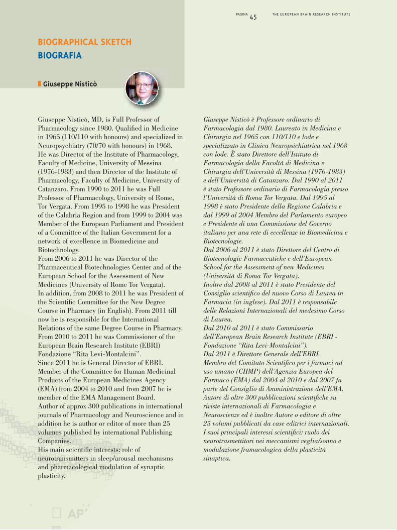
PAGINA 45 THE EUROPEAN BRAIN RESEARCH INSTITUTE
Giuseppe Nisticò, MD, is Full Professor ofPharmacology since 1980. Qualified in Medicinein 1965 (110/110 with honours) and specialized inNeuropsychiatry (70/70 with honours) in 1968.He was Director of the Institute of Pharmacology,Faculty of Medicine, University of Messina(1976-1983) and then Director of the Institute ofPharmacology, Faculty of Medicine, University ofCatanzaro. From 1990 to 2011 he was FullProfessor of Pharmacology, University of Rome,Tor Vergata. From 1995 to 1998 he was Presidentof the Calabria Region and from 1999 to 2004 wasMember of the European Parliament and Presidentof a Committee of the Italian Government for anetwork of excellence in Biomedicine andBiotechnology.From 2006 to 2011 he was Director of thePharmaceutical Biotechnologies Center and of theEuropean School for the Assessment of NewMedicines (University of Rome Tor Vergata).In addition, from 2008 to 2011 he was President ofthe Scientific Committee for the New DegreeCourse in Pharmacy (in English). From 2011 tillnow he is responsible for the InternationalRelations of the same Degree Course in Pharmacy.From 2010 to 2011 he was Commissioner of theEuropean Brain Research Institute (EBRI)Fondazione “Rita Levi-Montalcini”.Since 2011 he is General Director of EBRI.Member of the Committee for Human MedicinalProducts of the European Medicines Agency(EMA) from 2004 to 2010 and from 2007 he ismember of the EMA Management Board.Author of approx 300 publications in internationaljournals of Pharmacology and Neuroscience and inaddition he is author or editor of more than 25volumes published by international PublishingCompanies. His main scientific interests: role ofneurotransmitters in sleep/arousal mechanismsand pharmacological modulation of synapticplasticity.
Giuseppe Nisticò è Professore ordinario diFarmacologia dal 1980. Laureato in Medicina eChirurgia nel 1965 con 110/110 e lode especializzato in Clinica Neuropsichiatrica nel 1968con lode. È stato Direttore dell’Istituto diFarmacologia della Facoltà di Medicina eChirurgia dell’Università di Messina (1976-1983)e dell’Università di Catanzaro. Dal 1990 al 2011è stato Professore ordinario di Farmacologia pressol’Università di Roma Tor Vergata. Dal 1995 al1998 è stato Presidente della Regione Calabria edal 1999 al 2004 Membro del Parlamento europeoe Presidente di una Commissione del Governoitaliano per una rete di eccellenze in Biomedicina eBiotecnologie.Dal 2006 al 2011 è stato Direttore del Centro diBiotecnologie Farmaceutiche e dell’EuropeanSchool for the Assessment of new Medicines(Università di Roma Tor Vergata).Inoltre dal 2008 al 2011 è stato Presidente delConsiglio scientifico del nuovo Corso di Laurea inFarmacia (in inglese). Dal 2011 è responsabiledelle Relazioni Internazionali del medesimo Corsodi Laurea.Dal 2010 al 2011 è stato Commissariodell’European Brain Research Institute (EBRI -Fondazione “Rita Levi-Montalcini”).Dal 2011 è Direttore Generale dell’EBRI.Membro del Comitato Scientifico per i farmaci aduso umano (CHMP) dell’Agenzia Europea delFarmaco (EMA) dal 2004 al 2010 e dal 2007 faparte del Consiglio di Amministrazione dell’EMA.Autore di oltre 300 pubblicazioni scientifiche suriviste internazionali di Farmacologia eNeuroscienze ed è inoltre Autore o editore di oltre25 volumi pubblicati da case editrici internazionali. I suoi principali interessi scientifici: ruolo deineurotrasmettitori nei meccanismi veglia/sonno emodulazione framacologica della plasticitàsinaptica.
❚ Giuseppe Nisticò
BIOGRAPHICAL SKETCHBIOGRAFIA

SELECTED PUBLICATIONSPUBBLICAZIONI
PAGINA 46 THE EUROPEAN BRAIN RESEARCH INSTITUTE
1. Marley E. and Nisticò G. (1972). Effects ofcatecholamines, and adenosine derivatives given intothe brain of fowls. Brit. J. Pharmac. 46, 619-636.
2. Marley E. and Nisticò G. (1975). Tryptamines andsome other substances affecting waking and sleep infowls. Br. J. Pharmac. 53,193-205.
3. Marley E. and Nisticò G. (1975). Central effects ofclonidine [2-(2,6-dichlorophenylamino)-2-imidazolinehydrochloride] in fowls. Br. J. Pharmac. 55,459-473.
4. Carruba M., Nisticò G. and Gargiulo G. (1978). Effectsof central nervous system-acting drugs after selectivedestruction by neurotoxins of 5-hydroxytryptaminefibers in the brain. Ann. N. Y. Acad. Sci. 305,242-258.
5. Gargiulo G. and Nisticò G. (1978). Time course of 5,6-dihydroxytryptamine neurotoxic effects on fowldiencephalon and upper brain stem monoaminergicpathways. J. Anatomy. 126,261-274.
6. Racagni G., Apud J.A., Locatelli V., Cocchi D., NisticòG., Di Giorgio R.M. and Muller E.E. (1979). GABA ofCNS origin in the rat anterior pituitary inhibitsprolactin secretion. Nature. 281,575-578.
7. De Sarro G.B., Ascioti C., Froio F., Libri V. and NisticòG. (1987). Evidence that locus coeruleus is the sitewhere clonide and drugs acting at alfa-1 and alfa-2-adrenoceptors affect sleep and arousal mechanisms.Br. J. Pharmac. 90, 675-685.
8. De Sarro G.B., Meldrum B.S. and Nisticò G. (1988).Anticonvulsant effects of some calcium entry blockersin DBA/2 mice. Br. J. Pharmac. 93,247-256.
9. Bagetta G., De Sarro G.B., Priolo E. and Nisticò G.(1988). Ventral segmenta area, site through whichdopamine D-2 receptors agonists evoke behaviouraland electrocortical sleep in rats. Br. J. Pharmac.95,860-866.
10. De Sarro G.B., Bagetta G., Ascioti C., Libri V. andNisticò G. (1988). Microinfusion of clonidine andyohimbine into locus coeruleus alters EEG powerectrum effects of aging and reversal byphosphatidylserine. Br. J. Pharmac. 95,1278-1286.
11. De Sarro G.B., Bagetta G., Ascioti C., Libri V. andNisticò G. (1989). Effects of pertussis toxin on thebeahvioural and ECoG spectrum changes induced bycloni dine and yohimbine after their microinfusion intothe locus coeruleus. Br. J. Pharmac. 96,59-64.
12. Mollace V., Masuda Y., Pelaggi T. and Nisticò G.(1990). The effects of phosphatidylserine on thedecreased cardiovascular response to clonidinemicroinfused into the nucleus tractus solitarii of oldrats. Br. J. Pharmac. 100,547-551.
13. Bagetta G., De Sarro G.B., Sakurada R., Rispoli V. andNisticò G. (1990). Different profile of electrocorticalpower spectrum changes after microinfusion into thelocus coeruleus of selective agonists at various opioidreceptor subtypes in rats. Br. J. Pharmac. 101,655-661.
14. Nisticò G. and De Sarro G.B. (1991). Is interleukin 2 aneuromodulator in the brain? Trends in Neurosci. 14,146-150.
15. Mollace V., De Francesco E.A., Fersini G. and NisticòG. (1991). Age-dependent changes in cardiovascularresponses induced by muscimol infused into thenucleus tractus solitarii and nucleus parabrachialismedialis in rats. Br. J. Pharmac. 103, 1802-1806.
16. Nisticò G., De Sarro G.B., Bagetta G. and Mollace V.(1992). Altered sensitivity of α2-adrenoreceptors inthe brain during age in rats. Proc. Ann. N.Y. Acad. Sci.673, 206-213.
17. Bagetta G., Corasanti M.T., Aloe L., Berliocchi L.,Costa N., Finazzi-Agrò A., Nisticò G. (1996).Intracerebral injection of human immunodeficiencyvirus type 1 coat protein gp 120 differentially affectsthe expression of nerve growth factor and nitric oxidesynthase in the hippocampus of rat. Proc. Natl. Acad.Sci. USA, 93, 928-33.
18. Nisticò R., Piccirilli S., Sebastianelli L., Nisticò G.,Bernardi G., Mercuri N.B. (2007). The blockade ofK(+)-ATP channels has neuroprotective effects of an invitro model of brain ischemia. Int. Rev. Neurobiol. 82,383-395.
19. Levi-Montalcini R., Knight R.A., Nicotera P., NisticòG., Bazan N., Melino G. (2011). Rita’s 102!! Mol.Neurobiol. 2011 Apr. 43(2):77-9.
20. Bornstein S., Ehrhart-Bornstein M., Androutsellis-Theotokis A., Eisenhofer G., Licinio J., Wong M.L.,Calissano P., Nisticò G., Levi-Montalcini R. (2012).Chromaffin Cells - The Peripheral Brain, MolecularPsychiatry, 17, 354-358.

SELECTED VOLUMESVOLUMI
PAGINA 47 THE EUROPEAN BRAIN RESEARCH INSTITUTE
1. Muller E.E., Nisticò G. and Scapagnini U. (1997).Neurotrasmitters and Anterior Pituitary Function.Academic Press. New York. pp. 1-435.
2. Nisticò G. and Bolis L. (Eds.) (1983). Progress inNonmammalian Brain Research. CRC Press. BocaRaton, Florida. Vol. I pp. 1-185.
3. Nisticò G. and Bolis L. (Eds.) (1983). Progress inNonmammalian Brain Research. CRC Press. BocaRaton, Florida. Vol. II pp. 1-224.
4. Nisticò G. and Bolis L. (Eds.) (1983). Progress inNonmammalian Brain Research. CRC Press. BocaRaton, Florida. Vol. III pp. 1-245.
5. Nisticò G., Di Pierri R. and Meinardi H. (Eds.) (1983).Epilepsy, an update on research and therapy. Alan LissInc. New York. pp. 1-381.
6. Nisticò G., Morselli P.L., Lloyd K.G., Fariello R.G. andEngel J. (Eds.) (1985.) Neurotrasmitters, seizures andEpilepsy III. Raven Press. New York. pp. 1-505.
7. Muller E.E. and Nisticò G. (Eds.) (1989). Brainmessengers and the Pituitary, Acdemic Press. NewYork. pp. 1-711.
8. Vane J., Higgs A., Marsico S.A. and Nisticò G. (Eds.)(1989). Asthma H.S. Basic mechanism and therapeuticperspectives. Pythagora Press. Roma-Milano. Pp. 1-252.
9. Placidi G.F., Dell’Osso L., Nisticò G. and Akiskal H.S.(Eds.) (1993). Recurrent Mood Disorders. Springer-Verlag, berlin. Pp.1-304.
10. Moncada S., Nisticò G. and Higgs A.E. (Eds.) (1993).Nitric oxide: Brain and Immune System. PortlandPress. London. Pp. 1-293.
11. Moncada S., Nisticò G., Bagetta G., Higgs A.E. (Eds.)(1998). Nitric oxide and the Cell, Portland Press,London pp. 1-305.
12. Nisticò G., McGiff J., Born G. (Eds.) (2008). InMemory of Sir John Vane, Exòrma, Roma.
13. Negri L., Melchiorri P., Hökfelt T., Nisticò G. (2009).In Memory of Vittorio Erspamer, Exòrma, Roma.
14. Pepeu G.C., Mugelli A., Moroni F., Nisticò G. (2011).In Memory of Alberto Giotti, Exòrma, Roma.

Alessandro Usiello
❚ Alessandro Usiello, Visiting Fellow(CEINGE, Second University of Naples, Italy)
BEHAVIOURAL STUDIES UNIT
PAGINA 48 THE EUROPEAN BRAIN RESEARCH INSTITUTE
function of D-aspartic acid has remained unclear.D-aspartate is widely expressed in the brainduring early development and in newborns, whileit decreases to trace levels a few weeks after birth.A marked difference in the amount of D-aspartatehas been also found in pathological conditionssince the brains of Alzheimer’s disease patientsdisplay a drastic decline of D-aspartic acid levelscompared to controls. The only enzyme known sofar to metabolize free bicarboxilic D-amino acidsin vitro and thus potentially able to regulate theirlocal concentration in tissues is D-AspartateOxidase (DDO). Anatomically, a reciprocaltopographical localization of DDO and itssubstrate, D-Aspartate, has been found throughoutthe CNS, which let hypothesize that DDO mightinactivate the intracellular amino acid or depletethe synaptically released D-aspartic acid.Recently, in the attempt to understand thebiological consequences of an altered D-aspartateand NMDA signalling, a genetic mouse model hasbeen generated in which the DDO enzyme hasbeen removed through a gene-targeting approach.Mutated animals, available in our laboratory,reveal a dramatic increase in the amount ofD-aspartic acid in all tissues examined.The purpose of our project is to elucidate in vivothe contribution of altered levels of these D-aminoacids in neural functions using mutant Ddo-/-
mice. In detail, we are carrying out behavioraltests in Ddo-/- mice to explore diverse centralnervous system functions, namely motor activity,motor coordination, anxiety- related responses,depression, sensorimotor gating and learning andmemory abilities.
For a long time it has been assumed that L-formsof aminoacids exclusively constitute free aminoacids pool in mammals while free D-aminoacidswere considered unnatural and only deriving fromintestinal bacterial flora. However, in the last twodecades, advances in the analytical methodologiesfor separating chiral aminoacids have shown thatD-forms, namely D-aspartate, N methyl D-asparticacid (NMDA) and D-serine, occur in themammalian Central Nervous System (CNS) andperipheral tissues. Extensive studies generatedmany experimental evidences for defining D-serineas a neurotransmitter, while the physiological

UNITÀ DI STUDI COMPORTAMENTALI
PAGINA 49 THE EUROPEAN BRAIN RESEARCH INSTITUTE
Per lungo tempo si è pensato che la forma degliaminoacidi levogiri (L-aminoacidi) costituisse laforma esclusiva con cui gli aminoacidi eranopresenti nei mammiferi, mentre gli aminoacididestrogiri (D-aminoacidi) erano considerati nonnaturali o solamente derivanti dalla flora batterica.Nell’ultimo ventennio, gli sviluppi nelle metodologiedi chimica analitica hanno mostrato cheD-aspartato, acido N-metil-D-aspartico (NMDA) eD-serina, sono presenti nel Sistema Nervoso Centrale(SNC) e nei tessuti periferici. Recenti studi hannogenerato evidenze sperimentali per i quali laD-serina è oggi considerata una molecola ad azioneneuromodulatrice, al contrario, la funzionefisiologica dell’acido D-aspartico nel SNCrimangono ancora ignote. Di rilievo, tuttavia,èl’osservazione che una significativa riduzione nellaquantità del D-aspartato si ritrova nei cervelli dipazienti affetti dal morbo di Alzheimer relativamentea cervelli normali. Il solo enzima conosciuto, capacedi metabolizzare i D-aminoacidi bicarbossilici liberiin vitro, è la D-Aspartato Ossidasi (DDO).Anatomicamente, una localizzazione reciproca delDDO e del suo substrato, D-Aspatato, lasciaipotizzare che il controllo di questo D-aminoacidopossa avere un ruolo a livello sinaptico.Nel tentativo di capire le conseguenze biologiche diun’alterazione nei livelli di D-aspartato, un modellogenetico murino è stato generato, nel quale l’enzimaDDO è stato rimosso attraverso un approccio diablazione genica. Gli animali mutati, disponibili nelnostro laboratorio, mostrano un forte aumento dellaquantità di acido D-aspartico in tutti i tessutiesaminati. Lo scopo di questo progetto è di chiarirein vivo il contributo di livelli alterati di questiD-aminoacidi nelle funzioni neuronali usando
mutanti murini Ddo-/-. In dettaglio, noi intendiamocaratterizzare, mediante l’utilizzo di specifici testcomportamentali, le funzioni centrali di questi topitransgenici relative all’attività motoria, alle rispostedi ansia e depressione e ai meccanismi diapprendimento e memoria.

Silvia Marinelli
NEUROPATHIC PAIN LABORATORY
PAGINA 50 THE EUROPEAN BRAIN RESEARCH INSTITUTE
Neuropathic pain (NP) is a form of chronic pain inwhich a sustained activation of nociceptivepathway cause a “maladaptive” response/plasticityof the pain circuitry in which the nociceptivetransmission is enhanced and altered so that painis felt in the absence of stimuli and the response toinnocuous (allodynia) and noxious stimuli areenhanced (hyperalgesia).It can persist years and even decades after its firstexpression so that it is considered itself adisabling disease. In addition to its nociceptive and the nocifensivecomponent NP is also considered by theemotional–affective and cognitive aspects whichare crucial for a whole knowledge of the painmechanisms. In fact pain can induce impairmentof cognitive processes that in turn modulate painperception. In other words, chronic painconditions cause depression and anxiety and viceversa patients with depression and/or anxiety feel
pain intensely. To this regard, specific brain areas,such as the cingulate and somatosensory cortex,undergochanges following persistent activationofthe nociceptive circuits and the synaptic strengthincreased due to a synaptic excitatory facilitationor reduction in inhibition. In particular, astructural reorganization, recruitment of additionalbrain areas, alteration of neurochemistry anddisruption of the functional connectivity betweendifferent cortical regions are the main mechanismsof the chronic NP.The major interest of our laboratory is the study ofthe basic mechanisms of NP in the cingulate andsomatosensorycortex and the involvement ofcannabinoid receptors (CBRs) and vanilloidreceptors type-1 (TRPV1) in the emotional-affective, cognitive and sensory aspects of pain.Using a combination of electrophysiological,cellular, pharmacological and morphologicaltechniques in a murine model of NP (the chronicconstriction injury) we aim at identifyingalterations of excitatory and inhibitoryneurotransmission onto cortical pyramidalneurons, with a focus on changes ofendocannabinoid signaling.
Infrared image of a pyramidal neuron (1)and its electrical activity (2)
Immagine ad infrarosso di un neurone piramidale (1)e sua attività elettrica (2)
1
❚ Silvia Marinelli, Group Leader
❚ Michela Giustizieri, Postdoc
❚ Gioacchino Lonobile, Postdoc
❚ Maria Cristina Marrone, Postdoc
PATHOPHYSIOLOGICAL ROLE OF ENDOCANNABINOID SYSTEM

LABORATORIO DOLORE NEUROPATICO
PAGINA 51 THE EUROPEAN BRAIN RESEARCH INSTITUTE
Il dolore neuropatico (DN) è una forma di dolorecronico in cui l’attivazione persistente del circuitonocicettivo provoca un’alterata risposta/plasticitàdei circuiti nervosi del dolore causandoun’aumentata trasmissione nocicettiva.In particolare, il dolore cronico è dovuto ad unostimolo che normalmente non lo provoca (allodinia)oppure è causato da un’eccessiva risposta dolorosa astimoli algogeni di lieve entità (iperalgesia).Il DN può persistere anni e anche decenni dopo lasua prima espressione ed è per questo consideratouna malattia invalidante.Oltre alla componente nocicettiva e nocifensiva, ilDN è caratterizzato anche dall’aspetto emotivo-affettivo e cognitivo, quest’ultimo fondamentale peruna completa conoscenza dei meccanismi deldolore. Infatti, il dolore può indurre lacompromissione dei processi cognitivi che a lorovolta modulano la percezione del dolore.In altri termini, le condizioni di dolore cronicospesso causano depressione e ansia e viceversa, ipazienti affetti da depressione e/o ansia sentonodolore intensamente.
A tale proposito, specifiche aree cerebrali, come lacorteccia cingolata esomatosensoriale, subisconocambiamenti strutturali e funzionali in seguitoall’attivazione persistente dei circuiti nocicettivi.Inoltre la forza sinaptica è aumentata e ciò puòessere dovuto ad una facilitazione sinapticaeccitatoria o ad una riduzione dell’inibizione. L’interesse principale del nostro laboratorio è lostudio dei meccanismi di base del DN conparticolare riferimento alla corteccia cingolataesomatosensoriale, e il coinvolgimento dei recettoridei cannabinoidi di tipo 1 e 2, e dei recettorivanilloidi di tipo 1 (TRPV1) negli aspettiemotivi-affettivi, cognitivi e sensoriali del dolore.Usando una combinazione di tecnicheelettrofisiologiche, farmacologiche e diimmunofluorescenza in un modello murino di DN(la costrizione cronica del nervo sciatico) miriamo aindividuare modificazioni della neurotrasmissioneeccitatoria e inibitoria su neuroni corticalipiramidali, con particolare interesse ad alterazionidel sistema degli endocannabinoidi.
RUOLO FISIOPATOLOGICO DEL SISTEMADEGLI ENDOCANNABINOIDI
Pyramidal neurons of the anterior cingulate cortex stainedwith the SMI-32 primary antibody
Neuroni piramidali di corteccia cingolata di topo marcaticon l’anticorpo primario SMI-32
2

PAGINA 52 THE EUROPEAN BRAIN RESEARCH INSTITUTE
After obtaining her Master’s Degree inPharmacology at the University of Rome“La Sapienza” in 1996, Silvia Marinelli continuedher graduate student training with Prof. P.Calabresi at the University of Rome “Tor Vergata”(1997-1998) and subsequently did her PhD in thelaboratory of Prof. N.B. Mercuri at the FondazioneSanta Lucia in Rome. From 1999 to 2001 sheworked in the Department of Pharmacology -University of Sydney (Australia) in the laboratoryof Prof. Mac D. Christie. In 2002 she returned tothe laboratory of Prof. Mercuri to continue herPostdoctoral research. From 2006-2010 sheworked as Senior Scientist in the laboratory of
Alberto Bacci at the European Brain ResearchInstitute (EBRI) in Rome. Her research studieshave focused mainly a) on the role of vanilloidreceptor type 1 in different brain areas,b) functional and anatomical characterization ofserotoninergic neurons involved in the descendingnociceptive pathways and c) the endocannabinoidinvolvement in a form of self-inhibition ofneocortical principal and GABAergic neurons.In 2012 she was appointed Junior Group Leader ofthe Neuropathic Pain laboratory(Pathophysiological role of the endocannabinoidsystem) at the EBRI. She is recipient of a younginvestigator grant by the Italian Minister of Health.
❚ Silvia Marinelli
BIOGRAPHICAL SKETCHBIOGRAFIA
Dopo aver conseguito la laurea in Farmacia pressol’Università di Roma “La Sapienza” nel 1996,Silvia Marinelli ha continuato la sua formazionecon il Prof. P. Calabresi presso l’Università di Roma“Tor Vergata” (1997-1998) e successivamente hafatto il suo dottorato di ricerca nel laboratorio delProf. N.B. Mercuri alla Fondazione Santa Lucia diRoma. Dal 1999 al 2001 ha lavorato presso ilDipartimento di Farmacologia - Università diSydney (Australia) nel laboratorio del Prof. Mac D.Christie. Nel 2002 ha continuato la sua ricercacome post-doc nel laboratorio del Prof. Mercuri.Dal 2006-2010 ha lavorato come Senior Scientistnel laboratorio di Alberto Bacci presso l’IstitutoEuropean Brain Research Institute (EBRI) di
Roma. I suoi studi si sono concentratiprincipalmente a) sul ruolo del recettore vanilloidedi tipo 1 in diverse aree del cervello, b) sullacaratterizzazione funzionale e anatomica deineuroni serotoninergici coinvolti nel circuitodiscendente antinocicettivo e c) sul coinvolgimentodegli endocannabinoidi in una forma diauto-inibizione dei neuroni piramidali eGABAergicidella neocorteccia.Nel 2012 è stata nominata presso l’EBRI JuniorGroup Leader del Laboratorio Dolore Neuropatico(Ruolo fisiopatologico del sistema degliendocannabinoidi).Inoltre Silvia Marinelli è beneficiaria di un grant“Giovani Ricercatori” del Ministro della Salute.

PAGINA 53 THE EUROPEAN BRAIN RESEARCH INSTITUTE
SELECTED PUBLICATIONSPUBBLICAZIONI
1. Manseau F., Marinelli S., Schwaller B., Mendez P.,Prince D.A., Huguenard J.R., Bacci A. (2010).Desynchronization of neocortical networks byasynchronous release of GABA at autaptic andsynaptic contacts from fast-spiking interneurons.Plos Biology. Vol 8 (9).
2. Marinelli S., Pacioni S., Cannich A., Marsicano G.,Bacci A. (2009). Self-modulation of neocorticalglutamatergic neurons by endocannabinoids.Nature Neurosci. 12:1488-90.
3. Marinelli S., Pacioni S., Bisogno T., Di Marzo V.,Prince D.A., Huguenard J.R., Bacci A. (2008). Theendocannabinoid 2-AG is responsible for the slowself-inhibition in neocortical interneurons. J. Neurosci.28:13532-13541.
4. Marinelli S., Di Marzo V., Florenzano F., Viscomi M.T.,Fezza F., Van Der Stelt M., Bernardi G., Molinari M.,Maccarroneand Mercuri N.B. (2007). N-arachidonoyldopamine tunes synaptic transmissiononto dopaminergic neurones by activating bothcannabinoid and vanilloid receptors.Neurospychopharmacology. 32, 298-308.
5. Marinelli S., Connor M., Schnell S.A., Christie M.J.,Wessendorf M.W., Vaughan C.W. (2005). Delta-opioidreceptor-mediated actions on rostral ventromedialmedulla neurons. Neuroscience. 132(2):239-44.
6. Marinelli S., T. Pascucci, G. Bernardi, S. Puglisi-Allegra, and Mercuri N.B. (2005 May) Activation ofTRPV1 in the VTA excites dopaminergic neurons andincreases chemical- and noxious-induced dopaminerelease in the nucleus accumbens.Neuropsychopharmacology. 30(5):864-70.
7. Marinelli S., Schnell S.A., Hack S.P., Christie M.J.,Wessendorf M.W. and Vaughan C.W. (2004 Dec).Serotonergic and nonserotonergic dorsal raphe neuronsare pharmacologically and electrophysiologicallyheterogeneous. J. Neurophysiol. 92(6):3532-7.
8. Marinelli S., Di Marzo V., Berretta N., Matias I.,Maccarrone M., Bernardi G. and Mercuri N.B. (2003).
Presynaptic facilitation of glutamatergic synapses todopaminergic neurons of the rat substantia nigra byendogenous stimulation of vanilloid receptors.J. Neuroscience. 23(8):3136-3144.
9. Marinelli S., Schnell S., Vaughan C.W., WessendorfM.W., Christie M.J. (2002). Rostral ventromedialmedulla neurons that project to the spinal cord expressmultiple opioid receptor phenotypes. J. Neuroscience,22(24): 10847-10855.
10. Marinelli S., Vaughan C.W., Christie M.J., Connor M.(2002). Capsaicin activationof glutamatergic synaptictransmission in the rat locus coeruleus in vitro. J.Physiology. 543, pp. 531-540.
11. Vaughan C.W., Connor M., Jennings E.A., Marinelli S.,Allen R.G., Christie M.J. (2001). Actions ofnociceptin/orphanin FQ and other prepronociceptinproducts on rat rostral ventromedial medulla neuronsin vitro. J. Physiology. 543(3), pp.849-859.
12. Marinelli S., Federici M., Bernardi G., Mercuri N.B.(2001). Hypoglycaemia enhances ionotropic butreduced metabotropic glutamate current in ratsubstantia nigra dopaminergic neurons. J.Neurophysiology. 85, pp.1159-1166.
13. Marinelli S., Bernardi G., Giacomini P., Mercuri N.B.(2000). Pharmacological identification of the K+currents mediating the hypoglycaemichyperpolarisation of rat midbrain dopaminergicneurons. Neuropharmacology. Vol 39 (6), pp. 1021-1028.
14. Marinelli S., Gatta F., Sagratella S. (2000). Effect ofGYKI 52466 and some 2,3-benzodiazepinederivateson hippocampal in vitro basal neuronal excitability and4-aminopiridine epileptic activity. Eur. J. Pharmacol.391(1-2), pp75-80.
15. Sagratella S. and Marinelli S. (2000). Effect on someGABA and NMDA antagonists on a model ofpresynaptic hippocampal paired pulse inhibition: Prog.Neuropsycopharmacol. Biol. Psychiatry. Vol 24 (2), pp.327-336.

❚ Michelangelo Campanella, Group Leader
❚ Stefania Cocco, Postdoc
❚ Ramona Lupi, Postdoc
❚ Valerio De Biase, PhD Student
❚ Cinzia Agostini, Lab technician
METABOLISM IN BRAIN DISEASES LABORATORY
MITOCHONDRIA TO READ OUT ANDUNDERSTAND METABOLIC ANDFUNCTIONAL DEFICIENCY OF BRAINPATHOLOGIES
purpose of improving the understanding of biologyof degenerative phenomena in neurons and glia, byapplying our advanced methods of investigation forcellular biology characterization, and through theintegrated study of regulatory pathways ofmitochondrial homeostasis.The techniques evolved in years allow us to studywith accuracy the mitochondrial physiology inintact cells and monitor the metabolic profilethrough the study of enzymatic pathways inphysiology and pathology. Imaging protocolscombined with luminescence approaches allowthis by preserving cell integrity, so increasingquality and effectiveness of our studies.These methods complemented with standardassays of biochemistry and immuno-istochemistryconstitute a technical background that very fewcenters worldwide have and/or apply in the CNSstudy. In addition, the use of transgenic animals(available at the hosting Institution) allows us torun appropriate controls and so realize theambitious experimental program.Two are the major lines of research currentlyfollowed:I. the regulation of mitochondrial enzymes in the
pathogenesis of neuronal death.II. the molecular mechanisms of mitophagy in the
study of brain tumors.Line I aims at studying the glycolytic andmitochondrial metabolism during the reversion ofthe F1Fo-ATPsynthase, by the analysis of itsmolecular inhibitor IF1 and its cross-talk with theglycolitic enzymes susceptible to the F1Fo-ATPsynthase activity. This study is conducted both“in vivo” on experimental models of Alzheimerdisease, using pathological transgenic animalmodels (Tg2576 and AD11 mice), and “in vitro”on neuronal and glial treated with b-amyloid.Data obtained so fare have revealed an increasedexpression of IF1 in pathological conditions,suggesting a role of the protein in the metabolic
Several studies have demonstrated the role ofmitochondria in the pathophysiology of centralnervous system (CNS), precisely on toxicity andcell death in the development of associatedneurodegenerative diseases. Impairment ofmitochondrial energetic activity and its regulationof biological events defined by intra and interneuronal signaling is well known and, in some ofthe most common types of neurodegeneration, thevery cause of the disease.In Alzheimer’s as well as in Parkinson’s, thanks tothe terrific work of functional characterization inone and site-specific mutations in the other, themitochondrion has been considered the commondenominator in the onset of cellular pathology. Andmitochondrial damage is a well-defined featureeven in Amyotrophic Lateral Sclerosis (ALS).In the light of this, the work of our unit is placed inat the center of modern neuroscience, and has the
Michelangelo Campanella
PAGINA 54 THE EUROPEAN BRAIN RESEARCH INSTITUTE
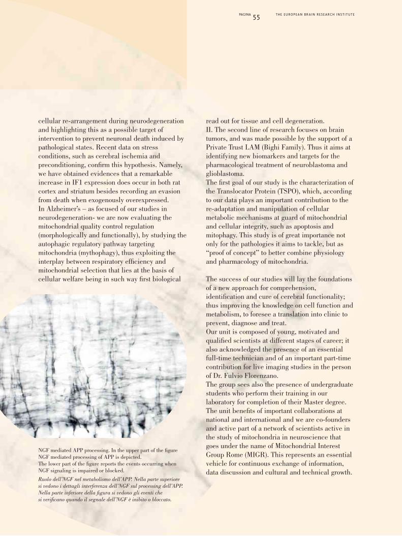
cellular re-arrangement during neurodegenerationand highlighting this as a possible target ofintervention to prevent neuronal death induced bypathological states. Recent data on stressconditions, such as cerebral ischemia andpreconditioning, confirm this hypothesis. Namely,we have obtained evidences that a remarkableincrease in IF1 expression does occur in both ratcortex and striatum besides recording an evasionfrom death when exogenously overexpressed.In Alzheimer’s – as focused of our studies inneurodegeneration- we are now evaluating themitochondrial quality control regulation(morphologically and functionally), by studying theautophagic regulatory pathway targetingmitochondria (mythophagy), thus exploiting theinterplay between respiratory efficiency andmitochondrial selection that lies at the basis ofcellular welfare being in such way first biological
read out for tissue and cell degeneration. II. The second line of research focuses on braintumors, and was made possible by the support of aPrivate Trust LAM (Bighi Family). Thus it aims atidentifying new biomarkers and targets for thepharmacological treatment of neuroblastoma andglioblastoma. The first goal of our study is the characterization ofthe Translocator Protein (TSPO), which, accordingto our data plays an important contribution to there-adaptation and manipulation of cellularmetabolic mechanisms at guard of mitochondrialand cellular integrity, such as apoptosis andmitophagy. This study is of great importance notonly for the pathologies it aims to tackle, but as“proof of concept” to better combine physiologyand pharmacology of mitochondria.
The success of our studies will lay the foundationsof a new approach for comprehension,identification and cure of cerebral functionality;thus improving the knowledge on cell function andmetabolism, to foresee a translation into clinic toprevent, diagnose and treat.Our unit is composed of young, motivated andqualified scientists at different stages of career; italso acknowledged the presence of an essentialfull-time technician and of an important part-timecontribution for live imaging studies in the personof Dr. Fulvio Florenzano. The group sees also the presence of undergraduatestudents who perform their training in ourlaboratory for completion of their Master degree.The unit benefits of important collaborations atnational and international and we are co-foundersand active part of a network of scientists active inthe study of mitochondria in neuroscience thatgoes under the name of Mitochondrial InterestGroup Rome (MIGR). This represents an essentialvehicle for continuous exchange of information,data discussion and cultural and technical growth.
NGF mediated APP processing. In the upper part of the figureNGF mediated processing of APP is depicted.The lower part of the figure reports the events occurring whenNGF signaling is impaired or blocked.
Ruolo dell’NGF nel metabolismo dell’APP. Nella parte superioresi vedono i dettagli interferenza dell’NGF sul processing dell’APP.Nella parte inferiore della figura si vedono gli eventi chesi verificano quando il segnale dell’NGF è inibito o bloccato.
PAGINA 55 THE EUROPEAN BRAIN RESEARCH INSTITUTE

LABORATORIO METABOLISMO PATOLOGIECEREBRALI
MITOCONDRIO PER INTERPRETARE E COMPRENDERE DEFICIT METABOLICI E FUNZIONALI DI PATOLOGIE CEREBRALI
Queste metodiche, unite a saggi standard dibiochimica e immuno-istochimica, compongono unbagaglio tecnico che pochissimi centri nel mondohanno e/o applicano agli studi del SNC. Inoltre,l’utilizzo di animali transgenici permette al nostrolavoro di avvalersi di adeguati controlli e realizzarel’ambizioso programma sperimentale.Due sono le principali linee di ricerca seguite: I. regolazione degli enzimi mitocondriali nella
patogenesi della morte neuronale. II. meccanismi molecolari di mitofagia nello studio
dei Tumori Cerebrali.La linea I verte a chiarire l’adattamento delmetabolismo mitochondriale e glicolitico durantela reversione dell’enzima mitocondrialeF1Fo-ATPsintasi, avvalendosi dello studio del suomodulatore molecolare IF1 e del cross talk conenzimi glicolitici sensibili all’attività dell’enzimaF1Fo-ATPsintasi. Tale lavoro viene svoltoprincipalmente in modelli sperimentali diAlzheimer, “in vivo” utilizzando animali transgeniciche manifestano la malattia (linee di topi Tg2576 eAD11), ed “in vitro”, utilizzando modelli cellularineuronali e gliari esposti a b−amyloide.Gli studi compiuti sino ad ora indicano unincremento specifico di espressione della proteinaIF1 in condizioni patologiche. Tale aumentosuggerisce un protettivo della proteina neimeccanismi di adattamento metabolico cellulareche intervengono durante insulti neurodegenerativi,ed essendo quindi un potenziale target di studio perprevenire la morte neuronale. A sostegno di taleipotesi abbiamo condotti degli studi in condizioni distress, quali ischemia cerebrale e preconditioning.In particolare, in quest’ultima abbiamo verificatoun significativo aumento dell’espressione dellaproteina IF1 in tessuto corticale e striatale di rattoche si associa ad un marcata protezione dalla mortequando la proteina è sovraespressa. In modelli diAlzheimer’s stiamo anche valutando l’efficienza diregolazione della qualità mitocondriale
Recentemente diversi studi hanno dimostrato ilruolo del mitocondrio nella fisiopatologia delsistema nervoso centrale (SNC), e in particolarenello sviluppo della tossicità e della morte cellularein patologie neurodegenerative. La deregolazione dell’attività energeticamitocondriale e la regolazione degli eventi biologicimitocondriali, mediate dal signalling intra ed interneuronale, è ormai acclamata e, in alcune tra leneurodegenerazioni più note, causa stessa dellamalattia. Nelle patologie di Alzheimer e Parkinson,grazie alla formidabile opera di caratterizzazionefunzionale nell’uno, e mutazioni sito-specifichenell’altra, il mitocondrio si è dimostrato esserecomun denominatore nello sviluppo della patologia;così come per la patogenesi della Sclerosi LateraleAmiotrofica (SLA).Alla luce di ciò, il lavoro della nostra unità sicolloca in un punto nevralgico delle neuroscienzemoderne, e ha la finalità di migliorare lacomprensione della biologia dei fenomenidegenerativi di neuroni e glia mediante l’uso dimetodiche di indagine di avanguardia per lacaratterizzazione bio-cellulare, e attraverso lo studiointegrato di pathways regolatorie dell’omeostasimitocondriale.Le tecniche affinate in anni di lavoro ci permettonodi studiare con precisione la fisiologiamitocondriale in cellule intatte, e di monitorare ilprofilo metabolico grazie allo studio delle vieenzimatiche in fisiologia e patologia. Protocolli diimaging, uniti ad approcci di luminescenza,permettono di analizzare signalling e mofologiamantendo l’integrità cellulare, incrementando cosìqualità ed efficacia degli studi.
PAGINA 56 THE EUROPEAN BRAIN RESEARCH INSTITUTE

(morfologica e funzionale), attraverso lo studiodella via regolatoria autofagica targettata aimitocondri (mitofagia), ritenendo che l’interplaytra efficienza respiratoria e selezione dei mitocondrisia alla base del “welfare” cellulare e che la suaalterazione sia un segnale biologico che preceda esostenga l’insorgere della degenerazione cellulare etessutale.II. La seconda linea di ricerca, relativa allo studiodei tumori cerebrali, è stata possibile grazie al supporto del Trust Privato LAM (Famiglia Bighi), epunta all’identificazione di nuovi bio-markers ebersagli per il trattamento farmacologico delNeuroblastoma e Glioblastoma. Il primo obiettivo del nostro studio è lacaratterizzazione della proteina TranslocatorProtein (TSPO), che contribuisce in manieraimportante al ri-adattamento metabolico cellulare ealla manipolazione dei meccanismi a guardiadell’integrità mitocondriale e cellulare, qualiapoptosi e mitofagia. Questo studio è di grandeimportanza, non solo per le patologie alle quali èfinalizzato, ma perché rappresenta una “proof ofconcept” per coniugare meglio fisiologia efarmacologia del mitocondrio.
Il successo della nostra attività di ricerca getterà lebasi per un nuovo approccio per la comprensione,identificazione e cura delle malattie che attaccanola funzionalità cerebrale; migliorando laconoscenza funzionale e metabolica sarà possibilesupportare una futura translabilità nella clinica dinuovi protocolli per prevenzione, diagnosi etrattamento.La nostra unità si compone di motivati e capacigiovani scienziati a diversi punti di carriera, di unsupporto tecnico essenziale full-time e di unimportante contributo part-time per gli studi diimaging, nella persona del Dr Fulvio Florenzano.Il gruppo giova anche della presenza di studentiuniversitari che svolgono nel nostro laboratorio illoro tirocinio sperimentale per il completamentodella laurea specialistica. Beneficia, inoltre, diimportanti collaborazioni sul territorio nazionale edinternazionale. Siamo anche tra i fondatori di unNetwork di Scienziati attivi nello studio deimitocondri nelle neuroscienze, denominatoMitochondrial Interest Group Rome (MIGR), veicolofondamentale per il continuo scambio diInformazioni, confronti sui dati ottenuti e crescitaculturale e tecnica continua.
PAGINA 57 THE EUROPEAN BRAIN RESEARCH INSTITUTE
Neurons in cell culture
Neuroni in coltura cellulare
Neurons from rat cerebral cortex
Neuroni di corteccia cerebrale di ratto

PAGINA 58 THE EUROPEAN BRAIN RESEARCH INSTITUTE
Michelangelo Campanella graduated in Italy(University of Ferrara) in July 2001 and obtained aPhD in Pharmacology in the same University inFebruary 2005. He then moved to the UnitedKingdom to work with Prof. M R Duchen at theUniversity College of London (UCL) and increasehis knowhow in mitochondrial physiology andcellular bio-energetic supported by the Accademiadei Lincei/Royal Society, European MolecularBiology Organization (EMBO) and Marie CurieActions as Long Term Research Fellow. In 2008
has become Tenured in Pharmacology (RoyalVeterinary College) and Principal Investigatoraffiliated of the UCL Consortium for MitochondrialResearch. He is now acting as Ambassador of theprestigious Biochemical Society. In December2010, he become Head of the Research Unit“Metabolism in Brain Diseases” at the EuropeanBrain Research Institute (EBRI)-Rita Levi-Montalcini Foundation. In 2011, he received theNew Investigator Award of the Biotechnology andBiological Sciences Research Council (BBSRC).
❚ Michelangelo Campanella
BIOGRAPHICAL SKETCHBIOGRAFIA
Michelangelo Campanella, laureato all’Universitàdi Ferrara nel 2001, ha ottenuto il Dottorato inFarmacia presso la stessa Università nel 2005.Si è trasferito nel Regno Unito per lavorare con ilprof. M.R. Duchen all’University College di Londra(UCL) ed accrescere le sue conoscenze in fisiologiamitocondriale e bioenergetica cellulare grazieanche ai fondi Accademia dei Lincei/Royal Society,European Molecular Biology Organization(EMBO) e Marie Curie Actions come “ricercatore a
lungo termine”. Nel 2008 è divenuto Docente diFarmacologia (Royal Veterinary College) ePrincipal Investigator affiliato al Consorzio per laRicerca Mitocondriale. È attualmente Ambasciatoredella prestigiosa Società Biochimica. Dal Dicembre2010 è a capo del Laboratorio “Metabolism inBrain Diseases” all’European Brain ResearchInstitute (EBRI). Nel 2011 ha ricevuto il PremioNuovo Ricercatore del Consiglio di Ricerca delleScienze Biologiche e Biotecnologiche (BBSRC).

PAGINA 59 THE EUROPEAN BRAIN RESEARCH INSTITUTE
1. Severinatne M., Faccenda D., De Biase V. andCampanella M. In Bcl-2 knock-down cells, PK11195inhibits mitophagy targeting the F1Fo-ATPsynthase.Current Molecular Medicine in press.
2. Gatliff J. and Campanella M. (2011). The 18KdaTranslocator Protein (TSPO): a New Perspective inMitochondrial Biology. Current Molecular Medicine inpress.
3. Gastaldello A., Gami P., Callaghan H. and CampanellaM. (2010 July). Ca2+ dependent autophagy is enhancedby the Pharmacological Agent PK11195. Autophagy.6;6(5). 2010.
4. Campanella M., Parker N., Tan C.H., Hall A.M. andDuchen M.R. (2009 Jul). IF1: setting the pace of the
F1Fo-ATP synthase. Trends in Biochemical Sciences(TiBs). 34(7):343-50.
5. Campanella M., Seraphin A., Abeti R., Casswell E.,Echave P. and Duchen M.R. (2009). IF1, the F1Fo-ATPsynthase endogenous regulator, definesmitochondrial volume fraction in HeLa cells byregulating basal autophagy. Biochimica and BiophysicaActa (BBA); 1787(5):393-401.
6. Campanella M., Farah Z., Casswell E., Chong S.,Wieckowski M.R., Abramov A.Y., Tinker A. andDuchen M.R. (2008 July). Regulation of MitochondrialStructure and Function by the F(1)F(o)-ATPaseInhibitor Protein, IF(1). Cell Metabolism. 8(1):13-25.
SELECTED PUBLICATIONSPUBBLICAZIONI
Epithelial cells from the basal layerexpressing NGF receptor
Cellule epiteliali che esprimono ilrecettore dell’NGF nello strato basale

Cristina Marchetti
❚ Cristina Marchetti, Junior Project Leader
❚ Hélène Marie, Former Unit Leader
MECHANISMS OF NEURONAL AND SYNAPTICPLASTICITY UNIT
Our unit is interested in understanding whatchanges take place in the brain when we learn orstore a memory. Brains are in fact plastic, allowingfor changes in their pattern of activity in responseto a given stimulus (e.g. a learning event).Such changes can happen at the level of neurons,the basic units of the neural circuit, or at the levelof synapses, the connections between neurons.Several different molecular mechanisms areinvolved in these processes, which can be altereddue to normal aging or to several pathologies likeAlzheimer’s disease.In physiological conditions, we study the neuronaladaptations resulting from the involvement of twomain intracellular mechanisms: 1) the pathwayregulated by CREB, a transcription factor knownto be activated during synaptic plasticity andduring learning, and 2) the role of microRNAs inregulating these plasticities. To this end, we use acombination of techniques, including in vivo viralmediated gene transfer, that allows expression ofrecombinant proteins in the rodent brain, andelectrophysiology. With these tools, we expressproteins of the CREB-dependent transcriptionpathway or alter microRNA functioning in thebrain and we investigate the characteristics ofinfected neurons by whole-cell in vitroelectrophysiology.To investigate how synaptic plasticity is altered inpathologies such as Alzheimer’s disease, we recordsynaptic currents in vitro in brain slices fromtransgenic mouse models. Our aim is to pinpointthe alterations in synaptic transmission andneuronal output which are ultimately responsiblefor memory deficits.
PAGINA 60 THE EUROPEAN BRAIN RESEARCH INSTITUTE

UNITÀ DI MECCANISMI DI PLASTICITÀNEURONALE E SINAPTICA
L’interesse della nostra unità è la comprensione deimeccanismi del cervello alla base della memoria edell’apprendimento. Il cervello è infatti in grado dimodificare la sua attività elettrica in risposta adiversi stimoli (ad esempio un dato damemorizzare). Tali modifiche possono avvenire sia alivello dei neuroni, le cellule che costituiscono leunità di base dei circuiti cerebrali, sia a livello disinapsi, le connessioni tra i neuroni stessi. Moltimeccanismi molecolari sono coinvolti in questiprocessi, che possono essere alterati a causadell’invecchiamento o di patologie come la malattiadell’Alzheimer.In condizioni fisiologiche, studiamo qualialterazioni neuronali risultano dall’attivazione didue meccanismi intracellulari: 1) quello regolatodal CREB, un fattore di trascrizione coinvolto nellaplasticità sinaptica e nell’apprendimento, e 2) il
sistema dei microRNA presenti nelle sinapsi.A tale scopo, utilizziamo un insieme di tecniche,che comprendono l’espressione di una proteina diinteresse nel cervello tramite infezione virale esuccessive registrazioni di segnali elettrici(elettrofisiologia). In questo modo, esprimiamoproteine del pathway CREB-dipendente o alteriamola funzionalità del microRNA, per poi studiare lecaratteristiche dei segnali elettrici emessi deineuroni di interesse, con la tecnica del patch clampin vitro.Nell’ambito delle patologie, utilizziamo modellitransgenici della malattia di Alzheimer edeffettuiamo registrazioni di field extracellulare e dipatch clamp in vitro, per studiare quali alterazionidi correnti sinaptiche e di potenziali di membranasono responsabili dei deficit di memoria.
Hippocampus brain slice, showing CA1pyramidal cells infected with greenfluorescent protein (GFP). This methodallows identifying cells that that expressthe protein of interest.
Cellule piramidali dell’ippocampo infettatecon la green fluorescent protein (GFP).Questo metodo permette di individuarequali cellule esprimono la proteina diinteresse.
Recording of spontaneous synaptic current events recorded from a hippocampal CA1pyramidal cell with the whole cell patch clamp technique.
Registrazione in patch clamp di correnti sinaptiche spontanee proveniente da una cellula CA1di ippocampo.
Evoked synaptic currents recorded from a hippocampal CA1 pyramidal cell, stimulated witha paired-pulse protocol and recorded with the whole cell patch clamp technique. Even withthe same stimulation intensity, the response has a different amplitude, due to synapticplasticity properties.Correnti sinaptiche evocate da una stimolazione a doppio impulso e registrate da una cellulaCA1 in patch clamp. A parità di stimolazione, l’intensità della risposta cambia, a causa delleproprietà di plasticità sinaptica.
PAGINA 61 THE EUROPEAN BRAIN RESEARCH INSTITUTE

PAGINA 62 THE EUROPEAN BRAIN RESEARCH INSTITUTE
Inhibitory neurons play several key roles inneocortical function. For example, they shapesensory receptive fields and drive several highfrequency network oscillations.On the other hand, defects in their function canlead to devastating diseases, such as epilepsy andschizophrenia.The questions we are working on are: what is therole of Fast Spiking Interneurons in coordinatingfast network synchrony? How does a realistic inputinto a network in a high conductance-in vivo likestate shape the correlation between neurons?In our laboratory we recently set up a dynamicclamp system. The dynamic clamp is a powerfultechnique that offers the advantage of providing aneuron with an artificial (i.e. computer generated)and realistic stimulus that features all theappropriate waveform details.
❚ Antonio Pazienti, Researcher
❚ Alberto Bacci, Former Unit Leader
Antonio Pazienti
PROJECT OUTLINE – 2011-2013
Spectrogram of gamma oscillations in hippocampus
Spettrogramma di oscillazioni gamma registrate nell’ippocampo
CORTICAL MICROCIRCUITS UNIT
The amplitude of these waveforms is computedand updated in real-time taking into accountmembrane potential fluctuations, and thus thedriving force of one or more synapticconductances. In particular, the dynamic clampwill be used to recreate in vitro high conductancestates typical of in vivo conditions.By integrating patch-clamp, field potentialelectrophysiology and dynamic clamp techniques,we will examine the role played by fast andprecisely timed feedback inhibition of fast-spikinginterneurons in shaping the spike timing of thenetwork during in vivo-like stimuli.Previous studies have been solely obtained fromquiescent brain slices. We will test the functional role of neurotransmissionin cortical oscillations induced in vitropharmacologically. The outcome of theseexperiments will define the pivotal role of autaptictransmission during cortical rhythms underlyingsensory processing and will provide important cluesfor understanding pathologies like epilepsy andschizophrenia.

PAGINA 63
I neuroni inibitori svolgono diversi ruoli chiavenelle funzioni neocorticali. Determinano peresempio i campi recettivi sensoriali e guidanodiverse stati oscillatori della rete. D’altra parte, i difetti nella loro funzione puòportare a malattie devastanti, come l’epilessia e laschizofrenia.Le domande su cui stiamo lavorando sono: qual è ilruolo degli interneuroni Fast Spiking nelcoordinamento della sincronia della rete?Come viene plasmata la correlazione tra i neuronida input realistici in una rete in stato di altaconduttanza (high-conductance state)?Nel nostro laboratorio abbiamo recentementeappronatato un sistema per il dynamic clamp. Il dynamic clamp è una potente tecnica che offre ilvantaggio di fornire ai neuroni stimoli artificiale(cioè generati al computer) ma realistico, checaratterizzato da tutti i dettagli fisiologici. Gli input vengono calcolati e aggiornati in temporeale, tenendo conto delle fluttuazioni dei potenzialidi membrana, e della forza motrice determinatadalla differenza tra potenziale di membrana epotenziale di riposo di uno o più conduttanzesinaptiche. In particolare, il dynamic clamp saràutilizzato per ricreare in vitro high-conductance
states tipici di condizioni in vivo.Grazie all’integrazione di patch-clamp, fieldpotential e dynamic clamp, esamineremo il ruolosvolto dalla inibizione rapida e perfettamentesincronizzata degli interneuroni fast spiking nelplasmare il grado di coordinazione della retedurante lo stato simil in-vivo. Gli studi precedentisono stati esclusivamente ottenuti da fettine dicervello di riposo.Metteremo inoltre alla prova il ruolo funzionaledella neurotrasmissione nel oscillazioni corticaliindotta in vitro farmacologicamente.I risultati di questi esperimenti definiranno meglioil ruolo centrale della trasmissione autapticadurante ritmi corticali, che sono alla base dielaborazione sensoriale, e forniranno indiziimportanti per la comprensione di patologie comel’epilessia e la schizofrenia.
Dynamic clamp setup
PROGETTO – 2011-2013
A neocortex neuron
Un neurone della corteccia cerebrale
THE EUROPEAN BRAIN RESEARCH INSTITUTE
UNITÀ DI MICROCIRCUITICORTICALI

Mara D’Onofrio
❚ Mara D’Onofrio, Head of Facility
❚ Ivan Arisi, Bioinformatics
❚ Rosella Brandi, Researcher
GENOMICS FACILITY
PAGINA 64 THE EUROPEAN BRAIN RESEARCH INSTITUTE
The EBRI Genomics Facility provides resources toinvestigate gene expression and functions with afocus on applying RNA/DNA microarraytechnologies. The Facility combines state of the arttechnology and expertise in genomics andbioinformatics. The Facility, in collaboration with Cattaneo’sgroup, focuses on the understanding of thecomplex molecular mechanisms related tocognition and synaptic dysfunction as early eventsof neurodegeneration process, with main intereston Alzheimer’s disease. The aim of this project isto provide new knowledge on molecules that act atthe early stage of neurodegenerative phenomena,focusing on synaptic functions. The microarray technology of the EBRI Facility isbased on the Agilent platform, that provides high-
density microarrays chips with 60-mer oligossynthesized in-situ, allowing the analysis of geneexpression profiles, mRNA and microRNA,genomic aberrations on DNA samples (CGH,Comparative Genomic Hybridization), methylation.Projects currently ongoing at EBRI Facility incollaboration with Cattaneo’s group:• Alzheimer’s Disease project: mRNA and
microRNA gene expression profiles in animalmodels and human samples of Alzheimer’sDisease for the identification of molecularchanges and biomarkers for an early diseasediagnosis;
• microRNA, synapses and neurodegeneration;• Role of early inflammation in Alzheimer’s disease;• Spinocerebellar Ataxia: for identification of
high-density genomic aberration profiles in theDNA sequence of specific genes;
• Data mining techniques for analysis of genomicsand clinical dataset.
The Facility offers collaborations to all EBRIgroups.
The Genomics Facility would like to thank AgilentTechnologies for their collaboration andcontinued support.
Probe intensities in a 2-color microarray study from a chip with44000 probes, where each probe corresponds to a coding mRNA.

FACILITY DI GENOMICA
PAGINA 65 THE EUROPEAN BRAIN RESEARCH INSTITUTE
Heterozygous 0.3 Mb DNA deletion from a patient affected by mental retardation, from a 2-colormicroarray CGH study. Each dot corresponds to the signal of a 5 kb DNA segment
La Facility di Genomica EBRI mette a disposizionerisorse per lo studio dell’espressione e delle funzionidei geni, con l’utilizzo di tecnologie di microarrayapplicate a RNA/DNA.La Facility combina tecnologia e competenza ingenomica e bioinformatica. La Facility, in collaborazione con il gruppo delProf. Cattaneo, si propone di comprendere icomplessi meccanismi molecolari all’origine didisfunzioni sinaptiche e cognitive, come eventiprecoci di processi neurodegenerativi, con maggiorinteresse per la malattia di Alzheimer.Lo scopo di questo progetto è inoltre produrre nuoveconoscenze sulle molecole che svolgono un ruolonelle fasi precoci del fenomeno neurodegenerativo,focalizzandosi sulle funzioni sinaptiche.La tecnologia microarray della Facility EBRI,basata sulla piattaforma Agilent, che fornisce chipper microarray ad alta densità con oligo 60-mersintetizzati in situ, permette l’analisi di profili diespressione genica, mRNA e microRNA, aberrazionigenomiche su campioni di DNA (CGH,Comparative Genomic Hybridization), metilazione eprofili dei promotori di DNA (ChIP on chip).
Progetti in corso in collaborazione con il Prof.Cattaneo ed altri gruppi:
• Progetto malattia di Alzheimer: profili diespressione (mRNA e microRNA) in modellianimali e campioni umani per lacaratterizzazione dei meccanismi molecolari allabase delle malattie neurodegenerative e laidentificazione di marcatori precoci di malattia.
• microRNA, sinapsi e neuro degenerazione• Ruolo della infiammazione precoce nella malattia
di Alzheimer• Atassia Spinocerebellare: per l’identificazione di
profili di aberrazioni genomiche specifiche (incollaborazione con il Dr. Veneziano)
• Data mining applicato a grandi insiemi di dati
La Facility Genomics ringrazia AgilentTechnologies per la loro collaborazione e il lorocontinuo supporto

❚ Fulvio Florenzano, Head of Facility
CONFOCAL MICROSCOPY FACILITY
number of cells in their entirety. The confocalmicroscopy unit has been created to providesupport for scientists who wish to implementconfocal techniques in their research programs.The confocal microscopy unit is composed by twoconfocal systems, one fluorescence microscopeand two softwares for image analysis. One invertedconfocal microscope: Leica SP5 (EBRI-CNR-S.Lucia Foundation), equipped with fourfluorescence channels plus a transmitted lightchannel. One upright confocal microscope: NikonC1 (EBRI), three fluorescence channels plus acamera for widefield imaging.In addition an inverted fluorescence microscope(EBRI) equipped with a camera for imageacquisition. An incubation chamber formicroscopes allow to perform live imagingexperiments. For image analysis and processingtwo softwares are available: LUCIA from Nikonand Imaris from BitPlane. An histology room isavailable for preparation of fluorescent andimmunoperoxidase slides of both tissue and cells.The confocal microscopes are equipped to performroutine imaging on fixed and live cells as well asbiophysical analytical techniques such as Försterresonance energy transfer (FRET) andfluorescence recovery after photobleaching(FRAP) microscopy. The confocal microscopy unitis open to all researches working at the institute:we train users to drive instrumentationindependently or we offer personalized assistance.We also offer users comprehensive training (dataacquisition and image analysis), assistance in thedesign of experimental protocols and ongoingconsultation and support.
The optic microscope is an instrument which usesthe light as energy source to observe small objectsmagnified. It has been the first modern scientificinstrument and deeply influenced the scientificknowledge and the culture. The history of themicroscopy is deeply linked to that of histologyand anatomy. This is particularly true for thesciences of the nervous system, in which thecontributions of the microscope to the physiologyand pathology of the nervous system have beenfundamental. For example, Camillo Golgi, the firstItalian to receive the Nobel Prize in Physiology orMedicine, experimented a metal impregnation ofnervous tissue which allowed to stain a limited
Fulvio Florenzano
PAGINA 66 THE EUROPEAN BRAIN RESEARCH INSTITUTE
Hippocampus dentate gyrus: immunofluorescence evidenceimmediate early genes expression in neurons
Giro dentato dell’ippocampo: evidenza con immunofluorescenzadell’espressione immediata di geni precoci nei neuroni
PC12 cells differentiated by NGF treatment. Note neurite development
Differenziazione di cellule PC12 dopo trattamento con NGF. È evidentelo sviluppo dei neuriti

equipaggiato con quattro canali a fluorescenza piùun canale a luce trasmessa. Un microscopioconfocale diritto: Nikon C1 (EBRI), tre canali afluorescenza più una fotocamera per immagini afluorescenza non confocali.Inoltre un microscopio a fluorescenza invertito(EBRI) equipaggiato con una fotocamera per leacquisizioni d’immagini. Una camerad’incubazione per microscopio permette di eseguireesperimenti di live imaging. Per l’analisi ed ilprocessamento delle immagini è disponibile ilprogramma LUCIA della Nikon. Una stanza perl’istologia è disponibile per la preparazione divetrini in immunoperossidasi ed in fluorescenza siadi tessuto che di cellule. I microscopi confocali sonoequipaggiati per eseguire imaging di routine sucellule fissate o vive o per eseguire tecnicheanalitiche biofisiche in microscopia come la Förster
resonance energy transfer (FRET) e lafluorescence recovery after
photobleaching (FRAP). L’unità dimicroscopia confocale è aperta
all’accesso di ricercatori daqualsiasi laboratoriodell’istituto: noi istruiamogli utenti all’utilizzo dellastrumentazione in manieraindipendente oppureoffriamo assistenza
personalizzata. Il periodo diformazione comprende nozioni
sull’acquisizione delle immaginie sulla loro analisi, assistenza nel
disegno dei protocolli sperimentali edurante lo svolgimento degli esperimenti.
Domande riguardanti le tecniche di fissazione dellecellule e dei tessuti, la selezione dei fluorofori ed iprotocolli di immunocolorazione sono semprebenvenute, come le richieste per i vari approcci perl’acquisizione di immagini di cellule e tessuti.
PAGINA 67 THE EUROPEAN BRAIN RESEARCH INSTITUTE
Il microscopio ottico è uno strumento che usa laluce come sorgente di energia per osservare piccolioggetti ingranditi. È stato il primo strumentoscientifico moderno e ha profondamente influenzatola conoscenza scientifica e la cultura.La storia della microscopia è profondamente legataa quella dell’istologia e dell’anatomia. Questo èparticolarmente vero per le scienze del sistemanervoso, e i contributi del microscopio allafisiologia e patologia del sistema nervoso sono statimolto importanti. Camillo Golgi, il primo premioNobel italiano per la medicina, mise a punto unatecnica istologica basata sull’impregnazione consali metallici del tessuto nervoso che permetteva dicolorare, nella loro interezza, un numero limitato dicellule. L’unità di microscopia confocale è stata creata perfornire supporto agli scienziati che desideranoimplementare le tecniche di microscopiaconfocale nei loro programmi diricerca. L’unità di microscopiaconfocale è composta da duesistemi confocali, unmicroscopio a fluorescenza edue programmi perl’analisi d’immagine.Un microscopio confocaleinvertito: Leica SP5(EBRI-CNR-S. LuciaFondazione),
FACILITY DI MICROSCOPIACONFOCALE
Cell culture of cerebellar granules
Colture cellulari di granuli del cervelletto

ACTIVITIESATTIVITÀ
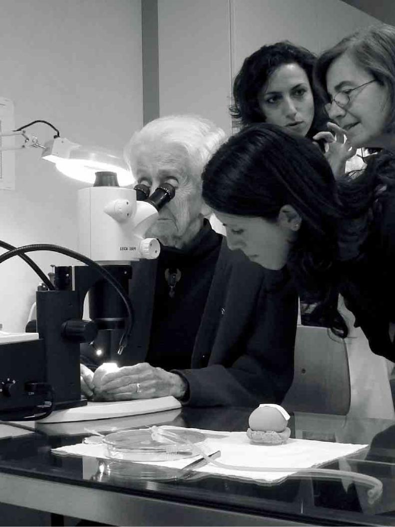

SCIENTIFIC COLLABORATIONS
In addition to the independent researchundertaken at EBRI, many collaborative researchagreements and projects with outside instituteshave been implemented. EBRI is very interestedin establishing collaborations between the bestresearchers in Neurosciences, bringing togetherleading scientists to explore new researchdirections with integrated and cross-disciplinaryapproaches, and formulating collaborativeresearch projects in areas of common interest. EBRI is currently collaborating with Italian andforeign institutes and has various ongoing researchprojects that are financially supported by nationaland international organizations.At the national level, ongoing collaborationsinclude the Italian National Research Council(CNR), Chiesi Pharmaceutics, Scuola NormaleSuperiore (Pisa), University of Rome“Tor Vergata”, University of Florence, Universityof Catania, University of Catanzaro, and CeingeInstitute in Naples, Istituto Neurologico Besta(Milano), IRCCS Fatebenefratelli (Brescia).Internationally, we have established collaborations
with Dresden University (German), the EdmondLily Safra Center for Brain Sciences (HebrewUniversity of Jerusalem), University CollegeLondon (The Wolfson Institute), Xiamen BiowayBiotech (China).EBRI has coordinated two international projects,which have just been completed, the first fundedby the European Commission (project MEMORIES- “Development, characterization and validation ofnew original models for Alzheimer’s Disease) andthe second one co-financed by Italian Ministry ofUniversity and Research and the MassachusettsInstitute of Technology.Group leaders of EBRI are part of the EuropeanNeuroscience Initiative (ENI) network, acollaborative network made up of a selected numberof prestigious neuroscience institutes in Europe.Over the years, the Institute has received financialsupport from Italian Ministry of Education andResearch, the Italian Ministry of Health, the LazioRegion, the Italian Institute of Technology (IIT),FILAS, European Commission, Alzheimer’sAssociation, Telethon Foundation.
HUVEC cells with evident cytoskeleton
Cellule HUVEC con in evidenza il citoscheletro
PAGINA 70 THE EUROPEAN BRAIN RESEARCH INSTITUTE

COLLABORAZIONI SCIENTIFICHE
Oltre ai progetti di ricerca indipendenti portatiavanti da EBRI, molti altri ne sono stati sviluppatiin collaborazione con altri Istituti. EBRI infatti sipone l’obbiettivo di stabilire collaborazioni con imigliori ricercatori in Europa nel campo delleNeuroscienze, per esplorare nuove linee e indirizzidi ricerca tramite un approccio interdisciplinare edintegrato, e formulare nuovi progetti incollaborazione, in settori di interessecomune.Attualmente EBRI collabora conIstituzioni italiane e straniere e gestisce diversiprogetti di ricerca finanziati da istituzioninazionali ed internazionali.Al livello nazionale EBRI collabora con ilConsiglio Nazionale della Ricerca (CNR),Chiesi Farmaceutici, Scuola Normale Superiore(Pisa), l’Università di Roma “Tor Vergata”,l’Università di Firenze, l’Università di Catania,l’Università di Catanzaro, e Ceinge,University ofFlorence, University of Catania, University ofCatanzaro, and Ceinge – Biotecnologie Avanzate,Istituto Neurologico Besta (Milano), IRCCSFatebenefratelli (Brescia).
A livello internazionale abbiamo collaborazioni conUniversità di Dresden (Germania), Edmond LilySafra Center for Brain Sciences (Hebrew Universityof Jerusalem), University College London (TheWolfson Institute), Xiamen Bioway Biotech (China).EBRI è stato il coordinatore di due progettiinternazionali che si sono conclusi di recente, ilprimo finanziato dalla Comunità Europea (progettoMemories “Sviluppo, caratterizzazione e validazionedi nuovi modelli originali per la malattia diAlzheimer), ed il secondo co-finanziato dalMinistero dell’Istruzione, Università e RicercaScientifica e l’Istituto di Tecnologia delMassachusetts.I Group Leaders dell’EBRI fanno parte della ENInetwork (European Neuroscience Initiative), unarete collaborativa composta da un numero diprestigiosi Istituti di Neuroscienze in Europa.Negli anni, l’EBRI ha ricevuto supporti finanziaridal Ministero dell’Istruzione e Ricerca, il Ministerodella Sanità, la Regione Lazio e l’Istituto Italiano diTecnologia (IIT), FILAS, la Commissione Europea,l’Alzheimer’s Association, la Fondazione Telethon.
PAGINA 71 THE EUROPEAN BRAIN RESEARCH INSTITUTE

PAGINA 72 THE EUROPEAN BRAIN RESEARCH INSTITUTE
EBRI SUPPORTING MEMBERS
As of 2011, EBRI also relies on the generosityof its Supporting Members to continue to moveforward in brain research and advance ourunderstanding of how the brain works.EBRI is grateful to our Supporting Members:Alliance Boots is a leading international,pharmacy-led health and beauty group deliveringa range of products and services to customers.Working in close partnership with manufacturersand pharmacists, Alliance Boots is committed toimproving health in the local communities andhelping customers and patients to look and feeltheir best. Alliance Boots is dedicated tosupporting the development of a new generation ofpharmacists. In 2008 a cooperation agreement wassigned in Rome, with the University of Tor Vergataand the University of Nottingham, to support a newpharmacy degree course with a stronginternational focus and a modern understanding ofthe profession. Chiesi Farmaceutici SpA is a research-focusedinternational Group that develops and put into themarket innovative pharmaceutical products toimprove the quality of human life.With a high quality entrepreneurial team
characterized by self-confidence and acollaborative spirit, Chiesi Farmaceutici aims tocombine commitment to results with integrity,operating in a socially and environmentallyresponsible manner. The company is fullycommitted to the treatment of pulmonary diseases.In addition to improving asthma treatment, theGroup is currently engaged in identifying neweffective treatments for Chronic ObstructivePulmonary Disease (COPD). Chiesi Farmaceuticiis also involved in advanced therapies forophthalmic and neurological diseases (includingstem cells for corneal lesions, and new drugs forthe treatment of Alzheimer’s disease)Marfy creates a collection of high fashion patternsfor haute couture and has produced 13,000creations becoming an Italian leader in the field.Along with its creativity, professionalism andunmistakable elegance, they are exporting styleand patterns throughout the world. Marfy,committed to supporting scientific research, iscurrently sponsoring a research project at EBRIentitled “Bighi L.A.M.” aimed at the identificationand comprehension of molecular mechanismsunderlying the development of brain tumours.

PAGINA 73 THE EUROPEAN BRAIN RESEARCH INSTITUTE
SOCI SOSTENITORI DELL’EBRI
Dal 2011, EBRI beneficia inoltre della generositàdei suoi Supporting Members per continuare a fareprogressi nella ricerca del cervello e perfezionare lacomprensione del funzionamento del cervello.EBRI è grato ai suoi Supporting Members:Alliance Boots è una società nel settorefarmaceutico della salute e della bellezza a livellointernazionale che fornisce una gamma di prodottie servizi ai clienti. Lavorando in strettacollaborazione con i farmacisti, Alliance Boots siimpegna a migliorare la salute nelle comunità eaiutare i clienti e i loro pazienti a stare meglio.Alliance Boots si dedica a sostenere lo sviluppo diuna nuova generazione di farmacisti.Nel 2008 un accordo di cooperazione è statofirmato a Roma con l’Università di Tor Vergata el’Università di Nottingham per sostenere un nuovocorso di laurea in Farmacia con un forteorientamento internazionale e una comprensionemoderna della professione.Chiesi Farmaceutici SpA è un gruppointernazionale incentrato sulla ricerca, in gradodi sviluppare e commercializzare soluzioniterapeutiche innovative che migliorino la qualitàdella vita delle persone.
Con un team imprenditoriale di elevata qualità,caratterizzato da spirito di collaborazione esicurezza in se stesso, l’obbiettivo della ChiesiFarmaceutici è unire impegno verso i risultati eintegrità, operando con responsabilità sia dal puntodi vista sociale, sia da quello ambientale.L’Azienda dedica il massimo impegno alla curadelle malattie polmonari. Oltre a migliorare lacura dell’asma, il Gruppo è impegnato aindividuare nuovi trattamenti efficaci per labroncopneumopatia cronica ostruttiva (BPCO).La Chiesi Farmaceutici sta anche sviluppandoterapie avanzate con l’uso di cellule staminali peril controllo di lesioni della cornea che portano acecità e l’impiego di farmaci innovativi per iltrattamento della malattia di Alzheimer.Marfy crea cartamodelli di alta sartoria per l’altamoda e ha realizzato circa 13.000 creazioni.Inoltre è leader in Italia nel proprio settore ed esportail suo stile e i suoi modelli in tutto il mondo insiemead una creatività, professionalità ed eleganzainconfondibili. Attualmente sta sponsorizzando pressol’EBRI il progetto “BIGHI L.A.M.” volto acomprendere i meccanismi molecolari alla base dellosviluppo di tumori cerebrali.

PAGINA 74 THE EUROPEAN BRAIN RESEARCH INSTITUTE
2012
10 feb Andrey Y. Abramov, Institute of Neurology - UCL, UKInterplay between calcium, mitochondria and reactive oxygen speciesin mechanism of neurodegeneration of Parkinson’s disease
13 feb Paolo ScoppolaMovimenti corporei e onde cerebrali: un nuovo modello di interazione uomo-macchina
29 mar Alessandro Morelli, Università di Genova, ItalySintesi extramotocondriale di ATP: prospettive nello studio della conduzione nervosa,della memoria, del sonno e delle malattie neurodegenerative
2011
24 jan Andreas Androutsellis-Theotokis, University of Dresden, Germany A transplantation-free stem cell therapy strategy for neurodegnerative disorders
15 feb Davide Tampellini, Weill Medical College of Cornell University, New York, USASynaptic modulation of beta-amyloid homeostatsis in models of Alzheimer’s disease
21 june Annalisa Pastore, The National Institute for Medical Research The Ridgeway, London, UK Polyglutamine and neurodegeneration
11 july Solomon Snyder, John Hopkins Medical School, Baltimore, USANovel Neural Messengers
30 sept Marco Canossa, Italian Institute of Technology (IIT); University of Bologna, ItalyTemporal expression of neurotrophin receptors regulates newborn neuron integration into hippocampal circuits
04 oct Stefan Bornstein, Department of Medicine University of Dresden, GermanyNew regenerative therapies for diabetes
04 nov Kevin Shakesheff, School of Pharmacy, University of Nottingham, UKThree-dimensional templates for tissue regeneration
10 nov Philippe Busquin, Former European Commissioner, Bruxelles, BelgiumEuropean Space of Research
29 nov Hermona Soreq, Hebrew University of Jerusalem, Israel MicroRnas in the interface between inflammation and neurodegeneration
SEMINARS AND EVENTS

PAGINA 75 THE EUROPEAN BRAIN RESEARCH INSTITUTE
2010
15 feb Alison Barth, Carnegie Mellon University, Pittsburgh, USACellular and Synaptic substrates of perceptual plasticity
14 may Gerry Melino, University Tor Vergata; University of Leicester, UKP73 neurodevelopment and degradation
17 may Tommaso Russo, University of Naples Federico II; CEINGE, Italythe APP-Fe65 complex and its role in DNA damage response
28 may Tommaso Fellin, Italian Institute of Technologies; University of Genoa, ItalyAstrocyte modulation of neuronal networks in the somatosensory cortex in vivo
25 june Alan Carleton, University of Geneva, SwitzerlandAnalysis of the mechanisms underlying sensory perceptionand odor coding in the mammalian olfactory bulb
23 july Michele Giugliano, University of Antwerp, BelgiumNanomaterials/Neuronal hybrid system: first step in nano-neuroscience
02 oct Lucio Annunziato, University of Naples, ItalyThe The Na+/Ca++ The Na+/Ca++ exchanger,a druggable target for brain ischemia
14 oct Michelangelo Campanella, Royal Veterinary College, UK Setting the pace of mitochondrial key enzymatic pathwaysto modulate cellular life and death
SEMINARI ED EVENTI

EBRI PUBLICATIONS 2005-2012
PAGINA 76 THE EUROPEAN BRAIN RESEARCH INSTITUTE
2012Barbato C., Ruberti F. (2012) The role of microRNA
regulation of neuronal differentiation and plasticity,In: Mallick B (eds) Regulatory RNAs: Springer-VerlagBerlin Heidelberg.
Bianchi D., Marasco A., Limongiello A., Marchetti C.,Marie H., Tirozzi B., Migliore M. (2012) On themechanisms underlying the depolarization block in thespiking dynamics of CA1 pyramidal neurons. Journalof Computational Neuroscience, in press.
Bornstein S.R., Ehrhart-Bornstein M., Androutsellis-Theotokis A., Eisenhofer G., Licinio J, Wong M.L.,Calissano P., Nisticò G., Levi-Montalcini R. (2012)Chromaffin Cells - The Peripheral Brain. MolecularPsychiatry, 17:354-358.
Calissano P. (2012) NGF and APP cross talk indevelopment and in Alzheimer Disease. EuropeanJournal of Neurodegenerative Diseases, in press.
Campanella M. (2012) The Physiology and Pharmacologyof the mitochondrial 18-kd translocator protein(TSPO): an emerging molecular target for diagnosisand therapy. Current Molecular Medicine, in press.
Campanella M. (2012) Impaired Mitochondrial Metabolismin Neurodegeneration. European Journal ofNeurodegenerative Diseases, in press.
Capsoni S., Carucci N.M., Cattaneo A. (2012) Pathogenfree conditions slow the onset of neurodegeneration ina mouse model of Nerve Growth Factor deprivation. J.Alzheimers Dis., in press.
Ceci M., Welshhans K., Ciotti M.T., Brandi R., Parisi C.,Paoletti F., Pistillo L., Bassell G.J., and Cattaneo A.(2012) RACK1 is a ribosome scaffold protein for b-actin mRNA/ ZBP1complex. PLoS One, in press.
Cherubini A., Nisticò G., Pani L. et al. (2012) Subtresholddepression in older subjects: an unmet therapeuticneed. J. Nutr., Health and Aging, in press.
Covaceuszach S., Marinelli S., Krastanova I., Ugolini G.,Pavone F., Lamba D., and Cattaneo A. (2012) SingleCycle Structure-based Humanization of an Anti-NerveGrowth Factor Therapeutic Antibody. PLoS One, in press.
Falco A., Festa M., Basile A., Rosati A., Pascale M.,Florenzano F., Nori S.L., Nicolin V., Di Benedetto M.,Vecchione M.L., Arra C., Barbieri A., De Laurenzi V.,Turco M.C. (2012) BAG3 controls angiogenesis throughregulation of ERK phosphorylation. Oncogene, in press.
Gatliff J. and Campanella M. (2012) The 18Kdatranslocator Protein (TSPO): a new perspectivein mitochondrial biology. Current Molecular Medicine,in press.
Klionsky D.J. (2012) Guidelines for the use andinterpretation of assays for monitoring autophagy.Autophagy, in press.
Lisi S., Gamucci O., Vottari T., Scabia G., Funicello M.,Marchi M., Galli G., Arisi I., Brandi R., D’Onofrio M.,Cattaneo A., Pinchera A., Santini F., Maffei M. (2012)Obesity associated hepatosteatosis and impairment ofglucose homeostasis are attenuated by Haptoglobindeficiency. Diabetes, in press.
Manca A., Capsoni S., Di Luzio A., Vignone D., MalerbaF., Paoletti F., Brandi R., Arisi I., Cattaneo A., andLevi-Montalcini R. (2012) Nerve growth factorregulates axial rotation during early stages of chickembryo development. Proc. Nat. Acad. Sci.,109:2009-14.
Maya-Vetencourt J.F., Baroncelli L., Viegi A., TiraboschiE., Castren E., Cattaneo A. and Maffei L. (2012)IGF-1 restores visual cortex plasticity in adult life byreducing local GABA levels. Neural Plasticity,in press.
Nisticò R., Collingridge G. (2012) The synaptic basisof Alzheimer’s disease. European Journal ofNeurodegenerative Diseases, in press.
Nisticò R., Pignatelli M., Piccinin S., Mercuri N.B.,Collingridge G. (2012) Targeting synaptic disfunctionin Alzheimer’s disease therapy. Mol. Neurobiol.,in press.
Nisticò R., Cavallucci V., Piccinin S., Macrì S., PignatelliM., Mehdawy B., Laviola G., Mercuri N.B., D’AmelioM. (2012) Insulin receptor haploinsufficiency impairslong-term plasticity in the hippocampus. Neuromol.Med., in press.
Severinatne M., Faccenda D., De Biase V., Campanella M.(2012) Mitochondrial degradation is limitedby PK11195: functional implications for theF1Fo-ATPsynthase in Mitophagy. Current MolecularMedicine, in press.
Su T., Scardigli R., Fasulo L., Paradiso B., Barbieri M.,Binaschi A., Bovolenta R., Zucchini S., Cossu G.,Cattaneo A., Simonato M. (2012) By-stander effecton brain tissue of mesoangioblasts producingneurotrophins. Cell Transplantation, in press.

PAGINA 77 THE EUROPEAN BRAIN RESEARCH INSTITUTE
PUBBLICAZIONI EBRI 2005-2012
2011Amadoro G., Corsetti V., Ciotti M.T., Florenzano F., Capsoni
S., Amato G., Calissano P. (2011) Endogenous Aβcauses cell death via early tau Hyperphosphorylation.Neurobiology of Aging, 32: 969-90.
Arisi I., D’Onofrio M., Brandi R., Di Mambro A., FelsaniA., Capsoni S., Drovandi G., Felici G., Weitschek E.,Bertolazzi P. and Cattaneo A. (2011) Logic miningfrom gene expression profile in the brain of the AD11anti-NGF mice model of Alzheimer’s Disease.J. Alzheimers Dis., 24: 721-38.
Balducci C., Mehdawy B., Mare L., Giuliani A., LorenziniL., Sivilia S., Giardino L., Calzà L., Lanzillotta A.,Sarnico I., Pizzi M., Usiello A., Viscomi A.R.,Ottonello S., Villetti G., Imbimbo B.P., Nisticò G.,Forloni G., Nisticò R. (2011) The gamma-secretasemodulator CHF5074 restores memory andhippocampal synaptic plasticity in plaque-free Tg2576mice. J. Alzheimers Dis., 24: 799-816.
Capsoni S., Brandi R., Arisi I., D’Onofrio M. and CattaneoA. (2011) A dual mechanism linking NGF/proNGFimbalance and early inflammation to Alzheimer’sdisease Neurodegeneration in the AD11 anti-NGFmouse model. CNS Neurol. Disord. Drug Targets,10:635-47.
D’Amelio M., Cavallucci V., Middei S., Marchetti C.,Pacioni S., Ferri A., Diamantini A., De Zio D., CarraraP., Battistini L., Moreno S., Marie H., Bacci A.,Ammassari-Teule M., Cecconi F. (2011) Non-apoptoticcaspase-3 activity triggers synaptic degeneration at theonset of Alzheimer’s Disease. Nature Neuroscience,14: 69-76.
D’Onofrio M., Paoletti F., Arisi I., Brandi R., Malerba F.,Fasulo L., Cattaneo A. (2011) NGF and proNGFactivate functionally distinct mRNAs in PC12 cells:an early gene expression profiling. PLoS One, 6.
Iannetti L., Accorinti M., Malagola R., Bozzoni-PantaleoniF., Da Salt S., Nicoletti F., Gradini R., Traficante A.,Campanella M. and Pivetti-Pezzi P. (2011) Role of theintravitreal growth factors in the pathogenesis ofidiopathic epiretinal membrane. Invest. Ophthalmol.Vis. Sci. 2011 Jul 29; 52: 5786-9.
Levi-Montalcini R., Knight R.A., Nicotera P., Nisticò G.,Bazan N., Melino G. (2011) Rita’s 102!!. Mol.Neurobiol., 43: 77-79.
Malerba F., Paoletti F., Capsoni S., Cattaneo A. (2011)Intranasal delivery of therapeutic proteins forneurological diseases. Expert Opin. Drug Deliv.,8: 1277-96.
Marchetti C., Tafi E., Marie H. (2011) Viral-mediatedexpression of a constitutively active form of cAMPresponse element binding protein in the dentate gyrusincreases long term synaptic plasticity, Neuroscience,190: 21-6.
Marchetti C., Marie H. (2011) Hippocampal synapticplasticity in Alzheimer’s disease: what have we learnedso far from transgenic models? Rev. Neurosci.,22: 373-402.
Natoli M., Leoni B.D., D’Agnano I., Brandi R., Arisi I.,D’Onofrio M., Zucco M.F., Felsani A. (2011) CellGrowing Density Affects the Structural and FunctionalProperties of Caco-2 Differentiated Monolayer. J. Cell.Physiol., 226: 1531-43.
Nisticò G. (2011) Orphan drugs assessment in thecentralized procedure. Ann. Ist. Super. Sanità, 47: 98-99.
Paoletti F., Malerba F., Kelly G., Noinville S., Lamba D.,Cattaneo A., Pastore A. (2011) Conformationalplasticity of proNGF. PLoS one, 6.
Ruberti F., Barbato C., Cogoni C. (2011) Targeting microRNAsin neurons: tools and perspectives, ExperimentalNeurology, doi:10.1016/j.expneurol.2011.10.031.
Ruberti F., Pezzola S. and Barbato C. (2011) Advancesin MicroRNAs and Alzheimer’s Disease Research,In: Alzheimer’s Disease Pathogenesis-Core concepts,Shifting Paradigms and Therapeutic Targets, SuzanneDe La Monte (Ed.), ISBN: 978-953-307-690-4, InTech.
Vetere G., Marchetti C., Benevento M., Tafi E., Marie H.,Ammassari-Teule M. (2011) Viral-mediated expressionof a constitutively active form of CREB in the dentategyrus does not induce abnormally enduring fearmemory, Behav. Brain Res., 222: 394-6.
2010Amadoro G., Corsetti V., Ciotti M.T., Florenzano F.,
Capsoni S., Amato G., Calissano P. (2010) EndogenousAbeta causes cell death via early tauhyperphosphorylation. Neurobiol. Aging, July 21,2009.
Amadoro G., Corsetti V., Stringaro A., Colone M.,D’Aguanno S., Meli G., Ciotti M., Sancesario G.,

PAGINA 78 THE EUROPEAN BRAIN RESEARCH INSTITUTE
Cattaneo A., Bussani R., Mercanti D., Calissano P.(2010) A NH2 Tau Fragment Targets NeuronalMitochondria at AD Synapses: Possible Implications forNeurodegeneration. J. Alzheimers Dis. 21: 445-70.
Barbato C., Ruberti F., Pieri M., Vilardo E., Costanzo M.,Ciotti M.T., Zona C., Cogoni C. (2010) MicroRNA-92modulates K(+) CI(-) co-transporter KCC2 expressionin cerebellar granule neurons. J. Neurochem.113: 591-600.
Batassa E.M., Costanzi M., Saraulli D., Scardigli R.,Barbato C., Cogoni C., Cestari V. (2010) RISC activityin hippocampus is essential for contextual memory.Neurosci. Lett. 471:185-88.
Capsoni S., Tiveron C., Amato G., Vignone D., Cattaneo A.(2010) Peripheral neutralization of nerve growth factorinduces immunosympathectomy and centralneurodegeneration in transgenic mice. J. AlzheimersDis. 20:527-46.
Capsoni S., Tiveron C., Vignone D., Amato G., Cattaneo A.(2010) Dissecting the involvement of tropomyosin-related kinase A and p75 neurotrophin receptorsignaling in NGF deficit-induced neurodegeneration.Proc. Natl. Acad. Sci. USA. 107:12299-304.
Cattaneo A. (2010) Tanezumab, a recombinant humanizedmAb against nerve growth factor for the treatment ofacute and chronic pain. Curr. Opin. Mol. Ther.12: 94-106.
De Arcangelis V., Serra F., Cogoni C., Vivarelli E., MonacoL., Naro F. (2010) β1-syntrophin modulation by miR-222 in mdx mice. PLoS One. Aug. 10: 5-8.
Houeland G., Romani A., Marchetti C., Amato G.,Capsoni S., Cattaneo A. and Marie H. (2010)Transgenic mice with chronic NGF deprivation andAlzheimer’s disease-like pathology, displayhippocampal region-specific impairments in short- andlong-term plasticities. J. Neurosci. 30:13089-94.
Lagostena L., Rosato-Siri M., D’Onofrio M., Brandi R.,Arisi I., Capsoni S., Franzot J., Cattaneo A., CherubiniE. (2010) In the adult hippocampus, chronic nervegrowth factor deprivation shifts GABAergic signalingfrom the hyperpolarizing to the depolarizing direction.J. Neurosci. 30:885-93.
Manseau F., Marinelli S., Mendez P., Schwaller B., PrinceD.A., Huguenard J.R., Bacci A. (2010)Desynchronization of Neocortical Networks byAsynchronous Release of GABA at Autaptic and
Synaptic Contacts from Fast-Spiking Interneurons.PLoS Biol. 8(9).
Marie H. and Nolan M. (2010) Virus-based technologiesfor investigating function and pathology of the nervoussystem. Front. Cell Neurosci. 7: 3-16.
Marchetti C., Tafi E., Middei S., Rubinacci M., Restivo L.,Ammassari-Teule M., and Marie H. (2010) Synapticadaptations of CA1 pyramidal neurons induced by ahighly effectivecombinational antidepressant therapy.Biol. Psychiatry. 67:146-54.
Middei S., Berretta N., Roberto A., Panico M.B., Lista S.,Bernardi G., Mercuri N.B., Ammassari-Teule M.,Nisticò R. (2010) Learning discloses abnormalstructural and functional plasticity at hippocampalsynapses in the APP23 mouse model of Alzheimer’sdisease, Learn. Mem. 17: 236-40.
Nisticò S., Paolillo N., Minella D., Piccirilli S., Rispoli V.,Giardina E., Biancolella M., Chimenti S., Novelli G.,Nisticò G. (2010) Effects of TNF α and IL1β on theactivation of genes related to inflammatory, immuneresponses and cell death in immortalized humanHaCat keratinocytes, Int. J. Immunopath. Pharmacol.23: 1057-1072.
Paoletti F., Ainger K., Donati I., Scardigli R., Vetere A.,Cattaneo A., Campa C. (2010) Novel fluorescentcycloheximide derivatives for the imaging of proteinsynthesis. Biochem. Biophys. Res. Commun. 396:258-64.
Ruberti F., Barbato C., Cogoni C. (2010) Post-transcriptional regulation of amyloid precursor proteinby microRNAs and RNA binding proteins. Commun.Integr. Biol. 3:499-503.
Vilardo E., Barbato C., Ciotti M., Cogoni C., Ruberti F.(2010) MicroRNA-101 regulates amyloid precursorprotein expression in hippocampal neurons. J. Biol.Chem. 285:344-51.
2009Amadoro G., Corsetti V., Ciotti M.T., Florenzano F.,
Capsoni S., Amato G., Calissano P. (2009)Endogenously Aβ causes cell death via early tauhyperphosphorylation. Neurobiol. Aging 32: 969-90.
Barbato C., Ruberti F., Cogoni C. (2009) Searching forMIND: MicroRNAs, In Neurodegenerative Diseases. J.Biomed. Biotechnol. 871313.

PAGINA 79 THE EUROPEAN BRAIN RESEARCH INSTITUTE
Barbato C., Arisi I., Frizzo M.E., Brandi R, Da Sacco L.,Masotti A. (2009) Computational Challenges inmiRNA Target Predictions: To Be or Not to Be a TrueTarget? J. Biomed Biotech. 803069.
Biamonte F., Assenza G., Marino R., D’Amelio M., PanteriR., Caruso D., Scurati S., Garcia Yague J., Garcia-Segura L.M., Cesa R., Strata P., Melcangi R.C. (2009)Interactions between neuroactive steroids and reelinhaploinsufficiency in Purkinje cell survival. Neurobiol.Dis. 36:103-15.
Capsoni S., Covaceuszach S., Ugolini G., Spirito F.,Vignone D., Stefanini B., Amato G., Cattaneo A. (2009)Delivery of NGF to the brain: intranasal versus ocularadministration in anti-ngf transgenic mice. J.Alzheimers Dis. 16: 371-88.
Cecere G., Cogoni C. (2009) Quelling targets the rDNAlocus and functions in rDNA copy number control.BMC Microbiol. 9:44.
Cesa R., Strata P. (2009). Axonal competition in thesynaptic wiring of the cerebellar cortex duringdevelopment and in the mature cerebellum. Neurosci.162:624-32.
Colussi C., Gurtner A., Rosati J., Illi B., Ragone G.,Piaggio G., Moggio M., Lamperti C., D’Angelo G.,Clementi E., Minetti G., Mozzetta C., Antonini A.,Capogrossi M.C., Puri P.L., Gaetano C. (2009) NitricOxide Deficiency Determines Global ChromatinChanges in Duchenne Muscular Dystrophy. FASEB J.23: 2131-41.
Covaceuszach S., Capsoni S., Ugolini G., Spirito F.,Vignone D., Cattaneo A. (2009) Development of a noninvasive NGF-based therapy for Alzheimer’s disease.Curr. Alzheimer Res. 6:158-70.
D’Onofrio M., Ambrosini A., Di Mambro A., Arisi I.,Santorelli F.M., Grieco G.S., Nicoletti F., Nappi G.,Pierelli F., Schoenen J., Buzzi M.G. (2009) Theinterplay of two single nucleotide polymorphisms inthe CACNA1A gene may contribute to migrainesusceptibility. Neurosci. Lett. 453:12-5.
D’Onofrio M., Arisi I., Brandi R., Di Mambro A., FelsaniA., Capsoni S., Cattaneo A. (2009) Earlyinflammation and immune response mRNAs in thebrain of AD11 anti-NGF mice. Neurobiology ofageing 32:1007-22.
FANTOM Consortium, Suzuki H., Forrest A.R., VanNimwegen E., Daub C.O., Balwierz P.J., Irvine K.M.,
Lassmann T., Ravasi T., Hasegawa Y., De Hoon M.J.,Katayama S., Schroder K., Carninci P., Tomaru Y.,Kanamori-Katayama M., Kubosaki A., Akalin A.,Ando Y., Arner E., Asada M., Asahara H., Bailey T.,Bajic V.B., Bauer D., Beckhouse A.G., Bertin N.,Björkegren J., Brombacher F., Bulger E., Chalk A.M.,Chiba J., Cloonan N., Dawe A., Dostie J., EngströmP.G., Essack M., Faulkner G.J., Fink J.L., Fredman D.,Fujimori K., Furuno M., Gojobori T., Gough J.,Grimmond S.M., Gustafsson M., Hashimoto M.,Hashimoto T., Hatakeyama M., Heinzel S., Hide W.,Hofmann O., Hörnquist M., Huminiecki L., Ikeo K.,Imamoto N., Inoue S., Inoue Y., Ishihara R., IwayanagiT., Jacobsen A., Kaur M., Kawaji H., Kerr M.C.,Kimura R., Kimura S., Kimura Y., Kitano H., Koga H.,Kojima T., Kondo S., Konno T., Krogh A., Kruger A.,Kumar A., Lenhard B., Lennartsson A., Lindow M.,Lizio M., Macpherson C., Maeda N., Maher C.A.,Maqungo M., Mar J., Matigian N.A., Matsuda H.,Mattick JS, Meier S., Miyamoto S,. Miyamoto-Sato E.,Nakabayashi K., Nakachi Y., Nakano M., Nygaard S.,Okayama T., Okazaki Y., Okuda-Yabukami H.,Orlando V., Otomo J., Pachkov M., Petrovsky N.,Plessy C., Quackenbush J., Radovanovic A., Rehli M.,Saito R., Sandelin A., Schmeier S., Schönbach C.,Schwartz A.S., Semple C.A., Sera M., Severin J.,Shirahige K., Simons C., St Laurent G., Suzuki M.,Suzuki T., Sweet M.J., Taft R.J., Takeda S., TakenakaY., Tan K., Taylor M.S., Teasdale R.D., Tegnér J.,Teichmann S., Valen E., Wahlestedt C., Waki K.,Waterhouse A., Wells C.A., Winther O., Wu L.,Yamaguchi K., Yanagawa H., Yasuda J., Zavolan M.,Hume D.A. Riken Omics Science Center, Arakawa T.,Fukuda S,. Imamura K., Kai C., Kaiho A., KawashimaT., Kawazu C., Kitazume Y., Kojima M., Miura H.,Murakami K., Murata M., Ninomiya N., Nishiyori H.,Noma S., Ogawa C., Sano T., Simon C., Tagami M.,Takahashi Y., Kawai J., Hayashizaki Y. (2009) Thetranscriptional network that controls growth arrest anddifferentiation in a human myeloid leukemia cell line.Nature Genetics. 41:553-62.
Faulkner G.J., Kimura Y., Daub C.O., Wani S., Plessy C.,Irvine K.M., Schroder K., Cloonan N., Steptoe A.L.,Lassmann T., Waki K., Hornig N., Arakawa T.,Takahashi H., Kawai J., Forrest A.R., Suzuki H.,Hayashizaki Y., Hume D.A., Orlando V., Grimmond

PAGINA 80 THE EUROPEAN BRAIN RESEARCH INSTITUTE
S.M., Carninci P. (2009) The regulated retrotransposontranscriptome of mammalian cells. Nature Genetics.41:563-71.
Glickstein M., Strata P., Voogd J. (2009) Cerebellum:History. Neurosci. 162:549-59
Guasconi V., Puri P.L. (2009) Chromatin: the interfacebetween extrinsic cues and the regulation of muscledifferentiation and regeneration. Trends Cell Biol.19:286-94
Huang Y.H., Lin Y., Mu P., Lee B., Brown T., Wayman G.,Marie H., Liu W., Yan Z., Sorg B.A., Schluter O,. ZukinR.S., Dong Y. (2009) In vivo cocaine experiencegenerates silent synapses. Neuron 63:40-47.
Mandolesi G., Autuori E., Cesa R., Premoselli F., CesareP., Strata P. (2009) GluRdelta2 expression in themature cerebellum of hotfoot mice promotes parallelfiber synaptogenesis and axonal competition. PLoSOne. 4(4).
Mandolesi G., Vanni V., Cesa R., Grasselli G., Puglisi F.,Cesare P., Strata P. (2009) Distribution of the SNAP25and SNAP23 synaptosomal-associated protein isoformsin rat cerebellar cortex. J. Neurosci. 164:1084-1096.
Marinelli S., Pacioni S., Cannich A., Morsicano G., BacciA. (2009). Self-modulation of neocortical pyramidalneurons by endocannabinoids, Nature Neuroscience 12:1488-1490.
Matrone C., Marolda R., Ciafrè S., Ciotti M.T., Mercanti D.,Calissano P. (2009) Tyrosine kinase nerve growth factorreceptor switches from prosurvival to proapoptoticactivity via Abeta-mediated phosphorylation. Proc. Nat.Acad. Sci. 105: 11358-11363.
Meli G., Visintin M,. Cannistraci I., Cattaneo A. (2009)Direct in vivo intracellular selection of conformation-sensitive antibody domains targeting Alzheimer’samyloid-beta oligomers. J. Mol. Biol. 387:584-606.
Mozzetta C., Minetti G., Puri P.L. (2009) Regenerativepharmacology in the treatment of genetic diseases: theparadigm of muscular dystrophy. Int. J. Biochem. CellBiol. 41:701-10.
Origlia N., Capsoni S., Cattaneo A., Fang F., Arancio O.,Yan S.D., Domenici L. (2009) Aβ-dependentinhibition of LTP in different intracortical circuits ofthe visual cortex: the role of RAGE. J. Alzheimers Dis.17:59-68.
Paoletti F., Covaceuszach S., Konarev P.V., Gonfloni S.,Malerba F., Schwarz E., Svergun D.I., Cattaneo A.,
Lamba D. (2009) Intrinsic structural disorder of mouseproNGF. Proteins. 75:990-1009.
Peng Y., He S., Marie H., Zeng S., Ma J., Tan Z., Lee S.Y.,Malenka R.C., Yu X. (2009) Coordinated changes indendritic arborization and synaptic strength duringneural circuit development. Neuron 61:71-84.
Restivo L., Tafi E., Ammassari-Teule M., Marie H. (2009)Viral-mediated expression of a constitutively activeform of CREB in hippocampal neurons increasesmemory. Hippocampus 19:228-234.
Sacchetti B., Scelfo B., Strata P. (2009) Cerebellum andemotional behaviour. Neuroscience 162:756-62.
Strata P. (2009) David Marr’s theory of cerebellar learning:40 years later. J. Physiol. 587.23:5519-5520
Strata P., Thach W.T., Ottersen O.P. (2009) New insights incerebellar function. Neurosci. 162:545-83.
Taft R.J., Glazov E.A., Cloonan N., Simons C., Stephen S.,Faulkner G.J., Lassmann T., Forrest A.R., GrimmondS.M., Schroder K., Irvine K., Arakawa T., NakamuraM., Kubosaki A., Hayashida K., Kawazu C., MurataM., Nishiyori H., Fukuda S., Kawai J., Daub CO,Hume D.A., Suzuki H., Orlando V., Carninci P.,Hayashizaki Y., Mattick J.S. (2009) Tiny RNAsassociated with transcription start sites in animals.Nature Genetics 41:572-8.
Tiranti V., Viscomi C., Hildebrandt T., Di Meo I., Mineri R.,Tiveron C., D. Levitt M., Prelle A., Fagiolari G., RimoldiM., Zeviani M. (2009) Loss of ETHE1, a mitochondrialdioxygenase, causes fatal sulfide toxicity in ethyl-malonic encephalopathy. Nat. Med. 15:200-205.
2008Atlante A., Amadoro G., Bobba A., De Bari L., Corsetti V.,
Pappalardo G., Marra E., Calissano P., Passarella S.(2008). A peptide containing residues 26-44 of tauprotein impairs mitochondrial oxidativephosphorylation acting at the level of the adeninenucleotide translocator. Biochem. Biophys. Acta1777:1289-1300.
Barbato C., Giorgi C., Catalanotto C., Cogoni C. (2008)Thinking about RNA? microRNAs in the brain.Mamm. Genome 19:541-51.
Buontempo S., Barbato C., Bruno T., Corbi N., Ciotti M.T.,Floridi A., Fanciulli M., Passananti C. (2008) Che-1enhances cyclin-dependent kinase 5 expression and
PAGINA 81 THE EUROPEAN BRAIN RESEARCH INSTITUTE
interacts with the active kinase-complex. Neuroreport26;19:531-35
Cattaneo A., Capsoni S., Paoletti F. (2008) Towards noninvasive nerve growth factor therapies for Alzheimer’sdisease. J. Alzheimers Dis. 15:255-83.
Cesa R., Morando L., Strata P. (2008). Transmitter-receptor mismatch in GABAergic synapses in theabsence of activity. Proc. Natl. Acad. Sci. USA.105:18988-93.
Colussi C., Mozzetta C., Gurtner A., Illi B., Straino S.,Ragone G., Pescatori M., Zaccagnini G., Rosati G.,Minetti G, Martelli F., Ricci E., Piaggio G., GallinariP., Steinkulher C., Capogrossi M.C., Puri P.L., Carlo G.(2008) A common epigenetic mechanism underliesnitric oxide donors and histone deacetylase inhibitorseffect in duchenne muscular dystrophy. Proc. Natl.Acad. Sci. 105, 19183-87.
Corsetti V., Amadoro G., Gentile A., Capsoni S., CiottiM.T., Cencioni M.T., Atlante A., Canu N., Rohn T.T.,Cattaneo A., Calissano P. (2008) Identification of acaspase-derived N-terminal tau fragment in cellularand animal Alzheimer’s disease models. Mol. CellNeurosci. 38:381-92.
Covaceuszach S., Cassetta A., Konarev P.V., Gonfloni S.,Rudolph R., Svergun D.I., Lamba D., Cattaneo A.(2008) Dissecting NGF interactions with its TrkA andp75 receptors by structural and functional studies ofan antiNGF neutralizing antibody. J. Mol. Biol.381:881-96.
Delaunay A., Bromberg K.D., Hauashi Y., Mirabella M.,Burch D., Kirkwood B., Serra C, Malicdan M.C.,Mizisin A.P., Morosetti R., Broccolini A., Guo L.T.,Jones S.N., Lira SA, Puri P.L., Shelton D., Ronai Z.(2008) The ER-bound RING finger protein 5(RNF5/RMA1) causes degenerative myopathy intransgenic mice and is deregulated in inclusion bodymyosytis. PLoS One. 3 (2).
De Luca G., Russo M.T., Degan P., Tiveron C., Zijno A.,Meccia E., Ventura I., Mattei E., Nakabeppu Y.,Crescenzi M., Pepponi R., Pezzola A., Popoli P.,Bignami M. (2008) A role for oxidized DNA precursorsin Huntington’s disease-like striatalneurodegeneration. PLoS Genet. 4(11).
Di Certo M.G, Batassa E.M., Casella I., Serafino A,. FloridiA., Passananti C., Molinari P., Mattei E. (2008)Delayed internalization and lack of recycling in a
beta2-adrenergic receptor fused to the G protein alpha-subunit. BMC Cell Biol. 9:56.
Farioli-Vecchioli S., Saraulli D., Costanzi M., Pacioni S.,Cinà D., Aceti M., Micheli L., Bacci A., Cestari V.,Tirone F. (2008) The timing of differentiation of adulthippocampal neurons is crucial for spatial memory.PLoS Biol. 6(10).
Gentile A., Amadoro G., Corsetti V., Ciotti M.T., SerafinoA., Cailssano P. (2008) Spontaneous aggregation andaltered intracellular distribution of endogeneous alpha-synuclein during neuronal apoptosis. J. Alzheimers Dis.13:151-60.
Guasconi V., Puri P.L. (2008) Epigenetic drugs in thetreatment of skeletal muscle atrophy. Curr. Opin. Clin.Nutr. Metab. Care 11:233-41.
Manseau F., Goutagny R., Danik M., Williams S. (2008)The hippocamposeptal pathway generates rhythmicfiring of GABAergic neurons in the medial septum anddiagonal bands: an investigation using a completeseptohippocampal preparation in vitro. J. Neurosci.28:4096-107.
Marinelli S., Pacioni S., Bisogno T., Di Marzo V., PrinceD.A., Huguenard J.R., Bacci A. (2008) Theendocannabinoid 2-AG is responsible for the slow selfinhibition in neocortical interneurons. J. Neurosci.28:13532-41.
Matrone C., Ciotti M.T., Mercanti D., Marolda R.,Calissano P. (2008) NGF and BDNF signalling controlamyloidogenic route and Abeta production inhippocampal neurons. Proc. Natl. Acad. Sci. USA.105:13139-44.
Matrone C., Di Luzio A., Meli G., D’Aguanno S., SeveriniC., Ciotti M.T., Cattaneo A., Calissano P. (2008)Activation of the amyloidogenic route by NGFdeprivation induces apoptotic death in PC12 cells. J.Alzheimers Dis. 13:81-96.
Nolan T., Cecere G., Mancone C., Alonzi T., Tripodi M.,Catalanotto C., Cogoni C. (2008) The RNA-dependentRNA polymerase essential for post-transcriptionalgene silencing in Neurospora crassa interacts withreplication protein A. Nucleic Acids Res. 36:532-8.
Origlia N., Righi M., Capsoni S., Cattaneo A., Fang F,.Stern D.M., Chen J.X., Schmidt A.M., Arancio O., YanS.D., and Domenici L. (2008) Receptor for advancedglycation end product-dependent activation of p38mitogen-activated protein kinase contributes to

PAGINA 82 THE EUROPEAN BRAIN RESEARCH INSTITUTE
amyloid-β-mediated cortical synaptic dysfunction. J.Neurosci. 28:3521-30.
Patrizi A., Scelfo B., Viltono L., Briatore F., Fukaya M.,Watanabe M., Strata P., Varoqueaux F., Brose N.,Fritschy J.M., Sassoè-Pognetto M. (2008) Synapseformation and clustering of neuroligin-2 in the absenceof GABAA receptors. Proc. Nat. Acad. Sci. 105:13150-13155.
Terzibasi E., Valenzano D.R., Benedetti M,. Roncaglia P.,Cattaneo A., Domenici L., Cellerino A. (2008) Largedifferences in aging phenotype between strains of theshort-lived annual fish Nothobranchius furzeri. PLoSOne. 3(12).
Visintin M., Melchionna T., Cannistraci I. and Cattaneo A.(2008) In vivo selection of intrabodies specificallytargeting protein-protein interactions: a generalplatform for an “undruggable” class of disease targets.J. Biotech. 135:1-15.
Zacchi P., Dreosti E., Visintin M., Marchionni I., Moretto-Zita M., Cannistraci I., Kasap Z., Betz H., Cattaneo A.,Cherubini E. (2008) Gephyrin selective intrabodies asa new strategy for studying inhibitory receptorclustering. J. Mol. Neurosci. 34:141-8.
2007Barbato C., Ciotti M.T., Serafino A., Calissano P., Cogoni
C. (2007) Dicer expression and localization in post-mitotic neurons Brain Res. 1175:17-27.
Berardi N., Braschi C., Capsoni S., Cattaneo A., Maffei L.(2007) Environmental enrichment prevents the onset ofmemory deficits and reduces neuropathologicalhallmarks in a mouse model of Alzheimer-likeneurodegeneration. J. Alzheimers Dis. 11 359-70.
Cicconi S., Gentile A., Ciotti M.T., Parasassi T., SerafinoA.L., Calissano P. (2007) Apoptotic death inducesAbeta production and fibril formation to a much largerextent than necrotic-like death in CGNs. J. AlzheimersDis. 12:211-220.
Dell’Agnello C., Leo S., Agostino A., Szabadkai G.,Tiveron C., Zulian A., Prelle A., Roubertoux P.,Rizzuto R., Zeviani M. (2007) Increased longevity andrefractoriness to Ca(2+)-dependent neurodegenerationin Surf1 knockout mice. Hum. Mol. Genet. 16:431-44.
Langevin L.M., Mattar P., Scardigli R., Roussigné M.,Logan C., Blader P., Schuurmans C. (2007) Validating
in utero electroporation for the rapid analysis of generegulatory elements in the murine telencephalon. Dev.Dyn. 236:1273-86.
Marconi B., Koch G., Pecchioli C., Cavallai P., CaltagironeC. (2007) Breakdown of inhibitory effects induced byfoot motor imagery on hand motor area in lower-limbamputees. Clin. Neurophysiol. 118:2468-78.
Marconi B., Pecchioli C., Koch G., Caltagirone C. (2007)Functional overlap between hand and forearm motorcortical representations durino motor cognitive tasks.Clin. Neurophysiol. 118:1767-75.
Martellli C., Giansanti A., Arisi I., Rosato V. (2007)Asymptotic states and topological structure of anactivation-deactivation chemical network. J. Theor.Biol. 245:423-432.
Melchionna T., Cattaneo A. (2007) A protein silencingswitch by ligand-induced proteasome targetingintrabodies. J. Mol. Biol. 374:641-54.
Simonato M., Latella L., Puri P.L. (2007) DNA damage andcellular differentiation: More questions than responses.J. Cell Physiol. 213:642-8.
Ugolini G., Marinelli S., Covaceuszach S., Cattaneo A.,Pavone F. (2007). The function neutralizing anti-TrkAantibody MNAC13 reduces inflammatory andneuropathic pain. Proc. Natl. Acad. Sci. USA.104:2985-90.
2006Arisi I., Cattaneo A. and Rosato V. (2006) Parameter
estimate of signal transduction pathways. BMCNeuroscience. 7 (Suppl I):S6.
Berrera M., Cattaneo A., Carloni P. (2006) Molecularsimulation of the binding of nerve growth factorpeptide mimics to the receptor tyrosine kinase A.Biophys. J. 15; 91:2063-71.
Capsoni S., Cattaneo A. (2006) On the molecular basislinking Nerve Growth Factor (NGF) to Alzheimer’sdisease. Cell Mol. Neurobiol. 25: 619-33.
Forlino A., Gualeni B., Pecora F., Della Torre S., PiazzaR., Tiveron C., Tatangelo L., Superti-Furga A., CettaG., Rossi A. (2006) Insights from a transgenic mousemodel on the role of SLC26A2 in health and disease.Novartis Foundation Symposium 273.
Iacovelli L., Arcella A., Battaglia G., Pazzaglia S., AronicaE., Spinsanti P., Caruso A., De Smaele E., Saran A.,

PAGINA 83 THE EUROPEAN BRAIN RESEARCH INSTITUTE
Gulino A., D’Onofrio., Giangaspero F., Nicoletti F.(2006) Pharmacological Activation of mGlu4Metabotropic Glutamate Receptors Inhibits the Growthof Medulloblastomas. The Journal of Neuroscience26:8388-8397.
Levi-Montalcini R., Calissano P. (2006) The scientificchallenge of the 21st century: from a reductionist to aholistic approach via systems biology. BMCNeuroscience 7 (Suppl 1):S1.
Origlia N., Capsoni S., Domenici L., Cattaneo A. (2006)Time window in cholinomimetic ability to rescue long-term potentiation in neurodegenerating anti-nervegrowth factor mice. J. Alzheimers Dis. 9:59-68.
Paoletti F., Konarev P.V., Covaceuszach S., Schwarz E.,Cattaneo A., Lamba D., Svergun D.I. (2006) Structuraland functional properties of mouse proNGF. Biochem.Soc. Trans. 34: 605-606.
Rende M., Pistilli A., Stabile A.M., Terenzi A., CattaneoA., Ugolini G., Sanna P. (2006) Role of nerve growthfactor and its receptors in non-nervous cancer growth:efficacy of a tyrosine kinase inhibitor (AG879) andneutralizing antibodies antityrosine kinase receptor Aand antinerve growth factor: an in-vitro and in-vivostudy. Anticancer Drugs 17: 929-41.
Rosato-Siri M., Cattaneo A., Cherubini E. (2006) Nicotine-induced enhancement of synaptic plasticity atCA3-CA1 synapses requires GABAergic interneurons
in adult anti-NGF mice. J. Physiol. 576:361-77.Sola E., Capsoni S., Rosato-Siri M., Cattaneo A.,
Cherubini E. (2006) Failure of nicotine-dependentenhancement of synaptic efficacy at Schaffer-collateralCA1 synapses of AD11 anti-nerve growth factortransgenic mice. Eur. J. Neurosci. 24:1252-64.
Valenzano D.R., Terzibasi E., Genade T., Cattaneo A.,Domenici L., Cellerino. A. (2006) Resveratrol prolongslifespan and retards the onset of age-related markers ina short-lived vertebrate. J. Physiol. 576:361-77.
Valenzano D.R., Terzibasi E., Genade T., Cattaneo A.,Domenici L., Cellerino A. (2006) Temperature affectslongevity and age-related locomotor and cognitivedecay in the short-lived fish Nothobranchius furieri.Ageing Cell. 16: 296-300.
2005Cheung V.C.K., D’Avella A., Tresch M.C., Bizzi E. (2005)
Central and sensory contributions to the activation andorganization of muscle synergies during natural motorbehaviors, J. Neurosci. 25:6419-6434.
Saltiel P., Wyler-Duda K., D’Avella A., Ajemian R.J., BizziE. (2005) Localization and connectivity in spinalinterneuronal networks: the adduction-caudalextension-flexion rhythm in the frog. J. Neurophysiol.94: 2120-2138.

PAGINA 84 THE EUROPEAN BRAIN RESEARCH INSTITUTE
In 2012 the European Brain Research Institutehas launched a new journal, European Journalof Neurodegenerative Diseases (EJND), withBiolife Publisher. This exciting new journal coversall aspects of neurodegenerative diseases:morphological, ultrastructural, physiological,pathophysiological, molecular, biochemical,bioenergetic, pharmacological, therapeutic,epidemiological, translational, biomarkersand regulatory aspects.
Published every four months, the EuropeanJournal of Neurodegenerative Diseases aims topublish high quality review articles on a timelymanner and based on a rigorous peer-reviewprocess. The first issue of EJND has beenpublished in April 2012.
Visit the website www.biolifesas.org for moreinformation and submission guidelines.Email of the Journal: [email protected]
THE EBRI JOURNAL
European Journalof Neurodegenerative Diseases
EBRI

PAGINA 85 THE EUROPEAN BRAIN RESEARCH INSTITUTE
The European Brain Research Institute considersthe promotion and communication of scientificknowledge to young students of high priority.With this perspective a collaboration has beenestablished between EBRI and the Foundationfor the Advancement of Research in MolecularMedicine (FARMM) for the purpose of bringingstudents closer to the scientific world.The FARMM in collaboration with the Institute ofNeurobiology & Molecular Medicine (CNR) andwith the support of Prof. Rita Levi-Montalcini, in2004 launched an informative scientific program
(Bioform project) “Know your DNA”, in the fieldof biology, directed mainly to high schoolstudents in Rome. The project directed by Prof.Ettore D’Ambrosio, offers, to about 1500 studentseach year, the opportunity to carry out anexperiment of individual genetic profilingthrough the analysis of his/her own DNA usingthe same techniques, instruments and substancesas those used in advanced bio-molecularresearch. The courses are held at the EuropeanBrain Research Institute. Visit the website www.farmm.it
L’EBRI considera di fondamentale importanzala promozione e la comunicazione della conoscenzascientifica presso i giovani. In questa prospettivasi è stabilita una collaborazione tra l’EBRIe la FARMM allo scopo di avvicinare gli studential mondo della Scienza.La FARMM, in collaborazione con l’IMNN del CNRe con il supporto di Rita Levi-Montalcini halanciato nel 2004 un programma di informazionescientifica (progetto Bioform) in campo biologico
“Conosci il tuo DNA”, rivolto agli studenti delleScuole superiori di Roma e del Lazio. Il progetto,diretto da Ettore D’Ambrosio, offre a circa 1500studenti ogni anno la possibilità di effettuare unesperimento di profilo genetico individuale tramitel’analisi del proprio DNA utilizzando le stessetecniche, strumenti e reagenti impiegati nellaricerca bio-molecolare avanzata. I Corsi vengonotenuti presso i Laboratori Didattici dell’EBRI.Visita il sito www.farmm.it
SCHOOLS IN THE LAB
SCUOLE IN LABORATORIO

HOW YOU CAN SUPPORT EBRI
PAGINA 86 THE EUROPEAN BRAIN RESEARCH INSTITUTE
As a non-profit institute, EBRI depend ongovernment, institutional and private fundings.Funds received from awarded grants, anddonations are used to support research programs,purchase state-of-the-art equipment, and sustainresearch personnel costs.
Make a donationDonations to EBRI are tax deductible and may beoffered via bank transfer to the following bankaccount:Banca Nazionale del Lavoro - Agenzia 24Via Cristoforo Colombo, 279 - 00147 RomaC/C: 18743CIN: Z - ABI: 1005 - CAB: 03224IBAN: IT44Z0100503224000000018743
A bequest in your willBequests are gifts made through your will to thecharitable causes that are important to you. We areextremely grateful to those who have been so kindto remember the European Brain ResearchInstitute in their will. Through the generosity ofbequests, we can promote and advanceneurobiological and neurophysiological researchwith the aim of finding new therapies againstAlzheimer, ALS, Parkinson’s diseases and otherneurological pathologies.
Designate part of your taxes “5x1000”Italian tax residents may designate part of theirtaxes to non-profit organizations by simplyindicating the fiscal code of EBRI - 97272740586- in their Italian income tax declaration.
Mailing address for all donationsand enquiresEuropean Brain Research Institute“Rita Levi-Montalcini”Via del Fosso di Fiorano 64, 00143 Rome(www.ebri.it - email: [email protected])Checks should be made payable to:Fondazione EBRI “Rita Levi-Montalcini”
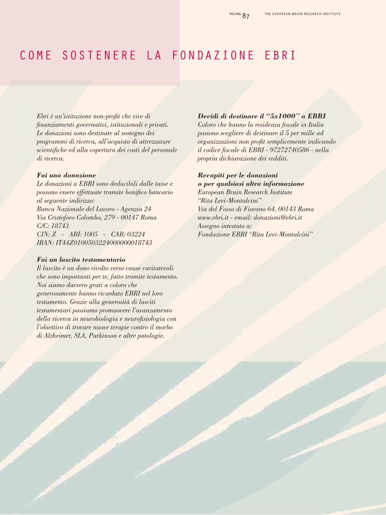
PAGINA 87 THE EUROPEAN BRAIN RESEARCH INSTITUTE
Ebri è un’istituzione non-profit che vive difinanziamenti governativi, istituzionali e privati.Le donazioni sono destinate al sostegno deiprogrammi di ricerca, all’acquisto di attrezzaturescientifiche ed alla copertura dei costi del personaledi ricerca.
Fai una donazioneLe donazioni a EBRI sono deducibili dalle tasse epossono essere effettuate tramite bonifico bancarioal seguente indirizzo: Banca Nazionale del Lavoro - Agenzia 24Via Cristoforo Colombo, 279 - 00147 RomaC/C: 18743CIN: Z - ABI: 1005 - CAB: 03224IBAN: IT44Z0100503224000000018743
Fai un lascito testamentarioIl lascito è un dono rivolto verso cause caritatevoliche sono importanti per te, fatto tramite testamento.Noi siamo davvero grati a coloro chegenerosamente hanno ricordato EBRI nel lorotestamento. Grazie alla generosità di lascititestamentari possiamo promuovere l’avanzamentodella ricerca in neurobiologia e neurofisiologia conl’obiettivo di trovare nuove terapie contro il morbodi Alzheimer, SLA, Parkinson e altre patologie.
Decidi di destinare il “5x1000” a EBRIColoro che hanno la residenza fiscale in Italiapossono scegliere di destinare il 5 per mille adorganizzazioni non profit semplicemente indicandoil codice fiscale di EBRI - 97272740586 - nellapropria dichiarazione dei redditi.
Recapiti per le donazionio per qualsiasi altra informazioneEuropean Brain Research Institute“Rita Levi-Montalcini”Via del Fosso di Fiorano 64, 00143 Romawww.ebri.it - email: [email protected] intestato a:Fondazione EBRI “Rita Levi-Montalcini”
COME SOSTENERE LA FONDAZIONE EBRI

PAGINA 88 THE EUROPEAN BRAIN RESEARCH INSTITUTE
With G. Nisticò, T. Hökfelt and L. Frati at the openingof the workshop “In Memory of Vittorio Erspamer”,Sapienza University of Rome, June 13, 2009.
With Sir S. Moncada and C. Cavazza at theopening of the new Faculty of Pharmacy,University of Rome Tor Vergata, 2008
Departure to USA, 1947
1940
Group photograph with Viktor Hamburger and scientists,Chicago, 1949
With Viktor Hamburger, St. Louis, 1978
St. Louis, USA, 1963
Stokholm Nobel Ceremony, 1986
Photographs of Rita Levi-Montalcini atvarious fundamental periods of her life.

PAGINA 89 THE EUROPEAN BRAIN RESEARCH INSTITUTE
Rita Levi-Montalcini at the microscope with some young researchers
Rita Levi-Montalcini al microscopio con alcune giovani ricercatrici, 2010

EBRIEuropean Brain Research InstituteRita Levi-Montalcini Foundation
Via del Fosso di Fiorano, 6400143 Rome - Italy
web: www.ebri.it - email: [email protected]: +39 06 501703024 - fax: +39 06 501703335
Graphics: omgrafica - Roma


