30
Evaluation and Management of Ataxic Disorders AN OVERVIEW FOR PHYSICIANS Susan L. Perlman, MD for the National Ataxia Foundation
$5.00
National Ataxia Foundation2600 Fernbrook Lane, Suite 119Minneapolis, MN 55447-4752Telephone: 763-553-0020Fax: 763-553-0167E-mail: [email protected]: www.ataxia.org
Evaluat ionand
Managementof
AtaxicDisorders
AN OVER V I EWFOR PHYS I C I ANS
Susan L. Perlman,MDfor the
National Ataxia Foundation
Axatia Disorders book cover.indd 1 2/19/16 3:15 PM

Axatia Disorders book cover.indd 2 2/19/16 3:15 PM

E v a l u a t i o n a n d M a n a g e m e n t o f
AtaxicDisorders
A n O v E r v i E w f O r P h y s i c i A n s
susan L. Perlman, MDfor the National Ataxia Foundation
i
Ataxia Disorders book.indd 1 2/24/16 2:04 PM
Evaluation and ManagEMEnt of ataxic disordErsan overview for Physicians
national ataxia foundation2600 fernbrook lane, suite 119Minneapolis, Mn 55447-4752
telephone 763-553-0020fax 763-553-0167Email [email protected] www.ataxia.org
©2016 national ataxia foundation
all rights reserved
Printed in the united states of america
isBn: 0-943218-14-4
library of congress control number: 2007923539
ii
Ataxia Disorders book.indd 2 2/24/16 2:04 PM

ContentsAbout the author . . . . . . . . . . . . . . . . . . . . . . . . . . . . . . . . . . . . . . . . . . . ivAcknowledgements . . . . . . . . . . . . . . . . . . . . . . . . . . . . . . . . . . . . . . . . . vPreface . . . . . . . . . . . . . . . . . . . . . . . . . . . . . . . . . . . . . . . . . . . . . . . . . . . viiIntroduction . . . . . . . . . . . . . . . . . . . . . . . . . . . . . . . . . . . . . . . . . . . . . . . viiiEvaluation of the ataxic patient . . . . . . . . . . . . . . . . . . . . . . . . . . . . . . 1Characteristics of ataxia . . . . . . . . . . . . . . . . . . . . . . . . . . . . . . . . . . . . . . 1Basic ataxia phenotypes . . . . . . . . . . . . . . . . . . . . . . . . . . . . . . . . . . . . . 2Evaluation . . . . . . . . . . . . . . . . . . . . . . . . . . . . . . . . . . . . . . . . . . . . . . . . 2-3
Table 1—identifiable causes of nongenetic ataxia . . . . . . . . . . . . . . . . . . . 2Table 2—Key features of examination that may provide clues to the diagnosis of ataxia . . . . . . . . . . . . . . . . . . . . . . . . . . . . . . 3Table 3—Workup for the ataxic patient with or without a family history . . . . . . . . . . . . . . . . . . . . . . . . . . . . . . . . . . . . . . . . . . . . . . 4
Autosomal dominant cerebellar ataxia . . . . . . . . . . . . . . . . . . . 5Table 4—the dominantly inherited ataxias—Molecular genetics . . . . 6-9Table 5—the dominantly inherited ataxias—associated features in differential diagnosis . . . . . . . . . . . . . . . . . . . . . . . . . . . . . 10Table 6—the dominantly inherited ataxias—Prioritizing genetic testing as tests continue to become available . . . . . . . . . . . . . 11
Recessively inherited ataxias . . . . . . . . . . . . . . . . . . . . . . . . . . 12Table 7—the recessively inherited ataxias—differential diagnosis . . . 13Table 8—the recessively inherited ataxias—Molecular genetics . . . . . 14
Maternally inherited ataxias (X-linked and mitochondrial) ..... 14Table 9—Maternally inherited ataxias— x-linked and mitochondrial . . . . . . . . . . . . . . . . . . . . . . . . . . 15
Sporadic ataxias . . . . . . . . . . . . . . . . . . . . . . . . . . . . . . . . . . . . . . . . . . . 16Table 10—classification of the sporadic ataxias . . . . . . . . . . . . . . . . . . . 16Figure 1—Hot cross bun sign in pons . . . . . . . . . . . . . . . . . . . . . . . . . . . 17
Treatment of the ataxic patient . . . . . . . . . . . . . . . . . . . . . . . . . . . . . 17
Resources to aid in the evaluation of the ataxic patient . . . . . . 18
References for treatment of the ataxic patient . . . . . . . . . . . . . . 19
References . . . . . . . . . . . . . . . . . . . . . . . . . . . . . . . . . . . . . . . . . . . . . . . . . . . . . 19-20
iii
Ataxia Disorders book.indd 3 2/24/16 2:04 PM
About the Author dr. susan l. Perlman is clinical Professor of neurology at the david geffen school of Medicine at ucla. she received her Md degree from the state university of new York at stony Brook in 1975 and completed her neurology residency and research fellowship at ucla in 1981. Her research was in the biochemistry of friedreich’s ataxia. dr. Perlman has been the director of the ucla ataxia clinic since 1986, where she has engaged in clinical research and has published numerous articles on the inherited and sporadic ataxias. since 1993 she has been a member of the Medical research advisory Board of the national ataxia foundation and is a founding member of the cooperative ataxia group (now the crc-sca). in 2009, dr. Perlman was appointed as the Medical director of the national ataxia foundation.
iv
Ataxia Disorders book.indd 4 2/24/16 2:04 PM

Acknowledgements this booklet was made possible through the immeasurable dedication of susan Perlman, Md. dr. Perlman has been a tremendous asset to the national ataxia foundation and to those affected by ataxia. dr. Perlman’s commitment to ataxia is extraordinary, and she took time out of a very busy schedule to write this handbook.
the national ataxia foundation would like to extend a special thank you to dr. susan Perlman for her time and commitment in writing this handbook.
v
Ataxia Disorders book.indd 5 2/24/16 2:04 PM

vi
Ataxia Disorders book.indd 6 2/24/16 2:04 PM

Preface this book is intended to inform and guide family practice and other physicians who may be caring for patients with ataxic symptoms or who have been diagnosed with ataxia.
the goals of this book are threefold:
1) to provide health care practitioners with a vocabulary to aid in their understanding of what is and is not ataxia.
2) to provide diagnostic protocols for use in defining the types and causes of ataxia that are seen in medical practice.
3) to provide resources for use in counseling and managing the ataxic patient.
there is nothing more discouraging for a patient or family member than to be given a specific diagnosis, and then be told that “there is nothing that can be done.” Physicians are equally disheartened to see exponential progress in the understanding of the pathophysiology of complex disorders, but little being made available that will yield direct benefits for the treatment of their patients. over the past 20 years, molecular genetic research has completely revolutionized the way the progressive cerebellar ataxias are classified and diagnosed, but has yet to produce effective gene-based, neuroprotective, or neurorestorative therapies. We are fortunate that in the past five years, several pharmaceutical companies, partnering with research groups, have begun to add drugs to their pipelines that may have application to cerebellar ataxia in the near future.
the current treatment of cerebellar ataxia remains primarily a neurorehabilitation challenge (physical, occupational, and speech/swallowing therapy; adaptive equipment; driver safety training; nutritional counseling; psychosocial counseling), with modest additional gains made with the use of symptomatic medications.
Even in a situation where there really appears to be nothing else to offer, sharing of information and seeking new information together can provide strength and encouragement to the patient and family, which is the true foundation of the therapeutic relationship.
thank you to my patients and their families for their willingness to work with me and to share with me their ideas and hopes.
vii
Ataxia Disorders book.indd 7 2/24/16 2:04 PM

Introduction ataxia is incoordination or clumsiness of movement that is not the result of muscle weakness. it is caused by cerebellar, vestibular, or proprioceptive sensory (large fiber/posterior column) dysfunction. cerebellar ataxia is produced by lesions of the cerebellum or its afferent or efferent connections in the cerebellar peduncles, red nucleus, pons, medulla, or spinal cord. a unilateral cerebellar lesion causes ipsilateral cerebellar ataxia. crossed connections between the frontal cerebral cortex and the cerebellum may allow unilateral frontal disease to mimic a contralateral cerebellar lesion.
viii
Ataxia Disorders book.indd 8 2/24/16 2:04 PM
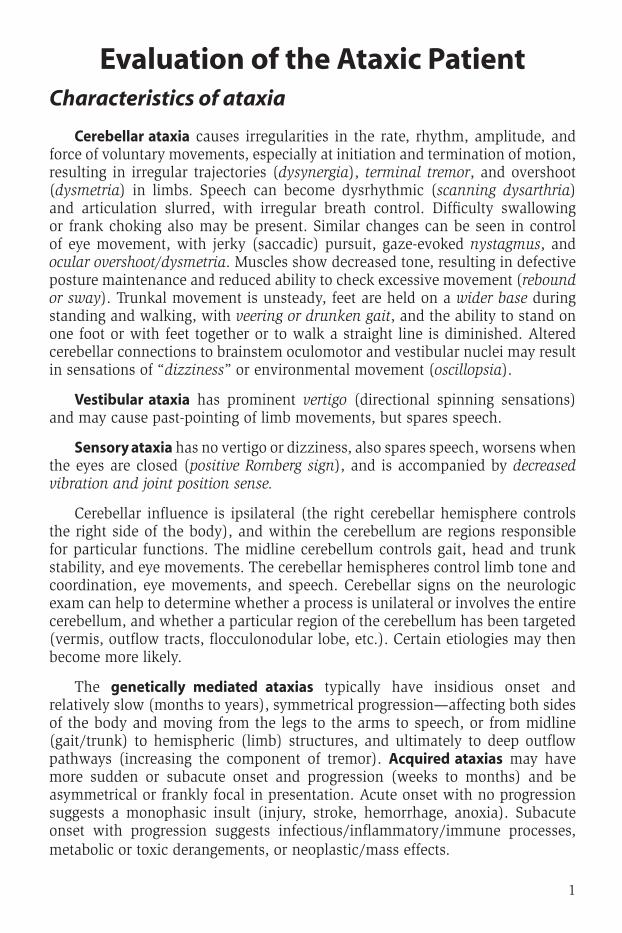
Evaluation of the Ataxic PatientCharacteristics of ataxia Cerebellar ataxia causes irregularities in the rate, rhythm, amplitude, and force of voluntary movements, especially at initiation and termination of motion, resulting in irregular trajectories (dysynergia), terminal tremor, and overshoot (dysmetria) in limbs. speech can become dysrhythmic (scanning dysarthria) and articulation slurred, with irregular breath control. difficulty swallowing or frank choking also may be present. similar changes can be seen in control of eye movement, with jerky (saccadic) pursuit, gaze-evoked nystagmus, and ocular overshoot/dysmetria. Muscles show decreased tone, resulting in defective posture maintenance and reduced ability to check excessive movement (rebound or sway). trunkal movement is unsteady, feet are held on a wider base during standing and walking, with veering or drunken gait, and the ability to stand on one foot or with feet together or to walk a straight line is diminished. altered cerebellar connections to brainstem oculomotor and vestibular nuclei may result in sensations of “dizziness” or environmental movement (oscillopsia).
Vestibular ataxia has prominent vertigo (directional spinning sensations) and may cause past-pointing of limb movements, but spares speech.
Sensory ataxia has no vertigo or dizziness, also spares speech, worsens when the eyes are closed (positive Romberg sign), and is accompanied by decreased vibration and joint position sense.
cerebellar influence is ipsilateral (the right cerebellar hemisphere controls the right side of the body), and within the cerebellum are regions responsible for particular functions. the midline cerebellum controls gait, head and trunk stability, and eye movements. the cerebellar hemispheres control limb tone and coordination, eye movements, and speech. cerebellar signs on the neurologic exam can help to determine whether a process is unilateral or involves the entire cerebellum, and whether a particular region of the cerebellum has been targeted (vermis, outflow tracts, flocculonodular lobe, etc.). certain etiologies may then become more likely.
the genetically mediated ataxias typically have insidious onset and relatively slow (months to years), symmetrical progression—affecting both sides of the body and moving from the legs to the arms to speech, or from midline (gait/trunk) to hemispheric (limb) structures, and ultimately to deep outflow pathways (increasing the component of tremor). Acquired ataxias may have more sudden or subacute onset and progression (weeks to months) and be asymmetrical or frankly focal in presentation. acute onset with no progression suggests a monophasic insult (injury, stroke, hemorrhage, anoxia). subacute onset with progression suggests infectious/inflammatory/immune processes, metabolic or toxic derangements, or neoplastic/mass effects.
1
Ataxia Disorders book.indd 9 2/24/16 2:04 PM

Basic ataxia phenotypes there are seven basic phenotypes: • Autosomal dominant cerebellar ataxia/spinocerebellar ataxia (SCA) • Friedreich’s ataxia-like syndromes • Early onset cerebellar ataxia (EOCA) • Mitochondrial syndromes • Multiple system atrophy picture • Idiopathic late onset cerebellar syndromes • Hereditary spastic paraplegia/ataxia (not discussed in this booklet)
Evaluation the neurological history may provide clues to cause relating to associated illnesses, medication use, or lifestyle/environmental exposures (see Table 1). the neurological examination can be supplemented by neural imaging (magnetic resonance scanning/Mri or computed tomography/ct of the brain or spine) and electrophysiologic studies (electromyogram and nerve conduction/EMg-ncv; evoked potential testing—visual/vEr, brainstem/
2
Table 1. IdEntIFIAblE CAuSES OF nOngEnEtIC AtAxIAType Cause
Congenital Developmental
Mass lesion of a specific typeTumor, cyst, aneurysm, hematoma, abscess, normal pressure or partial obstructive hydrocephalus
Vascular Stroke, hemorrhage; subcortical vascular disease
Infectious/Post-infectious/Post-vaccination
Anthrax; Epstein-Barr; enterovirus; HIV; HTLV; prion disease; Lyme disease; syphilis; measles, rubella, varicella; Whipple’s disease; progressive multifocal leukoencephalopathy
Post-anoxic, post-hyperthermic, post-traumaticChronic epilepsy
MetabolicAcute thiamine (B1) deficiency; chronic vitamin B12 and E deficiencies; autoimmune thyroiditis and low thyroid levels
Toxic Drug reactions
Environmental
Amiodarone, cytosine arabinoside, 5-fluorouracil, lithium, phenytoin, valproic acid, and othersAcrylamide, alcohol, organic solvents, organo-lead/mercury/tin, inorganic bismuth/mercury/thallium
Immune-mediated Vasculitis Paraneoplastica
Other autoantibodies
Anti-immune therapies used in reported cases of immune-mediated cerebellar ataxia
Behcet’s, giant cell arteritis, lupus, and others
Anti-Yo, Hu, Ri, MaTa, CV2, Zic4; anti-calcium channel; anti-CRMP-5, ANNA-1,2,3, mGluR1, TR
Anti-GluR2, GADb, MPP1, GQ1b ganglioside; anti-gliadin (most common–reported also in the inherited syndromes as a possible secondary factor; treated with gluten-free diet)c-e
Steroids, plasmapheresis, IVIG, rituximab, mycophenolatemofetil, methotrexate, and others
a Bataller, L., and J. Dalmau. Paraneoplastic neurologic syndromes: approaches to diagnosis and treatment. semin neurol, 2003 23(2): p. 215-24.b Mitoma, h., et al. Presynaptic impairment of cerebellar inhibitory synapses by an autoantibody to glutamate decarboxylase. J neurol sci, 2000. 175(1): p. 40-44.c Bushara, K.O., et al. Gluten sensitivity in sporadic and hereditary cerebellar ataxia. Ann neurol, 2001. 49(4): p. 540-43d hadjivassiliou, M. et al. Dietary treatment of gluten ataxia. J neurol neurosurg Psychiatry, 2003. 74(9): p. 1221-24 e hadjivassiliou, M. et al. Gluten ataxia in perspective: epidemiology, genetic susceptibility and clinical characteristics. Brain, 2003. 126(Pt 3): p. 685-91.
Ataxia Disorders book.indd 10 2/24/16 2:04 PM

BaEr, somatosensory/ssEr; electronystagmography of oculomotor and vestibular pathways/Eng; electroencephalogram/EEg). these can confirm the anatomic localization of the process and often the actual etiology (mass lesion of a specific type—e.g. tumor, cyst, hematoma, abscess; stroke or hemorrhage; subcortical vascular disease; inflammation/ infection or vasculitis; demyelination; characteristic regional atrophy, hypo- or hyperintensities; normal pressure or partial obstructive hydrocephalus). Additional laboratory studies can then be ordered (blood; urine; spinal fluid; biopsy of muscle, nerve, or brain). there may be key features on examination that will provide clues to a specific cause for the ataxia (see Table 2).
the presence of a known genetic disorder does not rule out the presence of additional acquired insults that might alter the presentation and course of the symptoms of ataxia and warrant independent investigation.
similarly, the absence of a clear family history does not rule out the role of genetic factors in an apparently sporadic disorder. there may be no family history because the history wasn’t taken, because the information is unavailable (adoption, loss of contact, noncooperation, paternity issues), because of nondominant inheritance patterns (recessive, x-linked, maternal/mitochondrial), or because of specific genetic processes that modify disease presentation in the pedigree (anticipation, incomplete penetrance, mosaicism).
3
Table 2. KEY FEAtuRES OF ExAMInAtIOn tHAt MAY PROVIdE CluES tO tHE dIAgnOSIS OF AtAxIA
Type Features
neurological Features
Ataxia with parkinsonism and autonomic dysfunction suggest multiple system atrophy (MSA)
Accompanying dementia, seizures, ophthalmoplegia, or chorea suggest something other than MSA
Non-neurologic features
Cardiac (examples: cardiomyopathy, conduction disturbances) – Friedreich’s ataxia (FRDA), mitochondrial disease
Skeletal (examples: scoliosis, foot deformities) – FRDA, ataxia-telangiectasia, variants of Charcot-Marie-Tooth disease, late-on set inborn errors of metabolism
Endocrine – diabetes (FRDA/mitochondrial, Wilson’s disease), adrenalin sufficiency (adrenoleukodystrophy, or ALD; adrenomyeloneuropathy, or AMN)
Liver/metabolic – inborn errors of metabolism
Skin–phakomatoses (neurofibromatosis), ataxia-telangiectasia, inborn errors (vitamin E deficiency, sialidosis, ALD/AMN, Hartnup’s, cerebrotendinous xanthomatosis [CTX])
Mitochondrial disorders seem to have more features beyond ataxia than do the other ataxic illnesses
Distinctive neurologic features: dementia, dystonia, exercise intolerance, hearing loss, migraine myelopathy, myoclonus, myopathy, neuropathy, ophthalmoplegia, optic neuropathy, pigmentary retinopathy, seizures, stroke-like episodesDistinctive non-neurologic features: adrenal dysfunction, anemia, cardiomyopathy, cataracts, diabetes mellitus, other endocrine dysfunction, exocrine pancreas dysfunction, intestinal pseudo- obstruction, lactic acidosis, renal disease, rhabdomyalysis, short stature
Ataxia Disorders book.indd 11 2/24/16 2:04 PM
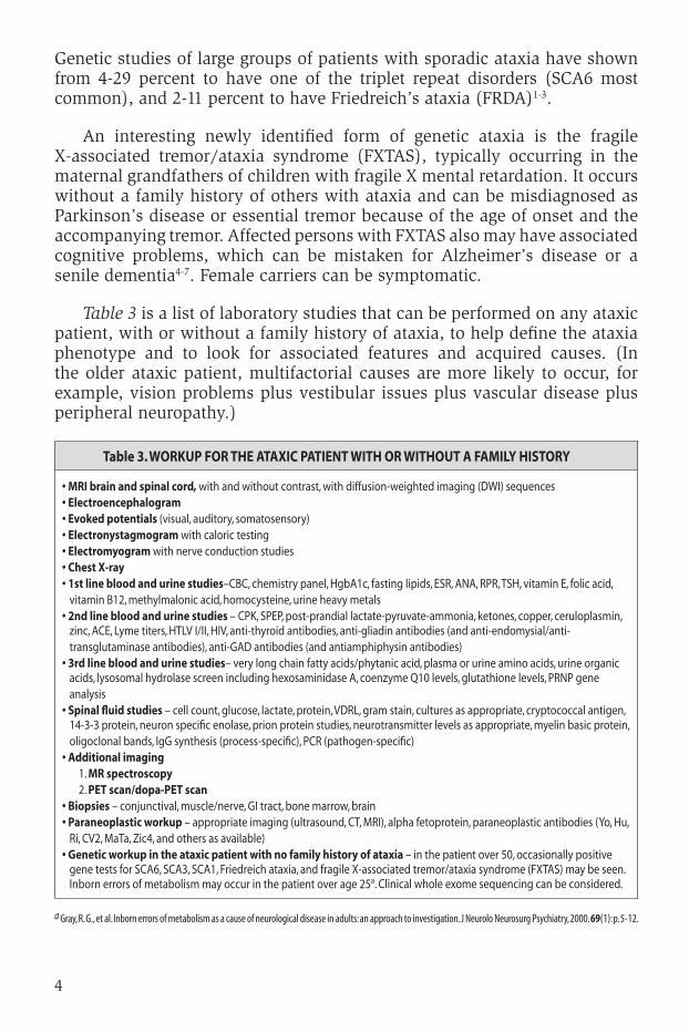
genetic studies of large groups of patients with sporadic ataxia have shown from 4-29 percent to have one of the triplet repeat disorders (sca6 most common), and 2-11 percent to have friedreich’s ataxia (frda)1-3.
an interesting newly identified form of genetic ataxia is the fragile x-associated tremor/ataxia syndrome (fxtas), typically occurring in the maternal grandfathers of children with fragile x mental retardation. it occurs without a family history of others with ataxia and can be misdiagnosed as Parkinson’s disease or essential tremor because of the age of onset and the accompanying tremor. affected persons with fxtas also may have associated cognitive problems, which can be mistaken for alzheimer’s disease or a senile dementia4-7. female carriers can be symptomatic.
Table 3 is a list of laboratory studies that can be performed on any ataxic patient, with or without a family history of ataxia, to help define the ataxia phenotype and to look for associated features and acquired causes. (in the older ataxic patient, multifactorial causes are more likely to occur, for example, vision problems plus vestibular issues plus vascular disease plus peripheral neuropathy.)
4
Table 3. WORKuP FOR tHE AtAxIC PAtIEnt WItH OR WItHOut A FAMIlY HIStORY
• MRI brain and spinal cord, with and without contrast, with diffusion-weighted imaging (DWI) sequences• Electroencephalogram• Evoked potentials (visual, auditory, somatosensory)• Electronystagmogram with caloric testing• Electromyogram with nerve conduction studies• Chest x-ray• 1st line blood and urine studies–CBC, chemistry panel, HgbA1c, fasting lipids, ESR, ANA, RPR, TSH, vitamin E, folic acid,
vitamin B12, methylmalonic acid, homocysteine, urine heavy metals• 2nd line blood and urine studies – CPK, SPEP, post-prandial lactate-pyruvate-ammonia, ketones, copper, ceruloplasmin,
zinc, ACE, Lyme titers, HTLV I/II, HIV, anti-thyroid antibodies, anti-gliadin antibodies (and anti-endomysial/anti-transglutaminase antibodies), anti-GAD antibodies (and antiamphiphysin antibodies)
• 3rd line blood and urine studies– very long chain fatty acids/phytanic acid, plasma or urine amino acids, urine organic acids, lysosomal hydrolase screen including hexosaminidase A, coenzyme Q10 levels, glutathione levels, PRNP gene analysis
• Spinal fluid studies – cell count, glucose, lactate, protein, VDRL, gram stain, cultures as appropriate, cryptococcal antigen, 14-3-3 protein, neuron specific enolase, prion protein studies, neurotransmitter levels as appropriate, myelin basic protein, oligoclonal bands, IgG synthesis (process-specific), PCR (pathogen-specific)
• Additional imaging 1. MR spectroscopy 2. PEt scan/dopa-PEt scan• biopsies – conjunctival, muscle/nerve, GI tract, bone marrow, brain• Paraneoplastic workup – appropriate imaging (ultrasound, CT, MRI), alpha fetoprotein, paraneoplastic antibodies (Yo, Hu,
Ri, CV2, MaTa, Zic4, and others as available)• genetic workup in the ataxic patient with no family history of ataxia – in the patient over 50, occasionally positive
gene tests for SCA6, SCA3, SCA1, Friedreich ataxia, and fragile X-associated tremor/ataxia syndrome (FXTAS) may be seen. Inborn errors of metabolism may occur in the patient over age 25a. Clinical whole exome sequencing can be considered.
a Gray, r. G., et al. inborn errors of metabolism as a cause of neurological disease in adults: an approach to investigation. J neurolo neurosurg Psychiatry, 2000. 69(1): p. 5-12.
Ataxia Disorders book.indd 12 2/24/16 2:04 PM
Autosomal dominant cerebellar ataxia the dominantly inherited ataxic disorders have an incidence of 1-5 in 100,000. they include the typical spinocerebellar ataxias (scas), which now number 40; the episodic ataxias (Ea 1-8); and the atypical spinocerebellar ataxias (dentatorubral-pallidoluysian atrophy/drPla and gerstmann-straussler-scheinker/gss disease), which may have prominent features other than ataxia. Pathogenetic classification would group scas 1-3, 6, 7, 12, 17, and drPla as polyglutamine (triplet repeat or cag repeat) disorders; scas 8, 10, 31, and 36 as other repeat types; scas 4, 5, 11-16, 23, 27, and gss as resulting from other mutation types; and scas 6, 13, 19, 22, and Ea-1, 2, and 5 as primary channelopathies. the molecular bases of the remaining scas are still unknown (see Table 4).
the average age of onset is in the third decade, and, in the early stages, most of these dominantly inherited disorders may be indistinguishable from one another, except by genetic testing (see tables 5 and 6). there have been efforts to develop algorithms to prioritize genetic testing, with the most statistically sound using Baysian analysis to help predict which of the most common scas (scas 1, 2, 3, 6, 7, 8) could be expected in a particular clinical situation8.
5
Ataxia Disorders book.indd 13 2/24/16 2:04 PM

76
Table 4. The DominanTly inheriTeD aTaxias – molecular GeneTics See http://neuromuscular.wustl.edu/ataxia/domatax.html or http://www.ncbi.nlm.nih.gov/pubmed/?term=autosomal+dominant+cerebellar+ataxias for most current references
ataxic Disorder Gene locus Gene/Product mutation Prevalence
TyPical DominanT
SCA16p23 Ataxin-1 CAG expansion/coding exon. Normal <39 repeats. Disease-causing >44. If no CAT
interruption, disease-causing 39-446-27% of dominant ataxias worldwide
SCA2 12q24 Ataxin-2CAG expansion/coding exon. Normal <33 repeats, with CAA interruption. Disease-causing >33, with no CAA interruption (two patients with interrupted 34 expansion)
13-18% of dominant ataxias worldwide
SCA3/Machado-Joseph disease
14q24.3-q31 Ataxin-3 CAG expansion/coding exon. Normal <41 repeats. Disease-causing >45. Homozygous mutant genes cause earlier onset, more severe disease
23-36% of dominant ataxias worldwide
SCA4 16q22.1Puratrophin-1. Functions in intracellular signaling, actin dynamics. Targeted to the Golgi apparatus. Mutant protein associated with aggregates in Purkinje cells
Single-nucleotide C-T substitution in 5’ untranslated regionFamilies in Utah and Germany; six families in Japan with later onset pure cerebellar syndrome
SCA5 11p11-q11ß-III Spectrin stabilizes the glutamate transporter EAAT4 at the surface of the plasma membrane
Inframe deletions; missense (Leu253Pro) Lincoln family in US; families in Germany and France
SCA6 19p13CACNa1A/P/Q type calcium channel subunit (disease mechanisms may result from both CAG repeat and channelopathy processes)
CAG expansion/coding exon. Normal <19 repeats. Disease-causing >19. Homozygous mutant genes cause earlier onset, more severe disease. Allelic with EA-2 (gene truncations) and hemiplegic migraine (missense mutations)
10-30% of dominant ataxias worldwide
SCA7 3p21.1-p12Ataxin-7. Component of TFTC-like transcriptional complexes (disease mechanisms may result from both CAG repeat and transcriptional dysregulatory processes)
CAG expansion/coding exon. Normal <28 repeats. Disease-causing >37. Intermediate 28-36, may expand into disease range, especially with paternal transmission
2-5% of dominant ataxias worldwide; may be more common in Sweden and Finland
SCA8 13q21Normal product is an untranslated RNA that functions as a gene regulator. Evidence for a translated polyglutamine protein (Ataxin-8) from an anti-parallel transcript has also been found
CTG expansion at 3’ end. Normal <80 repeats. Disease-causing 80-300, although expansions in this range occur in non-ataxic persons and in other neurologic diseases. Expansions >300 may not cause disease in SCA8 pedigrees
2-4% of dominant ataxias worldwide; genetic testing results may be open to interpretation
SCA9 Unknown Unknown UnknownOne American-English family; ophthalmoplegia, optic atrophy, upper motor neuron, Parkinsonism, posterior column features
SCA10 22q13 Ataxin-10. Gene product essential for cerebellar neuronal survivalPentanucleotide repeat (ATTCT) expansion in intron 9, probable loss of function mutation. Normal <22 repeats. Disease 800-4500. Intergenerationally more likely to contract than expand
Mexican families (ataxia and epilepsy); five Brazilian families (no epilepsy)
SCA11 15q15.2 Tau tubulin kinase 2 (TTBK2) Stop, frameshift, insertion, or deletion Two British families
SCA12 5q31-q33PPP2R2B/brain specific regulatory subunit of protein phosphatase 2A (serine/threonine phosphatase)
CAG expansion in 5’ untranslated region of gene, possibly upstream from transcription start site and affecting gene transcription. Minimal intergenerational instability
German-American family; may account for up to 7% of ADCA in India
SCA13 19q13.3-q13.4KCNC3 voltage-gated potassium channel associated with high-frequency firing in fast-spiking cerebellar neurons
Two missense mutations found (R420H and F448L)French family–seven of eight affected members were women, early-onset with cognitive decline. Filipino family with adult-onset ataxia
SCA14 19q13.4-qter PRKCG/protein kinase Cy (serine/threonine kinase)
Missense mutations in conserved residues of C1/exon 4–regulatory domain and in catalytic domain of the enzyme. Increased intrinsic activity of mutant enzyme moves intraneuronal distribution from cytosol to plasma membrane. May reduce expression of ataxin-1 in Purkinje cells, and mutant ataxin-1 may reduce expression of PRKCG
Japanese (axial myoclonus), English/Dutch, Dutch, and French (broader age of onset, cognitive impairment) families described. Incomplete penetrance
SCA15 3p26.1 Inositol 1,4,5-triphosphate receptor, type 1 (ITPR1)Same locus as SCA16, SCA29 Large deletions, missense. Australian, French & Japanese families; 1% of SCA
SCA16 See SCA15
SCA17/Huntington disease-like 4
6q27
TATA box-binding protein (DNA binding subunit of RNA polymerase II transcription factor D [TFIID]), essential for the expression of all protein-encoding genes; disease mechanisms may result from both CAG repeat and transcriptional dysregulatory processes)
CAG/CAA expansion. Normal <42 repeats. Disease-causing >45. Intermediate 43-48, with incomplete penetrance. Minimal intergenerational instability. Homozygous mutant genes cause earlier-onset, more severe disease. Variable phenotypes include similarities to Huntington’s disease, Parkinson’s disease, Alzheimer’s disease, and variant Jakob-Creutzfeldt disease
Japanese, German, Italian, and French families
SCA18 7q22-q32 Unknown Linkage studies with DNA polymorphisms point to location One Irish-American family
SCA19 1p13.2 KCND3Allelic with SCA22 Missense Several Dutch families
SCA20 11q12 Contiguous gene duplication syndrome: Region contains ≥ 12 genes 260-kb duplication at 11q12 Anglo-Celtic family in Australia
SCA21 1p36.33 Transmembrane protein 240 (TMEM240) Missense and stop 2% of French SCA
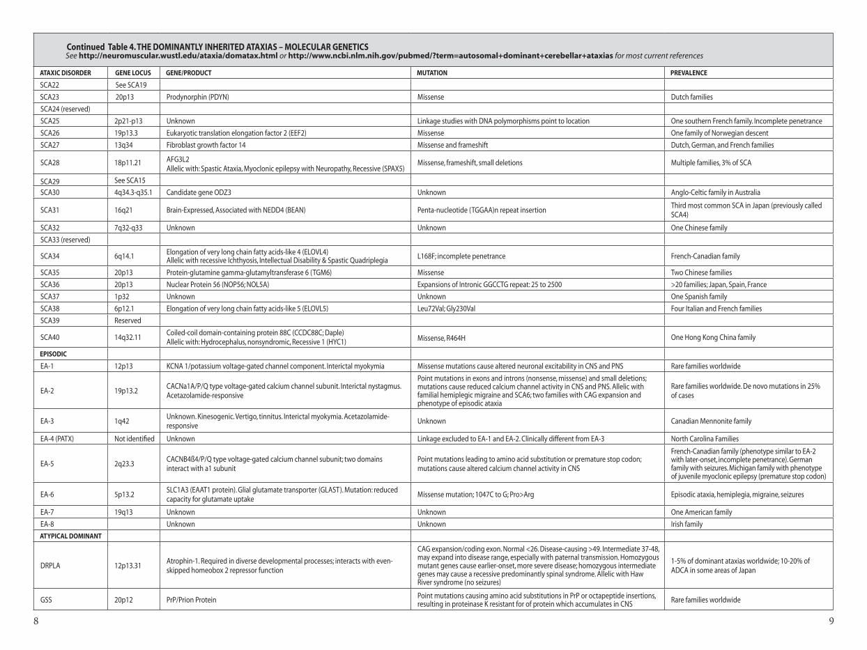
98
Continued Table 4. The DominanTly inheriTeD aTaxias – moleCular GeneTiCs See http://neuromuscular.wustl.edu/ataxia/domatax.html or http://www.ncbi.nlm.nih.gov/pubmed/?term=autosomal+dominant+cerebellar+ataxias for most current references
aTaxiC DisorDer Gene loCus Gene/ProDuCT muTaTion PrevalenCe
SCA22 See SCA19
SCA23 20p13 Prodynorphin (PDYN) Missense Dutch families
SCA24 (reserved)
SCA25 2p21-p13 Unknown Linkage studies with DNA polymorphisms point to location One southern French family. Incomplete penetrance
SCA26 19p13.3 Eukaryotic translation elongation factor 2 (EEF2) Missense One family of Norwegian descent
SCA27 13q34 Fibroblast growth factor 14 Missense and frameshift Dutch, German, and French families
SCA28 18p11.21 AFG3L2 Allelic with: Spastic Ataxia, Myoclonic epilepsy with Neuropathy, Recessive (SPAX5)
Missense, frameshift, small deletions Multiple families, 3% of SCA
SCA29 See SCA15
SCA30 4q34.3-q35.1 Candidate gene ODZ3 Unknown Anglo-Celtic family in Australia
SCA31 16q21 Brain-Expressed, Associated with NEDD4 (BEAN) Penta-nucleotide (TGGAA)n repeat insertionThird most common SCA in Japan (previously called SCA4)
SCA32 7q32-q33 Unknown Unknown One Chinese family
SCA33 (reserved)
SCA34 6q14.1 Elongation of very long chain fatty acids-like 4 (ELOVL4)Allelic with recessive Ichthyosis, Intellectual Disability & Spastic Quadriplegia L168F; incomplete penetrance French-Canadian family
SCA35 20p13 Protein-glutamine gamma-glutamyltransferase 6 (TGM6) Missense Two Chinese families
SCA36 20p13 Nuclear Protein 56 (NOP56; NOL5A) Expansions of Intronic GGCCTG repeat: 25 to 2500 >20 families; Japan, Spain, France
SCA37 1p32 Unknown Unknown One Spanish family
SCA38 6p12.1 Elongation of very long chain fatty acids-like 5 (ELOVL5) Leu72Val; Gly230Val Four Italian and French families
SCA39 Reserved
SCA40 14q32.11Coiled-coil domain-containing protein 88C (CCDC88C; Daple)Allelic with: Hydrocephalus, nonsyndromic, Recessive 1 (HYC1) Missense, R464H One Hong Kong China family
ePisoDiC
EA-1 12p13 KCNA 1/potassium voltage-gated channel component. Interictal myokymia Missense mutations cause altered neuronal excitability in CNS and PNS Rare families worldwide
EA-2 19p13.2CACNa1A/P/Q type voltage-gated calcium channel subunit. Interictal nystagmus. Acetazolamide-responsive
Point mutations in exons and introns (nonsense, missense) and small deletions; mutations cause reduced calcium channel activity in CNS and PNS. Allelic with familial hemiplegic migraine and SCA6; two families with CAG expansion and phenotype of episodic ataxia
Rare families worldwide. De novo mutations in 25% of cases
EA-3 1q42Unknown. Kinesogenic. Vertigo, tinnitus. Interictal myokymia. Acetazolamide-responsive
Unknown Canadian Mennonite family
EA-4 (PATX) Not identified Unknown Linkage excluded to EA-1 and EA-2. Clinically different from EA-3 North Carolina Families
EA-5 2q23.3CACNB4ß4/P/Q type voltage-gated calcium channel subunit; two domains interact with a1 subunit
Point mutations leading to amino acid substitution or premature stop codon; mutations cause altered calcium channel activity in CNS
French-Canadian family (phenotype similar to EA-2 with later-onset, incomplete penetrance). German family with seizures. Michigan family with phenotype of juvenile myoclonic epilepsy (premature stop codon)
EA-6 5p13.2SLC1A3 (EAAT1 protein). Glial glutamate transporter (GLAST). Mutation: reduced capacity for glutamate uptake
Missense mutation; 1047C to G; Pro>Arg Episodic ataxia, hemiplegia, migraine, seizures
EA-7 19q13 Unknown Unknown One American family
EA-8 Unknown Unknown Irish family
aTyPiCal DominanT
DRPLA 12p13.31Atrophin-1. Required in diverse developmental processes; interacts with even-skipped homeobox 2 repressor function
CAG expansion/coding exon. Normal <26. Disease-causing >49. Intermediate 37-48, may expand into disease range, especially with paternal transmission. Homozygous mutant genes cause earlier-onset, more severe disease; homozygous intermediate genes may cause a recessive predominantly spinal syndrome. Allelic with Haw River syndrome (no seizures)
1-5% of dominant ataxias worldwide; 10-20% of ADCA in some areas of Japan
GSS 20p12 PrP/Prion Protein Point mutations causing amino acid substitutions in PrP or octapeptide insertions, resulting in proteinase K resistant for of protein which accumulates in CNS Rare families worldwide
10
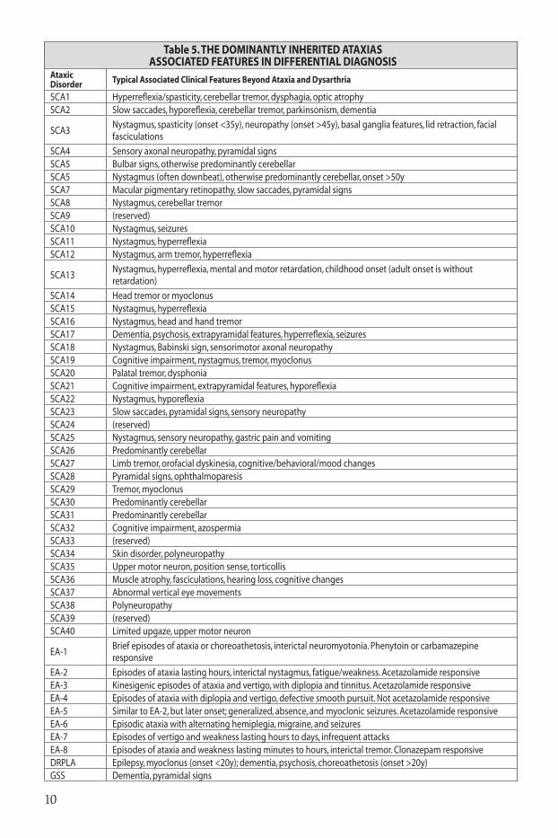
Table 5. tHE dOMInAntlY InHERItEd AtAxIASASSOCIAtEd FEAtuRES In dIFFEREntIAl dIAgnOSIS
Ataxic Disorder typical Associated Clinical Features beyond Ataxia and dysarthria
SCA1 Hyperreflexia/spasticity, cerebellar tremor, dysphagia, optic atrophySCA2 Slow saccades, hyporeflexia, cerebellar tremor, parkinsonism, dementia
SCA3Nystagmus, spasticity (onset <35y), neuropathy (onset >45y), basal ganglia features, lid retraction, facial fasciculations
SCA4 Sensory axonal neuropathy, pyramidal signsSCA5 Bulbar signs, otherwise predominantly cerebellarSCA5 Nystagmus (often downbeat), otherwise predominantly cerebellar, onset >50ySCA7 Macular pigmentary retinopathy, slow saccades, pyramidal signsSCA8 Nystagmus, cerebellar tremorSCA9 (reserved)SCA10 Nystagmus, seizuresSCA11 Nystagmus, hyperreflexiaSCA12 Nystagmus, arm tremor, hyperreflexia
SCA13Nystagmus, hyperreflexia, mental and motor retardation, childhood onset (adult onset is without retardation)
SCA14 Head tremor or myoclonusSCA15 Nystagmus, hyperreflexiaSCA16 Nystagmus, head and hand tremorSCA17 Dementia, psychosis, extrapyramidal features, hyperreflexia, seizuresSCA18 Nystagmus, Babinski sign, sensorimotor axonal neuropathySCA19 Cognitive impairment, nystagmus, tremor, myoclonusSCA20 Palatal tremor, dysphoniaSCA21 Cognitive impairment, extrapyramidal features, hyporeflexiaSCA22 Nystagmus, hyporeflexiaSCA23 Slow saccades, pyramidal signs, sensory neuropathySCA24 (reserved)SCA25 Nystagmus, sensory neuropathy, gastric pain and vomitingSCA26 Predominantly cerebellarSCA27 Limb tremor, orofacial dyskinesia, cognitive/behavioral/mood changesSCA28 Pyramidal signs, ophthalmoparesisSCA29 Tremor, myoclonusSCA30 Predominantly cerebellarSCA31 Predominantly cerebellarSCA32 Cognitive impairment, azospermiaSCA33 (reserved)SCA34 Skin disorder, polyneuropathySCA35 Upper motor neuron, position sense, torticollisSCA36 Muscle atrophy, fasciculations, hearing loss, cognitive changesSCA37 Abnormal vertical eye movementsSCA38 PolyneuropathySCA39 (reserved)SCA40 Limited upgaze, upper motor neuron
EA-1Brief episodes of ataxia or choreoathetosis, interictal neuromyotonia. Phenytoin or carbamazepine responsive
EA-2 Episodes of ataxia lasting hours, interictal nystagmus, fatigue/weakness. Acetazolamide responsiveEA-3 Kinesigenic episodes of ataxia and vertigo, with diplopia and tinnitus. Acetazolamide responsiveEA-4 Episodes of ataxia with diplopia and vertigo, defective smooth pursuit. Not acetazolamide responsiveEA-5 Similar to EA-2, but later onset; generalized, absence, and myoclonic seizures. Acetazolamide responsiveEA-6 Episodic ataxia with alternating hemiplegia, migraine, and seizuresEA-7 Episodes of vertigo and weakness lasting hours to days, infrequent attacksEA-8 Episodes of ataxia and weakness lasting minutes to hours, interictal tremor. Clonazepam responsiveDRPLA Epilepsy, myoclonus (onset <20y); dementia, psychosis, choreoathetosis (onset >20y)GSS Dementia, pyramidal signs
Ataxia Disorders book.indd 18 2/24/16 2:04 PM
11
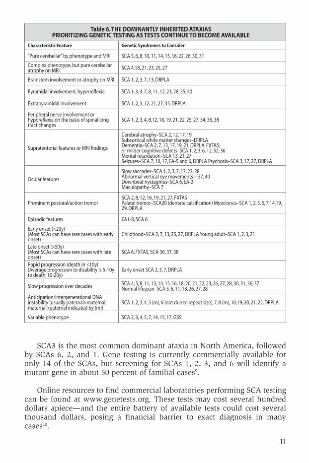
Table 6. tHE dOMInAntlY InHERItEd AtAxIASPRIORItIzIng gEnEtIC tEStIng AS tEStS COntInuE tO bECOME AVAIlAblE
Characteristic Feature genetic Syndromes to Consider
“Pure cerebellar” by phenotype and MRI SCA 5, 6, 8, 10, 11, 14, 15, 16, 22, 26, 30, 31
Complex phenotype, but pure cerebellar atrophy on MRI SCA 4,18, 21, 23, 25, 27
Brainstem involvement or atrophy on MRI SCA 1, 2, 3, 7, 13, DRPLA
Pyramidal involvement, hyperreflexia SCA 1, 3, 4, 7, 8, 11, 12, 23, 28, 35, 40
Extrapyramidal involvement SCA 1, 2, 3, 12, 21, 27, 35, DRPLA
Peripheral nerve involvement or hyporeflexia on the basis of spinal long tract changes
SCA 1, 2, 3, 4, 8,12, 18, 19, 21, 22, 25, 27, 34, 36, 38
Supratentorial features or MRI findings
Cerebral atrophy–SCA 2, 12, 17, 19Subcortical white matter changes–DRPLADementia–SCA 2, 7, 13, 17, 19, 21, DRPLA, FXTAS; or milder cognitive defects–SCA 1, 2, 3, 6, 12, 32, 36 Mental retardation–SCA 13, 21, 27 Seizures–SCA 7, 10, 17, EA-5 and 6, DRPLA Psychosis–SCA 3, 17, 27, DRPLA
Ocular features
Slow saccades–SCA 1, 2, 3, 7, 17, 23, 28 Abnormal vertical eye movements—37, 40Downbeat nystagmus–SCA 6, EA-2 Maculopathy–SCA 7
Prominent postural/action tremorSCA 2, 8, 12, 16, 19, 21, 27, FXTASPalatal tremor–SCA20 (dentate calcification) Myoclonus–SCA 1, 2, 3, 6, 7,14,19, 29, DRPLA
Episodic features EA1-8, SCA 6
Early onset (<20y)(Most SCAs can have rare cases with early onset)
Childhood–SCA 2, 7, 13, 25, 27, DRPLA Young adult–SCA 1, 2, 3, 21
Late onset (>50y)(Most SCAs can have rare cases with late onset)
SCA 6, FXTAS, SCA 36, 37, 38
Rapid progression (death in <10y)(Average progression to disability is 5-10y; to death, 10-20y)
Early onset SCA 2, 3, 7, DRPLA
Slow progression over decades SCA 4, 5, 8, 11, 13, 14, 15, 16, 18, 20, 21, 22, 23, 26, 27, 28, 30, 31, 36, 37 Normal lifespan–SCA 5, 6, 11, 18, 26, 27, 28
Anticipation/intergenerational DNA instability (usually paternal>maternal; maternal>paternal indicated by (m))
SCA 1, 2, 3, 4, 5 (m), 6 (not due to repeat size), 7, 8 (m), 10,19, 20, 21, 22, DRPLA
Variable phenotype SCA 2, 3, 4, 5, 7, 14, 15, 17, GSS
sca3 is the most common dominant ataxia in north america, followed by scas 6, 2, and 1. gene testing is currently commercially available for only 14 of the scas, but screening for scas 1, 2, 3, and 6 will identify a mutant gene in about 50 percent of familial cases9.
online resources to find commercial laboratories performing sca testing can be found at www.genetests.org. these tests may cost several hundred dollars apiece—and the entire battery of available tests could cost several thousand dollars, posing a financial barrier to exact diagnosis in many cases10.
Ataxia Disorders book.indd 19 2/24/16 2:04 PM
12
the clinical use of next-generation exome sequencing is becoming more widespread. it might prove a less expensive and more comprehensive technology for the identification of non-repeat disorders. However, there is limited information available to direct clinicians in identifying which patients would most benefit from such testing. findings suggest that patients with chronic progressive cerebellar ataxia would benefit diagnostically from exome sequencing irrespective of a positive family history or early age at onset. strategies for the integration of genomic testing into the clinical evaluation and effective bioinformatics methods of data analysis are being developed11,
12, 13.
Recessively inherited ataxias14,15
• Friedreich’s ataxia-like syndromes16
• Early onset cerebellar ataxia (EOCA)17
the recessive ataxias are most often onset before the age of 25. the most common of the recessively inherited ataxias is friedreich’s ataxia (frda), with an incidence of 1 in 30,000-50,000. carrier frequency is 1 in 60-110. it is rare in asian and african pedigrees. in some populations, ataxia with oculomotor apraxia types 1 and 2 (AOA1, AOA2) also are found with high frequency.
Before the age of 5, Ataxia-telangiectasia is the most common recessively inherited cause of cerebellar ataxia18,19.
in young children, however, the most common cause of ataxia remains acute viral/post-viral cerebellar ataxia, which is self-limited and recovers in most within three to four weeks20.
the diagnostic criteria for these disorders are listed in table 7, with the molecular genetic features listed in table 8.
Ataxia Disorders book.indd 20 2/24/16 2:04 PM
13
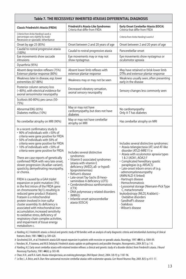
Table 7. tHE RECESSIVElY InHERItEd AtAxIAS dIFFEREntIAl dIAgnOSIS
Classic Friedreich’s Ataxia (FRdA) Friedreich’s Ataxia-like SyndromesCriteria that differ from FRDA
Early Onset Cerebellar Ataxia (EOCA)Criteria that differ from FRDA
Criteria from Anita Harding’s work a(percentages vary slightly by study)Recessive or sporadic inheritance
Criteria from Anita Harding’s work d
Onset by age 25 (85%) Onset between 2 and 20 years of age Onset between 2 and 20 years of ageCaudal to rostral progressive ataxia (100%) Caudal to rostral progressive ataxia Pancerebellar onset
Eye movements show saccade intrusions
Eye movements may or may not show nystagmus
Eye movements show nystagmus or oculomotor apraxia
Dysarthria (95%)
Absent deep tendon reflexes (75%) Extensor plantar response (80%)
Absent lower limb reflexes with extensor plantar response
May have retained or brisk lower limb DTRs and extensor plantar response
Weakness later in disease, esp. lower extremities (67-88%) Weakness may or may not be seen Weakness usually seen, often presenting
early in the disease
Posterior column sensory loss(~80%), with electrical evidence for axonal sensorimotor neuropathy
Decreased vibratory sensation, axonal sensory neuropathy Sensory changes less commonly seen
Scoliosis (60-80%); pes cavus (50-75%)
Abnormal EKG (65%) Diabetes mellitus (10%)
May or may not have cardiomyopathy, but does not have diabetes
No cardiomyopathy Only A-T has diabetes
No cerebellar atrophy on MRI (90%) May or may not have cerebellar atrophy on MRI Has cerebellar atrophy on MRI
In a recent confirmatory study b• 90%ofindividualswith>50%of
criteria were gene positive for FRDA• 50%ofindividualswith50%of
criteria were gene positive for FRDA• 10%ofindividualswith<50%of
criteria were gene positive for FRDA
There are case reports of genetically confirmed FRDA with very late onset, slower progression (Acadian variant), spasticity, demyelinating neuropathy, or chorea.
FRDA is caused by a GAA triplet expansion or point mutation (3%) in the first intron of the FRDA gene on chromosome 9q13, resulting in reduced gene product (frataxin). Frataxin is a mitochondrial protein involved in iron-sulfur cluster assembly. Its deficiency is associated with mitochondrial iron accumulation, increased sensitivity to oxidative stress, deficiency of respiratory chain complex activities, and impairment of tissue energy metabolism c.
Includes several distinctive syndromes:
• VitaminE-associatedsyndromes(ataxia with vitamin E deficiency [AVED], aß- or hypoß-lipoproteinemias)
• Refsum’sdisease• Late-onsetTay-Sachs(ß-hexo-
saminidase A deficiency�LOTS)• Cerebrotendinousxanthomatosis
(CTX)• DNApolymeraseyrelateddisorders
(MIRAS)• Infantileonsetspinocerebellar
ataxia (IOSCA)
Includes several distinctive syndromes:• Ataxia-telangiectasia(AT)andAT-like
disorder (ATLD-MRE11) e• Ataxiawithoculomotorapraxiatypes
1 & 2 (AOA1, AOA2) f• Complicatedhereditaryspastic
paraplegias (e.g. ARSACS)• Late-onsetinbornerrorsof(e.g.- adrenomyeloneuropathy(AMN/ALD-X linked)- Hartnup’s disease- Hemochromatosis- Lysosomal storage (Niemann-Pick Type
C, metachromaticleukodystropy [MLD], Krabbe’s) -
Oxidative disorders- Sandhoff’s disease- Sialidosis- Wilson’s disease
a harding, A.E. friedreich’s ataxia: a clinical and genetic study of 90 families with an analysis of early diagnostic criteria and intrafamilial clustering of clinical features. Brain, 1981. 104(3): p. 589-620.b Geschwind, D.h., et al. friedreich’s ataxia GAA repeat expansion in patients with recessive or sporadic ataxia. Neurology, 1997. 49(4): p. 1004-09.c voncken, M., P. ioannou, and M.B. Delatycki. friedreich ataxia-update on pathogenesis and possible therapies. Neurogenetics, 2004. 5(1): p. 1-8.d harding, A.E. Early onset cerebellar ataxia with retained tendon reflexes: a clinical and genetic study of a disorder distinct from friedreich’s ataxia. J Neurol Neurosurg Psychiatry, 1981. 44(6): p. 503-08.e chun, h.h., and r.A. Gatti. Ataxia-telangiectasia, an evolving phenotype. DNA Repair (Amst), 2004. 3(8-9): p. 1187-96.f Le Ber, i., A. Brice, and A. Durr. new autosomal recessive cerebellar ataxias with oculomotor apraxia. Curr Neurol Neurosci Rep, 2005. 5(5): p. 411-17.
Ataxia Disorders book.indd 21 2/24/16 2:04 PM

14
Table 8. tHE RECESSIVElY InHERItEd AtAxIAS MOlECulAR gEnEtICS
Phenotype Ataxic Disorder Disease Abbr. gene/Protein gene
Abbr locus Protein Function
Friedreich Ataxia-like
Friedreich’s ataxia FRDA Frataxin FXN 9q13 Mitochondrial iron metabolism
Ataxia with vitamin E deficiency AVED a-Tocopherol
transfer protein TTPA 8q13.1-q13.3 Vitamin E homeostasis
Abetalipoproteinemia ABLMicrosomal triglyceride transfer protein
MTP 4q22-q24
Lipoprotein Metabolism
Refsum’s disease -Phytanoyl-CoA hydroxylase PHYH 10oter-
p11.2 Fatty acid oxidation
Peroxisome biogenesis factor 7 PEX7 6q22-
q24Peroxisomal protein importation
Friedreich Ataxia-like with cerebellar atrophy
Late-onset Tay-Sachs disease LOTX ß-Hexosaminidase
A HEXA 15Q23-Q24
Glycosphingolipid metabolism
Cerebrotendinous xanthomatosis CTX Sterol-27
hydroxylase CYP27 2q33-qter Bile acid syntheses
DNA polymerase y related disorders MIRAS DNA polymerase
y-1 POLG1 15q24-q26
Mitochondrial DNA repair/replication
Infantile onset spinocerebellar ataxia IOSCA Twinkle, Twinky C10orf2 10q24 DNA replication,
unknown
Early onset cerebellar ataxia with retained reflexes (EOCARR)
Ataxia-telangiectasia ATAtaxia-telangiectasia, mutated
ATM 11q22-q23
DNA damage response
Ataxia-telangiectasia-like disorder ATLD Meiotic
recombination 11 MRE11 11q21 DNA damage response
Ataxia with oculomotor apraxia type 1 AOA1 Aprataxin APTX 9p13.3 DNA repair,
?RNA processing
Ataxia with oculomotor apraxia type 2 AOA2 Senataxin SETX 9Q34
?DNA repair,?DNA transcription,?RNA processing
Autosomal recessive ataxia of Charlevoix-Saguenay
ARSACS Sacsin SACS 13q12 ?Protein folding
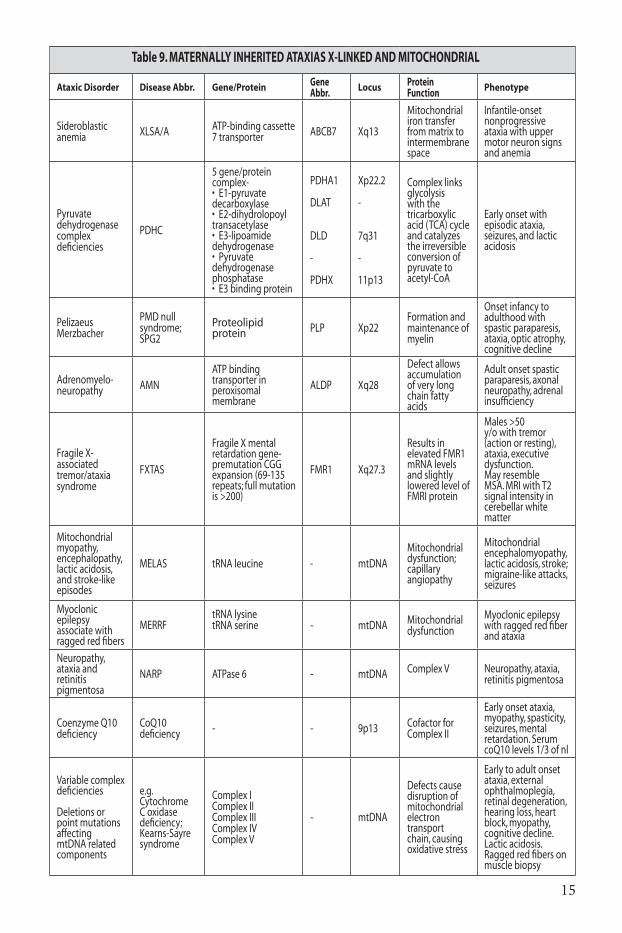
Maternally inherited ataxias (X-linked and mitochondrial)
these forms of ataxia are suspected when the defective genetic material seems always to come down from the mother’s side of the family. she may or may not be symptomatic herself. Her sons and daughters are equally at-risk to inherit the disease gene. in x-linked disorders the female carriers may not develop symptoms. affected males with x-linked disorders will never pass the defective gene on to their sons (no male-to-male transmission), but will always pass it on to their daughters, who then become carriers and may or may not develop symptoms. the presence of male-to-male transmission rules out an x-linked ataxia. in genetic disorders of the mitochondrial genome, phenotype severity depends on the ratio of abnormal to normal mitochondria. as all mitochondria are inherited from the mother, an affected male can never pass on the disease to his children. Most mitochondrial genes, however, are coded in the autosomal genome, causing disorders that can be transmitted like other dominant or recessive diseases21,22.
the most common maternally inherited ataxias are outlined in Table 9.
Ataxia Disorders book.indd 22 2/24/16 2:04 PM
15
Table 9. MAtERnAllY InHERItEd AtAxIAS x-lInKEd And MItOCHOndRIAl
Ataxic Disorder Disease Abbr. gene/Protein gene Abbr. locus Protein
Function Phenotype
Sideroblastic anemia XLSA/A ATP-binding cassette
7 transporter ABCB7 Xq13
Mitochondrial iron transfer from matrix to intermembrane space
Infantile-onset nonprogressive ataxia with upper motor neuron signs and anemia
Pyruvate dehydrogenase complex deficiencies
PDHC
5 gene/protein complex-•E1-pyruvatedecarboxylase•E2-dihydrolopoyltransacetylase•E3-lipoamidedehydrogenase•Pyruvatedehydrogenase phosphatase•E3bindingprotein
PDHA1
DLAT
DLD
-
PDHX
Xp22.2
-
7q31
-
11p13
Complex links glycolysis with the tricarboxylic acid (TCA) cycle and catalyzes the irreversible conversion of pyruvate to acetyl-CoA
Early onset with episodic ataxia, seizures, and lactic acidosis
Pelizaeus Merzbacher
PMD null syndrome; SPG2
Proteolipid protein PLP Xp22
Formation and maintenance of myelin
Onset infancy to adulthood with spastic paraparesis, ataxia, optic atrophy, cognitive decline
Adrenomyelo-neuropathy AMN
ATP binding transporter in peroxisomal membrane
ALDP Xq28
Defect allows accumulation of very long chain fatty acids
Adult onset spastic paraparesis, axonal neuropathy, adrenal insufficiency
Fragile X-associated tremor/ataxia syndrome
FXTAS
Fragile X mental retardation gene-premutation CGG expansion (69-135 repeats; full mutation is >200)
FMR1 Xq27.3
Results in elevated FMR1 mRNA levels and slightly lowered level of FMRI protein
Males >50 y/o with tremor (action or resting), ataxia, executive dysfunction. May resemble MSA. MRI with T2 signal intensity in cerebellar white matter
Mitochondrial myopathy, encephalopathy, lactic acidosis, and stroke-like episodes
MELAS tRNA leucine - mtDNAMitochondrial dysfunction;capillary angiopathy
Mitochondrial encephalomyopathy, lactic acidosis, stroke; migraine-like attacks, seizures
Myoclonic epilepsy associate with ragged red fibers
MERRFtRNA lysinetRNA serine - mtDNA Mitochondrial
dysfunctionMyoclonic epilepsy with ragged red fiber and ataxia
Neuropathy, ataxia and retinitis pigmentosa
NARP ATPase 6 - mtDNA Complex V Neuropathy, ataxia, retinitis pigmentosa
Coenzyme Q10 deficiency
CoQ10 deficiency - - 9p13 Cofactor for
Complex II
Early onset ataxia, myopathy, spasticity, seizures, mental retardation. Serum coQ10 levels 1/3 of nl
Variable complex deficiencies
Deletions or point mutations affecting mtDNA related components
e.g. Cytochrome C oxidase deficiency;Kearns-Sayre syndrome
Complex IComplex IIComplex IIIComplex IVComplex V
- mtDNA
Defects cause disruption of mitochondrial electron transport chain, causing oxidative stress
Early to adult onset ataxia, external ophthalmoplegia, retinal degeneration, hearing loss, heart block, myopathy, cognitive decline. Lactic acidosis. Ragged red fibers on muscle biopsy
Ataxia Disorders book.indd 23 2/24/16 2:04 PM

16
the most reliable approach to sporadic ataxia is to assign a phenotype by history and physical, imaging, and electrodiagnostics; obtain a detailed family and environmental history; rule out known acquired causes; and consider genetic testing. then, wait and watch, and treat bothersome symptoms23-25.
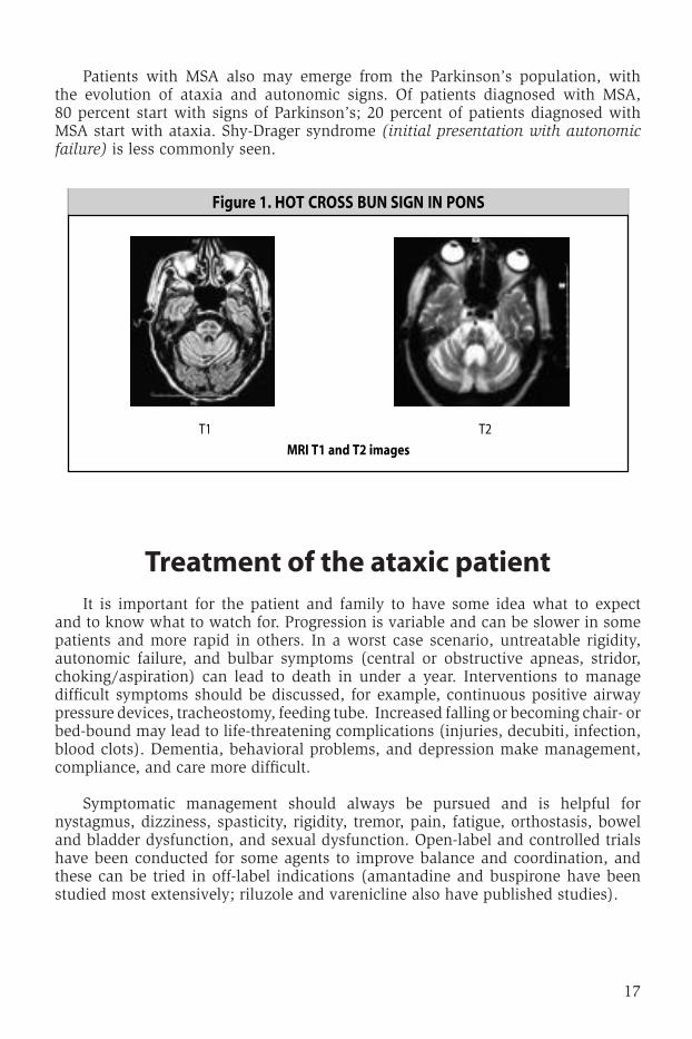
of patients with idiopathic late-onset cerebellar ataxia (iloca), 25 percent will go on to develop multiple system atrophy (Msa), with the emergence of symptoms of l-dopa-unresponsive parkinsonism and autonomic failure26-29. autonomic involvement will be confirmed by orthostatic blood pressure changes, lower motor neuron bowel and bladder dysfunction, and abnormalities in testing for heart rate variability, tilt table, sympathetic skin response/sweating, and cardiac i-123-MiBg-sPEct. rEM sleep disturbances or erectile dysfunction may precede ataxia by 5-10 years. obstructive sleep apnea and stridor are common. notable cerebellar disability is seen within 2-3 years. dopa-PEt scans will confirm basal ganglia involvement, but Mri scanning may show the earliest signs of impending Msa. Hot cross bun sign in pons and hyper/ hypo-intensities in putamen correlate strongly with Msa (see Figure 1). the presence of dementia, ophthalmoplegia, or chorea suggest something other than Msa.
Sporadic ataxias in all age ranges and populations, nongenetic ataxia is more common than inherited ataxia, often by a ratio of 2:1. (See Table 1 for identifiable nongenetic etiologies.) With thorough evaluation (see Table 3), a treatable cause might be found, but the majority of these syndromes remain idiopathic. their classification is outlined in Table 10.
Table 10. ClASSIFICAtIOn OF tHE SPORAdIC AtAxIAS
Type Classification
Sporadic ataxia with identifiable genetic cause (2-29% in various studies)
SCA 1-28, with missing family history (SCA6 most commonly found)
Any recessively inherited ataxia (FRDA, AOA 1 or 2, ataxia-telangiectasia most commonly found)
Any X-linked or mitochondrially inherited ataxia (FXTAS most commonly found)
Sporadic ataxia with known acquired cause (see Table1)
Idiopathic cerebellar ataxia, according to HardingaType A – with dementia; ddx-parenchymatous cerebellar cortical atrophy, prion diseases, Whipple’s disease, inborn errors of metabolism
Type B – with tremor; ddx-FXTAS
Type C – sporadic olivopontocerebellar atrophy; multiple system atrophy; other Parkinson-plus syndromes (PSP)
Idiopathic late-onset cerebellar atrophy (ILOCA)b
a harding, A.E. “idiopathic” late onset cerebellar ataxia. A clinical and genetic study of 36 cases. J Neurol Sci, 1981. 51(2): 259-71.b Klockgether, T. sporadic ataxia with adult onset: classification and diagnostic criteria. Lancet Neurol. 2010 Jan;9 (1):94-104.
Ataxia Disorders book.indd 24 2/24/16 2:04 PM
17
Treatment of the ataxic patient it is important for the patient and family to have some idea what to expect and to know what to watch for. Progression is variable and can be slower in some patients and more rapid in others. in a worst case scenario, untreatable rigidity, autonomic failure, and bulbar symptoms (central or obstructive apneas, stridor, choking/aspiration) can lead to death in under a year. interventions to manage difficult symptoms should be discussed, for example, continuous positive airway pressure devices, tracheostomy, feeding tube. increased falling or becoming chair- or bed-bound may lead to life-threatening complications (injuries, decubiti, infection, blood clots). dementia, behavioral problems, and depression make management, compliance, and care more difficult.
symptomatic management should always be pursued and is helpful for nystagmus, dizziness, spasticity, rigidity, tremor, pain, fatigue, orthostasis, bowel and bladder dysfunction, and sexual dysfunction. open-label and controlled trials have been conducted for some agents to improve balance and coordination, and these can be tried in off-label indications (amantadine and buspirone have been studied most extensively; riluzole and varenicline also have published studies).
Patients with Msa also may emerge from the Parkinson’s population, with the evolution of ataxia and autonomic signs. of patients diagnosed with Msa, 80 percent start with signs of Parkinson’s; 20 percent of patients diagnosed with Msa start with ataxia. shy-drager syndrome (initial presentation with autonomic failure) is less commonly seen.
Ataxia Disorders book.indd 25 2/24/16 2:04 PM

18
there are as yet no approved disease-modifying therapies for any of the genetic ataxias, although research has been aggressive and will provide such therapies in the upcoming years. acquired ataxias can be treated specific to the cause (infectious, inflammatory, immune-mediated, toxic, metabolic), but neuronal loss cannot be restored at this time. research in growth factors and stem cells will provide possible replacement strategies in the future.
rehabilitation resources are widely available and very helpful in most ataxic illnesses. these could include physical, occupational, and speech/swallowing therapy; aids to gait and activities of daily living; safety interventions; individual educational programs with schools; nutrition counseling; ophthalmology assessment; home health assistance; genetic and psychosocial counseling; legal aid; support groups; and special assistance and support for the caregiver.
sincere effort should be applied to answering the patient’s and family’s questions as honestly and completely as possible (What do I have? What is the cause? Are my children at risk? Can it be cured? Will it get worse? How bad will it get? How soon? Is there any research being done?) no one should be told that there is nothing that can be done.
Resources to aid in the evaluation of the ataxic patient• NCBI PubMed Website:www.ncbi.nlm.nih.gov/entrez/query
• Online Mendelian Inheritance in Man/OMIM Website:www.ncbi.nlm.nih.gov/omim
• GeneReviews Website:www.genetests.org
• Neuromuscular Disease Center NeuromuscularDivisionBox8111—Neurology 660SouthEuclidAvenue,SaintLouis,MO63110 Telephone:314-362-6981 Website:www.neuro.wustl.edu/neuromuscular
• National Ataxia Foundation 2600FernbrookLane,Suite119,Minneapolis,MN55447 Telephone:763-553-0020 Website:www.ataxia.org
• Friedreich’s Ataxia Research Alliance P.O.Box1537,Springfield,VA22151 Telephone:703-426-1576 Website:www.curefa.org
Ataxia Disorders book.indd 26 2/24/16 2:04 PM
19
References for treatment of the ataxic patient
Thefollowingreferencematerialsprovidehelpfulinformation.Despitevigorousresearch,therearestillnodisease-modifyingtherapiesorapprovedsymptomatictreatmentsforcerebellarataxia
•G.Grimaldi,G.PArgyropoulos,A.Boehringeretal.Non-invasivecerebellarstimulation--aconsensuspaper.Cerebellum.2014Feb;13(1):121-38.
•S.Manek,MD,andM.F.Lew,MD.“GaitandBalanceDysfunctioninAdults.”InMovement Disorders,2003,5:177-185.
•M.Nance,MD.Living With Ataxia: An Information and Resource Guide.2ndedition.NationalAtaxiaFoundation,2003.
•M.Ogawa.“PharmacologicalTreatmentsofCerebellarAtaxia.”InCerebellum,2004,3:107-11.
•S.L.Perlman,MD.“CerebellarAtaxia.”InCurrent Treatment Options in Neurology,2000,2:215-224.
•S.L.Perlman,MD.“SymptomaticandDisease-ModifyingTherapyfortheProgressiveAtaxias.”InThe Neurologist,2004,10:275-89.
•G.N.Rangamani,PhD,CCC-SLP.“Managing Speech and Swallowing Problems: A Guidebook for People With Ataxia.”2ndedition.NationalAtaxiaFoundation,2006.
•M.SynofzikandW.Ilg.“Motortrainingindegenerativespinocerebellardisease:ataxia-specificimprovementsbyintensivephysiotherapyandexergames.”InBiomed Res Int.2014;2014:583507.
•M.M.Trujillo-Martín,P.Serrano-Aguila,F.Monton-Alvarezetal.“Effectivenessandsafetyoftreatmentsfordegenerativeataxias:asystematicreview.”InMov Disord.2009Jun15;24(8):1111-24.
•B.P.VandeWarrenburg,J.vanGaalen,S.Boesch,etal.“EFNS/ENSConsensusonthediagnosisandmanagementofchronicataxiasinadulthood.”InEur J Neurol.2014Apr;21(4):552-62.
References1.Moseley,M.L.,etal.IncidenceofdominantspinocerebellarandFriedreichtripletrepeats
among361ataxiafamilies.Neurology,1998.51(6):p.1666-71.2.Schols,L.,etal.Geneticbackgroundofapparentlyidiopathicsporadiccerebellarataxia.Hum
Genet,2000.107(2):p.132-37.3.Abele,M.,etal.Theaetiologyofsporadicadult-onsetataxia.Brain,2002.125(Pt5):p.961-68.4.Hall,D.A.,etal.InitialdiagnosesgiventopersonswiththefragileX-associatedtremor/ataxia
syndrome(FXTAS).Neurology,2005.65(2):p.299-301.5.VanEsch,H.,etal.ScreeningforFMR-1premutationsin122olderFlemishmalespresenting
withataxia.Eur J Hum Genet,2005.13(1):p.121-23.6.Hagerman,P.J.,andR.J.Hagerman.FragileX-associatedtremor/ataxiasyndrome(FXTAS).Ment
Retard Dev Disabil Res Rev,2004.10(1):p.25-30.7.Hagerman,R.J.,etal.FragileX-associatedtremor/ataxiasyndrome(FXTAS)infemaleswiththe
FMR1premutation.Am J Hum Genet,2004.74(5):p.1051-56.
Ataxia Disorders book.indd 27 2/24/16 2:04 PM
20
8. Maschke,M.,etal.Clinicalfeatureprofileofspinocerebellarataxiatype1-8predictsgeneticallydefinedsubtypes.Mov Disord,2005.20(11):p.1405-12.
9. ShakkottaiVG,FogelBL.Clinicalneurogenetics:autosomaldominantspinocerebellarataxia.Neurol Clin.2013Nov;31(4):987-1007
10. FogelBL,etal.Utilizationofgenetictestingpriortosubspecialistreferralforcerebellarataxia.Genet Test Mol Biomarkers.2013Aug;17(8):588-94.
11. LeeH,etal.ClinicalexomesequencingforgeneticidentificationofrareMendeliandisorders.JAMA.2014Nov12;312(18):1880-7.
12. Fogel,BL,etal.Exomesequencingintheclinicaldiagnosisofsporadicorfamilialcerebellarataxia.JAMA Neurol.2014Oct;71(10):1237-46.
13. Fogel,BL.Interpretationofgenetictesting:variantsofunknownsignificance.Continuum (Minneap Minn).2011Apr;17(2Neurogenetics):347-52.
14. FogelBL,PerlmanS.Clinicalfeaturesandmoleculargeneticsofautosomalrecessivecerebellarataxias.Lancet Neurol.2007Mar;6(3):245-57.
15. Fogel,BL.Childhoodcerebellarataxia.J Child Neurol.2012Sep;27(9):1138-45.16. Collins,A.Clinicalneurogenetics:friedreichataxia.Neurol Clin.2013Nov;31(4):1095-120.17. Poretti,A.,etal.Differentialdiagnosisofcerebellaratrophyinchildhood.Eur J Paediatr
Neurol.2008May;12(3):155-67.Epub2007Sep14.18. AmbroseM,GattiRA.Pathogenesisofataxia-telangiectasia:thenextgenerationofATM
functions. Blood.2013May16;121(20):4036-45.19. ChaudharyMW,Al-BaradieRS.Ataxia-telangiectasia:futureprospects.Appl Clin Genet. 2014
Sep10;7:159-67.20. Poretti,A.etal.Acuteataxiainchildren:approachtoclinicalpresentationandroleof
additionalinvestigations.Neuropediatrics.2013Jun;44(3):127-41.21. ToscanoA,MusumeciO.MitochondrialDisordersinAdults.Curr Mol Med. 2014Oct10.[Epub
aheadofprint]22. ArdissoneA,,etal.MitochondrialDiseasesinChildhood.Curr Mol Med.2014Oct10.[Epub
aheadofprint]23. FogelBL,PerlmanS.Anapproachtothepatientwithlate-onsetcerebellarataxia.Nat Clin
Pract Neurol.2006Nov;2(11):629-3524. Fogel,BL,etal.Mutationsinrareataxiagenesareuncommoncausesofsporadiccerebellar
ataxia.Mov Disord.2012Mar;27(3):442-6.25. Sedel,F.etal.Therapyinsight:inbornerrorsofmetabolisminadultneurology--aclinical
approachfocusedontreatablediseases.Nat Clin Pract Neurol.2007May;3(5):279-90.26. Gilman,S.,etal.Evolutionofsporadicolivopontocerebellaratrophyintomultiplesystem
atrophy.Neurology,2000.55(4):p.527-32.27. Gilman,S.etal.Secondconsensusstatementonthediagnosisofmultiplesystematrophy.
Neurology.2008Aug26;71(9):670-6.28. Lin,DJ.,etal.Multiplesystematrophyofthecerebellartype:clinicalstateoftheart.Mov
Disord. 2014Mar;29(3):294-304.29. Fogel,BL,etal.Theneurogeneticsofatypicalparkinsoniandisorders.Semin Neurol.2014
Apr;34(2):217-24.
Ataxia Disorders book.indd 28 2/24/16 2:04 PM

Axatia Disorders book cover.indd 3 2/19/16 3:15 PM

$5.00
National Ataxia Foundation2600 Fernbrook Lane, Suite 119Minneapolis, MN 55447-4752Telephone: 763-553-0020Fax: 763-553-0167E-mail: [email protected]: www.ataxia.org
Evaluat ionand
Managementof
AtaxicDisorders
AN OVER V I EWFOR PHYS I C I ANS
Susan L. Perlman,MDfor the
National Ataxia Foundation
Axatia Disorders book cover.indd 4 2/19/16 3:15 PM


