EVALUATION OF AUTOMATIC CLASS III DESIGNATION FOR CLEARLLAB REAGENTS DECISION SUMMARY A. DEN Number: DEN160047 B. Purpose for Submission: De novo request for evaluation of automatic class III designation of the ClearLLab Reagents C. Measurand: Cluster of Differentiation (CD) Surface Markers on white blood cells D. Type of Test: Immunophenotyping, qualitative, flow cytometric assay E. Applicant: Beckman Coulter Inc. F. Proprietary and Established Names: Trade Name: ClearLLab T1, T2, B1, B2, M Common Name: ClearLLab G. Regulatory Information: 1. Regulation section: 21 CFR 864.7010 2. Classification: Class II (Special Controls) 3. Product code: PWD 4. Panel: 81-Hematology
Transcript
EVALUATION OF AUTOMATIC CLASS III DESIGNATION FOR CLEARLLAB REAGENTS
DECISION SUMMARY
A. DEN Number:
DEN160047 B. Purpose for Submission:
De novo request for evaluation of automatic class III designation of the ClearLLab Reagents C. Measurand:
Cluster of Differentiation (CD) Surface Markers on white blood cells D. Type of Test:
Immunophenotyping, qualitative, flow cytometric assay E. Applicant:
Beckman Coulter Inc.
F. Proprietary and Established Names:
Trade Name: ClearLLab T1, T2, B1, B2, M Common Name: ClearLLab
G. Regulatory Information:
1. Regulation section:
21 CFR 864.7010
2. Classification:
Class II (Special Controls)
3. Product code: PWD
4. Panel:
81-Hematology
CLSI EP28-A3c, Defining, Establishing, and Verifying Reference Intervals in the Clinical Laboratory CLSI H26-A2, Validation, Verification, and Quality Assurance of Automated Hematology Analyzers CLSI EP17-A2, Protocols for Determination of Limits of Detection and Limits of Quantitation CLSI EP25-A, Evaluation of Stability on In Vitro Diagnostic Reagents CLSI EP09-A3 Measurement Procedure Comparison and Bias Estimation Using Patient Samples
CLSI EP12-A2, User Protocol for Evaluation of Qualitative Test Performance
CLSI H43-A2, Clinical Flow Cytometric Analysis of Neoplastic Hematolymphoid Cells
K. Test Principle:
Beckman Coulter’s ClearLLab product is comprised of pre-cocktailed, lineage-driven combinations of the consensus Cluster of Differentiation (CD) reagents for evaluation of B and T cell neoplasia and the consensus limited CD reagents for myelomonocytic neoplasia as described in Table 3 of “2006 Bethesda International Consensus Recommendations on the Immunophenotypic Analysis of Hematolymphoid Neoplasia by Flow Cytometry: Optimal Reagents and Reporting for the Flow Cytometric Diagnosis of Hematopoietic Neoplasia”1. The ClearLLab product is intended for use on the FC 500 flow cytometer with instrument set-up performed with Flow-Check Pro Fluorospheres, Flow-Set Pro Fluorospheres, and QuickComp color compensation reagents for alignment, voltage standardization, and compensation. This test depends on the ability of a monoclonal or polyclonal antibody to bind to the surface of cells expressing discrete antigenic determinants. Specific cell staining is accomplished by incubating specimens prepared for staining with the appropriate antibody reagent. The ClearLLab Reagents are composed of five panels containing four or five monoclonal or polyclonal antibody reagents, each specific for a different cell surface antigen and conjugated to a specific fluorochrome. After sample preparation, the specimens are analyzed on the flow cytometer with manual gating. The FC 500 flow cytometer running under the control of CXP Software applies the principles of flow cytometry to analyze a whole blood, bone marrow or lymph node sample. Samples are prepared and stained with a monoclonal antibody reagents followed by lysis of red blood cells prior to introduction into the instrument. Cellular populations are identified based on the specific monoclonal and polyclonal antibodies and fluorochromes used in the different panels. Detection of fluorescent antibodies bound to cells utilizes the capability of the FC 500 flow cytometer to
1 2006 Bethesda International Consensus recommendations on the immunophenotypic analysis of hematolymphoid neoplasia by flow cytometry: optimal reagents and reporting for the flow cytometric diagnosis of hematopoietic neoplasia. Wood BL, Arroz M, Barnett D, DiGiuseppe J, Greig B, Kussick SJ, Oldaker T, Shenkin M, Stone E, Wallace P. Cytometry B Clin Cytom. 2007; 72 Suppl 1:S14-22
detect fluorescence with one laser, a blue laser with a 488 nm excitation and five different detection channels (FL1-FL5).
The CXP SYSTEM application software (K030828) provides automated instrument setup of light scatter and fluorescence intensity standardization, color compensation, and verification when used with the quality control reagents. The CXP Software has an Auto-Set Panel which automatically standardizes the cytometer, adjusts compensation settings, passes cytometer settings to designated test protocols, and verifies cytometer setup and antibody performance. Compensation settings are determined using CD45-FITC, CD45-PE, and CD45-ECD supplied in the QuickComp 4 Kit and CD45-PC5.5 and CD45-PC7 single color reagents. The existing FC 500 CXP software will support the ClearLLab reagents for use on the FC 500 flow cytometer without any further modifications from the original clearance in K030828.
Sample Preparation & Analysis Whole Blood (WB) and Bone Marrow (BM) preparation: Specimens are prepared manually with three pre-wash steps using phosphate buffered saline (PBS) containing 2% Heat Inactivated Fetal Calf Serum (HI-FCS). The remaining white blood cell pellet is then resuspended in PBS containing HI-FCS. The washed specimen is stained with the ClearLLab 5-Color Reagents. VersaLyse Lysing Solution for lysing red blood cells and the IOTest 3 Fixative Solution are used for red blood cell lysis and fixation prior to analysis.
Lymph Node preparation: Lymph node specimens require disaggregation of the tissue into a single cell suspension. Single cell suspensions prepared from lymphoid tissues may not require washing prior to staining if the specimen was washed during the disaggregation process. If washing steps were not performed for removal of residual soluble proteins, or if the cells were re-suspended in a buffer containing human serum or serum proteins, then pre-washing is necessary.
Sample analysis: White blood cells (WBCs) are analyzed on an FC 500 flow cytometer. Data are analyzed using a sequential gating strategy. The total WBC gate is defined in the first histogram (Side Scatter vs. CD45-PC7) by the inclusion of all CD45+ events with low, medium and high Side Scatter (SSC). A second histogram defines specific leukocyte subsets such as lymphocytes, monocytes, and granulocytes. The low SSC/bright CD45+ population identifies lymphocytes. Applicable markers are then displayed in subsequent histograms gated on lymphocytes. This process is repeated for mid SSC/medium CD45+ populations to identify monocytes and for high SSC/medium CD45+ to identify granulocytes.
L. Interpretation of Results
Flow cytometric immunophenotyping is multiparametric and relies on simultaneous identification and characterization of both normal and candidate malignant cell populations. Normal cells, which vary in type and relative numbers depending on the sample type, patient age, and clinical setting, display highly conserved and reproducible patterns of cell surface marker expression and light scatter. These normal internal control patterns may serve as both process controls for sample type, sample degradation, sample preparation and staining, data acquisition, and data analysis as well as references against which candidate malignant populations may be compared. Candidate malignant populations may display aberrant loss of a marker, aberrant gain of a marker, or aberrant over- or under-expression of a marker. Any population that does not conform to the normal internal control pattern is evaluated for
malignancy. Interpretation of these patterns requires expert knowledge and training in the technique. It is not possible to predict the phenotype of a particular patient’s hematolymphoid malignancy and apply fixed templates for samples for detection of specific phenotypes. Even well-characterized diseases such as chronic lymphocytic leukemia may display case-to-case immunophenotypic variability and it is therefore the role of the expert interpreter to assess the significance of findings and to interpret them together with clinical history, morphological assessment, and other ancillary techniques in order to arrive at a final diagnosis. The device labeling states that interpretation of specimens should be performed by a pathologist or equivalent professional who has the appropriate training. Finally, a small number of rare samples contain no or very few normal cells and require careful evaluation and correlation with other sources of information such as clinical history and other laboratory findings to obtain the appropriate final diagnosis.
M. Performance Characteristics:
All results provided below met the manufacturer’s pre-specified acceptance criteria. 1. Analytical Performance:
a. Precision and Reproducibility: Precision studies were conducted according to the
methodology presented in CLSI EP05-A3, Evaluation of Precision Performance of Quantitative Measurement Procedures; Approved Guideline – Third Edition.
i. Precision (repeatability): Thirty-eight specimens across the three specimen types were
evaluated. For Whole Blood (WB) and Bone Marrow (BM), testing was conducted at three sites, with each of the three sites using a different anticoagulant according to their current clinical testing procedures. For lymph node specimens (LN), testing was conducted at two sites. Since LN specimens are not collected using an anticoagulant, this was not a variable for lymph node specimen analysis.
A minimum of one specimen for each specimen type and lineage were collected at each site. Lineage is defined by the tubes used, where T lineage is comprised of T1 and T2 tubes, B lineage is comprised of B1 and B2 tubes, and M lineage is comprised of Myeloid tube. Specimens were selected to cover the markers in each lineage tube regardless of the disease. The same specimen was analyzed twice each day with one time point in the morning and one time point in the afternoon. Six sample preparations per time point were made for a total of 12 replicates per specimen per lineage tube.
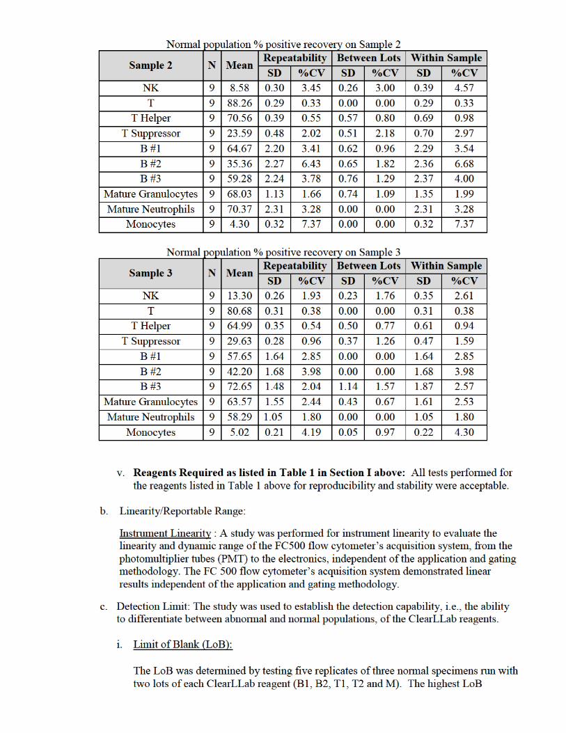
A qualitative approach was applied to repeatability assessment where the presence or absence of an abnormal phenotype was reported. An analysis of the % abnormal population, if present, or pre-selected lineage-specific normal populations was assessed for repeatability and reproducibility. The specimens were tested in 12 replicates. The expected result was to obtain the same number of positive or negatives for all replicates. The total actual number positive or negative was divided by the total expected number positive or negative to calculate the % positive or % negative results. The qualitative results for both abnormal and normal populations met 100% agreement for all sites and anticoagulants for expected and actual results with respect to the presence or absence of an abnormal phenotype.
observed from the two lots was 0.27% population of abnormal cells.
ii. Limit of Detection (LoD):
The LoD was determined using one representative clinical specimen collected in K2 or K3 EDTA for each ClearLLab reagent. Each clinical specimen was spiked into a normal specimen to target low levels of abnormal population as a percentage of the total percentage of leukocytes in the sample. The percentages used were 2%, 1% and 0.5% of abnormal cells in the total population of white cells measured.
The Limit of Detection defined as 1% population of abnormal cells for the ClearLLab application is therefore confirmed.
d. Analytical Specificity:
i. Interfering Substances:
Not applicable. Adequate washing with phosphate buffered saline (PBS) before staining with reagents removes residual plasma and interfering substances.
ii. Assay Carryover:
Carryover studies were conducted on three FC 500 flow cytometry systems according to the methodology presented in CLSI H26-A2: Validation, Verification, and Quality Assurance of Automated Hematology analyzers; Approved Standard-Second Edition, Section 5.7 with regard to high or low value sample order. Two studies were conducted to evaluate specimen and reagent carryover.
Specimen Carryover
Three samples with high WBC counts (HTv1, HTv2, HTv3) were prepared from one clinical whole blood specimen containing a high level of WBCs. The samples were prepared to target 20,000 cells /μL WBCs. CD34 marker analysis was performed using a CD34+ clinical whole blood specimen.
Three samples with low WBC counts (LTv1, LTv2, LTv3) were prepared by spiking a diluted (1:10,000) whole blood specimen with a red blood cell pool which has been depleted of white blood cells and diluted 4:1 with Immuno-Trol storage buffer. This resulted in a contrived specimen that had a red blood cell concentration similar to whole blood with very low concentrations of white blood cells.
The reagent selected for specimen carryover was the ClearLLab M reagent. M reagent was chosen as a representative reagent since it contains all the different types of fluorochrome conjugates used in the device (FITC, PE, ECD, PC5.5 and PC7) as well as markers that allow identification and assessment of recovery of all leukocyte populations (lymphocytes, monocytes and granulocytes). Testing with M reagent also assesses the impact of lysing and fixative reagents on integrity of cell membranes for all relevant cell subsets and the affinity of various conjugated antibodies that is representative of all ClearLLab reagents.
Three HTv samples (HTv1, HTv2, HTv3) were analyzed consecutively followed immediately by the three LTv samples (LTv1, LTv2, LTv3). This was repeated three
Whole Blood and Bone Marrow: A total of 204 specimens including 35 whole blood specimens with normal hematology CBC/Diff and 169 specimens with abnormal hematology CBC/Diff were tested. This multi-center anticoagulant study for whole blood and bone marrow was conducted across four sites covering hematopoietic malignancies as well as those with hematological abnormalities but no malignancy. Peripheral blood and bone marrow collected in K2EDTA were analyzed within 24 hours. Specimens collected in ACD and Sodium Heparin were analyzed within 48 hours. Specimens were also stained with reference/comparator reagents containing previously FDA cleared CD marker assays. Markers with a direct comparator were used to determine the performance between anticoagulants in bone marrow and whole blood. Equivalent performance was demonstrated between different anticoagulants for both whole blood and bone marrow specimens.
2. Clinical Performance:
A multi-center, retrospective study was conducted at four sites comparing the diagnostic accuracy of the ClearLLab reagents to detect the presence or absence of an abnormal phenotype to the clinical outcome of “malignant” or “non-malignant” based on the clinical sites’ final patient diagnosis. Patients with abnormal B-cell, T-cell, Natural Killer (NK) cell, and myeloid cell populations were tested. Residual samples were tested from a diverse population of patients covering hematopoietic malignancies as well as those with hematological abnormalities but no malignancy. This study included whole blood, bone marrow and lymph node specimen types. Specimen types tested reflected the distribution of the diseases and /or clinical indications encountered in the population of patients that are routinely encountered in evaluations of patients suspected of having hematopoietic neoplasia by flow cytometry. Consecutive specimens evaluated for flow cytometric immunophenotyping of leukemia and lymphoma and meeting the target disease categories were enrolled at these sites.
The study was designed to combine data from four sites to get an adequate representation of diseases. The specimen mix of the abnormal specimens consisted of peripheral blood (approximately 60%), bone marrow (approximately 30%) and lymph nodes (approximately 10%) for both hematopoietic malignant and nonmalignant categories. The samples consisted of a distribution of approximately 50% hematologically abnormal with no malignancy and approximately 50% malignant.
Clinical sites provided final diagnosis (“malignant” or “non-malignant”) for all subjects and all specimen types based on the clinical testing performed at the respective sites regardless of the specimen type sent to the flow cytometry laboratory for testing as per WHO guidelines.7 Two qualified flow experts evaluated the ClearLLab flow cytometry results independently of each other and were blinded to the final diagnosis. Each flow expert was asked to evaluate the ClearLLab results for the presence or absence of abnormal cell populations and provide the percentage of abnormal population as well as the phenotype associated with the abnormal population. The result was designated as “malignant” if an abnormal population was identified, and “non-malignant” if an abnormal population was not identified.
7 Swerdlow, S, et al. WHO Classification of Tumours of Haematopoietic and Lymphoid Tissues 4th Edition. . Lyon: International Agency for Research on Cancer, 2008.
The sample size was determined based on the approach recommended by Burderer.8 For this study the assumption was prevalence =1 (design was not prevalence driven). The target outcome for all specimen types that were identified as positive by flow cytometry was 80%±10% agreement with the clinical diagnosis and for all specimen types that were identified as negative by flow cytometry was 90%±10%. For whole blood specimens only, the target outcomes for both positive and negative was 90%±10%.
A total of 279 abnormal hematologic specimens were enrolled: 137 hematologically abnormal but no malignancy and 142 with hematolymphoid malignancy per site’s final diagnosis. The demographic information regarding the 279 subjects is shown in the graphic below.
8 Statistical methodology. I. Incorporating the prevalence of disease into the sample size calculation for sensitivity and specificity. NM, Burderer. s.l. : Acad. Emerg. Med, 1996, Vols. 3(9): 895-900.
patient specimens, 137 patients presenting for flow cytometry immunophenotyping which were found to be non-malignant and 142 patient specimens found to have hematologic malignancy. The patients with hematologic malignancy included 16% acute leukemia, 25% chronic leukemia, 44% lymphoma, 6% plasma cell neoplasm and 9% Others (includes MDS, MPN and eosinophilic hyperplasia). As shown in Comparison of ClearLLab Phenotypes vs. Diagnosis of Malignancy/Non-Malignancy, the clinical trial demonstrated the performance of this reagent panel to identify abnormal cell populations indicative of hematologic malignancy and to aid in demonstrating the absence of disease. The results of two independent flow cytometry experts demonstrated sensitivity of 82% (75–88%, 95% CI) for Flow Expert #1 and 86% (79–91%) for Flow Expert #2 and a specificity of 94% (89–97%) for Flow Expert #1 and 93% (87–96%) for Flow Expert #2. The PPV of ClearLLab reagents to identify malignancy was 94% (88–97%) and 92% (87–96%), respectively for each Flow Expert. Agreement between experts was 98% for positive specimens and 94% for negative specimens.
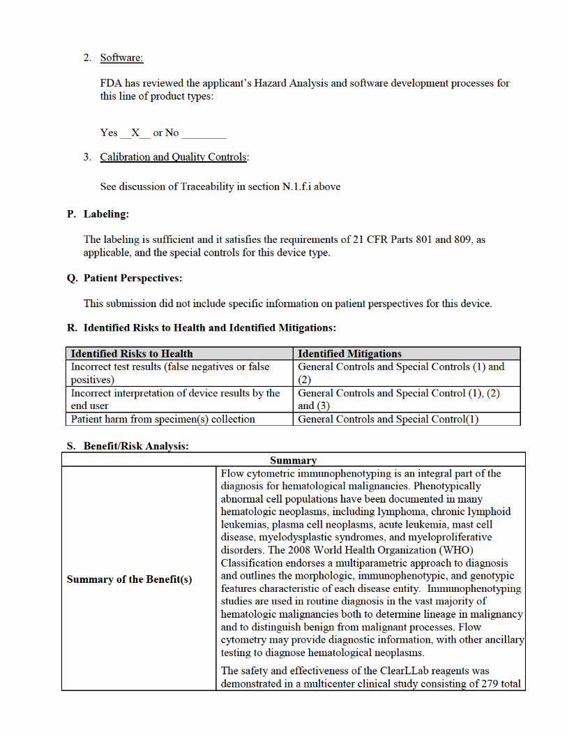
Summary of Risk(s)
There is minimal potential risk associated with use of this device given the combination of required general controls and special controls. The recognized risks for these assays are 1) possible erroneous results (false positive or false negative), 2) incorrect interpretation of device results by the pathologist or equivalent professional, and 3) related to patient harm from specimen(s) collection.
Summary of Other Factors
With respect to risk of potential harm to the patient while obtaining specimen(s), all flow cytometry studies are conducted ex-vivo and are independent of the assay configuration. The only intervention is blood drawing and/or obtaining tissues (bone marrow, lymph node) by well-established biopsy procedures.
Conclusions Do the probable benefits outweigh the probable risks?
Yes, the probable benefits of this device outweigh the probable risks, given the combination of required general controls and special controls established for this device.
T. Conclusion:
The information provided in this de novo submission is sufficient to classify this device into class II under regulation 21 CFR 864.7010. FDA believes that the special controls, along with the applicable general controls, including design controls, provide reasonable assurance of the safety and effectiveness of the device type. The device is classified under the following:
Product Code: PWD Device Type: Flow Cytometric Test System for Hematopoietic Neoplasms Class: II (special controls) Regulation: 21 CFR 864.7010
a. Identification. A flow cytometric test system for hematopeietic neoplasms is a device that consists of reagents for immunophenotyping of human cells in relation to the level of expression, antigen density, and distribution of specific cellular markers. These reagents are used as an aid in the differential diagnosis or monitoring of hematologically abnormal patients having or suspected of having hematopoietic neoplasms. The results should be interpreted by a pathologist or equivalent professional in conjunction with other clinical and laboratory findings.
b. Classification. Class II (special controls). A flow cytometric test system for hematopoietic
neoplasms must comply with the following special controls:
1. Premarket notification submissions must include the following information:
i. The indications for use must indicate the clinical hematopoetic neoplasms for which the assay was designed and validated, for example, chronic leukemia or lymphoma.
ii. A detailed device description including the following: (A) A detailed description of all test components, all required reagents, and all
instrumentation and equipment, including illustrations or photographs of nonstandard equipment or methods.
(B) Detailed documentation of the device software including, but not limited to, standalone software applications and hardware-based devices that incorporate software.
(C) A detailed description of methodology and assay procedure. (D) A description of appropriate internal and external quality control materials that are
recommended or provided. The description must identify those control elements that are incorporated into the testing procedure, if applicable.
(E) Detailed specifications for sample collection, processing, and storage. (F) Detailed specification of the criteria for test results interpretation and reporting
including pre-established templates. (G) If applicable, based on the output of the results, a description of the specific number of
events to collect, result outputs, and analytical sensitivity of the assay that will be reported.
iii. Information that demonstrates the performance characteristics of the test, including: (A) Device performance data from either a method comparison study comparing the
specific lymphocyte cell markers to a predicate device or data collected through a clinical study demonstrating clinical validity using well-characterized clinical specimens. Samples must be representative of the intended use population of the device including hematologic neoplasms and the specific sample types for which the test is indicated for use.
(B) If applicable, device performance data from a clinical study demonstrating clinical validity for parameters not established in a predicate device of this generic type using well-characterized prospectively obtained clinical specimens including all hematologic neoplasms and the specific sample types for which the device is indicated for use.
(C)Device precision data using clinical samples to evaluate the within-lot, between-lot, within-run, between run, site-to-site and total variation using a minimum of three sites, of which at least two sites must be external sites. Results shall be reported as the standard deviation and percentage coefficient of variation for each level tested.
(D) Reproducibility data generated using a minimum of three lots of reagents to evaluate mean fluorescence intensity and variability of the recovery of the different markers and/or cell populations.
(E) Data from specimen and reagent carryover testing performed using well-established methods (e.g., CLSI H26-A2).
(F) Specimen and prepared sample stability data established for each specimen matrix in the anticoagulant combinations and storage/use conditions that will be indicated.
(G) A study testing anticoagulant equivalency in all claimed specimen type/anticoagulant combinations using clinical specimens that are representative of the intended use population of the device.
(H) Analytic sensitivity data using a dilution panel created from clinical samples. (I) Analytical specificity data, including interference and cross-contamination. (J) Device stability data, including real-time stability of reagents under various storage
times and temperatures. (K) For devices that include polyclonal antibodies, Fluorescence Minus One (FMO) studies
to evaluate non-specific binding for all polyclonal antibodies. Each FMO tube is compared to reagent reference to demonstrate that no additional population appears when one marker is absent. Pre-specified acceptance criteria must be provided and followed.
(L) For devices indicated for use as a semi-quantitative test, linearity data using a dilution panel created from clinical samples.
(M) For devices indicated for use as a semi-quantitative test, clinically relevant analytical sensitivity data, including limit of blank, limit of detection, and limit of quantification.
iv. Identification of risk mitigation elements used by the device, including a detailed description of all additional procedures, methods, and practices incorporated into the instructions for use that mitigate risks associated with testing the device.
2. The 21 CFR 809.10 compliant labeling must include the following:
i. The intended use statement in the 21 CFR 809.10(a)(2) and 21 CFR 809.10(b)(2) compliant labeling must include a statement that the results should be interpreted by a pathologist or equivalent professional in conjunction with other clinical and laboratory findings. The intended use statement must also include information on what the device detects and measures whether the device is qualitative, semi-quantitative, and/or quantitative, the clinical indications for which the device is to be used, and the specific population(s) for which the device is intended.
ii. A detailed description of the performance studies conducted to comply with paragraph (1)(iii) and a summary of the results.
3. As part of the risk management activities performed under 21 CFR 820.30 design controls,
product labeling and instruction manuals should include clear examples of all expected phenotypic patterns and gating strategies using well-defined clinical samples representative of both abnormal and normal cellular populations. These samples must be selected based upon the indications described in paragraph (1)(i) of this section.