Abstract Lyonia ovalifolia has been used in a folk medicine for the treatment of wounds, cuts, burns, and scabies by different communities in Nepal. The present study is intended to evaluate the phytochemical constituents present in the leaf of plant and to subject methanol and hexane extract of plant leaf to phytochemical screening, GC-MS analysis, HPLC and FTIR analysis. Phytochemical screening of methanol extract revealed the presence of phenolic compounds, flavonoids, glycosides, tannins, xanthoprotein, quinones and saponins. Hexane extracts showed the presence of resins and quinones. The bioactive compounds of L. ovalifolia leaves have been evaluated using GC-MS, HPLC and FTIR. Fourteen chemical constituents have been identified through GC-MS analysis, among that the major constituents are Cyclopental [c] pyran-1(3H)one, hexahydro4,7-dimetyl (27.88%), Cyclopentene, 1-(2-propenyl) (16.52%), Diglycerol (14.99%) and others. The compounds identified through GC-MS analysis were probably helpful for the cure of many infections. The mass spectra of the compounds formed by the methanol extract are matched with NIST library. Keywords: Lyoniaovalifolia, bioactive compound, HPLC analysis, FTIR analysis
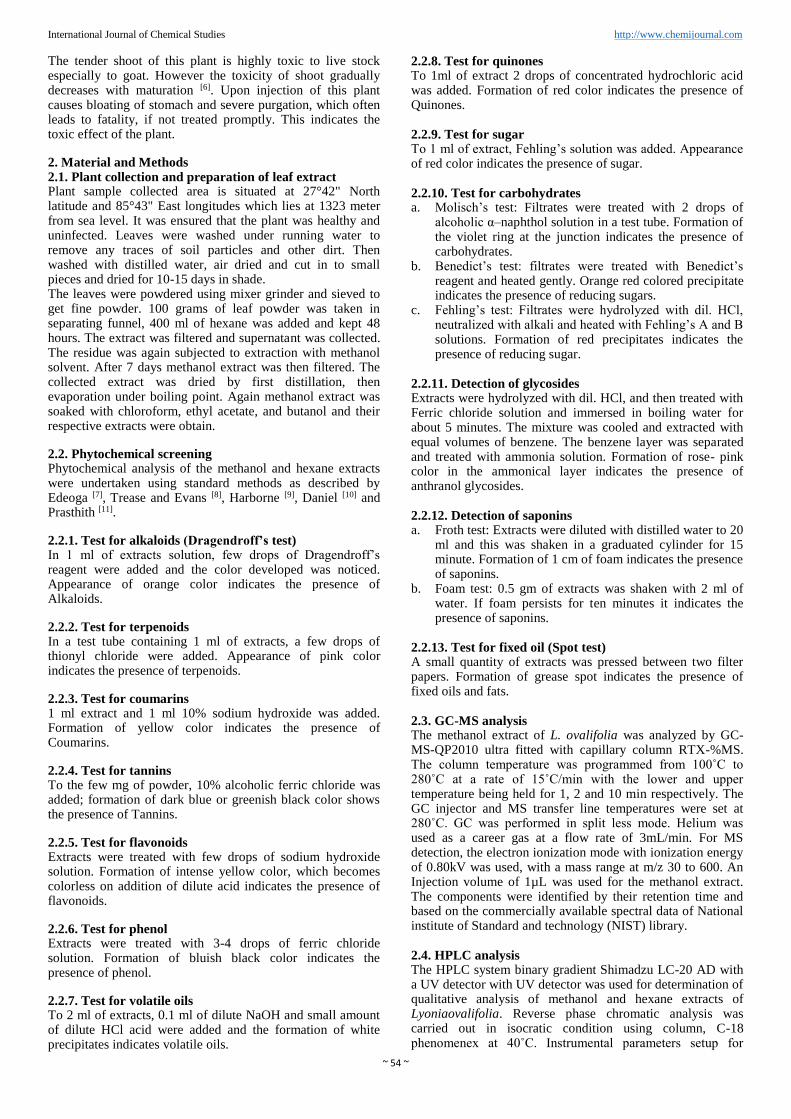
1. Introduction Lyonia ovalifolia is a deciduous or evergreen, shrubs or trees, ranging up to 4.5 m tall. Distributed throughout Bangladesh, Bhutan, Cambodia, India, Japan, Laos, Malaysia, Myanmar, Nepal, Pakistan, Sikkim, Thailand and Vietnam. The tree has brown bark, peeling in narrow strips. Ovate leaves are leathery, short-stalked, acute or long-pointed, having length of 8-15 cm. Small white flask-shaped flowers are born in almost horizontal clusters in leaf axils, constricted at the mouth and are finely hairy. L. ovalifolia is a plant of ethno medicinal relevance used to cure wounds, burns and scabies by the practitioners of traditional medicine in Nepal [1, 2]. Some previous studies have suggested its in-vitro antibacterial activities [3, 4]. The cyclic adenosine monophosphate (cAMP), formed from ATP, regulates several biological process. In human body, cAMP have impact on higher order of thinking, neurogenesis, memory, emotional disorder and cognitive function. The intracellular concentration of cAMP is regulated by two membrane-bound enzymes, adenylate cyclase and phosphor-diesterase. The palntmay induce the paralysis of nerve centers and motor nerve terminals, the cAMP regulation activity of the compounds isolated from this plant was evaluated by Alpha-Screen assay. Two compounds (secorhodomollolides A and D) found in L. ovalifolia significantly decrease the cAMP level at a concentration of 50mm in N1E-115 neuroblastoma cell indicating a neuropharmacological efficiency of this plant [5].
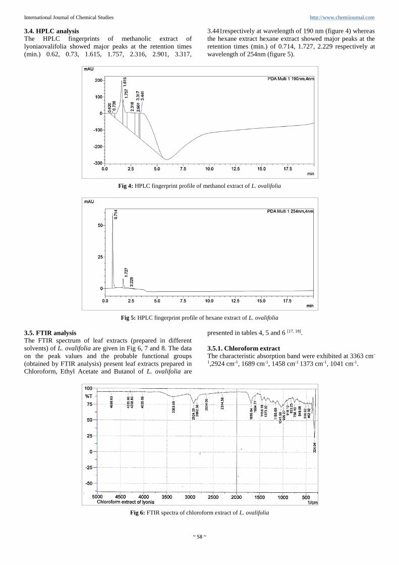
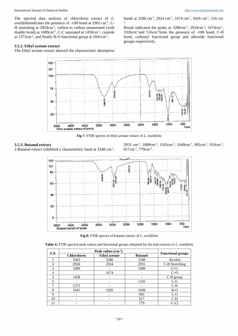
International Journal of Chemical Studies http://www.chemijournal.com
The tender shoot of this plant is highly toxic to live stock especially to goat. However the toxicity of shoot gradually decreases with maturation [6]. Upon injection of this plant causes bloating of stomach and severe purgation, which often leads to fatality, if not treated promptly. This indicates the toxic effect of the plant.
2. Material and Methods 2.1. Plant collection and preparation of leaf extract Plant sample collected area is situated at 27°42" North latitude and 85°43" East longitudes which lies at 1323 meter from sea level. It was ensured that the plant was healthy and uninfected. Leaves were washed under running water to remove any traces of soil particles and other dirt. Then washed with distilled water, air dried and cut in to small pieces and dried for 10-15 days in shade. The leaves were powdered using mixer grinder and sieved to get fine powder. 100 grams of leaf powder was taken in separating funnel, 400 ml of hexane was added and kept 48 hours. The extract was filtered and supernatant was collected. The residue was again subjected to extraction with methanol solvent. After 7 days methanol extract was then filtered. The collected extract was dried by first distillation, then evaporation under boiling point. Again methanol extract was soaked with chloroform, ethyl acetate, and butanol and their respective extracts were obtain.
2.2. Phytochemical screening Phytochemical analysis of the methanol and hexane extracts were undertaken using standard methods as described by Edeoga [7], Trease and Evans [8], Harborne [9], Daniel [10] and Prasthith [11].
2.2.1. Test for alkaloids (Dragendroff’s test) In 1 ml of extracts solution, few drops of Dragendroff’s reagent were added and the color developed was noticed. Appearance of orange color indicates the presence of Alkaloids.
2.2.2. Test for terpenoids In a test tube containing 1 ml of extracts, a few drops of thionyl chloride were added. Appearance of pink color indicates the presence of terpenoids.
2.2.3. Test for coumarins 1 ml extract and 1 ml 10% sodium hydroxide was added. Formation of yellow color indicates the presence of Coumarins.
2.2.4. Test for tannins To the few mg of powder, 10% alcoholic ferric chloride was added; formation of dark blue or greenish black color shows the presence of Tannins.
2.2.5. Test for flavonoids Extracts were treated with few drops of sodium hydroxide solution. Formation of intense yellow color, which becomes colorless on addition of dilute acid indicates the presence of flavonoids.
2.2.6. Test for phenol Extracts were treated with 3-4 drops of ferric chloride solution. Formation of bluish black color indicates the presence of phenol.
2.2.7. Test for volatile oils To 2 ml of extracts, 0.1 ml of dilute NaOH and small amount of dilute HCl acid were added and the formation of white precipitates indicates volatile oils.
2.2.8. Test for quinones To 1ml of extract 2 drops of concentrated hydrochloric acid was added. Formation of red color indicates the presence of Quinones. 2.2.9. Test for sugar To 1 ml of extract, Fehling’s solution was added. Appearance of red color indicates the presence of sugar. 2.2.10. Test for carbohydrates a. Molisch’s test: Filtrates were treated with 2 drops of
alcoholic α–naphthol solution in a test tube. Formation of the violet ring at the junction indicates the presence of carbohydrates.
b. Benedict’s test: filtrates were treated with Benedict’s reagent and heated gently. Orange red colored precipitate indicates the presence of reducing sugars.
c. Fehling’s test: Filtrates were hydrolyzed with dil. HCl, neutralized with alkali and heated with Fehling’s A and B solutions. Formation of red precipitates indicates the presence of reducing sugar.
2.2.11. Detection of glycosides Extracts were hydrolyzed with dil. HCl, and then treated with Ferric chloride solution and immersed in boiling water for about 5 minutes. The mixture was cooled and extracted with equal volumes of benzene. The benzene layer was separated and treated with ammonia solution. Formation of rose- pink color in the ammonical layer indicates the presence of anthranol glycosides. 2.2.12. Detection of saponins a. Froth test: Extracts were diluted with distilled water to 20
ml and this was shaken in a graduated cylinder for 15 minute. Formation of 1 cm of foam indicates the presence of saponins.
b. Foam test: 0.5 gm of extracts was shaken with 2 ml of water. If foam persists for ten minutes it indicates the presence of saponins.
2.2.13. Test for fixed oil (Spot test) A small quantity of extracts was pressed between two filter papers. Formation of grease spot indicates the presence of fixed oils and fats. 2.3. GC-MS analysis The methanol extract of L. ovalifolia was analyzed by GC-MS-QP2010 ultra fitted with capillary column RTX-%MS. The column temperature was programmed from 100˚C to 280˚C at a rate of 15˚C/min with the lower and upper temperature being held for 1, 2 and 10 min respectively. The GC injector and MS transfer line temperatures were set at 280˚C. GC was performed in split less mode. Helium was used as a career gas at a flow rate of 3mL/min. For MS detection, the electron ionization mode with ionization energy of 0.80kV was used, with a mass range at m/z 30 to 600. An Injection volume of 1µL was used for the methanol extract. The components were identified by their retention time and based on the commercially available spectral data of National institute of Standard and technology (NIST) library. 2.4. HPLC analysis The HPLC system binary gradient Shimadzu LC-20 AD with a UV detector with UV detector was used for determination of qualitative analysis of methanol and hexane extracts of Lyoniaovalifolia. Reverse phase chromatic analysis was carried out in isocratic condition using column, C-18 phenomenex at 40˚C. Instrumental parameters setup for
International Journal of Chemical Studies http://www.chemijournal.com
analysis was: injection volume 2µL; Solvent system were methanol (HPLC grade) and water (40:60); flow rate; 1 mL/min. The chromatogram was monitored at 180 nm to 800nm for both methanol and hexane extracts. Samples were filtered through an HPLC filter membrane using nylon 6, 6 filter paper. 2.5. Fourier transform infrared spectrophotometer (FTIR) analysis The chloroform, butanol and ethyl acetate fraction of methanol extract of L. ovalifolia were loaded in FTIR spectrometer (SHIMADZU IR Prestige- 21) with a scan range
from 5000-400 cm-1. Thus obtained FTIR spectra were analyzed. 3. Results and Discussions 3.1. Extraction: Hexane and methanol extract of young leaf of L. ovalifolia were prepared using cold percolation method. During the process 1.09g hexane extract and 19.56g of methanol extract were obtained. 3.2. Phytochemical screening analysis The result of phytochemical screening of leaf extract of L. ovalifoliais presented in Table 1.
Table 1: Phytochemical screening of leaf extract of Lyonia ovalifolia
S.N. Phytochemical constituents Hexane Methanol
1 Phenols + +
2 Flavonoids + +
3 Alkaloids - -
4 Carbohydrates - -
5 Glucosides - +
6 Tannins - +
7 Xanthoprotein - +
8 Resin + +
9 Quinone + +
10 Emodin - +
11 Saponin - +
(+)= Present, (-) = Absent
The methanol extract detected the presence of phenols, flavonoids, tannins, glycosides, xanthoprotein, resin, quinone, emodin and saponins. The hexane extract showed the presence of phenols, flavonoids, resin and quinone. From the results, it was confirmed that the plant Lyonia ovalifolia has a large number of chemical constituents which may be responsible for many pharmacological actions and medicinal properties. Flavonoids found in plant are therapeutically applicable for some biological actions and pharmacological effects like anti-oxidant, anti-inflammatory, anti-cancer, anti-diabetic, anti-viral, anti-allergic and diuretic activities [12]. Saponins detected during analysis are reported to have numerous properties which includes pharmacological activities such as anti-microbial, anti-inflammatory as well as sweetness and bitterness, foaming and emulsifying properties. These properties suggest that saponins act as a chemical barrier against potential pathogens in plants [13, 14].
Tannins are found to possess spasmolytic activity, free radical scavenger properties and anti-oxidant activities. Plants rich in phenolic contents have been reported to have effective beneficial effects such as, anti-microbial, anti-inflammatory, anti-viral, anti-mutagenic, anti-tumor [15], anti-oxidant activity [16], and chemo protective effects.
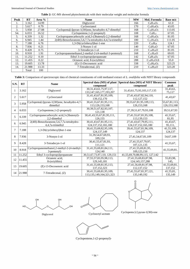
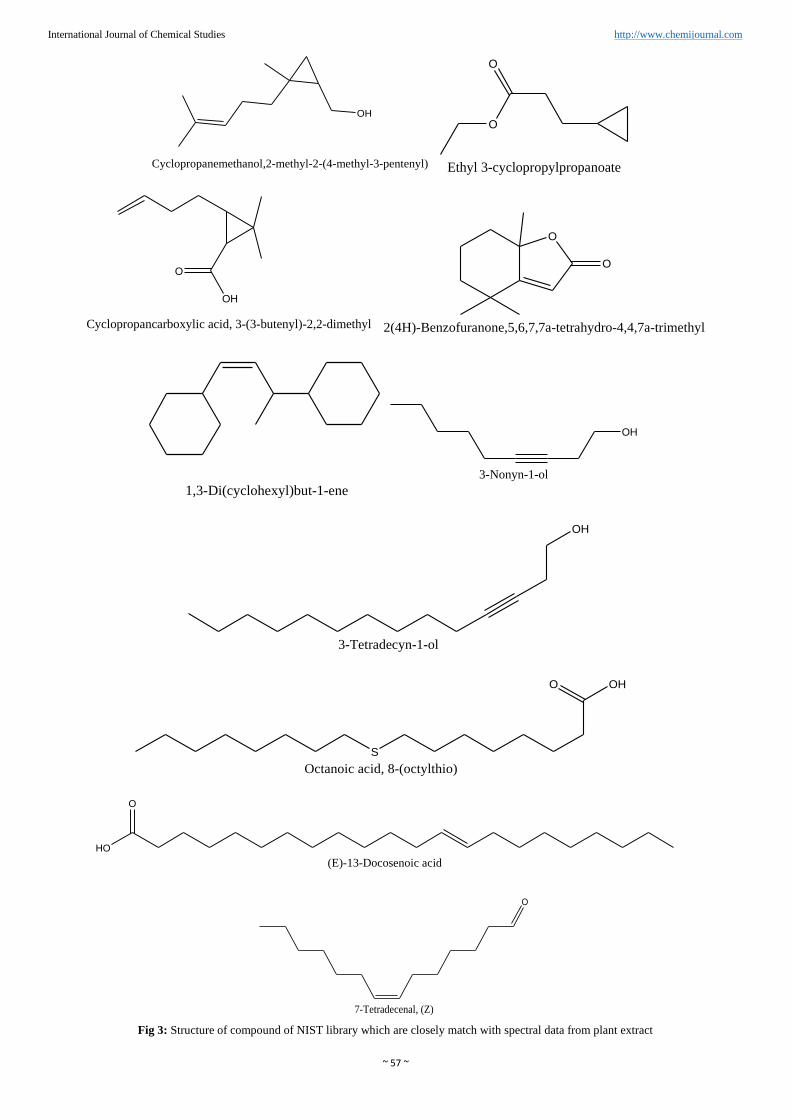
3.3. GC-MS analysis GC-MS chromatogram of the methanol extract of L. ovalifolia (Figure 2) showed 14 peaks which indicated the presence of fourteen major phytochemical constituents. The identification of the phytochemical compound was confirmed based on the peak area, retention time and molecular formula. The active principles with their Retention time (RT), Molecular formula, Molecular weight (MW) and peak area in percentage are presented in table 2. The spectrum of the unknown component was compared with the spectrum of the component stored in the NIST library.
Fig 2: GC-MS chromatogram of methanol extract of L. ovalifolia GC-MS analysis of methanol extract of L. ovalifolia identified the presence of carboxylic acid compounds, alcohol
compound, diterpenes, Fatty acid ester compound and ester compound.