RESEARCH Evaluation of Expression of the PTEN Gene, Oestrogen and Progesterone Receptors as Diagnostic and Predictive Factors in Endometrial Cancer Dariusz Samulak & Patrycja Grosman-Dziewiszek & Magdalena M. Michalska & Ewa Mojs & Katarzyna Samulak & Hanna Romanowicz & Beata Smolarz Received: 4 October 2012 /Accepted: 30 July 2013 /Published online: 13 September 2013 # The Author(s) 2013. This article is published with open access at Springerlink.com Abstract Endometrial cancer belongs to the commonest ma- lignancy in females after breast cancer, malignant neoplasm of female genitals in Europe and North America but there is still not significant improvement as far as the curability of this neoplasm is concerned, especially its advanced forms. That is why there is need to define new factors that could be not only diagnostic but also predictve factors. In present study we analyzed the mRNA PTEN expression by quantitative real- time polymerase chain reaction (Q-PCR) in 123 women of endometrial carcinoma and 14 women of control group. Moreover we assessed oestrogen (ER) and progesterone re- ceptors (PgR) in all cases. We defined the correlation between expression of PTEN gene and receptors and between PTEN expression and maturity grade of cancer. Neoplasm advance- ment grade G1 was diagnosed in 82.11 % of patients (n =101), G2 in 9.76 % of patients (n =12) and G3 in 8.13 % of patients (n =10). Presence of ER and PgR and decreased expression of PTEN gene was found in majority of patients with endome- trial cancer (79.12 % and 59.34 % respectively) and the most numerous group was with weak expression of ER and strong expression of PgR. There was no statistically significant dif- ference in gene expression depending on receptors expression nor maturity grade of cancer (p >0.05). Evaluation of expres- sion of PTEN gene may turn out to be a very useful tool aimed at qualifying patients for different therapies of endometrial cancer and at searching of new diagnostic and therapeutic methods of this cancer independently on its receptor status nor maturity grade of cancer. Keywords Endometrial cancer . Oestrogen and progesterone receptors . PTEN gene . Predictive factor Introduction Endometrial cancer (EC) is the most frequent, after breast cancer, malignant neoplasm of female genitals in Europe and North America and the fourth among all malignant neoplasms in women, after breast cancer, lung cancer and large intestine cancer [1–3]. EC cancerogenesis is not fully recognized pro- cess regarding many risk factors. Prognostic factors common- ly used to identification of endometrial cancer present an incomplete picture of the tumour biology of endometrial cancer [4]. Therefore, investigation of other prognostic factors D. Samulak Cathedral of Mother’ s and Child’ s Health, Poznan University of Medical Sciences, ul. Polna 33, 60-535 Poznań, Poland D. Samulak : M. M. Michalska Department of Obstetrics and Gynaecology, Regional Hospital in Kalisz, ul. Toruńska 7, 62-800 Kalisz, Poland D. Samulak State Higher Vocational School in Kalisz, ul. Nowy Świat 4, 62-800 Kalisz, Poland P. Grosman-Dziewiszek Faculty and Institute of Histology and Embryology, Academy of Medical Sciences in Wrocław, ul. Chałubińskiego 6a, 50-368 Wrocław, Poland E. Mojs Department of Clinical Psychology, Poznan University of Medical Sciences, Poznań, Poland K. Samulak Student Scientific Society, University of Medical Sciences in Poznan, ul. Polna 33, 60-535 Poznań, Poland H. Romanowicz : B. Smolarz (*) Department of Pathology, Laboratory of Molecular Genetics, Institute of Polish Mothers Memorial Hospital, Rzgowska 281/289, 93-338 Lodz, Poland e-mail: [email protected]Pathol. Oncol. Res. (2014) 20:191–196 DOI 10.1007/s12253-013-9684-3
Transcript
RESEARCH
Evaluation of Expression of the PTEN Gene, Oestrogenand Progesterone Receptors as Diagnostic and PredictiveFactors in Endometrial Cancer
Dariusz Samulak & Patrycja Grosman-Dziewiszek & Magdalena M. Michalska &
Ewa Mojs & Katarzyna Samulak & Hanna Romanowicz & Beata Smolarz
Received: 4 October 2012 /Accepted: 30 July 2013 /Published online: 13 September 2013# The Author(s) 2013. This article is published with open access at Springerlink.com
Abstract Endometrial cancer belongs to the commonest ma-lignancy in females after breast cancer, malignant neoplasm offemale genitals in Europe and North America but there is stillnot significant improvement as far as the curability of thisneoplasm is concerned, especially its advanced forms. That iswhy there is need to define new factors that could be not onlydiagnostic but also predictve factors. In present study weanalyzed the mRNA PTEN expression by quantitative real-time polymerase chain reaction (Q-PCR) in 123 women of
endometrial carcinoma and 14 women of control group.Moreover we assessed oestrogen (ER) and progesterone re-ceptors (PgR) in all cases. We defined the correlation betweenexpression of PTEN gene and receptors and between PTENexpression and maturity grade of cancer. Neoplasm advance-ment grade G1was diagnosed in 82.11% of patients (n =101),G2 in 9.76 % of patients (n =12) and G3 in 8.13 % of patients(n =10). Presence of ER and PgR and decreased expression ofPTEN gene was found in majority of patients with endome-trial cancer (79.12 % and 59.34 % respectively) and the mostnumerous group was with weak expression of ER and strongexpression of PgR. There was no statistically significant dif-ference in gene expression depending on receptors expressionnor maturity grade of cancer (p >0.05). Evaluation of expres-sion ofPTEN genemay turn out to be a very useful tool aimedat qualifying patients for different therapies of endometrialcancer and at searching of new diagnostic and therapeuticmethods of this cancer independently on its receptor statusnor maturity grade of cancer.
Keywords Endometrial cancer . Oestrogen and progesteronereceptors . PTEN gene . Predictive factor
Introduction
Endometrial cancer (EC) is the most frequent, after breastcancer, malignant neoplasm of female genitals in Europe andNorth America and the fourth among all malignant neoplasmsin women, after breast cancer, lung cancer and large intestinecancer [1–3]. EC cancerogenesis is not fully recognized pro-cess regarding many risk factors. Prognostic factors common-ly used to identification of endometrial cancer present anincomplete picture of the tumour biology of endometrialcancer [4]. Therefore, investigation of other prognostic factors
D. SamulakCathedral of Mother’s and Child’s Health, Poznan Universityof Medical Sciences, ul. Polna 33, 60-535 Poznań, Poland
D. Samulak :M. M. MichalskaDepartment of Obstetrics and Gynaecology, Regional Hospitalin Kalisz, ul. Toruńska 7, 62-800 Kalisz, Poland
D. SamulakState Higher Vocational School in Kalisz, ul. Nowy Świat 4,62-800 Kalisz, Poland
P. Grosman-DziewiszekFaculty and Institute of Histology and Embryology,Academy of Medical Sciences in Wrocław, ul. Chałubińskiego 6a,50-368 Wrocław, Poland
E. MojsDepartment of Clinical Psychology, Poznan University of MedicalSciences, Poznań, Poland
K. SamulakStudent Scientific Society, University ofMedical Sciences in Poznan,ul. Polna 33, 60-535 Poznań, Poland
H. Romanowicz :B. Smolarz (*)Department of Pathology, Laboratory of Molecular Genetics,Institute of Polish Mothers Memorial Hospital, Rzgowska 281/289,93-338 Lodz, Polande-mail: [email protected]
is of special clinical relevance, particularly in view of theunexpectedly progressive course of the disease and frequentrelapses in some cases. PTEN is the most frequently mutatedgene identified yet in endometrial cancers [5, 6].
PTEN (Phosphatase and Tensin Homologue) is a supressorgene located in chromosome 10q23. It codes lipid and proteinphosphatase (PTEN) and contributes to the control of theproliferation, differentiation and apoptosis process [7]. ThePTEN tumor suppressor gene regulates the oncogenic phos-phatidylinositol 3-kinase (PI3K ) signaling pathway that isinvolved in carcinogenesis. Downstream of those two path-ways is AKT, a serine-threonine kinase that is regulated byPI3K and influences apoptosis and cell proliferation [7].Alterations in the PTEN-PI3K-AKT pathways have been re-ported for hormone-related tumors among women, includingbreast, ovarian [8, 9] and endometrial [5, 6] cancers. Until nowit has been found that PTEN mutations are the most frequentgenetic changes in endometrial cancer of type I and occur in25–83 % of tumours, including tumours with microsatelliteinstability [10]. PTEN inactivation was described most fre-quently in early stages of endometrial cancer whilst in othertypes of neoplasms it occurs in their more advanced stages andis connected with cancer cells metastases [11]. Due to this fact,PTEN may be a good endometrial cancer marker, already inthe early stage of its development [12, 13].
PTEN expression correlates negatively with neoplasm ad-vancement grade. PTEN mutations, occurring in an earlydevelopment stage of the tumour, result in lack of cell differ-entiation, which might be connected with a more aggressiveneoplasm type [14, 15].
In light of substantial evidence that the progression of en-dometrial cancer can be associated with PTEN , it seems reasonable to check a possible correlation between the expression ofthis gene and clinical status of endometrial cancer patients.
Material and Methods
Patients
Endometrium was obtained from 137 women who underwenthysterectomy with adnexectomy in the Gynaecological andObstetric Clinical Hospital of the University of Medical Sci-ences in Poznan between 2004 and 2006. Tumour tissues wereobtained from women with endometrial adenocarcinoma.Clinical data for the patients and histological data were regis-tered. There were 123 women and their mean age was 60,6±10.0 years (range: 34–85 years). Endometrium from age-matched, cancer-free women (n =14) served as control (themean age 60,1±7.6). Endometrial carcinomas were classifiedaccording to the criteria of the International Federation ofGynecology and Obstetrics (FIGO). Histological typing andgrading were done according to the WHO classification.
Before the surgery each patient underwent a subject exam-ination, a gynaecological examination, an ultrasonographicexamination by means of an intravaginal probe and preoper-ative laboratory tests. Moreover, a conscious written consentto operation and uptake of segments of the endometrium forthe sake of scientific research was signed by every patient.
Tissue material obtained after the operation from eachpatient was divided into two parts. From one part wax blockswere created, and after that specimens for histopathologicalassessment dyed with hematoxilin and eosin (H + E). Theother part of the material was frozen in −80 °C. The proce-dures, used in the study, were approved by the Ethical Com-mittee of the Medical University of Poznań (Poland).
Evaluation of ER and PR
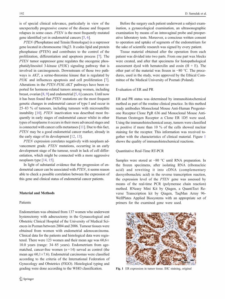
ER and PR status was determined by immunohistochemicalmethod as part of the routine clinical practice. In this methodready antibodies Monoclonal Mouse Anti-Human Progester-one Receptor Clone PgR 636 and Monoclonal Mouse Anti-Human Oestrogen Receptor α Clone ER 1D5 were used.Using the immunohistochemical assay, tumors were classifiedas positive if more than 10 % of the cells showed nuclearstaining for the receptor. This information was received to-gether with the characteristics of clinical material. Figure 1shows the quality of immunohistochemical reactions.
Quantitative Real-Time RT-PCR
Samples were stored at −80 °C until RNA preparation. Inthe frozen specimens, after isolating RNA (ribonucleicacid) and rewriting it into cDNA (complementarydeoxyribonucleic acid) in the reverse transcription reaction,the expression level of the PTEN gene was assessed bymeans of the real-time PCR (polymerase chain reaction)method. RNeasy Mini Kit by Qiagen, a QuantiTect Re-verse Transcription kit by Qiagen, TaqMan Array 96-WellPlates Applied Biosystems with an appropriate set ofprimers for the examined gene were used.
Fig. 1 ER expression in tumor tissue. IHC staining, original
192 D. Samulak et al.
18S rRNA (ribosomal ribonucleic acid) constituted theinternal control of the reaction. The reaction was repeatedthree times for each specimen. On the basis of the Ct value(threshold cycle—the number of reaction cycles after whichfluorescence exceeds the defined threshold) of the examinedgene and of the internal control gene the relative expressionlevel of RNA was calculated according to the ΔΔCt (delta,delta Ct) approximation method.
Statistical Analysis
The occurrence of statistically significant differences betweenexpression of PTEN gene depending on receptors expressionand on neoplasm maturity grade was checked by means ofKruskal—Wallis test.
The tests were carried out after obtaining a consent fromthe Bioethical Commission of the Academy of Medical Sci-ences of. Karol Marcinkowski in Poznań (Resolution number240/04 from 05.02.2004).
The tests were carried out in the Patomorphological Labo-ratory of the Gynaecological and Obstetric Clinical Hospitalof the University of Medical Sciences in Poznań and in theFaculty and Institute of Histology and Embryology of theAcademy of Medical Sciences in Wrocław.
The statistical analysis was carried out in the Faculty andInstitute of IT (informatics) and Statistics of the University ofMedical Sciences in Poznań. For the sake of the analysisSTATISTICA v. 8 was used, for multiple comparisonsKruskal—Wallis test was applied, U Mann–Whitney testwas used to carry out the descriptive statistics for individualgroups.
Results
In the final results of the histopathological examination neo-plasm advancement grade G1 was diagnosed in 82.11 % ofpatients with endometrial cancer (n =101), G2 in 9.76 % ofpatients (n =12) and G3 in 8.13 % of patients (n =10).
In 29 patients with endometrial cancer, it was impossible tomark receptors in the examined material. Out of the remaining94, in 7 patients the lack of ER was detected and in 2 patientsthe lack of PgR. Expression of PTEN gene was possible toevaluate in 118 patients with endometrial cancer. Whereforethe number of patients in whom receptors and gene weremarked together was 91. Taking these results into accound itwas possible to divide patients with endometrial cancer intothe following subgroups according to receptor expressionintensification level. There were 17 patients in subgroupER++/PgR + (strong expression of ER and strong expressionof PgR), 6 patients in subgroup ER++/PgR0 (strong expres-sion of ER and weak or no expression of PgR), 44 patients inER+/PgR + (weak expression of ER and strong expression of
PgR), 17 patients in ER+/PgR0 (weak expression of ER andweak or no expression of PgR), 5 in ER0/PgR + (no expres-sion of ER and strong expression of PgR) and 2 patients insubgroup ER0/PgR0 (no expression of ER and weak or noexpression of PgR). Dependecies of the presence of PTENexpression on the receptor status of patients with endometrialcancer are displayed in Table 1. In each receptor subgroup itwas determined whether PTEN gene expression in the exam-ined material was higher or lower than gene expression in thecontrol group. Figure 2 shows summary of expression ofPTEN gene depending on expression of ER and PgR.
By means of the Kruskal—Wallis test for multiple com-parisons the occurrence of statistically significant differencesbetween expression of PTEN gene in particular receptor sub-group was checked. No statistically significant difference inthe field of the examined parameters was found.
Statistically significant differences between groups G1, G2and G3 were examined in the event ofΔΔCt for gene PTEN .The assumed significance level was p <0.05 (Table 2).
No statistically significant difference in gene expressiondepending on neoplasm maturity grade was found.
Discussion
In this paper, apart from expression of PTEN and ER and PgRreceptors in endometrial cancer, the following issues wereexamined: the occurrence of dependencies between intensifi-cation ofPTEN expression and expression of receptors as wellas the occurrence of statistically significant differences in geneexpression depending on malignancy grade G of endometrialcancer.
Loss of PTEN expression is connected, through trail PI3K/AKT, with loss of control of cell proliferation and apoptosisand with the promotion of neoplasm development [16] .Reduction of PTEN expression ensues most frequentlythrough point mutations, loss of heterozygosity and promoterhypermethylation [17]. Somatic mutations of PTEN are com-mon in endometrial cancer but occur mainly in type I cancer(in ca. 83 %) [10, 18–20]. Germinal mutations of PTEN occurin Crowden syndrome in 80 % of instances [21]. Loss ofheterozygosity occur in 40 % of instances of endometrialcancer [22–25] and hypermethylation in circa 20 %, mainlyin highly advanced cancers [26]. Matias-guiu et al. [23] reportthe occurrence of mutation and reduction of PTEN expressionalready during the hyperplasia stage. Similar data regardingPTEN inactivation, during the hyperplasia stage with or with-out atypy, occur in reports of Prat et al. [24] as well as Dollet al. [12]. Mutter et al. [11] found decreased expression in75% of instances of the pre-cancer stage and in 95% of type Icancer and in 25 % of type II cancer, which let them draw thefollowing conclusion: disorders in PTEN expression are anearly incidents in the development process of endometrioid
PTEN gene expression and oestrogen and progesterone receptors status in endometrial cancer 193
endometrial cancer. Salvesen et al. [27] found the occurrenceof promoter hypermethylation and decreased PTEN expres-sion linked to this fact in 26 out of 138 patients (19 %) withendometrial cancer. Tests showed statistical significance re-garding the occurrence of hypermethylation in advanced can-cer stages. Latta et al. [28] found mutation of gene PTEN bothin the normal endometrium exposed to oestrogens, pre-cancerstages as well as endometrial cancer. However, the percentageof patients with decreased expression of PTEN increased withthe advancement of changes within the endometrium andamounted to 18–55 % for pre-cancer stages and 26–80 %for cancer. Salvesen et al. [29] in their further research report-ed the presence of mutations in 54 % of incidences of cancerand statistically significant dependency between the occur-rence of mutations and young age of patients, low advance-ment according to FIGO, endometrioid cancer type, highmaturity grade G, the occurrence of microsatellite instabilityas well as poor prognosis. Also Mackay et al. [30] noticed intheir research on 123 patients beneficial prognosis in instancesof decreased PTEN expression in women with an advancedendometrial cancer. However, Erkanli et al. [31], finding moresignificant reduction of expression in cancers than in
hyperplasia or normal endometrium, report the occurrence ofstatistically significant correlation between the reduction ofexpression and shorter survival time. Comparisons of expres-sion degree of PTEN depending on neoplasm maturity weremade by Inab et al. [32] as well as Kagan et al. [33]. Theypresented data which indicates a statistically significant de-pendency between maturity grade G and gene expression.Both testes showed more decreased expression in grade G1and G2 than in G3. Moreover, Kagan found lower PTENexpression in specimens with positive expression of oestrogenand progesterone receptors. This difference was statisticallysignificant. Sobczuk et al. [34] examining expression of thisgene in 70 patients with cancer and 68 with normal endome-trium found a significant difference in the reduction of PTENexpression between these two groups. Bogusiewicz et al. [35]examining among others PTEN expression in 45 women withprimary endometrioid endometrial cancer found the reductionof PTEN level in 33 % of incidences and lack of correlationbetween the reduction of expression of this gene and theoccurrence of endometrial cancer.
In this paper decreased expression of gene PTEN wasfound in 59.34 % of examined patients (n =54), which
confirms the thesis about the contribution of these gene topathogenesis of endometrial cancer. However, a significantdifference was noticed between the percentage of patientswith overexpression and decreased expression of gene PTENin individual receptor subgroups. Low expression was foundin 76.47 % of patients from subgroup ER+/PgR0 and in81.81 % of women from subgroup ER+/PgR+. In subgroupsER++/PgR+, ER++/PgR0 and ER0/PgR + the amount ofPTEN mRNA was decreased in 5.88 %, 33.34 % and 20 %of incidences respectively, which in comparison with worldreports is not without any meaning either. Subgroup ER0/PgR0, in which 50 % of patients had decreased expression,requires some explanations. However, due to the fact that thissubgroup includes only 2 people, the results might not bereliable. Despite such a big percentage of patients with decreasedexpression of gene PTEN no statistically significant dependencyregarding histological maturity grade has been found.
In research on the theory based on molecular rules of devel-opment of endometrial cancer attention has been drawn to agroup of pharmaceuticals beingmTOR inhibitors. mTOR (mam-malian target of the rapamycin) is a protein (serine/threonineprotein kinase) which constitutes a part of the trail regulating cellproliferation, growth and apoptosis (mTOR-AKT-PI3K-PTEN).In vitro tests showed sensitivity of endometrial cancer cells withPTEN inactivation to mTOR inhibitors. It happens because lossof PTEN leads to activation of many trails enhancing the activityof mTOR and at the same time uncontrolled cell growth [36].Pharmaceuticals in this group include: temsirolimus (CCI-779),everolimus (RAD001) and deforolimus (AP23573) [10]. Intests carried out in 19 patients in an advanced endometrialcancer stage a partial response to treatment in 5 women(26 %) was found as well as inhibition of the development ofthe disease in 12 patients (63%) [36, 37]. Other pharmaceuticalsthat are taken into account whenever PTEN expression isdecreased include PI3K inhibitors (enzastaurin) and AKT in-hibitors (triciribine) [10]. Due to this fact it seems justified toevaluate expression of PTEN and to begin a therapy based onthis evaluation in patients suffering from endometrial cancer,especially in advanced incidences where other methods becomeineffective or impossible to carry out.
Tests regarding expression of PTEN gene presented in thispaper are in most cases compliant with the results obtained byother scientists and indicate their significant usefulness in theprocess of search for new endometrial cancer therapeuticmethods as well as create new possibilities regarding use ofPTEN markers as a predictive factor. However, it is a noveltyto compare gene expression in hormone-dependent andhormone-independent groups. This gives a possibility to workout a treatment method individualized for each patient bymeans of e.g. mTOR inhibitors, PI3K inhibitors and AKTinhibitors as well as hormonotherapy.
Conclusions
1. Decreased expression of PTEN gene and expression ofoestrogen and progesterone receptors occurs in the ma-jority of patients with endometrial cancer.
2. Expression of PTEN gene and oestrogen and progester-one receptors is not dependent on each other.
3. Expression of PTEN gene is not dependent on the matu-rity grade of cancer.
4. Evaluation of expression of PTEN gene may turn out tobe a very useful tool aimed at qualifying patients fordifferent therapies of endometrial cancer and at searchingof new diagnostic and therapeutic methods of this cancer,especially in relation with rare neoplasms and those with apoor prognosis, independently on their receptor status normaturity grade of cancer.
Conflict of Interest Statement Authors declare that they have noconflict of interest.
Open Access This article is distributed under the terms of the CreativeCommons Attribution License which permits any use, distribution, andreproduction in any medium, provided the original author(s) and thesource are credited.
2. Jemal A, Murray T, Ward E, Samuels A, Tiwari RC, Ghafoor A et al(2005) Cancer statistics 2005. CA Cancer J Clin 55:10–30
3. Bray F, Loos AH, Oostindier M, Weiderpass E (2005) Geographicand temporal variations in cancer of the corpus uteri: incidence andmortality in pre- and postmenopausal women in Europe. Int J Cancer117:123–131
4. Salvesen HB, Akslen LA (2002) Molecular pathogenesis and prog-nostic factors in endometrial carcinoma. APMIS 110:673–689
5. Hayes MP, Wang H, Espinal-Witter R, Douglas W, Solomon GJ,Baker SJ, Ellenson LH (2006) PIK3CA and PTEN mutations inuterine endometrioid carcinoma and complex atypical hyperplasia.Clin Cancer Res 12:5932–5935
Table 2 Analysis of PTEN gene expression according to histologicalstage of endometrial cancer
Dependent: ΔΔCt PTEN Value p for multiple comparisons (bilateral);ΔΔCt PTEN Independent variable(grouping): maturity grade G Kruskal-Wallis:H ( 2, N =118)=5.422665 p=.0664
G 1 G 2 G 3
G 1 0.151903 0.855077
G 2 0.151903 0.076082
G 3 0.855077 0.076082
PTEN gene expression and oestrogen and progesterone receptors status in endometrial cancer 195
6. Oda K, Stokoe D, Taketani Y, McCormick F (2005) High frequencyof coexistent mutations of PIK3CA and PTEN genes in endometrialcarcinoma. Cancer Res 65:10669–10673
7. Blanco-Aparico C, Renner O, Leal J, Carnero A (2007) PTEN, morethan AKT pathway. Carcicnogenesis 28:1379–1386
8. Stemke-Hale K, Gonzalez-Angulo AM, Lluch A, Neve RM, KuoWL, Davies M, Carey M, Hu Z, Guan Y, Sahin A, Symmans WF,Pusztai L, Nolden LK, HorlingsH, Berns K, HungMC, van deVijverMJ, Valero V, Gray JW, Bernards R, Mills GB, Hennessy BT (2008)An integrative genomic and proteomic analysis of PIK3CA, PTEN,and AKT mutations in breast cancer. Cancer Res 68:6084–6091
9. Levine DA, Bogomolniy F, Yee CJ, Lash A, Barakat RR, Borgen PI,Boyd J (2005) Frequent mutation of the PIK3CA gene in ovarian andbreast cancers. Clin Cancer Res 11:2875–2878
10. Bansal N, Yendluri V, Wenham RM (2009) The molecular biology ofendometrial cancers and the implications for pathogenesis, classifi-cation, and targeted therapies. Cancer Control 16:8–13
11. Mutter GL, Lin MC, Fitzerald JT, Kum JB, Baak JPA, Lees JA,Wenig LP, Eng C (2000) Altered PTEN expression as a diagnosticmarker for the earliest endometrial precancers. J Natl Cancer Inst 92:924–931
12. Doll A, Abal M, Rigau M, Monge M, Gonzalez M, Demajo S, ColasE, Llaurado M, Alazzouzi H, Planaguma J, Lohmann MA, Garcia J,Castellvi S, Ramon yCajal J, Gil-MorenoA,Xercavins J, Alameda F,Reventos J (2008) Novel molecular profiles of endometria cancer-new light through old windows. J Steroid Biochem Mol Biol 108:221–229
14. Piekarski J (2005) Should we start routine assessment of expressionof PTEN protein in patients with breast cancer? J Oncol 55:480–484
15. Maxwell GL, Risinger JI, Gumbs C, Shaw H, Bentley RC, BarrettJC, Berchuck A, Futreal PA et al (1998)Mutation of the PTEN tumorsuppressor gene in endometrial hyperplasias. Cancer Res 58:2500–2503
16. Sarbassov D, Guertin DA, Ali SM, Sabatin M (2005) Phosphorylationand regulation of AKT/PKB by the Rictor-mTOR Complex. Science304:1098–1101
17. Samarnthai N, Hall K, Yeh IT (2010) Molecular profiling ofendometrial malignancies. Obstet Gynecol Int 2010:162363
18. Lax SF (2007) Molecular genetic changes in epithelial, stromal andmixed neoplasms of the endometrium. Pathology 39:46–54
19. Lax SF (2004) Molecular genetic pathways in various types ofendometrial carcinoma: from a phenotypical to a molecularbasedclassification. Virchows Arch 444:213–223
20. Hecht JL, Mutter GL (2006) Molecular and pathologic aspects ofendometrial carcinogenesis. J Clin Oncol 24:4783–4791
21. Pilarski R (2009) Cowden syndrome: a critical review of the clinicalliterature. J Genet Couns 18:13–27
22. Llobet D, Pallares J, Yeramian A, Santacana M, Eritja N, Velasco A,Dolcet X, Matias-Guiu X (2009)Molecular pathology of endometrial
carcinoma: practical aspects from the diagnostic and therapeuticviewpoints. J Clin Pathol 62:777–785
23. Matias-guiu X, Catasus L, Bussaglia E, LagardaH, Garcia A, Pons C,Muñoz J, Argüelles R, Machin P, Prat J (2001) Molecular pathologyof endometrial hyperplasia and carcinoma. Hum Pathol 32:569–577
24. Prat J, Gallardo A, Cuatrecasas M, Catasús L (2007) Endometrialcarcinoma: pathology and genetics. Pathology 39:72–87
25. Tashiro H, Blazes MS, Wu R, Cho KR, Bose S, Wang SI, Li J,Parsons R, Ellenson LH (1997) Mutations in PTEN are frequent inendometrial carcinoma but rare in other common gynecologicalmalignancies. Cancer Res 57:3935–3940
26. Liu FS (2007) Molecular carcinogenesis of endometrial cancer. Tai-wan J Obstet Gynecol 46:26–32
27. Salvesen HB, MacDonald N, Ryan A, Jacobs IJ, Lynch ED, AkslenLA, Das S (2001) PTEN methylation is associated with advancedstage and microsatellite instability in endometrial carcinoma. Int JCancer 91:22–26
28. Latta E, Chapman WB (2002) PTEN mutations and evolving con-cepts in endometrial neoplasia. Curr Opin Obstet Gynecol 14:59–65
29. Salvesen HB, Stefansson I, Kretzschmar EI, Gruber P, MacDonaldND, Ryan A, Jacobs IJ, Akslen LA, Das S (2004) Significance ofPTEN alterations in endometrial carcinoma: a population-based stud-y of mutations, promoter methylation and PTEN protein expression.Int J Oncol 25:1615–1623
30. Mackay HJ, Gallinger S, Tsao MS, McLachlin CM, Tu D, Keiser K,Eisenhauer EA, Oza AM (2010) Prognostic value of microsatelliteinstability (MSI) and PTEN expression in women with endometrialcancer: results from studies of the NCIC Clinical Trials Group (NCICCTG). Eur J Cancer 46:1365–1373
31. Erkanli S, Kayaselcuk F, Kuscu E, Bagis T, Bolat F, Haberal A,Demirhan B (2006) Expression of survivin, PTEN and p27 in normal,hyperplastic, and carcinomatous endometrium. Int J Gynecol Cancer16:1412–1418
32. Inaba F, Kawamata H, Teramoto T, Fukasawa I, Inaba N, Fujimori T(2005) PTEN and p53 abnormalities are indicative and predictivefactors for endometrial carcinoma. Oncol Rep 13:17–24
33. Kagan J, Srivastava S (2005) Mitochondria as a target for earlydetection and diagnosis of cancer. Crit Rev Clin Lab Sci 42:453–472
34. Sobczuk A, Smolarz B, Romanowicz-Makowska H, Pertyński T(2006) MMAC/PTEN gene expression in endometrial cancer: RT-PCR studies. Pol J Pathol 57:137–140
35. Bogusiewicz M, Semczuk A, Gogacz M, Skomra D, Jakowicki JA,Rechberger T (2006) Lack of correlation between leptin receptorexpression and PI3-K/Akt signaling pathway proteins immunostain-ing in endometrioid-type endometrial carcinomas. Cancer Lett 238:61–68
36. Temkin SM, Fleming G (2009) Current treatment of metastaticendometrial cancer. Cancer Control 16:38–45
37. Gadducci A, Tana R, Cosio S, Fanucchi A, Genazzani AR (2008)Molecular target therapies in endometrial cancer: from the basicresearch to the clinic. Gynecol Endocrinol 24:239–249