This article was downloaded by: [Moskow State Univ Bibliote] On: 18 October 2013, At: 03:56 Publisher: Taylor & Francis Informa Ltd Registered in England and Wales Registered Number: 1072954 Registered office: Mortimer House, 37-41 Mortimer Street, London W1T 3JH, UK Journal of Toxicology and Environmental Health: Current Issues Publication details, including instructions for authors and subscription information: http://www.tandfonline.com/loi/uteh19 Evaluation of hepatocytes isolated by a nonperfusion technique in a prescreen for cytotoxicity Charles A. Tyson a , Chozo Mitoma b & Jeanne Kalivoda b a Life Sciences Division , SRI International , 333 Ravenswood Avenue, Menlo Park, California b Life Sciences Division , SRI International , Menlo Park, California Published online: 19 Oct 2009. To cite this article: Charles A. Tyson , Chozo Mitoma & Jeanne Kalivoda (1980) Evaluation of hepatocytes isolated by a nonperfusion technique in a prescreen for cytotoxicity, Journal of Toxicology and Environmental Health: Current Issues, 6:1, 197-205 To link to this article: http://dx.doi.org/10.1080/15287398009529842 PLEASE SCROLL DOWN FOR ARTICLE Taylor & Francis makes every effort to ensure the accuracy of all the information (the “Content”) contained in the publications on our platform. However, Taylor & Francis, our agents, and our licensors make no representations or warranties whatsoever as to the accuracy, completeness, or suitability for any purpose of the Content. Any opinions and views expressed in this publication are the opinions and views of the authors, and are not the views of or endorsed by Taylor & Francis. The accuracy of the Content should not be relied upon and should be independently verified with primary sources of information. Taylor and Francis shall not be liable for any losses, actions, claims, proceedings, demands, costs, expenses, damages, and other liabilities whatsoever or howsoever caused arising directly or indirectly in connection with, in relation to or arising out of the use of the Content. This article may be used for research, teaching, and private study purposes. Any substantial or systematic reproduction, redistribution, reselling, loan, sub-licensing, systematic supply, or distribution in any form to anyone is expressly forbidden. Terms & Conditions of access and use can be found at http://www.tandfonline.com/page/ terms-and-conditions
Transcript
This article was downloaded by: [Moskow State Univ Bibliote]On: 18 October 2013, At: 03:56Publisher: Taylor & FrancisInforma Ltd Registered in England and Wales Registered Number: 1072954 Registered office: Mortimer House,37-41 Mortimer Street, London W1T 3JH, UK
Journal of Toxicology and Environmental Health:Current IssuesPublication details, including instructions for authors and subscription information:http://www.tandfonline.com/loi/uteh19
Evaluation of hepatocytes isolated by a nonperfusiontechnique in a prescreen for cytotoxicityCharles A. Tyson a , Chozo Mitoma b & Jeanne Kalivoda ba Life Sciences Division , SRI International , 333 Ravenswood Avenue, Menlo Park, Californiab Life Sciences Division , SRI International , Menlo Park, CaliforniaPublished online: 19 Oct 2009.
To cite this article: Charles A. Tyson , Chozo Mitoma & Jeanne Kalivoda (1980) Evaluation of hepatocytes isolated by anonperfusion technique in a prescreen for cytotoxicity, Journal of Toxicology and Environmental Health: Current Issues, 6:1,197-205
To link to this article: http://dx.doi.org/10.1080/15287398009529842
PLEASE SCROLL DOWN FOR ARTICLE
Taylor & Francis makes every effort to ensure the accuracy of all the information (the “Content”) contained in thepublications on our platform. However, Taylor & Francis, our agents, and our licensors make no representationsor warranties whatsoever as to the accuracy, completeness, or suitability for any purpose of the Content. Anyopinions and views expressed in this publication are the opinions and views of the authors, and are not theviews of or endorsed by Taylor & Francis. The accuracy of the Content should not be relied upon and should beindependently verified with primary sources of information. Taylor and Francis shall not be liable for any losses,actions, claims, proceedings, demands, costs, expenses, damages, and other liabilities whatsoever or howsoevercaused arising directly or indirectly in connection with, in relation to or arising out of the use of the Content.
This article may be used for research, teaching, and private study purposes. Any substantial or systematicreproduction, redistribution, reselling, loan, sub-licensing, systematic supply, or distribution in any form to anyoneis expressly forbidden. Terms & Conditions of access and use can be found at http://www.tandfonline.com/page/terms-and-conditions
EVALUATION OF HEPATOCYTES ISOLATEDBY A NONPERFUSION TECHNIQUEIN A PRESCREEN FOR CYTOTOXICITY
Charles A. Tyson, Chozo Mitoma, Jeanne Kalivoda
Life Sciences Division, SRI International,
Menlo Park, California
Twenty-three chemicals, differing widely in cytotoxic (hepatotoxic) potency in vivo,were examined to determine their ability to release glutamic-oxaloacetic trans-aminase (GOT) from hepatocytes isolated by a nonperfusion method from rat liver.The test chemicals were carbon tetrachloride, chloroform, 1,1,2- and 1,1,1-trichloro-ethane, six bromobenzene analogs, tri-n-butyl tin, chlorpromazine, tetracycline,halothane, phenobarbital, L-ethionine, acetaminophen, thioacetamide, allyl alcohol,ethanol, ascorbic acid, dimethyl sulfoxide, and acetone. In all but two cases-thioacetamide and allyl alcohol-there was a good correspondence between chemicalsactive in the assay as now performed and those that elevate serum transaminase andcause liver injury on short-term exposure in vivo. These results indicate that with furthereffort it may be possible to develop an effective, inexpensive, and rapid prescreen toidentify drugs and environmental chemicals that are potentially cytotoxic to animals andhumans.
INTRODUCTION
Although several in vitro cytotoxicity screens are in use today, they areused generally for dose-ranging preliminary to mutagenicity/carcinogenicitytests and are little used to thoroughly evaluate cytotoxic parameters.Further, these screens do not include the hepatocyte and hence somenoxious compounds may escape detection. Since the liver is the organ mostfrequently affected in subchronic and chronic toxicity studies(Environmental Protection Agency, 1979) and since some pharmacologicagents have toxic effects on this organ, a cytotoxic screen using the liver orfunctional elements from it would be highly relevant to in vivo situations.For example, such a screen could assist in the early detection of high-riskdrugs and chemicals when only small quantities of the chemicals are availableand in the establishment of priorities for animal testing when there are manychemicals to be tested and speed and cost considerations are important(Fogarty Conference, 1977; Muul et al., 1977).
The authors are indebted to Drs. G. W. Newell and W. A. Skinner for their support.This work was supported by SRI International Internal Research and Development Program
70508.Requests for reprints should be sent to Charles A. Tyson, SRI International, 333 Ravenswood
Of the several types of systems—organ perfusion, tissue, cellular, andsubcellular—that might serve as a practical screen, isolated hepatocytesuspensions or cultures hold the most promise (Fry and Bridges, 1976). Twolaboratories reported good correlations between liver injury caused byseveral chemicals in vivo and released transaminases induced by the samechemicals from isolated hepatocyte suspensions, a sign of altered cellfunction or injury (Zimmerman et a!., 1974; Stacey et al., 1978); a thirdlaboratory reported similar correlations for five hepatotoxins in cell cultures(Acosta et al., 1978).
We surveyed 23 chemicals, with widely different in vivo cytotoxicpotencies, to determine their ability to release transaminases from isolatedhepatocyte suspensions under standardized assay conditions. Unlike thecommonly used perfusion procedures, the nonperfusion approach permitspreparation of cell isolates from a wide variety of animal species and theirsubsequent evaluation in the screen for the hepatocyte system(s) that bestpredict the human response.
METHODS
Animals
Male Sprague-Dawley rats (210-260 g) were given water and PurinaRodent Chow (Ralston-Purina Co., St. Louis, Mo.) ad libitum before use.The rats were killed by decapitation between 8 and 10 a.m. on the day ofthe experiment. The livers were immediately excised, trimmed, and placed inabout 20 ml Dulbecco's Ca2 + - and Mg2 + -free phosphate-buffered saline in apetri dish on ice.
Hepatocytes
Liver lobes were blotted dry, weighed, and sliced thinly by hand in strips0.5-1.0 mm thick. The method of Fry et al. (1976) for the preparation ofcell isolates was followed, but we used 20 ml buffers through the enzymedigest stage and 10 ml thereafter; incorporated 0.2 m/V7 methionine into themedium in the enzyme-digest stage, which increased cell viability slightly,presumably by assisting in the maintenance of endogenous glutathione levelsduring this stage (Reed and Orrenius, 1977); and used Brunswick type VIIgauze (20 X 12) (Johnson and Johnson, New Brunswick, N.J.) to filter outdebris and undigested tissue fragments. After washing, the cells wereresuspended in Mg2 + -free Hanks balanced salt solution containing 5 mA7CaCI2 (HBSS), and the cell count and percent viability were determined bytrypan blue exclusion. Approximately 2 h were required to process the liverof one rat. Cell viability averaged 90%, with a usual range of 85 to 94%, andthe yields of viable cells were 2.5-9.3 X 10⁶ per gram of liver. We consideredthis range of cell viability acceptable and did not attempt to increase it. Thecells were used for the test assay immediately.
Dow
nloa
ded
by [
Mos
kow
Sta
te U
niv
Bib
liote
] at
03:
56 1
8 O
ctob
er 2
013
HEPATOCYTE PRESCREEN FOR CYTOTOXICITY 199
Test Assay
Various experimental conditions and media were evaluated, and thefollowing test procedure showed satisfactory cell stability with time at 37°C,response to test chemicals, and reproducibility. A 0.5-1.0 ml sample of cellsuspension and enough HBSS, degassed first with carbogen (95% O2 and 5%CO2), were mixed in a 25-ml erlenmeyer flask to a final volume of 2.0 mlcontaining 1.4-1.7 X 10⁶ viable cells/ml. The air above each solution wasdisplaced with carbogen for 1.5 min with the stopper for the flask barelyajar, the test chemical was added by syringe directly into the solution at theend of this period, and the flask was immediately stoppered tightly. Flaskswere transferred in sets of 4 to a water bath kept at 37°C and shaken for 120min at 70 oscillations/min. Aliquots (0.5 ml) were taken after 30 and 120min of agitation, cells were separated in a Sorvall RC-5B centrifuge at 200^,and 20/JI of the supernatant was assayed spectrophotometrically, using theEskalab test and reagent for serum GOT (SGOT) (SmithKline Instruments,Inc., Sunnyvale, Calif.). In some experiments glutamic-pyruvic transaminase(GPT) was also assayed, but the activity of GPT was lower than that of GOTin the hepatocytes and thus the GPT assay was less sensitive.
Chemicals
The sources of the test chemicals were: bromobenzenes, AldrichChemical Co. (Milwaukee, Wise); 1,1,2- and 1,1,1-trichloroethanes andthioacetamide, Eastman-Kodak Co. (Rochester, N.Y.); carbon tetrachloride,chloroform, acetone, and dimethyl sulfoxide, Mallinckrodt Inc. (St. Louis,Mo.); phenobarbital, L-ethionine, and ascorbic acid, Sigma Chemical Co. (St.Louis, Mo.); allyl alcohol, Shell Chemical Co. (New York, N.Y.); anhydrousethanol, U.S. Industrial Chemicals Co. (Tuscola, III.); halothane, AyerstLaboratories (New York, N.Y.); tri-/7-butyl tin, Alfa Products (Danvers,Mass.); tetracycline, Wyeth Laboratories (Philadelphia, Pa.); chlorpromazine,Smith Kline & French (Philadelphia, Pa.); and acetaminophen, Matheson,Coleman and Bell (E. Rutherford, N.J.). Collagenase and hyaluronidase weretype I and type II grades, respectively, from Sigma Chemical. Waymouth'smedium MB 752/1, used in some experiments, was obtained from Gibco(Grand Island Biological Co., Santa Clara, Calif.). All other chemicals werereagent grade.
RESULTS
Characteristics
Better than 70% cell viability, as measured by trypan blueimpermeability, was retained in either Hanks buffer or Waymouth's MB752/1 medium, and GOT activity in the medium was only 15-20% of thetotal cellular GOT activity, after 2 h at 37°C. The cells could incorporateamino acids into protein at a linear rate for 4 h. They also carried out
Dow
nloa
ded
by [
Mos
kow
Sta
te U
niv
Bib
liote
] at
03:
56 1
8 O
ctob
er 2
013
200 C. A. TYSON ET AL.
hydroxylation of aniline (forming p-aminophenol) and conjugation ofacetaminophen to the sulfate, glucuronide, and mercapturic acid derivativesin the Waymouth medium.
Under a light microscope, the cells were predominantly large and roundwith smooth membrane surfaces and were presumably parenchymal inorigin. They were usually isolated but there were occasional clusters, aspreviously reported (Fry et al., 1976). Some smaller cells were present, butfew erythrocytes (< 5%) were seen.
The responsiveness of the cells to different chemicals was compared withthat reported in the literature from studies using the same chemicals andcells prepared by perfusion techniques. The half-maximal concentration forchlorpromazine-induced release of GOT from the cells was 0.25 m/W,virtually the same as the value found for hepatocytes prepared by perfusionof the liver (Abernathy et al., 1977). The half-maximal concentrations forCCI4 and bromobenzene were approximately 10 and 3 m/W,respectively—higher than those required to release the same percentage ofGOT from cells prepared by perfusion techniques (Lindstrom et al., 1978;Thor et al., 1978). In the perfusion studies the rats were first treated withphenobarbita! and/or diethyl maleate, which increase the hepatotoxicity ofboth test chemicals as a result of enhanced formation of toxic metabolites ofCCI4 and bromobenzene. The half-maximal concentration of CCI4 was muchlower (< 1 m/W) when the toxicant was allowed to diffuse into the reactionmedium as a vapor and the reproducibility from run to run, as reported byothers (Stacey and Priestley, 1978), was somewhat improved for CCI4.
Assay
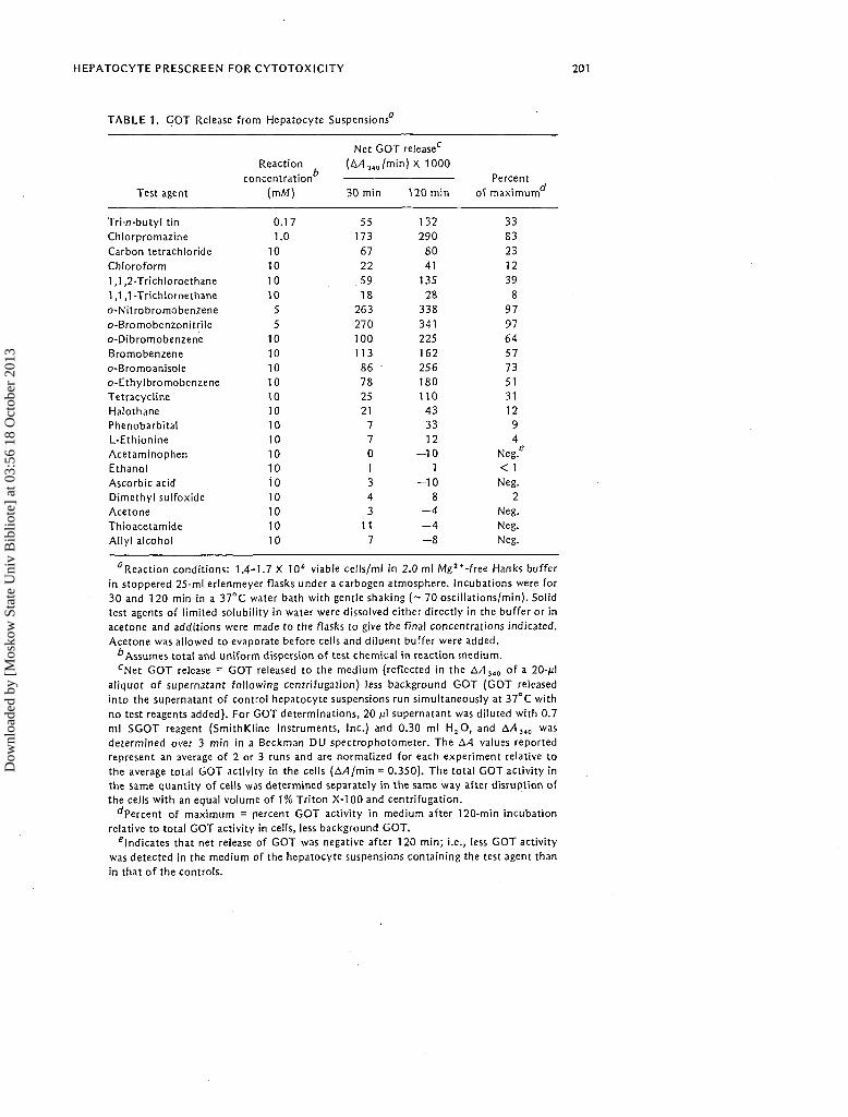
The ability of 23 chemicals to release GOT from the cells was evaluated.The results are shown in Table 1. At a concentration of 10 m/W or lower, 13of the chemicals produced an appreciable release of GOT (>10%) after 120min. These were tri-n-butyl tin, chlorpromazine, carbon tetrachloride,chloroform, 1,1,2-trichloroethane, all six bromobenzene analogs,tetracycline, and halothane. The response to 1,1,1-trichloroethane andphenobarbital was weaker but clearly distinguishable. All of these chem-icals released GOT in a concentration-dependent manner in separateexperiments. L-Ethionine was marginally active at 10 m/W. Sevenchemicals—acetaminophen, ethanol, dimethyl sulfoxide, allyl alcohol,thioacetamide, ascorbic acid, and acetone—were inactive at the cutoffpoint (10 m/W) used to discriminate inactive from active chemicals.Acetaminophen, tested at 10 m/W with hepatocytes prepared from mouselivers in the same way, was found to release 20% of the cytoplasmic GOTabove background after 2 h under the assay conditions shown in Table 1.None of the test chemicals at the test concentrations used appreciablyaffected GOT activity when incubated with the enzyme alone for 2 h at37°C in the assay medium.
Dow
nloa
ded
by [
Mos
kow
Sta
te U
niv
Bib
liote
] at
03:
56 1
8 O
ctob
er 2
013
HEPATOCYTE PRESCREEN FOR CYTOTOXICITY 201
TABLE 1. GOT Release from Hepatocyte Suspensions
Test agent
Tri-n-butyl tin
Chlorpromazine
Carbon tetrachloride
Chloroform
1,1,2-Trichloroethane1,1,1-Trichloroethane
o-Nitrobromobenzene
o-Bromobenzonitrileo-Dibromobenzene
Bromobenzeneo-Bromoanisoleo-Ethylbromobenzene
TetracyclineHalothanePhenobarbitalL-Ethionine
Acetaminophen
EthanolAscorbic acidDimethyl sulfoxide
AcetoneThioacetamideAllyl alcohol
Reaction
concentration
(m/W)
0.171.0
10
10
1010
5
510
1010
10
10101010
1010
io101010
10
Net
(A/1 j ,
GOT release0
0/min) X 1000
30 min 120 min
55173
6722
59
18263
270100
113
86782521
770
134
311
7
132
290
8041
135
28
338
341225162256180
110
4333
12- 1 0
1
- 1 08
- 44
- 8
Percent
of maximum
3383
23
12
39
8
97
9764
5773
513112
94
Neg/< 1Neg.
2
Neg.Neg.Neg.
^Reaction conditions: 1.4-1.7 X 10 ' viable cells/ml in 2.0 ml Mg2 + -free Hanks bufferin stoppered 25-ml erlenmeyer flasks under a carbogen atmosphere. Incubations were for30 and 1 20 min in a 37°C water bath with gentle shaking (~ 70 oscillations/min). Solidtest agents of limited solubility in water were dissolved either directly in the buffer or inacetone and additions were made to the flasks to give the final concentrations indicated.Acetone was allowed to evaporate before cells and diluent buffer were added.
''Assumes total and uniform dispersion of test chemical in reaction medium.cNet GOT release = GOT released to the medium (reflected in the AAiM of a 20-MI
aliquot of supernatant following centrifugation) less background GOT (GOT releasedinto the supernatant of control hepatocyte suspensions run simultaneously at 37°C withno test reagents added). For GOT determinations, 20 fi\ supernatant was diluted with 0.7ml SGOT reagent (SmithKline Instruments, Inc.) and 0.30 ml H2O, and A/l340 wasdetermined over 3 min in a Beckman DU spectrophotometer. The A-4 values reportedrepresent an average of 2 or 3 runs and are normalized for each experiment relative tothe average total GOT activity in the cells (AAjm'm = 0.350). The total GOT activity inthe same quantity of cells was determined separately in the same way after disruption ofthe cells with an equal volume of 1% Triton X-100 and centrifugation.
^Percent of maximum = percent GOT activity in medium after 120-min incubationrelative to total GOT activity in cells, less background GOT.
indicates that net release of GOT was negative after 120 min; i.e., less GOT activitywas detected in the medium of the hepatocyte suspensions containing the test agent thanin that of the controls.
Dow
nloa
ded
by [
Mos
kow
Sta
te U
niv
Bib
liote
] at
03:
56 1
8 O
ctob
er 2
013
202 C. A. TYSON ET AL.
TABLE 2. GOT Release Induced by Weakly Active or InactiveChemicals at 20 mM from Hepatocyte Suspensions0
"Experimental conditions and method of determining GOT releasewere as described in Table 1, except that the concentration of thetest chemicals was 20 m/W in the reaction medium, based on theamount added.
^Percent of maximum is the percent GOT activity in the mediumafter 120-min incubation relative to total GOT activity in the cellsless background GOT.
When the concentrations of some of the more weakly active orinactive compounds in the test assay were increased to 20 mM, the resultsin Table 2 were obtained. Halothane and 1,1,1-trichloroethane weresubstantially more effective in producing GOT release, and phenobarbitaland L-ethionine were slightly active. Dimethyl sulfoxide produced aborderline release of GOT from the cells at both 10 and 20 m/W that wasnot considered significant under the experimental conditions. Acetamino-phen and acetone produced a detectable release of GOT at a testconcentration of 20 mM (Table 2), but ethanol and ascorbic acid remainedinactive. Leakage of GOT to the medium was not increased by eitherthioacetamide or allyl alcohol at 20 mM, nor was there any increase atconcentrations down to 0.5 m/W (determined in separate experiments).
DISCUSSION AND CONCLUSIONS
Rat hepatocyte suspensions prepared by the nonperfusion method ofFry et al. (1976) responded qualitatively to chemical challenge in a simpleassay system in a manner that suggests a possible correlation with thecytotoxicity of the chemicals to liver cells reported in vivo. Thus,tri-/7-butyl tin, a potent cytotoxin and uncoupler of oxidative phosphoryla-tion (Piver, 1973), was effective in releasing GOT from the cells at the
Dow
nloa
ded
by [
Mos
kow
Sta
te U
niv
Bib
liote
] at
03:
56 1
8 O
ctob
er 2
013
HEPATOCYTE PRESCREEN FOR CYTOTOXICITY 203
lowest concentration of any chemical evaluated in the assay. The halo-alkanes and bromobenzenes, except o-bromoanisole and o-ethylbromo-benzene (see below, however), were active as expected (Gehring, 1968;Zimmerman, 1976; Toranzo et al., 1977). 1,1,1-Trichloroethane, weaklyhepatotoxic in vivo (Zimmerman, 1976), was also weakly hepatotoxic inour assay. L-Ethionine, which perturbs cell function by an indirectmechanism and induces marginal SGOT elevation in vivo (Zimmerman,1976), and phenobarbital, which is not considered to be a potenthepatotoxin (Schiff, 1975), were even less potent in vitro. Acetaminophen,which is hepatotoxic in vivo, was not so in the assay with rat hepatocytes,but was, at the same concentration, with mouse hepatocytes; this resultmay be attributed to the relative hepatotoxicity of acetaminophen in thesespecies (Mitchell et al., 1976). Ethanol, which was not considered ashort-term hepatotoxin, was ineffective in our assay. This chemical ishepatotoxic on long-term exposure and is known to release GOT fromhepatocyte cell cultures (Acosta et al., 1978); therefore, longer-terminteraction with the cells may be required for its effect to becomemanifest. Thus, the only serious discrepancy between results and expecta-tions was the inactivity of thioacetamide and ally I alcohol—hepatotoxinsknown to elevate serum GPT (SGPT) in rats after single exposures (Malinget al., 1974)—in the assay.
Two bromobenzene analogs—o-bromoanisole and o-ethylbromobenzene—that were not observed to elevate SGPT or cause liver damage in a pre-vious study (Toranzo et al., 1977) did so in the in vitro assay (Table 1).When we reexamined these chemicals in rats, using higher doses (0.15-0.35ml ip per kilogram of body weight),1 both were found to increase SGOT(and SGPT) activity more than twofold over control (same volume ofsaline ip) levels after 6 h and altered the livers of the test animals-evidenced by lowered glycogen stores, vacuolated cells, and/or franknecrosis—by 24 h. We conclude that these analogs are also hepatotoxic invivo.
On the basis of the data reported here and those of other investigators(Zimmerman et al., 1974; Stacey et al., 1978; Acosta et al., 1978) and thefact that isolated hepatocyte suspensions are increasingly used for toxicitystudies, we believe that it may be possible to develop an assay that ispredictive enough to serve as a cytotoxic screen for humans. However, aconsiderable amount of developmental work remains to be done. There isa need to (1) refine the assay conditions to eliminate false negatives andcover a wider range of cytotoxins, (2) optimize the assay conditions forassessing the relative cytotoxic potency of chemicals in vivo and evaluatethe correlation experimentally, and (3) determine whether other types ofhepatotoxic reactions (e.g., drug-induced hypersensitivity reactions,cholestasis) can be predicted in the assay with appropriate indicators.
1 0.35 ml/kg is a lethal dose of o-bromoanisole.
Dow
nloa
ded
by [
Mos
kow
Sta
te U
niv
Bib
liote
] at
03:
56 1
8 O
ctob
er 2
013
204 C. A. TYSON ET AL.
We have prepared hepatocyte suspension with viabilities of 90% or more,using the basic nonperfusion technique developed by Fry et al. (1976). Asthose investigors noted, the percent viability obtained depends on the skillsand training of the technician or investigator. The. yields and viabilities of theisolated cells obtained here were not as good as those reported for perfusiontechniques,2 but the opportunity to experiment with cell systems from thelivers of animals larger than the rodent and the short preparation times(<2.5 h) more than compensate for these deficiencies. These advantages arealso important in other applications for which hepatocytes prepared bynonperfusion approaches could be used, particularly in vitro comparativemetabolic, toxicity, or therapeutic studies.
REFERENCES
Abernathy, C. O., Lukacs, L., and Zimmerman, H. J. 1977. Adverse effects of chlorpromazine
metabolites on isolated hepatocytes (39833). Proc. Soc. Exp. Biol. Med. 155:474-478.Acosta, D., Anuforo, D., and Smith, R. V. 1978. The use of primary liver cell cultures to study
hepatotoxic agents. Abstr. 16th Annu. Mut. Soc. Toxicol., San Francisco, March 12-16, p. 87.Bellemann, P., Gebhardt, R., and Mecke, D. 1977. An improved method for the isolation of hepatocytes
from liver slices. Selective removal of trypan blue-dyeable cells. Anal. Biochem. 81:408-415.Environmental Protection Agency. 1979. Subcronic Toxicity Workshop, Denver, Colorado, May 9.
Washington, D.C.: EPA.Fogarty International Center Conference on Guidelines for Hepatotoxicity Due to Drugs and
Chemicals. 1977. National Institutes of Health, Bethesda, Md., November 14-16.Fry, J. R. and Bridges, J. W. 1976. The metabolism of xenobiotics in cell suspensions and cell cultures.
Prog. Drug. Metab. 2:71-118.Fry, J. R., Jones, C. A., Wiebkin, P., Bellemann, P., and Bridges, J. W. 1976. The enzymic isolation of
adult rat hepatocytes in a functional and viable state. Anal. Biochem. 71:341-350.Gehring, P. J. 1 968. Hepatotoxic potency of various chlorinated hydrocarbon vapours relative to their
narcotic and lethal potencies in mice. Toxicol. Appl. Pharmacol. 13:287-298.Lindstrom, T. D., Anders, M. W., and Remmer, H. 1978. Effect of phenobarbital and diethyl maleate
on carbon tetrachloride toxicity in isolated rat hepatocytes. Exp. Mol. Pathol. 28:48-57.Maling, H. M., Highman, B., Williams, M. A., Saul, W., Butler, W. M., Jr., and Brodie, B. B. 1974.
Reduction by pretreatment with dibenamine of hepatotoxicity induced by carbon tetrachloride,thioacetamide, or dimethylnitrosamine. Toxicol. Appl. Pharmacol. 27:380-394.
Mitchell, J. R., Snodgrass, W. R., and Gillette, J. R. 1976. The role of biotransformation in
chemical-induced liver injury. Environ. Health Perspect. 15:27-38.
Muul, I., Hegyeli, A. F., and Dacre, J. C. 1977. Toxicological testing dilemma. Science 193:834.Piver, W. J. 1973. Organotin compounds: Industrial applications and biological investigation. Environ.
Health Perspect. 4:61-79.Reed, D. J. and Orrenius, S. 1977. The role of methionine in glutathione biosynthesis by isolated
Schiff, L. 1975. Diseases of the Liver, 4th ed., pp. 649-650. Philadelphia, Pa.: Lippincott.Stacey, N. and Priestley, B. G. 1978. Dose-dependent toxicity of CCl4 is isolated rat hepatocytes and
the effects of hepatoprotective treatments. Toxicol. Appl. Pharmacol. 45:29-39.Stacey, N. H., Priestly, B. G., and Hall, R. C. 1978. Toxicity of halogenated volatile anesthetics in
isolated rat hepatocytes. Anesthesiology 48:17-22.
2 However, cells with better than 97% viability have reportedly been isolated by the nonperfusion
approach by use of a brief trypsin digest after the collagenase-hyaluronidase stage (Bellemann et al.,
1977).
Dow
nloa
ded
by [
Mos
kow
Sta
te U
niv
Bib
liote
] at
03:
56 1
8 O
ctob
er 2
013
HEPATOCYTE PRESCREEN FOR CYTOTOXICITY 205
Thor, H., Moldeus, P., Kristoferson, A., Hogberg, J., Reed, D. J., and Orrenius, S. 1978. Metabolicactivation and hepatotoxicity. Metabolism of bromobenzene in isolated hepatocytes. Arch.Biochem. Biophys. 1 8 8 : 1 1 4 - 1 2 1 .
Toranzo, E. G., Gillesse, T., Mendenhall, M., Traiger, G. J., Riley, P. G., Hanzlik, R. P., and Wiley, R.
A. 1977. Effect of substituents on arene oxide-mediated liver toxicity among substituted
bromobenzenes. Toxicol. Appl. Pharmacol. 40:415-425.Zimmerman, H. J. 1976. Various forms of chemically induced liver injury and their detection by
diagnostic procedures. Environ. Health Perspect. 15:3-12.Zimmerman, H. J., Kendler, J., Libber, S., and Lukacs, L. 1974. Hepatocyte suspensions as a model
for demonstration of drug hepatotoxicity. Biochem. Pharmacol. 23:2187-2189.
Received August 27, 1979Accepted September 20, 1979