12 Evaluation of Oxidative Stress and the Efficacy of Antioxidant Treatment in Diabetes Mellitus Nemes-Nagy Enikő et al. * University of Medicine and Pharmacy, Tîrgu-Mureş, Medical Biochemistry Department, Romania 1. Introduction Studies on the efficacy of antioxidant treatment in type 1 diabetes mellitus is an interesting, actual research subject. Reactive oxygen species (ROS) are continuously produced and eliminated by living organisms normally maintaining ROS at certain steady-state levels. Under some circumstances, the balance between ROS generation and elimination is disturbed leading to enhanced ROS level causing oxidative stress Lushchak, 2011. Oxidative stress is involved in the development of several important diseases (cancer, ulcer, atherosclerosis, autoimmune diseases, ischaemia-reperfusion injury, emphysema, inflammation, etc.). Oxidative stress due to increased production of reactive oxygen species and/or impaired antioxidant capacity of the body plays a special, important role in the development of type 1 diabetes mellitus and its complications Baynes, 1991, Giugliano et al., 1995, Krippeit-Drews et al., 1994, Nakazaki et al., 1995, Nomikos et al., 1989, Raes et al, 1995, Shinn, 1998. Under normal conditions, oxidative tissue damage is prevented by enzymatic and non- enzymatic antioxidants. Superoxide dismutase (SOD), glutathione peroxidase (GPx) and catalase (CAT) are three enzymes involved in detoxification of reactive oxygen species (superoxide radical, hydroxyl radical, and hydrogen peroxide). Pancreatic cells have a poor antioxidant defence system, so they are very vulnerable to oxidative stress, especially to superoxide mediated radical damage Dejica, 2000. Evaluation of oxidative status can be made by various methods, most of them very sophisticated, requiring laboratories with modern equipment, and are used almost exclusively for research purposes. * V. Balogh-Sămărghiţan 1 , Elena Cristina Crăciun 2 , R. Morar 3 , Dana Liana Pusta 3 , Fazakas Zita 1 , Szőcs-Molnár Terézia 4 , Dunca Iulia 4 , Sánta Dóra 5 and Minodora Dobreanu 6 1 University of Medicine and Pharmacy, Tîrgu-Mureş, Medical Biochemistry Department, Romania 2 University of Medicine and Pharmacy "Iuliu Haţieganu", Cluj-Napoca, Pharmaceutical Biochemistry and Clinical Laboratory Department, Romania 3 Faculty of Veterinary Medicine, University of Agricultural Sciences and Veterinary Medicine, Cluj-Napoca, Romania 4 II. Clinical Hospital of Pediatrics, Tîrgu-Mureş, Romania 5 Santa Medical Unit, Tîrgu-Mureş, Romania, 6 University of Medicine and Pharmacy, Tîrgu-Mureş, Clinical Biochemistry Department, Romania www.intechopen.com
Transcript
12
Evaluation of Oxidative Stress and the Efficacy of Antioxidant Treatment in Diabetes Mellitus
Nemes-Nagy Enikő et al.* University of Medicine and Pharmacy, Tîrgu-Mureş, Medical Biochemistry Department,
Romania
1. Introduction
Studies on the efficacy of antioxidant treatment in type 1 diabetes mellitus is an interesting, actual research subject. Reactive oxygen species (ROS) are continuously produced and eliminated by living organisms normally maintaining ROS at certain steady-state levels. Under some circumstances, the balance between ROS generation and elimination is disturbed leading to enhanced ROS level causing oxidative stress Lushchak, 2011. Oxidative stress is involved in the development of several important diseases (cancer, ulcer, atherosclerosis, autoimmune diseases, ischaemia-reperfusion injury, emphysema, inflammation, etc.).
Oxidative stress due to increased production of reactive oxygen species and/or impaired antioxidant capacity of the body plays a special, important role in the development of type 1 diabetes mellitus and its complications Baynes, 1991, Giugliano et al., 1995, Krippeit-Drews et al., 1994, Nakazaki et al., 1995, Nomikos et al., 1989, Raes et al, 1995, Shinn, 1998. Under normal conditions, oxidative tissue damage is prevented by enzymatic and non-enzymatic antioxidants. Superoxide dismutase (SOD), glutathione peroxidase (GPx) and catalase (CAT) are three enzymes involved in detoxification of reactive oxygen species (superoxide radical, hydroxyl radical, and hydrogen peroxide). Pancreatic cells have a poor antioxidant defence system, so they are very vulnerable to oxidative stress, especially to superoxide mediated radical damage Dejica, 2000. Evaluation of oxidative status can be made by various methods, most of them very sophisticated, requiring laboratories with modern equipment, and are used almost exclusively for research purposes. * V. Balogh-Sămărghiţan1, Elena Cristina Crăciun2, R. Morar3, Dana Liana Pusta3, Fazakas Zita1, Szőcs-Molnár Terézia4, Dunca Iulia4, Sánta Dóra5 and Minodora Dobreanu6
1 University of Medicine and Pharmacy, Tîrgu-Mureş, Medical Biochemistry Department, Romania 2 University of Medicine and Pharmacy "Iuliu Haţieganu", Cluj-Napoca, Pharmaceutical Biochemistry and Clinical Laboratory Department, Romania 3 Faculty of Veterinary Medicine, University of Agricultural Sciences and Veterinary Medicine, Cluj-Napoca, Romania 4 II. Clinical Hospital of Pediatrics, Tîrgu-Mureş, Romania 5 Santa Medical Unit, Tîrgu-Mureş, Romania, 6 University of Medicine and Pharmacy, Tîrgu-Mureş, Clinical Biochemistry Department, Romania
www.intechopen.com
Oxidative Stress and Diseases
282
Exploring the body oxidative status can be made through the following ways:
1. Free radical measurement by absorption spectroscopy with electronic spin resonance (ESR) and electronic paramagnetic resonance (EPR) Olinescu, 1994.
2. Measurement of chemical uptake (chemical trapping) by quantitative determination of the elimination of specific derivatives of salicylic acid, hydroxylated or nitrosylated compounds.
3. Measuring the antioxidant capacity of each antioxidant in part or total plasma antioxidant capacity.
4. Determination of antioxidant enzyme activites (SOD, CAT, GPX) and non-enzymatic antioxidants (tocopherols and tocotrienols, vitamin C, ubiquinone, glutathione, carotenoids and vitamin A, bilirubin, melatonin, uric acid, ceruloplasmin, vitamin K, lipoic acid) Dejica, 2000, 2001.
5. The measurement of biological compounds resulting from oxidative processes: - lipid compounds - conjugated dienes, hydroperoxides, aldehydes (malondialdehyde,
hydroxynonenal) - ethane and pentane measurements in expired gases - DNA beta-hydroxydeoxyguanosine - protein derivatives (carbonyl or thiol groups) - amino acids - methionine sulfoxide, ortho-tyrosine, dityrosine, nitrotyrosine,
chlorotyrosine. 6. Measurement of the antioxidants / oxidizing substances ratio (eg. ascorbic acid /
Determination of trace elements is also of interest because some are in the active center of antioxidant enzymes: for example Cu, Zn and Mn are found in the structure of SOD, and Se is present in the active center of GPX, but also possesses antioxidant effect independent of this enzyme Olinescu, 1994. The variety of antioxidant substances in the body, the difficulty of measuring their individual level and the interactions between them require methods for measuring total antioxidant capacity (TAOC) in different biological samples Cadenas & Packer, 2002. By measuring TAOC one can assess not only the interaction effects of antioxidants known, but also the antioxidant action of unidentified components present in human plasma. In most methods uric acid is the major contributor to the TAOC of plasma, so increasing plasma levels of uric acid can mask the depletion of ascorbate or other antioxidants in some pathological conditions if only TAOC measurement is carried out.
TAOC are methods used to measure indirect inhibition involving a prooxidant (typically a free radical) and an oxidizable substrate. The prooxidant induces oxidative deterioration of the substrate, which is inhibited in the presence of the antioxidant.
Antioxidant capacity can be defined as the ability of a compound to reduce prooxidants. In biological systems, prooxidants are usually defined as toxic substances causing oxidative damage to lipids, proteins, nucleic acids, leading to a variety of pathological events, but not
www.intechopen.com
Evaluation of Oxidative Stress and the Efficacy of Antioxidant Treatment in Diabetes Mellitus
283
every prooxidant is necessary a toxic compound. Antioxidants are substances that, in low concentrations compared to an oxidizable substrate, prevent or delay oxidation initiated by a prooxidant Dejica, 2001. Several vitamins are well known for their antioxidant properties: vitamin C exhibits its effect in hydrophilic environment, while vitamin A and E protect especially the cell membranes, and act in hydrophobic phase. Many other, more complex antioxidant substances are found in plants.
3. Implication of oxidative stress and glycation process in diabetes mellitus
In type 2 diabetic patients, besides the relative insulin deficiency, there is a certain grade of insulin resistance. The relationship between reactive oxygen species and the effect of insulin has been studied, and the results showed that in elderly people, presenting intense exposure to oxidative stress, the ratio between GSH/GSSG is reduced, leading to intensified lipoperoxidation. This phenomenon might exhibit a negative influence on the integrity of plasma membranes, leading to their disfunction, regarding for instance the transmembrane glucose transport. There is a high probability that the periferal action of insulin is disturbed by the negative effect of reactive oxygen species on membrane ATP-ase activity Dejica, 2000. Peroxidation of lipids (especially LDL) plays an important role in inducing macrovascular lesions found in both diabetes and in atherosclerotic disease. Susceptibility to oxidation of lipoproteins seems to be a key element in the initiation and propagation of the atherogenic process Dobreanu M., Módy, 1998. In diabetic patients, lipid oxidation affects circulating lipids, and also those present in cell membranes or myelin layers. Hyperglycemia, excessive autooxidation and decreased antioxidant capacity registered in diabetic patients are responsible for intensified oxidative processes.
The speed of non-enzymatic glycation is proportional to the blood sugar level. Glucose (and fructose and galactose) are attached to the extreme N-terminal peptide chain, initially forming a Schiff base (aldimine), which is unstable, then by Amadori rearrangement a stable cetoimine is formed, and advanced glycation end products, leading to alterations in configuration, of the electronegative charge and molecular recognition of proteins.
Amadori-products can also be oxidized, by reactions catalyzed by transition metals, releasing eritronic acid and forming carboxymethylated lysine. Its level is double in the collagen of the skin of diabetic patients compared to non-diabetic subjects, and it is positively correlated with the presence of retino- and nephropathy in diabetic patients Wolff, 1993. All structural proteins and those circulating in the body can suffer non-enzymatic glycation processes. They can alter protein structure and function of vessels, nerves, liver, skin and other organs. Glycosylation of LDL lipoprotein particles decreases their catabolism and accelerates HDL catabolism, disorders that may explain in part the modifications present in macroangiopathy. Glycosylated proteins are more susceptible to attack by reactive oxygen species.
Enzymatic glycosylation of proteins is also important in development of chronic diabetic complications. The collagen molecules are thus glycosylated, glycoproteins and
www.intechopen.com
Oxidative Stress and Diseases
284
proteoglycans suffer similar processes. At the level of nerves, lens, livers or other organs, glycosylation of proteins is involved in the occurrence of diabetic neuropathy, cataracts, dysmetabolic hepatopathy Gherasim, 1998, Kovács, 2001. Based on these evidences, antioxidant treatment is a promising approach for complementary therapy in diabetes mellitus. Several phytotherapeutical products contain a complexity of free radical scavengers and exhibit no side effects on long term treatment.
4. Evaluation of oxidative stress and the efficacy of antioxidant treatment in diabetes mellitus
4.1. Studies on animals
Some of the plants known for their high antioxidant power are: Allium sativa, Ricinus communis, Securinega virosa, Viscum album, pomegranates, berries (including strawberries, blueberries, and raspberries), walnuts, sunflower seeds, ginger and several plants of Indian traditional medicine: Emblica officinalis L., Curcuma longa L., Mangifera indica L., Momordica charantia L., Santalum album L, Swertia chirata Buch-Ham, Withania somnifera and Cassia auriculata Capasso et al, 2003, Dejica, 2001, Moshi & Mbwambo, 2003, Pari & Latha, 2003, Pietta, 1998, Scartezzini & Speroni, 2000, Varga et al., 2001. Our studies carried out in streptozotocin diabetic rats demonstrated that treatment with the blueberry (Eridiarom®) or blueberry and sea buckthorn concentrate (Diavit®) for two months had regenerative effect on pancreatic beta cells Crăciun et al., 2007, Morar et al., 2004. The therapeutic properties of blueberry are attributed to its anthocyanosides which belong to a class of substances known as plant bioflavonoids. Pharmacologically, anthocyanosides are thought to decrease vascular permeability and improve microcirculation. They are also thought to have antioxidant activity.
Diavit® is a dietary supplement with a more complex composition compared to Eridiarom®, it contains quinolizidine alkaloids, anthocyanosides, sugars, carotenoids, vitamins (C, E, PP, B1, B2, folic acid), minerals (K, Ca, P, S, Mg, Cl, Mn, Fe), organic acids, flavonoids, etc..
Carotenoids are best recognized for their antioxidant capacity, they are considered the most potent biological quenchers of singlet oxygen Morar, 2003, Paiva & Russell, 1999, Pizzorno & Murray, 2003, Rombi, 1998, Slosse & Hootele, 1981, Timberlake & Henry, 1988, Verette, 1984, Zeb, 2004. The experiment was carried out on 5 groups of male Wistar rats, the first five group received Streptozotocin i.v. 4 mg/100 g body weight to induce diabetes.
The toxicity of Streptozotocin can be counteracted by desferioxamine, suggesting that oxidative reactions catalyzed by transition metals could be responsible for the toxicity of this substance. Streptozotocin, being one of the glico-nitrosureas, could exhibit its diabetes-inducing effect also by inadequate NO release Giugliano et al., 1995. The first group served as a diabetic witness, the second group was treated with Siofor® ½ tablet/day/group (equivalent to 12 mg/kg), the third group received Meguan® (2 tablets of
www.intechopen.com
Evaluation of Oxidative Stress and the Efficacy of Antioxidant Treatment in Diabetes Mellitus
285
0.5 g/group/day), the 4th group was treated with Eridiarom® (1.2 g/group/day), and the fifth group was given Diavit® (1.2 g/group/day). Healthy, normally fed rats formed the 6th, non streptozotocin-treated witness group.
After 5 days, and than each month glycaemia was measured, and after 100 days histopathologic examination (pancreas, liver, kidney, heart, muscles, eyes) were performed on 2 animals of each group, and the rest of the rats were kept under observation.
During the experiment one animal in each treated group died, and in the group treated with Siofor two animals died.
The dynamics of glycaemia increases at 7 days after the induction of subclinical diabetes (176.8 – 185.5 mg/dl +/- 3.0 – 5.6 SD) in each of the streptozotocin-treated groups compared to the initial values (103.2 – 105.3 mg/dl +/- 1.6 – 2.2 SD).
After 30 days the glycaemia is close to normal (120.1 – 127.8 mg/dl +/- 2.0 – 2.1 SD) in groups 4 and treated with the phytotherapeutic products Eridiarom® and Diavit®5 (14.6- 22.6% higher than the initial values), and it is high (151.6 mg/dl +/- 4.0 – 4.1 SD) in groups 2 and 3 treated with the antidiabetic sulphamides Siofor® and Meguan® (45.46% higher than the initial values), but lower compared to the first, witness group (164.6 mg/dl +/- 4.2 SD). After 90 days the glycaemia is practically normalized in groups 4 and 5 (102.8 – 114.3 mg/dl +/- 1.5 -2.3 SD) (the final value 1.37% lower, and 9.6% higher than the initial values) and it is slightly increased in the first 3 groups (135.0 – 133.3 mg/dl +/- 3.3 – 4.0 mg/dl) (29,1-27.9% higher than the initial values).
The histopathologic examination of the pancreas (Trichromic stain) revealed a strong destructive action of streptozotocin against the endocrine and exocrine pancreas.
In the first witness group we can observe pancreatic cytonecrosis, atrophy with hypofunction and disturbing of the ratio between and secretory cells after administering streptozotocin. 100 days later the Langerhans islets of the rats in the first, witness group present partial regeneration and recovery of pseudolobules with cells, general interstitial oedema, atrophy of the acini’s level with the significant reducing of the cellular secretory pole.
In group 2, treated with Siofor®, 100 days after the beginning of the experiment partial recovery can be observed in the Langerhans islets, with cells during the pseudolobules’ reconstruction, and partial recovery of the exocrine secretor function by acinary and lobular hyperfunction.
In group 3 treated with Meguan®, 100 days after the initiation of the experiment regenerated Langerhans islets can be seen with and cells’ reorganisation and the formation of sinusoidal capillary, and acinus-lobulary hyperfunction with the hypertrophy of the secretor pole in pancreatic acini.
In group 4, treated with Eridiarom®, 100 days after the beginning of the experiment, Langerhans islets with regenerated and cells can be observed, with increased mitotic index and complete recovery, and generalised acinus-lobulary functional hypertrophy.
In group 5, treated with Diavit®, 100 days after the initiation of the experiment, Langerhans islets with complete recovery of the cell-architectonics can be observed, specific for normal
www.intechopen.com
Oxidative Stress and Diseases
286
functioning Langerhans cells, and hyperthrophy with general acinus-lobulary moderate secretion.
Based on these experimental data we can conclude that the two phytotherapeutic products exhibited a powerful regenerative effect on the pancreatic cells, presenting a better efficacy compared to the widely used antidiabetic sulphamides Crăciun et al., 2007, Morar et al., 2004. 4.2 Studies on human subjects
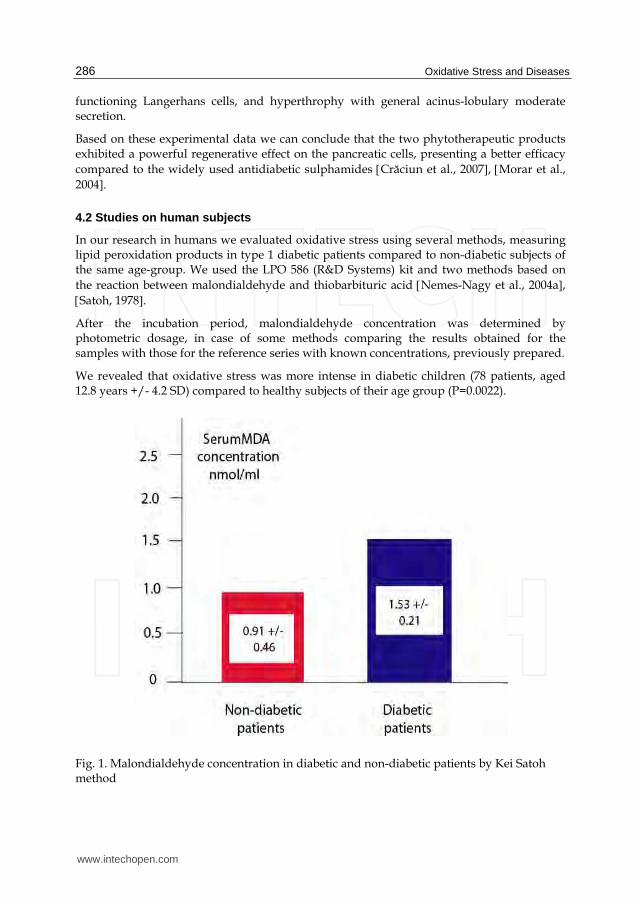
In our research in humans we evaluated oxidative stress using several methods, measuring lipid peroxidation products in type 1 diabetic patients compared to non-diabetic subjects of the same age-group. We used the LPO 586 (R&D Systems) kit and two methods based on the reaction between malondialdehyde and thiobarbituric acid Nemes-Nagy et al., 2004a, Satoh, 1978. After the incubation period, malondialdehyde concentration was determined by photometric dosage, in case of some methods comparing the results obtained for the samples with those for the reference series with known concentrations, previously prepared.
We revealed that oxidative stress was more intense in diabetic children (78 patients, aged 12.8 years +/- 4.2 SD) compared to healthy subjects of their age group (P=0.0022).
Fig. 1. Malondialdehyde concentration in diabetic and non-diabetic patients by Kei Satoh method
www.intechopen.com
Evaluation of Oxidative Stress and the Efficacy of Antioxidant Treatment in Diabetes Mellitus
287
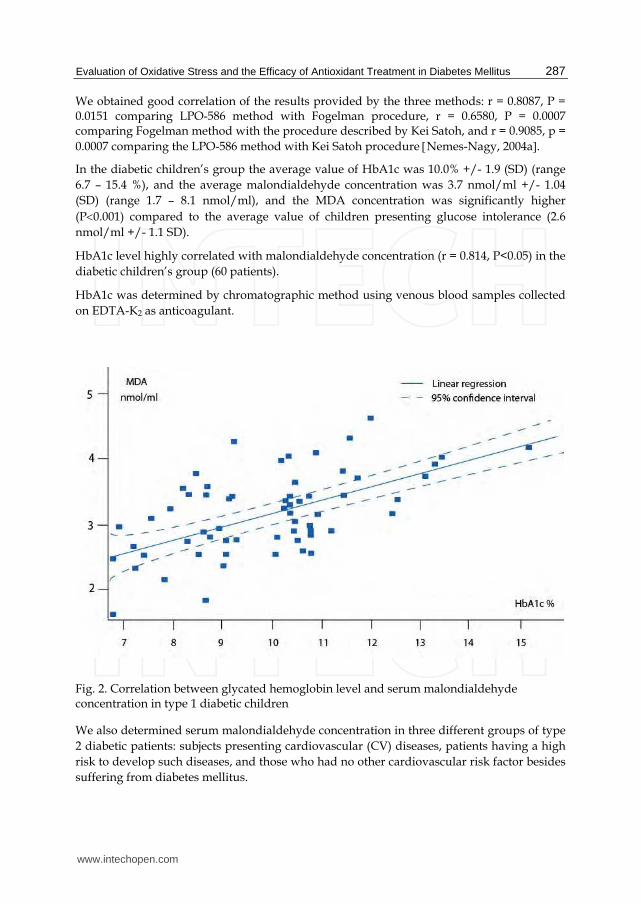
We obtained good correlation of the results provided by the three methods: r = 0.8087, P = 0.0151 comparing LPO-586 method with Fogelman procedure, r = 0.6580, P = 0.0007 comparing Fogelman method with the procedure described by Kei Satoh, and r = 0.9085, p = 0.0007 comparing the LPO-586 method with Kei Satoh procedure Nemes-Nagy, 2004a. In the diabetic children’s group the average value of HbA1c was 10.0% +/- 1.9 (SD) (range 6.7 – 15.4 %), and the average malondialdehyde concentration was 3.7 nmol/ml +/- 1.04 (SD) (range 1.7 – 8.1 nmol/ml), and the MDA concentration was significantly higher (P0.001) compared to the average value of children presenting glucose intolerance (2.6 nmol/ml +/- 1.1 SD).
HbA1c level highly correlated with malondialdehyde concentration (r = 0.814, P<0.05) in the diabetic children’s group (60 patients).
HbA1c was determined by chromatographic method using venous blood samples collected on EDTA-K2 as anticoagulant.
Fig. 2. Correlation between glycated hemoglobin level and serum malondialdehyde concentration in type 1 diabetic children
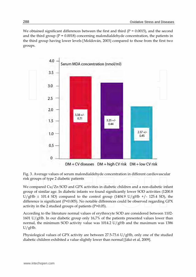
We also determined serum malondialdehyde concentration in three different groups of type 2 diabetic patients: subjects presenting cardiovascular (CV) diseases, patients having a high risk to develop such diseases, and those who had no other cardiovascular risk factor besides suffering from diabetes mellitus.
www.intechopen.com
Oxidative Stress and Diseases
288
We obtained significant differences between the first and third (P = 0.0015), and the second and the third group (P = 0.0018) concerning malondialdehyde concentration, the patients in the third group having lower levels Moldován, 2003 compared to those from the first two groups.
Fig. 3. Average values of serum malondialdehyde concentration in different cardiovascular risk groups of type 2 diabetic patients
We compared Cu/Zn SOD and GPX activities in diabetic children and a non-diabetic infant group of similar age. In diabetic infants we found significantly lower SOD activities (1200.8 U/gHb 101.4 SD) compared to the control group (1404.9 U/gHb +/- 125.4 SD), the difference is significant (P<0.005). No notable differences could be observed regarding GPX activity in the 2 studied groups of patients (P>0.05).
According to the literature normal values of erythrocyte SOD are considered between 1102-1601 U/gHb. In our diabetic group only 16,7% of the patients presented values lower than normal, the minimum SOD activity value was 1014.2 U/gHb and the maximum was 1386 U/gHb.
Physiological values of GPX activity are between 27.5-73.6 U/gHb, only one of the studied diabetic children exhibited a value slightly lower than normal Jákó et al, 2009.
www.intechopen.com
Evaluation of Oxidative Stress and the Efficacy of Antioxidant Treatment in Diabetes Mellitus
289
Our research team demonstrated the powerful antioxidant and hypoglycemic effect of a blueberry (Vaccinium myrtillus) concentrate (Eridiarom®) in diabetic children (initially this product was used for treatment of diarrhoea in humans). We selected 29 infants presenting poor carbohydrate metabolic balance to participate to the study.
Fig. 4. Erythrocyte Cu-Zn superoxide dismutase activity in type 1 diabetic children
Fig. 5. Whole blood glutathione peroxidase dismutase activity in type 1 diabetic children
www.intechopen.com
Oxidative Stress and Diseases
290
The average glycemic level at the beginning of the study was 179.4 mg/dl 39.2 (SD), after 3 months of Eridiarom® treatment the value decreased to 159.1 mg/dl +/- 40.7 (SD), the difference is significant (P<0.005).
Insulin doses (UI/kg) could be lowered in 78.6% of the patients. Initially the average dose was 0.98 UI/kg 0.2 (SD) and after 3 months of treatment 0.91 UI/kg 0.2 (SD), the difference is significant (P<0.05). This result can be explained by the regenerative effect of this phytotherapeutic product on the pancreatic beta cells, improving their insulin secretion.
We observed that the longer the treatment with this dietary supplement, better the results are.
After 3 months of Eridiarom® treatment HbA1c values presented decrease in 71.43% of the patients. Using the Student paired t test, we obtained significant differences (P<0.05) regarding the HbA1c values at the beginning of the study (9.6% 1.6 SD) and 8.5% 1.5 (SD) after 3 months of Eridiarom® treatment.
After 3 months of Eridiarom® treatment MDA concentration decreased in 92.9% of the patients, 7.1% showed practically no modification of the MDA level.
Fig. 6. Serum MDA concentration before and after Eridiarom® treatment
www.intechopen.com
Evaluation of Oxidative Stress and the Efficacy of Antioxidant Treatment in Diabetes Mellitus
291
The average MDA concentration before the study was 6.7 nmol/ml 0.7 (SD), after 3 months of treatment the average value was 4.5 nmol/ml 0.8 (SD), the difference is significant (P0.0001).
Prior to the study the serum magnesium and calcium concentrations were under the normal range in 43% of the patients. The average magnesium level at the beginning of the study was 1.7 mg/dl, while after 3 months of Eridiarom® treatment in case of all these patients the magnesium level turned to normal, the average value being 2.2 mg/dl; the difference is significant (P0.0001) Balogh-Sămărghiţan et al., 2004, Nemes-Nagy et al., 2006.
Fig. 7. Dynamics of serum magnesium concentration under blueberry containing Eridiarom® treatment
Several studies were made on the implications of magnesium in diabetes mellitus. Certain key enzymes of carbohydrate metabolism (glucokinase, hexokinase, glucose-6-phosphate dehydrogenase) and lipid metabolism (mevalonate kinase, lecithin-cholesterol acyl-transferase) are magnesium-dependent.
Insulin, along with catecholamines, has major effects on intracellular homeostasis of magnesium, being, besides vitamin D and taurine, an important magnesium-linking substance. Insulin receptors exhibit a magnesium-dependent kinase activity Vereşiu, 2000. Studies evaluating magnesium levels in diabetic patients compared to healthy controls showed decreases in case of diabetic subjects, and other studies have shown an increased risk for this disease in patients with magnesium deficiency. Possible correlation of Mg with chronic diabetes complications have been the subject of other studies, that have found lower values in patients with retinopathy De Valk, 1999 and neuropathy DeLourdes, 1998.
www.intechopen.com
Oxidative Stress and Diseases
292
Effects of magnesium administration in patients with diabetes to improve glycemic control and prevention of chronic complications are conflicting and awaiting further confirmation.
A few years later we studied the effect of another dietary supplement (Diavit®), containing blueberry (Vaccinum myrtillus) and sea buckthorn (Hippophae rhamnoides) concentrate with a complex composition previously presented.
We compared glycaemic profile, glycated hemoglobin (HbA1C) after 2 and 3 months of treatment, C peptide level and changes in antioxidant enzyme activity (Cu/Zn superoxide dismutase and glutation peroxidase) after two months of treatment with the Diavit® dietary supplement versus after placebo treatment.
Values for activity of erythrocyte Cu/Zn SOD, a scavenger of superoxide radicals, were significantly higher (P<0.05) in diabetic patients after two months of treatment with the concentrate (1260.9 66.9 U/g Hb) compared to those obtained before treatment (1201.6 105.6 U/g Hb).
There was no significant difference in GPX activity before (40.8 9.2 U/g Hb) and after the study (43.9 13.9 U/g Hb), only a slight, not significant increase could be observed (P>0.05) Capasso et al, 2003, Nemes-Nagy et al., 2007, 2008, 2010, Paglia & Valentine, 1967, Szőcs-Molnár et al., 2006.
Fig. 8. Dynamics of blood SOD activity under blueberry and sea buckthorn containing Diavit® treatment
www.intechopen.com
Evaluation of Oxidative Stress and the Efficacy of Antioxidant Treatment in Diabetes Mellitus
293
Hydrogen peroxide, formed by the reaction catalyzed by SOD, is decomposed by two enzymes: GPX and CAT. Dosage of blood catalase activity could have helped to have a better picture on the enzymatic antioxidant equipment of these diabetic patients under the treatment with this dietary supplement.
HbA1C levels were significantly lower after treatment with the dietary supplement (9.2 1.6 % versus 4the initial 10.2 2.3 %; P<0.05) and C-peptide average value increased significantly (P<0.05) after 2 month of treatment with this dietary supplement (0.2 ng/ml 0.1 SD) compared to the initial average value (0.04 ng/ml 0.02 SD). Insulin requirement reduced significantly from the average of 0.96 0.27 IU/kg bodyweight to 0.89 0.28 IU/kg after 2 months of treatment with the product (P<0.05), insulin doses were reduced in 66.7% of the patients.
Lower blood glucose and HbA1c values may be due to a regenerative effect that the product has on pancreatic beta cells. Significantly higher C-peptide levels after 2 months of treatment with the extract support this hypothesis Nemes-Nagy et al., 2008. A scientific team in Harvard University, Howard Hughes Medical Institute, under the leadership of Prof. Douglas Melton, published their findings about the capacity of pancreatic beta cells to regenerate by self-duplication due to some latent embrional cell remains or adult stem cells Zhou et al., 2008, and this regenerative process might explain our results with the product studied. We suppose that better results could be obtained if the treatment with the concentrate begins soon after the diabetic disease is diagnosed, maybe because long term insulin treatment causes the atrophy of pancreatic beta-cells, similar to corticosteroid-caused hypofunction of corticosuprarenals. This hypothesis should be verified in latter studies. Based on our experimental data it seems that the longer the treatment with these dietary supplements, the better the results are.
Hyperglycemia in diabetes mellitus produces increased oxidative stress via non-enzymatic glycation, glucose autooxidation, and alteration in polyol pathway activity. This is characterized by increased lipid peroxide production and decreased antioxidative defence (e.g. inactivation of SOD by glycation) which affects the entire body. Two months using Diavit® lead to a significant increase in SOD which may have occurred as a result of its antioxidant and hypoglycemic effects. The antioxidant effect of this product might be partially due to anthocyanosides, known as scavengers of superoxide anions, inhibitors of lipid peroxidation. Lower glycaemic levels during the study might cause lower superoxide radical production and decrease the inactivation rate of this antioxidant enzyme, leading to higher SOD levels than before.
Protection of free radical scavengers might help to maintain higher levels of antioxidants under treatment with this concentrate, and several components of the product (carotenoids, vitamin E, C) show important protective role against oxidative stress. According to recent studies, it might be a link between GPX and ascorbate: intracellular vitamin C cooperates in enhancing glutathione recovery after oxidative challenge thus providing cells with enhanced survival potential, while extracellular vitamin C is recycled through a mechanism involving the simultaneous neutralization of oxidant species Montecinos et al., 2007. Several data suggest that oxygen metabolits are involved in the pathogenesis of autoimmune destruction of pancreatic beta cells, involving inflammatory process, and especially superoxide
www.intechopen.com
Oxidative Stress and Diseases
294
radical is required for the expression of the disease. Pancreatic beta cells are particularly sensitive to superoxide mediated radical damage, having a poor antioxidant defence system. Superoxide itself or derivative radicals may be the direct cause of cell damage. Radical generation leads to breakage of cell DNA, which initiates the repair process resulting in depletion of cellular NADH + H+ levels, leading to an inhibition of pro-insulin synthesis and renders the cell more sensitive to radical damage because NAD is involved in the electron-transport process required for radical scavenging by the cell. Oxygen radicals are also involved in the production of the cytokines (IL-1, TNF) by the cells of the inflammatory focus that could be involved in the cell damage Nemes-Nagy et al., 2008. Another important component of Diavit® is the phytoestrogen called resveratrol, which is a natural polyphenolic compound found largely in the skin of red grapes, but also in blueberries.
Growing evidence suggests that resveratrol may play an important role in the prevention of many human diseases. Many of the biological actions of this polyphenol have been attributed to its antioxidant properties, it exhibits anticoagulant, vasodilator, antiinflammatory effects, inhibits oxidation of LDL-cholesterol particles, thus preventing atherosclerosis, and also increases sensitivity to insulin.
Certain studies evaluated the effect of resveratrol on intracellular reduced glutathione (GSH) and membrane sulphydryl groups in erythrocytes subjected to oxidative stress in vitro to test the efficacy of the antioxidant effect of resveratrol on human erythrocytes. In one of these studies subjecting erythrocytes to oxidative stress (in vitro) by incubating them with t-BHP (10 micromolar) caused a significant decrease in the intracellular GSH level and membrane -SH content compared with basal values.
Incubation of erythrocytes/membranes with resveratrol (1-100 micromolar final concentration) resulted in significant protection against the t-BHP-induced oxidative stress as evidenced by the increase in GSH level and membrane -SH content. It was observed that the effect of resveratrol is dose/concentration and time-dependent, it protects erythrocytes experimentally exposed to oxidative stress. Since resveratrol is naturally present in many fruits and vegetables, a diet rich in resveratrol, or dietary supplements containing this substance may provide protection against degenerative diseases and prevent diabetes complications [Pandey & Rizvi, 2010].
A new promising study on humans demonstrated the effect of regular consumption of a resveratrol supplement and the health of patients with impaired glucose tolerance. Resveratrol has been tested in relation to diabetes before, but only in animal subjects or on cell lines. Those studies have repeatedly shown promising effects on insulin secretion, insulin sensitivity and glucose tolerance, leading to the initiation of this first-of-its-kind pilot clinical study on humans [Crandall, 2010].
5. Studies on antioxidant components of medicinal teas, fruit and vegetable juices
We also determined the flavonoid content and the antioxidant capacity of several medicinal teas used by diabetic patients. We found the highest amount of flavonoids in Juglandis
www.intechopen.com
Evaluation of Oxidative Stress and the Efficacy of Antioxidant Treatment in Diabetes Mellitus
295
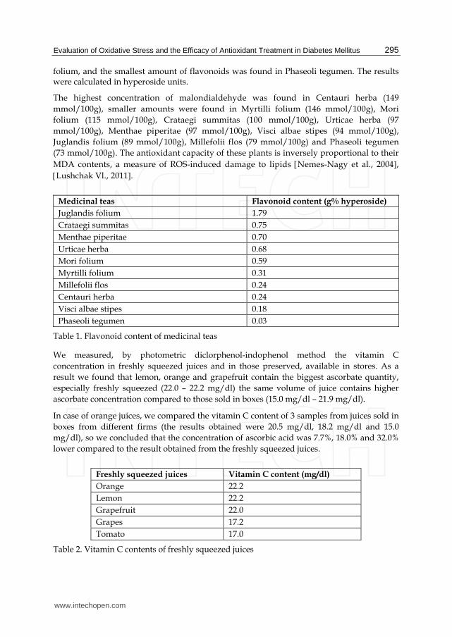
folium, and the smallest amount of flavonoids was found in Phaseoli tegumen. The results were calculated in hyperoside units.
The highest concentration of malondialdehyde was found in Centauri herba (149 mmol/100g), smaller amounts were found in Myrtilli folium (146 mmol/100g), Mori folium (115 mmol/100g), Crataegi summitas (100 mmol/100g), Urticae herba (97 mmol/100g), Menthae piperitae (97 mmol/100g), Visci albae stipes (94 mmol/100g), Juglandis folium (89 mmol/100g), Millefolii flos (79 mmol/100g) and Phaseoli tegumen (73 mmol/100g). The antioxidant capacity of these plants is inversely proportional to their MDA contents, a measure of ROS-induced damage to lipids Nemes-Nagy et al., 2004, Lushchak Vl., 2011.
We measured, by photometric diclorphenol-indophenol method the vitamin C concentration in freshly squeezed juices and in those preserved, available in stores. As a result we found that lemon, orange and grapefruit contain the biggest ascorbate quantity, especially freshly squeezed (22.0 – 22.2 mg/dl) the same volume of juice contains higher ascorbate concentration compared to those sold in boxes (15.0 mg/dl – 21.9 mg/dl).
In case of orange juices, we compared the vitamin C content of 3 samples from juices sold in boxes from different firms (the results obtained were 20.5 mg/dl, 18.2 mg/dl and 15.0 mg/dl), so we concluded that the concentration of ascorbic acid was 7.7%, 18.0% and 32.0% lower compared to the result obtained from the freshly squeezed juices.
Table 2. Vitamin C contents of freshly squeezed juices
www.intechopen.com
Oxidative Stress and Diseases
296
6. Vitamin C dynamics in human milk
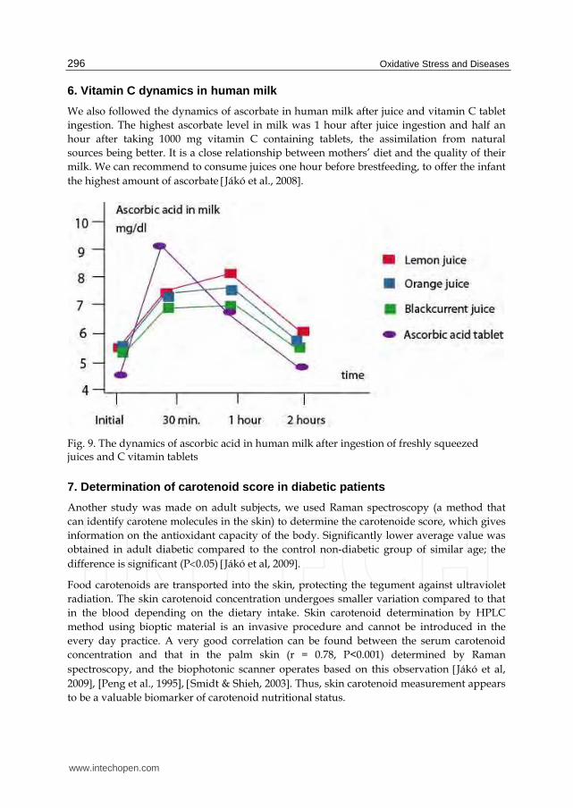
We also followed the dynamics of ascorbate in human milk after juice and vitamin C tablet ingestion. The highest ascorbate level in milk was 1 hour after juice ingestion and half an hour after taking 1000 mg vitamin C containing tablets, the assimilation from natural sources being better. It is a close relationship between mothers’ diet and the quality of their milk. We can recommend to consume juices one hour before brestfeeding, to offer the infant the highest amount of ascorbate Jákó et al., 2008.
Fig. 9. The dynamics of ascorbic acid in human milk after ingestion of freshly squeezed juices and C vitamin tablets
7. Determination of carotenoid score in diabetic patients
Another study was made on adult subjects, we used Raman spectroscopy (a method that can identify carotene molecules in the skin) to determine the carotenoide score, which gives information on the antioxidant capacity of the body. Significantly lower average value was obtained in adult diabetic compared to the control non-diabetic group of similar age; the difference is significant (P0.05) Jákó et al, 2009. Food carotenoids are transported into the skin, protecting the tegument against ultraviolet radiation. The skin carotenoid concentration undergoes smaller variation compared to that in the blood depending on the dietary intake. Skin carotenoid determination by HPLC method using bioptic material is an invasive procedure and cannot be introduced in the every day practice. A very good correlation can be found between the serum carotenoid concentration and that in the palm skin (r = 0.78, P<0.001) determined by Raman spectroscopy, and the biophotonic scanner operates based on this observation Jákó et al, 2009, [Peng et al., 1995], Smidt & Shieh, 2003. Thus, skin carotenoid measurement appears to be a valuable biomarker of carotenoid nutritional status.
www.intechopen.com
Evaluation of Oxidative Stress and the Efficacy of Antioxidant Treatment in Diabetes Mellitus
297
Raman scattering spectroscopy is a highly specific method for skin carotenoid determination. This method is able to discern carotenoids from other potentially interfering compounds present in the skin due to its ability to identify molecules with long conjugated double-bond structures. Raman spectroscopy involves a blue, low-energy laser light source of 470 – 490 nm, directed onto the surface of the skin, where Raman resonance light scattering events cause the carotenoids to emit a green signal at 510 – 530 nm, which is detected and quantified.
Fig. 10. Comparision of skin carotenoid score in diabetic and non-diabetic patients
Studies showed that the Raman spectroscopic method accurately reflects the presence of carotenoids in the human skin with high reproducibility. Significant differences in carotenoid concentrations were found between five different skin sites, the highest concentration being found in the palm.
An epidemiological study performed on a large number of healthy volunteers determined that the Raman spectroscopic method of measuring skin carotenoids is not really affected by gender, age or skin pigmentation.
Raman intensity measurements were positively related to the amount of fruit and vegetable intake and use of carotenoid-containing dietary supplements, and inversely related to body fat content and smoking. This study confirmed that skin carotenoids follow similar dietary and demographic patterns as in case of serum or plasma carotenoid measurements.
Before the measurement, every patient has to fill in a special questionnaire containing questions regarding its lifestyle, dietary habits and other aspects that could influence the result.
www.intechopen.com
Oxidative Stress and Diseases
298
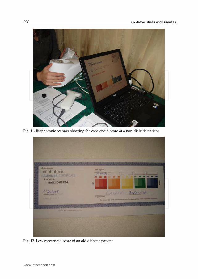
Fig. 11. Biophotonic scanner showing the carotenoid score of a non-diabetic patient
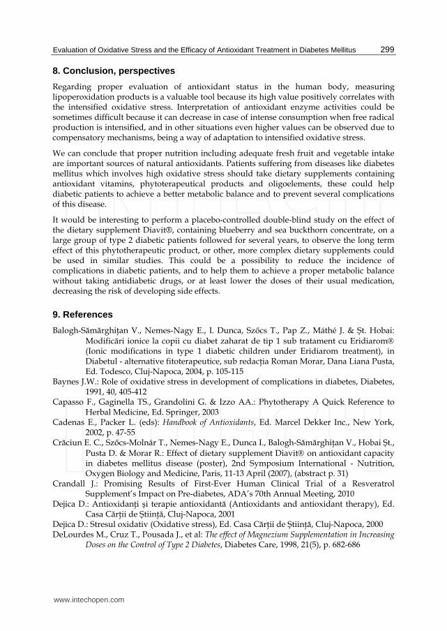
Fig. 12. Low carotenoid score of an old diabetic patient
www.intechopen.com
Evaluation of Oxidative Stress and the Efficacy of Antioxidant Treatment in Diabetes Mellitus
299
8. Conclusion, perspectives
Regarding proper evaluation of antioxidant status in the human body, measuring lipoperoxidation products is a valuable tool because its high value positively correlates with the intensified oxidative stress. Interpretation of antioxidant enzyme activities could be sometimes difficult because it can decrease in case of intense consumption when free radical production is intensified, and in other situations even higher values can be observed due to compensatory mechanisms, being a way of adaptation to intensified oxidative stress.
We can conclude that proper nutrition including adequate fresh fruit and vegetable intake are important sources of natural antioxidants. Patients suffering from diseases like diabetes mellitus which involves high oxidative stress should take dietary supplements containing antioxidant vitamins, phytoterapeutical products and oligoelements, these could help diabetic patients to achieve a better metabolic balance and to prevent several complications of this disease.
It would be interesting to perform a placebo-controlled double-blind study on the effect of the dietary supplement Diavit®, containing blueberry and sea buckthorn concentrate, on a large group of type 2 diabetic patients followed for several years, to observe the long term effect of this phytotherapeutic product, or other, more complex dietary supplements could be used in similar studies. This could be a possibility to reduce the incidence of complications in diabetic patients, and to help them to achieve a proper metabolic balance without taking antidiabetic drugs, or at least lower the doses of their usual medication, decreasing the risk of developing side effects.
9. References
Balogh-Sămărghiţan V., Nemes-Nagy E., I. Dunca, Szőcs T., Pap Z., Máthé J. & Şt. Hobai: Modificări ionice la copii cu diabet zaharat de tip 1 sub tratament cu Eridiarom (Ionic modifications in type 1 diabetic children under Eridiarom treatment), in Diabetul - alternative fitoterapeutice, sub redacţia Roman Morar, Dana Liana Pusta, Ed. Todesco, Cluj-Napoca, 2004, p. 105-115
Baynes J.W.: Role of oxidative stress in development of complications in diabetes, Diabetes, 1991, 40, 405-412
Capasso F., Gaginella TS., Grandolini G. & Izzo AA.: Phytotherapy A Quick Reference to Herbal Medicine, Ed. Springer, 2003
Cadenas E., Packer L. (eds): Handbook of Antioxidants, Ed. Marcel Dekker Inc., New York, 2002, p. 47-55
Crăciun E. C., Szőcs-Molnár T., Nemes-Nagy E., Dunca I., Balogh-Sămărghiţan V., Hobai Şt., Pusta D. & Morar R.: Effect of dietary supplement Diavit on antioxidant capacity in diabetes mellitus disease (poster), 2nd Symposium International - Nutrition, Oxygen Biology and Medicine, Paris, 11-13 April (2007), (abstract p. 31)
Crandall J.: Promising Results of First-Ever Human Clinical Trial of a Resveratrol Supplement’s Impact on Pre-diabetes, ADA’s 70th Annual Meeting, 2010
Dejica D.: Antioxidanţi şi terapie antioxidantă (Antioxidants and antioxidant therapy), Ed. Casa Cărţii de Ştiinţă, Cluj-Napoca, 2001
Dejica D.: Stresul oxidativ (Oxidative stress), Ed. Casa Cărţii de Ştiinţă, Cluj-Napoca, 2000 DeLourdes M., Cruz T., Pousada J., et al: The effect of Magnezium Supplementation in Increasing
Doses on the Control of Type 2 Diabetes, Diabetes Care, 1998, 21(5), p. 682-686
www.intechopen.com
Oxidative Stress and Diseases
300
De Valk H., Hardus L.L.J., Van Rijn J.M. & Erkelens W.D.: Plasma Magnesium Concentration and Progression of Rethinopathy, Diabetes Care, 1999, 22(5), p. 864-865
Dobreanu M., Módy E.: Studiul influenţei vitaminelor antioxidante asupra oxidării in vitro a lipoproteinelor cu densitate scăzută, Revista de Medicină şi Farmacie, Târgu-Mureş, 1998, 44, p. 3-4
Gherasim L.: Medicină internă, vol. III - Bolile cardiovasculare şi metabolice, Ed. Medicală, Bucureşti, 1998, p. 1167-1297
Giugliano, D., Ceriello, A., et. al.- Diabetes mellitus, hypertension and cardiovascular disease, which role for oxidative stress ? Metabolism, 1995, 44, 363-368
Jákó Zs., Nemes-Nagy E., Balogh-Sămărghiţan V., Crăciun E.C., Baki L.B., Kósa B., Zöld G., Czédula A., Szőcs K., Szilveszter M., Sánta D. & Dobreanu M.: Az oxidatív stressz felmérésének módozatai cukorbetegeknél (Evaluation of oxidative stress in diabetic patients), Orvostudományi Értesítő, vol 82, nr, 4/2009 (ISSN 1453-0953, CNCSIS B/2008, cod 274), p. 261-264
Jákó Zs., Nemes-Nagy E., Szabó K.D., Szabó A., Balogh A., Baki L.B., Al-Aissa Z., Kósa B., Balogh-Sămărghiţan V. & Fazakas Z.: Aszkorbinsav meghatározása gyümölcs-és zöldséglevekből, valamint a C-vitamin koncentráció dinamikája az anyatejben (Determination of ascorbic acid concentration in fruit and vegetable juices, and following the dynamics of vitamin C in the human milk), EME Orvostudományi Értesítő, (ISSN 1453-0953), Cluj Napoca, 2008, 81(2), p. 52-53
Kovács L.G.: Modern Aspects of Laboratory Diagnosis and Monitoring of Diabetes Mellitus, Klin. Kísérl. Lab. Med., 2001, 28, p. 98-107
Krippeit-Drews P., Lang F., Haussinger D. & Drews G.: H2O2 induced hyperpolarisation of pancreatic beta-cells, Pflugers Arch., 1994, 426, p. 552-554
Lushchak Vl.: Adaptive response to oxidative stress: Bacteria, fungi, plants and animals, Comp Biochem Physiol C Toxicol Pharmacol, 2011, 153(2), p. 175-190
Moldován M., Nemes-Nagy E., V. Balogh-Sămărghiţan, Máthé J. & Pap Z.: A szérum malondialdehid koncentráció és érelmeszesedés összefüggése 2 típusú cukorbetegeknél (Correlation of serum malondialdehyde concentration and atherosclerosis in type 2 diabetic patients), EME Orvostudományi Értesítő, Cluj Napoca, vol. 76(3), 2003, p. 479-481
Montecinos V., Guzmán P. & Barra V.: Vitamin C Is an Essential Antioxidant That Enhances Survival of Oxidatively Stressed Human Vascular Endothelial Cells in the Presence of a Vast Molar Excess of Glutathione, J. Biol. Chem., 2007, Vol. 282, Issue 21, 15506-15515
Morar R.: Eridiarom, Erisol, Polivitarom sau alternative fitoterapeutice (Eridiarom, Erisol, Polivitarom or phytotheraperutical alternatives), Ed. Todesco, Cluj-Napoca, 2003.
Morar R., Pusta D, Toader S, Orboi A., Miclăuş V., Pasca I. & Cîmpean A.: Rezultate histologice şi biochimice obţinute în tratamentul cu unele sulfamide hipoglicemiante şi produse fitoterapeutice originale în diabetul provocat (The histological and biochemical results obtained in the treatment with some hypoglycemiant sulphamides and original phytoterapeutic products of induced diabetes), In: Diabetul - alternative fitoterapeutice, eds: Roman Morar, Dana Liana Pusta, Ed. Todesco, Cluj-Napoca, 2004, p. 91-104.
Moshi M.J., Mbwambo Z.H.: Experience of Tanzanian traditional healers in the management of non-insulin dependent diabetes mellitus, Pharmaceutical Biology, 2003, 40(7), 552-560.
www.intechopen.com
Evaluation of Oxidative Stress and the Efficacy of Antioxidant Treatment in Diabetes Mellitus
301
Nakazaki M., Kakei M., Koriyama N. & Tanaka H.: Involvement of ATP-sensitive K+ channels in free radical-mediated inhibition of insulin secretion in rat pancreatic beta-cells, Diabetes, 1995, 44, 878-883.
Nemes-Nagy E., Balogh-Sămărghiţan V., Dunca I., Szőcs T., Pap Z., Máthé J. & Hobai Şt.: Áfonyakivonattal (Eridiarom) történő kezelés hatékonysága 1. típusú cukorbetegeknél (Efficacy of treatment with a blueberry concentrate (Eridiarom) in type 1 diabetic patients), EME Orvostudományi Értesítő, Cluj-Napoca, 2006, 79(1), 49-53
Nemes-Nagy E., E.C. Crăciun, R. Morar, D.L. Pusta, Szőcs-Molnár T., Dunca I., M. Dobreanu & V. Balogh-Sămărghiţan: Efficacy of phytotherapeutical products (Eridiarom, Diavit) in treatment of type 1 diabetes mellitus, p. 491-501, in: Handbook of Type I Diabetes Mellitus: Etiology, Diagnosis and Treatment, Leon Aucoin, Tristan Prideux (eds), Ed. Nova Science Publishers, Inc, New York, SUA, 2010, ISBN 978-1-60741-311-0
Nemes-Nagy E., Dobreanu M., Balogh-Sămărghiţan V., Simionescu A., Pap Z. & Hobai Şt.: Trei metode de determinare a produşilor de lipoperoxidare la copiii diabetici (Three methods for determination of lipid peroxidation products in diabetic children), Infomedica, Bucureşti, 2004, 2(120), p. 28-31
Nemes-Nagy E., Máthé J., V. Balogh-Sămărghiţan, Moldován M. & Hobai Şt.: Cukorbetegek által használt gyógyteák antioxidáns összetevőinek vizsgálata (Study on antioxidant components in medicinal teas used by diabetic patients), EME Orvostudományi Értesítő, Cluj-Napoca, 77(3), 2004, p. 348-351.
Nemes-Nagy E., Szőcs-Molnár T., Dunca I., V. Balogh-Sămărghiţan, Morar R., Kósa B., Ferenczi Attila, Gyenge O. & Şt. Hobai: A Diavit táplálékkiegészítővel történő hosszú távú kezelés hatékonysága cukorbeteg gyermekeknél (The efficacy of long-term therapy with the dietary supplement Diavit in diabetic children), EME Orvostudományi Értesítő, Cluj-Napoca, 80(2), 2007, p. 124-126
Nemes-Nagy E., Szőcs-Molnár T., Dunca I., V. Balogh-Sămărghiţan, Şt. Hobai, R. Morar, D.L. Pusta & E.C. Crăciun: Effect of a dietary supplement containing blueberry and sea buckthorn concentrate on antioxidant capacity in type 1 diabetic children, Acta Physiologica Hungarica, Akadémiai Kiadó, Budapest, vol. 95(4), 2008, p. 383-393, DOI-number: 10.1556/APhysiol.95.2008.4.5.
Nomikos I.N., Wang Y. & Lafferty K.J.: Involvement of O2 radicals in “autoimmune” diabetes. Immunol Cell Biol., 1989, 67, 85-87.
Olinescu R: Radicali liberi în fiziopatologia umană (Free radicals in human physiopathology), Ed. Tehnică, 1994, Bucureşti
Paglia D.E., Valentine W.N.: Studies on the quantitative and qualitative characterisation of erythrocyte glutathione peroxidase, J. Lab. Clin. Med., 1967, 70, 158-169.
Paiva S., Russell R.: Beta-carotene and other carotenoids as antioxidans, J Am Coll Nutr., 1999, 18, 426-433
Pandey K.B., Rizvi S.I.: Protective effect of resveratrol on markers of oxidative stress in human erythrocytes subjected to in vitro oxidative insult, Phytother Res, 2010, 24 Suppl. 1, p. 11-14
Pari L., Latha M.: Antidiabetic activity of Cassia auriculata flowers: effect on lipid peroxidation in streptozotocin diabetes rats. Pharmaceutical Biology., 2003, 40(7), 512-517
www.intechopen.com
Oxidative Stress and Diseases
302
Peng Y.M., Peng Y.S., Lin Y. et al: Concentrations and plasma-tissue-diet relationships of carotenoids, retinoids and tocopherols in humans, Nutrition and Cancer, 1995, 23:233-246
Pietta P., Simonetti P. & Mauri P. - Antioxidant activity of selected medicinal plants, J. Agricultural Food Chem., 1998, 46, p. 4487-4490
Pizzorno J. Jr, Murray M., eds: A textbook of natural medicine. Vol 1. WA: Bastyr University Publication, Seattle, 1993
Prior R.L., Cao G.: In vivo total antioxidant capacity: comparison of different analytical methods, Free Radic. Biol. Med., 1999, 27, p. 1173-1181
Raes M., Renard P., Remacle J.: Free radicals as second messengers. C R Seances Soc Biol Fil., 1995, 189, 355-366.
Rombi M.: 100 plantes médicinales. Composition, mode d’action et intérêt thérapeutique. Deuxième édition. Édition Romart, 1998, pp. 199-201
Satoh K.: Serum lipid peroxide in cerebrovascular disorders determined by a new colorimetric method, Clinica Chimica Acta, 1978, 90:37-43
Scartezzini P., Speroni E.: Review on some plants of Indian traditional medicine with antioxidant activity, J Ethnopharmacol, 2000, 71(1-2):23-24
Shinn S.H.: Oxidative stress and diabetic vascular complications. In: Recent advances on pathogenesis and management of diabetes mellitus, 1st ed, Elsevier Science Co., Singapore, 1998, pp 3-8.
Slosse P., Hootele C.: Myrtine and epimyrtine quinolizidine alkaloids from Vaccinium myrtillus. Tetrahedron, 1981, 37, 4287-4292.
Smidt C.R., Shieh D.: Non-invasive biophotonic assessment of skin carotenoids as a biomarker of human antioxidant status, FASEB Journal, 2003, 17:11-15
Szőcs-Molnár T., Nemes-Nagy E., Dunca I., Truţa I., Ferenczi A., Morar R., Balogh-Sămărghiţan V. & Sin A.: Influenţa suplimentului alimentar Diavit asupra echilibrului metabolic la copii cu diabet zaharat de tip 1 (Influence of dietary supplement Diavit on metabolic balance in type 1 diabetic children), Revista Română de Medicină de Laborator, 2006, 4(3), p. 36-39
Timberlake C., Henry B.: Anthocyanins as natural food colorants. In: Plant flavonoids in biology and medicine II: biochemical, cellular, and medicinal properties, eds. Cody V, Middleton E Jr, Harborne JB, Beretz A, New York, NY: Alan R. Liss, Inc; 1988, pp. 107-121.
Varga E., Kovács M., Dernovics M., Csedő K. & Pais I.: Szelénnel kezelt fokhagyma (Allium sativa L.) vizsgálata (Study on garlic (Allium sativa L.) treated with selenium). EME Orvostudományi Értesítő, Cluj-Napoca, 2001, 74, 347-351.
Vereşiu I.A.: Implicaţiile magneziului în diabetul zaharat (Implications of magnesium in diabetes mellitus), Infomedica, 2000, 3(73), p. 10-12
Verette E.: Fractionnement des composés polyphénoliques de la myrtille (Vaccinium myrtillus). Étude de leur activité antiradicalaire, determination des anthocyanes monomères. Thèse de Doctorat de Troisième cycle. Montpellier I (Pharmacie), 1984.
Wolff S.R.: Free radicals, transition metals and oxidative stress in the aethiology of diabetes mellitus and complications, Br Med Bull, 1993, 49, p. 642-652
Zeb A.: Chemical and nutritional constituents of sea buckthorn juice. Pakistan Journal of Nutrition, 2004, 3(2), 99-106
Zhou Q., Brown J., Kanarek A., Rajagopal J. Melton D.A.: In vivo reprogramming of adult pancreatic exocrine cells to beta cells, Nature, 2008, 455, p. 627-632
www.intechopen.com
Oxidative Stress and DiseasesEdited by Dr. Volodymyr Lushchak
ISBN 978-953-51-0552-7
Hard cover, 610 pages
Publisher InTech
Published online 25, April, 2012
Published in print edition April, 2012
InTech EuropeUniversity Campus STeP Ri
Slavka Krautzeka 83/A
51000 Rijeka, Croatia
Phone: +385 (51) 770 447
Fax: +385 (51) 686 166
www.intechopen.com
InTech ChinaUnit 405, Office Block, Hotel Equatorial Shanghai
No.65, Yan An Road (West), Shanghai, 200040, China
Phone: +86-21-62489820
Fax: +86-21-62489821
The development of hypothesis of oxidative stress in the 1980s stimulated the interest of biological and
biomedical sciences that extends to this day. The contributions in this book provide the reader with the
knowledge accumulated to date on the involvement of reactive oxygen species in different pathologies in
humans and animals. The chapters are organized into sections based on specific groups of pathologies such
as cardiovascular diseases, diabetes, cancer, neuronal, hormonal, and systemic ones. A special section
highlights potential of antioxidants to protect organisms against deleterious effects of reactive species. This
book should appeal to many researchers, who should find its information useful for advancing their fields.
How to referenceIn order to correctly reference this scholarly work, feel free to copy and paste the following:
Nemes-Nagy Enikő, V. Balogh-Sămărghiţan, Elena Cristina Crăciun, R. Morar, Dana Liana Pusta, Fazakas
Zita, Szőcs-Molnár Terézia, Dunca Iulia, Sánta Dóra and Minodora Dobreanu (2012). Evaluation of Oxidative
Stress and the Efficacy of Antioxidant Treatment in Diabetes Mellitus, Oxidative Stress and Diseases, Dr.
Volodymyr Lushchak (Ed.), ISBN: 978-953-51-0552-7, InTech, Available from: