Page 1
The Egyptian Journal of Hospital Medicine (2012) Vol. 49; 960– 975
960
Evaluation of Serum Complement C3 and C4 Levels as biomarkers
for Systemic Lupus Erythromatosus
Fayez Muhammad Shaldoum*, Yousra Refaey Abdo Mohammed, Naglaa
Mohamed El Wakeel and Abeer Saad Gawish**
*Department of Zoology, Faculty of Science, University of El-Azhar, Cairo, Egypt
**Department of Radiology, Faculty of Dental Medicine, University of El-Azhar, Cairo, Egypt
Abstract
Background: Systemic Lupus Erthematosis (SLE) is a chronic autoimmune disorder that
affects multiple organ systems and also affects the skin and oral mucosa, with the exact cause
is unknown. Many hypotheses try to explain the role of the complement C3, C4 in the
pathogenesis of SLE. The aim of this study is to determine levels of serum complement C3
and C4 in patient with SLE, so that we may explain its role in diagnosis and pathogenesis of
the disease.
Methods: Twenty patients were informed from outcome patients of Dermatology Unit in El-
Azhar University suffering from SLE. All the patients included in this study fulfilled 4 or
more of the American Rheumatism Association classification Criteria for SLE. Blood
samples from These 20 SLE patients (18 females and 2 males) aged from 20 to 45 years old
were collected. Complement C3 and C4 were measured using radial immunodiffusion plates
system technique. Clinical parameters such as Erythrocyte Sedimentation Rate (ESR), Total
Protein (TPR), Serum Creatinine and Antinuclear Antibody (ANA) of those patients were
considered in order to compare and explain the data obtained for the levels of C3 and C4. The
data were collected and statistically analyzed.
Results: Most of patients were female 90% and only 10% male. Of all patients, 60% have
low level of serum C4, 40% have normal level of serum C4, 25% have abnormal level of
serum C3, and 75% have normal level of serum C3.Statistical analysis of the data on the
correlation between C4, and disease activity revealed significant (P> 0.05) correlation,
however no significant correlation was found between C3 and disease activity. Analysis on
the correlation between C3 and C4 with TPR, S. creatinne, and ESR, showed no significant
correlation. No significant relationship was also found between C3 and C4.All patients have
had high TPR, S. creatinne and ESR. All patients have had positive ANA which is an
important marker of SLE as an auto immune disease.
Conclusions: Patients showed different degrees of oral and systemic manifestations, which
exacerbate and become acute with decreased level of complement C4 and instability of C3
level. Accordingly, the low level of C4 was associated with the development and exacerbation
of SLE. Increased C3 levels is solely due to activity through the alternative pathway in SLE
patients
Key words: Complement, C3, C4, SLE,
Page 2
Fayez Shaldoum et al
961
Introduction
Systemic lupus erythromatosus (SLE) is a
prototypic autoimmune disease
characterised by the production of
antibodies to components of the cell
nucleus in association with a diverse array
of clinical manifestations (Bhaviya et al.,
2010;Yang et al., 2009 and Bernknopf et
al., 2011). Clinically, SLE usually affects
adults, women of childbearing age (20 to
40 years), female to male ratio is 9:1
approximately 8% to 15% of SLE cases
occur in children (Rahman et al., 2008).
Estrogen (mainly 17-oestradiol, estradiol)
and their metabolites seem to play an
important role in SLE (Lords et al., 2002
Weidler et al., 2004; and Ali Khan et al.,
2009). Gonadotrophin releasing hormone
(GnRH) has been shown to exacerbate
lupus. There is preliminary evidence that
there is a defective hypothalamo-pituitary-
adrenal (HPA) axis in SLE patients (Juha
Kere, 2004). The primary pathological
findings in patients with SLE are those of
inflammation, vasculitis, immune complex
deposition, and vasculopathy (Bernatsky et
al., 2008; Habibi et al., 2011 and Löfgren
et al., 2012).
The exact aetiology of SLE is
unknown. SLE shows a strong familial
aggregation, with a much higher frequency
among first degree relatives of patients.
Moreover, in extended families, SLE may
coexist with other organ specific
autoimmune diseases such as haemolytic
anaemia, immune thrombocytopenic
purpura, and thyroiditis. The genetic
factors play an important role in the
predisposition of the disease (Lee and Bae,
2010; Yuan et al., 2010; Krishnamurthy
and Mahadevan, 2011). However, most
cases of SLE are sporadic without
identifiable genetic predisposing factors,
suggesting that multiple environmental
factors may also be responsible
(Bernknopf et al., 2011).
The immune system is a network of
molecules, cells, tissues, and organs that
work together to defend the body against
attacks by “foreign” invaders. Sometimes
the immune system’s recognition
apparatus breaks down, and the body
begins to manufacture antibodies directed
against self antigens in its own cells and
tissues (Kelly, 2007 and Anaya, 2010).
The complement system consists of a
tightly regulated network of proteins that
play an important role in host defense and
inflammation. Some of these proteins are
soluble plasma constituents and others are
cellmembrane proteins. A main task of
complement components is tostop
invasion by microbes; membrane
complement proteins actas receptors for
target-bound complement (Janeway et al.,
2001). Complement activation results in
opsonization of pathogens and their
removal by phagocytes, as well as cell
lysis (Liu et al., 2011). Complement
Page 3
Evaluation of Serum Complement C3 and C4 Levels….
962
activation is known to occur through three
different pathways: classical, alternate and
lectin involving proteins that mostly exist
as inactive zymogens that are then
sequentially cleaved and activated. All the
pathways converge at C3 (which is the
most abundant complement protein found
in the blood), resulting in the formation of
the activation products, C3a, C3b, C5a and
the membrane attack complex (C5b-9)
(Sarma and Ward, 2011). Regulators of
complementactivation protect selftissue
from damage (Zipfel et al., 2006 and
DeFranco et al., 2007). During
previousyears it has become more and
more obvious that the complementsystem
is not only involved in killing of invasive
pathogens, but also contains important
regulators and activators of several
humoral and cellular immune functions
(Sturfelt and Truedsson, 2005).
The association between the
complement system and SLE is
contradictory. The complement system has
long been known to be activated in
exacerbations of SLE (Chen et al.,
2009).There are many hypotheses try to
explain the role of the complement in the
pathogenesis of SLE (Hussain et al.,
2008):
First, as complement is known as a
mediator of inflammation, complement
deficiency might predispose to the
development of SLE. Inherited
complement C4 deficiency, whether
partial or complete, confer a high risk to
developing SLE, whereas C3 deficiency is
only rarely associated with SLE-like
illness (Cohen, 2004).The deficiency of
complement leading to the inappropriate
clearance of apoptotic cells which lead to
formation of immune complexes. Impaired
handling of immune complexes is a major
pathogenetic factor in SLE (Horák, 2009).
Second, Complement deficiency is
strongly associated with SLE, while on the
other hand the activation of complement
plays a major role in the inflammation.
Furthermore, the complement system may
become a target of the adaptive immune
response as autoantibodies against several
complement components (Flierman and
Daha, 2007).In patients with SLE,
autoantibodies against components of the
classical pathway are often found. SLE
patients have elevated serum of
Immunocoaglutinins which are
autoantibodies against C3 or C4
fragments. Anti-C1q autoantibodies are
found in approximately 30–50% of SLE
patients C3 and C4 nephritic factors are
IgG autoantibodies that stabilize the
alternative pathway C3 convertase and the
classical pathway C3 convertase,
respectively(Peter, 2011).
The third theory postulates the possible
positive role of the complement in
induction of autotolerance by
determination of activation thresholds of B
and T. the complement system protects
against immune response to
autoantigensby enhancing the elimination
of self-reactive lymphocytes
(Trendelenburg, 2005).
Page 4
Fayez Shaldoum et al
963
It has been detected that Serial estimation
of anti-dsDNA titre, C3 and C4 levels help
in diagnose of lupus flare and make
appropriate therapeutic decisions in
patients with high SLEDAI score (Yang et
al., in 2009 and Col et al., 2010).
Patients and Methods
Patients: twenty patients were randomly
selected from the Dermatology
Department, El-Azhar University with
confirmed diagnosis of SLE were included
in the study, and filled an informed
consent. The age of the patients were 19-
45 years old (Mean age 28.72 7.32).
All the patients included in this study
fulfilled the inclusion and exclusion
criteria. Inclusion criteria are: all the
patients included in this prospective study
were evaluated and fulfilled 4 or more of
the American Rheumatism Association
classification Criteria for SLE, all the
patients confirmed to had SLE by clinical
& laboratory examination, all patients
were stopping their immunosuppressive
therapy for at least 3 months and
Patients'age ranged from 19 to 45Y.
Exclusion criteria were toexclude Cases
with drug-induced LE from this study
when suspected.
Serum collection: Consent was taken
from all patients before blood sampling.
Serum samples were obtained in an empty
vacutainer tube for the preparation of
serum.The serum was obtained by
allowing the blood to clot at room
temperature for two hours and the tube
was then centrifuged. Then serum was
removed and aliquoted and stored at -80
till all samples were collected.
Laboratory examination: ESR has been
measured by Westergren’s method. ANA
titer and profile done by ELISA technique
(positive >120 units/ml), using Stanbio
reagent supplied by stanbio laboratory
,Texas USA for titre and its pattern
depending on a method by Cabral and
Alarcon (1998). TPR and S. Creatninine
are measured by using method of Bowers
(1980).
Detection of C3 and C4 Using diagnostic
reagents: N Antiserum to Human
Complement Factors (C3, C4) "N AS:
C3","N AS: C4" for the quantitative
determination of complement factors (C3
and C4) in human serum prepared before
by means of immunonephlometry on the
BN ProSpec Systems.
Calculation of results
Data are coded, entered and checked to
SSPS software version 10. Quantitative
data are presented as mean standard
deviation or presented as mean& range
when data are not normally distributed.
Qualitative data are expressed in number
and percentages.
Comparisons regard the activity is
estimated by student t test. Comparison of
median of S. creatinineand TPR is
estimated by Mann Whitney test.
Correlation coefficient r is calculated for
estimating the association between
2quantitative variables. P is considered
significant when it is less than 0.05.
Page 5
Evaluation of Serum Complement C3 and C4 Levels….
964
Results
All patients examined and
included in the study fulfilled the selection
criteria. Clinical and laboratory
characteristics of studied SLE patients are
shown in table (1).
The patient sex
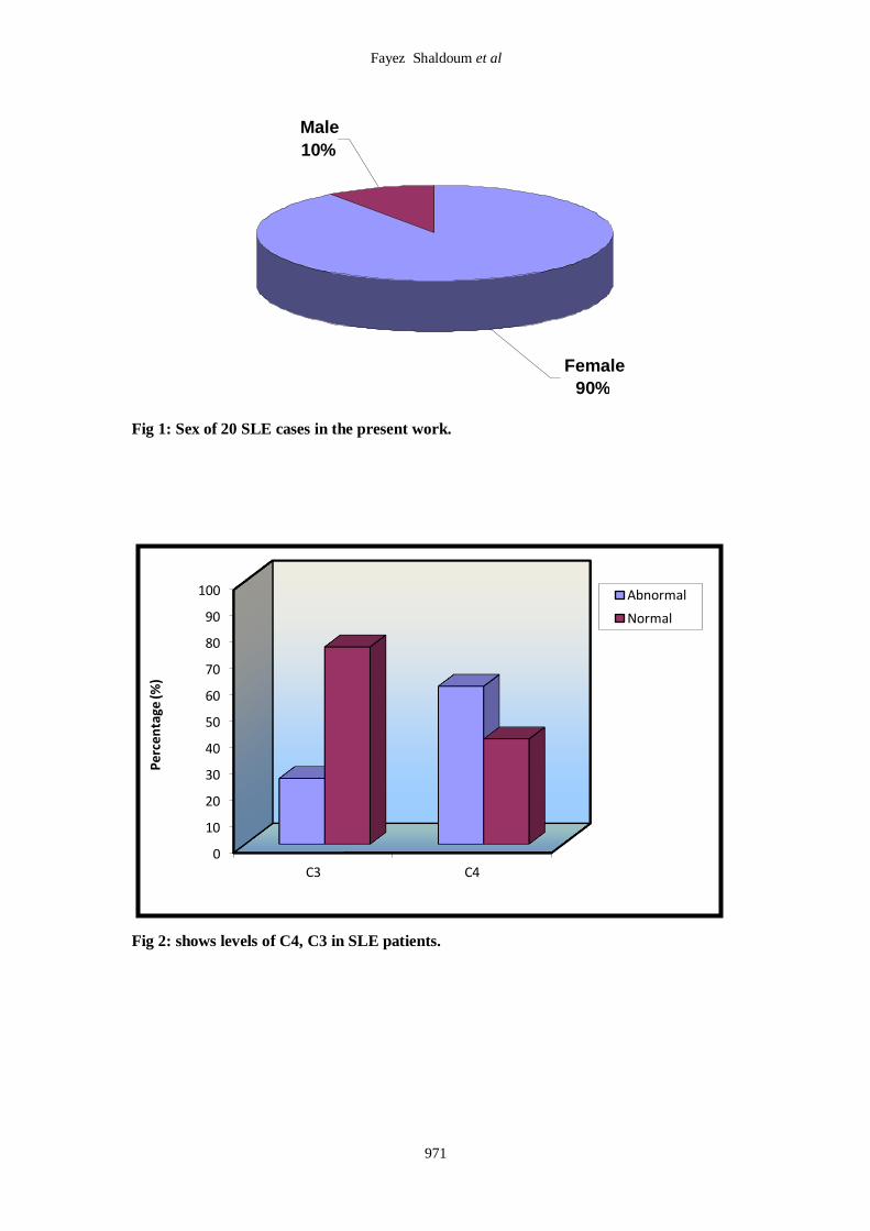
In the present study it was found
that most of patients were female 90% and
only 10% were male (Figure 1).
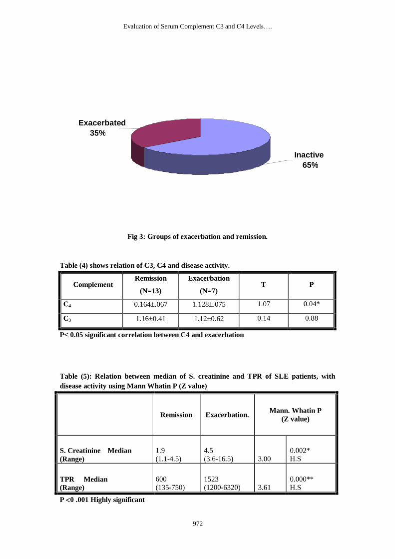
Levels of serum complement C4, C3 in
SLE patients
In the present study, the
complement C3 and C4 has been
measured. It has been found that 60% of
all patients have low level of serum C4,
40% have normal level of serum C4, 25%
have abnormal level of serum C3, and
75% have normal level of serum C3 as
shown in Table 2, Figure2.
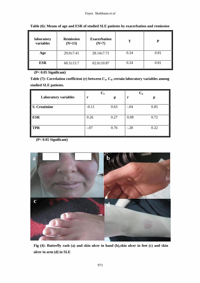
In the current study, patients were
divided into 2 groups, exacerbation and
remission (Table 3 and Figure 3).
Moreover, a positive correlation (P> 0.05)
between low level of C4 and disease
activity was noticed in all SLE
participating patients, While, that was not
the situation in case of C3 (Table 4).
Furthermore, clinical and
laboratory parameters were taken in this
study in order to study and describe the
relation between the level of complement
C3, C4 and the exacerbation and remission
of the disease.The relation between S.
creatinine and TPR, and SLE activity is
shown in table 5. In all the SLE
participants, the relations between C3, C4
with certain laboratory parameters
including: S. creatinine, ESR, TPR and
age were listed in tables (5, 6). An increase
in ESR values in all of SLE patients was
noticed.
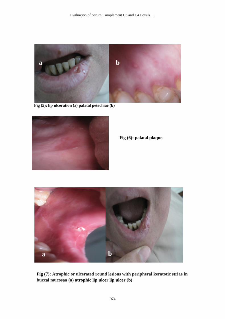
The relation between C4, oral
manifestations and disease activity
Thorough skin examination of
SLE participants, in 60% of them, it was
noticed that active disease manifestations
were as following: butterfly malar rash and
photosensitivity (figure 4a), skin
ulcerations in leg fingers, arm and hand
(figures4b, 4c and 4d).
Active disease group:
From this study, it was found that
the low level of C4 was significantly
correlated (P> 0.05) with disease activity
as shown in table (4). Also acute and
subacute oral manifestations were revealed
in this group of patients meaning that the
decrease in the level of C4 is correlated
with presence of these acute oral
manifestations.
Acute lesions:
In this category of patients 40%,
oral lesions ranged from erythema in the
palatal mucosa and buccal mucosa to
purpuric macules. Petechiae and
Ulcerations were also found in palatal
mucosa and buccal mucosa. Bullous LE:
labial blisters, intra-oral intact or ruptured
blisters, (Figure 5a and 5b).
Subacute lesions:
20% of SLE patients revealed
discrete red patches, which diffusely and
Page 6
Fayez Shaldoum et al
965
discretely arranged in labial and palate
(Figure 6).
Inactive disease group
Through the current study, normal
or increased level of C4 was associated
with decreased disease activity (40%)
which means that the increase in the level
of C4 was correlated with the remission of
the disease. Interestingly, it was noticed
that chronic oral manifestations were
found in 50% of this group of patients
(about 20% of total SLE patients), tables 2
and 4.
Chronic lesions
20% of SLE patients revealed
atrophic or ulcerated round lesions with
peripheral keratoticstriae asymmetrically
distributed in buccal mucosa. Ulcers
ranged from linear with keratoticstriae,
intensely keratotic lesions were also found
on palatal mucosa, (Figure 7a &7b).
DISCUSSION
Systemic lupus erythematosis (SLE) is an
autoimmune disease of uncertain etiology
that is complex in multiple aspects. At the
clinical level, SLE has been described as a
wide array of disorders and patients
manifestations ranging from immediately
life threatening disease of major organs
such as heart, lungs or kidney. Deficiency
of C4, whose gene is found on
chromosome 6 in the MHC III region,
imparts a risk of SLE (Bajaj et al., 2010).
Complement system has important
protective functions in both the innate and
the adaptive immune systems but can also,
when inappropriately activated, cause
tissue damage. Complement deficiency
predisposes to infection and also to
development of autoimmune disease,
especially SLE, and complement is at the
same time involved in the pathogenesis of
this disease (Sturfelt and Truedsson,
2005).
There are three major complement
activation routes: classical, alternative and
lectin pathways. Regardless of how these
pathways are initiated, the complement
activity leads to proteolytic activation and
deposition of the major complement
proteins C4 and C3, which induces
phagocytosis, and the subsequent
assembly of the membrane attack complex
which lyses the invading microbes.
However, complement is a double-edged
sword; adequate complement activation is
necessary for killing the bacteria and
removing the apoptotic cells, while
excessive complement activation can harm
the host by generating inflammation and
exacerbating tissue injury (Chen et al.,
2009).The evidence that products of
complement activation really contribute to
tissue damage in SLE is somewhat
circumstantial (Sturfelt and Truedsson,
2005).
Attention has been paid to other
factors beside C3 and C4 as: age, sex,
ESR, TPR and S. Creatinine in relation to
SLE, in this study. Furthermore, Clinical
studies suggested that the gender in SLE is
influenced by sex hormones, such as
Page 7
Evaluation of Serum Complement C3 and C4 Levels….
966
estrogen and androgen; in agreement with
results obtained from numerous studies
(Weidler et al., 2004; Horák et al., 2009;
Bernknopf et al.,2011).
In the current study C4 and C3
were measured in serum of patients
suffering from SLE. C4 was chosen to be
measured as a component of the classical
or lectin pathway of complement
activation; the easiest to measure is C4.
Additionally, C3 was measured in the
current study because its measurement
together with C4 could enhance the
understanding of the mechanisms involved
and aid in the clinical definition of SLE
(Hussain et al., 2008).
In this work 60% of all SLE
patients participated in the study had low
levels of C4, all those patients presented
criteria of disease flare, while 40% had
normal C4 levels and those patients
presented criteria of disease remission.
These results were in agreement with
results obtained from previous studies
(Hussian et al., 2008; Horák et al., 2009
and Peter et al., 2011).
Hussian et al, (2008) suggested
that low C4 levels may falsely be regarded
as classical pathway activation and they
reported that several other factors may
explain low C4 level such as: Partial
defects or homozygous defects in either
C4A or/and C4B will result in reduced
levels of total C4, reduced synthesis or
increased catabolism of C4 without
corresponding complement activation may
also explain low C4 values.
In 2002, Eniav found that the
development of severe SLE in the absence
of both classical and alternative
complement pathways suggests that it is
the absence of C4, and not the presence of
C3 that is critical in SLE pathogenesis.
Thus, this study supports that complement
C4 provides an important protective role
against the development of SLE.
In the present study 25% of the
patients had low C3, while, 75% had
normal C3. Cameron et al. (1973) studied
55 samples of SLE and he also found that
low plasma C4 concentrations were
commonest in lupus group, but a C3
concentration of below 20% of reference
normal serum was not seen in their study;
in contrast to our data.
. It was found that more than 75%
of all individuals with deficiency of C4
have SLE, which is commonly severe.
Deficiency of Complement may lead to
abnormal in vivo processing of dying cells
that, in the context of an inflammatory
response, could initiate and drive an
autoimmune response leading to the
development of SLE (Carroll, 2004).
Another mechanism in the
pathogenesis of SLE associated with
Complement deficiency that Complement
may be involved in the recognition of self
by B cells and thereby defects in
Complement might result in failure of B
cell negative selection. This failure allows
auto-reactive B cells to survive and
propagate when they would normally
undergo apoptosis (Botto et al., 2009).
Page 8
Fayez Shaldoum et al
967
Moreover, analysis of serologic
factors in SLE has revealed that in many
patients, lower C4 levels occur before the
depression of other complement
components. After the induction of
remission, C4 has a tendency to return to
normal levels more slowly than C3;
decreased C4 levels are mainly present in
those with exacerbation of SLE. Prolonged
decrease in serum C3 levels is associated
with the development of chronic SLE
(Ceribell et al., 2009).
Clinically, most patients included
in this study presented with multiple oral
lesions. Locations more frequently
affected were buccal mucosa, hard palate
and lower lips. Some patients had lesions
affecting more than one oral site. These
findings agree with previous studies, in
which buccal mucosa, palate and
vermilion of lips (more the lower than the
upper lip) are referred as the commonest
sites for lupus oral lesions (Lourenço et
al., 2007 and Simonsen et al., 2008).
Considering morphologic aspects,
the oral lesions examined presented varied
clinical aspects, ranging from the classic
plaques with central erythema surrounded
by a white rim with radiating
keratoticstriae and occasionally
telangiectasias. In this study classic lesions
were present in nearly half of the patients.
This reflects the importance of considering
other clinical hypotheses when examining
oral lesions suggestive of lupus
erythematosus and shows that in many
circumstances their diagnosis is
challenging (Chen et al., 2009 and Peter et
al., 2011).
The data presented in this work
has not found significant correlation
between S. Creatinine, TPR and C3 or C4.
However, a significant correlation between
S. Creatinine, TPR and disease activity has
been found. The decreased levels of C4
could either be due to the low production
or its consumption in a classical activity of
the complement (Hussain et al., 2008).
No authors have discussed a
correlation between TPR and C4 or C3. In
this study, no significant correlation has
been found between the increased level of
TPR and the high activity of C3. These
findings indicated that the decreased levels
of C4 are merely because of the low
production. These findings also explained
that the complement activation is mainly
through the alternative pathway in the
active SLE. An obvious increase in ESR in
all participants was revealed.However, the
statistical analysis of the data presented
here revealed neither significant
correlation between ESR and C3 nor
between ESR and C4. ESR is used as a
screening test to identify patients with
inflammatory conditions. It is a useful
marker to monitor disease activity and is
commonly elevated in patients with SLE
(Luis et al., 2005).
In addition, findings in this study
have shown that all patients had positive
ANA which is an important marker of
SLE. It has been found that all patients
with SLE have a positive ANA test, with a
Page 9
Evaluation of Serum Complement C3 and C4 Levels….
968
sensitivity of 93% to 95% (Wallace,
2007).
Ultimately, SLE is a complex
autoimmune disease with multiple
pathogenic mechanisms. In this study,
serum level of C3 and C4 were assessed,
and a correlation between complement and
different clinical aspects including; oral
manifestations has been established. This
correlation suggests possible use of
decreased level of C4 as a biomarker for
SLE. This study also suggests that the
increased level of C3 is due to alternative
activation of complement pathway in SLE
patients.
REFERENCSES
Ali Khan W, Uddin M, Mohd. Ali Khan W
and Harvinder S. Chabbra (2009): Catecholoestrogens: possible role in systemic
lupus erythematosus. J Oxford, 48: 1345–
1351.
Anaya JM (2010): The autoimmune
tautology. Arthritis Res Ther; 12: 147.
Bajaj DR, Devrajani BR and Matlani BL
(2010): Discoid Lupus Erythematosus:A
Profile. College of Physicians and Surgeons
Pakistan J., 20: 361-364.
Bernatsky S, Joseph L, Boivin JF (2008): The relationship between cancer and medicate
on exposures in systemic lupus erythaematosus: a case-cohort study. Ann
Rheum Dis., 67: 74–9.
Bernknopf A, Rowley K, and Bailey T
(2011): A review of systemic lupus
erythematosus and current treatment options.
Formulary, 46:178–194.
Bhaviya BS, Bindina B, Danu S, Anish TS,
and Rajasi R S (2010): Clinical and
epidemiological features of Systemic lupus
erythematosus and its impact on childbearing.
Calicut Medical Journal, 8: 1-5.
Botto M, Kirschfink M, Macor P,
Pickering MC, Wurzner R, Tedesco F
(2009): Complement in human diseases:
Lessons from complement deficiencies. Mol
Immunol., 46: 2774-83.
Bowers L D(1980): Kinetic serum creatinine
assays I. The role of various
factors in determining specificity. Clin.
Chem., 26: 551-554.
Cabral A R, Cabiedes J, Alarcon-Segovia
D. (2004): Tween 20 detaches cardiolipin
from ELISA plates and makes anticardiolipin
antibodies undetectable regardless of the presence of β2-glycoprotein-I. J Immunol
Methods,175:107–114.
Carroll M. (1994): A protective role for
innate immunity in systemic lupus
erythematosu. Nat Rev Immunol., 4: 825–30.
Ceribell A, Andreoli L, Cavazzana I,
Franceschini F (2009): Complement
Cascade in Systemic Lupus Erythematosus.
Annals, 1173:427–434.
Chen M, Mohamed R, Cees GM (2009): The complement system in systemic
autoimmune disease. Journal of Autoimmunity, 34: 1-11.
Cohen M. (2004): Systemic Lupus
Erythematosus: diagnosis and classification.
Intern Med J., 34: 701-2.
DeFranco AL, Locksley RM, Robertson M
(2007):Immunity: The Immune Response in
Infectious and Inflammatory Disease.
London; Sunderland, MA: New Science
Press; Sinauer Associates, ISBN
9780953918102.
Eniav S, Pozdnyakova O, Ma M, Carroll MC. (2002): Complement C4 is protective
for Lupus Disease Independent of C3. Am
Assoc Immunol., 168: 1036-1941.
Flierman R, Daha R (2007): Pathogenic
role of anti C1q autoantibodies in the
development of lupus nephritis a hypotesis.
Mol Immunol., 44: 133-38.
Habibi S, Saleem MA and Ramanan AV
(2011): Juvenile Systemic Lupus
Erythematosus: Review of Clinical Features
and Management. Indian Pediatrics J., 48:
879-887.
Horák P, Zadrazil J, Ciferská H and
Hermanová Z (2009): Antibodies against
Complement System in SLE. Current
Rheumatology Reviews, 5: 1- 59.
Hussain N, Jaffery G, Hasnain S (2008):
Serum Complement C3 and C4 Levels in
Relation to Diagnosis of Lupus Nephritis.
Tropical Journa Pharmaceutical Research, 7:
1117-1121.
Janeway CA Jr, Travers P, Walport M,
Shlomchik MJ (2001):Immunobiology. (5th Ed.). Garland Publishing.ISBN 0-8153-3642-
X.
Juha Kere (2004): Mapping of susebtability
genes for Systemic lupus erthematosis (SLE).
Yliopistopaino Helsinki, ISBN 952-10-1676-
0.
Kelly J (2007): Understanding the Immune
System How It Works. National Institute of
Page 10
Fayez Shaldoum et al
969
Allergy and Infectious Diseases Science
Education, United States.
Krishnamurthy S and Mahadevan S
(2011); Systemic Lupus Erythematosus:
Recent Concepts in Genomics, Pathogenetic
Mechanisms and Therapies.ISRN Immunology, vol. 2011, Article ID 868964, 7
pages, 2011. doi:10.5402/2011/868964
Lee H S and Bae S C, (2010): What can we
learn from genetic studies of systemic lupus
erythematosus? Implications of genetic
heterogeneity among populations in SLE.
Lupus, 19: 1452–1459.
Liu B, Zhang J, Tan PY, Hsu D, Blom
AM, et al (2011): A Computational and
Experimental Study of the Regulatory
Mechanisms of the Complement System.
PLoS Comput Biol., 7: 1-16.
Löfgren SE, Frostegård J, Truedsson L,
Pons-Estel BA, D'Alfonso S, Witte T(2012): Genetic association of miRNA-146a with
systemic lupus erythematosus in Europeans
through decreased expression of the gene.
Gene immune, 13: 268-74.
Lords RS, Bongiovanni B, Bralley JA
(2002): Estrogen metabolism and the diet-
cancer connection: rational for assessing the
ration of urinary hydroxylated estrogen
metabolites. Altern Med Rev., 7:112–29.
Lourenço SV, Carvalho FRG, Boggio P,
Sotto MN, Vilela MAC, Rivitti EA, Nico
MMS (2007): Lupus erythematosus: clinical
and histopathological study of oral
manifestations and immunohistochemical
profile of the inflammatory infiltrate. J Cutan
Pathol., 34: 558-64.
Luis M V, Graciela S A, Gerald M J, Holly
M. B, Barri J F, and John DR (2005):
Elevation of Erythrocyte Sedimentation Rate
Is Associated with Disease Activity and
Damage Accrual. J Rheumatol., 32:2150-2155.
Peter M H, Cole C, Crawford F, John
WK, Joshua M T, Donna LB,Susan AB
and Karla DK (2011): IgG Autoantibodies
against Deposited C3 Inhibit Macrophage-
Mediated Apoptotic Cell Engulfment in
Systemic Autoimmunity. Immunology J.,
187: 2101-2111.
Rahman A and David A Isenberg
(2008):Systemic Lupus Erythematosus. N
Engl J Med., 358: 929–939. Sarma V and Peter A. Ward (2011): The
complement system. Cell Tissue Res., 343:
227–235.
Simonsen MM, Apparecida M, Vilela C,
Rivitt VA and Lourenco SV (2008): Oral
lesions in lupus erythematosus correlation
with cutaneous lesions. Eur J Dermatol., 18:
376-81.
SturfeltG and TruedssonL (2005): Complement and its breakdown products in
SLE. Rheumatology, 44: 1227-1232.
Trendelenburg M. (2005): Antibodies against C1q in patients with systemic lupus
erythematosus. Springer Semin
Immunopathol., 27: 276-85.
Wallace, Daniel J., and Hahn B H (2007):
Lupus Erythematosus. 7th ed., Lippincott
Williams & Wilkins, Philadelphia, PA, 1441
pp.
Weidler C, Harle P, Schedel J, Schmidt M,
Scholmerich J, Straub RH. (2004): Patients
with rheumatoid arthritis and systemic lupus
erythematosus have increased renal excretion of mutagenic estrogen In relation to
endogenous antiestrogen. J Rheumatol., 3:
489–94.
Yang DH, Chang DM, Lai JH, Lin FH
and Chen CH (2009): Usefulness of
erythrocyte-bound C4d as a biomarker to
predict disease activity in patients with
systemic lupus erythematosus. Oxford
Journals, 48: 1083-1087.
Yuan YJ, Luo XB and Shen N (2010): Current advances in lupus genetic and
genomic studies in Asia. Lupus, 19: 1374–1383.
Zipfel PF, Misselwitz J, Licht C, Skerka C
(2006): The role of defective complement
control in hemolytic uremic syndrome.
Semin.Thromb. Hemost., 32: 146–54.
Page 11
Evaluation of Serum Complement C3 and C4 Levels….
970
Table (1): Clinical and laboratory characteristics of studied SLE patients.
Sex
Female NO (%) Male NO (%)
18 (90.0) 2 (10.0)
Age (year)
X SD Range
28.77.32 19-4
Serum Creatinine
Median
Range
3.55
(1.10-16.5)
TPR
Median
Range
692.5
(135-6320)
ESR
X SD Range
61.0512.51 35.0-80.0
Table (2): Means of serum complements C4, C3 values of SLE patients.
Complement
X SD Range Normal value Abnormal
C4 0.150.07 0.10-.30 8 (40.0) 12 (60.0)
C3 1.150.48 0.10-2.20 15 (75.0) 5 (25.0)
Table (3): Frequency of exacerbation and remission among studied SLE patients.
Activity N = 20
Remission 13 (65.0)
Exacerbation 7 (35.0)
Page 12
Fayez Shaldoum et al
971
Fig 1: Sex of 20 SLE cases in the present work.
Fig 2: shows levels of C4, C3 in SLE patients.
0
10
20
30
40
50
60
70
80
90
100
C3 C4
Pe
rcen
tage
(%)
Abnormal
Normal
Male
10 %
Female
90 %
Page 13
Evaluation of Serum Complement C3 and C4 Levels….
972
Fig 3: Groups of exacerbation and remission.
Table (4) shows relation of C3, C4 and disease activity.
Complement Remission
(N=13)
Exacerbation
(N=7) T P
C4 0.164.067 1.128.075 1.07 0.04*
C3 1.160.41 1.120.62 0.14 0.88
P> 0.05 significant correlation between C4 and exacerbation
Table (5): Relation between median of S. creatinine and TPR of SLE patients, with
disease activity using Mann Whatin P (Z value)
Remission Exacerbation. Mann. Whatin P
(Z value)
S. Creatinine Median
(Range)
1.9
(1.1-4.5)
4.5
(3.6-16.5) 3.00
0.002*
H.S
TPR Median
(Range)
600 (135-750)
1523 (1200-6320) 3.61
0.000** H.S
P 0 .001 Highly significant
Exacerbated
35 %
Inactive
65 %
Page 14
Fayez Shaldoum et al
973
Table (6): Means of age and ESR of studied SLE patients by exacerbation and remission
laboratory
variables
Remission
(N=13)
Exacerbation
(N=7) T P
Age 29.07.41 28.147.71 0.24 0.81
ESR 60.513.7 62.010.87 0.24 0.81
(P> 0.05 Significant)
Table (7): Correlation coefficient (r) between C3, C4, certain laboratory variables among
studied SLE patients.
Laboratory variables
C3
r p
C4
r p
S. Creatinine -0.11 0.63 -.04 0.85
ESR 0.26 0.27 0.08 0.72
TPR -.07 0.76 -.28 0.22
(P> 0.05 Significant)
b a
c d
Fig (4): Butterfly rash (a) and skin ulcer in hand (b),skin ulcer in feet (c) and skin
ulcer in arm (d) in SLE
Page 15
Evaluation of Serum Complement C3 and C4 Levels….
974
Fig (5): lip ulceration (a) palatal petechiae (b)
a b
Fig (6): palatal plaque.
a b
Fig (7): Atrophic or ulcerated round lesions with peripheral keratotic striae in
buccal mucosaa (a) atrophic lip ulcer lip ulcer (b)
b a
Page 16
Fayez Shaldoum et al
975
كدالالت بيولوجية على الذئبة الحمراء C3, C4اإلسترشاد بمستويات المتمم **عبير سعد جاويش, **نجالء محمد الوكيل, يسرا رفاعى عبده, *فايز محمد أحمد شلضوم
.مصر -القاهرة -جامعة األزهر -كلية العلوم -قسم علم الحيوان*
.مصر -القاهرة -.جامعة األزهر -األسنان كلية طب -الطب اإلشعاعىقسم **
في دم المرضى المصابين بمرض الذئبة C3,C4 هدفت هذه الدراسة إلى تحديد مستويات المتمم
.مما ساعدنا في تفسير دور المتمم في تشخيص و نشأة هذا المرضالحمراء
الذئبة الحمراء مرض التهابي مزمن بسبب هجوم الجسم المناعي وهذا يعني أن خاليا الجسم المناعية
و مريض الذئبة , تتعرف على بعض أنسجة الجسم على أنها أجسام غريبة فتهاجمها مسببة تلف هذه األنسجة
أجسام مضادة في الدم يكون هدفها تدمير أنسجة حيوية له ومن هذه األنسجة الجلد و القلب والرئة الحمراء ينتج
.والكلية والمفاصل واألعصاب
وحيث أن الذئبة الحمراء مرض مناعي فمن المحتمل أن تكون هناك عالقه قوية بين التغيرات في نسبة
يمكننا من اإلستعانة بمستويات المتمم في الدم كمرشد على جزيئات المتمم بالدم وتطور المرض في شكل قد
.حدوث الذئبة الحمراء و نشأتها
ضمت هذه الدراسة عشرون مريضا بمرض الذئبة الحمراء طبقا للمعايير جمعية مرضى الروماتيزم
.Radio-Immunoassayوذلك بإستخدام تقنية C3,C4 األمريكية وتم قياس مستويات المتمم
لهما دور هام في نشأة المرض و تطوره وايضا لهما دور في ظهور C3,C4 من نتائج هذه الدراسة ان المتتمم
.االعراض الفمية
.بدم المريض كداللة على مرض الذئبة الحمراء C4وأوضحت الدراسة امكانية استخدام اإلنخفاض فى مستوى


![Association between Serum Matrix Metalloproteinase- (MMP ... · Systemic lupus erythematosus (SLE) is a multisystemic autoimmune disease [1]. Although the pathogenesis of SLE remains](https://static.documents.pub/doc/80x56/5fcc017e5ec16209cf240aa6/association-between-serum-matrix-metalloproteinase-mmp-systemic-lupus-erythematosus.jpg)


