Page 1
Journal of Dental & Oro-facial Research Vol 15. Issue 01 Jan. 2019 JDOR
RUAS 3
Evaluation of Superficial Cervical Plexus Block for
Incision and Drainage of Facial Space Infections: A
Prospective Clinical Study Gyanendra Misra 1, *Kavitha Prasad 2, Lalitha R.M.3, Ranganath Krishnappa4, Rajanikanth B.R.5, Sejal K.M.6,
Parimala Sagar7, Vineeth Kumar8 and Prathibha Gopalraju9
*Corresponding Author E - Mail: [email protected]
Contributors:
1Ex Post Graduate Student, 2Professor and Head, 3Ex Senior
Professor, 4Professor, 5,6,7,8Associate
Professor, Assitant Professor,
Department of Oral and
Maxillofacial Surgery, Faculty of
Dental Sciences, M.S. Ramaiah
University of Applied Sciences,
Bengaluru - 560054
Abstract
Background: Odontogenic
infections are the most common
source for spreading facial space
infections. The infections of these
potential spaces in the facial planes
include acute cellulitis of the soft
tissue with or without true abscess
formation. Aggressive incision and
drainage (I&D) of all the involved
spaces is considered necessary to
assure fast resolution of the infection
and to eventually maintain a patent
airway. An adequate level of
anesthesia has been a critical
component of the treatment plan.
Skin in front of, above, below the ear
and anterior aspect of neck is supplied by cervical plexus. A superficial cervical
plexus block is an option worth considering in these cases. The injection site is
usually far enough away from the intended area for I&D to be performed safely.
Material and methods: The study protocol involved incision and drainage of facial
space infection involving submandibular, submental, sublingual and/or pharyngeal
spaces, performed under MAC and superficial cervical plexus block with
concomitant inferior alveolar nerve block. Clinical parameters evaluated were
intraoperative pain (During incision, drainage, drain fixation and immediately after
the procedure in recovery room), amount of sedation required during the procedure
and complications if any. Results: During incision 80% patients scored the pain on
VAS to be 2, and rest two patients gave a score of 3 and four. During exploration
and drainage of the involved spaces the average pain score was 4.4 where 60%
patients scored it below 4, 30% scored it to be 5 and one patient had extreme pain
and scored it to be 9. The pain scores during drain fixation was 2 or less in 90%
cases and 3 in remaining 30%. Immediately after the incision and drainage all the
patients had a significant pain relief in the recovery room and scored the pain to be
4 or less on VAS scale. 9 of 10 patients did not have any complications during the
procedure. One patient had seizure and cardiac arrest during exploration of the
space, emergency tracheostomy was done and I&D was completed successfully.
Conclusion: Bilateral SCPB with inferior alveolar nerve block provides significant
intraoperative pain control for drainage of submandibular, submental, sublingual
and or pharyngeal space infections. It offers an effective alternate to general
anesthesia which is easy to learn, has low complication rate and a high success rate,
thereby reducing the total length of hospital stay and overall cost of the treatment.
Keywords: Odontogenic Infection, Facial Space Infection, Superficial Cervical
Plexus Block
1. INTRODUCTION
The majority of infections that manifest in the
orofacial region that is 90-95% are odontogenic
in origin. Of these, approximately 70% present as
periapical inflammation or abscess and arise in an
otherwise healthy individual as a result of pulp
necrosis caused by caries, trauma, periodontal
infections or pericoronitis and account for the
most common source for spreading facial space
infections.
Most of the odontogenic infections are self-
limiting and may drain spontaneously. However
these infections may drain into facial spaces
adjacent to the oral cavity and spread
aggressively leading to more severe infection of
these potential spaces in the facial planes, causing
acute cellulitis of the soft tissues with or without
true abscess formation. Later it may spread into
respiratory passages, requiring a timely effort to
establish a patent airway; in addition to the
debridement, incision and drainage and
appropriate antibiotic therapy.
This fast-spreading, indurated cellulitis occurring
in the suprahyoid soft tissues leads to pain,
dysphagia, trismus, swelling, and potential fatal
airway obstruction1. Aggressive Incision and
Drainage (I&D) of all the involved spaces is
Page 2
Journal of Dental & Oro-facial Research Vol 15. Issue 01 Jan. 2019 JDOR
RUAS 4
considered necessary to assure fast resolution of
the infection and to eventually maintain a patent
airway2.
An adequate level of anesthesia has been a critical
component of the treatment plan. Awake
fiberoptic intubation is often the safest option,
although this may be associated with its own
complications2. Some other options available are
inhalational induction and intubation without
muscle relaxants, or tracheostomy under local
anesthesia. Incision and drainage under local
infiltration would be difficult as the anesthesia
can be suboptimal, hence may not permit a
thorough exploration of the wound.
A Superficial Cervical Plexus (SCP) block is an
option worth considering in these cases. The
injection site is usually far enough away from the
intended area for I&D to be performed safely.3
This technique of locoregional anesthesia lowers
the cost of patient care, improves the
effectiveness of treatment and patient comfort.
Hence, the present study was undertaken to
evaluate the use of superficial cervical plexus
block annexed with an inferior alveolar nerve
block for I&D of facial space infections.
2. MATERIALS AND METHODS
The study conducted was an observational study
on patients with orofacial infections attending the
Department of Oral and Maxillofacial Surgery,
Faculty of Dental Sciences, MSRUAS, M.S.
Ramaiah Memorial Hospital, and M.S. Ramaiah
Medical Teaching Hospital Bangalore, from
01/01/2011 to 30/10/2012 with clinically
diagnosed infection of submandibular,
submental, sublingual and/or pharyngeal spaces.
A total of ten cases satisfying the inclusion
criteria were included and the patients were
treated with incision and drainage under
superficial cervical plexus block. The case
history was recorded as per the proforma. The
various parameters which were assessed were
pain, amount of sedative required and
complications.
Inclusion Criteria
Patients with the infection of facial
spaces involving submandibular,
submental, sublingual and/or pharyngeal
spaces
Patients with Ludwig’s angina
Patients with significant medical history
contraindicated for general anesthesia
Exclusion Criteria
Patients with significant respiratory
diseases
Physically fit patients who are highly
apprehensive for the procedure under
local anesthesia
Patients with known allergy to local
anesthetic agents
Facial space Infections involving deep
neck and mediastinal spaces
Surgical Technique
A. Patient Positioning: The patient was
placed in a supine position, with his head
turned to the side contrary to the one to
be blocked
B. Equipment: The basic armamentarium to
perform SCP locoregional anesthesia
included sterile towels and gauze packs,
20-mL syringes with local anesthetic
solution, 1.5ʺ, 25 - gauge needles, sterile
gloves, and a marking pen. O2 pulse
oxymeter, ECG, intubation and
resuscitation equipment
C. Regional Anatomy and Landmarks: The
cervical plexus is formed by the anterior
divisions of the four upper cervical
nerves. Situated on the anterior surface of
the four upper cervical vertebrae, it rests
on the levator anguli scapulae and
scalenus medius muscles, and is covered
by the sternocleidomastoid muscle.
Emerging through the intervertebral
foramen, the dorsal and ventral roots
combine to form spinal nerves. The
anterior rami of C2 through C4 form the
cervical plexus (the C1 root is primarily
a motor nerve and it is not blocked by this
technique). The cervical plexus gives off
both superficial (SCP) and deep branches
(deep cervical plexus). The branches of
Page 3
Journal of Dental & Oro-facial Research Vol 15. Issue 01 Jan. 2019 JDOR
RUAS 5
the SCP emerge as four distinct nerves
from the posterior border of the
sternocleidomastoid muscle and supply
innervation to the skin and superficial
structures of the head, neck, and
shoulder. The deep branches of the
cervical plexus innervate the deeper
structures of the neck, including the
muscles of the anterior neck and the
diaphragm (phrenic nerve). The third and
fourth cervical nerves typically send a
branch to the spinal accessory nerve, or
directly into the deep surface of the
trapezius to supply sensory fibers to this
muscle (Fig.1 and 2).
Figure 1: Landmarks and technique of SCPB4
Figure 2: Diagrammatic cross-sectional drawing
of anatomy of neck at vertebral level (C4)5
D. Landmarks: Mastoid Clavicular insertion of
the SCM muscle; Sternal insertion of the
SCM muscle Procedure: All patients were
positioned in neck extension for the ease of
surgery and for correct localization of
landmarks. Oxygen was administered via a
nasal cannula. Against gentle resistance from
the anesthetist's hand, the patient was
instructed to lift his or her head. A
simultaneous slight Valsalva's maneuver was
encouraged to help outline the
sternocleidomastoid muscle and locate the
external jugular vein. The midpoint of the
posterior border of the sternocleidomastoid
muscle was located and marked. This
corresponds with the external jugular vein as
it crosses the posterior border of the muscle
(Erb’s point). The needle was inserted
perpendicular to the skin at Erb’s point for all
its length (1.5 cm), avoiding muscular (SCM)
or vascular (external jugular vein) puncture,
without looking for any bony contact (Fig.3).
The volume of 15 ml (7.5ml 0.5%
Ropivacaine + 7.5ml 2% lignocaine with
1:200000 adrenalin) local anesthetic was
injected over 5 min with multiple aspiration
tests and maintenance of verbal contact with
the patient. An intraoral inferior alveolar
nerve block was also given with 2ml 2%
lignocaine with 1:200000 adrenalin by
classical or Vazirani Akinosi technique. All
the patients were given minimal sedation
with 40µg Fentanyl+1mg Midazolam.
Incision and drainage was performed under
superficial cervical plexus block. If the intra
operative pain control was inadequate
additional 50µg Fentanyl and 1mg
Midazolam was given intravenously.
Figure 3: Localization of landmarks and direction
for performing blockage of superficial branches of
cervical plexus6
The cervical plexus block was performed by the
same technique and by same surgeon in all the
patients and the primary outcome variable was
pain which was assessed using a 10 point Visual
Analog Scale (VAS) recorded during incision,
exploration of the involved space, drain fixation
and post-operatively in the recovery room. The
heart rate, blood pressure and peripheral oxygen
saturation was measured at all times. Clinical
parameters measured were pain (during the
Page 4
Journal of Dental & Oro-facial Research Vol 15. Issue 01 Jan. 2019 JDOR
RUAS 6
incision, exploration of space, drain fixation,
post-operatively in the recovery room), additional
sedation required and complications if any.
3. RESULTS
A total of 10 patients with infection of
odontogenic origin involving submandibular,
submental, sublingual and or pharyngeal spaces
satisfying inclusion and exclusion criteria who
reported to M.S. Ramaiah Medical Teaching /
Memorial Hospital, M.S. Ramaiah Dental
College were included in the study.
Out of these ten patients, 30% (3) were female
and 70% (7) were males and age ranging from 16-
58 years. Mean age was 37.7 years. The most
common source of the infection was 1st and 2nd
mandibular molars in 60% cases and mandibular
3rd molars in 30% cases; in one case the etiology
was found to be maxillary 3rd molar.
90% of the cases with submandibular space
infection showed multiple space infection. With
a predilection towards right side. 40% cases
presented with right submandibular and
submental space infection, three cases with
Ludwig’s angina where the etiology was right 1st
or 2nd molar. Only 3 cases presented to the
hospital with left submandibular and submental
space infection.
50% of patients at the time of presentation were
febrile. The total leucocyte count ranged from
10.2 ×10³ -18.6 ×10³. The mean TLC was 13.6
×103. All the patients were started on empirical
antibiotics (i.e. Inj. amoxicillin clavulanic acid
and Inj. metronidazole) after admission and were
continued postoperatively. In three patients of
Ludwig’s angina, additionally Inj. amikacin was
given.
9 of 10 cases presented with swelling of the
submandibular region with difficulty in
swallowing and speech. One patient also had
trismus along with these symptoms.
During incision 80% patients scored the pain on
VAS to be 2, and rest two patients gave a score of
3 and 4. During exploration and drainage of the
involved spaces the average pain score was 4.4
where 60% patients scored it below 4, 30%
scored it to be 5 and one patient had extreme pain
and scored it to be 9. The pain scores during drain
fixation was 2 or less in 90% cases and 3 in
remaining 30%. Immediately after the incision
and drainage all the patients had a significant pain
relief in the recovery room and scored the pain to
be 4 or less on VAS scale. An additional 50µg
Fentanyl and 1 mg midazolam was given to first
three patients.
Amongst the ten patients, nine did not have any
complications during the procedure. One patient
had seizure and cardiac arrest during exploration
of the space, emergency tracheostomy was done
and I&D was completed successfully.
4. DISCUSSION
Maxillofacial infections are a public health
concern that arise in otherwise healthy patients as
a sequel to pulp necrosis caused by caries or
trauma. Periodontal infections, pericoronitis, and
surgery are other sources. Many odontogenic
infections drain spontaneously, but drainage may
need to be established.7, 8 Predisposing factors
such as alcoholism, immunosuppression,
uncontrolled diabetes mellitus and multiple
underlying medical conditions are reported to
increase the risk of odontogenic infection.8
The anatomy of maxillofacial structures can
influence the spread of odontogenic infection.9 It
is essential for the diagnosis and treatment to
specify the cause and to evaluate the extent of
infection.10 Complications, such as
retropharyngeal spread, suppurative mediastinal
extension, airway obstruction, pleuropulmonary
suppuration, and hematogenous dissemination to
distant organs, clearly indicate the potentially
serious nature of these infections.11
Successful treatment requires an understanding
of the microflora, the regional anatomy, the
disease process, treatment methods available, and
interdisciplinary team collaboration.12 Prolonged
hospitalization can also become an economic
factor for both the patient and society.
The management of odontogenic infections
includes the use of high-dose intravenous
Page 5
Journal of Dental & Oro-facial Research Vol 15. Issue 01 Jan. 2019 JDOR
RUAS 7
bactericidal antibiotics. The recommended
antibiotics are penicillin-metronidazole,
ampicillin - sulbactum, or clindamycin. Certain
cephalosporins may also be useful in selected
patients. Early surgical intervention i.e
aggressive incision and drainage of all of the
involved spaces is necessary to assure early
resolution of the infection. Continual airway
monitoring and the establishment of surgical
airways is the final portion of the treatment
triad.13
In an exhaustive review of the literature, from
1945 to 1979, 75 cases of Ludwig Angina were
treated, and the authors strongly advocated
elective tracheostomy under local anesthesia.
However, there may be good reason to avoid
tracheostomy. Cellulitis of the neck involving the
tracheostomy site makes it a more difficult
procedure. Moreover, surgical dissection of the
facial planes in the neck may actually open and
contaminate the pathways, leading to life-
threatening mediastinal invasion. Other options
for airway management may include orotracheal,
blind nasotracheal, and fiber optic intubation or
cricothyroidotomy with jet insufflation.14
Nevertheless, general anesthesia has its down
sides; high economic cost, a number of highly
trained personnel required, morbidity and
mortality, and high cost equipment.2 Regional
anaesthesia lowers costs of patient care because
of the shorter duration of recovery and
procedure.3 The use of locoregional anesthesia of
the Superficial Cervical Plexus (SCP) is
commonly and frequently used in a variety of
disciplines; that is, carotid endarterectomy,
thyroidectomy and vocal cord surgery.2
The General Anesthetic versus Local Anesthetic
(GALA) study tested the hypothesis that local
anesthesia is safer than general anesthesia in a
large population undergoing carotid
endarterectomy.4 The effective use of local
anesthetics with sedative hypnotic- amnestic
agents can provide both patient comfort and
safety to perform deeper spaces I&D.2
The SCP, by way of the anterior rami of C-2, C-
3, and C-4, distributes sensory innervation to the
skin, starting at the base of the skull and covering
the anterior and lateral neck from the mandible to
the clavicle and the superficial aspect of the
shoulder.15
The SCPB is simple and easy to perform. SCPB
provides the same sensory (dermatome)
anesthesia as the DCPB, which simply
incorporates the motor component of the Cervical
Plexus at the nerve roots before the sensory and
motor aspects separate.15
Mukhopadhyay et al. in their study successfully
used bilateral SCPB for excision of thyroglossal
cyst and fistula or branchial fistula, lymph node
excision or biopsy, thyroidectomy and other neck
swellings like sebaceous cyst, lipoma and
granuloma.4
A SCPB is an option worth considering for I&D
of odontogenic infections. It is easy to perform
and the success rate is high. The injection site is
usually far enough away from the infected area
for it to be performed safely. A SCPB alone may
be adequate for most neck and face abscesses, but
the auriculotemporal nerve can also be blocked if
necessary.3 For I&D of an abscess which is
restricted to the submandibular area, there is need
for a long buccal nerve block. Also if there is
submental spread, an inferior alveolar nerve
block is required.2
Shteif et al. also concluded that superficial
cervical plexus block with concomitant
mandibular nerve block has a high success rate,
low complication rate and high patient
acceptance rate for the drainage of submandibular
and submental abscesses.2
Cervical plexus block is associated with a
frequent incidence of patient anxiety, discomfort,
and pain during carotid endarterectomy because
of irregular innervation of the operative field. To
improve patients’ comfort and cooperation,
supplemental IV analgesia and/or sedation is
often administered, which may impair mental
status evaluation and compromise respiratory and
cardiovascular function,16 if Ramifentanyl and
Propofol are used at anesthetic doses. However,
in our study Fentanyl and Midazolam were used
and only in subanesthetic doses. We did not
observe any impairment of mental function or
Page 6
Journal of Dental & Oro-facial Research Vol 15. Issue 01 Jan. 2019 JDOR
RUAS 8
compromised respiratory and cardiovascular
function in our patients.
The present study included ten patients with
infection of facial spaces involving
submandibular, submental, sublingual and or
pharyngeal spaces or Ludwigs Angina. Patients
with significant respiratory diseases, who were
highly apprehensive for the procedure to be
performed under local anesthesia, with
involvement of mediastinal spaces and with
specific allergies to the local anesthetic agent
were excluded from the study. Among these 30%
(3) patients were female and 70% (7) were male
with age ranging from 16 - 58 years and mean age
of 37 years. This is similar to a study by
Kannangara et al who reported a male
predominance in his study, finding 40 (66%)
males and 21 females (34%), with an age range
of 6-79 years and most patients being between
20- 29 years old. A survey by Sethi and Stanley
identified a slight predilection for males, but did
not list exact numbers for each gender, also
reported age range of 3- 87 years with an average
of 45.5 years. In contrast, a study by Hunt et al
noted a female predominance with 30 males
(41%) and 43 females (59%).17,18
The submandibular space is considered to be
important in odontogenic infection, as infection
in this space is often accompanied by changes in
other surgical spaces. Ariji et al reported that
75.8% patients of submandibular space infection
also showed involvement in multiple spaces.19
The findings of the present study corroborates
with the above mentioned observations. In our
study 90% of patients with infection of
submandibular space also showed multiple space
involvement. Involvement of the
submandibular space causes severe symptoms
such as neck rigidity, trismus, dysphagia,
respiratory distress, sialorrhoea, and pyrexia.19
and difficulty in speech.2, 20 The most common
clinical presentation in our study was pain,
dysphagia, trismus, difficulty in speech and
pyrexia.
In the present study, ten cases of submandibular
space infection were included, nine cases
presented with multiple space involvement. Of
which these cases, three cases were diagnosed
with Ludwigs Angina. A similar finding was
reported by Labriola et al in their study wherein
the most frequent single space infection was
Submandibular (26%), followed by Buccal
(21%), Masticator (15%), and Canine (13%).18 In
contrast, a five year retrospective study by Wang
et al reported that in an adult population the most
commonly involved site for odontogenic
infection was maxilla (46%) followed by
mandible (40%), and both in 4% cases.8 Further,
none of the above mentioned studies provided
any information regarding multispace infections.
In this regard, it is plausible to suggest that space
infection of the perimandibular region constitutes
a major percentage of maxillofacial infections;
hence the knowledge and application of SCPB
annexed with a local block of mandibular region
may bring about significant pain relief in patients.
In the present study the teeth involved (focus of
infection) were 1st and 2nd mandibular molars in
60% cases and mandibular 3rd molars in 30% and
maxillary 3rd molars in 10% of cases. Chow et al
in their review reported mandibular molars to be
the most frequently involved teeth in odontogenic
infections18. All the patients included in the study
presented with the chief complaint of pain and
swelling in submandibular region with or without
difficulty in swallowing. On admission 50% of
the patients were febrile, routine blood
investigations were sent and patients were started
on empirical antibiotics i.e Inj Amoxycillin
clavulanic acid 1.2gm and Metronidazole 100 ml.
The total WBC count ranged from 18.7×10³/µL
to 10.2×10³/µL with an average of 13.6×10³/µL.
I & D under SCPB and MAC (Monitored
Anesthetic Care) was planned after a routine
radiographic investigation to identify the
causative tooth.
The I&D was planned after the first sign of nerve
blockage i.e. the loss of pin prick sensation in the
area of distribution of the representative
components of superficial cervical plexus (5-10
mins after local anesthetic injection deep to
superficial cervical fascia i.e the ICPB) with a
mixture of 2% Lignocaine with 1:200000
adrenaline and 0.5% ropivacaine under MAC.
Ropivacaine was chosen as it is known to be less
cardio toxic after intravenous administration than
bupivacaine, even at an equipotent dose. This
Page 7
Journal of Dental & Oro-facial Research Vol 15. Issue 01 Jan. 2019 JDOR
RUAS 9
characteristic is an important advantage when
relatively large volumes of local anesthetic are
administered in a highly vascularized area.21 Also
Akerman et al reported Ropivacaine to have a
more rapid onset of action in comparison with
bupivacaine due to its weaker binding to
extraneural fat and tissues and to its greater
availability for transfer to the site of action in the
nerve. Conversely, the long duration of action of
Ropivacaine may be related partially to its
vasoactivity, as a broad range of Ropivacaine
concentrations can cause vasoconstriction.
Addition of lignocaine was to reduce the potential
toxicity of using a large volume of a local
anesthetic agent and further hasten the onset of
action.
In the existing literature various techniques /
methods have been described to block SCP.
Murphy, Scott, Prys-Roberts, and Katz have all
described the superficial injection as being
simply `subcutaneous'. However, Chaikof and
colleagues have suggested it should be
`intradermal' (i.e. even more superficial). In
contrast, Yerzingatsian advised that the injection
should properly be made into the body of the
sternomastoid muscle, so that the solution is
deposited below the investing fascia.22
A superficial plexus block should properly
involve injection below the investing fascia of the
neck, and it is only then that the injectate enters
the deep cervical space.5 Tonkovic et al in their
study called such an injection as intermediate
cervical plexus block, wherein the needle was
inserted at the Erb’s point or punctum nervosum,
to a depth of 1–1.5 cm perpendicular to the skin
until a loss of resistance (past the investing layer
of the deep cervical fascia) was obtained. In his
opinion, ICPB combined best features of
traditional two regional techniques (SCPB and
DCPB) simplicity, reliability and low
complication rate.23
The simple subcutaneous injection for SCPB can
either be performed by a two injection (caudal
and rostral directions) or a three injection
technique (caudal, rostral and a third
subcutaneous transverse injection to ensure the
block of the transverse cervical branches) as
described by Herbland et al.21 In the present study
we used an Intermediate cervical plexus block
with mixture of Ropivacaine and Lignocaine with
adrenaline and intraoral inferior alveolar nerve
block. In the operating room to alleviate anxiety,
all patients were sedated with 40µg Fentanyl and
1mg midazolam. As per the initial study design
SCPB was administered at Erb’s point to a depth
of 1.5 cm with a mixture of 2.5 ml 2% lignocaine
with 1:200000 adrenalin and 2.5 ml 0.5%
Ropivacaine unilaterally on the side of space
involvement. Oxygen was administered to all
patients via a simple nasal cannula. Monitoring of
blood pressure and heart rate,
electrocardiography, and pulse oximetry was
done in the intraoperative period. The first patient
scored the pain during incision as 4 on a 10 point
VAS scale, however experienced extreme pain
during exploration and scored it to 9. To
successfully drain the involved space, an
additional 50 µg Fentanyl and 2 mg Midazolam
had to be administered.
Since the first patient experienced extreme pain
during exploration, it was decided to give
bilateral SCPB irrespective of the involvement of
spaces (Unilateral/Bilateral) in the following
cases. This was in accordance to the study by
Krovvidi et al.24 who reported that while
achieving adequate analgesia in the cervical
plexus region, several problems may arise due to
innervation from the contralateral side, variant
sensory fibers from the vagus or ansa cervicalis
or high dissection of the carotid artery by the
surgeon, which leads to pain in submandibular
and dental region.
We observed that the pain scores with bilateral
SCPB were better but the following two patients
were still not comfortable during the procedure
and scored the pain to be 3 and 5 during incision
and drainage respectively. Therefore, additional
sedation was required in these two patients. As
the literature suggests we attributed this lesser
pain control to the lesser amount of solution used.
After first three patients it was decided to give
bilateral SCPB with the local anesthetic volume
increased to 15 ml of prepared mixture of local
anesthetic (7.5 ml 2% lignocaine with 1:200000
adrenaline + 7.5 ml 0.5% Ropivacaine). The
bilateral superficial cervical plexus block with
7.5 ml of solution on each side effectively
Page 8
Journal of Dental & Oro-facial Research Vol 15. Issue 01 Jan. 2019 JDOR
RUAS 10
reduced intra operative pain. Following 7 patients
scored the pain to be below 2 and 4 on VAS scale
respectively during incision and drainage. No
patient in the present study thereafter received
more than 40µg of Fentanyl and 1 mg of
midazolam, indicating a sufficient level of pain
control.
A wide variation ( 3.6 ml- 30 ml) exists between
diverse authors regarding the amount of solution
used for the bilateral SCPB.6 This was the reason
for our initial difficulty in choosing the volume of
anesthetic. We observed that the pain score was
higher in 30% patients (VAS -8) who received 10
ml of the anesthetic as compared to the rest 70%
patients (VAS-3,4) who received 15 ml of the
prepared solution. A study by NYSORA
observed that a low concentration may suffice
when the needle is ideally placed in the vicinity
of the cervical plexus nerves, this is often not the
case and the higher concentration results in both
a higher success rate and a longer duration of
blockade.
In our study we observed that the pain control was
very good during incision and drain fixation but a
higher pain score was recorded during
exploration of the spaces (Figure 4) probably due
to presence of infection or the fact that this block
may not be able to cover the deeper
compartments of the neck to alleviate pain during
swallowing as suggested by Mukopadhyay et al.4
in their study on thyroid surgeries. Therefore in
our opinion, combining the SCPB with
mandibular nerve block instead of inferior
alveolar nerve block may further increase the
patient comfort. To the best of our knowledge
from the existing literature there are not sufficient
studies in Oral and Maxillofacial Surgery on pain
experienced by the patients during I & D of facial
space infection with or without the SCPB for
appropriate comparison for its effectiveness in
pain control.
The pain score during drain fixation was 2 or less
in 90% cases and 3 in remaining 10%,
immediately after I&D, in the recovery room all
patients had a significant pain relief, and gave a
score less than 4 on VAS scale. The post-
operative recovery was uneventful in all the
patients. Although existing literature lists the
possible complications of the block which
includes infection, hematoma, phrenic nerve
blockade, local anaesthetic toxicity, nerve injury
and spinal anaesthesia, there are only isolated
case reports of such complications.
Mukhopadhyay et al.4 describe that blockade of
the phrenic nerve does not occur after superficial
cervical plexus block, but is common with deep
cervical plexus block. Thus highly stressed
patients as well as patients with significant
respiratory disease may be considered a
contraindication for superficial cervical plexus
block.14
In our study of ten patients, three patients
presented with Ludwigs angina, two of whom had
respiratory distress at the time of presentation. Of
these three patients, two were managed
successfully under bilateral SCPB and MAC
whereas third patient had to undergo
tracheostomy. This patient was a 55 year old male
who presented with Ludwigs angina, with a
history of empyema drained under general
anesthesia 15 days ago. The patient started
rapidly desaturating, so was taken up for
emergency decompression under SCPB with
tracheostomy setup standby. The patient seized
and arrested during exploration of the involved
space. Emergency tracheostomy was done and
I&D was completed successfully.
Conversion to general anesthesia is considered as
a complication of the SCPB but in this instance it
cannot be directly attributed to the block or the
technique. As multiple aspirations were
confirmed before depositing the solution it is very
unlikely for the solution to have been deposited
intravascularly to have caused seizure and cardiac
arrest. In our opinion patients preexisting
respiratory condition along with longer period of
desaturation might have triggered the event.
All except one patient in our study had an
uneventful procedure and were discharged in 0 to
10 days of hospital stay. Peters et al in their study
on maxillofacial infection suggested that LOS is
best predicted by knowing the patient’s status
with respect to an underlying medical condition
and the infection location. After accounting for
these variables, if a patient actually requires
operative management, only an extra day is added
Page 9
Journal of Dental & Oro-facial Research Vol 15. Issue 01 Jan. 2019 JDOR
RUAS 11
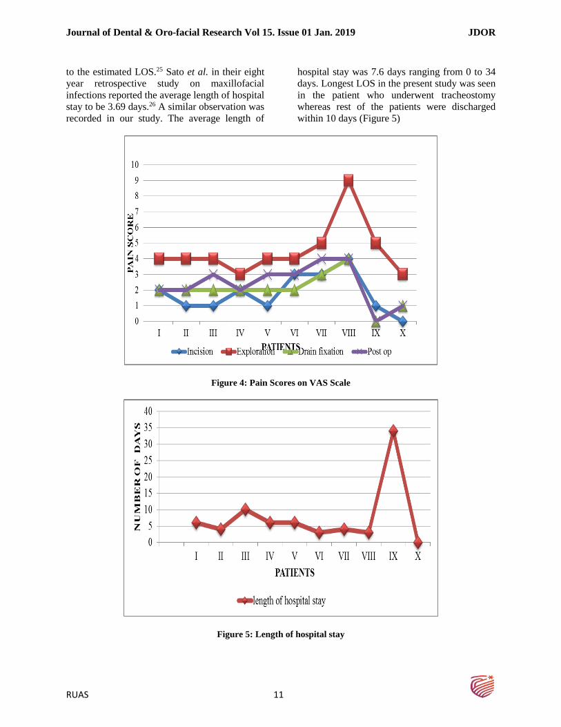
to the estimated LOS.25 Sato et al. in their eight
year retrospective study on maxillofacial
infections reported the average length of hospital
stay to be 3.69 days.26 A similar observation was
recorded in our study. The average length of
hospital stay was 7.6 days ranging from 0 to 34
days. Longest LOS in the present study was seen
in the patient who underwent tracheostomy
whereas rest of the patients were discharged
within 10 days (Figure 5)
Figure 4: Pain Scores on VAS Scale
Figure 5: Length of hospital stay
Page 10
Journal of Dental & Oro-facial Research Vol 15. Issue 01 Jan. 2019 JDOR
RUAS 12
In the present study on ten patients, we observed
that the intermediate cervical plexus block
combined with inferior alveolar nerve block with
15 ml of prepared anesthetic solution caused a
significant reduction in the intraoperative pain
during I&D of submandibular, submental,
sublingual space infection.
By our understanding the SCP can be blocked by
the following techniques- Superficial,
intermediate and deep (Fig. 6). Pandit et al. in a
cadaveric study reported that subcutaneous
injection for the SCPB alone is unlikely to be
clinically effective and concluded that a SCPB
should properly involve injection below the
investing fascia of the neck, and it is only then
that the injectate enters the deep cervical space.5
Figure 6: Proposed method of classifying
Cervical Plexus Block
Various authors have used different
concentration, type and volume of the anesthetic
solution for the block, comparative studies show
that the duration and onset of anesthesia produced
by 0.5 and 0.75% Ropivacaine fails to show any
significant difference.21, 27 Addition of certain
drugs like Clonidine or Fentanyl to the local
anesthetic mixture can also be considered in
future studies to increase the effectiveness and/or
the duration of the block.
In our study we chose to give 0.5% Ropivacaine
with 2% Lignocaine, and a total volume of 15 ml
of local anesthetic for bilateral SCPB. In our
opinion a volume greater than 15 ml of local
anesthetic when injected might diffuse through
the deep cervical fascia and also cause the
blockade of deep cervical plexus as demonstrated
by a cadaveric study by Pandit et al.5 Bilateral
SCPB is an easy to learn technique and provides
sufficient anesthesia for the I&D of
submandibular, submental, sublingual and or
pharyngeal space infections to be performed with
significant pain relief. It has a very low
complication rate and a high success rate,
although it is most commonly supplemented with
sedation in our experience I&D of these spaces
can be performed under SCPB with minimal or
no sedation. It is an effective technique which
avoids general anesthesia and its complications
reducing the total length of hospital stay and the
overall cost.
The limitations of the present study were the
small sample size and the change in the protocol
during the study period. This can be attributed to
the existing confusion in the literature regarding
the volume, concentration and type of anesthetic
to be used for the block. Also, in our opinion use
of mandibular nerve block would have provided
better pain control during exploration close to the
mylohyoid muscle and body of mandible. There
is a need for further study with a larger sample
and a study design suitable to compare the pain
scores during incision and drainage with and
without the block. Also randomized studies are
required to determine the quantity of local
anesthetic that should be used and technique to be
used to block the SCP to perform incision and
drainage successfully.
5. CONCLUSION
The SCPB provides adequate anesthesia for
performing incision and drainage of
submandibular, submental, sublingual and
pharyngeal spaces. However, it is better to
combine it with mandibular nerve block when
exploration is close to the mylohyoid muscle. The
patients’ pain scores are significantly less with
this block and therefore are more co-operative
and allow complete drainage of the facial spaces.
Page 11
Journal of Dental & Oro-facial Research Vol 15. Issue 01 Jan. 2019 JDOR
RUAS 13
REFERENCES
[1] Flynn TR, Shanti RM, Levi MH, Adamo AK,
Kraut RA, Trieger N., Severe Odontogenic
Infections, Part 1: Prospective Report. J Oral
Maxillofac Surg. 2006; 64(7):1093-103.
[2] Shteif M, Lesmes D, Hartman G, Ruffino S,
Laster Z. The Use of the Superficial Cervical
Plexus Block in the Drainage of Submandibular
and Submental Abscesses-An Alternative for
General Anesthesia. J. Oral Maxillofac Surg.
2008; 66(12):2642-5.
[3] Ling KU HM, Ha KO, Wang CY. Superficial
Cervical Plexus Block Combined with
Auriculotemporal Nerve Block for Drainage of
Dental Abscess in Adults with Difficult
Airways. Anaesth Intensive Care. 2009;
37(1):124-6.
[4] Mukhopadhyay S, Niyogi M, Dutta M, Ray R,
Gayen GC, Mukherjee M, Bilateral Superficial
Cervical Plexus Block with or without Low-
Dose Intravenous Ketamine Analgesia:
Effective, Simple, Safe, and Cheap Alternative
To Conventional General Anesthesia for
Selected Neck Surgeries. Local and Regional
Anesthesia. 2012; 5:1-7.
[5] Pandit JJ DD, Morris JF. Spread of Injectate
with Superficial Cervical Plexus Block in
Humans: An Anatomical Study. British Journal
of Anaesthesia. 2003; 91(5):733-5.
[6] Troncoso GA NR, Araya JE, Galdames IC.
Efficacy of Anesthetic Blockage of Superficial
Branches of the Cervical Plexus. International
J Odontostomat. 2008; 2(1):77-81.
[7] Gill Y SC. Orofacial Odontogenic Infections:
Review of Microbiology and Current
Treatment. Oral Surc Oral Med Oral Pathol.
1990; 70(2):155-8.
[8] Wang J, Ahani A, Pogrel MA. A Five-Year
Retrospective Study of Odontogenic
Maxillofacial Infections in a Large Urban
Public Hospital. International Journal of Oral
and Maxillofacial Surgery. 2005;34(6):646-9.
[9] Ohshima A, Ariji Y, Goto M, Izumi M, Naitoh
M, and Kurita K, Anatomical Considerations
for the Spread of Odontogenic Infection
Originating from the Pericoronitis of Impacted
Mandibular Third Molar: Computed
Tomographic Analyses. Oral Surgery, Oral
Medicine, Oral Pathology, Oral Radiology,
and Endodontology. 2004; 98(5):589-97.
[10] Obayashi N, Ariji Y, Goto M, Izumi M, Naitoh
M, Kurita K, Spread of Odontogenic Infection
Originating in the Maxillary Teeth:
Computerized Tomographic Assessment. Oral
Surgery, Oral Medicine, Oral Pathology, Oral
Radiology, and Endodontology. 2004;
98(2):223-31.
[11] Zeitoun IM DP. Cervical Cellulitis and
Mediastinitis Caused by Odontogenic
Infections: Report of Two Cases and Review of
Literature. J Oral Maxillofac Surg. 1995;
53:203-8.
[12] Marilyn E. Levi VDE. Oral Infections and
Antibiotic Therapy. Otolaryngologic Clinics of
North America. 2011; 44(1):57-78.
[13] Peterson Lj. Contemporary Management of
Deep Infections of the Neck. J Oral Maxillofac
Surg. 1993; 51:226-31.
[14] Gupta AK DV, Rudagi VK, Gupta A. Drainage
of Ludwig’ Angina under Superficial Cervical
Plexus Block in Pediatric Patient. Anestesia
Pediatrica e Neonatale. 2009; 7(3).
[15] Masters R, Castresana E, Castresana M.
Superficial and Deep Cervical Plexus Block:
Technical Considerations. Journal of the
American Association of Nurse Anesthetists.
1995; 63(3):235-43.
[16] Sindjelic RP, Vlajkovic GP, Davidovic LB,
Markovic DZ, Markovic MD. The Addition of
Fentanyl to Local Anesthetics Affects the
Quality and Duration of Cervical Plexus Block:
a Randomized, Controlled Trial. Anesth Analg.
2010; 111(1):234-7.
[17] Richard h. Haug mjH, a. Thomas indresano. An
Epidemiologic and Anatomic Survey of
Odontogenic Infections. J Oral Marillofac
Surg. 1991; 49:976-80.
[18] Storoe W, Haug RH, Lillich TT. The Changing
Face of Odontogenic Infections. J Oral
Maxillofac Surg. 2001; 59(7):739-48;
discussion 48-9.
[19] Ariji Y, Gotoh M, Kimura Y, Naitoh M, Kurita
K, and Natsume N, et al. Odontogenic Infection
Pathway to the Submandibular Space: Imaging
Page 12
Journal of Dental & Oro-facial Research Vol 15. Issue 01 Jan. 2019 JDOR
RUAS 14
Assessment. International journal of Oral and
Maxillofacial Surgery. 2002; 31(2):165-9.
[20] Mehrotra M MS. Decompression of Ludwig
Angina under Cervical Block. Anesthesiology.
2002; 97(6):1625-6.
[21] Herbland A, Cantini O, Reynier P, Valat P,
Jougon J, Arimone Y, The Bilateral Superficial
Cervical Plexus Block with 0.75% Ropivacaine
Administered before or after Surgery does not
Prevent Postoperative Pain after Total
Thyroidectomy. Regional Anesthesia and Pain
Medicine. 2006; 31(1):34-9.
[22] Correct Nomenclature of Superfcial Cervical
Plexus Blocks [Internet]. 2012 [cited Oct 17
2012]. Available
from:http://bja.oxfordjournals.org.
[23] Tonkovi, JPD, Sakan S, Baronica R.
Intermediate Cervical Plexus Block for Carotid
Endarterectomy in High Risk Patients.
Periodicum Biologorum. 2009; 111(2):231-4.
[24] Krovvidi H, Thomas W, Danks J.
Supplementary Intraoral Inferior Alveolar
Block Improves the Quality of Regional
Anesthesia during Carotid Endarterectomy:
Experience with 100 cases. Journal of Clinical
Anesthesia. 2008; 20(5):406.
[25] Edward s. Peters bf, David w. Wormuth,
Stephen T. Sonis. Risk Factors Affecting
Hospital Length of Stay in Patients with
Odontogenic Maxillofacial Infections. J Oral
Maxillofac Surg. 1996; 54:1386-91.
[26] Sato FR, Hajala FA, Freire Filho FW, Moreira
RW, de Moraes M. Eight-Year Retrospective
Study of Odontogenic Origin Infections in a
Postgraduation Program on Oral and
Maxillofacial Surgery. J Oral Maxillofac Surg.
2009; 67(5):1092-7.
[27] Sharrawy EE YJ. Anesthetic Efficacy of
Different Ropivacaine Concentrations for
Inferior Alveolar Nerve Block. Anesth Prog.
2006; 53:3-7.