Page 1
IOSR Journal of Applied Chemistry (IOSR-JAC)
e-ISSN: 2278-5736.Volume 10, Issue 1 Ver. I (Jan. 2017), PP 05-24
www.iosrjournals.org
DOI: 10.9790/5736-1001010524 www.iosrjournals.org 5 |Page
Evaluation of Validated Herbal Medicine Formulation for Anti-
Proliferative and Apoptotic Activities Used In the Tribal
Medicine System
G. Panduranga Murthy1*, K. B. Chandrasekhar
2, D.K. Ravishankar
3,
B.C. Leelaja4, S. Lokesh
5, B. Manju
6
*1Department of Engineering Chemistry, Maharaja Institute of Technology Thandavapura,
(Visvesvaraya Technological University, Belagavi), Nanjanagud Taluk, Mysore district-571 302 (Karnataka), India 2Department of Chemical Engineering and Biotechnology, Oil Technological and Pharmaceutical Research
Institute Jawaharlal Nehru Technological University (JNTUA), Ananthapuramu-515 002, India. 3Department of Chemistry, Sri Mahadeshwara Govt. First Grade College (University of Mysore),
Kollegal-571 440, Chamaraja Nagara district (Karnataka), India. 4Bhoomigeetha Institute of Research and Development (BIRD) B.H. Road, Tumkur-572 102, India.
5Department of Studies in Biotechnology, University of Mysore, Manasagangothri, Mysore-560006, India.
6Department of Engineering Chemistry, Maharaja Institute of Technology,
Belavadi, Srirangapatna, Mysore-590 018.
Abstract: The current study was carried out to appraise the Anti-proliferative activity of Herbal Medicine
Formulation (HMF) comprising different components of ethno-medicinal plant drugs which are practiced by the
local healers in the Tribal Medicine System (TMS). The medicine formulation was subjected for validation with
the authorized Ayurvedic Medical Practitioner in order to ascertain the active resources for novel lead
constituents present in the drug. In addition, the validated HMF will also facilitate the movement of indigenous
practices of traditional medicines to the public domain. The analysis was made on preliminary phyto-chemicals
using fractions of HMF drug which reveals the active presence and were known to demonstrate both biomedical
and physiological activities. Subsequently, the analysis of Thin Layer Chromatographic confirms the presence of
diversified bioactive ingredients in the different components of HMF drug which indicates the efficacy of the HMF
drug. The active fractions were prepared by serial extraction with ethanol, methanol, petroleum ether and ethyl
acetate respectively based on the standard protocols and tested against MCF-7 (Mammarain Cancer cells)
followed by HeLa cancer cells lines in vitro and their probable mechanism of action was critically analyzed.
Further, the anti-proliferative activity was evaluated with these diversified HMF drug fractions against cancer
cell lines by MTT assay, Trypan Blue assay and Hoechst’s staining methods respectively. The apoptotic effects
was determined and correlated for its anti-cancer activity in the extracts of HMF drug. The TLC of extracts
showed that, chloroform extract moved to the maximum distance of 15cm followed by ethyl acetate extract with a
14.7 cm as retention factor. Whereas, the ethanolic extract of HMF drug moved the least distance with 6.5cm
retention factor and aqueous extract did not show any movement on the stationary phase. The analysis for MTT
assay demonstrated that, the cell viability was diminished with the increased concentration of the HMF drug. The
inhibition concentration value (IC50) for the MTT assay at 24hrs was found to be 5.1µg/ml and for 48hrs at
5.4µg/ml. Later, the attentiveness of HMF drug against both MCF-7 and HeLa cancer cells was instituted to
appraise the anti-proliferative activity. The arresting of cell proliferation and growth of these cancerous cells was
observed even in very low concentration of the HMF drug, which may be due to the presence of bio-active
constituents in the medicine formulation. Subsequently, the apoptotic assays showed that, the increased fractions
of HMF drug (60, 75, 80 and 95%) induced mitochondrial depolarization in MCF cells correspondingly, at those
fractions which could have triggered the apoptosis in mitochondrial pathway. Eventually, the ability of curbing
and inhibition capability of the free radicals was assessed in the different fractions of HMF drug through
biochemical method like, ABTS scavenging assays respectively. The result reveals; significant ABTS free radical
scavenging activity of 7± 0.74 was evident in the extract. Hence, the HMF drug was found to be most effective in
deactivating these Cancer Cells and justifies the tested herbal formulation, HMF being practiced by the tribal
healers in their traditional medicine system. Further, the formulation needs to be evaluated for its clinical trials
through the absolute purification process in order to propose the medicine as most potential anticancer herbal
drug.
Key Words: Anti-proliferative activity; Apoptosis; Antioxidant activity; Ethno-medicinal plant drugs, Herbal
Medicine Formula; HeLa and MCF-7 Cell lines
Page 2
Evaluation of validated herbal medicine formulation for Anti-proliferative and apoptotic activities ..
DOI: 10.9790/5736-1001010524 www.iosrjournals.org 6 |Page
I. Introduction
The natural products derived mainly from plants and propose a diverse sequence of active constituents
which plays a key role by regulating the biological organization with these natural antioxidants. The remedy using
chief constituents of herbal formulation has been significantly accomplished which can facilitate development of
most desired drug by breakthrough technology for the benefit of mankind. The large interest is currently being
paid by the researchers to explore most potent natural products for their interesting counterpart activities against
different ailments apart from anticancer drug research (Van, 1993).
Furthermore, the implication of wide range of medicines towards management and prevention of cancer
and associated ailments are explicitly derived either from indigenous or aboriginal plants contribute around 25%
of its total effectiveness. It was estimated that, nearly 60-80% of the drugs approved for cancer related
therapeutics are derived from plants only. This has protracted the pursuit of efficient antioxidant and anticancer
agents from natural sources particularly medicinal plants which are practiced by some ethnic group (Mishra et al.,
2008; Boopathy and Karthiresen, 2010). However, the investigations on lead constituents from pant drugs have
been recuperated the status by means of getting superior perceptive of their biological importance such as
antimicrobial, antioxidant, radical scavenging, anti-proliferative activities (Manian et al., 2008; Caamal, 2011; Gul
et al., 2013).
The ethno-medicinal plants were appraised all the way through systematic screening protocols and
possess an imperative position in the drug invention and many innovative drugs have been justified with respect to
effectiveness of their formulations in different cultures of traditional medicine system. Even though, the beneficial
aspects of both synthetic and chemo-typing profile along with molecular modeling are in the front line. Similarly,
the medicinal plants remain an essential source of any new-fangled drugs, which further leads to new chemical
lead constituents along with the vital role in the biological system. Therefore, bio-prospecting of antitumor drugs
from natural products is escalated now and receiving outstanding interest globally due to its target specificity and
have no side effects in the therapeutic strategies (Sharma et al., 2011; Sanaz et al., 2012).
Generally, cancer’ is a very serious health setback and patients are confronted with undesirable side
effects resulting from conventional treatments. The complementary and alternative medicines are optional choices.
Some herbal or traditional formulations from oriental medicines in some parts of India were found to be evidence
for anti-cancer activities such as, anti-proliferation, anti-angiogenesis and apoptosis (Van, 1993; Manian et al.,
2008; Atjanasuppat et al., 2009; Al-Rashidi et al., 2011). In Karnataka, many herbal, folklore medicines, and
traditional medicine formulas have been used extensively as complementary medicines without any proven
evidence of their effectiveness in the biological system. Hence, after interviewing traditional or tribal medicine
men in the province of Biligirirangana Hills (Chamarajanagara district of Karnataka, it was noticed practically
that, an herbal formulation (pre-prepared) used to treat cancer related ailments such as, breast, lung and liver
cancers was preferred in the study. This tribal herbal formulation merits investigation for the anti-proliferative
activity of its crude-aqueous extracts and its each plant component of the Herbal Medicine formulation against
human cancer cells (Ravishankar and Murthy, 2011).
Mammarian cancer or breast cancer is the most numerous malignancies among women are the leading
cause of death due to cancer related ailments and the consequential of the metastatic development of primary stage
of cell tumors are of great concern (Jemal et al., 2006). The possible exploration of plants based medicine
formulations in the treatment of different diseases together with cancers is inevitable. This would be the very basis
for innovations via modern medical science, as they are considered to be vast sources of new-fangled drugs (Jones
et al., 2006; Kim, 2008; Aune et al., 2009; Hasan et al., 2011; Woo and Kim, 2011). The discovery and
production of drugs has been dominated by the synthetic chemistry which further facilitate both target specific and
non-target drugs. Therefore, the specific protocol was established to ascertain and production of the potential
drugs from bioactive plants and their extracts of active fractions will create a platform to provide new and novel
products for disease management via strategic treatments but, prevention is still sizeable. Besides, the antitumor
area has the greatest impact by the active constituents derived from plant drugs, where drugs like vinblastine,
vincristine, taxol, and camptothecin have improved the chemotherapy of some cancers in a remarkable way.
The plants based medicines are stable which have unlimited capacity to produce active constituents that
in turn attract researchers in the quest for innovative, novel and active chemotherapeutics (Lampronti et al., 2003;
Ravelo et al., 2004; Itharat, 2004; Jemal, 2006; Aune et al., 2009). The long-lasting search for new-fangled
anticancer lead molecules in plant medicines and traditional foods is a realistic and promising strategy for its
prevention (Anusha and Murthy, 2010) in the daily life. But, in accordance with this worldwide trend of cancer
related ailments and increased mortality rate, the current study was undertaken to evaluate the extracts of validated
Herbal Medicine Formulation (HMF) for anti-proliferative and apoptotic activities which are being practiced by
Tribal healers in the Traditional Medicine System (TMS) for Cancer and related ailments.
Page 3
Evaluation of validated herbal medicine formulation for Anti-proliferative and apoptotic activities ..
DOI: 10.9790/5736-1001010524 www.iosrjournals.org 7 |Page
II. Materials and Methods Ethno-medicinal plant drugs
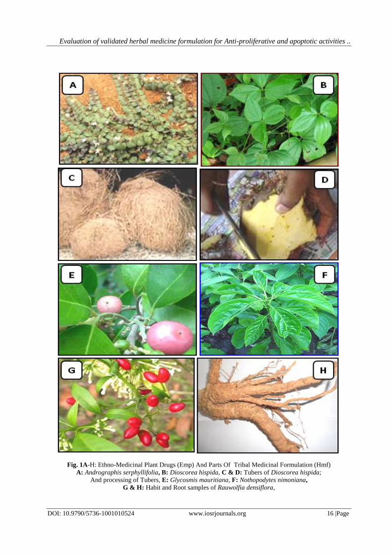
The different parts of five ethno-medicinal plant drugs, A. serphyllifolia (leaves), D. hispida (tubers); G.
mauritiana (leaves); N. nimmoniana (leaves) and R. densiflora (whole plant) were collected from different
tracts/regions of B.R. Hills of Chamaraja Nagara districts of Karnataka (Fig. 1A-H).
Instruments
The instruments such as, Electric blender, Microscope (Olympus), Muffle furnace (Meta lab, Scientific
industries, Mumbai), Centriguge (REMI R-4C & 8RC Centrifuge Machine Bengaluru), Soxhlets Apparatus
(Multiple Units), Flash Evaporator, Shaker incubator, Laminar Air Flow (Meditech, Chennai), Spectrophotometer
(UV/Visible, Elico Limited, Hyderabad) etc. were employed in the study.
Chemicals
In the study , the standard chemicals of analytical grade were used namely, Picric acid, α-naphthol,
Benedict’s reagent, 5% Ferric chloride, 1%Gelatin, 10% sodium hydroxide, Alcohol, Biuret’s reagent, Ninhydrine
reagent, Lead acetate, NaOH, Conc.H2SO4, copper sulphate, dimethylsulfoxide (DMSO), di-sodium hydrogen
orthophosphate (Na2HPO4), ethylenediamine tetracetic acid (EDTA), Folin Ciocalteu’s phenol (FC) reagent.
Besides, the chemicals used in the study were of, 3(4,5-dimethylthiazol-2-yl)2,5-diphenyl-tetrazolium bromide
(MTT), rutin, gallic acid followed by ferric chloride (FeCl3), hydrogen peroxide (H2O2), Hydroxylamine
hydrochloride, sodium dodecyl sulphate (SDS), magnesium sulphate (MgSO4), potassium chloride (KCl),
magnesium chloride (MgCl2), sodium hydroxide (NaOH), sodium chloride (NaCl), sodium dihydrogen-ortho-
phosphate (NaH2PO4), trichloroacetic acid (TCA), and Tris-HCl were obtained from Sisco Research Lab.,
(Mumbai, India). Additionally, other specific solvents and reagents of analytical grade were used in the studies,
which were procured from authorized S.D. Fine chemicals Pvt. Ltd., followed by Merck, India. The media like,
agar, cholesterol, hypochlorite solution, proteose peptone and yeast extract were procured from Hi-Media,
(Mumbai), India (Deng et al., 2006; Zakaria et al., 2011).
Interaction with Tribal Medicine Men and collection of Ethno-medicinal plants
The interactions were conducted intermittently with Tribal Medicine Men at the different locale of
Biligirirangana Hills, Karnataka (during the period, 2013-2014) with a semi-structured questionnaire. The data on
Medicine formulation and responsible plant components were documented and the Herbal Medicine Formulations
(HMF) and individual ethno-medicinal plant materials were obtained from the Tribal Medicine Men (Fig. 3A-J).
The particulars on atypical practiced medicine formulation comprising different parts of ethno-medicinal plants
such as, Andrographis serphyllifolia, Vahl (leaves), Discorea hispida, Dennst, (tubers); Glycosmis mauritiana
Tanaka, (leaves); Nothapodytes nimmoniana Blume (leaves) and Rawolfia densiflora (Wall.) Benth & Hook
(whole plant) respectively were explicitly collected during the interaction. The plants species were identified and
authenticated by consulting a taxonomist followed by standard flora, correspondingly, the plant materials were
deposited at Bhoomigeetha Institute of Research & Development, (Tumkur), Karnataka, India. The baseline
informations of selected ethno-medicinal plants are represented in the Table 1.
Validation of Tribal Medicine Formulation (HMF)
The different plants samples of Tribal medicine formulation were scientifically validated based on their
physical characteristics in association with an authorized Ayurvedic practitioner, Nisarga Ayurveda Research
Foundation, Sakaleshpur, Hassan district, India (Table 1). The standard protocols were identified and the
methodology was employed in the present study based on the descriptions of Chaithra (2013).
Preparation and Processing of HMF
The collected ethno-medicinal plant materials of HMF were subjected for unraveling different desirable
parts like, leaves, stem, root/ tubers from the main plants or whole plant parts. The different parts of the selected
ethno-medicinal plant drug materials were subjected for shade drying for 20 days to ensure that, the active
constituents were free from decomposition and possibility of photo-chemical degradation was also monitored. The
whole HMF formula and its components were extracted by following the traditional process i.e., boiling with
water for 15 minutes followed by filtering the same using muslin cloth then, the filtrates were dried by means of
lyophilization processes. Further, the dried extracts of HMF were examined to determine antioxidant, anti-
proliferative and apoptotic activities as per the standard analytical procedures (Anonymous, 2002).
Preparation of Solvent extracts
The air-dried components of the HMF drug were powdered using a suitable mechanical grinder to obtain
a coarse powder, which was then subjected to successive solvent extraction with ethanol, methanol and petroleum
ether in a soxhlet apparatus. The material was dried in hot air oven at 40°C each time before extracting with the
Page 4
Evaluation of validated herbal medicine formulation for Anti-proliferative and apoptotic activities ..
DOI: 10.9790/5736-1001010524 www.iosrjournals.org 8 |Page
next solvent. The extracts were then filtered through a Whatman No.1 filter paper and concentrated to the dry
mass using rotary evaporator. The extraction process was repeated for three times at different time intervals. The
yield of each extract was measured and residues were stored in dark glass tubes for further analysis.
Phyto-chemical analysis of EMP and HMF
The extracts from ethno-medicinal plant (EMP) drugs and Tribal Medicine Formulation (HMF) were
used for the Phyto-chemical analysis qualitatively for the detection of carbohydrates, proteins followed by the
secondary metabolites like alkaloids, flavonoids, terpenoids, steroids, tannins, saponins and total phenols etc. The
aqueous extracts of the plant was subjected to qualitative chemical screening for the identification of the alkaloids,
flavonoids and tannins using standard procedures (Trease, G. E.; and Evans 1996; Kokate et al., 2004; Mondal, et
al.,2013).
ABTS radical scavenging activity The ABTS assay (2, 2-azinobis 3-ethylbenzothiazoline-6-sulfonic acid) was performed by preparing a
stock solution which is consisting of 7 mM ABTS solution and 2.45 mM potassium persulfate solution at equal
proportion. This was subjected for incubation at room temperature for 12h during dark condition that was further
yielded a dark colored solution which contains radicals of ABTS. Subsequently, to perform each assay, the fresh
working solution was prepared by mixing stock solution with methanol (50%) and the initial absorbance was
0.700 (± 0.02) at 745 nm at 30°C temperature. Further, the extracts of ethno-medicinal plant drugs and tribal
medicine formulation were used at variable concentrations (50-3000μg/ml). Then, these concentrations were
subjected for reaction with known volume (3 ml) of ABTS solution and the absorbances were taken at 734 nm.
Meanwhile, the ascorbic acid was taken as positive control. Finally, the radical scavenging activity was assessed
based on the percent activity of ABTS and calculated the value as per the standard formula (Re et al., 1999).
Thin Layer chromatography (TLC)
Thin layer chromatography (TLC) is a chromatography technique used to separate diverse concoction of
bioactive constituents. Thin layer chromatography is performed on a sheet of glass, plastic, or aluminum foil,
which is coated with a thin layer of adsorbent material, usually silica gel, aluminium oxide, or cellulose. This layer
of adsorbent is known as the stationary phase. After the sample has been applied on the plate, a solvent or solvent
mixture (known as the mobile phase) is drawn up the plate via capillary action. Because different analytes ascend
the TLC plate at different rates, separation is achieved and the compounds in the extracts gets separated based on
their affinity. The fluorescence bands were observed at 254nm (short wavelength) in UV light followed by 366 nm
(long wavelength) using UV light in the respective plates. After the run, plates were dried and sprayed with
NP/PEG reagents were used to detect the bands on the TLC plates and the observation of chromatograms was
done under long wavelength UV followed by visible light. The movement of the active compound was expressed
by its retention factor (Rf), values were calculated for different samples (Wagner and Bladt, 2009).
Cell Lines and Cell Culture Preparation
The Cell- lines used in this study were of estrogen receptor–positive ‘MCF-7’ breast cancer cells and
cervical ‘HeLa’ cancer cells. The MCF-7cancer cells were cultured in 89% DMEM and 10% FBS along with 1%
penicillin/streptomycin. Similarly, the HeLa, cells were cultured in 89% RPMI 1640 with 10% FBS and 1%
penicillin/streptomycin. All the cells were cultured at 37 °C at 95% humidity and 5% CO2 for 3 days as they
reached 80%–90% confluency. Subsequently, the spent medium was removed and replaced with fresh medium
and incubated again for 24 h. The cell cultures were then washed with PBS 1–2 times and were suspended using
trypsin-EDTA and finally, fresh medium was added to the cells (Evan and Vousden, 2001).
Anti-proliferative activity
Anti-proliferative activity was measured by MTT assay and that was performed using 96-well plate at a
cell density of 3 x 10³ cells/well. The cancer cells, MCF-7 and HeLa were subjected for analysis using pre-
cultured by Minimum Essential Medium (MEM) containing 10% Fetal Bovine Serum (FBS) and as per their
specificity described above. All the cell lines were pre-cultured for 24 h before confronting with variable
concentration of the extracts i.e., 10, 30, 100, 300, and 1,000 μg/ml. Then, phosphate buffer saline (PBS) was used
as negative control and adriamycin was used as positive control, finally subjected for incubation as per the
ABTS radical scavenging activity (%) = Control OD – Sample OD × 100
Control OD
Rf = Distance travelled by the solute
Distance travelled by the solvent front TLC plates
Page 5
Evaluation of validated herbal medicine formulation for Anti-proliferative and apoptotic activities ..
DOI: 10.9790/5736-1001010524 www.iosrjournals.org 9 |Page
protocol explained above (Nesaretnam et al., 1998; Sun et al., 2002; Giri et al., 2006; Siripong et al., 2006; Hu et
al., 2011). After the incubation period (72 h), the treated cells were added with MTT reagent then incubated again
for 3h and the formozan salts were dissolved with DMSO. The absorption was measured at 550 nm. The
concentration that inhibited 50% cell growth (IC50) was calculated using curve fitting and the each experiment was
done in 3 replicates and reported as IC50 ± SD (Elumalai et al., 2012).
MTT bioassay
The cytotoxic effect in different active fractions of HMF drug was evaluated against MCF-7 using MTT
bioassay (Sanaz et al., 2012). The human breast cancer MCF-7 Cell line was cultured explicitly as mentioned in
the procedure. Accordingly, the cells were seeded in 96-well microtitre plate (200 μl/well) with concentration of
4×10⁴ cells/cm2. The cultivated cells were exposed to various concentration of the methanolic extract (1, 0.75,0.5,
0.25, 0.1, 0.075, 0.05, 0.025, 0.01 mg/mL) prepared in 1% dimethyl sulfoxide (DMSO) as they reached 40-50%
confluency and were then incubated for different periods of time (24, 48 and 72 h). The control groups received
the same amounts of DMSO with four wells remained untreated as control. After the treatment, normal culture
medium was replaced with 200 μl fresh media and 50 μl MTT reagent (2 mg/mL in PBS), except the cell-free
blank control wells. The cells were maintained vi incubation as per the procedure mentioned above subsequently,
the MTT solution was substituted with 200 μl of DMSO and 25 μl sorenson buffer (0.1M NaCl, 0.1M glycine
regulated to pH: 10.5 with 1M NaOH), incubated for 15 min at 37°C. Eventually, the optical density of the wells
was measured at 570 nm by means of a spectrophotometric plate reader of standard firm. The growth of tumoral
cells and viability of the cells was determined using the formula.
Viability % = (optical density of sample/optical density of control) ×100
Furthermore, the cyto-toxicity of the HMF extract was determined by plotting of the percent cyto-toxicity index,
CI % = [1-(optical density of sample/optical density of control)] × 100, versus concentrations of the fractions of
Tribal medicine formulation.
Evaluation of Anticancer activity from HMF drug formula
The concept of making dilution which is a process of declining the concentration by adding of a solution such as
water. The crude extracts of HMF drug was diluted according to the requirements and the extracts were syringe
flittered to avoid contamination prior to use.
Trypan Blue Assay
Trypan blue is an imperative stain applied to examine the dead tissues or the cells which takes blue
colour selectively. This assay is used to determine the dead cell count as well as the living cell count. The living
cells will have an intact membrane which does not allow the dye to pass since the cells are very selective in
compounds. The dead cell does not process an intact membrane and takes up the stain.
Hoechst Stain Assay
This assay was done to check that the cell death has occurred due to apoptosis (the cell death due to
destruction in the actual functions of membrane followed by cells that in turn leads to inflammation) or not.
Besides, Hoechst Stain assay was done to confirm that, the cell death has occurred explicitly due to apoptosis
only.
MTT Assay
The MTT assay was performed to reduce the yellow composite called 3-(4,4-dimethylthiazol-2-yl)-2,5-
diphenyl tetrazolium bromide (MTT) under the influence of succinate dehydrogenase enzyme in the complex-II at
the electron transport chain that occurs in Mitochondria. The turn down of MTT can occur only physiologically
active cells so, the activity will be determined based on the viability status of the cells. The whole reaction
comprises, the MTT which go into the cell subsequently to the mitochondria wherein, MTT was reduced to form
an insoluble Formosan product which appears in dark purple color. Meanwhile, the cells are solubilized through
DMSO solvent thus, the released Formosan product was assessed through spectrophotometric method (Mosmann,
1983).
III. Results The present study was carried out at the Department of Engineering Chemistry, Maharaja Institute of
Technology, (Visvesvaraya Technological University), Thandavapura, Nanjanagud taluk, Mysuru dist - 571302,
(Karnataka), India and the analytical study was conducted at the department of Biotechnology and applied
sciences, SIET, Tumkur. The herbal/tribal medicine formulation was validated in association with Bhoomigeetha
Institute of Research and Development (BIRD), Tumkur. The anti-cancer study was executed in association with
Sri Raghavendra Biotechnologies, Bengaluru. The HMF (Tribal Medicine Formula) or Herbal Medicine Formula
Page 6
Evaluation of validated herbal medicine formulation for Anti-proliferative and apoptotic activities ..
DOI: 10.9790/5736-1001010524 www.iosrjournals.org 10 |Page
(HMF) was procured from traditional practitioner at Biligirirangana Hills, Chamarajanagara district, Karnataka
(Table 1 and Fig 1).
Phyto-chemical analysis
The phyto-chemical screening of aqueous extract and solvent extracts of EMP and HMF demonstrated
the presence of Carbohydrate, proteins, alkaloids, flavonoids, saponins, tannins, gums & mucilages, coumarins,
terpenoids, tannins, steroids, glycosides, phyto sterols, fixed oils and fats, phenols, saponin etc in Cold water, hot
water followed by solvent extracts. The presence of these phyto-chemicals suggested to taking part in synergistic
role to exert the observed pharmacological activity. The fact that strong synergism of several constituents in the
crude drug may prove more potent and effective than any single purified compound, is always overlooked.
Moreover, this may help to nullify the toxic effects (if any) of individual constituents (Table 2).
Antioxidant activity
The method used, ABTS radical scavenging assay, gave the measure of antioxidant activity of the HMF
drug extract determined by the decolorization of the ABTS+, through measuring the reduction of the radical cation
as the percentage inhibition of absorbance at 734nm. The effects of HMF drug aqueous extract on ABTS free
radical scavenging activities was assayed at various concentrations. The IC50 value was found to be 07± 0.74µl/ml
was evident in this extract (Table 3).
Chromatographic studies
This was done in order to purify the HMF drug solvent extracts and the TLC chromatograms were
developed using the solvent system, toluene, chloroform and ethanol. The bioactive compound moved on the
stationary phase was measured and the retention factor was calculated. There were two to five bands of whole
HMF drug extract at Rf values of 0.32 and 0.44 in A. serpyllifolia; 0.52 and 0.26 in D.hispida; 0.59 and 0.23 in G.
mauritiana; 0.34, 0.14, 0.16, 0.62 and 0.95 in N.nimmoniana similarly, in case of R. densiflora 0.76 and 0.85
were present. The extracts showed variable Rf values as compared among these extracts (Table 4 and Fig.2A&B).
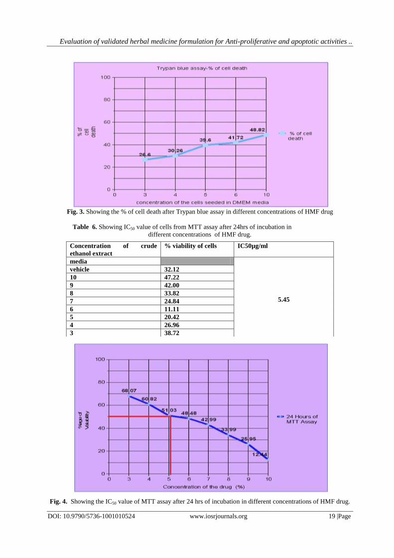
Anticancer activity
The partially purified extract of HMF drug was tested for anticancer property against both HeLa and
MCF-7 cell lines respectively. The inhibition concentration (IC50) as observed from the graph lies between 3 and 4
in 24 hours of trypan blue assay (Table 5 and Fig. 3, 8, 10 & 11). The cells show positive Hoechst assay as the
DNA has acquired the Hoechst stain and the same is seen under the fluorescent microscope as observed (Fig. 14 &
17).
The result analysis for MTT assay shows that, the cell viability decreases with the increase in
concentration of the HMF drug. The inhibition concentration value (IC50) for MTT assay at 24 hours showed to be
3% and for 48 hours (Table 6 & 7) lies in between 3 and 4 (Fig. 4,5,6,7 and 9A &B). The positivity of the MTT
Assay can be observed in the fig 5 and 7 by the formation of crystals.
Anticancer activity against MCF-7 Cell Lines
The cells were cultured on the suitable Animal cell media under controlled condition and the various
concentrations of the formulation was taken and tested for its action on the growth of HeLa cells and Mammarian
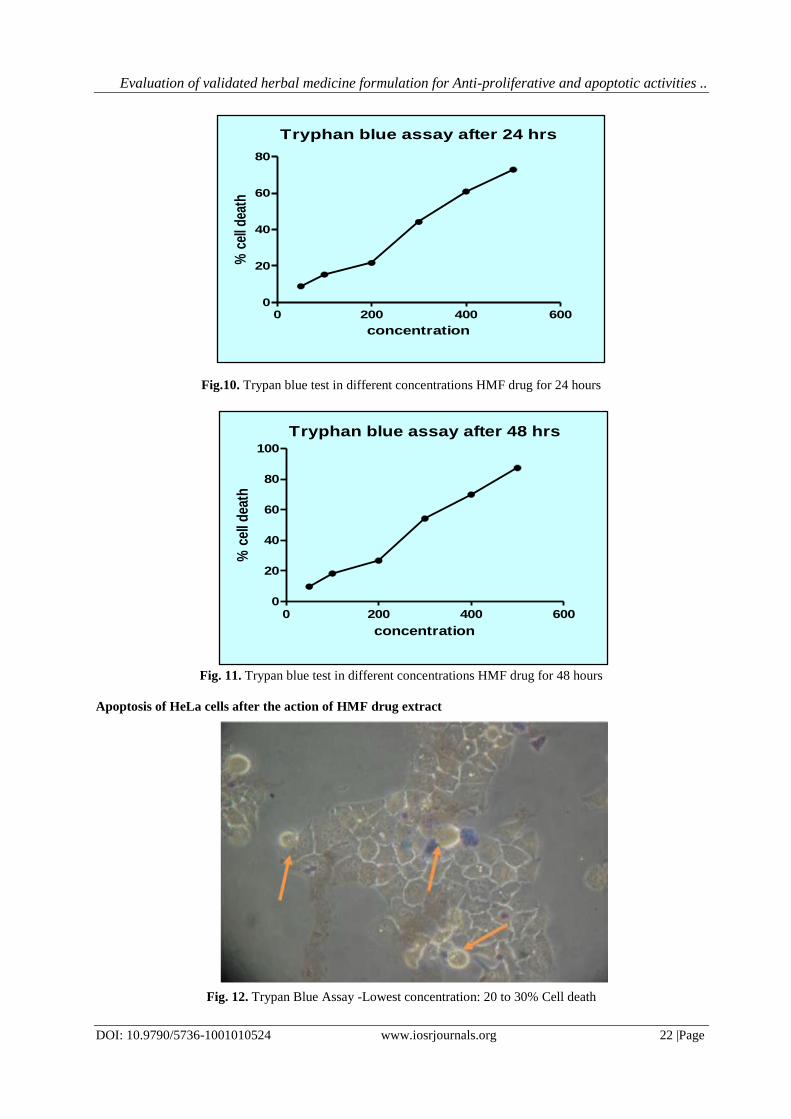
Cancer cells (MCF-7). The Anticancer activity was determined using the ‘Tryphan Blue assay’ in Table 5. This
result revealed that, profound activity was noticed by the end of 24 hours than 48 hours shown in Fig. 8, 10, 11,12
& 16. Then, the Cell viability count was determined through MTT assay along with a media control contained
growing cells with no drug. Similarly, the vehicle control was set up in presence of the respective solvents. The
ethanolic extract of HMF drug was tested against HeLa cells followed by MCF-7 cell lines (Fig. 4,5, 6,7 and 9A
&B). The HMF drug extract was found to be superior as compared to other extracts. The result reveals the
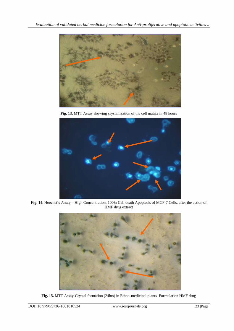
apoptosis of both cancer cells indicating the most positive action of the Herbal Medicine formulation (Fig.12,13
&14).
The ‘Hoechst staining’ technique was showed the apoptotic state of cells and DNA, through fluorescent
microscopy. The remarkable activity was noticed in the HMF drug tested in its crude form compared to the
purified form which confirms that, the HMF drug works best in its crude form which is being currently practiced
by the Traditional healers (Fig. 15, 16 & 17).
IV. Discussion The presence of active secondary metabolites in the extracts of ethno-medicinal plants may have
profound activity and justifies the status for preparation of crude potential drug by the tribal people. The phyto-
chemical screening of the ethno-medicinal plants showed the presence of alkaloids, flavonoids, terpenoids,
saponins, tannins, phenolic compounds and reducing sugars. A. serphyllifolia and D. hispida did not contain
Page 7
Evaluation of validated herbal medicine formulation for Anti-proliferative and apoptotic activities ..
DOI: 10.9790/5736-1001010524 www.iosrjournals.org 11 |Page
cardiac glycosides and coumarins while, G. mauritiana, N. nimmoniana and R. densiflora showed the presence of
glycosides, tannins and alkaloids. The phyto-chemical analysis conducted on the plant extracts revealed the
presence of constituents which are known to exhibit medicinal as well as physiological activities (Sofowora,
1993). The TLC result in the different components of HMF drug exhibited the presence of diversified bioactive
constituents and confirms the overall potentiality of the HMF drug.
The MTT assay on the cytotoxic activity of the partially purified extract of HMF plant drug on MCF-7
cell line apart from experimentation with HeLa cells suggested that, the extract was fairly cytotoxic to MCF-7
cells in a dose and time dependent manner. The findings of the present study will provide an insight into a new
implication of the traditional usage of HMF drug in the tribal medicine system found to be as a most potential,
efficacious and novel cancer chemo-preventive agent, where integration of the different herbal compositions
remedy may help in preventing or reducing the risk of breast cancer and associated oxidative stress diseases. The
similar expressions were also made in traditional medicinal plants (Abdul et al., 2009; Althunibat et al., 2009;
Rajesh et al., 2011; Cieckiewicz et al., 2012).
Several studies in this filed have shown that, the ethno-medicinal plants are of potential value for
identifying anti-proliferative agents followed by determination of the anti-inflammatory and anti-proliferative
activity of herbal medicine formulation where, the suppression of cell growth as well as induction of apoptosis in
human breast cancer cells was observed (Conforti et al., 2008; Nurhanan et al., 2008; Hogan et al., 2010;
Mbaveng et al., 2011; Lin et al., 2012). Elsewhere, metabolites like, flavonoides, di-terpenoids and poly phenols
from Herbal Medicine formulation was appraised for their Cytotoxic effects against both MCF-7 and HeLa cancer
cell lines respectively. This expression from the study revealing that, the HMF has strong tumor regression
potentiality even for a broad range of tumor cells depicted in the ailment. This is in accordance with the reports of
Sun et al., (2002); Matito et al, (2003); Giri et al, (2006); Hu et al, (2011); Elumalai et al., (2012).
In addition, this study has clearly shown that, HMF drug has relatively substantial antioxidant activity
with regard to the potential constituents present in it that might be responsible for the chemo-preventive effects of
the HMF drug extract. The protective role and consistency of these active compounds of the HMF drug might
have connected to the ailment in controlling the various oxidative stress factors (Braca et al., 2002; Matito et al.,
2003; Aiyegoro et al., 2009; Tyagi et al., 2010; Murthy et al., 2011; Hu et al., 2011; Hazirah et al., 2013). The
different endogenous antioxidants produced by the human body have potential health benefits against oxidative
stress and related ailments, but also naturally supplemented herbal antioxidant compounds such as, phenolic acids,
polyphenols and flavonoids scavenge free radicals like peroxide, hydroperoxide or lipid peroxyl are most
promising and inhibits the oxidative mechanisms to prevent the system from degenerative diseases (Bhandari and
Kawabata, 2004; Barriera et al., 2008; Aiyegoro and Okoh, 2010; Verma et al., 2009; Hogan et al., 2010; Yang et
al., 2010).
In addition, this study has clearly shown that, the active fractions of HMF drug has antioxidant activity
and comparatively high with regard to the potential radical scavenging ability of its methanolic extract. Since, the
HMF drug are normally comprising complex mixtures of different active metabolites i.e., flavonoids, alkaloids
and other phenolic compounds present in the methanolic extract of HMF drug which further donates a hydrogen
atom for scavenging the stable DPPH radical, would be beneficial in quantifying the presumed charge of them as
antioxidant and anti-cancer agents (Oskoveian et al., 2011; Hazirah et al., 2013).
The exceptional fact that, anticancer activities of ethno-medicinal plants have been verified critically to
be coupled with a diverse range of phyto-chemicals, such as polyphenols, flavonoids, terpenoides, steroids,
catechins etc (Tung et al., 2009). Many researchers have also opined that, the more phenolic content of different
plant components increases the antioxidant activity in the system since, there is a linear correlation between
phenolic contents and antioxidant activity (Prasad et al., 2009; Hogan et al., 2010; Jain and Jain, 2011; Nagmoti et
al., 2012). Although, the phenolics of plant sources are very basis for antioxidants, in which they have differential
expressions along with a specific ability to quench various free radicals. As a result, verifying the role of these
phyto-chemicals in HMF drug, certain kind of an oxidative stress related diseases is of substantial importance. In
the study, there is a correlation between total active phyto-chemicals with special reference to phenolic and
flavonoids contents as well as radical scavenging activity along with the anti-prolifreative activity of breast cancer
in the active fractions of HMF drug was explored.
By taking into account, the results on cyto-toxicity in the extracts of HMF drug at MCF-7 cells was found
to be superior with increased contents of both phenolics and flavonoids and thereby, higher radical scavenging
activity in higher concentrations was noticed. On the whole, this study suggests, the potentialities of both
antioxidant and cytotoxic activities of HMF extract which could be helpful in preventing or curbing the progress
of various oxidative stress-related diseases such as breast cancer. This was evident in the approaches on bioassay-
guided fractionation in HMF drug, further it would be of value to purify and identify the foremost active
constituents in the extract which is responsible for inhibiting the proliferation of MCF-7 cells in the system
(Ksouri et al., 2009; Fu et al., 2010). Similarly, as some phenolic antioxidants have a specific role in suppressing
the growth and proliferation of transformed or malignant cells through induction of programmed cell death or
Page 8
Evaluation of validated herbal medicine formulation for Anti-proliferative and apoptotic activities ..
DOI: 10.9790/5736-1001010524 www.iosrjournals.org 12 |Page
apoptosis, it materializes to demand the need for additional investigation on cell cycle analysis and determination
of the distinctive mechanism of action for providing anti-prolifreative activity in the extract of Herbal Medicine
formulation (Puneeth kumar et al., 2011; Oskoueian et al., 2011; Sanaz et al., 2012). The authors believe that, the
current objectives of the study could furnish the appropriate background for detailed examination on anticancer
properties of this HMF drug. Besides, it is the very first report on the analysis of the anti-proliferative (MCF-7)
and antioxidant activities of Herbal Medicine formulation being practiced in the Tribal Medicine System (TMS).
V. Conclusion Finally, it can be concluded that, the Herbal medicine formulation has potent antimicrobial and
antioxidant activities apart from their antiproliferative activities. Hence, with this basis, the formulation showed
the highest anticancer activity against MCF-7 Cell lines and justifies their practice of Herbal Formulation in their
TMS. Further, the susceptibility of mammarian cancer cells has been influenced by HMF drug has been
substantiated based on the outcome of the study. In addition, the significance of these observations and
recommendations in the light of previous studies with asynchronous population of MCF-7 cells also has been
discussed.
Acknowledgements The authors would like to express thanks to the authorities of Jawaharalal Nehru Technological
University, Ananthapuramu (AP), India for providing the opportunity to carry-out this Research study. The first
Author would like to thank the authorities of Department of Chemical Engineering and Biotechnology along
with Oil Technological and Pharmaceutical Research Institute (OTPRI) for their guidance and cooperation.
Further, sincere thanks to Maharaja Institute of Technology, Thandavapura (Visvesvaraya Technological
University, Belagavi), Nanjanagud Tq, Mysuru dist-571302, India for providing basic infrastructure for
conducting preliminary studies. In addition, the author is also thankful to Sri Raghavendra Biotechnologies,
Bengaluru for their association with human cancer cell lines and laboratory facilities to complete the objectives
successfully. The authors are finally extending their grateful thanks to Bhoomigeetha Institute of Research &
Development (BIRD), Tumkur for providing the ethno-medicinal plant materials with taxonomical identification
and formulations (HMF / HMF) and for their assistance in scientific validation.
References
[1]. Abdul, A.B., Abdelwahab, S.I., Fong, H.K., Mohan, S.M., Al-Zubairi, A.S., Elhassan, M.M. 2009. In vitro
response of cancer cells to the growth-inhibitory effects of dichloromethane extract of Goniothalamus
umbrosus. Res. J. Pharmacol., 3, 1–6.
[2]. Adedapo, A.A., Jimoh, F.O., Afolayan, A.J, Masika, P.J. 2008. Antioxidant activities and phenolic contents
of the methanol extracts of the stems of Acokanthera oppositifolia and Adenia gummifera. BMC
Complement Altern Med, 8, 54.
[3]. Aiyegoro, O.A., Okoh, A.I. 2009. Phytochemical screening and polyphenolic antioxidant activity of
aqueous crude leaf extract of Helichrysum pedunculatum. Int. J. Mol. Sci., 10, 4990–5001.
[4]. Aiyegoro, O.A., Okoh, A.I. 2010. Preliminary phytochemical screening and in vitro antioxidant activities of
the aqueous extract of Helichrysum longifolium DC. BMC Complement. Altern. Med., 10, 21.
[5]. Akim, A., Ling, L.C., Rahmat, A. and Zakaria, Z.A. 2011. Antioxidant and anti-proliferative activities of
Roselle juice on Caov-3, MCF-7. MDA-MB-231 and HeLa cancer cell lines. Afr. J. Pharm. Pharmacol., 5,
957–965.
[6]. Al-Rashidi, W., Mat Supri, N.N., Manshoor, N. 2011. Cytotoxic activity of crude extract from Costus
malortieanus (Costaceae). Am.Eurasian J. Toxicol. Sci., 3, 63–66.
[7]. Althunibat, O.Y., Hashim, R.B., Taher, M., Daud, J.M., Ikeda, M.A., Zali, B.I. 2009. In vitro antioxidant
and antiproliferative activities of three Malaysian sea cucumber species. Eur. J. Sci. Res., 37, 376–387.
[8]. Anonymous. 2002. Wealth of India. First supplementary Series, Vol-3, (D-I), Raw materials. Niscom. 130.
[9]. Anusha Kulkarni., Ramya, M.C and Panduranga Murthy, G. 2010. Evaluation of Tribal Medicine and
Active Principle of Glycosmis Mauritiana (Lam) Tanaka for Antioxidant and Anticancer activity against
Pc-3 Cell Lines. 34rd
Series, SPP KSCST Projects, 45-46.
[10]. Atjanasuppat, K., Wongkham, W., Meepowpan, P., Kittakoop, P., Sobhon, P., Bartlett, A., Whitfield, P.J.
2009. In vitro screening for anthelmintic and antitumour activity of ethnomedicinal plants from Thailand. J.
Ethnopharmacol., 123, 475–482.
[11]. Aune, D., De Stefani E, Ronco, A. 2009. Fruits, vegetables and the risk of cancer: a multisite case-control
study in Uruguay. Asian. Pac. J. Cancer Prev., 10, 419-28.
Page 9
Evaluation of validated herbal medicine formulation for Anti-proliferative and apoptotic activities ..
DOI: 10.9790/5736-1001010524 www.iosrjournals.org 13 |Page
[12]. Barreira, J.C.M., Ferreira I.C.F.R., Oliveira, M.B.P.P., Pereira, J.A. 2008. Antioxidant activities of the
extracts from chestnut flower, leaf, skins and fruit. Food Chem, 107, 1106–1113.
[13]. Bhandari MR, Kawabata M. 2004. Organic acid, phenolic content and antioxidant activity of wild yam
(Dioscorea spp.) tubers of Nepal. Food Chem, 88:163–168.
[14]. Boopathy NS, Kathiresan K. 2010. Anticancer drugs from marine flora: an overview. J Oncol, 214186:18.
[15]. Braca A, Sortino C, Politi M. 2002. Antioxidant activity of flavonoids from Licania licaniae flora. J
Ethnopharmacol, 79:379–381.
[16]. Caamal-Fuentes, E., Torres-Tapia, L.W., Simá-Polanco, P., Peraza-Sánchez, S.R., Moo-Puc, R. 2011.
Screening of plants used in Mayan traditional medicine to treat cancer-like symptoms. J. Ethnopharmacol.,
135, 719–724.
[17]. Chaithra, D, Nisaraga Ayurvedic Research Foundation, Sakaleshpur, Hassan district (India): Validated
Tribal Medicine formulation (HMF): Ref. No.176/2013.
[18]. Cieckiewicz, E., Angenot, L., Gras, T., Kiss, R., Frédérich, M. 2012. Potential anticancer activity of young
Carpinus betulus leaves. Phytomedicine, 19, 278–283.
[19]. Conforti, F., Ioele, G., Statti, G.A., Marrelli, M., Ragno, G., Menichini, F. 2008. Antiproliferative activity
against human tumor cell lines and toxicity test on Mediterranean dietary plants. Food Chem. Toxicol., 46,
3325–3332.
[20]. Deng XK, Yin W, Li WD, Yin FZ, Lu XY, Zang XC, Hua ZC, Cai BC. 2006. The antitumor effects of
alkaloids from the seeds of Strychnos nux-vomica on HepG2 cells and its possible mechanism. J
Ethnopharmacol, 106:179–186.
[21]. Diwanay, S., Chitre, D., Patwardhan, B. 2004. Immunoprotection by botanical drugs in cancer
chemotherapy J Ethnopharmacol., 90: 49-55.
[22]. Elumalai, P., Gunadharini, D.N., Senthilkumar, K., Banudevi, S., Arunkumar, R., Benson, C.S., Sharmila,
G., Arunakaran, J. 2012. Ethanolic neem (Azadirachta indica A. Juss) leaf extract induces apoptosis and
inhibits the IGF signaling pathway in breast cancer cell lines. Biomed. Prev. Nutr., 2, 59–68.
[23]. Evan, G.I, Vousden, K.H. Proliferation, cell cycle and apoptosis in cancer. Nature 2001, 411:342–348.
[24]. Fu, W., Chen, J., Cai, Y., Lei, Y., Chen, L., Pei, L., Zhou, D., Lang, X., Ruan, J. 2010. Antioxidant, free
radical scavenging, anti-inflammatory and hepatoprotective potential of the extract from Parathelypteris
nipponica (Franch. et Sav.) Ching. J. Ethnopharmacol., 130, 521–528.
[25]. Giri, B., Gomes, A., Debnath, A., Saha, A., Biswas, A.K., Dasgupta, S.C. 2006. Antiproliferative, cytotoxic
and apoptogenic activity of Indian toad (Bufo melanostictus, Schneider) skin extract on U937 and K562
cells. Toxicon, 48, 388–400.
[26]. Gul MZ, Bakhshu LM, Ahmed F, Kondapi AK, Qureshi IA, Ghazi IA. 2011. Evaluation of Abelmoschus
moschatus extracts for antioxidant, free radical scavenging, antimicrobial and antiproliferative activities
using in vitro assays. BMC Complement Altern Med, 11:64.
[27]. Hasan TN, B LG, Shafi G, Al-Hazzani AA, Alshatwi AA. 2011. Anti-proliferative effects of organic
extracts from root bark of Juglans regia L. (RBJR) on MDA-MB-231 human breast cancer cells: role of
Bcl-2/Bax, caspases and Tp53. Asian Pac J Cancer Prev, 12, 525-30.
[28]. Hazirah, A., Zainal, B., Abdah, M. 2013. Total phenolic content, antioxidant and cytotoxic activity of cocoa
(Theobroma cacao L.) polyphenols extracts on cancer cell lines. Malays. J. Nutr., 19, 223–232.
[29]. Hinneburg I, Dorman HJD, Hiltunen R. 2006. Antioxidant activities of extracts from selected culinary
herbs and spices. Food Chem, 97:122–129.
[30]. Hogan S, Chung H, Zhang L, Li J, Lee Y, Dai Y, Zhou K. 2010. Antiproliferative and antioxidant
properties of anthocyanin-rich extract from acai. Food Chem, 118:208–214.
[31]. Hossain, M.A., Shah, M.D., Gnanaraj, C., Iqbal, M. 2011. In vitro total phenolics, flavonoids contents and
antioxidant activity of essential oil, various organic extracts from the leaves of tropical medicinal plant
tetrastigma from Sabah. Asian Pac. J. Trop. Med., 4, 717–721.
[32]. Hu, W., Yu, L., Wang, M.-H. 2011. Antioxidant and antiproliferative properties of water extract from
Mahonia bealei (Fort.) Carr. leaves. Food Chem. Toxicol., 49, 799–806.
[33]. Itharat, A., Houghton, P.J, Eno-Amooquaye E, Burke, P.J, Sampson, J.H, Raman, A. In vitro cytotoxic
activity of Thai medicinal plants used traditionally to treat cancer. J Ethnopharmacol, 2004, 90: 33-38.
[34]. Jain, R., Jain, S.K. 2011. Screening of in vitro cytotoxic activity of some medicinal plants used traditionally
to treat cancer in Chhattisgarh state, India. Asian Pac. J. Trop. Biomed., 1, S147–S150.
[35]. Jalil, A.M.M., Ismail, A., Pei, C.P., Hamid, M., Kamaruddin, S.H.S. 2008. Effects of cocoa extract on
glucometabolism, oxidative stress, and antioxidant enzymes in obese-diabetic (Ob-db) rats. J. Agric. Food
Chem., 56, 7877–7884.
[36]. Jemal, A., Siegel, R., Ward, E. 2006. Cancer Statistics. CA Cancer J Clin, 56, 106-30.
Page 10
Evaluation of validated herbal medicine formulation for Anti-proliferative and apoptotic activities ..
DOI: 10.9790/5736-1001010524 www.iosrjournals.org 14 |Page
[37]. Jones, W., Chin, Y., Kinghorn, A. 2006. The role of pharmacognosy in modern medicine and pharmacy.
Curr Drug. Targets, 7, 247-64.
[38]. Kim, J .2008. Protective effects of Asian dietary items on cancers - soy and ginseng. Asian Pac J Cancer
Prev, 9, 543-8.
[39]. Kokate, C.K., Purohit, A.P., and Gokhale, S.B., Practical Pharmacognasy, Vallabh Prakashan, Delhi. 2(ed.),
2004.
[40]. Ksouri, R., Falleh, H., Megdiche, W., Trabelsi, N., Mhamdi, B., Chaieb, K., Bakrouf, A., Magné, C.,
Abdelly, C: 2009. Antioxidant and antimicrobial activities of the edible medicinal halophyte Tamarix
gallica L. and related polyphenolic constituents. Food Chem Toxicol, 47:2083–2091.
[41]. Lampronti, I., Martello, D., Bianchi, N., Borgatti, M., Lambertini, E., Piva, R., Jabbar, S., Choudhuri,
M.S.K., Khan, M.T.H., Gambari, R. 2003. In vitro antiproliferative effects on human tumor cell lines of
extracts from the Bangladeshi medicinal plant Aegle marmelos., Correa Phytomedicine, 10: 300-308.
[42]. Lin, H.-H., Chan, K.-C., Sheu, J.-Y., Hsuan, S.-W., Wang, C.-J., Chen, J.-H. 2012. Hibiscus sabdariffa leaf
induces apoptosis of human prostate cancer cells in vitro and in vivo. Food Chem., 132, 880–891.
[43]. Ludwiczuk, A., Saha, A., Kuzuhara, T., Asakawa, Y. 2011. Bioactivity guided isolation of anticancer
constituents from leaves of Alnus sieboldiana (Betulaceae). Phytomedicine, 18, 491–498.
[44]. Manian, R., Anusuya, N., Siddhuraju, P., Manian, S. 2008. The antioxidant activity and free radical
scavenging potential of two different solvent extracts of Camellia sinensis (L.) O. Kuntz, Ficus bengalensis
L. and Ficus racemosa L. Food Chem, 107:1000–1007.
[45]. Matito, C., Mastorakou, F., Centelles, J.J., Torres, J.L., Cascante, M. 2003. Antiproliferative effect of
antioxidant polyphenols from grape in murine Hepa-1c1c7. Eur J Nutr, 42:43–49.
[46]. Mbaveng, A.T., Kuete, V., Mapunya, B.M., Beng, V.P., Nkengfack, A.E., Meyer, J.J.M., Lall, N. 2011.
Evaluation of four Cameroonian medicinal plants for anticancer, antigonorrheal and antireverse
transcriptase activities. Environ. Toxicol. Pharmacol., 32, 162–167.
[47]. Mishra, KP, Ganju L, Sairam M, Banerjee PK, Sawhney RC. 2008. A review of high throughput
technology for the screening of natural products. Biomed Pharmacother, 62:94–98.
[48]. Moncada S, Palmer RMJ, Higgs EA. 1991. Nitric oxide: physiology, pathophysiology, and pharmacology.
Pharmacol Rev., 43:109–142.
[49]. Mondal, S., Marouthu, I., Pushyami, P., and Suresh, P., Toxicity studies of ethanol extract from Ixora
pavetta and rews leaf. World J. Pharm. Pharma. Sci. 3(1), 2013, 350-360.
[50]. Mosmann, T. 1983. Rapid colorimetric assay for cellular growth and survival: application to proliferation
and cytotoxicity assays. J Immunol Methods, 65:55–63.
[51]. Nagmoti, D.M., Khatri, D.K., Juvekar, P.R., Juvekar, A.R. 2012. Antioxidant activity and free radical-
scavenging potential of Pithecellobium dulce Benth seed extracts. Free Radic. Antioxid., 2, 37-43.
[52]. Nakayama, T.1994. Suppression of hydroxyperoxide-induced cytotoxicity by polyphenols. Cancer Res,
54:1991–1993.
[53]. Nesaretnam, K., Stephen R, Dils R, Darbre P. 1998. Tocotrienols inhibit the growth of human breast cancer
cells irrespective of estrogen receptor status. Lipids, 33:461–469.
[54]. Nisa, S., Bibi, Y., Waheed, A., Zia, M., Sarwar, S., Ahmed, S., Chaudhary, M.F. 2011. Evaluation of
anticancer activity of Debregeasia Salicifolia extract against estrogen receptor positive cell line. Afr. J.
Biotechnol., 10, 990–995.
[55]. Nurhanan, M.Y., Asiah, O., Mohd Ilham, M.A., Siti Syarifah, M.M., Norhayati, I., Lili Sahira, H. 2008.
Anti-proliferative activities of 32 Malaysian plant species in breast cancer cell lines. J. Trop. For. Sci., 20,
77–81.
[56]. Oskoueian, E., Abdullah, N., Saad, W.Z., Omar, A.R., Kuan, W.B., Zolkifli, N.A., Hendra, R., Ho, Y.W.
2011. Antioxidant, anti-inflammatory and anticancer activities of methanolic extracts from Jatropha curcas
Linn. J. Med. Plants Res., 5, 49–57.
[57]. Panduranga Murthy,G., Punith kumar, T.G., Suresh,A., Ravishankar, H.G., Chandrasekhar, K.B and
Lokesh, S.2011. Evaluation of ethanolic Leaf extract of Dioscorea hispida, Dennst. For Anti-inflamatory
and Anti-analgesic activities. International Journal of Pharma and Industrial Research, Vol-1(2): 83-87.
[58]. Prasad, K.N., Hao, J., Shi, J., Liu, T., Li, J., Wei, X., Qiu, S., Xue, S., Jiang, Y. 2009. Antioxidant and
anticancer activities of high pressure-assisted extract of longan (Dimocarpus longan Lour.) fruit pericarp.
Innov. Food Sci. Emerg. Technol., 10, 413–419.
[59]. Prasanna, R., Harish, C.C., Pichai, R., Sakthisekaran, D., Gunasekaran, P. 2009. Anti-cancer effect of
Cassia auriculata leaf extract in vitro through cell cycle arrest and induction of apoptosis in human breast
and larynx cancer cell lines. Cell Biol. Int., 33, 127–134.
Page 11
Evaluation of validated herbal medicine formulation for Anti-proliferative and apoptotic activities ..
DOI: 10.9790/5736-1001010524 www.iosrjournals.org 15 |Page
[60]. Punithkumar, T.G., Panduranga Murthy, G., Suresh, G., Suresh,V., Senthilkumar, N and Ravishankar, H.G.
2011. Evaluation of Antitumour activity and Antioxidant status in Dioscorea hispida, Dennst. leaves on
Ehrlich ascites carcinoma in Swiss albino mice. International Journal of Drug Development and Research,
Vol: 3(2):203-210.
[61]. Rajesh, R., Chitra, K., Paarakh, P.M., Chidambaranathan, N. 2011. Anticancer activity of aerial parts of
Aerva lanata Linn Juss ex Schult against Dalton’s Ascitic Lymphoma. Eur. J. Integr. Med., 3, e245–e250.
[62]. Ramljak, D., Romanczyk, L.J., Metheny-Barlow, L.J., Thompson, N., Knezevic, V., Galperin, M., Ramesh,
A., Dickson, R.B. 2005. Pentameric procyanidin from Theobroma cacao selectively inhibits growth of
human breast cancer cells. Mol. Cancer Ther., 4, 537–546.
[63]. Ravelo, Á.G, Estévez-Braun A, Chávez-Orellana H, Pérez-Sacau E, Mesa-Siverio D.2004. Recent studies
on natural products as anticancer agents Curr Top Med Chem, 4: 241-265.
[64]. Ravishankar, H.G and Panduranga Murthy, G. 2009. Ethno-medicinal wealth of Biligirirangana Hills (B.R.
Hills), Karnataka, India. M.Phil thesis:Annamalai University, Tamilnadu (India), Data Base: Pp-1-415.
[65]. Rice-Evans, C. 2004. Flavonoids and isoflavones: absorption, metabolism and bioactivity. Free Radic Biol
Med, 36:827–828.
[66]. Rice-Evans C.A, Miller N.J, Paganga G.1996. Structure-antioxidant activity relationships of flavonoids and
phenolic acids. Free Rad Biol Med, 20:933–956.
[67]. Sanaz Hamedeyazdan, Fatemeh Fathiazad, Simin Sharifi, Hossein Nazemiyeh. 2012. Antiproliferative
Activity of Marrubium persicum Extract in the MCF-7 Human Breast Cancer Cell LineAsian Pacific
Journal of Cancer Prevention, 13, 5843-5848.
[68]. Sharma, J.V.C., Pitchaiah, G., Satyavati, D., Rao, J.V., Sanjay, H. 2011. In vitro anticancer activity of
methanolic extract of roots of Glochidion zeylanicum (Gaertn.). Int. J. Res. Pharm. Biomed. Sci., 2, 760–
764.
[69]. Shoeb, M. 2006. Mini review anticancer agents from medicinal plants. Bangladesh J. Pharmacol., 1, 35–
41.
[70]. Sofowora, A., Medicinal Plants And traditional Medicine in Africa. Spectrum Books Ltd., Ibadan, Nigeria,
1993, 191-289.
[71]. Srinivasan P, Vadhanam MV, Arif JM, Gupta C. 2002. A rapid screening assay for antioxidant potential of
natural and synthetic agents in vitro. Int J Oncol, 20:983–986.
[72]. Suffness M, Pezzuto JM: Assays related to cancer drug discovery. In Methods in Plant Biochemistry:
Assays for Bioactivity, Volume 6. Edited by Hostettmann K. London: Academic, 1990:71–133.
[73]. Sun J, Chu YF, Wu X, Liu RH. 2002. Antioxidant and anti-proliferative activities of common fruits. J
Agric Food Chem, 50:7449–7454.
[74]. Szekanecz Z, Koch AE. 2007. Mechanisms of disease: angiogenesis in inflammatory diseases. Nat Clin
Pract Rheum, 3:635–643.
[75]. Trease, G. E., and Evans, W. C., Pharmacognosy, 1996, 89-122 (London: Bailliere Tindall).
[76]. Tung YT, Wu JH, Huang CY, Kuo YH, Chang ST. 2009. Antioxidant activities and phytochemical
characteristics of extracts from Acacia confuse bark. Bioresour Technol, 100:509–514.
[77]. Tyagi, S.N., Saxena, A., Patel, B.D. 2010. In vitro Antioxidant Activity of methanolic and aqueous extract
of Flacourtia indica Merr. Am.-Eurasian J. Sci. Res., 5, 201–206.
[78]. Van PG.1993. Carotenoids and cancer: an update with emphasis on human intervention studies. Eur J
Cancer, 29(A):1335–1344.
[79]. Verma AR, Vijayakumar M, Mathela CS, Rao CV. 2009. In vitro and in vivo antioxidant properties of
different fractions of Moring oleifera leaves. Food Chem Toxicol, 47:2196–2201.
[80]. Wangcharoen, W., Morasuk, W. 2007. Antioxidant capacity and phenolic content of chilies. Kasetsart J.
Nat. Sci., 41, 561–569.
[81]. Wagner H, and Bladt S. Plant Drug Analysis, A Thin Layer Chromatography Atlas. 2nd ed. Springer-
Verlag Berlin Heidelberg. 2009.
[82]. Woo, H.D, Kim, J. 2011. Nutritional epidemiology of cancer in Korea: recent accomplishments and future
direactions. Asian Pac J Cancer Prev, 12, 2377-83.
[83]. Yang, J, Paulino R, Janke-Stedronsky S, Abawi F. 2007. Free radical scavenging activity and total phenols
of noni (Morinda citrifolia L.) juice and powder in processing and storage. Food Chem, 102:302-308.
[84]. Yang, QM., Pan ,X.H., Kong, W.B., Yang, H., Su YD., Zhang, L., Zhang, Y., Yang, Y., Ding, L., Liu, G.
2010. Antioxidant activities of malt extract from barley (Hordeum vulgare L.) toward various oxidative
stress in vitro and in vivo. Food Chem, 118:84–89.
[85]. Zakaria, Z.A., Rofiee, M.S., Mohamed, A.M., Teh, L.K., Salleh, M.Z. 2011. In vitro antiproliferative and
antioxidant activities and total phenolic contents of the extracts of Melastoma malabathricum leaves. J.
Acupunct. Meridian Stud., 4, 248–256.
Page 12
Evaluation of validated herbal medicine formulation for Anti-proliferative and apoptotic activities ..
DOI: 10.9790/5736-1001010524 www.iosrjournals.org 16 |Page
Fig. 1A-H: Ethno-Medicinal Plant Drugs (Emp) And Parts Of Tribal Medicinal Formulation (Hmf)
A: Andrographis serphyllifolia, B: Dioscorea hispida, C & D: Tubers of Dioscorea hispida;
And processing of Tubers, E: Glycosmis mauritiana, F: Nothopodytes nimoniana,
G & H: Habit and Root samples of Rauwolfia densiflora,
Page 13
Evaluation of validated herbal medicine formulation for Anti-proliferative and apoptotic activities ..
DOI: 10.9790/5736-1001010524 www.iosrjournals.org 17 |Page
Table 1:Validated Tribal Medicine formulation (HMF) and its components practiced for antiproliferative and anti-
inflammatory related ailments at Biligirirangana Hill Tracts, Karnataka.
Sl.
No.
Ethno-medicinal plants with
Vernacular Name.
Family Plant parts
used
Quantity
(powder)
(g/kg)
Validated
Quantity of
HMF (g)*
1 Andrographis serphyllifolia
Vahl. (A)
Vr. Name: Kasinasara
Acanthaceae Whole plant 20
(A) 20+
(D)15+
(G) 25+
(N) 25+
(R) 15+
(HMF)
ADGNR
= 100g
2 Dioscorea hispida
Dennst. (D)
Vr. Name: Noolana hambu
Dioscoreaceae Tubers 15
3 Glycosmis mauritiana (Lam)
Tanaka. (G)
Vr. Name: Orrange berry
Rutaceae leaves 25
4 Nothapodytes nimoniana,
Blume. (N)
Vr. Name:
Durvasane mara
Icacinaceae Leaves 25
5 Rauwolfia densiflora Benth &
Hook. R)
Vr. Name: Snake root
Apocynaceae Leaves 15
DOSAGE, DURATION AND MODE OF TREATMENTS OF TRIBAL MEDICINE FORMULATION
Paste of HMF
It is applied on affected part of the
wound, skin cut, infected region due to
tumour formation with few drops of
Lime juice as external application.
Duration:
Apply paste at wound area & cover with a thin
cloth 4times/week
Decoction of
HMF
Ground & juice boiled with warm
water & swallowed internally for
tumour related problems. Decoction
with warm water/ goat milk for
inflammation, skin destructions and
related ailments
Duration: One tsp two times a day for 7 days.
*HMF obtained from TMM was validated by Authorized Ayurvedic Practitioner
Table. 2: Phyto-chemical analysis in different extracts of HMF drug
A-Cold water, B-Hot water, C-Ethanol, D-Methanol, E-Acetone, F-Chloroform, G-petroleum ether,
H-Ethyl acetate, I-Hexane, J-NaCl
SL.
No.
Phytochemicals A B C D E F G H I J
1. Carbohydrate + + + - - + + - + +
2. Proteins + + - + - + + + - +
3. Alkoloids + + + + + - - - + +
4. Flavonoids + + + - - - - + - -
5. Terpenoids + + + + - - - - - +
6. Tannins + + - + - - - - - +
7. Phenol - + - - - - - - - -
8. Saponin + + - - - - - - - +
9. Oils& fats + + - - - - - - - +
Page 14
Evaluation of validated herbal medicine formulation for Anti-proliferative and apoptotic activities ..
DOI: 10.9790/5736-1001010524 www.iosrjournals.org 18 |Page
Table 3. Antioxidant activity (ABTS) free radical scavenging activity in HMF drug
Table 4. Showing column fractions of Crude drug in Column Chromatography
Column eluents
of ethanol extract
Distance moved by
the compound(cm)
Distance moved by
the solvent(cm)
Retention
factor(cm)
Crude sample 14.7 19.5 0.754
Fraction 1 16.1 19.5 0.826
Fraction 2 16.1 19.5 0.826
Fraction 3 16.2 19.5 0.83
Fraction 4 15.8 19.5 0.81
Fig. 2A & B. The TLC chromatograms of the individual components of HMF drug extracts
A: A. serpyllifolia, B: D.hispida; C: G. mauritiana; D: N.nimmoniana; E: R. densiflora
Stationary phase: Silica gel GF254; Mobile phase: toluene: chloroform: ethanol = 5: 5:2)
{(Observed under ultra violet light 254 nm (A) and 366nm (B)}
Evaluation of Anticancer activity
Table 5. Showing Tryphan Blue assay values in different concentrations of HMF drug
SL. No Concentration % Cell Death
1 Media 8.39
2 Vehicle 17.46
3 10% 48.82
4 6% 41.72
5 5% 39.6
6 4% 30.26
7 3% 26.6
Test
Variables
Plant Extrant IC50µg/ml
Sample HMF drug extract 7±0.74
Référence Gallic acid 1.72±0.03
Page 15
Evaluation of validated herbal medicine formulation for Anti-proliferative and apoptotic activities ..
DOI: 10.9790/5736-1001010524 www.iosrjournals.org 19 |Page
Fig. 3. Showing the % of cell death after Trypan blue assay in different concentrations of HMF drug
Table 6. Showing IC50 value of cells from MTT assay after 24hrs of incubation in
different concentrations of HMF drug.
Fig. 4. Showing the IC50 value of MTT assay after 24 hrs of incubation in different concentrations of HMF drug.
Concentration of crude
ethanol extract
% viability of cells IC50µg/ml
media
5.45
vehicle 32.12
10 47.22
9 42.00
8 33.82
7 24.84
6 11.11
5 20.42
4 26.96
3 38.72
Page 16
Evaluation of validated herbal medicine formulation for Anti-proliferative and apoptotic activities ..
DOI: 10.9790/5736-1001010524 www.iosrjournals.org 20 |Page
Table 7. Table showing IC50 value of cells from MTT assay after 48hrs of incubation in
different concentrations of HMF drug
Concentration of crude ethanol
extract
% Viability of cells IC50µg/ml
media
5.1
Vehicle 96.69
10 47.52
9 24.75
8 11.22
7 3.96
6 13.20
5 17.49
4 33.99
3 46.20
Fig. 5. Showing the IC50 value of MTT assay after 48 hrs of incubation in different concentrations of HMF drug
Fig. 6. MTT assay in different concentrations of aqueous extract of HMF drug
Page 17
Evaluation of validated herbal medicine formulation for Anti-proliferative and apoptotic activities ..
DOI: 10.9790/5736-1001010524 www.iosrjournals.org 21 |Page
Fig. 7. MTT assay in different concentrations of HMF drug for 24hrs
Fig. 8. Trypan Blue assay in different concentrations of HMF drug for 48hrs Anticancer activity from different
(Crude) concentrations of HMF drug
Fig. 9A & B. MTT Assay in different concentrations HMF drug (24 and 48 hrs)
Page 18
Evaluation of validated herbal medicine formulation for Anti-proliferative and apoptotic activities ..
DOI: 10.9790/5736-1001010524 www.iosrjournals.org 22 |Page
Tryphan blue assay after 48 hrs
concentration
% c
ell d
eath
0 200 400 6000
20
40
60
80
100
Tryphan blue assay after 24 hrs
concentration
% c
ell d
eath
0 200 400 6000
20
40
60
80
Fig.10. Trypan blue test in different concentrations HMF drug for 24 hours
Fig. 11. Trypan blue test in different concentrations HMF drug for 48 hours
Apoptosis of HeLa cells after the action of HMF drug extract
Fig. 12. Trypan Blue Assay -Lowest concentration: 20 to 30% Cell death
Page 19
Evaluation of validated herbal medicine formulation for Anti-proliferative and apoptotic activities ..
DOI: 10.9790/5736-1001010524 www.iosrjournals.org 23 |Page
Fig. 13. MTT Assay showing crystallization of the cell matrix in 48 hours
Fig. 14. Hoechst’s Assay – High Concentration: 100% Cell death Apoptosis of MCF-7 Cells, after the action of
HMF drug extract
Fig. 15. MTT Assay-Crystal formation (24hrs) in Ethno-medicinal plants Formulation HMF drug
Page 20
Evaluation of validated herbal medicine formulation for Anti-proliferative and apoptotic activities ..
DOI: 10.9790/5736-1001010524 www.iosrjournals.org 24 |Page
Fig. 16. Trypan Blue Assay-Lowest concentration: 40-50% Cell Death (24hrs) in Ethno-medicinal plants
formulation HMF drug
Fig. 17. Hoechst staining: To visualize the chromosomes after the action of the test sample HMF drug