eviCore healthcare Clinical Decision Support Tool Diagnostic Strategies: This tool addresses common symptoms and symptom complexes. Imaging requests for individuals with atypical symptoms or clinical presentations that are not specifically addressed will require physician review. Consultation with the referring physician, specialist and/or individual’s Primary Care Physician (PCP) may provide additional insight.
CPT® (Current Procedural Terminology) is a registered trademark of the American Medical Association (AMA). CPT® five digit codes, nomenclature and other data are copyright 2021 American Medical Association. All Rights Reserved. No fee schedules, basic units, relative values or related listings are included in the CPT® book. AMA does not directly or indirectly practice medicine or dispense medical services. AMA assumes no liability for the data contained herein or not contained herein.
Obstetrical Ultrasound Imaging Guidelines Abbreviations and Glossary for OB Ultrasound Imaging Guidelines 6OB-1: Obstetrical Ultrasound Imaging General Guidelines 7
OB-1.0: General Guidelines 8OB-1.1: Required Documentation 8OB-1.2: Inappropriate Use of OB Ultrasound 8OB-1.3: Ultrasound Code Selection 9
OB-2: Uncertain Dates 13OB-2.1: Uncertain Dates/Unknown Last Menstrual Period (LMP) 14
OB-8: Third Trimester Imaging 29OB-8.1: Third Trimester Imaging – Ultrasound 30
OB-9: High Risk Pregnancy 31OB-9.0: High Risk General Information 32OB-9.1: High Risk Group One – Risk Factors 33OB-9.2: High Risk Group Two – Findings on Ultrasound that May Require Further Imaging 35OB-9.3: High Risk Group Three – Pre-pregnancy BMI ≥30 kg/m2 36OB-9.4: High Risk Group Four – Macrosomia 37OB-9.5: High Risk Group Five – Zika and COVID-19 Virus 37OB-9.6: High Risk Group Six – Pre-Gestational Diabetes 39
OB-9.7: High Risk Group Seven Gestational Diabetes 40OB-9.8: Hypertensive Disorders in Pregnancy 41OB-9.9: History of Spontaneous Pre-Term Delivery/History of PPROM 43OB-9.10: History of Stillbirth 44
OB-10: High Risk Medications and Substances 47OB-10.1: Medications and Substances that Qualify for a Detailed Fetal Anatomic Scan 48
OB-11: Multiple Gestations 50OB-11.1: Suspected Multiple Gestations 51OB-11.2: Known Dichorionic Multiple Gestations 51OB-11.3: Known Monochorionic-Diamniotic or Monochorionic-Monoamniotic Multiple Gestations 52
OB-12: Fetal Echocardiography (ECHO) 55OB-12.1: Fetal Echocardiography – Coding 56OB-12.2: Fetal Echocardiography - Indications for Fetal Conditions 56OB-12.3: Fetal Echocardiography - Indications for Maternal Conditions 57OB-12.4: Fetal Echocardiography - Indications for Medication or Drug Exposure 58
OB-13: Fetal MRI 60OB-13.1: Indications for Fetal MRI 61
OB-14: Abnormal Fetal Position/ Presentation 64OB-14.1: Abnormal Fetal Position or Presentation 65
OB-15: Adnexal Mass/Uterine Fibroids and Uterine Anomalies 66OB-15.1: Adnexal Mass 67OB-15.2: Uterine Fibroids in Pregnancy 67OB-15.3: Uterine Anomalies in Pregnancy 68
OB-16: Alloimmunization/Rh Isoimmunization/ Other Causes of Fetal Anemia/Parvo/Hydrops 70
OB-16.1: Alloimmunization/Rh Isoimmunization 71OB-16.2: Exposure to Parvovirus B-19 72OB-16.3: Twin Anemia Polycythemia Sequence 72OB-16.4: Other Fetal Hydrops/Nonimmune Hydrops 72OB-16.5: Other Causes of Fetal Anemia 73
OB-18: Cervical Insufficiency/Current Preterm Labor 77 OB-18.1: Cervical Insufficiency 78 OB-18.2: Cerclage in Place in Current Pregnancy 78 OB-18.3: Current Preterm Labor 79
OB-19: No Fetal Heart Tones/Decreased Fetal Movement 81 OB-19.1: No Fetal Heart Tones 82 OB-19.2: Decreased Fetal Movement 82
OB-20: Fetal Growth Problems (FGR and Macrosomia) 83 OB-20.1: Fetal Growth Restriction Current Pregnancy 84 OB-20.2: Macrosomia – Large for Dates Current Pregnancy 85
OB-23: Preterm/Prelabor Rupture of Membranes 98 OB-23.1: Current Preterm/Prelabor Rupture of Membranes (PPROM) 99 OB-23.2: Current Prelabor Rupture of Membranes (PROM) 99
OB-24: Previous C-section or History of Uterine Scar 100 OB-24.1: Previous C-section or History of Uterine Scar 101
OB-25: Termination of Pregnancy – Imaging 102 OB-25.1: Imaging for Planned Pregnancy Termination 103
OB-26: Trauma 104 OB-26.1: Trauma – Imaging 105
OB-27: Unequal Fundal Size and Dates 107 OB-27.1: Unequal Fundal Size and Dates 108
OB-28: Procedure Coding Basics for Established Pregnancy 109 OB-28.1: Procedure Coding Basics for Established Pregnancy General Considerations 110 OB-28.2: Required Elements for Complete First Trimester Ultrasound 110 OB-28.3: Required Elements for Second or Third Trimester Fetal Anatomic Evaluation Ultrasound 111 OB-28.4: Required Elements for a Detailed Fetal Anatomic Evaluation Ultrasound 112 OB-28.5: Fetal Nuchal Translucency 113 OB-28.6: Limited and Follow-up Studies 114 OB-28.7: Obstetric Transvaginal Ultrasound 114 OB-28.8: Biophysical Profile (BPP) 114 OB-28.9: Fetal Doppler 115 OB-28.10: Duplex Scan 116 OB-28.11: Fetal Echocardiography 116 OB-28.12: 3D and 4D Rendering 117
Abbreviations and Glossary for OB Ultrasound Imaging Guidelines
ACOG American College of Obstetricians and Gynecologists AFI amniotic fluid index AFP alpha-fetoprotein CST contraction stress test B-mode (brightness)
two dimensional imaging procedure, B-mode ultrasound is the basis for all static and real time B-scan images
BPP Biophysical Profile includes the ultrasound variables: fetal breathing, muscle tone, and movement as well as amniotic fluid volume. BPP may be performed with or without a non-stress test (NST) which involves fetal heart rate (FHR) monitoring.
D & C/D & E dilatation and curettage/ Dilation and Evacuation
dichorionic twins
twins having distinct chorions (membrane that forms the fetal part of the placenta), including monozygotic twins (from one oocyte [egg]) separated within 72 hours of fertilization and all dizygotic twins (from two oocytes fertilized at the same time
Doppler involves measuring a change in frequency when the motion of vascular flow is measured
EDC Estimated Date of Confinement; determined from the first day of the last menstrual cycle
EDD Estimated Date of Delivery FHR fetal heart rate hCG human chorionic gonadotropin IDDM insulin-dependent diabetes mellitus
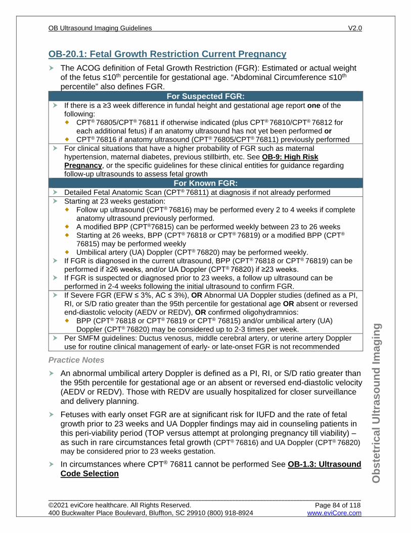
FGR Fetal growth restriction; an estimated weight of the fetus at or below 10th percentile for gestational age; and/or abdominal circumference of the fetus at or below 10th percentile for gestational age
M-mode ultrasound imaging technique in which structure movement can be depicted in a wave-like manner; primarily used in cardiac and fetal cardiac imaging
macrosomia estimated fetal weight of greater than 4000 or 4500 grams monochorionic twins
twins developed from one oocyte (egg) developing with a single chorions (membrane that forms the fetal part of the placenta)
NICU Neonatal Intensive Care Unit NST fetal non-stress test
oligohydramnios diminished amniotic fluid volume (AFV) for gestational age; definitions include: maximum deepest pocket of ≤2cm and/or AFI of ≤5cm or <the 5th percentile for gestational age if <30 weeks.
PACS Picture Archiving and Communications System polyhydramnios AFI ≥24cm or maximum vertical pocket of ≥8 cm PROM preterm rupture of membranes
quad screen alpha-fetoprotein (AFP), estriol, human chorionic gonadotropin (hCG), inhibin A
real time scan
considered the most common type of ultrasound; a 2-dimensional scan that reflects structure and motion over time, scanning and display of images are run at a sufficiently rapid rate so that moving structures can be viewed moving at their natural rate; frame rates ≥15 frames per second are considered “real time”
OB-1.0: General Guidelines This document offers an in-depth, indication driven guide to obstetrical imaging The use of a obstetrical CPT code is only indicated with a positive pregnancy test or
an otherwise confirmed pregnancy. It is not appropriate to report non-obstetrical pelvic ultrasound procedure codes (CPT® 76830, CPT® 76856, and CPT® 76857) with a positive pregnancy test or confirmed pregnancy
Ultrasound assessment is an accurate method of determining gestational age, fetal number, viability, and placental location, and it is recommended for all pregnant patients.
An evaluation of pregnancy with history and physical exam (an initial office visit) is necessary prior to obstetric ultrasound imaging requests
The following information must be submitted with each request: Expected date of delivery Gestational age at date of service Results of prior ultrasound studies if available
Normal (Low Risk) Pregnancy Imaging Per ACOG, in the absence of other specific indications, the optimal time for a
single ultrasound examination is at 18 to 22 weeks of gestation. This timing allows for a survey of fetal anatomy in most women and an accurate estimation of gestational age. Though fetal anatomy may be performed any time after 14 weeks, due to fetal size at earlier gestational age, fetal anatomical survey performed <16 weeks gestation may not be optimal. Report a fetal anatomy ultrasound CPT® 76805 if ≥16 weeks, for a normal/low
risk pregnancy. Current SMFM guidelines state that cervical length (CL) screening in singleton
gestations without a prior spontaneous preterm birth (PTB) cannot yet be universally mandated. Transvaginal ultrasound (CPT® 76817) may be considered if the
transabdominal CL is ≤3.6 cm or in certain circumstances of poor cervical visualization on transabdominal ultrasound
Fetal Nuchal Translucency (CPT® 76813) can be considered if Cell-Free DNA (cfDNA) is not planned or has not already been performed, as they are both screening tools for fetal aneuploidy
OB-1.1: Required Documentation See OB-1.0: General Guidelines
OB-1.2: Inappropriate Use of OB Ultrasound Obstetrical ultrasound studies cannot be authorized for payment for individuals who do not have a positive pregnancy test or clinical evidence of a pregnancy (fetal heart tones) Obstetrical ultrasound is not medically indicated for the following:
Sex determination only To provide a keepsake or souvenir picture
OB-1.3: Ultrasound Code Selection See OB-28: Procedure Coding Basics for Established Pregnancy
It is not appropriate to report non-obstetrical pelvic ultrasound procedure codes (CPT® 76830, CPT® 76856, and CPT® 76857) with a positive pregnancy test or confirmed pregnancy
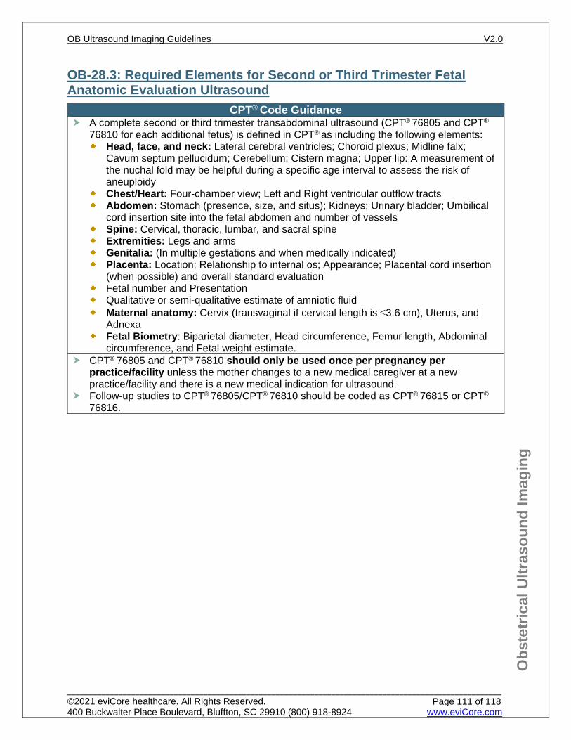
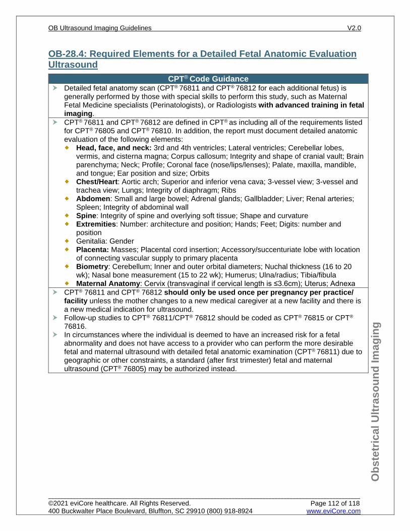
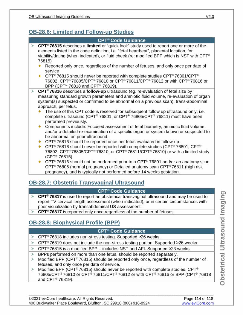
CPT® Code Guidance CPT® 76801 and CPT® 76802 (for each additional fetus) are reported for complete studies performed during the first trimester (<14 weeks). These codes should only be used once per pregnancy unless the mother changes to a new medical caregiver at a new office and there is a new medical indication for ultrasound. CPT® 76813 and CPT® 76814 (each additional fetus) are used to report nuchal translucency screening: an ultrasound measurement of the clear (translucent) space at the back of the fetal neck to assess risk for Down Syndrome (Trisomy 21), Trisomy 18, and other genetic disorders. CPT® 76805 and CPT® 76810 (for each additional fetus) are used to report complete studies (anatomy scan) performed during the second and third trimester, in a normal (low risk) pregnancy. These studies should only be used once per pregnancy unless the mother changes to a new medical caregiver at a new office and there is a new medical indication for ultrasound. CPT® 76811 and CPT® 76812 (for each additional fetus) describe a detailed fetal anatomic survey and are used only when the study includes this service. These studies should only be used once per pregnancy unless the mother changes to a new medical caregiver at a new office and there is a new medical indication for ultrasound. This detailed fetal anatomic evaluation is generally performed by those with special skills to perform this study, such as a Maternal Fetal Medicine specialist (Perinatologist), or a Radiologist with advanced training in fetal imaging. In circumstances where a detailed fetal anatomy (CPT® 76811) is indicated but access is limited due to geographic or other constraints, a standard fetal anatomy survey (CPT® 76805) may be authorized instead at the appropriate gestational age. CPT® 76817 is used to report a transvaginal ultrasound. The other OB ultrasound codes are used for transabdominal studies. CPT® 76816 is used to report a follow up study, such as a growth scan or follow up on anatomy when more than one area requires reexamination.
CPT® 76816 (should not be performed prior to a CPT® 76801 or an anatomy scan CPT® 76805 (normal pregnancy) or Detailed anatomy scan CPT® 76811 (high risk pregnancy)
CPT® 76816 should not be done on same date of service as CPT® 76815
CPT® Code Guidance CPT® 76815 describes a limited or ‘quick look’ study
It can be used at any gestational age for various indications, including quick look for AFI assessment, fetal heart-beat, fetal position, placental location etc.
It may be used specifically for ‘dating’ (when indicated) in those that don’t meet gestational age criteria for dating with CPT® 76801 or are too early for anatomy scan (i.e. >14 weeks but <16 weeks)
It is also used to report a modified BPP. Note: CPT® 76815 should never be reported with complete studies CPT® 76801/CPT®
76802, CPT® 76805/CPT® 76810, CPT® 76811/CPT® 76812, or with CPT® 76816 or BPP (CPT® 76818 and CPT® 76819).
CPT® 76818 (includes non-stress test) and CPT® 76819: are used to report a Biophysical profile (BPP), a test for antepartum fetal surveillance. CPT® 76820 describes Doppler velocimetry of the umbilical artery. CPT® 76821 describes Doppler velocimetry of the middle cerebral artery. CPT® 76825 describes fetal echocardiography and and CPT® 76827 describes the Doppler portion of the echocardiogram. These codes should only be used once per pregnancy unless the mother changes to a new medical caregiver at a new office or there is a new medical indication for ultrasound. CPT® 76826 describes a follow up fetal echocardiography and CPT® 76828 describes a follow up Doppler portion of the echocardiogram. CPT® 93325 may be added for color mapping in conjunction with fetal echocardiography procedures. CPT® 93976 describes a limited duplex scan and is used during pregnancy for characterizing the pattern and direction of blood flow in arteries and veins. It can be used to report fetal umbilical-placental flow evaluation (accreta or other placental or cord abnormalities). CPT® 74712 and CPT® 74713 (for each additional fetus) are used to report a fetal MRI (indicated for more in depth imaging of certain fetal abnormalities).
Practice Note ACOG recommendations for imaging during pregnancy and lactation:
Ultrasonography and magnetic resonance imaging (MRI) are not associated with risk and are the imaging techniques of choice for the pregnant patient, but they should be used prudently and only when use is expected to answer a relevant clinical question or otherwise provide medical benefit to the patient.
With few exceptions, radiation exposure through radiography (Xrays), computed tomography (CT) scan, or nuclear medicine imaging techniques is at a dose much lower than the exposure associated with fetal harm. If these techniques are necessary in addition to ultrasound or MRI or are more readily available for the diagnosis in question, they should not be withheld from a pregnant patient.
The use of gadolinium contrast with MRI should be limited; it may be used as a contrast agent in a pregnant woman only if it significantly improves diagnostic performance and is expected to improve fetal or maternal outcome.
With regards to iodinated IV contrast media, “it is generally recommended that contrast only be used if absolutely required to obtain additional diagnostic information that will affect the care of the fetus or woman during pregnancy”.
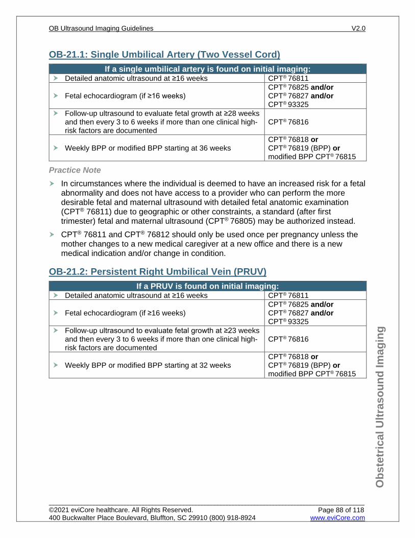
In circumstances where the individual is deemed to have an increased risk for a fetal abnormality and does not have access to a provider who can perform the more desirable fetal and maternal ultrasound with detailed fetal anatomic examination (CPT® 76811) due to geographic or other constraints, a standard (after first trimester) fetal and maternal ultrasound (CPT® 76805) may be authorized instead.
CPT® 76816 (should not be performed prior to a CPT® 76801 or an anatomy scan CPT® 76805 (normal pregnancy) or Detailed anatomy scan CPT® 76811 (high risk pregnancy), and is typically not performed prior to 14 weeks gestation.
Typically all components of the BPP (CPT® 76818 and CPT® 76819), such as breathing, are not present until ≥26 weeks gestation. However, a modified BPP (CPT® 76815) may be utilized sooner in certain high risk cases but should not be done prior to viability (23 weeks).
SMFM suggest that ductus venosus, middle cerebral artery, or uterine artery Doppler use for routine clinical management of early- or late-onset FGR is not recommended
The minimal use of color Doppler alone (CPT® 93976), when performed for anatomical structure identification, during a standard ultrasound procedure, is not separately reimbursable.
References 1. Reddy UM, Abuhamad AZ, Levine D, Saade GR. Fetal Imaging: Executive Summary of a Joint
Eunice Kennedy Shriver National Institute of Child Health and Human Development, Society for Maternal-Fetal Medicine, American Institute of Ultrasound in Medicine, American College of Obstetricians and Gynecologists, American College of Radiology, Society for Pediatric Radiology, and Society of Radiologists in Ultrasound Fetal Imaging Workshop. Obstetrical & Gynecological Survey. 2014;69(8):453-455. doi:10.1097/01.ogx.0000453817.62105.4a.
2. Practice Bulletin No. 175: Ultrasound in Pregnancy. Obstet Gynecol. 2016;128(6):e241-e256. Reaffirmed 2020.doi:10.1097/AOG.0000000000001815.
3. AIUM-ACR-ACOG-SMFM-SRU Practice Parameter for the Performance of Standard Diagnostic Obstetric Ultrasound Examinations. Journal of Ultrasound in Medicine. 2018;37(11). doi:10.1002/jum.14831.
4. AIUM Practice Parameter for the Performance of Detailed Second‐ and Third‐Trimester Diagnostic Obstetric Ultrasound Examinations. Journal of Ultrasound in Medicine. 2019;38(12):3093-3100. doi:10.1002/jum.15163.
5. AIUM Practice Parameter for the Performance of Limited Obstetric Ultrasound Examinations by Advanced Clinical Providers. Journal of Ultrasound in Medicine. 2018;37(7):1587-1596. doi:10.1002/jum.14677.
6. AIUM Practice Parameter for the Performance of Fetal Echocardiography. Journal of Ultrasound in Medicine. 2019;39(1). doi:10.1002/jum.15188.
7. Martins JG, Biggio JR, Abuhamad A. Society for Maternal-Fetal Medicine (SMFM) Consult Series #52: Diagnosis and Management of Fetal Growth Restriction. American Journal of Obstetrics and Gynecology. 2020. doi:10.1016/j.ajog.2020.05.010.
OB-2.1: Uncertain Dates/Unknown Last Menstrual Period (LMP) If there is a difference in the clinical size of the uterus on pelvic exam and the
estimated gestational age calculated by the LMP or there is an uncertain/unknown LMP or there have been irregular periods in the last year, one of the following may be indicated:
If <14 weeks by pelvic exam CPT® 76801 one time (plus CPT® 76802 for each additional fetus) and/or CPT® 76817 one time if a complete ultrasound has not yet been performed
If ≥14 weeks by abdominal exam CPT® 76815 or CPT® 76805 (CPT® 76811 if high risk) if complete fetal anatomic scan has not yet been performed
References 1. Practice Bulletin No. 175: Ultrasound in Pregnancy. Obstetrics & Gynecology. 2016;128(6):e241-
e256. Reaffirmed 2020. doi:10.1097/AOG.0000000000001815 2. Reddy UM, Abuhamad AZ, Levine D, Saade GR. Fetal Imaging: Executive Summary of a Joint
Eunice Kennedy Shriver National Institute of Child Health and Human Development, Society for Maternal-Fetal Medicine, American Institute of Ultrasound in Medicine, American College of Obstetricians and Gynecologists, American College of Radiology, Society for Pediatric Radiology, and Society of Radiologists in Ultrasound Fetal Imaging Workshop. Obstetrics & Gynecology. 2014;123(5):1070-1082. doi:10.1097/aog.0000000000000245.
3. ACOG Committee Opinion No 700: Methods for Estimating the Due Date. Obstetrics & Gynecology. 2017;129(5):e150-e154. doi:10.1097/AOG.0000000000002046.
4. ACOG Committee Opinion Number 688: Management of Suboptimally Dated Pregnancies, Obstetrics & Gynecology. 2017;129(3). Reaffirmed 2019. doi:10.1097/AOG.0000000000001949.
3. Prabhakaran S and Chuang A. In-office retrieval of intrauterine contraceptive devices with missing strings. Contraception. 2011;83(2):102-106. doi:10.1016/j.contraception.2010.07.004.
OB-4.1: History of Infertility If there is a history of infertility, or the current or a prior pregnancy was conceived
using an ovulation induction agent (for example Clomid) and/or by intrauterine insemination (IUI), or a past pregnancy with ART (IVF), a dating/viability US is indicated:
CPT® 76801 [plus CPT® 76802 for each additional fetus] and/or CPT® 76817 if <14 weeks or
CPT® 76815 if ≥14 weeks but <16 weeks Then, follow low risk imaging See OB-7.1: Fetal Anatomic Scan
Repeat ultrasound is not usually necessary unless there are new clinical indications
OB-4.2: Present Pregnancy with ART Treatment (IVF) Follow high risk imaging, See OB-9: High Risk Pregnancy
OB-4.3: Recurrent Pregnancy Loss Ultrasound imaging is supported if there is a history of at least 2 consecutive or 3
non-consecutive clinical miscarriages/losses at <20 weeks gestation Follow high risk imaging, See OB-9: High Risk Pregnancy
References 1. Kondapalli LA, Perales-Puchalt A. Low birth weight: is it related to assisted reproductive technology
or underlying infertility? Fertility and Sterility. 2013;99(2):303-310. doi:10.1016/j.fertnstert.2012.12.035.
2. Definitions of infertility and recurrent pregnancy loss: a committee opinion. Fertility and Sterility. 2020;113(3):533-535. doi:10.1016/j.fertnstert.2019.11.025.
3. ACOG Practice Bulletin No. 200. Early pregnancy loss. Obstetrics & Gynecology. 2018;132(5). doi:10.1097/aog.0000000000002899.
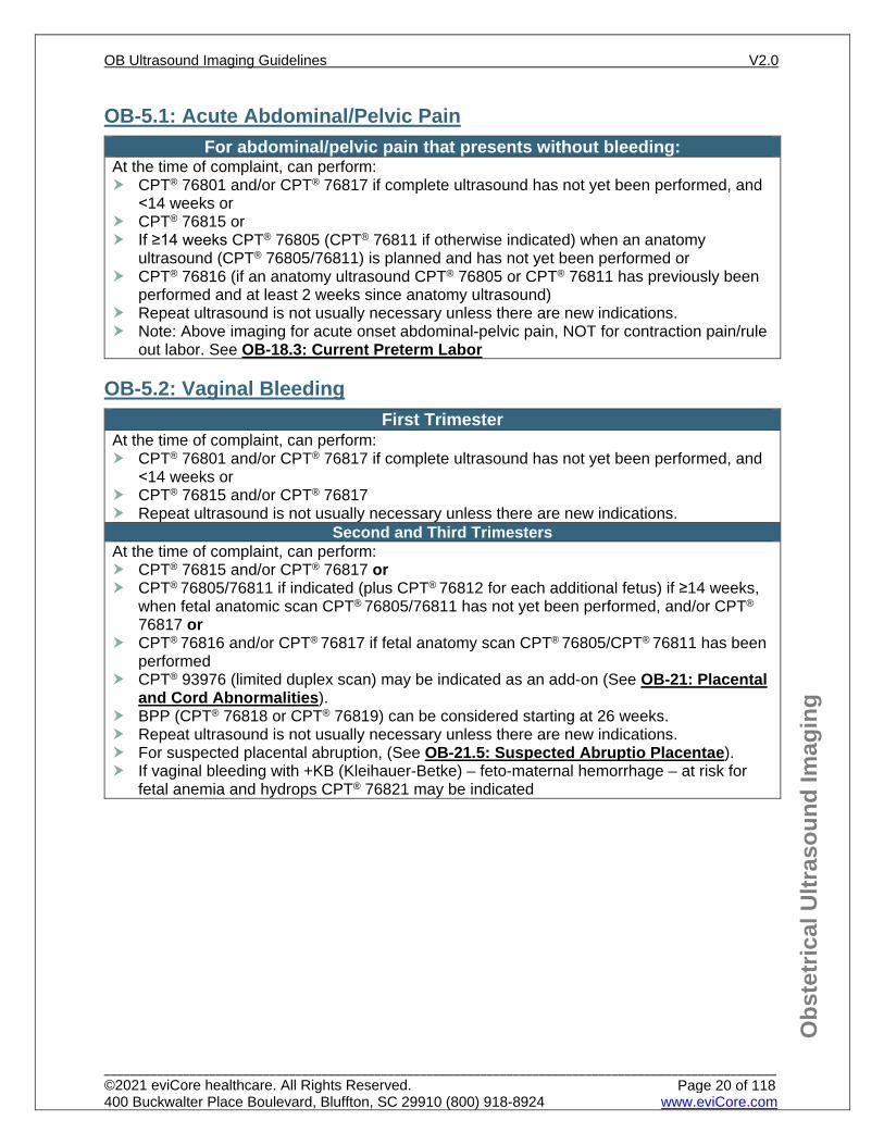
OB-5.1: Acute Abdominal/Pelvic Pain For abdominal/pelvic pain that presents without bleeding:
At the time of complaint, can perform: CPT® 76801 and/or CPT® 76817 if complete ultrasound has not yet been performed, and
<14 weeks or CPT® 76815 or If ≥14 weeks CPT® 76805 (CPT® 76811 if otherwise indicated) when an anatomy
ultrasound (CPT® 76805/76811) is planned and has not yet been performed or CPT® 76816 (if an anatomy ultrasound CPT® 76805 or CPT® 76811 has previously been
performed and at least 2 weeks since anatomy ultrasound) Repeat ultrasound is not usually necessary unless there are new indications. Note: Above imaging for acute onset abdominal-pelvic pain, NOT for contraction pain/rule
out labor. See OB-18.3: Current Preterm Labor
OB-5.2: Vaginal Bleeding First Trimester
At the time of complaint, can perform: CPT® 76801 and/or CPT® 76817 if complete ultrasound has not yet been performed, and
<14 weeks or CPT® 76815 and/or CPT® 76817 Repeat ultrasound is not usually necessary unless there are new indications.
Second and Third Trimesters At the time of complaint, can perform: CPT® 76815 and/or CPT® 76817 or CPT® 76805/76811 if indicated (plus CPT® 76812 for each additional fetus) if ≥14 weeks,
when fetal anatomic scan CPT® 76805/76811 has not yet been performed, and/or CPT®
76817 or CPT® 76816 and/or CPT® 76817 if fetal anatomy scan CPT® 76805/CPT® 76811 has been
performed CPT® 93976 (limited duplex scan) may be indicated as an add-on (See OB-21: Placental
and Cord Abnormalities). BPP (CPT® 76818 or CPT® 76819) can be considered starting at 26 weeks. Repeat ultrasound is not usually necessary unless there are new indications. For suspected placental abruption, (See OB-21.5: Suspected Abruptio Placentae). If vaginal bleeding with +KB (Kleihauer-Betke) – feto-maternal hemorrhage – at risk for
fetal anemia and hydrops CPT® 76821 may be indicated
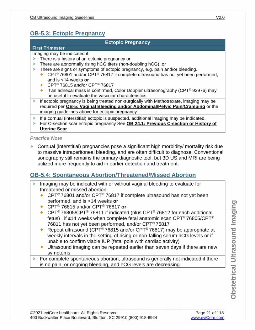
First Trimester Imaging may be indicated if: There is a history of an ectopic pregnancy or There are abnormally rising hCG titers (non-doubling hCG), or There are signs or symptoms of ectopic pregnancy, e.g. pain and/or bleeding.
CPT® 76801 and/or CPT® 76817 if complete ultrasound has not yet been performed, and is <14 weeks or
CPT® 76815 and/or CPT® 76817 If an adnexal mass is confirmed, Color Doppler ultrasonography (CPT® 93976) may
be useful to evaluate the vascular characteristics If ectopic pregnancy is being treated non-surgically with Methotrexate, imaging may be
required per OB-5: Vaginal Bleeding and/or Abdominal/Pelvic Pain/Cramping or the imaging guidelines above for ectopic pregnancy
If a cornual (interstitial) ectopic is suspected, additional imaging may be indicated. For C-section scar ectopic pregnancy See OB 24.1: Previous C-section or History of
Uterine Scar
Practice Note Cornual (interstitial) pregnancies pose a significant high morbidity/ mortality risk due
to massive intraperitoneal bleeding, and are often difficult to diagnose. Conventional sonography still remains the primary diagnostic tool, but 3D US and MRI are being utilized more frequently to aid in earlier detection and treatment.
OB-5.4: Spontaneous Abortion/Threatened/Missed Abortion Imaging may be indicated with or without vaginal bleeding to evaluate for
threatened or missed abortion. CPT® 76801 and/or CPT® 76817 if complete ultrasound has not yet been
performed, and is <14 weeks or CPT® 76815 and/or CPT® 76817 or CPT® 76805/CPT® 76811 if indicated (plus CPT® 76812 for each additional
fetus) , if ≥14 weeks when complete fetal anatomic scan CPT® 76805/CPT® 76811 has not yet been performed, and/or CPT® 76817
Repeat ultrasound (CPT® 76815 and/or CPT® 76817) may be appropriate at weekly intervals in the setting of rising or non-falling serum hCG levels or if unable to confirm viable IUP (fetal pole with cardiac activity)
Ultrasound imaging can be repeated earlier than seven days if there are new symptoms
For complete spontaneous abortion, ultrasound is generally not indicated if there is no pain, or ongoing bleeding, and hCG levels are decreasing.
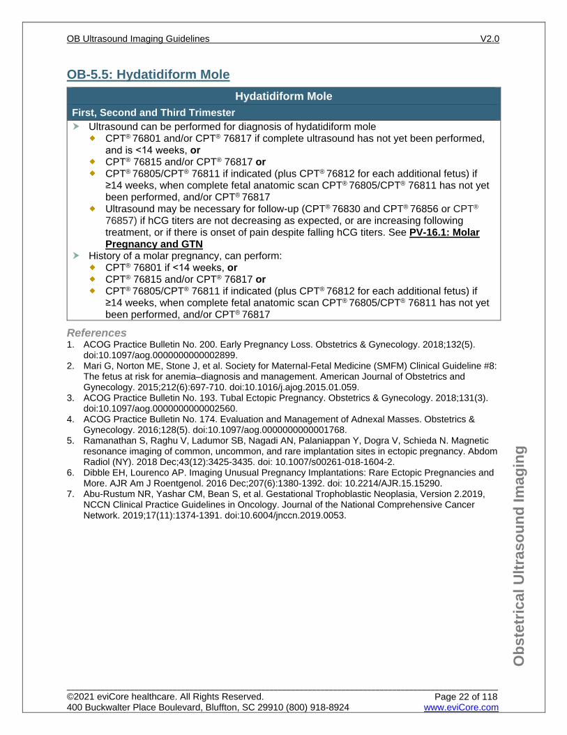
First, Second and Third Trimester Ultrasound can be performed for diagnosis of hydatidiform mole
CPT® 76801 and/or CPT® 76817 if complete ultrasound has not yet been performed, and is <14 weeks, or
CPT® 76815 and/or CPT® 76817 or CPT® 76805/CPT® 76811 if indicated (plus CPT® 76812 for each additional fetus) if
≥14 weeks, when complete fetal anatomic scan CPT® 76805/CPT® 76811 has not yet been performed, and/or CPT® 76817
Ultrasound may be necessary for follow-up (CPT® 76830 and CPT® 76856 or CPT® 76857) if hCG titers are not decreasing as expected, or are increasing following treatment, or if there is onset of pain despite falling hCG titers. See PV-16.1: Molar Pregnancy and GTN
History of a molar pregnancy, can perform: CPT® 76801 if <14 weeks, or CPT® 76815 and/or CPT® 76817 or CPT® 76805/CPT® 76811 if indicated (plus CPT® 76812 for each additional fetus) if
≥14 weeks, when complete fetal anatomic scan CPT® 76805/CPT® 76811 has not yet been performed, and/or CPT® 76817
References 1. ACOG Practice Bulletin No. 200. Early Pregnancy Loss. Obstetrics & Gynecology. 2018;132(5).
doi:10.1097/aog.0000000000002899. 2. Mari G, Norton ME, Stone J, et al. Society for Maternal-Fetal Medicine (SMFM) Clinical Guideline #8:
The fetus at risk for anemia–diagnosis and management. American Journal of Obstetrics and Gynecology. 2015;212(6):697-710. doi:10.1016/j.ajog.2015.01.059.
4. ACOG Practice Bulletin No. 174. Evaluation and Management of Adnexal Masses. Obstetrics & Gynecology. 2016;128(5). doi:10.1097/aog.0000000000001768.
5. Ramanathan S, Raghu V, Ladumor SB, Nagadi AN, Palaniappan Y, Dogra V, Schieda N. Magnetic resonance imaging of common, uncommon, and rare implantation sites in ectopic pregnancy. Abdom Radiol (NY). 2018 Dec;43(12):3425-3435. doi: 10.1007/s00261-018-1604-2.
6. Dibble EH, Lourenco AP. Imaging Unusual Pregnancy Implantations: Rare Ectopic Pregnancies and More. AJR Am J Roentgenol. 2016 Dec;207(6):1380-1392. doi: 10.2214/AJR.15.15290.
7. Abu-Rustum NR, Yashar CM, Bean S, et al. Gestational Trophoblastic Neoplasia, Version 2.2019, NCCN Clinical Practice Guidelines in Oncology. Journal of the National Comprehensive Cancer Network. 2019;17(11):1374-1391. doi:10.6004/jnccn.2019.0053.
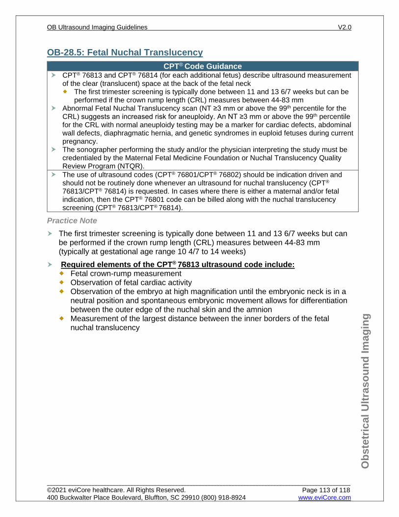
OB-6.1: First Trimester Screening First trimester screening includes biochemical markers and fetal nuchal translucency
(NT) (CPT® 76813) assessment. An increased Fetal Nuchal Translucency (NT ≥3.0 mm or above the 99th percentile for the CRL), may indicate a fetus with aneuploidy (e.g. Down’s syndrome, Trisomy 18) but may also indicate an increased risk for cardiac defects or other structural defects or genetic syndromes in euploid fetuses.
Nuchal translucency is most accurate when performed between 11 and 13 6/7 weeks, but can be performed if the crown rump length (CRL) measures between 44-83 mm.
First Trimester Screening:
Ultrasound CPT® 76813 (plus CPT® 76814 for each additional fetus) is the initial imaging for first trimester screening, to evaluate fetal nuchal translucency
If increased Fetal Nuchal Translucency (NT ≥3.0 mm or above the 99th percentile for the CRL): Fetal anatomic ultrasound (CPT® 76811) at ≥16 weeks Fetal echo (CPT® 76825 and/or CPT® 76827 and/or CPT® 93325) at ≥16 weeks cfDNA, Amniocentesis or CVS may be performed
Cell-Free DNA (cfDNA) can be performed any time after 10 weeks gestation and is currently the most sensitive screening test for Down’s syndrome per the American College of Medical Genetics and Genomics (99% accurate).
Fetal NT (CPT® 76813) is NOT recommended if cfDNA is planned or has already been performed, as they are both screening tools for fetal aneuploidy
Those with a positive cfDNA should be offered diagnostic testing (amniocentesis or CVS) and a detailed anatomy scan (CPT®76811) at ≥16 weeks. See OB-9.1: High Risk Group One – Risk Factors.
A “no call” or indeterminate result can occur (risk is higher with maternal obesity), which also has a higher risk of aneuploidy. These patients should managed as if positive.
Coding Notes CPT® 76813/CPT® 76814 can be performed once per pregnancy, and should be
performed only by those certified by the Fetal Medicine Foundation or Nuchal Translucency Quality Review Program (NTQR).
The use of ultrasound codes (CPT® 76801/CPT® 76802) should be indication driven and should NOT be routinely done whenever an ultrasound for nuchal translucency (CPT® 76813/CPT® 76814) is requested. In cases where there is either a maternal and/or fetal indication, then the CPT® 76801/CPT® 76802 code can indeed be billed along with the nuchal translucency screening (CPT® 76813/CPT® 76814).
OB-6.2: Second Trimester Screening Second Trimester Screening:
A fetal anatomy ultrasound (CPT® 76805) and/or QUAD screen can be performed during the second trimester to detect fetal aneuploidy, neural tube defects, and other anatomical defects. See OB-7.1: Fetal Anatomic Scan
If the quad screening is abnormal, a detailed anatomy ultrasound (CPT® 76811) may also be performed.
Practice Notes Multiple marker screening is used in the second trimester (15 to 22 6/7 weeks) to screen for aneuploidy as well as open neural tube defects (ONTD). Maternal serum alpha-fetoprotein (MSAFP) can be done at 15 to 20 weeks to screen
for neural tube defects in those that have had cfDNA or NT screen. The “quad” screen (AFP (alpha-fetoprotein), hCG (human chorionic gonadotropin),
uE (Unconjugated estriol), dimeric inhibin-A) is the most commonly used test for the second trimester.
A penta screen (quad screen markers + hyperglycosylated hCG) may be done in lieu of a quad screen.
Combined, integrated or sequential screening (first and second trimester screening) may also be used and provides a higher detection rate than a single screening.
Providers often wait for the results of the quad screen before ordering CPT® 76805. If the quad screen is abnormal, they may request CPT® 76811 in lieu of CPT® 76805.
References 1. American College of Obstetricians and Gynecologists’ Committee on Practice Bulletins—Obstetrics;
Committee on Genetics; Society for Maternal-Fetal Medicine. Screening for Fetal Chromosomal Abnormalities: ACOG Practice Bulletin, Number 226. Obstet Gynecol. 2020 Oct;136(4):e48-e69. doi: 10.1097/AOG.0000000000004084.
2. ACOG Practice Bulletin No. 175: Ultrasound in Pregnancy. Obstet Gynecol. 2016;128(6):e241-e256. Reaffirmed 2020 doi:10.1097/AOG.0000000000001815.
3. Gregg AR, Skotko BG, Benkendorf JL, et al. Noninvasive prenatal screening for fetal aneuploidy, 2016 update: a position statement of the American College of Medical Genetics and Genomics. Genetics in Medicine. 2016;18(10):1056-1065. doi:10.1038/gim.2016.97.
4. Norton ME, Biggio JR, Kuller JA, Blackwell SC. Society for Maternal-Fetal Medicine (SMFM) Consult Series | #42: The role of ultrasound in women who undergo cell-free DNA screening. American Journal of Obstetrics and Gynecology. 2017;216(3):B2-B7. doi:10.1016/j.ajog.2017.01.005.
5. Society for Maternal and Fetal Medicine (SMFM), coding committee, October 2017. SMFM’s white paper on billing combination of 76801 and 76813
6. ACOG Practice Bulletin No. 162 Prenatal diagnostic testing for genetic disorders. Obstetrics & Gynecology. 2016;127(5). Reaffirmed 2018. doi:10.1097/aog.0000000000001405.
7. Donofrio MT, Moon-Grady AJ, Hornberger LK, et al. Diagnosis and Treatment of Fetal Cardiac Disease. Circulation. 2014;129(21):2183-2242. doi:10.1161/01.cir.0000437597.44550.5d.
OB-7.1: Fetal Anatomic Scan Per ACOG, in the absence of other specific indications, the optimal time for a single
ultrasound examination is at 18 to 22 weeks of gestation. This timing allows for a survey of fetal anatomy in most women and an accurate estimation of gestational age. Though fetal anatomy may be performed any time after 14 weeks, due to fetal size at earlier gestational age, fetal anatomical survey performed at a gestational age <16 weeks may not be optimal.
Report a fetal anatomy ultrasound CPT® 76805 if ≥16 weeks, for a normal/low risk pregnancy.
If pregnancy is high risk report a detailed fetal anatomy ultrasound (CPT® 76811) if ≥16 weeks. This is generally performed by a Maternal Fetal Medicine (MFM)/Perinatologist, or a Radiologist at an AIUM or ACR accredited facility. See OB-9: High Risk Pregnancy
Current SMFM guidelines state that CL screening in singleton gestations without a prior spontaneous PTB cannot yet be universally mandated.
Transvaginal ultrasound (CPT® 76817) may be considered if the transabdominal cervical length (CL) is ≤3.6 cm or in certain circumstances of poor cervical visualization on transabdominal ultrasound. If cervical shortening identified – See OB-18.1: Cervical Insufficiency
OB-7.2: Fetal Anatomic Scan – Follow-up Follow-up ultrasounds (CPT® 76815 to assess a single item or CPT® 76816 if
multiple areas to be assessed) can be performed once for incomplete or equivocal finding on initial fetal anatomic scan.
CPT® 76816 (should not be performed prior to a CPT® 76801 or an anatomy scan CPT® 76805 (normal pregnancy) or Detailed anatomy scan CPT® 76811 (high risk pregnancy)
If pregnancy is high risk See OB-9: High Risk Pregnancy or other applicable high risk guideline.
Detailed anatomy ultrasound CPT® 76811 can be performed if not previously performed when initial fetal anatomic scan CPT® 76805 is abnormal. See OB-9: High Risk Pregnancy
References 1. AIUM-ACR-ACOG-SMFM-SRU Practice Parameter for the Performance of Standard Diagnostic
Obstetric Ultrasound Examinations. Journal of Ultrasound in Medicine. 2018;37(11). doi:10.1002/jum.14831.
2. AIUM Practice Parameter for the Performance of Detailed Second‐ and Third‐Trimester Diagnostic Obstetric Ultrasound Examinations. Journal of Ultrasound in Medicine. 2019;38(12):3093-3100. doi:10.1002/jum.15163.
3. Practice Bulletin No. 175: Ultrasound in Pregnancy. Obstet Gynecol. 2016;128(6):e241-e256. Reaffirmed 2020 doi:10.1097/AOG.0000000000001815.
4. AIUM Practice Parameter for the Performance of Limited Obstetric Ultrasound Examinations by Advanced Clinical Providers. Journal of Ultrasound in Medicine. 2018;37(7):1587-1596. doi:10.1002/jum.14677.
5. American Medical Association. CPT—Current Procedural Terminology. American Medical Association. https://www.ama-assn.org/practice-management/cpt. Published 2019. Copyright 1995 - 2019.
6. ACOG Practice Bulletin No.130: Prediction and Prevention of Preterm Birth. Obstet Gynecol. 2012;120(4):964-973. Reaffirmed 2016. doi:10.1097/AOG.0b013e3182723b1b.
7. Cho HJ, Roh H-J. Correlation Between Cervical Lengths Measured by Transabdominal and Transvaginal Sonography for Predicting Preterm Birth. Journal of Ultrasound in Medicine. 2016;35(3):537-544. doi:10.7863/ultra.15.03026.
8. Esplin MS, Elovitz MA, Iams JD, et al. Predictive Accuracy of Serial Transvaginal Cervical Lengths and Quantitative Vaginal Fetal Fibronectin Levels for Spontaneous Preterm Birth Among Nulliparous Women. JAMA. 2017;317(10):1047. doi:10.1001/jama.2017.1373.
9. Jain S, Kilgore M, Edwards RK, Owen J. Revisiting the cost-effectiveness of universal cervical length screening: importance of progesterone efficacy. American Journal of Obstetrics and Gynecology. 2016;215(1). doi:10.1016/j.ajog.2016.01.165
10. Blackwell SC, Gyamfi-Bannerman C, Biggio JR Jr, et al. 17-OHPC to Prevent Recurrent Preterm Birth in Singleton Gestations (PROLONG Study): A Multicenter, International, Randomized Double-Blind Trial. Am J Perinatol. 2020;37(2):127–136. doi:10.1055/s-0039-3400227.
11. Mcintosh J, Feltovich H, Berghella V, Manuck T. The role of routine cervical length screening in selected high- and low-risk women for preterm birth prevention. Society for Maternal-Fetal Medicine (SMFM) Consult Series #40. American Journal of Obstetrics and Gynecology. 2016;215(3). doi:10.1016/j.ajog.2016.04.027.
12. Friedman AM, Schwartz N, Ludmir J, Parry S, Bastek JA, Sehdev HM. Can transabdominal ultrasound identify women at high risk for short cervical length? Acta Obstetricia et Gynecologica Scandinavica. 2013;92(6):637-641. doi:10.1111/aogs.12111
OB-9: High Risk Pregnancy OB-9.0: High Risk General Information 32 OB-9.1: High Risk Group One – Risk Factors 33 OB-9.2: High Risk Group Two – Findings on Ultrasound that May Require Further Imaging 35
OB-9.2.1: Soft Markers for Aneuploidy 35 OB-9.2.2: Other Findings on Ultrasound that May Require Further Imaging 35
OB-9.3: High Risk Group Three – Pre-pregnancy BMI ≥30 kg/m2 36 OB-9.3.1: Class I Obesity - Pre-pregnancy BMI 30 to 34.9 36 OB-9.3.2: Class II Obesity - Pre-pregnancy BMI 35-39.9 36 OB-9.3.3: Class III Obesity - Pre-pregnancy BMI ≥40 36
OB-9.4: High Risk Group Four – Macrosomia 37 OB-9.4.1: Prior Pregnancy with Macrosomia 37 OB-9.4.2: Current Pregnancy with Suspected or Known Macrosomia 37
OB-9.5: High Risk Group Five – Zika and COVID-19 Virus 37 OB-9.5.1: Zika Virus 37 OB-9.5.2: COVID-19 Virus 38
OB-9.6: High Risk Group Six – Pre-Gestational Diabetes 39 OB-9.6.1: Pre-Gestational or Early Diagnosed (≤20 weeks) Diabetes - Not on Medication 39 OB-9.6.2: Pre-Gestational or Early Diagnosed (≤20 weeks) Diabetes - On Medication 39
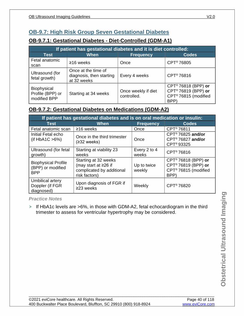
OB-9.7: High Risk Group Seven Gestational Diabetes 40 OB-9.7.1: Gestational Diabetes - Diet-Controlled (GDM-A1) 40 OB-9.7.2: Gestational Diabetes on Medications (GDM-A2) 40
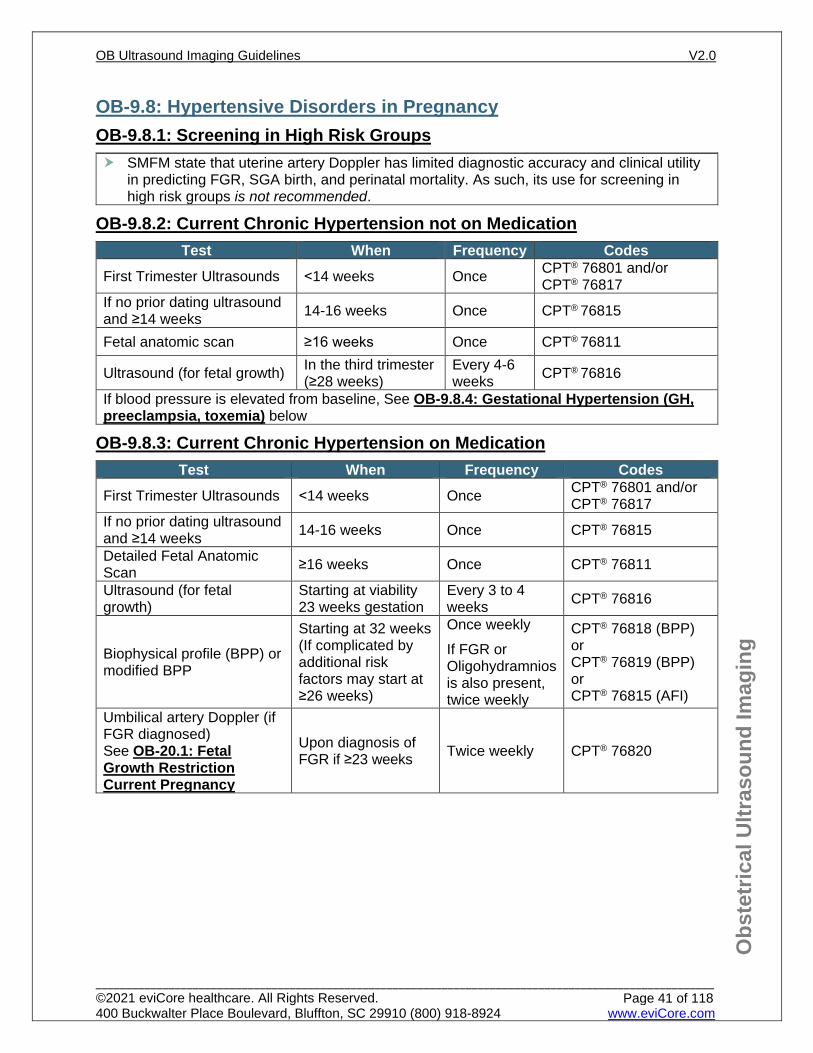
OB-9.8: Hypertensive Disorders in Pregnancy 41 OB-9.8.1: Screening in High Risk Groups 41 OB-9.8.2: Current Chronic Hypertension not on Medication 41 OB-9.8.3: Current Chronic Hypertension on Medication 41 OB-9.8.4: Gestational Hypertension (GH, preeclampsia, toxemia) 42
OB-9.9: History of Spontaneous Pre-Term Delivery/History of PPROM 43
OB-9.9.1: Spontaneous Preterm Delivery ≤34 Weeks; History of PPROM ≤34 weeks 43 OB-9.9.2: History of Spontaneous Preterm Delivery >34 weeks <37 weeks; History of PPROM >34 weeks <37 weeks 43
OB-9.0: High Risk General Information High Risk Pregnancy General Information: A Detailed Fetal Anatomic Scan (CPT® 76811/CPT® 76812 for each additional fetus)
is ideally performed between 18 to 20 weeks, but can be performed any time after 14 weeks when criteria is met. However, due to fetal size at earlier gestational age, fetal anatomical survey performed at a gestational age of <16 weeks may not be optimal.
This detailed fetal anatomic evaluation is generally performed by those with special skills to perform this study, such as a Maternal Fetal Medicine specialist (Perinatologist), or a Radiologist with advanced training in fetal imaging.
In circumstances where the individual is deemed to have an increased risk for a fetal abnormality and does not have access to a provider who can perform the more desirable fetal and maternal ultrasound with detailed fetal anatomic examination (CPT® 76811) due to geographic or other constraints, a standard (after first trimester) fetal and maternal ultrasound (CPT® 76805) may be authorized instead.
CPT® 76805, CPT® 76810, CPT® 76811, and CPT® 76812 should only be used once per pregnancy unless the mother changes to a new medical caregiver at a new office and there is a new medical indication and/or change in condition.
CPT® 76816 (should not be performed prior to a CPT® 76801 or an anatomy scan CPT® 76805 (normal pregnancy) or Detailed anatomy scan CPT® 76811 (high risk pregnancy)
Typically all components of the BPP (CPT® 76818 and CPT® 76819), such as breathing, are not present until ≥26 weeks gestation. However, a modified BPP (CPT® 76815) may be utilized sooner in certain high risk cases but should not be done prior to viability (23 weeks).
SMFM suggest that ductus venosus, middle cerebral artery, or uterine artery Doppler use for routine clinical management of early- or late-onset FGR is not recommended
OB-9.1: High Risk Group One – Risk Factors HIGH RISK PREGNANCY – Risk Factors
Socio-Demographic Risk Factors (maternal age) Age ≥35 years of age at the estimated date of confinement (EDC) Lifestyle Related Risk Factors (legal or illicit drug/alcohol use) Recreational drug (e.g. cocaine, amphetamines, opiates) or alcohol use during current
pregnancy (Excluding marijuana) For marijuana use -See OB-10.1: Medications and Substances that Qualify for a
Detailed Fetal Anatomic Scan Nicotine (≥10 cigarettes a day) Other nicotine exposure in pregnancy (e-cigs, vaping, chewing, patch) may also be
considered high risk Current Maternal IV drug abuse Current use of Suboxone, Subutex, Methadone Other High Risk medication or substance use - See OB-10.1: Medications and
Substances that Qualify for a Detailed Fetal Anatomic Scan
Practice Notes Several studies noted lower birth weights among offspring exposed to marijuana. These findings were more pronounced among women who used more marijuana, particularly during the first and second trimesters (at least weekly during the pregnancy). CPT® 76811 may be indicated (See OB-10.1: Medications and Substances that Qualify for a Detailed Fetal Anatomic Scan), however, given the limited evidence for antenatally detected abnormal growth, serial growth ultrasounds may not be indicated in the absence of other findings concerning for growth restriction Health Condition Related Risk Factors or Chronic medical condition that may affect fetal growth due to utero-placental insufficiency (maternal diseases or conditions) Anemia severe, <8 grams Hgb or 24% HCT Asthma (poorly controlled or steroid dependent) Autoimmune disease (e.g. Multiple Sclerosis, Immune Thrombocytopenic Purpura) Bariatric surgery Connective tissue disorders (lupus, RA, scleroderma, Sjogren’s, etc.) DVT/PE or Maternal thrombophilia (Antiphospholipid Syndrome, Factor V Leiden mutation,
Antithrombin III deficiency, Protein C/Protein S deficiency, Prothrombin gene mutation etc.) Genetic Carrier status e.g., Cystic Fibrosis/Known carrier of Spinal Muscular Atrophy (SMA),
CF, Tay-Sachs genetic diseases Heart disease (Maternal) – World Health Organization (WHO) Class II or greater Hemoglobinopathies (e.g. sickle cell disease, Alpha and Beta thalassemia minor (trait) or
major) History of endometrial ablation or Uterine Artery embolization Inflammatory Bowel Disease (Ulcerative colitis, Crohn’s Disease) Liver disease e.g. Hepatitis, Cholestasis of pregnancy (see imaging below) Maternal malnutrition (BMI <18.5) PKU Renal disease e.g. glomerulonephritis, persistent protein in the urine, renal insufficiency Seizure disorders – on antiepileptic medication Systemic malignancy Thyroid disorder (e.g. hyperthyroidism, poorly controlled hypothyroidism)
Previous pregnancy related risk factors Recurrent pregnancy loss (history of at least 2 consecutive or 3 non-consecutive clinical
miscarriages/losses at <20 weeks gestation) Prior pregnancy with adverse outcome (early onset preeclampsia ≤34 weeks, abruption,
accreta, previous uterine dehiscence or rupture, FGR at any gestational age, nonimmune hydrops, etc.).
Prior pregnancy with SGA (baby weighing <2500 grams at term or FGR less than the 10th percentile of expected weight)
For stillbirth See OB-9.10: History of Stillbirth Current pregnancy related risk factors Abnormal 1st or 2nd trimester screen (e.g. Abnormal MSAFP; Low PAPP_A; Low estriol;
Elevated inhibin A or Elevated B-hCG) Known chromosomal abnormalities; or abnormal cfDNA
Major Fetal anomaly such as gastroschisis, fetal ventriculomegaly, fetal hydronephrosis (>10mm), achondroplasias, fetal congenital heart disease, sustained fetal arrhythmias (See OB-16.5: Other Causes of Fetal Anemia)
ART Conception with assisted reproductive technologies (IVF) Grand multiparity: must have completed 5 or more pregnancies of greater than 20 weeks
gestation, living or stillbirth (does not include current pregnancy; twins count as 1 pregnancy)
Abnormal Fetal Nuchal Translucency ≥3.0mm; Thickened nuchal fold found on second trimester imaging ≥6mm up to 22 weeks
No prenatal care prior to the third trimester Maternal Infections (not exposure) Acquired Immune Deficiency Syndrome/HIV Positive Chicken Pox/Varicella Cytomegalovirus (CMV) Malaria Known parvovirus in current pregnancy post fetal treatment. See OB-16.2: Exposure to
Parvovirus B-19 Rubella Syphilis, untreated Toxoplasmosis Tuberculosis For Zika Virus and COVID-19 Virus See OB-9.5: High Risk Group Five: Zika and
COVID-19 Virus
Imaging For Above Conditions CPT® 76801 [plus CPT® 76802 for each additional fetus] if <14 weeks and a complete
ultrasound has not yet been performed, and/or CPT® 76817 for a transvaginal ultrasound CPT® 76815 can be performed for dating or quick look follow-up if ≥14 weeks but <16
weeks Detailed Fetal Anatomic Scan CPT® 76811 ≥16 weeks Starting at 23 follow-up growth scans (CPT® 76816) every 3 to 6 weeks Starting at 32 weeks, weekly BPP (CPT® 76818 or CPT® 76819) or modified BPP (CPT®
76815) Those diagnosed with intra-hepatic cholestasis of pregnancy (IHCP) can have weekly
BPP or modified BPP starting at the time of diagnosis (if ≥23 weeks).
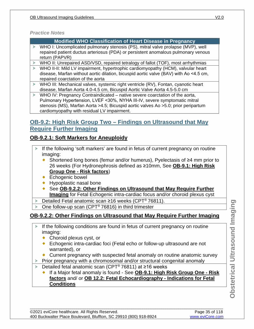
Practice Notes Modified WHO Classification of Heart Disease in Pregnancy
WHO I: Uncomplicated pulmonary stenosis (PS), mitral valve prolapse (MVP), well repaired patient ductus arteriosus (PDA) or persistent anomalous pulmonary venous return (PAPVR)
WHO II: Unrepaired ASD/VSD, repaired tetralogy of fallot (TOF), most arrhythmias WHO II-II: Mild LV impairment, hypertrophic cardiomyopathy (HCM), valvular heart
disease, Marfan without aortic dilation, bicuspid aortic valve (BAV) with Ao <4.5 cm, repaired coarctation of the aorta
WHO III: Mechanical valves, systemic right ventricle (RV), Fontan, cyanotic heart disease, Marfan Aorta 4.0-4.5 cm, Bicuspid Aortic Valve Aorta 4.5-5.0 cm
WHO IV: Pregnancy Contraindicated – native severe coarctation of the aorta, Pulmonary Hypertension, LVEF <30%, NYHA III-IV, severe symptomatic mitral stenosis (MS), Marfan Aorta >4.5; Bicuspid aortic valves Ao >5.0; prior peripartum cardiomyopathy with residual LV impairment.
OB-9.2: High Risk Group Two – Findings on Ultrasound that May Require Further Imaging OB-9.2.1: Soft Markers for Aneuploidy
OB-9.2.2: Other Findings on Ultrasound that May Require Further Imaging
If the following ‘soft markers’ are found in fetus of current pregnancy on routine imaging:
Shortened long bones (femur and/or humerus), Pyelectasis of ≥4 mm prior to 26 weeks (For Hydronephrosis defined as ≥10mm, See OB-9.1: High Risk Group One - Risk factors)
Echogenic bowel Hypoplastic nasal bone See OB-9.2.2: Other Findings on Ultrasound that May Require Further
Imaging for Fetal Echogenic intra-cardiac focus and/or choroid plexus cyst Detailed Fetal anatomic scan ≥16 weeks (CPT® 76811). One follow-up scan (CPT® 76816) in third trimester
If the following conditions are found in fetus of current pregnancy on routine imaging:
Choroid plexus cyst, or Echogenic intra-cardiac foci (Fetal echo or follow-up ultrasound are not
warranted), or Current pregnancy with suspected fetal anomaly on routine anatomic survey
Prior pregnancy with a chromosomal and/or structural congenital anomaly Detailed fetal anatomic scan (CPT® 76811) at ≥16 weeks
If a Major fetal anomaly is found - See OB-9.1: High Risk Group One - Risk factors and/ or OB 12.2: Fetal Echocardiography - Indications for Fetal Conditions
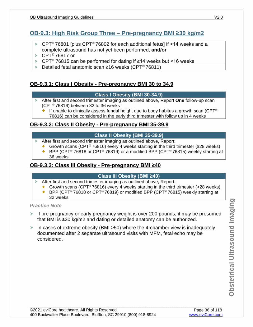
OB-9.3: High Risk Group Three – Pre-pregnancy BMI ≥30 kg/m2
OB-9.3.1: Class I Obesity - Pre-pregnancy BMI 30 to 34.9
OB-9.3.2: Class II Obesity - Pre-pregnancy BMI 35-39.9
OB-9.3.3: Class III Obesity - Pre-pregnancy BMI ≥40
Practice Note If pre-pregnancy or early pregnancy weight is over 200 pounds, it may be presumed
that BMI is ≥30 kg/m2 and dating or detailed anatomy can be authorized. In cases of extreme obesity (BMI >50) where the 4-chamber view is inadequately
documented after 2 separate ultrasound visits with MFM, fetal echo may be considered.
CPT® 76801 [plus CPT® 76802 for each additional fetus] if <14 weeks and a complete ultrasound has not yet been performed, and/or
CPT® 76817 or CPT® 76815 can be performed for dating if ≥14 weeks but <16 weeks Detailed fetal anatomic scan ≥16 weeks (CPT® 76811)
Class I Obesity (BMI 30-34.9) After first and second trimester imaging as outlined above, Report One follow-up scan
(CPT® 76816) between 32 to 36 weeks If unable to clinically assess fundal height due to body habitus a growth scan (CPT®
76816) can be considered in the early third trimester with follow up in 4 weeks
Class II Obesity (BMI 35-39.9) After first and second trimester imaging as outlined above, Report:
Growth scans (CPT® 76816) every 4 weeks starting in the third trimester (≥28 weeks) BPP (CPT® 76818 or CPT® 76819) or a modified BPP (CPT® 76815) weekly starting at
36 weeks
Class III Obesity (BMI ≥40) After first and second trimester imaging as outlined above, Report:
Growth scans (CPT® 76816) every 4 weeks starting in the third trimester (>28 weeks) BPP (CPT® 76818 or CPT® 76819) or modified BPP (CPT® 76815) weekly starting at
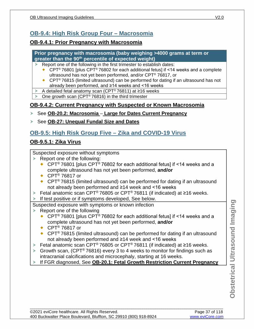
OB-9.4: High Risk Group Four – Macrosomia OB-9.4.1: Prior Pregnancy with Macrosomia
OB-9.4.2: Current Pregnancy with Suspected or Known Macrosomia See OB-20.2: Macrosomia – Large for Dates Current Pregnancy See OB-27: Unequal Fundal Size and Dates
OB-9.5: High Risk Group Five – Zika and COVID-19 Virus OB-9.5.1: Zika Virus
Prior pregnancy with macrosomia (baby weighing >4000 grams at term or greater than the 90th percentile of expected weight) Report one of the following in the first trimester to establish dates:
CPT® 76801 [plus CPT® 76802 for each additional fetus] if <14 weeks and a complete ultrasound has not yet been performed, and/or CPT® 76817, or
CPT® 76815 (limited ultrasound) can be performed for dating if an ultrasound has not already been performed, and ≥14 weeks and <16 weeks
A detailed fetal anatomy scan (CPT® 76811) at ≥16 weeks One growth scan (CPT® 76816) in the third trimester
Suspected exposure without symptoms Report one of the following:
CPT® 76801 [plus CPT® 76802 for each additional fetus] if <14 weeks and a complete ultrasound has not yet been performed, and/or
CPT® 76817 or CPT® 76815 (limited ultrasound) can be performed for dating if an ultrasound
not already been performed and ≥14 week and <16 weeks Fetal anatomic scan CPT® 76805 or CPT® 76811 (if indicated) at ≥16 weeks. If test positive or if symptoms developed, See below. Suspected exposure with symptoms or known infection Report one of the following
CPT® 76801 [plus CPT® 76802 for each additional fetus] if <14 weeks and a complete ultrasound has not yet been performed, and/or
CPT® 76817 or CPT® 76815 (limited ultrasound) can be performed for dating if an ultrasound
not already been performed and ≥14 week and <16 weeks Fetal anatomic scan CPT® 76805 or CPT® 76811 (if indicated) at ≥16 weeks. Growth scan, (CPT® 76816) every 3 to 4 weeks to monitor for findings such as
intracranial calcifications and microcephaly, starting at 16 weeks. If FGR diagnosed, See OB-20.1: Fetal Growth Restriction Current Pregnancy
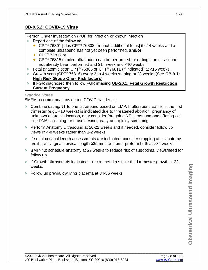
Practice Notes SMFM recommendations during COVID pandemic: Combine dating/NT to one ultrasound based on LMP. If ultrasound earlier in the first
trimester (e.g., <10 weeks) is indicated due to threatened abortion, pregnancy of unknown anatomic location, may consider foregoing NT ultrasound and offering cell free DNA screening for those desiring early aneuploidy screening
Perform Anatomy Ultrasound at 20-22 weeks and if needed, consider follow up views in 4-8 weeks rather than 1-2 weeks.
If serial cervical length assessments are indicated, consider stopping after anatomy u/s if transvaginal cervical length ≥35 mm, or if prior preterm birth at >34 weeks
BMI >40: schedule anatomy at 22 weeks to reduce risk of suboptimal views/need for follow up
If Growth Ultrasounds indicated – recommend a single third trimester growth at 32 weeks.
Follow up previa/low lying placenta at 34-36 weeks
Person Under Investigation (PUI) for infection or known infection Report one of the following:
CPT® 76801 [plus CPT® 76802 for each additional fetus] if <14 weeks and a complete ultrasound has not yet been performed, and/or
CPT® 76817 or CPT® 76815 (limited ultrasound) can be performed for dating if an ultrasound
not already been performed and ≥14 week and <16 weeks Fetal anatomic scan CPT® 76805 or CPT® 76811 (if indicated) at ≥16 weeks. Growth scan (CPT® 76816) every 3 to 4 weeks starting at 23 weeks (See OB-9.1:
High Risk Group One - Risk factors). If FGR diagnosed then follow FGR imaging OB-20.1: Fetal Growth Restriction
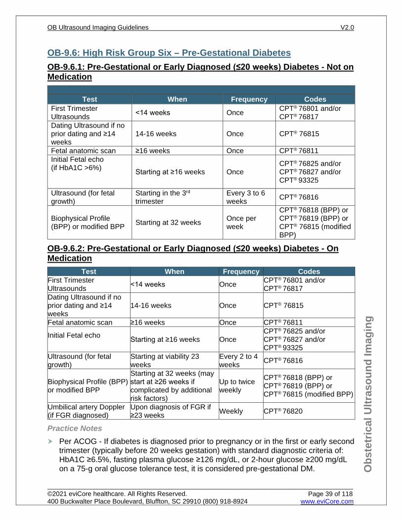
Starting at ≥16 weeks Once CPT® 76825 and/or CPT® 76827 and/or CPT® 93325
Ultrasound (for fetal growth)
Starting in the 3rd trimester
Every 3 to 6 weeks CPT® 76816
Biophysical Profile (BPP) or modified BPP Starting at 32 weeks Once per
week
CPT® 76818 (BPP) or CPT® 76819 (BPP) or CPT® 76815 (modified BPP)
OB-9.6.2: Pre-Gestational or Early Diagnosed (≤20 weeks) Diabetes - On Medication
Test When Frequency Codes First Trimester Ultrasounds <14 weeks Once CPT® 76801 and/or
CPT® 76817 Dating Ultrasound if no prior dating and ≥14 weeks
14-16 weeks Once CPT® 76815
Fetal anatomic scan ≥16 weeks Once CPT® 76811
Initial Fetal echo Starting at ≥16 weeks Once
CPT® 76825 and/or CPT® 76827 and/or CPT® 93325
Ultrasound (for fetal growth)
Starting at viability 23 weeks
Every 2 to 4 weeks CPT® 76816
Biophysical Profile (BPP) or modified BPP
Starting at 32 weeks (may start at ≥26 weeks if complicated by additional risk factors)
Up to twice weekly
CPT® 76818 (BPP) or CPT® 76819 (BPP) or CPT® 76815 (modified BPP)
Umbilical artery Doppler (if FGR diagnosed)
Upon diagnosis of FGR if ≥23 weeks Weekly CPT® 76820
Practice Notes Per ACOG - If diabetes is diagnosed prior to pregnancy or in the first or early second
trimester (typically before 20 weeks gestation) with standard diagnostic criteria of: HbA1C ≥6.5%, fasting plasma glucose ≥126 mg/dL, or 2-hour glucose ≥200 mg/dL on a 75-g oral glucose tolerance test, it is considered pre-gestational DM.
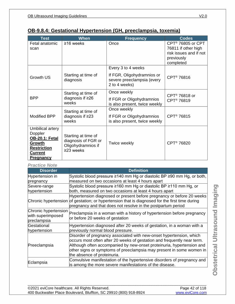
OB-9.8.4: Gestational Hypertension (GH, preeclampsia, toxemia) Test When Frequency Codes
Fetal anatomic scan
≥16 weeks Once CPT® 76805 or CPT 76811 if other high risk issues and if not previously completed
Growth US Starting at time of diagnosis
Every 3 to 4 weeks If FGR, Oligohydramnios or severe preeclampsia (every 2 to 4 weeks)
CPT® 76816
BPP Starting at time of diagnosis if ≥26 weeks
Once weekly If FGR or Oligohydramnios is also present, twice weekly
CPT® 76818 or CPT® 76819
Modified BPP Starting at time of diagnosis if ≥23 weeks
Once weekly If FGR or Oligohydramnios is also present, twice weekly
CPT® 76815
Umbilical artery Doppler OB-20.1: Fetal Growth Restriction Current Pregnancy
Starting at time of diagnosis of FGR or Oligohydramnios if ≥23 weeks
Twice weekly CPT® 76820
Practice Note Disorder Definition
Hypertension in pregnancy
Systolic blood pressure ≥140 mm Hg or diastolic BP ≥90 mm Hg, or both, measured on two occasions at least 4 hours apart
Severe-range hypertension
Systolic blood pressure ≥160 mm Hg or diastolic BP ≥110 mm Hg, or both, measured on two occasions at least 4 hours apart
Chronic hypertension Hypertension diagnosed or present before pregnancy or before 20 weeks of gestation; or hypertension that is diagnosed for the first time during pregnancy and that does not resolve in the postpartum period
Chronic hypertension with superimposed preclampsia
Preclampsia in a woman with a history of hypertension before pregnancy or before 20 weeks of gestation
Gestational hypertension
Hypertension diagnosed after 20 weeks of gestation, in a woman with a previously normal blood pressure.
Preeclampsia
Disorder of pregnancy associated with new-onset hypertension, which occurs most often after 20 weeks of gestation and frequently near term. Although often accompanied by new-onset proteinuria, hypertension and other signs or symptoms of preeclampsia may present in some women in the absence of proteinuria.
Eclampsia Convulsive manifestation of the hypertensive disorders of pregnancy and is among the more severe manifestations of the disease.
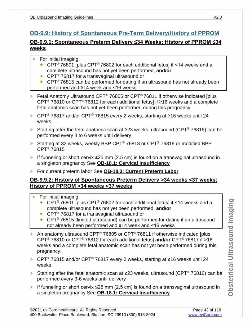
OB-9.9: History of Spontaneous Pre-Term Delivery/History of PPROM OB-9.9.1: Spontaneous Preterm Delivery ≤34 Weeks; History of PPROM ≤34 weeks
Fetal Anatomy Ultrasound CPT® 76805 or CPT® 76811 if otherwise indicated [plusCPT® 76810 or CPT® 76812 for each additional fetus] if ≥16 weeks and a completefetal anatomic scan has not yet been performed during this pregnancy.
CPT® 76817 and/or CPT® 76815 every 2 weeks, starting at ≥16 weeks until 24weeks
Starting after the fetal anatomic scan at ≥23 weeks, ultrasound (CPT® 76816) can beperformed every 3 to 6 weeks until delivery
Starting at 32 weeks, weekly BBP CPT® 76818 or CPT® 76819 or modified BPPCPT® 76815
If funneling or short cervix ≤25 mm (2.5 cm) is found on a transvaginal ultrasound ina singleton pregnancy See OB-18.1: Cervical Insufficiency
For current preterm labor See OB-18.3: Current Preterm LaborOB-9.9.2: History of Spontaneous Preterm Delivery >34 weeks <37 weeks; History of PPROM >34 weeks <37 weeks
An anatomy ultrasound CPT® 76805 or CPT® 76811 if otherwise indicated [plusCPT® 76810 or CPT® 76812 for each additional fetus] and/or CPT® 76817 if >16weeks and a complete fetal anatomic scan has not yet been performed during thispregnancy.
CPT® 76815 and/or CPT® 76817 every 2 weeks, starting at ≥16 weeks until 24weeks
Starting after the fetal anatomic scan at ≥23 weeks, ultrasound (CPT® 76816) can beperformed every 3-6 weeks until delivery
If funneling or short cervix ≤25 mm (2.5 cm) is found on a transvaginal ultrasound ina singleton pregnancy See OB-18.1: Cervical Insufficiency
For initial imaging:CPT® 76801 [plus CPT® 76802 for each additional fetus] if <14 weeks and a complete ultrasound has not yet been performed, and/or CPT® 76817 for a transvaginal ultrasound or CPT® 76815 can be performed for dating if an ultrasound has not already been performed and ≥14 week and <16 weeks
For initial imaging:CPT® 76801 [plus CPT® 76802 for each additional fetus] if <14 weeks and a complete ultrasound has not yet been performed, and/or CPT® 76817 for a transvaginal ultrasound or CPT® 76815 (limited ultrasound) can be performed for dating if an ultrasound not already been performed and ≥14 week and <16 weeks
For current preterm labor See OB-18.3: Current Preterm Labor
OB-9.10: History of Stillbirth
Fetal anatomic scan at ≥16 weeks (CPT® 76811) Follow up ultrasound (CPT® 76816) every 2 to 4 weeks to assess fetal growth
starting at 23 weeks or two weeks before prior pregnancy loss. Weekly BPP (CPT® 76818 or CPT® 76819 not to be performed prior to 26 weeks) or
modified BPP CPT® 76815 (not to be performed prior to 23 weeks) starting at 32weeks or two weeks before prior pregnancy loss
Practice Notes A history of stillbirth is not an indication for fetal echo. Per 2020 ACOG bulletin – no
mention of recommendation for echo – just detailed anatomy US. If demised fetushad a confirmed cardiac anomaly on autopsy, or if the detailed anatomy scan oneither the demised fetus or the current pregnancy had findings suspicious for cardiacanomaly, then echo may be indicated.
References 1. Practice Bulletin No. 175: Ultrasound in Pregnancy. Obstet Gynecol. 2016;128(6):e241-e256.
Reaffirmed 2020. doi:10.1097/01.AOG.0000451759.90082.7b.3. Reddy UM, Abuhamad AZ, Levine D, Saade GR. Fetal Imaging. Obstetrical & Gynecological Survey.
2014;69(8):453-455. doi:10.1097/01.ogx.0000453817.62105.4a4. AIUM Practice Parameter for the Performance of Detailed Second‐ and Third‐Trimester Diagnostic
Obstetric Ultrasound Examinations. Journal of Ultrasound in Medicine. 2019;38(12):3093-3100.doi:10.1002/jum.15163.
5. ACOG Practice Bulletin No. 200: Early Pregnancy Loss. Obstet Gynecol. 2018;132(5):e197-e207.doi:10.1097/AOG.0000000000002899.
6. Mcintosh J, Feltovich H, Berghella V, Manuck T. The role of routine cervical length screening inselected high- and low-risk women for preterm birth prevention. American Journal of Obstetrics andGynecology. 2016;215(3). doi:10.1016/j.ajog.2016.04.027.
7. Donofrio MT, Moon-Grady AJ, Hornberger LK, et al. Diagnosis and Treatment of Fetal CardiacDisease. Circulation. 2014;129(21):2183-2242. doi:10.1161/01.cir.0000437597.44550.5d .
8. Cavazos-Rehg PA, Krauss MJ, Spitznagel EL, et al. Maternal Age and Risk of Labor and DeliveryComplications. Maternal and Child Health Journal. 2014;19(6):1202-1211. doi:10.1007/s10995-014-1624-7.
9. ACOG Committee Opinion No. 807: Tobacco and Nicotine Cessation During Pregnancy. Obstetrics &Gynecology. 2020;135(5). doi:10.1097/aog.0000000000003822.
10. Machado JDB, Filho PV, Petersen GO, Chatkin JM. Quantitative effects of tobacco smoking exposureon the maternal-fetal circulation. BMC Pregnancy and Childbirth. 2011;11(1). doi:10.1186/1471-2393-11-24.
For initial imaging:CPT® 76801 [plus CPT® 76802 for each additional fetus] if <14 weeks and a complete ultrasound has not yet been performed, and/or CPT® 76817 for a transvaginal ultrasound or CPT® 76815 (limited ultrasound) can be performed for dating if an ultrasound has not already been performed and ≥14 week and <16 weeks
11. Hackshaw A, Rodeck C, Boniface S. Maternal smoking in pregnancy and birth defects: a systematicreview based on 173 687 malformed cases and 11.7 million controls. Human Reproduction Update.2011;17(5):589-604. doi:10.1093/humupd/dmr022.
12. Metz TD, Borgelt LM. Marijuana Use in Pregnancy and While Breastfeeding. Obstetrics &Gynecology. 2018;132(5):1198-1210. doi:10.1097/aog.0000000000002878.
13. ACOG Committee Opinion No. 722: Marijuana Use During Pregnancy and Lactation. Obstetrics &Gynecology. 2017;130(4). doi:10.1097/aog.0000000000002354.
14. ACOG Committee Opinion No. 711: Opioid Use and Opioid Use Disorder in Pregnancy. Obstetrics &Gynecology. 2017;130(2). doi:10.1097/aog.0000000000002235.
15. ACOG Committee Opinion No. 479: Methamphetamine Abuse in Women of Reproductive Age.Obstetrics & Gynecology. 2011;117(3):751-755. Reaffirmed 2017.doi:10.1097/aog.0b013e318214784e .
16. ACOG Practice Bulletin No. 90: Asthma in Pregnancy. Obstetrics & Gynecology. 2008;111(2, Part1):457-464. Reaffirmed 2019. doi:10.1097/aog.0b013e3181665ff4.
17. ACOG Practice Bulletin No. 212. Pregnancy and heart disease. Obstetrics & Gynecology2019;122:e320-56.
18. ACOG Practice Bulletin No. 78: Hemoglobinopathies in Pregnancy. Obstetrics & Gynecology.2007;109(1):229-238. Reaffirmed 2018. doi:10.1097/00006250-200701000-00055.
19. ACOG Practice Bulletin No. 223: Thyroid Disease in Pregnancy. Obstetrics & Gynecology.2020;135(6). doi:10.1097/aog.0000000000003894
20. Lee, RH; Greenberg, M; Metz, TD; et al. Society for Maternal-Fetal Medicine (SMFM) Consult Series#53: Intrahepatic cholestasis of pregnancy. February 2021.
21. Egan N, Bartels Ä, Khashan A, et al. Reference standard for serum bile acids in pregnancy. BJOG:An International Journal of Obstetrics & Gynaecology. 2012;119(4):493-498. doi:10.1111/j.1471-0528.2011.03245.x.
22. Getahun D, Fassett MJ, Longstreth GF, et al. Association between maternal inflammatory boweldisease and adverse perinatal outcomes. Journal of Perinatology. 2014;34(6):435-440.doi:10.1038/jp.2014.41.
23. ACOG Committee Opinion No. 776. Immune modulating therapies in pregnancy and lactation.Obstetrics & Gynecology. 2019;133(4):846-849. doi:10.1097/aog.0000000000003177.
24. ACOG Practice Bulletin No 156. Obesity in Pregnancy. Obstetrics & Gynecology. 2015;126(6).Reaffirmed in 2018. doi:10.1097/aog.0000000000001211.
25. Schuster M, Madueke-Laveaux OS, Mackeen AD, Feng W, Paglia MJ. The effect of the MFM obesityprotocol on cesarean delivery rates. American Journal of Obstetrics and Gynecology. 2016;215(4).doi:10.1016/j.ajog.2016.05.005.
26. ACOG. Committee Opinion No. 784: Management of Patients in the Context of Zika Virus. Obstetrics& Gynecology. 2019;134(3). doi:10.1097/aog.0000000000003399.
27. Boelig RC, Saccone G, Bellussi F, Berghella V. MFM guidance for COVID-19. American Journal ofObstetrics & Gynecology MFM. 2020:100106. doi:10.1016/j.ajogmf.2020.100106.
28. ACOG/SMFM Outpatient Assessment and Management for Pregnant Women With Suspected orConfirmed Novel Coronavirus (COVID-19).https://s3.amazonaws.com/cdn.smfm.org/media/2263/COVID-19_Algorithm5.pdf
29. Novoa RH, Quintana W, Llancarí P, Urbina-Quispe K, Guevara-Ríos E, Ventura W. Maternal clinicalcharacteristics and perinatal outcomes among pregnant women with coronavirus disease 2019. Asystematic review. Travel Med Infect Dis. 2021;39:101919. doi:10.1016/j.tmaid.2020.101919
35. Sciscione AC, Hayes EJ. Uterine artery Doppler flow studies in obstetric practice. American Journalof Obstetrics and Gynecology. 2009;201(2):121-126. doi:10.1016/j.ajog.2009.03.027.
36. Martins JG, Biggio JR, Abuhamad A. Society for Maternal-Fetal Medicine (SMFM) Consult Series#52: Diagnosis and Management of Fetal Growth Restriction. American Journal of Obstetrics andGynecology. 2020. doi:10.1016/j.ajog.2020.05.010.
37. ACOG. Practice Bulletin No. 171: Management of Preterm Labor. Obstetrics & Gynecology.2016;128(4). Reaffirmed 2018. doi:10.1097/aog.0000000000001711.
38. Yang J, Baer RJ, Berghella V, et al. Recurrence of Preterm Birth and Early Term Birth. Obstetrics &Gynecology. 2016;128(2):364-372. doi:10.1097/aog.0000000000001506.
39. Lengyel CS, Ehrlich S, Iams JD, Muglia LJ, Defranco EA. Effect of Modifiable Risk Factors onPreterm Birth: A Population Based-Cohort. Maternal and Child Health Journal. 2016;21(4):777-785.doi:10.1007/s10995-016-2169-8.
40. Practice Bulletin No. 130. Prediction and Prevention of Preterm Birth. Obstetrics & Gynecology.2012;120(4):964-973. Reaffirmed 2018. doi:10.1097/aog.0b013e3182723b1b.
41. SMFM Statement: Use of 17-alpha hydroxyprogesterone caproate for prevention of recurrent pretermbirth. American Journal of Obstetrics and Gynecology. 2020. doi:10.1016/j.ajog.2020.04.001.
42. Blackwell SC, Gyamfi-Bannerman C, Biggio JR Jr, et al. 17-OHPC to Prevent Recurrent PretermBirth in Singleton Gestations (PROLONG Study): A Multicenter, International, Randomized Double-Blind Trial. Am J Perinatol. 2020;37(2):127–136. doi:10.1055/s-0039-3400227
43. Obstetric Care Consensus No. 10: Management of Stillbirth. Obstetrics & Gynecology. 2020;135(3).doi:10.1097/aog.0000000000003719.
44. Gardosi J, Madurasinghe V, Williams M, Malik A, Francis A. Maternal and fetal risk factors forstillbirth: population based study. Bmj. 2013;346(jan24 3). doi:10.1136/bmj.f108.
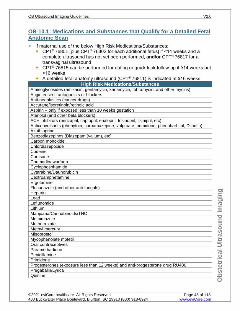
OB-10.1: Medications and Substances that Qualify for a Detailed Fetal Anatomic Scan If maternal use of the below High Risk Medications/Substances:
CPT® 76801 [plus CPT® 76802 for each additional fetus] if <14 weeks and a complete ultrasound has not yet been performed, and/or CPT® 76817 for a transvaginal ultrasound
CPT® 76815 can be performed for dating or quick look follow-up if ≥14 weeks but <16 weeks
A detailed fetal anatomy ultrasound (CPT® 76811) is indicated at ≥16 weeks High Risk Medications/Substances
Aminoglycosides (amikacin, gentamycin, kanamycin, tobramycin, and other mycins) Angiotensin II antagonists or blockers Anti-neoplastics (cancer drugs) Accutane/isoretinoin/retinoic acid Aspirin – only if exposed less than 10 weeks gestation Atenolol (and other beta blockers) ACE inhibitors (benzapril, captopril, enalopril, fosinopril, lisinipril, etc) Anticonvulsants (phenytoin, carbamazepine, valproate, primidone, phenobarbital, Dilantin) Azathioprine Benzodiazepines (Diazepam (valium), etc) Carbon monoxide Chlordiazepoxide Codeine Cortisone Coumadin/ warfarin Cyclophosphamide Cytarabine/Daunorubicin Dextroamphetamine Ergotamine Fluconazole (and other anti-fungals) Heparin Lead Leflunomide Lithium Marijuana/Cannabinoids/THC Methimazole Methotrexate Methyl mercury Misoprostol Mycophenolate mofetil Oral contraceptives Paramethadione Penicillamine Primidone Progesterones (exposure less than 12 weeks) and anti-progesterone drug RU486 Pregabalin/Lyrica Quinine
If another high risk indication see appropriate guideline for any further imaging Practice Note There may be other medications or drugs not included on this list that cause increased risk in pregnancy. Many of these medications/substances may be associated with fetal growth
restriction and/or other poor perinatal outcome and may require serial growth scans after 23 weeks and/or BPP assessments after 32 weeks See OB 9.1: High Risk Group One – Risk Factors.
Several studies noted lower birth weights among offspring exposed to marijuana. These findings were more pronounced among women who used more marijuana, particularly during the first and second trimesters (at least weekly during the pregnancy). CPT® 76811 may be indicated, however, given the limited evidence for antenatally detected abnormal growth, serial growth ultrasounds may not be indicated in the absence of other findings concerning for growth restriction.
In circumstances where the individual is deemed to have an increased risk for a fetal abnormality and does not have access to a provider who can perform the more desirable fetal and maternal ultrasound with detailed fetal anatomic examination (CPT® 76811) due to geographic or other constraints, a standard (after first trimester) fetal and maternal ultrasound (CPT® 76805) may be authorized instead.
References 1. Practice Bulletin No. 175: Ultrasound in Pregnancy. Obstet Gynecol. 2016;128(6):e241-e256.
Reaffirmed 2020. doi:10.1097/AOG.0000000000001815. 2. ACOG Practice Bulletin No. 92: Use of Psychiatric Medications During Pregnancy and Lactation.
Obstet Gynecol. 2008;111(4):1001-1020. Reaffirmed 2018. doi:10.1097/AOG.0b013e31816fd910. 3. Burkey BW, Holmes AP. Evaluating Medication Use in Pregnancy and Lactation: What Every
Pharmacist Should Know. The Journal of Pediatric Pharmacology and Therapeutics. 2013;18(3):247-258. doi:10.5863/1551-6776-18.3.247
4. ACOG Committee Opinion No. 711: Opioid Use and Opioid Use Disorder in Pregnancy. Obstetrics & Gynecology. 2017;130(2):e81-e94. doi:10.1097/aog.0000000000002235.
5. Schaefer C, Peters PWJ, Miller RK. Drugs during Pregnancy and Lactation: Treatment Options and Risk Assessment. 3rd ed. London: Elsevier/Academic Press; 2015.
6. ACOG Committee Opinion No. 776 Immune Modulating Therapies in Pregnancy and Lactation. Obstetrics & Gynecology. 2019;133(4):846-849. doi:10.1097/aog.0000000000003177.
7. ACOG Committee Opinion No. 722: Marijuana Use During Pregnancy and Lactation. Obstetrics & Gynecology. 2017;130(4). doi:10.1097/aog.0000000000002354.
OB-11.1: Suspected Multiple Gestations For Suspected multiple pregnancies:
CPT® 76801 [plus CPT® 76802 for each additional fetus] and/or CPT® 76817 if a complete ultrasound has not yet been performed and is <14 weeks, or
CPT® 76815 and/or CPT® 76817 can be performed for dating if an ultrasound not already been performed and ≥14 week and <16 weeks, or
CPT® 76805 and CPT® 76810 for each additional fetus if ≥14 weeks if a dating ultrasound or a complete anatomy ultrasound has not yet been performed during this pregnancy
OB-11.2: Known Dichorionic Multiple Gestations For Known dichorionic multiple pregnancies:
CPT® 76811 and CPT® 76812 for each additional fetus at ≥16 weeks if a complete detailed anatomic scan (CPT® 76811) has not yet been performed
Growth ultrasound (CPT® 76816) can be done every 4 to 6 weeks at ≥14 weeks. Universal cervical length (CL) screening with transvaginal ultrasound (CPT® 76817) is
NOT recommended in twin gestations. However, transvaginal ultrasound (CPT® 76817) may be considered if the cervical length (CL) is <3.6 cm on trans-abdominal ultrasound (as with singleton pregnancies – See OB-7.1: Fetal Anatomic Scan), or in certain circumstances of poor visualization with trans-abdominal ultrasound.
If cervical shortening identified – See OB-18.1: Cervical Insufficiency BPP (CPT® 76818 or CPT® 76819) or modified BPP (CPT® 76815) may be performed
weekly starting at 32 weeks or sooner if additional risk factors (eg. diabetes, or hypertensive disease - See OB-9: High Risk Pregnancy)
If FGR or growth discordance ≥20% is diagnosed, can perform: CPT® 76816 (growth ultrasound) every 2 to 4 weeks Modified BPP (CPT®76815) weekly between 23 to 26 weeks BPP (CPT® 76818 or CPT® 76819) or a modified BPP (CPT® 76815) weekly after 26
weeks UA Doppler (CPT® 76820) weekly (starting at ≥ 23 weeks) If UA Dopplers are abnormal (defined as a PI, RI, or S/D ratio greater than the
95th percentile for gestational age); or absent or reversed end diastolic flow is identified, then more frequent testing (usually twice per week but may be more often) with BPPs (CPT® 76818 or CPT® 76819 or CPT® 76815) and/or UA Dopplers (CPT® 78620) may be considered.
If IVF dichorionic twins, report an initial fetal echo as CPT® 76825 and/or CPT® 76827 with or without CPT® 93325. Trans-abdominal fetal echo is usually not performed prior to 16 weeks. See OB-12.3: Indications for Maternal Conditions
If other high risk factors, See OB-9: High Risk Pregnancy
OB-11.3: Known Monochorionic-Diamniotic or Monochorionic-Monoamniotic Multiple Gestations
For Known monochorionic-diamniotic or monochorionic-monoamniotic multiple pregnancies
CPT® 76811 and CPT® 76812 for each additional fetus if ≥16 weeks and a complete detailed anatomic scan (CPT® 76811) has not yet been performed.
Universal cervical length (CL) screening with transvaginal ultrasound (CPT® 76817) is NOT recommended in twin gestations. However, transvaginal ultrasound (CPT® 76817) may be considered if the cervical length (CL) is <3.6 cm on trans-abdominal ultrasound (as with singleton pregnancies – See OB-7.1: Fetal Anatomic Scan), or in certain circumstances of poor visualization with trans-abdominal ultrasound. If cervical shortening identified – See OB-18.1: Cervical Insufficiency
CPT® 76816 (growth ultrasound) every 2 to 4 weeks starting at 14 weeks Initial Fetal Echo (CPT® 76825 and/or CPT® 76827) with or without color Doppler (CPT®
93325) (usually not performed <16 weeks). MCA Doppler (CPT® 76821) is indicated every 2 weeks starting at 16 weeks until delivery
to monitor for Twin-Twin Transfusions Syndrome (TTTS) and/or Twin Anemia Polycythemia Sequence (TAPS). This may be performed with a limited ultrasound (CPT®
76815) or growth ultrasound (CPT® 76816). BPP (CPT® 76818 or CPT® 76819) or modified BPP (CPT® 76815) may be performed
weekly starting at 32 weeks or sooner if additional risk factors (eg. diabetes, or hypertensive disease - See OB-9: High Risk Pregnancy)
If TTTS is suspected or diagnosed, or if FGR or growth discordance ≥20% is diagnosed, more frequent testing (usually twice per week but may be more often) may be indicated. In these cases, you may perform: Limited ultrasound (CPT® 76815), or BPP (CPT® 76818 or CPT® 76819) if >26 weeks
UA Doppler (CPT® 76820) MCA Doppler (CPT® 76821)
If other high risk factors, See OB-9: High Risk Pregnancy
Triplets or higher order Multiple Pregnancy receive same imaging as monochorionic- diamniotic twins.
Practice Notes Birth weight discordance = (larger twin weight minus smaller twin weight) divided
larger twin weight × 100. Universal CL screening with transvaginal ultrasound (CPT® 76817) is NOT
recommended in twin gestations. In addition, Per ACOG - Cerclage placement (prophylactic or rescue) should be avoided in multifetal pregnancies. However, because several studies have shown that a one-time CL measurement ≤20 mm at 18-24 weeks may be an accurate predictor of preterm birth in multiple gestation, and because progesterone therapy might reduce the risk of neonatal morbidity and mortality associated with PTB, then a one-time transvaginal CL assessment may be considered if trans-abdominal CL measures ≤3.6 cm (as with singleton gestation- See: OB-7.1: Fetal Anatomic Scan). Transvaginal ultrasound (CPT® 76817) may be considered if the cervical length (CL) is ≤3.6 cm on trans-abdominal ultrasound (as with singleton pregnancies- See OB-7.1: Fetal Anatomic Scan), or in certain circumstances of poor visualization with trans-abdominal ultrasound.
TTTS is diagnosed by the ultrasound findings of polyhydramnios in one twin (the recipient) and oligohydramnios in the other twin (the donor). If AFI is discordant between the twins (low but not <2 cm in one and/or high but not >8 cm in the other); weekly imaging (MCA and/or limited US) may be indicated to ensure not developing TTTS.
Fetal loss of one twin during the first trimester does not appear to increase the risk of FGR or preterm delivery in the surviving twin, however, loss of one or more fetus(es) after 17 weeks gestation is associated with increased risk for FGR and PTB and should be imaged according to OB-11: Multiple Gestations. Monochorionic twin pregnancies with demise of one twin after 17 weeks have up to an 18% chance of major morbidity or mortality for the remaining fetus.
Typically all components of the BPP (CPT® 76818 and CPT® 76819), such as breathing, are not present until ≥26 weeks gestation. However, a modified BPP (CPT® 76815) may be utilized sooner in certain high risk cases but should not be done prior to viability (23 weeks).
In circumstances where CPT® 76811 cannot be performed See OB-1.3: Ultrasound Code Selection
References 1. ACOG Practice Bulletin No. 169: Multifetal Gestations: Twin, Triplet, and Higher-Order Multifetal
2. ACOG Practice Bulletin No. 175: Ultrasound in Pregnancy. Obstet Gynecol. 2016;128(6):e241-e256. Reaffirmed 2020. doi:10.1097/AOG.0000000000001815.
3. Khalil A, Rodgers M, Baschat A, et al. ISUOG Practice Guidelines: role of ultrasound in twin pregnancy. Ultrasound in Obstetrics & Gynecology. 2016;47(2):247-263. doi:10.1002/uog.1582.
4. ACOG Practice Bulletin No. 130: Prediction and Prevention of Preterm Birth. Obstet Gynecol. 2012;120(4):964-973. Reaffirmed 2016. doi:10.1097/AOG.0b013e3182723b1b.
5. ACOG Practice Bulletin No. 142: Cerclage for the Management of Cervical Insufficiency. Obstet Gynecol. 2014;123(2, PART 1):372-379. Reaffirmed 2019. doi:10.1097/01.aog.0000443276.68274.cc.
6. Rafael TJ, Berghella V, Alfirevic Z. Cervical stitch (cerclage) for preventing preterm birth in multiple pregnancy. Cochrane Database of Systematic Reviews. September 2014. doi:10.1002/14651858.cd009166.pub2.
7. Roman A, Rochelson B, Fox NS, Hoffman M, Berghella V, Patel V, Calluzzo I, Saccone G, Fleischer A. Efficacy of ultrasound-indicated cerclage in twin pregnancies. Am J Obstet Gynecol. 2015 Jun;212(6):788.e1-6. doi: 10.1016/j.ajog.2015.01.031.
8. Razaz N, Avitan T, Ting J, Pressey T, Joseph K. Perinatal outcomes in multifetal pregnancy following fetal reduction. Canadian Medical Association Journal. 2017;189(18). doi:10.1503/cmaj.160722.
9. Mcintosh J, Feltovich H, Berghella V, Manuck T. The role of routine cervical length screening in selected high- and low-risk women for preterm birth prevention. Society for Maternal-Fetal Medicine (SMFM) Consult Series #40. American Journal of Obstetrics and Gynecology. 2016;215(3). doi:10.1016/j.ajog.2016.04.027.
10. Practice Committee of American Society for Reproductive Medicine: Multiple gestation associated with infertility therapy: an American Society for Reproductive Medicine Practice Committee opinion. Fertility and Sterility. 2012;97(4):825-834. doi:10.1016/j.fertnstert.2011.11.048.
11. Slaghekke F, Pasman S, Veujoz M, et al. Middle cerebral artery peak systolic velocity to predict fetal hemoglobin levels in twin anemia-polycythemia sequence. Ultrasound in Obstetrics & Gynecology. 2015;46(4):432-436. doi:10.1002/uog.14925.
12. Lopriore E, Slaghekke F, Oepkes D, Middeldorp JM, Vandenbussche FP, Walther FJ. Clinical outcome in neonates with twin anemia-polycythemia sequence. American Journal of Obstetrics and Gynecology. 2010;203(1). doi:10.1016/j.ajog.2010.02.032.
13. Slaghekke F, Kist W, Oepkes D, et al. Twin Anemia-Polycythemia Sequence: Diagnostic Criteria, Classification, Perinatal Management and Outcome. Fetal Diagnosis and Therapy. 2010;27(4):181-190. doi:10.1159/000304512.
14. Tollenaar LSA, Slaghekke F, Middeldorp JM, et al. Twin Anemia Polycythemia Sequence: Current Views on Pathogenesis, Diagnostic Criteria, Perinatal Management, and Outcome. Twin Research and Human Genetics. 2016;19 (3):222-233. doi:10.1017/thg.2016.18.
15. ACOG Practice Bulletin No. 227: Fetal Growth Restriction. Obstet Gynecol. 2021;137(2):e16-e28 doi: 10.1097/AOG.0000000000004251.Martins JG, Biggio JR, Abuhamad A. Society for Maternal-Fetal Medicine (SMFM) Consult Series #52: Diagnosis and Management of Fetal Growth Restriction. American Journal of Obstetrics and Gynecology. 2020. doi:10.1016/j.ajog.2020.05.010
16. Simpson LL. Twin-twin transfusion syndrome. American Journal of Obstetrics and Gynecology. 2013;208(1):3-18. doi:10.1016/j.ajog.2012.10.880.
17. Lanna MM, Consonni D, Faiola S, et al. Incidence of Cerebral Injury in Monochorionic Twin Survivors after Spontaneous Single Demise: Long-Term Outcome of a Large Cohort. Fetal Diagnosis and Therapy. 2019;47(1):66-73. doi:10.1159/000500774.Hoskins. SMFM Special Statement: Updated checklists for management of monochorionic twin pregnancy. Am J Obstet Gynecol 2020
CPT® 76825/CPT® 76827 for Initial Fetal Echocardiography/Initial Doppler Echocardiography respectively
CPT® 76826/CPT® 76828 for Follow-up Fetal Echocardiography/Follow-up Doppler Echocardiography respectively
CPT® 93325 for Doppler color flow velocity mapping Transabdominal fetal echo is usually not performed prior to 16 weeks. Initial Fetal Echo, CPT® 76825 and Doppler Echo CPT® 76827 are performed only
once per fetus/per facility (i.e. Maternal Fetal Medicine versus Pediatric Cardiology request)
Follow-up-Fetal echo and/or Follow-up Doppler echo (CPT® 76826/CPT® 76828) may be indicated in certain rare circumstances.
Procedure code (CPT® 76827 or CPT® 76828) includes the evaluation of veins, arteries, and valves. Guidelines do not support the billing of an additional code (CPT® 76820 and/or CPT® 76821)
OB-12.2: Fetal Echocardiography - Indications for Fetal Conditions Initial Fetal echocardiography (CPT® 76825) and/or Doppler echocardiography (CPT® 76827) with or without Doppler color flow velocity mapping (CPT® 93325) can be performed if ≥16 weeks, for the indications listed below (See OB-12.1: Fetal Echocardiography – Coding):
Known or suspected abnormal fetal cardiac evaluation on fetal anatomic scan. Known or suspected abnormality must be documented as hard copy or
acknowledged verbally by provider of known or suspected fetal cardiac evaluation
Suboptimal cardiac evaluation alone is not an indication for fetal echogram. If the 4-chamber view is adequate and there is no other suspicion of a cardiac abnormality, a fetal echocardiogram is not considered medically necessary. A follow up ultrasound (CPT® 76815 or CPT® 76816) is indicated for suboptimal visualization.
Fetal cardiac arrhythmia; sustained fetal tachycardia or bradyarrhythmia Major fetal extra-cardiac anomaly, (excluding soft markers for aneuploidy: for
example shortened long bones, pyelectasis, echogenic bowel, hypoplastic nasal bone, cardiac echogenic foci and choroid plexus cyst) See OB-9.2: High Risk Group Two – Findings on Ultrasound that May Require Further Imaging.
Congenital heart disease (CHD) in a 1st degree relative of the fetus (i.e. CHD in the mother, father, or sibling of the fetus)
Known fetal chromosomal abnormalities (fetal aneuploidy) or ultrasound findings of a suspected chromosomal abnormality (excluding soft markers as only ultrasound findings)
Single umbilical artery Chorioangioma or Umbilical cord varix (if suspicion of fetal hydrops)
Causes of Fetal Anemia/Parvo/Hydrops Monochorionic twins/TTTS Abnormal Fetal Nuchal Translucency scan (NT ≥3.0mm or above the 99th
percentile for the CRL) during current pregnancy.
OB-12.3: Fetal Echocardiography - Indications for Maternal Conditions Initial Fetal echocardiography (CPT® 76825) and/or Doppler echocardiography (CPT® 76827) with or without Doppler color flow velocity mapping (CPT® 93325) can be performed if ≥16 weeks, for the indications listed below (See OB-12.1: Fetal Echocardiography – Coding):
Maternal Conditions: Maternal pre-gestational DM or early diagnosed GDM (1st or early 2nd trimester) Maternal gestational diabetes mellitus on medication, if HbA1C >6% Maternal connective tissue disease (SLE, RA, Sjogrens) with Anti-Ro/SSA or anti-La/SSB
OB-12.4: Fetal Echocardiography - Indications for Medication or Drug Exposure Initial Fetal echocardiography (CPT® 76825) and/or Doppler echocardiography (CPT® 76827) with or without Doppler color flow velocity mapping (CPT® 93325) can be performed if ≥16 weeks, for the indications listed below (See OB-12.1: Fetal Echocardiography – Coding): Lithium Excessive alcohol Anti-seizure medication, e.g. hydantoin Paroxetine (Paxil) Ace inhibitors Folate antagonists (methotrexate) Anticonvulsants Retinoic acid Thalidomide NSAIDS (Ibuprofen, Indomethacin) 2nd and 3rd trimester Venlafaxine (Effexor) Vitamin A greater than 10,000 units per day Other teratogen exposure to the fetus with a known association for cardiac
anomalies Practice Note In cases of extreme obesity (BMI> 50) where the 4-chamber view is inadequately
documented after 2 separate ultrasound visits with MFM, fetal echo may be considered.
With positive SSA/SSB antibodies, the most vulnerable period for the fetus is during the period from 18 to 24 weeks gestation. Normal sinus rhythm can progress to complete block in seven days during this high-risk period. New onset of heart block is less likely during the 26th through the 30th week, and it rarely develops after 30 weeks of pregnancy.
Fetal echocardiography may be indicated with severe, unexplained polyhydramnios if there are other suspicious findings on an anatomy scan.
The minimal use of color Doppler (CPT® 93325) alone, when performed for anatomical structure identification during a standard ultrasound procedure, is not separately reimbursable.
References 1. Donofrio MT, Moon-Grady AJ, Hornberger LK, et al. Diagnosis and Treatment of Fetal Cardiac
Disease. Circulation. 2014;129(21):2183-2242. doi:10.1161/01.cir.0000437597.44550.5d. 2. Brucato A. Prevention of congenital heart block in children of SSA-positive mothers. Rheumatology.
2008;47(Supplement 3):iii35-iii37. doi:10.1093/rheumatology/ken153. 3. Mcbride KL, Garg V. Impact of Mendelian inheritance in cardiovascular disease. Annals of the New
York Academy of Sciences. 2010;1214(1):122-137. doi:10.1111/j.1749-6632.2010.05791.x, 4. Reddy UM, Abuhamad AZ, Levine D, et al. Fetal imaging: executive summary of a joint Eunice
Kennedy Shriver National Institute Child Health and Human Development, Society for Maternal-Fetal Medicine, American Institute of Ultrasound in Medicine, American College of Obstetricians and Gynecologists, American College of Radiology, Society of Pediatric Radiology, and Society of Radiologists in Ultrasound Fetal Imaging Workshop. Obstet Gynecol Survey. 2014;69(8):453-455.
5. Lee W, Allan L, Carvalho JS, et al. ISUOG consensus statement: what constitutes a fetal echocardiogram? Ultrasound in Obstetrics and Gynecology. 2008;32(2):239-242. doi:10.1002/uog.6115.
6. Friedman DM, Kim MY, Copel JA, et al. Utility of Cardiac Monitoring in Fetuses at Risk for Congenital Heart Block. The PR Interval and Dexamethasone Evaluation (PRIDE) Prospective Study. Circulation. 2008;117(4):485-493. doi:10.1161/circulationaha.107.707661
7. AIUM Practice Parameter for the Performance of Fetal Echocardiography. Journal of Ultrasound in Medicine. 2019;39(1). doi:10.1002/jum.15188.
8. Sammaritano LR, Bermas BL, Chakravarty EE, et al. 2020 American College of Rheumatology Guideline for the Management of Reproductive Health in Rheumatic and Musculoskeletal Diseases. Arthritis Care & Research. 2020;72(4):461-488. doi:10.1002/acr.24130.
9. Jenkins KJ, Correa A, Feinstein JA, et al. Noninherited Risk Factors and Congenital Cardiovascular Defects: Current Knowledge. Circulation. 2007;115(23):2995-3014. doi:10.1161/circulationaha.106.183216.
10. Anderson KN, Lind JN, Simeone RM, Bobo WV, Mitchell AA, Riehle-Colarusso T, Polen KN, Reefhuis. Maternal Use of Specific Antidepressant Medications During Early Pregnancy and the Risk of Selected Birth Defects. J.JAMA Psychiatry. 2020 Aug 5;77(12):1246-55. doi: 10.1001/jamapsychiatry.2020.2453.
OB-13.1: Indications for Fetal MRI ACOG recommendations for imaging during pregnancy and lactation: Ultrasonography and magnetic resonance imaging (MRI) are not associated with risk
and are the imaging techniques of choice for the pregnant patient, but they should be used prudently and only when use is expected to answer a relevant clinical question or otherwise provide medical benefit to the patient.
The use of gadolinium contrast with MRI should be limited; it may be used as a contrast agent in a pregnant woman only if it significantly improves diagnostic performance and is expected to improve fetal or maternal outcome.
CPT® Code Guidance Fetal MRI (CPT® 74712); for each additional fetus (CPT® 74713) Do not report CPT® 74712 and CPT® 74713 in conjunction with CPT® 72195, CPT®
72196, CPT® 72197 If only placenta or maternal pelvis is imaged without fetal imaging, use MRI Pelvis (CPT®
72195)
Fetal MRI (CPT® 74712) [plus CPT® 74713 for each additional fetus] may be considered for assessment of known or suspected fetal abnormalities for surgical planning, and/or if an ultrasound is equivocal and additional information is needed for counseling purposes, for indications which may include the following:
Brain Congenital anomalies
ventriculomegaly corpus callosal dysgenesis holoprosencephaly posterior fossa anomalies malformations of cerebral cortical development microcephaly
Screening fetuses with a family risk for brain anomalies tuberous sclerosis corpus callosal dysgenesis malformations of cerebral cortical development
References 1. Saleem SN. Fetal MRI: An approach to practice: A review. Journal of Advanced Research.
2014;5(5):507-523. doi:10.1016/j.jare.2013.06.001. 2. Prayer D, Malinger G, Brugger PC, et al. ISUOG Practice Guidelines: performance of fetal magnetic
resonance imaging. Ultrasound in Obstetrics & Gynecology. 2017;49(5):671-680. doi:10.1002/uog.17412.
3. Reddy UM, Abuhamad AZ, Levine D, Saade GR. Fetal Imaging: Executive Summary of a Joint Eunice Kennedy Shriver National Institute of Child Health and Human Development, Society for Maternal-Fetal Medicine, American Institute of Ultrasound in Medicine, American College of Obstetricians and Gynecologists, American College of Radiology, Society for Pediatric Radiology, and Society of Radiologists in Ultrasound Fetal Imaging Workshop. Obstetrics & Gynecology. 2014;123(5):1070-1082. doi:10.1097/aog.0000000000000245.
4. American College of Radiology (ACR) and the Society for Pediatric Radiology (SPR). Practice Parameters by Modality | American College of Radiology: Practice Parameter for the Safe And Optimal Performance of Fetal Magnetic Resonance Imaging (MRI). American College of Radiology | American College of Radiology. https://www.acr.org/-/media/ACR/Files/Practice-Parameters/MR-Fetal.pdf. Published 2015. (Resolution 11).
5. American College of Obstetricians and Gynecologists Committee Opinion No. 723. Guidelines for diagnostic imaging during pregnancy and lactation. Obstetrics & Gynecology. 2017;130(4). doi:10.1097/aog.0000000000002355
OB-14.1: Abnormal Fetal Position or Presentation To confirm suspected abnormal fetal position or presentation (transverse or breech
presentation) at ≥36 weeks gestation, report one of the following: CPT® 76805 (plus CPT® 76810 for each additional fetus) when complete anatomy
scan has not yet been performed in the pregnancy or CPT® 76815 for limited ultrasound to check fetal position or CPT® 76816 if version is being considered and/or for delivery planning
Practice Note Fetal presentation should be assessed by abdominal palpation (Leopold’s) at 36
weeks or later, when presentation is likely to influence the plans for the birth. Routine assessment of presentation by abdominal palpation before 36 weeks is not always accurate. Suspected fetal malpresentation should be confirmed by an ultrasound assessment. An ultrasound can be performed at ≥36 weeks gestation to determine fetal position to allow for external cephalic version. Ultrasound to determine fetal position is not necessary prior to 36 weeks gestation unless delivery is imminent.
Though rarely used anymore, there may still be occasional planned vaginal delivery of a breech presentation. There is not enough evidence to support the use of X-ray pelvimetry for deciding on mode of delivery in women whose fetuses have a cephalic presentation. However, pelvimetry in breech presentation may allow for better selection of the delivery route, with a significantly lower emergency caesarean‐section rate noted in small trials.
doi:10.1097/aog.0000000000003837. 2. Safe Prevention of the Primary Cesarean Delivery. Obstetric Care Consensus No. 1. Obstetrics &
Gynecology. 2014;123(3):693-711. Reaffirmed 2018. doi:10.1097/01.aog.0000444441.04111.1d. 3. Pattinson RC, Cuthbert A, Vannevel V. Pelvimetry for fetal cephalic presentations at or near term for
deciding on mode of delivery. Cochrane Database of Systematic Reviews. 2017. doi:10.1002/14651858.cd000161.pub2
OB-15.1: Adnexal Mass For a known or suspected adnexal/pelvic mass, may perform:
CPT® 76801 [plus CPT® 76802 for each additional fetus] and/or CPT® 76817 to establish dates if <14 weeks or
CPT® 76815 and/or CPT® 76817 if a complete ultrasound was previously performed or
CPT® 76805 [plus CPT® 76810 if more than one fetus] if a complete fetal anatomic scan has not yet been performed and ≥14 weeks, or
CPT® 76816 if a complete anatomy scan was done previously. Following the initial ultrasound, follow up can be done once in each trimester
CPT® 76805 [plus CPT® 76810 for each additional fetus] if a complete fetal anatomic scan has not yet been performed, or
CPT® 76815 or CPT® 76816 if a complete ultrasound was previously performed. CPT® 76817 may be indicated for poor visualization of the adnexal mass in
certain circumstances. MRI Pelvis (CPT® 72195) without contrast can be done for indeterminate findings on
ultrasound; for surgical planning and/or for suspected malignancy. See PV-5: Adnexal Mass/Ovarian Cysts in the Pelvis Imaging Guidelines
Practice Note The majority of adnexal masses in pregnancy are benign, the most common
diagnoses are mature teratomas and corpus luteum or paraovarian cysts. Malignancy is reported in only 1.2-6.8% of pregnant patients with persistent mass.
Levels of CA-125 are elevated in pregnancy, a low-level elevation in pregnancy is not typically associated with malignancy.
ACOG recommendations for imaging during pregnancy and lactation: Ultrasonography and magnetic resonance imaging (MRI) are not associated with
risk and are the imaging techniques of choice for the pregnant patient, but they should be used prudently and only when use is expected to answer a relevant clinical question or otherwise provide medical benefit to the patient.
The use of gadolinium contrast with MRI should be limited; it may be used as a contrast agent in a pregnant woman only if it significantly improves diagnostic performance and is expected to improve fetal or maternal outcome.
OB-15.2: Uterine Fibroids in Pregnancy If more than one fibroid, total size of all fibroids should be used, i.e. one fibroid at 2
cm and one 3 cm is total of 5 cm and imaging would be indicated as below: Moderate (>5 cm) and large (>10 cm) fibroid(s): CPT® 76801 [plus CPT® 76802 for each additional fetus] and/or CPT® 76817
to establish dates if ≤14 weeks, or CPT® 76815 can be performed for dating if ≥14 weeks but <16 weeks Fetal anatomic scan (CPT® 76805 or CPT® 76811 if other high risk indication.
See OB-9: High Risk Pregnancy) at ≥16 weeks. If the fibroid is in the lower uterine segment or cervical fibroid then ultrasound
(CPT® 76815) and/or transvaginal ultrasound (CPT® 76817) every 2 weeks between 16 to 24 weeks, and
Follow up ultrasound (CPT® 76816) every 3 to 6 weeks, starting at 23 weeks. Submucosal fibroid(s) of any size:
First trimester: CPT® 76801 [plus CPT® 76802 for each additional fetus] and/or CPT® 76817 to establish dates if <14 weeks.
If placentation is over a submucosal fibroid: Follow up ultrasound (CPT® 76816) every 3 to 6 weeks, starting at 23 weeks
Practice Note Though pregnancy seems to have little or no effect on the overall size of fibroids,
Fibroids affect pregnancy and delivery in several ways, with abdominal pain, miscarriage, fetal malpresentation, and difficult delivery being the most frequent complications. These complications relate to preterm labor, placental abruption, fetal growth restriction, and fetal compression syndromes. The risk of preterm labor appears to correlate with the size of the fibroid (over 600 cm3) and/or the presence of multiple fibroids. Placental abruption has been reported to occur frequently in pregnancies complicated by fibroids, especially with placentation over a fibroid. Fibroid volumes >200 cm3 are more commonly associated with fetal growth restriction. Fetal compression syndrome is a direct result of large fibroids and is not commonly found with small fibroids. Finally, malposition or obstructed labor may be associated with fibroids of the lower uterine segment.
OB-15.3: Uterine Anomalies in Pregnancy For uterine septum, uterine didelphys, unicornuate uterus, bicornuate uterus:
CPT® 76801 [plus CPT® 76802 for each additional fetus] and/or CPT® 76817 to establish dates if <14 weeks if a complete ultrasound has not yet been performed, or
If a complete ultrasound was previously performed, CPT® 76815 and/or CPT®
76817 or CPT® 76805 or CPT® 76811 and/or CPT® 76817 at ≥16 weeks CPT® 76817 and/or CPT® 76815 every 2 weeks at 16 to 24 weeks (See OB-
18.1: Cervical Insufficiency) CPT® 76816 every 3 to 6 weeks starting at ≥23 weeks Starting at 32 weeks, weekly BPP (CPT® 76818 or CPT® 76819) or modified BPP
(CPT® 76815) Practice Note In circumstances where the individual is deemed to have an increased risk for a fetal
abnormality and does not have access to a provider who can perform the more desirable fetal and maternal ultrasound with detailed fetal anatomic examination (CPT® 76811) due to geographic or other constraints, a standard (after first trimester) fetal and maternal ultrasound (CPT® 76805) may be authorized instead.
CPT® 76811 and CPT® 76812 should only be used once per pregnancy unless the mother changes to a new medical caregiver at a new office and there is a new medical indication and/or change in condition.
References 1. ACOG Practice Bulletin No. 174: Evaluation and Management of Adnexal Masses. Obstet Gynecol.
2016;128(5):e210-e226. doi:10.1097/AOG.0000000000001768. 2. Stout M, Odibo A, Graseck A, et al. Leiomyomas at Routine Second-trimester Ultrasound
Examination and Adverse Obstetric Outcomes. Obstetric Anesthesia Digest. 2012;32(1):21-22. doi:10.1097/01.aoa.0000410780.41686.41.
3. Lee HJ, Norwitz ER, Shaw J. Contemporary management of fibroids in pregnancy. Reviews in obstetrics & gynecology. https://www.ncbi.nlm.nih.gov/pmc/articles/PMC2876319/. Published 2010.
4. Shavell VI, Thakur M, Sawant A, et al. Adverse obstetric outcomes associated with sonographically identified large uterine fibroids. Fertility and Sterility. 2012;97(1):107-110. doi:10.1016/j.fertnstert.2011.10.009.
5. Kase BA, Blackwell SC. SMFM consult: Fibroids in pregnancy: Meaning and Management. Contemporary OBGYN. http://www.contemporaryobgyn.net/modern-medicine-feature-articles/smfm-consult-fibroids-pregnancy-meaning-and-management. Published December 5, 2014.
6. Sei K, Masui K, Sasa H, Furuya K. Size of uterine leiomyoma is a predictor for massive haemorrhage during caesarean delivery. European Journal of Obstetrics & Gynecology and Reproductive Biology. 2018;223:60-63. doi:10.1016/j.ejogrb.2018.02.014
7. Penzias A, Bendikson K, Butts S, et al. Removal of myomas in asymptomatic patients to improve fertility and/or reduce miscarriage rate: a guideline. Fertility and Sterility. 2017;108(3):416-425. doi:10.1016/j.fertnstert.2017.06.034.
8. ACOG Practice Bulletin No. 142: Cerclage for the Management of Cervical Insufficiency. Obstet Gynecol. 2014;123(2, PART 1):372-379. Reaffirmed 2019. doi:10.1097/01.aog.0000443276.68274.cc.
9. Hua M, Odibo AO, Longman RE, Macones GA, Roehl KA, Cahill AG. Congenital uterine anomalies and adverse pregnancy outcomes. American Journal of Obstetrics and Gynecology. 2011;205(6). doi:10.1016/j.ajog.2011.07.022
10. Pfeifer S, Butts S, Dumesic D, et al. Uterine septum: a guideline. Fertility and Sterility. 2016;106(3):530-540. doi:10.1016/j.fertnstert.2016.05.014.
OB-16.1: Alloimmunization/Rh Isoimmunization Imaging for Alloimmunization/Rh Isoimmunization for any of the following indications: When any one of the following maternal antibody titers are ≥1:8 (critical titer):
Rhesus antibodies (Cc/Dd/Ee) Anti-Duffy (anti-fya) antibody Anti-Kidd antibody Other (atypical) antigens associated with hemolytic disease of the fetus and newborn
include: p antigen, MNS series, MSSsMT, Diego, Public antigens, Private antigens. Anti-Kell antibody (any antibody titer warrants additional evaluation) If evidence of fetal hydrops on previous imaging Prior pregnancy associated with HDFN (hemolytic disease of the fetus and newborn) The following imaging is indicated: CPT® 76801 [plus CPT® 76802 for each additional fetus] if <14 weeks and a complete
ultrasound has not yet been performed, and/or CPT® 76817 for a transvaginal ultrasound or CPT® 76815 can be performed for dating or quick look follow-up if ≥14 weeks but <16
weeks Detailed Fetal Anatomic Scan (CPT® 76811) ≥16 weeks Ultrasound CPT® 76816 every 2 to 4 weeks to assess fetal growth starting after fetal anatomic scan
(CPT® 76811) Fetal middle cerebral artery (MCA) Doppler (CPT® 76821) and CPT® 76815 every 1 to 2
weeks starting at 16 weeks BPP (CPT® 76818 or CPT® 76819) or modified BPP (CPT® 76815) weekly, starting at 32
weeks or sooner depending on fetal condition
Practice Note Fetal anemia and hydrops may be a result of immune conditions, such as red-cell or
Kell alloimmunization, non-immune hydrops caused by parvovirus B19 infection or any other known acquired or congenital causes of fetal anemia.
Rhesus isoimmunization/alloimmunization is the process through which fetal Rh+ red blood cells enter the circulation of an Rh negative mother causing her to produce antibodies which can cross the placenta and destroy the red blood cells of the current Rh+ fetus and/or in subsequent Rh+ pregnancies.
Atypical antigens not listed above, may be associated with hemolytic disease of the fetus and newborn and may require fetal assessment as in OB-16.1: Alloimmunization/Rh Isoimmunization if maternal antibody titers are ≥1:8. Atypical antigens include: MNSs series (M, N, S, s, U, Mi), MSSs-Mta, Diego (Dia, Dib), P- PPTj, Public antigen (Yt, En, Co2). Private antigens (Biles, Good, Heibel, Radin, Wrighta, and Zd). PP1Pk, Far, Good, Lan, LW.
Peak systolic velocity (PSV) of the fetal middle cerebral artery can be used as a substitute for amniocentesis to evaluate a fetus at risk for anemia due to Rhesus isoimmunization/alloimmunization. Measurements can be initiated as early as 16 weeks of gestation if there is a past history of early severe fetal anemia or very high titers. Because MCA-PSV increases across gestation, results are adjusted for gestational age.
In circumstances where the individual is deemed to have an increased risk for a fetalabnormality and does not have access to a provider who can perform the moredesirable fetal and maternal ultrasound with detailed fetal anatomic examination(CPT® 76811) due to geographic or other constraints, a standard (after firsttrimester) fetal and maternal ultrasound (CPT® 76805) may be authorized instead.
CPT® 76811 and CPT® 76812 should only be used once per pregnancy unless themother changes to a new medical caregiver at a new office and there is a newmedical indication and/or change in condition.
OB-16.2: Exposure to Parvovirus B-19 Parvovirus B-19 (Fifth Disease):
CPT® 76801 [plus CPT® 76802 for each additional fetus] if <14 weeks and a complete ultrasound has not yet been performed, and/or CPT® 76817 for a transvaginal ultrasound or CPT® 76815 can be performed for dating or quick look follow-up if ≥14 weeks but <16 weeks Ultrasound (CPT® 76816) every 2 to 4 weeks to assess fetal growth starting after performance of the fetal anatomic scan (CPT® 76811). Continue for 8 to 12 weeks post-exposure Starting at time of known exposure weekly limited ultrasound (CPT® 76815) until 26 weeks then weekly BPP (CPT® 76818 or CPT® 76819) or a modified BPP (CPT® 76815) if ≥26 weeks gestation and continuing for 8 to 12 weeks post-exposure Fetal middle cerebral artery (MCA) Doppler (CPT® 76821) every 1 to 2 weeks, starting at time of known exposure (if ≥16 weeks) and continuing for 8 to 12 weeks post-exposure
OB-16.3: Twin Anemia Polycythemia Sequence See OB-11.3: Known Monochorionic-Diamniotic or Monochorionic-
Monoamniotic Multiple Gestations
OB-16.4: Other Fetal Hydrops/Nonimmune Hydrops Hydrops from any cause should be imaged according to OB-16.1:
OB-16.5: Other Causes of Fetal Anemia MCA Doppler (CPT® 76821) assessment may be indicated if there is suspected fetal
anemia (e.g. fetus with heart failure, hydrops, alloimmunization), or if a fetus is at high risk for fetal anemia due to other pregnancy complications, e.g. chorioangioma, umbilical vein varix, or finding of sustained fetal tachyarrhythmia or bradyarrhythmia - See OB-12.2: Indications for Fetal Conditions; OB-16.1: Alloimmunization/Rh Isoimmunization, and OB-21: Placental and Cord Abnormalities.
Practice Notes In circumstances where the individual is deemed to have an increased risk for a fetal
abnormality and does not have access to a provider who can perform the more desirable fetal and maternal ultrasound with detailed fetal anatomic examination (CPT® 76811) due to geographic or other constraints, a standard (after first trimester) fetal and maternal ultrasound (CPT® 76805) may be authorized instead.
CPT® 76811 and CPT® 76812 should only be used once per pregnancy unless the mother changes to a new medical caregiver at a new office and there is a new medical indication and/or change in condition.
Typically all components of the BPP (CPT® 76818 and CPT® 76819), such as breathing, are not present until ≥26 weeks gestation. However, a modified BPP (CPT® 76815) may be utilized sooner in certain high risk cases but should not be done prior to viability (23 weeks).
References 1. ACOG Practice Bulletin No. 181. Prevention of Rh D alloimmunization. Obstet Gynecol 2017(2); 130:
57-70. doi:10.1097/aog.0000000000002232. 2. ACOG Practice Bulletin No. 192. Management of Alloimmunization During Pregnancy. Obstetrics &
Gynecology. 2018;131(3):e82-e90. doi:10.1097/aog.0000000000002528. 3. Reddy UM, Abuhamad AZ, Levine D, Saade GR. Fetal Imaging: Executive Summary of a Joint
Eunice Kennedy Shriver National Institute of Child Health and Human Development, Society for Maternal-Fetal Medicine, American Institute of Ultrasound in Medicine, American College of Obstetricians and Gynecologists, American College of Radiology, Society for Pediatric Radiology, and Society of Radiologists in Ultrasound Fetal Imaging Workshop. Obstetrical & Gynecological Survey. 2014;69(8):453-455. doi:10.1097/01.ogx.0000453817.62105.4a.
4. Mari G, Deter RL, Carpenter RL, et al. Noninvasive Diagnosis by Doppler Ultrasonography of Fetal Anemia Due to Maternal Red-Cell Alloimmunization. New England Journal of Medicine. 2000;342(1):9-14. doi:10.1056/nejm200001063420102.
5. Mari G, Norton ME, Stone J, et al. Society for Maternal-Fetal Medicine (SMFM) Clinical Guideline #8: The fetus at risk for anemia–diagnosis and management. American Journal of Obstetrics and Gynecology. 2015;212(6):697-710. doi:10.1016/j.ajog.2015.01.059.
6. Crane J, Mundle W, Boucoiran I, et al. Parvovirus B19 Infection in Pregnancy. Journal of Obstetrics and Gynaecology Canada. 2014;36(12):1107-1116. doi:10.1016/s1701-2163(15)30390-x.
7. ACOG. Practice Bulletin No. 151: Cytomegalovirus, parvovirus B19, varicella zoster, and toxoplasmosis in pregnancy. Obstetrics & Gynecology. 2015;125(6):1510-1525. Reaffirmed 2017. doi:10.1097/01.aog.0000466430.19823.53
8. Norton ME, Chauhan SP, Dashe JS. Society for Maternal-Fetal Medicine (SMFM) Clinical Guideline #7: nonimmune hydrops fetalis. American Journal of Obstetrics and Gynecology. 2015;212(2):127-139. doi:10.1016/j.ajog.2014.12. 018.
Practice Notes Polyhydramnios can be an early presenting finding of fetal hydrops associated with
fetal anemia. Middle cerebral artery Doppler is commonly used to diagnose whether this fetal anemia is present or not. See OB-16.1: Alloimmunization/Rh Isoimmunization.
Polyhydramnios may also present as a finding of cardiac dysfunction, fetal arrhythmias or cardiac malformation. Fetal echocardiography may be indicated if there are abnormal findings on an anatomy scan. See OB-12: Fetal Echocardiography (ECHO).
In circumstances where the individual is deemed to have an increased risk for a fetal abnormality and does not have access to a provider who can perform the more desirable fetal and maternal ultrasound with detailed fetal anatomic examination (CPT® 76811) due to geographic or other constraints, a standard (after first trimester) fetal and maternal ultrasound (CPT® 76805) may be authorized instead.
CPT® 76811 and CPT® 76812 should only be used once per pregnancy unless the mother changes to a new medical caregiver at a new office and there is a new medical indication and/or change in condition.
Typically all components of the BPP (CPT® 76818 and CPT® 76819), such as breathing, are not present until ≥26 weeks gestation. However, a modified BPP (CPT® 76815) may be utilized sooner in certain high risk cases but should not be done prior to viability (23 weeks).
For suspected polyhydramnios or oligohydramnios: Imaging may be indicated for unequal size and dates or suspected preterm/prelabor
rupture of membranes. See OB-27: Unequal Fundal Size and Dates and/or OB-23: Preterm/Prelabor Rupture of Membranes
For confirmed diagnosis of polyhydramnios: AFI ≥24cm or maximum deepest vertical pocket (MVP) ≥8cm. CPT® 76811 (Detailed Fetal Anatomy) at diagnosis, if not previously performed CPT® 76816 starting at ≥23 weeks
Every 3 - 4 weeks for mild polyhydramnios (AFI 24 - 30 cm or MVP 8 - 10 cm) Every 2 weeks for severe polyhydramnios (AFI >30 cm or MVP >10 cm)
CPT® 76815 may be indicated weekly for antepartum fetal surveillance from 23-26 weeks BPP (CPT® 76818 or CPT® 76819) or modified BPP (CPT® 76815) for AFI with NST
starting at 26 weeks Weekly for mild polyhydramnios Twice weekly for severe polyhydramnios
For confirmed diagnosis of oligohydramnios: AFI ≤5 cm or maximum vertical pocket ≤2 cm CPT® 76811 if not already performed; or CPT® 76816 every 2 to 4 weeks starting at ≥23 weeks CPT® 76815 weekly for antepartum fetal surveillance from 23 - 26 weeks CPT® 76818 or CPT® 76819 or a modified BPP (CPT® 76815), weekly, starting at 26
weeks. CPT® 76820, weekly starting at time of diagnosis if ≥23 weeks.
192. Reaffirmed 2020. doi:10.1097/01.AOG.0000451759.90082.7b. 3. AIUM Practice Parameter for the Performance of Detailed Second‐ and Third‐Trimester Diagnostic
Obstetric Ultrasound Examinations. Journal of Ultrasound in Medicine. 2019;38(12):3093-3100. doi:10.1002/jum.15163 .
4. Dashe JS, Pressman EK, Hibbard JU. SMFM Consult Series #46: Evaluation and management of polyhydramnios. American Journal of Obstetrics and Gynecology. 2018;219(4). doi:10.1016/j.ajog.2018.07.016.
5. Guidelines for Perinatal Care, 8th Edition; By AAP Committee on Fetus and Newborn and ACOG Committee on Obstetric Practice; Edited by Sarah J. Kilpatrick, Lu-Ann Papile and George A. Macones; Published in 2017.
6. Martins JG, Biggio JR, Abuhamad A. Society for Maternal-Fetal Medicine (SMFM) Consult Series #52: Diagnosis and Management of Fetal Growth Restriction. American Journal of Obstetrics and Gynecology. 2020. doi:10.1016/j.ajog.2020.05.010
OB-18.1: Cervical Insufficiency For any of the following:
History of cervical incompetence (one or more 2nd trimester loss related to painless cervical dilation at ≤24 weeks gestation)
History of cerclage in prior pregnancy History of prior precipitous delivery Over dilation of cervix during a termination of pregnancy Cervical obstetrical laceration from a previous delivery Surgical trauma to cervix (e.g. conization [CKC—cold-knife conization] or Loop
Electrosurgical Excision Procedure [LEEP]) CPT® 76801 [plus CPT® 76802 for each additional fetus] and/or CPT® 76817 if
<14 weeks and a complete ultrasound has not yet been performed or CPT® 76815 and/or CPT® 76817 for dating if ≥14 weeks and <16 weeks CPT® 76805 and/or CPT® 76817 at ≥16 weeks if a complete fetal anatomic scan
has not yet been performed during this pregnancy CPT® 76815 and/or CPT® 76817 every 2 weeks from 16 to 24 weeks Note: Lower uterine segment or cervical fibroid may also require CL screening
with (CPT® 76815) and/or transvaginal ultrasound (CPT® 76817) every 2 weeks between 16 to 24 weeks (See OB-15.2: Uterine Fibroids in Pregnancy)
If funneling or short cervix ≤25 mm (2.5 cm) is found on a transvaginal ultrasound CPT® 76816 may be performed after a complete ultrasound every 3 to 4 weeks
and/or CPT® 76817 and/or CPT® 76815 every 1 to 2 weeks until 32 weeks Note: CPT® 76815 should not be done on same date of service as CPT® 76816
OB-18.2: Cerclage in Place in Current Pregnancy CPT® 76801 [plus CPT® 76802 for each additional fetus] and/or CPT® 76817 once in
first trimester (<14 weeks) if a complete ultrasound has not yet been performed or CPT® 76815 and/or CPT® 76817 for dating if ≥14 weeks and <16 weeks CPT® 76805 or CPT® 76811 if other high risk factors [plus CPT® 76810/CPT® 76812
for each additional fetus] and/or CPT® 76817 once if a complete detailed fetal anatomic scan has not been performed
CPT® 76816 can be performed every 3 to 6 weeks starting after the fetal anatomic scan at 16 weeks.
If a prophylactic (history-indicated) cerclage is placed in the current pregnancy (usually done early in the second trimester due to a prior 2nd trimester loss after painless cervical dilation at ≤24 weeks gestation)
No further CL assessment with Transvaginal Ultrasound (CPT® 76817) after cerclage placement is necessary.
If a rescue cerclage (exam indicated) was placed due to cervical shortening or dilation found by ultrasound or physical exam in the current pregnancy
Transvaginal (CPT® 76817 and/or CPT® 76815) every 2 weeks, starting at ≥16 weeks until 32 weeks.
If funneling or further shortening of the cervix is found See OB 18.1: Cervical Insufficiency
OB-18.3: Current Preterm Labor Known preterm labor in current pregnancy (contractions PLUS cervical change)
CPT® 76805 [plus CPT® 76810 for each additional fetus] if a complete fetal anatomic scan has not yet been performed during this pregnancy; if a complete fetal anatomic scan was performed previously, CPT® 76815 or CPT® 76816 when symptomatic (CPT® 76816 no more than every 2 weeks)
CPT® 76817 once or when symptomatic Biophysical profile (BPP) (CPT® 76818 or CPT® 76819) or modified BPP (CPT®
76815), once when symptomatic starting at 30 weeks. For history of pre-term labor, See OB-9.9: History of Spontaneous Pre-Term
Delivery/History of PPROM Practice Notes In circumstances where the individual is deemed to have an increased risk for a fetal
abnormality and does not have access to a provider who can perform the more desirable fetal and maternal ultrasound with detailed fetal anatomic examination (CPT® 76811) due to geographic or other constraints, a standard (after first trimester) fetal and maternal ultrasound (CPT® 76805) may be authorized instead.
CPT® 76811 and CPT® 76812 should only be used once per pregnancy unless the mother changes to a new medical caregiver at a new office and there is a new medical indication and/or change in condition.
Typically all components of the BPP (CPT® 76818 and CPT® 76819), such as breathing, are not present until ≥26 weeks gestation. However, a modified BPP (CPT® 76815) may be utilized sooner in certain high risk cases but should not be done prior to viability (23 weeks).
References 1. ACOG Practice Bulletin No.142: Cerclage for the Management of Cervical Insufficiency. Obstet
Gynecol. 2014;123(2, PART 1):372-379.Reaffirmed 2020. doi:10.1097/01.aog.0000443276.68274.cc. 2. ACOG Practice Bulletin No. 130: Prediction and Prevention of Preterm Birth. Obstet Gynecol.
2012;120(4):964-973. Reaffirmed 2016. doi:10.1097/AOG.0b013e3182723b1b. 3. ACOG. Practice Bulletin No. 171: Management of Preterm Labor. Obstetrics & Gynecology.
2016;128(4).Reaffirmed 2018. doi:10.1097/aog.0000000000001711. 4. Practice Bulletin No. 175: Ultrasound in Pregnancy. Obstet Gynecol. 2016;128(6):e241-e256.
Reaffirmed 2020. doi:10.1097/AOG.0000000000001815. 5. Society for Maternal-Fetal Medicine. Progesterone and preterm birth prevention: translating clinical
trials data into clinical practice. Am J Obstet Gynecol. 2012;206(5):376-386.doi:10.1016/j.ajog.2012.03.010
6. Esplin MS, Elovitz MA, Iams JD, et al. Predictive Accuracy of Serial Transvaginal Cervical Lengths and Quantitative Vaginal Fetal Fibronectin Levels for Spontaneous Preterm Birth Among Nulliparous Women. JAMA. 2017;317(10):1047. doi:10.1001/jama.2017.1373.
7. Mcintosh J, Feltovich H, Berghella V, Manuck T. The role of routine cervical length screening in selected high- and low-risk women for preterm birth prevention. Society for Maternal-Fetal Medicine (SMFM) Consult Series #40. American Journal of Obstetrics and Gynecology. 2016;215(3). doi:10.1016/j.ajog.2016.04.027.
8. SMFM Statement: Use of 17-alpha hydroxyprogesterone caproate for prevention of recurrent preterm birth. American Journal of Obstetrics and Gynecology. 2020. doi:10.1016/j.ajog.2020.04.001.
9. Blackwell SC, Gyamfi-Bannerman C, Biggio JR Jr, et al. 17-OHPC to Prevent Recurrent Preterm Birth in Singleton Gestations (PROLONG Study): A Multicenter, International, Randomized Double-Blind Trial. Am J Perinatol. 2020;37(2):127–136. doi:10.1055/s-0039-3400227
The following is supported during the first trimester: Ultrasound imaging is supported if ≥12 weeks gestation and unable to obtain fetal heart
tones using a hand-held Doppler device. Ultrasound imaging is supported, if <12 weeks gestation, in the setting of absent fetal
heart tones, only if accompanied by other maternal signs or symptoms (such as cramping, vaginal bleeding, etc.) or if fetal heart tones had previously been heard using a fetal hand-held Doppler device, but now are unable to be heard by this method, regardless of symptoms.
Report one of the following: CPT® 76801 (plus CPT® 76802 for each additional fetus) and/or CPT® 76817 if a
complete ultrasound has not yet been performed; or CPT® 76815 and/or CPT® 76817
One of the following may be supported during the second and third trimester: CPT® 76815 for limited ultrasound or CPT® 76805 (plus CPT® 76810 for each additional fetus) if ≥14 weeks, when complete
fetal anatomic scan CPT® 76805 has not yet been performed or CPT® 76816 if ≥14 weeks and an anatomy ultrasound (CPT® 76805/CPT® 76811) was
OB-20.1: Fetal Growth Restriction Current Pregnancy The ACOG definition of Fetal Growth Restriction (FGR): Estimated or actual weight
of the fetus ≤10th percentile for gestational age. “Abdominal Circumference ≤10th percentile” also defines FGR.
For Suspected FGR: If there is a ≥3 week difference in fundal height and gestational age report one of the
following: CPT® 76805/CPT® 76811 if otherwise indicated (plus CPT® 76810/CPT® 76812 for
each additional fetus) if an anatomy ultrasound has not yet been performed or CPT® 76816 if anatomy ultrasound (CPT® 76805/CPT® 76811) previously performed
For clinical situations that have a higher probability of FGR such as maternal hypertension, maternal diabetes, previous stillbirth, etc. See OB-9: High Risk Pregnancy, or the specific guidelines for these clinical entities for guidance regarding follow-up ultrasounds to assess fetal growth
For Known FGR: Detailed Fetal Anatomic Scan (CPT® 76811) at diagnosis if not already performed Starting at 23 weeks gestation:
Follow up ultrasound (CPT® 76816) may be performed every 2 to 4 weeks if complete anatomy ultrasound previously performed.
A modified BPP (CPT®76815) can be performed weekly between 23 to 26 weeks Starting at 26 weeks, BPP (CPT® 76818 or CPT® 76819) or a modified BPP (CPT®
76815) may be performed weekly Umbilical artery (UA) Doppler (CPT® 76820) may be performed weekly.
If FGR is diagnosed in the current ultrasound, BPP (CPT® 76818 or CPT® 76819) can be performed if ≥26 weeks, and/or UA Doppler (CPT® 76820) if ≥23 weeks.
If FGR is suspected or diagnosed prior to 23 weeks, a follow up ultrasound can be performed in 2-4 weeks following the initial ultrasound to confirm FGR.
If Severe FGR (EFW ≤ 3%, AC ≤ 3%), OR Abnormal UA Doppler studies (defined as a PI, RI, or S/D ratio greater than the 95th percentile for gestational age OR absent or reversed end-diastolic velocity (AEDV or REDV), OR confirmed oligohydramnios: BPP (CPT® 76818 or CPT® 76819 or CPT® 76815) and/or umbilical artery (UA)
Doppler (CPT® 76820) may be considered up to 2-3 times per week. Per SMFM guidelines: Ductus venosus, middle cerebral artery, or uterine artery Doppler
use for routine clinical management of early- or late-onset FGR is not recommended
Practice Notes An abnormal umbilical artery Doppler is defined as a PI, RI, or S/D ratio greater than
the 95th percentile for gestational age or an absent or reversed end-diastolic velocity (AEDV or REDV). Those with REDV are usually hospitalized for closer surveillance and delivery planning.
Fetuses with early onset FGR are at significant risk for IUFD and the rate of fetal growth prior to 23 weeks and UA Doppler findings may aid in counseling patients in this peri-viability period (TOP versus attempt at prolonging pregnancy till viability) – as such in rare circumstances fetal growth (CPT® 76816) and UA Doppler (CPT® 76820) may be considered prior to 23 weeks gestation.
In circumstances where CPT® 76811 cannot be performed See OB-1.3: Ultrasound Code Selection
Typically all components of the BPP (CPT® 76818 and CPT® 76819), such as breathing, are not present until ≥26 weeks gestation. However, a modified BPP (CPT® 76815) may be utilized sooner in certain high risk cases but should not be done prior to viability (23 weeks).
OB-20.2: Macrosomia – Large for Dates Current Pregnancy The ACOG definition of macrosomia: Estimated fetal weight of greater than 4000
grams (DM) or 4500 grams (non-DM); ≥90th percentile for gestational age For history of a macrosomia see OB-9.4.1: Prior Pregnancy with Macrosomia
For Suspected Macrosomia:
At ≥23 weeks gestation, if there is a ≥3 week difference in fundal height and gestational age, may report one of the following: CPT® 76805 [plus CPT® 76810 for each additional fetus] if a complete fetal anatomic
scan has not yet been performed or CPT® 76816 if a complete ultrasound was done previously
See OB-27.1: Unequal Fundal Size and Dates
For Known Macrosomia ≥90th percentile In a low risk pregnancy, ultrasound is generally not indicated to estimate fetal weight
before 30 weeks gestation. As such, repeat imaging is generally not necessary unless needed to plan for delivery or if there are other high risk indications. Additional imaging recommendations are usually guided by the cause of the fetal
macrosomia (obesity, DM, etc.) See appropriate guideline for indication If no other high risk indication present, one CPT® 76816 ≥37 weeks to plan for delivery
Practice Notes Ultrasound is imprecise in predicting fetal macrosomia. Prospective studies have
shown that clinical estimates of macrosomia (Leopold’s) may be as predictive as estimates derived by ultrasonography
10.1097/AOG.0000000000004251Martins JG, Biggio, JR, Abuhamad, A. Society for Maternal-Fetal Medicine (SMFM) Consult Series #52: Diagnosis and Management of Fetal Growth Restriction. 2020 doi:10.1016/j.ajog.2015.01.059.
2. Copel JA, Bahtiyar MO. A Practical Approach to Fetal Growth Restriction. Obstetrics & Gynecology. 2014;123(5):1057-1069. doi:10.1097/aog.0000000000000232.
4. American Institute of Ultrasound in Medicine. AIUM practice guideline for the performance of detailed second-and–third-trimester diagnostic obstetric ultrasound examinations. Journal of Ultrasound Medicine 2019; 38:3093-3100.
5. ACOG Practice Bulletin No. 175: Ultrasound in Pregnancy. Obstet Gynecol. 2016;128(6):e241-e256. Reaffirmed 2020. doi:10.1097/AOG.0000000000001815.
6. Berkley E, Chauhan SP, Abuhamad A. Doppler assessment of the fetus with intrauterine growth restriction. American Journal of Obstetrics and Gynecology. 2012; 206(4):300-308. doi:10.1016/j.ajog.2012.01.022
7. American College of Obstetricians and Gynecologists; Society for Maternal-Fetal Medicine. Obstetric Care consensus No. 6: Periviable Birth. Obstet Gynecol. 2017 Oct;130(4):e187-e199. doi: 10.1097/AOG.0000000000002352. PMID: 28937572.
Follow-up ultrasound to evaluate fetal growth at ≥28 weeks and then every 3 to 6 weeks if more than one clinical high-risk factors are documented
CPT® 76816
Weekly BPP or modified BPP starting at 36 weeks CPT® 76818 or CPT® 76819 (BPP) or modified BPP CPT® 76815
Practice Note In circumstances where the individual is deemed to have an increased risk for a fetal
abnormality and does not have access to a provider who can perform the more desirable fetal and maternal ultrasound with detailed fetal anatomic examination (CPT® 76811) due to geographic or other constraints, a standard (after first trimester) fetal and maternal ultrasound (CPT® 76805) may be authorized instead.
CPT® 76811 and CPT® 76812 should only be used once per pregnancy unless the mother changes to a new medical caregiver at a new office and there is a new medical indication and/or change in condition.
OB-21.2: Persistent Right Umbilical Vein (PRUV) If a PRUV is found on initial imaging:
Detailed anatomic ultrasound at ≥16 weeks CPT® 76811
OB-21.3: Placental/Cord Abnormalities OB-21.3.1: Placental/Cord Abnormalities Circumvallate Placenta Placental hemangioma Succenturiate placenta or accessory lobe Hypo/Hyper-coiled Umbilical Cord Marginal Cord Insertion Umbilical cord cyst Velamentous Cord Insertion Fetal anatomic scan can be performed after 16 weeks (CPT® 76805/CPT® 76811)
with or without CPT® 93976 (limited duplex scan of arterial and venous) Ultrasound CPT® 76817 may be indicated to evaluate the placenta and/or cord in
relation to the cervix Ultrasound (CPT® 76816) with or without CPT® 93976 (limited duplex scan) every 3-
6 weeks starting at 28 weeks until delivery Weekly BPP or modified BPP (CPT® 76818/CPT® 76819 or CPT® 76815) starting at
32 weeks Practice Note Hypo/Hyper-coiled umbilical cord - Several studies have reported an increased
frequency of adverse pregnancy outcome, including congenital anomalies, growth restriction, fetal heart rate abnormalities, preterm birth, and intrauterine death in pregnancies with both hypocoiled and hypercoiled umbilical cord.
OB-21.3.2: Other Placental/Cord Abnormalities Amniotic Sheet/ Amniotic Band (Uterine Synechiae) Fetal anatomic scan can be performed after 16 weeks (CPT® 76805) One follow-up Ultrasound (CPT® 76816) may be considered in the 3rd trimester to
ensure that the band is not restricting fetal growth or movement. Amniotic Band Syndrome is a completely different entity, associated with an
increased risk of fetal anomalies and poor outcome. If Amniotic band syndrome is suspected or diagnosed, image as in OB-9: High Risk Pregnancy.
Chorioangioma and Umbilical cord varix Detailed fetal anatomic scan can be performed after 16 weeks (CPT® 76811) with or
without CPT® 93976 (limited duplex scan) Ultrasound (CPT® 76816) with or without CPT® 93976 (limited duplex scan) every 3-
6 weeks starting at the time of diagnosis until delivery Weekly BPP (CPT® 76818 or CPT® 76819) or a modified BPP (CPT® 76815) starting
Both chorioangioma and UVV can be associated with fetal anemia and/or low output heart failure. As such, MCA Dopplers (CPT® 76821) may be indicated e.g. If turbulence develops within the UVV
If suspected or known hydrops, Fetal ECHO (CPT® 76825, CPT® 76827, CPT®
93325) may be indicated. If fetal hydrops develops then image as per OB-16.1: Alloimmunization/Rh Isoimmunization
Practice Note Amniotic Bands visualized on ultrasound are often due to uterine synechiae
(intrauterine adhesions), residual gestation sac of a demised twin, fibrin strands s/p bleeding, chorioamniotic separation or may be noted with a circumvallate placenta. In general, they are benign entities and are not associated with adverse pregnancy outcome.
In circumstances where the individual is deemed to have an increased risk for a fetal abnormality and does not have access to a provider who can perform the more desirable fetal and maternal ultrasound with detailed fetal anatomic examination (CPT® 76811) due to geographic or other constraints, a standard (after first trimester) fetal and maternal ultrasound (CPT® 76805) may be authorized instead.
CPT® 76811 and CPT® 76812 should only be used once per pregnancy unless the mother changes to a new medical caregiver at a new office and there is a new medical indication and/or change in condition.
Ultrasound can be performed for follow-up of a known subchorionic hematoma or placental hematoma CPT® 76815 and/or CPT® 76817 if the last ultrasound was performed ≥7 days ago or CPT® 76816 and/or CPT® 76817 if a complete ultrasound scan was performed ≥2
weeks ago Imaging may be repeated earlier than seven days if there are new or worsening
symptoms such as an increasing amount of vaginal bleeding or increasing cramping or pain.
No further imaging is needed if the follow-up ultrasound shows that the hemorrhage has resolved.
If pregnancy is in second or third trimester follow OB-21.5: Suspected Abruptio Placentae
Second and Third Trimesters For suspected abruptio placentae:
CPT® 76805 [plus CPT® 76810 for each additional fetus] and/or CPT® 76817 if a complete fetal anatomic scan has not yet been performed during this pregnancy, with or without CPT® 93976 (limited duplex scan)
CPT® 76815 for limited ultrasound and/or CPT® 76817, or CPT® 76816 if anatomy ultrasound was done previously, and/or CPT® 76817 Vaginal bleeding with +KB (Kleihauer-Betke) – feto-maternal hemorrhage – at risk for
fetal anemia and hydrops CPT® 76821 may be indicated. Ultrasound is appropriate to follow-up a known abruption:
CPT® 76815 or CPT® 76816 if a complete ultrasound was done previously and/or CPT® 76817.
The number and frequency of follow-up ultrasounds will depend on the degree of abruption and the presence or absence of ongoing signs and symptoms.
OB-21.6: Previa (Placenta Previa and Vasa Previa) OB-21.6.1: Placenta Previa
Placenta Previa Second and Third Trimesters For suspected placenta previa (placental edge covers the internal cervical os) one of the
following ultrasound can be performed: CPT® 76805 [plus CPT® 76810 for each additional fetus] and/or CPT® 76817 if a
complete fetal anatomic scan has not yet been performed during this pregnancy with or without CPT® 93976 (limited duplex scan) or
CPT® 76815 for limited ultrasound and/or CPT® 76817 with or without CPT® 93976 (limited duplex scan) or
CPT® 76816 if a complete ultrasound was done previously and/or CPT® 76817 for a transvaginal ultrasound with or without CPT® 93976 (limited duplex scan)
For known placenta previa (placental edge covers the internal cervical os) or low lying placenta (placental edge <2 cm from internal os):
One routine follow-up ultrasound can be performed in the 3rd trimester (CPT® 76815 or CPT® 76816 and/or CPT® 76817) If placenta previa or low lying placenta is still present, one follow-up ultrasound
(CPT® 76815 or CPT® 76816 and/or CPT® 76817) can be performed in 3-4 weeks If persistent placenta previa (placental edge covers the internal cervical os), BPP
(CPT® 76818/ CPT® 76819 or modified BPP (CPT® 76815) weekly, starting at 32 weeks
Follow-up ultrasound can be performed at any time if bleeding occurs BPP (CPT®
76818 or CPT® 76819) or CPT® 76815 or CPT® 76816 if a complete ultrasound was done previously and/or CPT® 76817)
Practice Note For pregnancies beyond 16 weeks, if the placental edge is ≥2 cm away from the
internal os, the placental location should be reported as normal. If the placental edge is <2 cm from the internal os but not covering the internal os, it
should be labeled as low lying. If the placental edge covers the internal cervical os, the placenta should be labeled
as a placenta previa. “There is no evidence to guide the optimal time of subsequent imaging in
pregnancies thought to have placenta previa. In stable patients it is reasonable to perform a follow-up ultrasonogram at approximately 32 weeks of gestation. This allows adequate time for “resolution” of low-lying placentas and avoids potentially unnecessary studies. It may be worthwhile to perform an additional study at 36 weeks of gestation (if the previa persists) to determine the optimal route and timing of delivery. There is no clear benefit from more frequent ultrasonograms (eg, every 4 weeks) in stable cases.
OB-21.6.2: Vasa Previa Vasa previa occurs when fetal blood vessels that are unprotected by the umbilical
cord or placenta run through the amniotic membranes and cross over the internal cervical os.
If a Vasa Previa is found on initial imaging: Detailed anatomic ultrasound at ≥16 weeks CPT® 76811 Follow-up growth ultrasound every 2 to 4 weeks
starting at ≥23 weeks CPT® 76816
Once vasa previa is confirmed cervical length screening every 2 to 4 weeks starting at 28 weeks
CPT® 76817 and CPT® 76816 or CPT® 76815
BPP or modified BPP weekly starting at 32 weeks CPT® 76818 or CPT® 76819 (BPP) or CPT® 76815
OB-21.7: Placenta Accreta Spectrum (PAS): Accreta, Increta, Percreta PAS includes placenta accreta, placenta increta or placenta percreta.
OB-21.7.1: Suspected PAS For suspected PAS:
CPT® 76811 or CPT® 76805 and/or CPT® 76817 if a complete fetal anatomic scan has not yet been performed. May be performed with or without CPT® 93976 (limited duplex scan) or
CPT® 76816 (if a complete ultrasound was done previously) or CPT® 76815 and/or CPT® 76817 with or without CPT® 93976 (limited duplex scan)
If the ultrasound is inconclusive or equivocal then MRI Pelvis without contrast (CPT® 72195) may be indicated
Follow up growth ultrasounds can be performed every 2 to 4 weeks (CPT® 76816 if a complete ultrasound was done previously and/or CPT® 76817)
BPP (CPT® 76818 or CPT® 76819) or a modified BPP (CPT® 76815) weekly, starting at 32 weeks or sooner if indicated (other high-risk concerns)
Follow-up ultrasound can be performed at any time if bleeding occurs (CPT®
76815 and/or CPT® 76817) MRI Pelvis without contrast (CPT® 72195) if the ultrasound is indeterminate or
advanced imaging is needed for surgical planning. MRI Pelvis without contrast (CPT® 72195) is the appropriate code if only placenta or maternal pelvis is imaged without fetal imaging The uterus, tubes and ovaries arise out of the pelvis and are considered
pelvic organs. If the uterus rises out of the pelvic cavity, the imaging field can be determined on scout films. Imaging of the abdomen is not supported for problems suspected to arise from the pelvis.
The scout images (CT) and localizer images (MRI) are used to define the imaging field that is relevant to anatomical structures of clinical interest. The imaging field is defined by this clinical question, not by the imaging procedure code. The imaging code indicates the general anatomical region but does not define the specific imaging protocol or sequences.
Practice note ACOG recommendations for imaging during pregnancy and lactation:
Ultrasonography and magnetic resonance imaging (MRI) are not associated with risk and are the imaging techniques of choice for the pregnant patient, but they should be used prudently and only when use is expected to answer a relevant clinical question or otherwise provide medical benefit to the patient.
The use of gadolinium contrast with MRI should be limited; it may be used as a contrast agent in a pregnant woman only if it significantly improves diagnostic performance and is expected to improve fetal or maternal outcome.
2. AIUM Practice Parameter for the Performance of Limited Obstetric Ultrasound Examinations by Advanced Clinical Providers. Journal of Ultrasound in Medicine. 2018;37(7):1587-1596. doi:10.1002/jum.14677.
3. Lide B, Lindsley W, Foster MJ, Hale R, Haeri S. Intrahepatic Persistent Right Umbilical Vein and Associated Outcomes. Journal of Ultrasound in Medicine. 2016;35(1):1-5. doi:10.7863/ultra.15.01008.
4. Zangen R, Boldes R, Yaffe H, Schwed P, Weiner Z. Umbilical cord cysts in the second and third trimesters: significance and prenatal approach. Ultrasound in Obstetrics and Gynecology. 2010;36(3):296-301. doi:10.1002/uog.7576.
5. Predanic M, Perni SC, Chasen ST, et.al. Ultrasound evaluation of abnormal umbilical cord coiling in second trimester of gestation in association with adverse pregnancy outcome. Am J Obstet Gynecol. 2005 Aug;193(2):387-94. doi:10.1016/j.ajog.2004.12.092.
6. Laat MWMD, Franx A, Bots ML, Visser GHA, Nikkels PGJ. Umbilical Coiling Index in Normal and Complicated Pregnancies. Obstetrics & Gynecology. 2006;107(5):1049-1055. doi:10.1097/01.aog.0000209197.84185.15.
7. Mckinney J, Rac MW, Gandhi M. Society for Maternal-Fetal Medicine (SMFM) Fetal Anomalies Consult Series #2: December 2019. https://doi.org/10.1016/j.ajog.2019.09.
8. Jensen KK, Oh KY, Kennedy AM, Sohaey R. Intrauterine Linear Echogenicities in the Gravid Uterus: What Radiologists Should Know. Radiographics. 2018 Mar-Apr;38(2):642-657. doi: 10.1148/rg.2018170062.
9. Sistrom CL, Ferguson JE. Abnormal membranes in obstetrical ultrasound: incidence and significance of amniotic sheets and circumvallate placenta. Ultrasound Obstet Gynecol. 1993 Jul 1;3(4):249-55. doi: 10.1046/j.1469-0705.1993.03040249.x.
10. Weissmann-Brenner A, Simchen MJ, Moran O, Kassif E, Achiron R, Zalel Y. Isolated fetal umbilical vein varix-prenatal sonographic diagnosis and suggested management. Prenatal Diagnosis. 2009;29(3):229-233. doi:10.1002/pd.2219.
11. Zalel Y, Lehavi O, Heifetz S, et al. Varix of the fetal intra-abdominal umbilical vein: prenatal sonographic diagnosis and suggested in utero management. Ultrasound in Obstetrics and Gynecology. 2000;16(5):476-478. doi:10.1046/j.1469-0705.2000.00283.x.
12. Lee SW, Kim MY, Kim JE, Chung JH, Lee HJ, Yoon JY. Clinical characteristics and outcomes of antenatal fetal intra-abdominal umbilical vein varix detection. Obstetrics & Gynecology Science. 2014;57(3):181. doi:10.5468/ogs.2014.57.3.181.
14. Tuuli MG, Norman SM, Odibo AO, Macones GA, Cahill AG. Perinatal Outcomes in Women With Subchorionic Hematoma. Obstetrics & Gynecology. 2011;117(5):1205-1212. doi:10.1097/aog.0b013e31821568de .
15. Gyamfi-Bannerman C. Society for Maternal-Fetal Medicine (SMFM) Consult Series #44: Management of bleeding in the late preterm period. American Journal of Obstetrics and Gynecology. 2018;218(1). doi:10.1016/j.ajog.2017.10.019.
16. Heller HT, Mullen KM, Gordon RW, Reiss RE, Benson CB. Outcomes of pregnancies with a low-lying placenta diagnosed on second-trimester sonography. J Ultrasound Med. 2014 Apr;33(4):691-6. doi: 10.7863/ultra.33.4.691.
18. Sinkey RG, Odibo AO, Dashe JS. Society for Maternal-Fetal Medicine (SMFM) #37: Diagnosis and management of vasa previa. American Journal of Obstetrics and Gynecology. 2015;213(5):615-619. doi:10.1016/j.ajog.2015.08.031.
20. Kilcoyne A, Shenoy-Bhangle AS, Roberts DJ, Sisodia RC, Gervais DA, Lee SI. MRI of Placenta Accreta, Placenta Increta, and Placenta Percreta: Pearls and Pitfalls. American Journal of Roentgenology. 2017;208(1):214-221. doi:10.2214/ajr.16.16281.
21. SMFM Coding Committee White Paper: Coding for Placenta Accreta Spectrum. 22. Calì G, Giambanco L, Puccio G, Forlani F. Morbidly adherent placenta: evaluation of ultrasound
diagnostic criteria and differentiation of placenta accreta from percreta. Ultrasound in Obstetrics & Gynecology. 2013;41(4):406-412. doi:10.1002/uog.12385.
23. Jauniaux E, Collins S, Burton GJ. Placenta accreta spectrum: pathophysiology and evidence-based anatomy for prenatal ultrasound imaging. American Journal of Obstetrics and Gynecology. 2018;218(1):75-87. doi:10.1016/j.ajog.2017.05.067.
24. Mari G, Norton ME, Stone J, et al. Society for Maternal-Fetal Medicine (SMFM) Clinical Guideline #8: The fetus at risk for anemia–diagnosis and management. American Journal of Obstetrics and Gynecology. 2015;212(6):697-710. doi:10.1016/j.ajog.2015.01.059.
25. Shainker SA, Coleman B, Timor-Tritsch IE, Bhide A, Bromley B, Cahill AG, Gandhi M, Hecht JL, Johnson KM, Levine D, Mastrobattista J, Philips J, Platt LD, Shamshirsaz AA, Shipp TD, Silver RM, Simpson LL, Copel JA, Abuhamad A; Society for Maternal-Fetal Medicine. Electronic address: [email protected]. Special Report of the Society for Maternal-Fetal Medicine Placenta Accreta Spectrum Ultrasound Marker Task Force: Consensus on definition of markers and approach to the ultrasound examination in pregnancies at risk for placenta accreta spectrum. Am J Obstet Gynecol. 2021 Jan;224(1):B2-B14. doi: 10.1016/j.ajog.2020.09.001.
OB-22.1: Late-term/Post-term Pregnancy Ultrasound is supported at ≥41 weeks gestation
CPT® 76816 Twice weekly BPP (CPT® 76818 or CPT® 76819) or modified BPP CPT® 76815
Practice Note In post-date pregnancy, uterine artery Doppler velocimetry (CPT® 93976) has not been found to be useful. Per SMFM - uterine artery Doppler has limited diagnostic accuracy and clinical utility in predicting FGR, SGA birth, and perinatal mortality.
Reference 1. Practice Bulletin No. 146: Management of Late-Term and Postterm Pregnancies. Obstet Gynecol.
2014;124(2, PART 1):390-396. Reaffirmed 2020. doi:10.1097/01.AOG.0000452744.06088.48. 2. Martins JG, Biggio JR, Abuhamad A. Society for Maternal-Fetal Medicine (SMFM) Consult Series
#52: Diagnosis and Management of Fetal Growth Restriction. American Journal of Obstetrics and Gynecology. 2020. doi:10.1016/j.ajog.2020.05.010.
OB-23: Preterm/Prelabor Rupture of Membranes OB-23.1: Current Preterm/Prelabor Rupture of Membranes (PPROM) 99 OB-23.2: Current Prelabor Rupture of Membranes (PROM) 99
See OB-17: Amniotic Fluid Abnormalities/Oligohydramnios/Polyhydramnios
OB-23.1: Current Preterm/Prelabor Rupture of Membranes (PPROM) If suspected preterm/prelabor rupture of membranes (≤36 6/7 weeks) imaging may
be indicated to assess AFV: One time CPT® 76815 for quick look for AFI check, OR If confirmed oligohydramnios/PPROM further imaging may be indicated for
delivery planning (CPT® 76816). This is likely a hospital admission for evaluation and monitoring until delivery. Only in rare cases is outpatient monitoring performed
OB-23.2: Current Prelabor Rupture of Membranes (PROM) If ≥37 weeks - This will likely result in a hospital admission for delivery. Typically no
imaging is indicated in these circumstances. References 1. ACOG Practice Bulletin No.130: Prediction and Prevention of Preterm Birth. Obstetrics &
Gynecology. 2012;120(4):964-973. Reaffirmed 2016. doi:10.1097/AOG.0b013e3182723b1b. 2. ACOG Practice Bulletin No. 171: Management of Preterm Labor. Obstetrics & Gynecology.
2016;128(4):e155-e164. Reaffirmed 2020. doi:10.1097/AOG.000000000000171 3. ACOG Practice Bulletin No. 217: Prelabor Rupture of Membranes. Obstetrics & Gynecology. March
OB-24.1: Previous C-section or History of Uterine Scar
Practice Note Cesarean scar pregnancy is a complication in which an early pregnancy implants in
the fibrous tissue of a prior uterine scar. Because of the fibrous nature of scar tissue, a CSP is at risk for dehiscence, placenta accreta and hemorrhage. Women who consider pregnancy after a CSP are at significant risk of recurrence.
A combination of transvaginal and transabdominal, combined with color Doppler ultrasound imaging are recommended for diagnosis.
MRI Pelvis may also be considered in cases where ultrasound alone is inconclusive. References 1. Gyamfi-Bannerman C, Gilbert S, Landon MB, et al. Risk of Uterine Rupture and Placenta Accreta
With Prior Uterine Surgery Outside of the Lower Segment. Obstetrics & Gynecology. 2012;120(6):1332-1337. doi:10.1097/aog.0b013e318273695b.
2. ACOG Practice Bulletin No. 205: Vaginal Birth After Cesarean Delivery. Obstetrics & Gynecology. 2019;133(2):110-127. doi:10.1097/aog.0000000000003078.
3. Hamar BD, Levine D, Katz NL, Lim K-H. Expectant Management of Uterine Dehiscence in the Second Trimester of Pregnancy. Obstetrics & Gynecology. 2003;102(Supplement):1139-1142. doi:10.1097/00006250-200311001-00006.
4. Oyelese Y, Tchabo J-G, Chapin B, Nair A, Hanson P, Mclaren R. Conservative Management of Uterine Rupture Diagnosed Prenatally on the Basis of Sonography. Journal of Ultrasound in Medicine. 2003;22(9):977-980. doi:10.7863/jum.2003.22.9.977.
5. Miller R, Timor-Tritsch IE, Gyamfi-Bannerman C. Society for Maternal-Fetal Medicine (SMFM) Consult Series #49: Cesarean scar pregnancy. American Journal of Obstetrics and Gynecology. 2020;222(5):B2-B14. doi:10.1016/j.ajog.2020.01.030.
6. Ramanathan S, Raghu V, Ladumor SB, Nagadi AN, Palaniappan Y, Dogra V, Schieda N. Magnetic resonance imaging of common, uncommon, and rare implantation sites in ectopic pregnancy. Abdom Radiol (NY). 2018 Dec;43(12):3425-3435. doi: 10.1007/s00261-018-1604-2.
7. Dibble EH, Lourenco AP. Imaging Unusual Pregnancy Implantations: Rare Ectopic Pregnancies and More. AJR Am J Roentgenol. 2016 Dec;207(6):1380-1392. doi: 10.2214/AJR.15.15290.
Previous Cesarean section and/or uterine scar Report one of the following:
CPT® 76801 [plus CPT® 76802 for each additional fetus] if <14 weeks and a complete ultrasound has not yet been performed, and/or CPT® 76817, or
CPT® 76815 for dating if ≥14 weeks and <16 weeks Fetal anatomic scan CPT® 76805 ≥16 weeks. Growth scan (CPT® 76816) in the early third trimester (between 28-32 weeks) and CPT® 76816 once in the late third trimester for delivery planning (36-38 weeks) Transvaginal ultrasound, CPT® 76817 may be indicated for poor visualization of the lower
uterine segment or if uterine wall thinning (dehiscence) is suspected. If a cesarean scar pregnancy is suspected, additional imaging may be indicated.
OB-25.1: Imaging for Planned Pregnancy Termination For a planned pregnancy termination, ultrasound can be performed to determine
intrauterine pregnancy and gestational age. CPT® 76801 if <14 weeks and/or CPT® 76817, or CPT® 76815 and/or CPT® 76817, or If ≥14 weeks, CPT® 76805 (or CPT® 76811 if otherwise indicated) may be
performed (there may be State mandated imaging prior to termination). Routine follow-up is not usually necessary after uncomplicated medical abortion. For
suspected medical abortion complication see OB 5.4: Spontaneous Abortion/Threatened/Missed Abortion
Practice Note In general, most ultrasound requests are approvable for planned pregnancy
termination regardless of clinical information provided. Imaging may be indicated to confirm EGA, placenta location, and/or fetal anomalies.
References 1. ACOG Practice Bulletin No.225. Medication abortion up to 70 days of gestation Obstetrics &
Blunt trauma in the first trimester (prior to 13 weeks) generally does not cause pregnancy loss with the exception of profound hypotension:
No imaging is indicated unless there is cramping and/or bleeding. Between 13-20 weeks gestation:
If requested, CPT® 76815 and/or CPT® 76817 can be performed, or If requested, CPT® 76801 and/or CPT® 76817 if complete ultrasound has not yet been
performed, and is <14 weeks or If requested, CPT® 76805 (plus CPT® 76810 for each additional fetus) (or 76811/76812 if
otherwise indicated) if ≥14 weeks, when complete fetal anatomic scan has not yet been performed
After 20 weeks: CPT® 76805 (or CPT® 76811 plus CPT® 76812 for each additional fetus if otherwise
indicated) when a fetal anatomic scan has not yet been performed, or CPT® 76815 and/or CPT® 76817 or CPT® 76816 Additionally, starting at 26 weeks, BPP (CPT® 76818 or CPT® 76819) or modified BPP
(CPT® 76815) can be considered Vaginal bleeding with +KB (Kleihauer-Betke) (feto-maternal hemorrhage) at risk for fetal
anemia and hydrops, CPT® 76821 may be indicated. Other advanced imaging may be indicated. See AB-10.1: Blunt Abdominal Trauma
Practice Note Typically all components of the BPP (CPT® 76818 and CPT® 76819), such as
breathing, are not present until ≥26 weeks gestation. However, a modified BPP (CPT® 76815) may be utilized sooner in certain high risk cases but should not be done prior to viability (23 weeks).
ACOG recommendations for imaging during pregnancy and lactation: Ultrasonography and magnetic resonance imaging (MRI) are not associated with
risk and are the imaging techniques of choice for the pregnant patient, but they should be used prudently and only when use is expected to answer a relevant clinical question or otherwise provide medical benefit to the patient.
With few exceptions, radiation exposure through radiography (Xrays), computed tomography (CT) scan, or nuclear medicine imaging techniques is at a dose much lower than the exposure associated with fetal harm. If these techniques are necessary in addition to ultrasound or MRI or are more readily available for the diagnosis in question, they should not be withheld from a pregnant patient.
The use of gadolinium contrast with MRI should be limited; it may be used as a contrast agent in a pregnant woman only if it significantly improves diagnostic performance and is expected to improve fetal or maternal outcome.
With regards to iodinated IV contrast media, “it is generally recommended that contrast only be used if absolutely required to obtain additional diagnostic information that will affect the care of the fetus or woman during pregnancy”.
References 1. Jain V, Chari R, Maslovitz S, Farine D; Maternal Fetal Medicine Committee, Bujold E, Gagnon R,
Basso M, Bos H, Brown R, Cooper S, Gouin K, McLeod NL, Menticoglou S, Mundle W, Pylypjuk C, Roggensack A, Sanderson F. Guidelines for the Management of a Pregnant Trauma Patient. J Obstet Gynaecol Can. 2015 Jun;37(6):553-74. English, French. doi: 10.1016/s1701-2163(15)30232-2.
2. Greco PS, Day LJ, Pearlman MD. Guidance for Evaluation and Management of Blunt Abdominal Trauma in Pregnancy. Obstet Gynecol. 2019 Dec;134(6):1343-1357. doi: 10.1097/AOG.0000000000003585.
3. Mari G, Norton ME, Stone J, et al. Society for Maternal-Fetal Medicine (SMFM) Clinical Guideline #8: The fetus at risk for anemia–diagnosis and management. American Journal of Obstetrics and Gynecology. 2015;212(6):697-710. doi:10.1016/j.ajog.2015.01.
4. ACOG Practice Bulletin No. 211: Critical Care in Pregnancy. Obstet Gynecol. 2019 May;133(5):e303-e319. doi: 10.1097/AOG.0000000000003241.
References 1. Pay A, Frøen J, Staff A, Jacobsson B, Gjessing H. Prediction of small-for-gestational-age status by
symphysis-fundus height: a registry-based population cohort study. BJOG: An International Journal of Obstetrics & Gynaecology. 2016;123(7):1167-1173. doi:10.1111/1471-0528.13727.
2. Pay ASD, Wiik J, Backe B, Jacobsson B, Strandell A, Klovning A. Symphysis-fundus height measurement to predict small-for-gestational-age status at birth: a systematic review. BMC Pregnancy and Childbirth. 2015;15(1). doi:10.1186/s12884-015-0461-z.
Unequal fundal size is defined as a discrepancy between weeks of gestational age and fundal height measurement of ≥3 cm, at ≥23 weeks gestation One ultrasound can be performed (CPT® 76805) if complete fetal anatomic scan is
planned and has not been performed or CPT® 76816 if complete anatomy scan or detailed anatomy ultrasound (CPT® 76805/CPT®
76811) has been done previously Where fundus cannot be adequately palpated such as in obesity, leiomyomas, multiple
OB-28: Procedure Coding Basics for Established Pregnancy
OB-28.1: Procedure Coding Basics for Established Pregnancy General Considerations 110 OB-28.2: Required Elements for Complete First Trimester Ultrasound 110 OB-28.3: Required Elements for Second or Third Trimester Fetal Anatomic Evaluation Ultrasound 111 OB-28.4: Required Elements for a Detailed Fetal Anatomic Evaluation Ultrasound 112 OB-28.5: Fetal Nuchal Translucency 113 OB-28.6: Limited and Follow-up Studies 114 OB-28.7: Obstetric Transvaginal Ultrasound 114 OB-28.8: Biophysical Profile (BPP) 114 OB-28.9: Fetal Doppler 115 OB-28.10: Duplex Scan 116 OB-28.11: Fetal Echocardiography 116 OB-28.12: 3D and 4D Rendering 117
OB-28.1: Procedure Coding Basics for Established Pregnancy General Considerations All obstetric ultrasound studies require permanently recorded images which may be
stored on film or in a Picture Archiving and Communication System (PACS). Obstetric ultrasound services may not be billed without image recording.
Ultrasound procedure codes include the preparation of a required final written report which should be included in the patient’s medical record.
Each procedure code has specific required elements which are described in this section.
The report should document the results of the evaluation of each element or the reason any element is non-visualized.
Documentation of less than the required elements requires the billing of the “limited” code for that anatomic region.
Only one (1) limited exam may be billed per encounter. The use of a hand-held or any Doppler device that does not create a hard-copy
output is considered part of the physical examination and is not separately billable.
OB-28.2: Required Elements for Complete First Trimester Ultrasound
CPT® Code Guidance CPT® 76801 and CPT® 76802 (for each additional fetus) can be performed up to and
including 13 6/7 weeks gestation and is defined in CPT® as including the following elements:
Number and size of gestational sacs and fetuses Survey of visible fetal anatomic structures and placental evaluation when possible Qualitative assessment of amniotic fluid volume/gestational sac shape Examination of maternal uterus and adnexa
CPT® 76801 and CPT® 76802 should only be reported once per pregnancy/per practice/facility unless the mother changes to a new medical caregiver at a new practice/facility and there is a new medical indication for ultrasound.
Follow-up studies to CPT® 76801 and CPT® 76802 should be reported as CPT® 76815
OB-28.3: Required Elements for Second or Third Trimester Fetal Anatomic Evaluation Ultrasound
CPT® Code Guidance A complete second or third trimester transabdominal ultrasound (CPT® 76805 and CPT®
76810 for each additional fetus) is defined in CPT® as including the following elements: Head, face, and neck: Lateral cerebral ventricles; Choroid plexus; Midline falx;
Cavum septum pellucidum; Cerebellum; Cistern magna; Upper lip: A measurement of the nuchal fold may be helpful during a specific age interval to assess the risk of aneuploidy
Chest/Heart: Four-chamber view; Left and Right ventricular outflow tracts Abdomen: Stomach (presence, size, and situs); Kidneys; Urinary bladder; Umbilical
cord insertion site into the fetal abdomen and number of vessels Spine: Cervical, thoracic, lumbar, and sacral spine Extremities: Legs and arms Genitalia: (In multiple gestations and when medically indicated) Placenta: Location; Relationship to internal os; Appearance; Placental cord insertion
(when possible) and overall standard evaluation Fetal number and Presentation Qualitative or semi-qualitative estimate of amniotic fluid Maternal anatomy: Cervix (transvaginal if cervical length is ≤3.6 cm), Uterus, and
Adnexa Fetal Biometry: Biparietal diameter, Head circumference, Femur length, Abdominal
circumference, and Fetal weight estimate. CPT® 76805 and CPT® 76810 should only be used once per pregnancy per
practice/facility unless the mother changes to a new medical caregiver at a new practice/facility and there is a new medical indication for ultrasound.
Follow-up studies to CPT® 76805/CPT® 76810 should be coded as CPT® 76815 or CPT®
OB-28.4: Required Elements for a Detailed Fetal Anatomic Evaluation Ultrasound
CPT® Code Guidance Detailed fetal anatomy scan (CPT® 76811 and CPT® 76812 for each additional fetus) is
generally performed by those with special skills to perform this study, such as Maternal Fetal Medicine specialists (Perinatologists), or Radiologists with advanced training in fetal imaging.
CPT® 76811 and CPT® 76812 are defined in CPT® as including all of the requirements listed for CPT® 76805 and CPT® 76810. In addition, the report must document detailed anatomic evaluation of the following elements:
Head, face, and neck: 3rd and 4th ventricles; Lateral ventricles; Cerebellar lobes, vermis, and cisterna magna; Corpus callosum; Integrity and shape of cranial vault; Brain parenchyma; Neck; Profile; Coronal face (nose/lips/lenses); Palate, maxilla, mandible, and tongue; Ear position and size; Orbits
Chest/Heart: Aortic arch; Superior and inferior vena cava; 3-vessel view; 3-vessel and trachea view; Lungs; Integrity of diaphragm; Ribs
Abdomen: Small and large bowel; Adrenal glands; Gallbladder; Liver; Renal arteries; Spleen; Integrity of abdominal wall
Spine: Integrity of spine and overlying soft tissue; Shape and curvature Extremities: Number: architecture and position; Hands; Feet; Digits: number and
position Genitalia: Gender Placenta: Masses; Placental cord insertion; Accessory/succenturiate lobe with location
of connecting vascular supply to primary placenta Biometry: Cerebellum; Inner and outer orbital diameters; Nuchal thickness (16 to 20
wk); Nasal bone measurement (15 to 22 wk); Humerus; Ulna/radius; Tibia/fibula Maternal Anatomy: Cervix (transvaginal if cervical length is ≤3.6cm); Uterus; Adnexa
CPT® 76811 and CPT® 76812 should only be used once per pregnancy per practice/ facility unless the mother changes to a new medical caregiver at a new facility and there is a new medical indication for ultrasound.
Follow-up studies to CPT® 76811/CPT® 76812 should be coded as CPT® 76815 or CPT® 76816.
In circumstances where the individual is deemed to have an increased risk for a fetal abnormality and does not have access to a provider who can perform the more desirable fetal and maternal ultrasound with detailed fetal anatomic examination (CPT® 76811) due to geographic or other constraints, a standard (after first trimester) fetal and maternal ultrasound (CPT® 76805) may be authorized instead.
CPT® 76813 and CPT® 76814 (for each additional fetus) describe ultrasound measurement of the clear (translucent) space at the back of the fetal neck
The first trimester screening is typically done between 11 and 13 6/7 weeks but can be performed if the crown rump length (CRL) measures between 44-83 mm
Abnormal Fetal Nuchal Translucency scan (NT ≥3 mm or above the 99th percentile for the CRL) suggests an increased risk for aneuploidy. An NT ≥3 mm or above the 99th percentile for the CRL with normal aneuploidy testing may be a marker for cardiac defects, abdominal wall defects, diaphragmatic hernia, and genetic syndromes in euploid fetuses during current pregnancy.
The sonographer performing the study and/or the physician interpreting the study must be credentialed by the Maternal Fetal Medicine Foundation or Nuchal Translucency Quality Review Program (NTQR).
The use of ultrasound codes (CPT® 76801/CPT® 76802) should be indication driven and should not be routinely done whenever an ultrasound for nuchal translucency (CPT® 76813/CPT® 76814) is requested. In cases where there is either a maternal and/or fetal indication, then the CPT® 76801 code can be billed along with the nuchal translucency screening (CPT® 76813/CPT® 76814).
Practice Note The first trimester screening is typically done between 11 and 13 6/7 weeks but can
be performed if the crown rump length (CRL) measures between 44-83 mm (typically at gestational age range 10 4/7 to 14 weeks)
Required elements of the CPT® 76813 ultrasound code include: Fetal crown-rump measurement Observation of fetal cardiac activity Observation of the embryo at high magnification until the embryonic neck is in a
neutral position and spontaneous embryonic movement allows for differentiation between the outer edge of the nuchal skin and the amnion
Measurement of the largest distance between the inner borders of the fetal nuchal translucency
OB-28.6: Limited and Follow-up Studies CPT® Code Guidance
CPT® 76815 describes a limited or “quick look” study used to report one or more of the elements listed in the code definition, i.e. “fetal heartbeat”, placental location, for viability/dating (when indicated), or fluid check (re: modified BPP which is NST with CPT®
76815) Reported only once, regardless of the number of fetuses, and only once per date of
service CPT® 76815 should never be reported with complete studies CPT® 76801/CPT®
76802, CPT® 76805/CPT® 76810 or CPT® 76811/CPT® 76812 or with CPT® 76816 or BPP (CPT® 76818 and CPT® 76819).
CPT® 76816 describes a follow-up ultrasound (eg, re-evaluation of fetal size by measuring standard growth parameters and amniotic fluid volume, re-evaluation of organ system(s) suspected or confirmed to be abnormal on a previous scan), trans-abdominal approach, per fetus. The use of this CPT code is reserved for subsequent follow up ultrasound only; i.e.
complete ultrasound (CPT® 76801, or CPT® 76805/CPT® 76811) must have been performed previously.
Components include: Focused assessment of fetal biometry, amniotic fluid volume and/or a detailed re-examination of a specific organ or system known or suspected to be abnormal on prior ultrasound.
CPT® 76816 should be reported once per fetus evaluated in follow-up. CPT® 76816 should never be reported with complete studies (CPT® 76801, CPT®
76802, CPT® 76805/CPT® 76810, or CPT® 76811/CPT® 76810) or with a limited study (CPT® 76815).
CPT® 76816 should not be performed prior to a CPT® 76801 and/or an anatomy scan CPT® 76805 (normal pregnancy) or Detailed anatomy scan CPT® 76811 (high risk pregnancy), and is typically not performed before 14 weeks gestation.
CPT® 76817 is used to report an obstetrical transvaginal ultrasound and may be used to report TV cervical length assessment (when indicated), or in certain circumstances with poor visualization by transabdominal US assessment.
CPT® 76817 is reported only once regardless of the number of fetuses.
CPT® 76818 includes non-stress testing. Supported ≥26 weeks. CPT® 76819 does not include the non-stress testing portion. Supported ≥26 weeks CPT® 76815 is a modified BPP – includes NST and AFI. Supported ≥23 weeks BPPs performed on more than one fetus, should be reported separately. Modified BPP (CPT® 76815) should be reported only once, regardless of the number of
fetuses, and only once per date of service. Modified BPP (CPT® 76815) should never be reported with complete studies, CPT®
76805/CPT® 76810 or CPT® 76811/CPT® 76812 or with CPT® 76816 or BPP (CPT® 76818 and CPT® 76819).
Practice Note The BPP is designed to predict the presence or absence of fetal asphyxia and,
ultimately the risk of fetal death. A BPP is scored out of 10 with each parameter given a score of 0 or 2. A score ≥8 is considered normal. The following parameters are evaluated:
Fetal breathing movements Gross fetal body movements Fetal tone Amniotic fluid volume, at least one vertical pocket 2 x 2 cm Reactive FHR (non-stress testing portion)
CPT® 76815 is used to bill the AFI portion of a modified BPP (NST + AFI) Typically all components of the BPP (CPT® 76818 and CPT® 76819), such as
breathing, are not present until ≥26 weeks gestation. However, a modified BPP (CPT® 76815) may be utilized sooner in certain high risk cases but should not be done prior to viability (23 weeks).
If BPP ≤6, repeat BPP in ≤24 hours
OB-28.9: Fetal Doppler CPT® Code Guidance
CPT® 76820 describes Doppler velocimetry of the umbilical artery (UA Doppler) Utilized for known FGR; See OB-20.1: Fetal Growth Restriction Current Pregnancy
and known oligohydramnios See OB-17.1: Amniotic Fluid Abnormalities, and is typically performed ≥23 weeks gestation (viability).
May also be indicated with known twin to twin transfusion or known discordant twins (See OB-11: Multiple Gestations).
Its use to predict preeclampsia, and stillbirth is considered investigational.
CPT® 76821 describes Doppler velocimetry of the middle cerebral artery (MCA Doppler). Performed to evaluate a fetus at risk for anemia due to Rhesus
isoimmunization/alloimmunization (as a substitute for amniocentesis), Twin anemia polycythemia sequence and non-immune hydrops caused by parvovirus B19 infection or any other known acquired or congenital cause of fetal anemia. See OB-16.1: Alloimmunization/Rh Isoimmunization, OB-16.5: Other Causes of Fetal Anemia and OB-11: Multiple Gestations
MCA Doppler (CPT® 76821) assessment may also be indicated if a fetus is at high risk for fetal anemia due to other pregnancy complications, e.g. chorioangioma, umbilical vein varix, or finding of sustained fetal tachyarrhythmia or bradyarrhythmia or a known congenital heart defect with suspected heart failure in the fetus - See OB-12.2: Indications for Fetal Conditions, OB-16.1: Alloimmunization/Rh Isoimmunization, and OB-21: Placental and Cord Abnormalities.
SMFM suggest that ductus venosus, middle cerebral artery, or uterine artery Doppler use for routine clinical management of early- or late-onset FGR is not recommended
OB-28.10: Duplex Scan A Duplex scan describes an ultrasonic scanning procedure for characterizing the
pattern and direction of blood flow in arteries and veins. It produces real-time images integrating a B-mode two dimensional vascular structure, Doppler spectral analysis, and color flow Doppler imaging.
CPT® 93976 describes a limited duplex scan and is used during pregnancy to report uterine artery Doppler studies (done to report fetal umbilical-placental flow evaluation, accreta or other placental or cord abnormalities).
CPT® 93975 describes a complete duplex scan. This code is NOT used in obstetrical imaging.
The minimal use of color Doppler alone, when performed for anatomical structure identification, during a standard ultrasound procedure, is not separately reimbursable.
SMFM state that uterine artery Doppler has limited diagnostic accuracy and clinical utility in predicting FGR, SGA birth, and perinatal mortality. As such, its use for screening in high risk groups is not recommended
CPT® 76825 describes a complete fetal echocardiography. CPT® 76827 describes a complete Doppler echocardiography, fetal, pulsed wave and/or
continuous wave with spectral display It is usually billed along with CPT® 76825
CPT® 76826 describes a follow-up or repeat fetal echocardiogram It should never be billed with CPT® 76825 or more than once per fetus on any date of
service CPT® 76828 is a follow-up or repeat Doppler fetal echocardiogram Procedure code (CPT® 76827 or CPT® 76828) includes the evaluation of veins, arteries,
and valves, and covers Doppler evaluation of the ductus venosus, ductus arteriosus, and PR Interval measurement as well as other vessels. Guidelines do not support the additional billing of CPT® 76820 and/or CPT® 76821.
It is inappropriate to report codes CPT® 76825 – CPT® 76828 for the routine monitoring of fetal heart tones using a hand-held or any Doppler device that does not create a hard-copy output. Such fetal heart tone monitoring is considered part of the physical examination and is not separately billable.
CPT® 93325 is used to report color mapping in conjunction with fetal echocardiography procedures CPT® 76825 – CPT® 76828.
The use of color Doppler (CPT® 93325) alone, when performed for anatomical structure identification, during a standard ultrasound procedure, is not separately reimbursable.
OB-28.12: 3D and 4D Rendering There is currently insufficient data to generate appropriateness criteria for the use of
3D and 4D rendering in conjunction with Obstetrical ultrasound imaging. Per ACOG, despite the technical advantages of 3-dimensional ultrasonography,
proof of a clinical advantage of 3-dimensional ultrasonography in prenatal diagnosis, in general, is still lacking.
However, 3D (CPT® 76376 or CPT® 76377) rendering may be considered in certain situations of abnormal pregnancy implantation like suspected C-section scar pregnancies or suspected cornual (interstitial) ectopic pregnancy, or to locate an IUD. See OB-3.1: Locate an Intrauterine Device (IUD), OB 24.1: Previous C-section or History of Uterine Scar, OB 5.3: Ectopic Pregnancy, and PV-14.1: Uterine Anomalies in the Pelvis Imaging Guidelines
References 1. AIUM-ACR-ACOG-SMFM-SRU Practice Parameter for the Performance of Standard Diagnostic
Obstetric Ultrasound Examinations. Journal of Ultrasound in Medicine. 2018;37(11). doi:10.1002/jum.14831.
2. AIUM Practice Parameter for the Performance of Detailed Second‐ and Third‐Trimester Diagnostic Obstetric Ultrasound Examinations. Journal of Ultrasound in Medicine. 2019;38(12):3093-3100. doi:10.1002/jum.15163.
3. Reddy UM, Abuhamad AZ, Levine D, Saade GR. Fetal Imaging: Executive Summary of a Joint Eunice Kennedy Shriver National Institute of Child Health and Human Development, Society for Maternal-Fetal Medicine, American Institute of Ultrasound in Medicine, American College of Obstetricians and Gynecologists, American College of Radiology, Society for Pediatric Radiology, and Society of Radiologists in Ultrasound Fetal Imaging Workshop. Obstetrics & Gynecology. 2014;123(5):1070-1082. doi:10.1097/aog.0000000000000245
4. Practice Bulletin No. 175: Ultrasound in Pregnancy. Obstet Gynecol. 2016;128(6):e241-e256. Reaffirmed 2020. doi:10.1097/AOG.0000000000001815.
5. ACOG Practice Bulletin No. 163: Screening for Fetal Aneuploidy. Obstet Gynecol. 2016;127(5):e123-e137. Reaffirmed 2018. doi:10.1097/AOG.0000000000001406.
6. Society for Maternal and Fetal Medicine (SMFM), coding committee. SMFM Coding Committee White Paper: Billing of 76801 and/or 76813 with cfDNA. October 2017.
7. AIUM Practice Parameter for the Performance of Limited Obstetric Ultrasound Examinations by Advanced Clinical Providers. Journal of Ultrasound in Medicine. 2018;37(7):1587-1596. doi:10.1002/jum.14677
9. Sciscione AC, Hayes EJ. Uterine artery Doppler flow studies in obstetric practice. American Journal of Obstetrics and Gynecology. 2009;201(2):121-126. doi:10.1016/j.ajog.2009.03.027
10. ACOG Practice Bulletin No. 227: Fetal Growth Restriction. Obstet Gynecol. 2021;137(2):e16-e28 doi: 10.1097/AOG.0000000000004251Galan HL. Timing Delivery of the Growth-Restricted Fetus. Seminars in Perinatology. 2011;35(5):262-269. doi:10.1053/j.semperi.2011.05.009
11. Copel JA, Bahtiyar MO. A Practical Approach to Fetal Growth Restriction. Obstetrics & Gynecology. 2014;123(5):1057-1069. doi:10.1097/aog.0000000000000232.
12. Martins JG, Biggio JR, Abuhamad A. Society for Maternal-Fetal Medicine (SMFM) Consult Series #52: Diagnosis and Management of Fetal Growth Restriction. American Journal of Obstetrics and Gynecology. 2020. doi:10.1016/j.ajog.2020.05.010.
13. AIUM Practice Parameter for the Performance of Fetal Echocardiography. Journal of Ultrasound in Medicine. 2019;39(1). doi:10.1002/jum.15188.
14. Miller R, Timor-Tritsch IE, Gyamfi-Bannerman C. Society for Maternal-Fetal Medicine (SMFM) Consult Series #49: Cesarean scar pregnancy. American Journal of Obstetrics and Gynecology. 2020;222(5):B2-B14. doi:10.1016/j.ajog.2020.01.030.
15. Dibble EH, Lourenco AP. Imaging Unusual Pregnancy Implantations: Rare Ectopic Pregnancies and More. AJR Am J Roentgenol. 2016 Dec;207(6):1380-1392. doi: 10.2214/AJR.15.15290. Epub 2016 Aug 30. PMID: 27574902.