8735 W. Higgins Road, Suite 300 Chicago, IL 60631-2738 888.557.2266 International phone: 847.375.4733 Fax: 847.375.6430 [email protected] | www.AANN.org Evidence-Based Review: Nursing Care of Adults with Severe Traumatic Brain Injury Literature Review By Patricia A. Zrelak, Janice Eigsti, Anita Fetzick, Allison Gebhardt, Cristina Moran, Megan Moyer, Gennine Yahya
Transcript
8735 W. Higgins Road, Suite 300Chicago, IL 60631-2738
Clinical Practice Guidelines Editorial Board LiaisonKimberly Meyer, PhD RN ACNP-BC
LibrariansLaura Frater Lori Harding Richard James Mary Lou Klem Emme LopezMelissa A. Spangenberg
AANN National OfficeLeah ZamoraExecutive Director
Bryan O'DonnellManaging Editor
Archana PagudalaGraphic Designer
Tim UteschGraphic Designer
AcknowledgementsThe nursing profession and AANN are indebted to the volunteers who have devoted their time and expertise to this valuable resource, which was created for those who are committed to excellence in the care of patients with brain injury.
Conflict of Interest DisclosuresNone of the authors, editorial board members, or reviewers has any conflicts of interests to disclose.
Evidence-Based Strategies for Care of the Patient with Movement Disorders and Deep Brain Stimulation 3
Abstract
Purpose: The purpose of this review of the literature is to provide nurses with evidence‐based strategies to care for adult patients with severe traumatic brain injury (sTBI).Methods: Neuroscience nurse experts performed a criti-cal review of the published literature using Cochrane, PubMed, and the Cumulative Index of Nursing and Allied Health Literature (CINAHL) databases from January 2008 to November 2018, using a system-atic, librarian-assisted search strategy. The National Guidelines Clearinghouse also was searched prior to July 16, 2018, to include a review of published guidelines from national and international professional organizations.
Results: The literature search yielded 123 articles that were included in the reference list. Evidence was used to develop a summary of the literature addressing key nurs-ing management topics when caring for the adult patient with sTBI. Conclusions: This review of the literature identifies evidence-based nursing practices when caring for adult patients with sTBI. Keywords: Severe traumatic brain injury, primary brain injury, secondary brain injury, nursing care
Evidence-Based Review: Nursing Care of Adults with Severe Traumatic Brain Injury 4
IntroductionThe purpose of this literature review is to provide an evidence-based appraisal of the literature to assist the registered nurse (RN) in providing quality nursing care to patients with severe traumatic brain injury (sTBI). Although this review targets the care of the adult patient with sTBI during acute hospitalizations, this review may be used for caring for patients with sTBI in a variety of healthcare settings and across the continuum of care. Because of the impact of caring for a patient with sTBI in terms of morbidity, mortality, cost, and the incidence and prevalence of people affected, new treatments continue to emerge. Adherence to the content provided in this literature review must be balanced with patient and family preferences, considerations of the healthcare team, additional practice-specific resources, and emerging research recommendations. This literature review is not intended to replace formal learning but rather to augment the knowledge base of clinicians and provide a readily accessible reference tool for nurses and other clinicians caring for patients with sTBI. In addition, the content presented within this document is not inclusive of all activities that might improve outcomes but reflects select nursing-centered interventions scientifically examined within the past decade. This review targets activities independently performed by nurses and those interdependent activities that nurses are responsible for implementing and monitoring to help achieve positive patient outcomes in this high-risk population.
MethodsTo inform the literature review, computerized searches of PubMed and the Cumulative Index to Nursing and Allied Health Literature (CINAHL) were performed by a group of medical librarians using the keywords associ-ated with 29 structured questions using the Population, Intervention, Comparison, Outcome, Time (PICOT) for-mat. Specific search terms were used for all PICOT ques-tions as follows:
For PubMed: ((((((((((((((((((TBI[TIAB]) OR ((“brain inju-ries, traumatic”[MeSH Terms] OR (“brain”[TIAB] AND “injuries”[TIAB] AND “traumatic”[TIAB]) OR “trau-matic brain injuries”[TIAB] OR (“traumatic”[TIAB] AND “brain”[TIAB] AND “injury”[TIAB]) OR “traumatic brain injury”[TIAB]))) OR ((“brain injuries”[MeSH Terms] OR (“brain”[TIAB] AND “injuries”[TIAB]) OR “brain injuries”[TIAB] OR (“brain”[TIAB] AND “injury”[TIAB]) OR “brain injury”[TIAB]))) OR ((“brain contusion”[MeSH Terms] OR (“brain”[TIAB] AND “contusion”[TIAB]) OR “brain contusion”[TIAB]))) OR ((“brain concussion”[MeSH Terms] OR (“brain”[TIAB] AND “concussion”[TIAB]) OR “brain concussion”[TIAB]))) OR
((“brain injury, chronic”[MeSH Terms] OR (“brain”[TIAB] AND “injury”[TIAB] AND “chronic”[TIAB]) OR “chronic brain injury”[TIAB] OR (“chronic”[TIAB] AND “brain”[TIAB] AND “injury”[TIAB])))) OR ((“dif-fuse axonal injury”[MeSH Terms] OR (“diffuse”[TIAB] AND “axonal”[TIAB] AND “injury”[TIAB]) OR “dif-fuse axonal injury”[TIAB]))) OR ((“brain”[TIAB] AND “laceration”[TIAB]) OR “brain laceration”[TIAB]))) OR ((“brain haemorrhage”[TIAB] OR “intracranial hemorrhages”[MeSH Terms] OR (“intracranial”[TIAB] AND “hemorrhages”[TIAB]) OR “intracranial hemorrhages”[TIAB] OR (“brain”[TIAB] AND “hemorrhage”[TIAB]) OR “brain hemorrhage”[TIAB]))) OR ((“cerebral haemorrhage”[TIAB] OR “cerebral hemorrhage”[MeSH Terms] OR (“cerebral”[TIAB] AND “hemorrhage”[TIAB]) OR “cerebral hemorrhage”[TIAB]))) OR ((“craniocerebral trauma”[MeSH Terms] OR (“craniocerebral”[TIAB] AND “trauma”[TIAB]) OR “cra-niocerebral trauma”[TIAB]))))) OR ((((((“brain”[MeSH Terms] OR “brain”[TIAB])
For CINAHL: (MH “Neuroscience Nursing+”) OR (MH “Critical Care Nursing+”) OR “neuroscience nursing” OR “Critical care nursing” OR “Critical care” OR “acute care” OR “neurointensive” OR “neuro ICU” OR “neuro-acute” OR “neurocritical” OR “neuro critical” OR “step-down”).
In addition, the search associated with each of the 29 PICOT questions was further refined based on the spe-cific question and the quality of the literature. Each search was restricted to works published in English between January 2008 and November 2018 in which all or part of the sample included adults with sTBI. Although the searches focused on nursing care within the United States, they also included articles from other countries where resources and care may be similar. The target population was limited to adults. The reference lists of identified arti-cles were used to identify other pertinent studies.
In addition, the writing group searched the electronic National Guideline Clearinghouse, an initiative of the Agency for Healthcare Research and Quality, for guide-lines posted prior to July 16, 2018, as well as the Cochrane Library. Leading guidelines identified include Manage-ment of sTBI by the Brain Trauma Foundation (BTF),1 Best Practices in the Management of Traumatic Brain Injury from the American College of Surgeons Trauma Quality Improvement Program (TQIP),2 and the Emergency Neu-rological Life Support (ENLS) TBI algorithm published by the Neurocritical Care Society.3
The literature from articles meeting inclusion crite-ria for each topical area and PICOT question were sum-marized addressing the nursing management of patients with sTBI. Content was organized with the following top-ical headings: Prehospital and Emergency Department
Evidence-Based Review: Nursing Care of Adults with Severe Traumatic Brain Injury 5
Nursing Care of the Patient with sTBI, Nursing Manage-ment of the Patient with sTBI in the Intensive Care Unit (ICU), Acute Care Considerations, Rehabilitation Consid-erations, and Geriatric Considerations.
In alignment with BTF guidelines, sTBI is defined within this document as a brain injury incurred by a trau-matic mechanism of injury with a resultant level of con-sciousness (LOC) categorized by a Glasgow Coma Scale (GCS) score of 8 or less.1
Introduction to sTBIBackgroundsTBI is a leading cause of morbidity and mortality world-wide, particularly among young people.4 Because sTBI is caused by an external injury to the head that affects how the brain normally works, it most often is quantified as an initial GCS score of 8 or less.1,5-7 Patients with sTBI have significant structural and metabolic brain dysfunc-tion and are at high risk of developing secondary brain injury and, therefore, further deterioration.6 In addition, 56%–60% of patients with a GCS score of 8 or less will have one or more other injured organ systems, further complicating their care.7 Because nurses care for patients affected by the impact of sTBI and have the ability to alter a patient’s course of recovery, it is important that nurses have evidence-based resources to promote positive patient outcomes.
EpidemiologyApproximately 2.8 million people per year in the United States sustain a TBI.8 Actual numbers may be higher because mild TBI cases are underreported.8 Patients with sTBI represent between 6% and 8% of all TBI cases.9 Falls (47%), blunt injuries (15%), unknown or other causes (15%), motor vehicle accidents (14%), and assaults (9%) account for the largest portion of TBI in civilian popula-tions.8 Blood alcohol concentration is positive in 56% of patients with TBI, not limited to sTBI.10
Of the total number of reported TBI cases, roughly 282,000 people are hospitalized and 56,000 die.8,9 Falls among the elderly are responsible for the increasing number of hospital-related TBI admissions.8 Although outcomes vary based on the mechanism of injury, the patient’s age, concomitant injuries, and morbidities, over-all TBI mortality accounts for approximately one‐third of all injury‐related deaths.11 The 30‐day mortality for sTBI is 50%.8 Mortality is highest in patients with penetrating TBI (such as those injuries caused by firearms), account-ing for 34.8% of all TBI deaths, followed by motor vehicle accidents (31.4%) and falls (16.7%).12 Civilian TBI rates are highest among people age 15 to 24 years and those older
than age 65 years and occur 1.5 times more often in men than in women.13,14 However, TBIs can affect anyone at any age and may be associated with ongoing complica-tions and long-term disability. An estimated 5.3 million individuals are living with lifelong TBI-related functional, behavioral, and cognitive disability.13,15 Medical costs for TBI exceed $76.5 billion per year, with approximately 90% of these costs being attributed to the cost of fatal TBIs and TBIs requiring hospitalization, many of which are severe.14,15 Although recent estimates are not available, in the 1990s, the lifetime cost of caring for a person with sTBI exceeded $3 million.13
TBI PathophysiologyThe progression of sTBI occurs in two phases, known as primary and secondary injury. Primary brain injuries target the physical injuries occurring at the time of the traumatic event, which largely result in shearing and compression of the brain.16 This includes injuries such as cortical and brain stem contusions; lacerations; bone fragmentation; skull fracture; diffuse and focal axonal injuries; torn cerebral blood vessels, such as in epidural hematoma, subdural hematoma (SDH), and traumatic subarachnoid hemorrhage; and focal and petechial hem-orrhage.16 Broadly, primary brain injuries represent the initial injuries that follow the translation of the kinetic energy into tissue damage.17
Secondary injuries occur within minutes, hours, or days of the primary injury. These are the biochemical reactions and cascades induced by the primary injury or injuries.16 Secondary injuries include intracranial hema-tomas, vasogenic and cytotoxic cerebral edema, cerebral ischemia associated with increased intracranial pressure (ICP) and shock, hydrocephalus, metabolic abnormali-ties, neuroinflammation, meningitis, and vascular events such as vasospasm.18,19 The majority of secondary inju-ries are ischemic in nature. Nursing and medical inter-ventions in caring for a patient with sTBI primarily target the prevention and reduction of secondary injury, as these are thought to extend the amount of neuronal damage responsible for the severe physical and cognitive disabili-ties found in patients with sTBI.18,19 A summary of com-mon interventions to prevent secondary brain injury and promote positive patient outcomes is provided in Figures 1 and 2.
Evidence-Based Review: Nursing Care of Adults with Severe Traumatic Brain Injury 6
Figure 1. Intracranial and Cerebral Perfusion Pressure Management in sTBI
Figure 2. Maintenance Care in the Patient with sTBI
Table 1. Initial Assessment of Patients Using the ABCDE Trauma Assessment2,23
Initial Assessment Priorities
Key Assessment Features and Thresholds Rationale
Airway • Check consciousness.• Assess whether the airway is clear.• Assess for security of the airway.• Airway should be monitored while
maintaining spinal stabilization.
• Patients who have a decreased level of consciousness (LOC) (Glasgow Coma Scale [GCS] score < 8, not following commands) upon initial evaluation, rapidly declining mental status, or another severe extracranial injury that may compromise adequate oxygenation should receive rapid sequence intubation and assisted ventilation.
• In trauma patients, 10% have associated cervical spine injuries; until radiographic imaging is completed to rule out spinal instability, the c‐spine should be stabilized with a cervical collar or manual inline stabilization.
Breathing • Oxygenation should be measured using pulse oximetry with a goal of SaO2 > 90%.
• Hypoxia is associated with increased risk of mortality.
Prehospital and Emergency Nursing Care of the Patient with sTBIInitial Assessment of the Patient with sTBIFifty percent of TBI-related deaths occur within the first few hours of injury.9 Delays in appropriate care contrib-ute to secondary injury corresponding to increased mor-bidity and mortality. Therefore, rapid and appropriate assessment of sTBI in the field and emergency depart-ment (ED) is essential in facilitating timely care manage-ment to prevent debilitating secondary brain injury and sequelae.
During the prehospital or ED phase, emphasis is placed on assessment of the airway maintenance with cervical spine protection, breathing and ventilation, cir-culation with bleeding control, disability/neurological examination, and exposure and environmental control (also known as the ABCDE mnemonic for the prehospi-tal assessment of patients with trauma).2,20–23 This assess-ment is further expanded in Table 1. These assessments should be performed during triage and transport.22 The GCS score is a key component of the disability neuro-logic assessment, especially when evaluating patients with sTBI.7,20
Evidence-Based Review: Nursing Care of Adults with Severe Traumatic Brain Injury 7
• Assess maintenance of normocapnia—maintain normal breathing patterns.
• Monitor end-tidal CO2 (ETCO2) with a goal of 35–40mmHg.
• Hyperventilation should be avoided (ETCO2 < 35mmHg) unless there are signs of immediate herniation.
• Assess respiratory pattern for irregular respirations (a component of Cushing’s triad).
• Abnormal respiratory pattern may signal neurologic dysfunction, raised intracranial pressure (ICP), or injuries to the chest.
Circulation • Examine for life-threatening hemorrhage. • Assess blood pressure with a mean arterial pressure
(MAP) goal of at least 60 mmHg.
• Hypotension is associated with increased risk of mortality. • Avoidance of hypotension is especially important within the
first 6 hours of injury.• Until ICP monitoring is in place, aim to reach a target MAP
of 60 mmHg to ensure adequate cerebral perfusion pressure.• GCS exam may decline with hypotension and improve as
blood pressure is corrected.
• Assess for signs of herniation with Cushing’s response: hypertension, bradycardia (components of Cushing’s triad), and a widening pulse pressure.
• Cushing’s response is a late compensatory mechanism to maintain cerebral blood flow in the presence of raised ICP.
• Bradycardia is associated with increased ICP and cervical injury.
• Tachycardia • Tachycardia in the trauma patient can signal hypovolemic shock.
• In severe traumatic brain injury, tachycardia often is associated with an autonomic response to injury of the hypothalamus and may signal a terminal event.
Additional Assessment/Disability Neurologic Exam
• Verify mechanism of injury and assess LOC.• Verify GCS score at the scene.• Progressive decline in neurologic condition is
consistent with a decrease in GCS score >2 points.• GCS score should be calculated following
resuscitation but prior to receiving any paralytic or sedative agents.
• Presence of a GCS score <13 prompts evaluation transport to a designated trauma center for evaluation and possible neurosurgical consultation.
• Severe extracranial trauma or rapid deterioration of mental status also may indicate the need for intubation.
• According to Emergency Neurological Life Support guidelines, a GCS score ≤8 during the initial evaluation is an indication for endotracheal intubation.
• Risk of intracranial complications increases as the GCS score declines.
• The GCS should be performed repeatedly to identify worsening or improvement over time.
• The GCS exam performed in the emergency department may be a more reliable assessment of TBI severity compared to a prehospital GCS assessment, if the patient is free of paralytics or sedative agents.
• Pharmacologic sedation or paralytics can mimic or mask a neurologic deficit.
Evidence-Based Review: Nursing Care of Adults with Severe Traumatic Brain Injury 8
The Glasgow Coma ScaleThe GCS was first presented in 1974 by Graham Teasdale and Bryan Jennett to assess the LOC of patients with an acute brain injury.24 The GCS is widely used to record and trend the severity of brain injury based on neuro-logical assessment.25 As such, it also is used for clinical decision making, prognostication, and quantifying sever-ity of injury in research studies. The GCS examination evaluates several responses for eye opening (scored 1–4), verbal ability (scored 1–5), and best motor response of an upper extremity (scored 1–6). In 2014, 40 years after
its development, Teasdale updated the GCS examination scoring guidelines to address variations in technique.25 These are further described in Table 2. Note, when scor-ing the motor response, the patient’s score is based on the highest scoring response elicited in any single limb. If it is necessary to apply noxious stimuli, start with a central stimulus, such as sternal rub or supraorbital ridge pres-sure. Do not perform supraorbital ridge pressure in the presence of facial fractures. If there is no response to cen-tral noxious stimulus, apply peripheral stimulus, such as nailbed pressure.26
Initial Assessment Priorities
Key Assessment Features and Thresholds Rationale
• Check pupil size and functioning. • Pupil asymmetry is defined as >1mm difference in
diameter.• A fixed pupil is defined as <1mm response to bright
light.• Pupils should be assessed after resuscitation; note
any evidence of orbital injury.
• Pupil size and reactivity is a useful indicator of expanding intracranial lesion or herniation.
• Pupil asymmetry is a finding that prompts neurosurgical evaluation.
• Orbital trauma may mimic or mask signs of herniation and should be considered in each patient.
• Check seizure activity.• Verify whether the seizure was witnessed before or
after the trauma.
• Presence of seizure activity should prompt neurosurgical consultation.
• In some cases, a seizure may precede the trauma and may raise suspicion for a nontraumatic cause of decreased LOC.
Systemic Injury (Also Known as Exposure and Environment)
• Look for other nontraumatic causes of decreased LOC:
• Airway obstruction • Hypoxia caused by tension pneumothorax • Hypoglycemia • Opiate overdose • Neurologic event preceding the trauma, such as a spontaneous intracerebral hemorrhage, seizure, or stroke.
• Confounding factors may limit the accuracy of the neurologic exam and impact the management.
• Check for drug or alcohol intoxication.• Assess for severe extracranial trauma.
• American College of Surgeons Trauma Quality Improvement Program guidelines recommend transfer of a patient with any level of traumatic brain injury (GCS score 3–15) combined with severe extracranial injuries to a dedicated trauma center to allow for prompt neurosurgical and multidisciplinary assessment and intervention.
Table 1. Initial Assessment of Patients Using the ABCDE Trauma Assessment2,23 (continued)
Evidence-Based Review: Nursing Care of Adults with Severe Traumatic Brain Injury 9
Table 2. Updated CGS Assessment Components27
The Glasgow Coma Scale (GCS) Exam
Components of the GCS Term Used Score Key Points in Deciphering the Score
• Spontaneous opening should not be equated to alertness or awareness.• A response to a specific spoken command is not required when a patient
is determined to open eyes to “sound.”• To determine if a patient responds to a physical stimulus, graded pressure
should be applied using: • fingertip pressure • trapezius pinch • supraorbital notch.
• Factors that do not allow for eye opening, such as facial swelling or eye injury, are considered not testable.
VerbalResponse
Oriented
ConfusedWordsSoundsNoneNot testable
5
4321 NT
• An oriented verbal response requires that the patient must provide correct answers to the following:
o person (their name) o place (their location) o time (the month).
• The person is confused if any one of the three items is incorrect, even if communication is coherent.
• If the patient’s response lacks structured sentences or phrases, the classification words should be used.
• Presence of an endotracheal tube, tracheostomy, or physical injury that limits the ability to produce a verbal response should be classified as not testable.
• Classifying a patient as obeys commands means that the patient has made a specific response to a request and not an automatic or reflexive reaction. The instruction should be complex and specify movement in two parts:
o “Squeeze and release the examiner’s fingers.” o “Raise and lower your arms.”
• The standard for determining a localization response is to produce a connection between the location of the sensory input and specific movement made in response.
• A patient’s hand should be brought above the clavicle when a stimulus is applied to the head or neck.
• Crossing of the hand over to the opposite side of the body is not sufficient to determine a localizing response.
• Normal flexion of the elbow should be selected unless movement closely matches the features of an abnormal response.
• Abnormal flexion is consistent with: o slow movement o repetition (the same response each time) o arm moves across chest o forearm rotates, thumb clenched o leg extends.
• Straightening of the elbow is an example of an extension response.• Effects of other injuries (hemiplegia, spinal cord injury) or
pharmacological agents such as paralytics should be classified as not testable.
Evidence-Based Review: Nursing Care of Adults with Severe Traumatic Brain Injury 10
Guidelines advocate using the GCS for assessing neuro-logic impairment across the continuum for patients with sTBI because it has high reliability, especially when per-formed in a consistent manner by trained healthcare pro-fessionals.1,2 Repeated GCS examinations are important for detecting improvement or deterioration over time.
The GCS examination initially should be performed after airway, breathing, and circulation are assessed.20 According to ENLS guidelines, a GCS score of 8 or less during the initial evaluation is an indication for endo-tracheal intubation.23 When able, it is optimal to obtain a baseline GCS and neurological examination prior to intu-bation and the administration of sedative or paralytic medications. While under the effects of paralytic medi-cation, patients should not have noxious stimuli or ocu-lovestibular response tests performed because they will be unable to elicit a motor response as a result of muscle paralysis.28
When neuromuscular blocking agents (NMBAs) are given to block musculoskeletal activity, a periph-eral nerve stimulator—referred to as the “train of four” (TOF)—is used as part of the assessment to determine the degree of nerve paralysis.29 Placement on the wrist for ulnar nerve is preferred over the eyebrow for testing of the facial nerve or medial malleolus of the foot for test-ing of the posterior tibial nerve,30 as it best reflects move-ment of the diaphragm. Limb edema may limit the use-fulness of the device.31 The machine delivers four electri-cal impulses, one after the next. The operator looks at the patient’s pinkie finger (if the ulnar nerve is stimulated) or other corresponding stimulated anatomy and counts how many times the patient twitches.30 The goal is to find the lowest electrical impulse at which the patient twitches four out of four times. This is used as the patient’s base-line. The goal for an adequate level of paralysis is for the patient to twitch two of four times when the TOF is set at the patient’s baseline.30 Use of NMBAs are losing favor but still may be used in severely ill patients. Because NMBAs lack amnesic, sedative, and analgesic properties, simultaneous administration of analgesia or anxiolytics is mandatory.30 In conjunction with the TOF, the clinical assessment (i.e., vital signs, synchrony with the mechani-cal ventilator) should be used to evaluate the extent of paralysis.32
Although the GCS is widely used, it is not always fea-sible to accurately assess the eye and verbal components. For example, patients may have injuries affecting their ability to open their eyes or speak (e.g., those who have aphasia, are aphonic, have trauma to their vocal appa-ratus or artificial airway, or have a language barrier in which the verbal score is difficult to ascertain), be intoxi-cated by drugs or alcohol, or have a brain stem injury.
This has led to development of modified versions of the GCS, the most common being a version that focuses only on the motor component, the motor GCS (mGCS).33 Out-come discrimination at 3 months and 12 months post-injury between the mGCS and full GCS also are similar, supporting the simpler tool (the mGCS) for predicting outcomes.34 The highest mGCS within the first 24 hours post-injury has been shown to improve the performance of risk‐adjustment models for predicting sTBI mortal-ity.35 Currently, the mGCS score collected on arrival to the ED is used in select risk‐adjustment models for exter-nal benchmarking of sTBI mortality.33 Other research supports using a binary assessment of the mGCS of less than 6 (indicating that the patient does not follow com-mands) versus 6 as a predictor of serious injury instead of the full GCS because it is easier to calculate, especially in the field, and therefore may simplify prehospital trauma triage.36,37
Despite evidence suggesting the superiority of using the mGCS as a simpler, effective tool for predicting injury severity, the mCGS has not been uniformly accepted, and there are concerns regarding inter‐rater reliability and how to most accurately communicate physical exami-nation findings. When using the mGCS, a decline in motor function should be considered a signal of worsen-ing injury until determined otherwise. Because there are numerous scoring metrics for describing motor strength and function, institutions should be careful to adopt accu-rate and nonconflicting nomenclature for nurses to use when documenting physical findings to avoid confusion in how motor scoring is obtained and interpreted, regard-less of which version of the GCS used.38
The Full Outline of UnResponsiveness (FOUR) ScoreEvaluation of neurological status should drive clinical decision making and guide patient care–related goals. Although the GCS examination is extensively used in assessing the patient with an sTBI, there are shortcomings in its accuracy in certain types of patients, as mentioned in the previous section.
In 2005, Wijdicks et al. developed the Full Outline of UnResponsiveness (FOUR) score, an alternative assess-ment approach to address the limitations of the GCS exam (Table 3).39 The FOUR score measures eye move-ment, motor function, brain stem reflexes, and respiratory pattern. Each of the four components is scored ranging from 0 to 4, with higher scores representing higher neu-rologic function. Unlike the GCS, each component of the FOUR score carries equal weight so the total score is not skewed by specific assessment parameters. In addition,
Evidence-Based Review: Nursing Care of Adults with Severe Traumatic Brain Injury 11
the FOUR score measure enables evaluation of brain stem function in varying patient types, including those unable to provide verbal communication.
Evaluation of both eye opening and eye tracking pro-vides additional insight into LOC (especially mini-mal consciousness) and may help clinicians differenti-ate between patients in a vegetative state versus those with cerebromedullospinal disconnection (also known as “locked‐in” syndrome or pseudocoma). Cerebromedul-lospinal disconnection results in total paralysis of volun-tary muscles, except for those responsible for vertical eye movement. It is important to ask patients who appear to be in a comatose state to look up and down (while hold-ing their eyelids open). Patients with “locked‐in” syn-drome are conscious and aware of their surroundings, situation, and what is being said, although they are para-lyzed and unable to talk. They hear, see, feel, smell, taste, and think. It is important that nurses and other mem-bers of the healthcare team acknowledge this and are cog-nizant about bedside discussions in the presence of the patient and avoid unnecessary application of noxious stimuli. Incorporating evaluation of brain stem function and respiratory pattern can help facilitate recognition of herniation syndromes.
A systematic review of the literature supports the use-fulness of the FOUR score as an outcome predictor for many types of patients with altered LOC that has good inter‐rater reliability between physicians and nurses.40,41 The FOUR score is comparable to the GCS score in pre-dicting short-term mortality, and a prospective cohort study found that both discharge GCS and FOUR scores correlated with long-term outcomes after hospital discharge.42–44
Although the FOUR score addresses many of the limi-tations of the GCS, it may not be used as often because of the additional training required to accurately complete
the examination and decrease the variability in scoring among members of the healthcare team.
Nursing Assessment of the Patient with sTBI in the ICUThe neurologic examination remains fundamental in monitoring and guiding care for patients with sTBI. Basic components of the clinical neurologic examination in the patient with sTBI include serial evaluations of LOC, pupil evaluation, pertinent cranial nerve assessments, evaluation of sensory function, and assessment of best motor response. However, the neurological examination should be individualized to the patient and clinical cir-cumstance.18 A full assessment also should be completed because patients may have other injuries. The head and neck should be inspected for evidence of trauma with special attention to the ears (otorrhea), the nose (rhinor-rhea), Battle’s sign (retroauricular hematoma), or rac-coon’s eyes (periorbital ecchymoses)—indicators of poten-tial basilar skull fracture. Prior to performing appropriate assessments, radiographic evaluation for spinal cord injury (SCI) should be performed because of the high rate of comorbid injury.
Upon admission to the ICU, the nurse should perform a neurologic examination, compare it with the assess-ment performed in the ED, and establish an assessment baseline for detecting neurologic changes. It is impor-tant to use a consistent approach to aid in the detection of changes during serial examinations.
During the initial treatment of a patient with trauma in the ED, placement of an ICP monitoring device may not occur. Therefore, upon arrive to the ICU, the RN should be prepared to assess for signs and symptoms indicating the need for invasive ICP monitoring.23
Table 3. FOUR Score Assessment39
Score Eye Response Motor Response Brain Stem Reflexes Respiration
4 Eyes open, tracking, or blinking on command
Thumbs‐up, fist, or peace sign
Pupil and corneal reflexes present Not intubated, regular breathing pattern
3 Eyes open but not tracking
Localizing to pain One pupil is wide and fixed Not intubated, Cheyne‐Stokes breathing pattern
2 Eyes closed but open to loud voice
Flexion response to pain
Pupil or corneal reflexes absent Not intubated, irregular breathing
1 Eyes closed but open to pain
Extension response to pain
Both pupil and corneal reflexes absent Breathes above ventilator rate
0 Eyes remain closed with pain
No response to pain Absent pupil, corneal, or cough reflex Breathes at ventilator rate or apnea
Evidence-Based Review: Nursing Care of Adults with Severe Traumatic Brain Injury 12
Pupil ExaminationAssessment of pupil size and reactivity are essential clini-cal parameters in monitoring patients with sTBI. The pupillary light reflex assessment evaluates the functional ability of the optic and oculomotor cranial nerves (cranial nerves II and III, respectively). Clinical evaluation of the pupils focuses on four characteristics: size, reactivity to light, shape, and presence of anisocoria (unequal pupils). Of these, changes in pupil diameter or the development of anisocoria in the patient with sTBI often is the most concerning.
It is important to perform serial pupil examinations at the scene of the injury, in the ED, and at frequent inter-vals in the ICU. Pupil assessment findings may provide
valuable information about the severity and progres-sion of the injury, as well as information on the location of injury and brain function. For example, a dilated and nonreactive pupil requires immediate attention, because it signals compression of the third cranial nerve, signaling a potential midline shift or uncal herniation (Figure 3). Although pupillary abnormalities often indicate increased ICP associated with progression of hemorrhage or cere-bral edema, several other factors may result in an abnor-mal pupil response, as outlined in Table 4. Acute pupil changes should be reported immediately because other diagnostic tools such as an emergent computed tomogra-phy (CT) scan or continuous ICP monitoring along with immediate interventions to correct the underlying prob-lem may be warranted.
Table 4. Common Pupillary Finding in Patients with sTBI45–47
Asymmetric pupils Often presents as a fixed and dilated pupil; however, it is important to determine whether the larger pupil is dilated or the smaller pupil is constricted. In mydriasis, the affected pupil is dilated and nonreactive to light.
• Asymmetric pupils usually indicate a structural lesion. In TBI, this is most commonly caused by compression of cranial nerve (CN) III as it passes between the posterior cerebral and superior cerebellar arteries, resulting in ipsilateral pupil dilation associated with herniation. It also can be caused by a posterior communicating aneurysm, a defect in the efferent pathway, or direct trauma to the nerve endings of the sphincter muscle of the iris.
• The first clinical sign of CN III compression is pupil dilation, because the parasympathetic fibers are located on the outside of the nerve and are inactivated first by compression. This often occurs prior to any eye movement abnormality. As the compression continues, complete third nerve paralysis may occur. With further herniation progression, the contralateral oculomotor nerve may be compressed, producing bilateral pupil dilation.
• A paradoxical unilateral dilation of the pupil on the side opposite the lesion may occur with subdural or intraparenchymal hemorrhage.
With Adie’s pupil, the affected pupil slowly constricts to prolonged light exposure and slowly dilates in the dark. Accommodation is sluggish.
• Adie’s pupil usually is caused by parasympathetic denervation of the afflicted pupil.
Evidence-Based Review: Nursing Care of Adults with Severe Traumatic Brain Injury 13
Asymmetric pupils (continued) In Horner’s syndrome, the affected pupil is smaller than the other and does not immediately respond to direct light or to accommodation. Ptosis and, depending on the level of injury, loss of sweating may be present on the ipsilateral side of the face.
• Horner’s syndrome is caused by a deficiency of sympathetic activity. This may be caused by (but is not limited to) cervical dissection, chest or cervical trauma, infection, ischemia to the medulla or hypothalamus, or cavernous sinus thrombosis.
Varied • Other causes of unilateral pupil dilation include medication effects or postsurgical pupil
Pinpoint (or constricted) pupils In pinpoint pupils, also known as abnormal miosis, both pupils are like pinpoints and are too small for nurses to visually observe their reactions to light.
• This occurs as the result of parasympathetic stimulation or a disruption in the sympathetic pathway. This can be caused by pontine or intracranial hemorrhage, opioid use, organophosphate poisoning, clonidine overdose, pilocarpine eye drop use, and occasionally from mirtazapine or olanzapine.
• Hypothermia also can cause small, unreactive pupils.
Nonreactive, dilated pupils Both pupils are dilated with no direct or consensual light reflex and no response to accommodation. This is known as mydriasis.
• This often is an ominous sign of brain anoxia and brain death.
• Midposition unreactive pupils result from lesions affecting both sympathetic and parasympathetic pathways, such as in central transtentorial herniation.
• Abnormally shaped pupils suggest a midbrain lesion.
• Bilateral large, unreactive pupils that display hippus or dilate with neck scratching suggest a tectal or pretectal (midbrain) lesion.
• Drug-induced mydriasis may occur from hallucinogens, antihistamines, amphetamines, anticholinergics, dopamine, and barbiturates, as well as ophthalmic mydriatics administered in intraocular examinations (such as atropine or scopolamine).
• Reactive pupils in coma help to distinguish metabolic from a structural coma.
Equal pupils with abnormal response
Characterized by spasmodic, rhythmic, but regular dilating and contracting pupillary movements
• Although hippus often is benign, in the presence of TBI, hippus can indicate frontal lobe injury, compression of CN III, a lesion on or injury to the midbrain, or barbiturate toxicity.
Marcus Gunn pupil, also known as the relative afferent pupillary defect (RAPD), is seen during the swinging-flashlight test. The pupils dilate when a bright light is swung from the unaffected eye to the affected eye. The affected pupil will have a sluggish response to direct light with an intact consensual reflex. When the light is reshown into the affected pupil, the pupil will dilate.
• RAPD usually is caused by ischemia or infection of the optic or retinal nerves, retinal detachment, or severe macular degeneration.
Evidence-Based Review: Nursing Care of Adults with Severe Traumatic Brain Injury 14
Figure 3. Herniation Syndromes with Common Corresponding Signs and Symptoms
Reprinted with permission: Wikimedia Commons, Commons is a freely licensed media file repository, https://en.wikipedia.org/wiki/File:Brain_herniation_types-2.svg
Pupil size is measured in millimeters with the mean pupil diameter ranging from 2 mm to 6 mm.48 A stan-dardized measurement tool, such as a pupil gauge or automated pupillometer, should be used to decrease sub-jectivity, especially with serial measurements. Although most people have equal pupils, a discrepancy of less than 1.0 mm is considered within normal range.48 Up to 20% of the noninjured population will have unequal pupils at baseline.48 If in doubt about the cause of unequal pupils, asking family members about the patient’s baseline may be helpful in determining causation and urgency.
When assessing the pupils, the nurse should instruct the conscious patient to open their eyes and focus on a distant and straight-ahead object. In a comatose patient, the nurse will need to gently lift the upper eyelids to evaluate the pupils. The nurse should inspect each pupil for size, shape, and reactivity to light (testing both the direct and consensual reflex), while comparing the two pupils for equality. Pupil size should be assessed before and after the pupil responds to direct light from a pen-light. Reactivity to direct light is assessed by shining a low‐beam penlight inward from the outer canthus of each eye, checking each eye individually. A penlight is preferred to use of a flashlight when manually checking the pupils. Do not shine the light directly into the pupil because the glare or reflection may obscure visualization. The speed of pupillary reactivity is recorded as brisk, sluggish, or nonreactive. A sluggish or slow pupillary response may indicate increased ICP. Nonreactive pupils
often are associated with severe increases in ICP or severe brain damage. The consensual light reflex is when the opposite pupil constricts during the direct light assess-ment of the targeted pupil.
Although the pupillary assessment is an important component of serial neurological examinations, studies have demonstrated poor intra- and inter-rater reliability in manual examinations.49–51 Clinicians may subjectively underestimate pupil size, recognize anisocoria, or incor-rectly classify pupil reactivity and often are inconsistent with their own assessments (poor intra‐reliability).51 Use of automated pupillometry is an alternative to manual assessment of the pupil light response.52 This noninvasive tool is low cost, simple to use, and easy to interpret, and it may provide more objective and earlier measurements of clinical worsening, such as subtle pupillary changes. Automated pupillometry has been shown to detect a pupillary light reflex in 66.7% of pupils scored as “nonre-active” by practitioners.53,54 These devices provide a vari-ety of measures of pupil size and reactivity, including maximum and minimum size (measured in millimeters to the nearest 100th), constriction velocity (CV) (measured in millimeters per second and calculated as the amount of constriction divided by the duration during which the pupil remains constricted), latency (time from light stim-ulus until the start of constriction), and the neuropupil-lary index.54 Derived by comparing output from a math-ematical algorithm obtained from normal healthy volun-teers, the neuropupillary index ranges from 0 to 5 and is a comparison of the response of the patient to normal responses.55 A neuropupillary index value greater than 3.0 is considered normal, whereas neuropupillary index val-ues less than 3 are considered abnormal and are associ-ated with intracranial hypertension or increased ICP.51,52 A neuropupillary index of zero indicates a fixed pupil (absent pupillary reflex).55,56
Initial neurological pupil index values have been shown to correlate with GCS score in patients with sTBI, with good sensitivity and specificity in predicting clini-cal outcome at 1 month post-injury, and may have poten-tial as a prognostic indicator.57 A single-site, prospective cohort study found that CV and neuropupillary index values correlate with ICP values, suggesting that auto-mated pupillometry could serve as an adjunct to tradi-tional invasive neuromonitoring, although replicated studies are needed.44 This is further supported by a sec-ondary analysis that examined the relationship between ICP values and serial pupillometer readings in the first 72 hours of ICU admission and reported correspond-ing trends in pupillometry readings and ICP elevations in the absence of invasive monitoring.58 There is high inter‐device and inter‐rater reliability among individual
Evidence-Based Review: Nursing Care of Adults with Severe Traumatic Brain Injury 15
pupillometers of the same brand, suggesting that they can be used interchangeably and are not user dependent.59
Although several prospective studies have validated automated pupillometry as a superior method of accu-rately assessing pupil size and reactivity compared to manual assessments, studies demonstrating an impact on intermediate and long‐term patient outcomes when using pupillometry compared to manual pupil assessments in sTBI are lacking.54,60 Although using an automated pupil-lometer has its advantages, it is important to note that a systematic approach to implementation may be needed to support routine use,56 and readings may be inaccurate or difficult to obtain in some cases, such as when a patient
moves and in patients with periorbital edema, cataracts, or a prosthetic eye.53
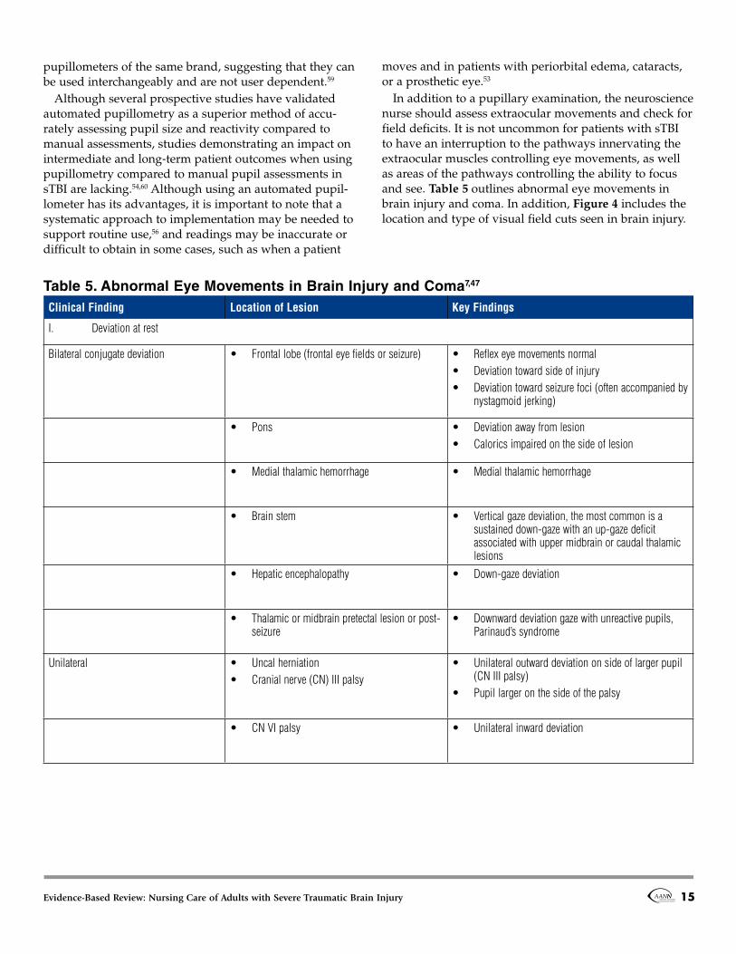
In addition to a pupillary examination, the neuroscience nurse should assess extraocular movements and check for field deficits. It is not uncommon for patients with sTBI to have an interruption to the pathways innervating the extraocular muscles controlling eye movements, as well as areas of the pathways controlling the ability to focus and see. Table 5 outlines abnormal eye movements in brain injury and coma. In addition, Figure 4 includes the location and type of visual field cuts seen in brain injury.
Table 5. Abnormal Eye Movements in Brain Injury and Coma7,47
Clinical Finding Location of Lesion Key Findings
I. Deviation at rest
Bilateral conjugate deviation • Frontal lobe (frontal eye fields or seizure) • Reflex eye movements normal• Deviation toward side of injury • Deviation toward seizure foci (often accompanied by
nystagmoid jerking)
• Pons • Deviation away from lesion • Calorics impaired on the side of lesion
• Brain stem • Vertical gaze deviation, the most common is a sustained down-gaze with an up-gaze deficit associated with upper midbrain or caudal thalamic lesions
• Hepatic encephalopathy • Down-gaze deviation
• Thalamic or midbrain pretectal lesion or post-seizure
• Downward deviation gaze with unreactive pupils, Parinaud’s syndrome
Unilateral • Uncal herniation • Cranial nerve (CN) III palsy
• Unilateral outward deviation on side of larger pupil (CN III palsy)
• Pupil larger on the side of the palsy
• CN VI palsy • Unilateral inward deviation
Evidence-Based Review: Nursing Care of Adults with Severe Traumatic Brain Injury 16
Table 5. Abnormal Eye Movements in Brain Injury and Coma7,47 (continued)
Clinical Finding Location of Lesion Key Findings
Skew deviation • CN III or IV nerve or nucleus lesion • Reflex eye movements normal• Deviation toward side of injury • Deviation toward seizure foci (often accompanied by
nystagmoid jerking)
II. Spontaneous eye movement
Random roving conjugate eye movement • Metabolic/toxic encephalopathy most likely • Can occur with bilateral lesions above the
brain stem.
• Indicates an intact CN III nucleus and medial longitudinal fasciculus
• Also known as “windshield wiper eyes”• Roving eye movements in early coma cannot be
mimicked and their presence excludes psychogenic unresponsiveness.
• With depending coma, roving eye movement will disappear, followed by the oculocephalic response and the oculovestibular reflex.
Periodic alternating gaze • Usually bilateral cerebral dysfunction • Eye deviates side to side with frequency of approximately three to five movements per second (pausing 2–3 seconds in each direction)
Ocular bobbing • Pons• Metabolic or toxic disorder
• Repetitive rapid vertical deviation downward with slow return to neural position
• Note: If patient can close eyes or blink, the pons is intact.
III. Internuclear ophthalmoplegia (INO) and variations
Disconjugate movements • Medial longitudinal fasciculus (MLF)• CN III or VII pathway
• INO is associated with lesion in medical longitudinal fasciculus.
• Eye ipsilateral to the MLF lesion does not adduct.
IV. Reflex eye movements (maneuvers to test brain stem)
Oculocephalic reflex (doll’s eye movements)
Brain stem reflexes • The eyes should move conjugately in the direction opposite to the movement. An abnormal response (absent or asymmetric) implies brain stem disease.
• Do not perform when neck instability is suspected.• Supratentorial lesions and metabolic processes
usually do not affect this reflex, except for metabolic encephalopathy.
Oculovestibular reflex (caloric test) Brain stem reflexes • Normal response is tonic deviation toward the side of the irrigated ear (cold water). Warm water causes the opposite response.
• Lack of response suggests sedative-hypnotic drug intoxication, structural lesion of the brain stem, or brain death.
• Loss of reflex eye movement with preserved pupillary reactivity is diagnostic of drug toxicity.
Evidence-Based Review: Nursing Care of Adults with Severe Traumatic Brain Injury 17
Figure 4. Visual Field Cuts Associated with Brain Injury
Key Visual field Area of lesion Clinical findings
1
Retina Visual field defects of various patternsScotoma
2 Optic nerve Ipsilateral monocular visual lossLoss of pupillary light reflex in both eyes when light is shone in the left eye
3 Midline optic chiasm Bitemporal May have relative afferent pupillary defect (RAPD)
4 Optic tract Contralateral homonymous hemianopia May have RAPD
5 Lateral optic chiasm Binasal hemianopia May have RAPD
Evidence-Based Review: Nursing Care of Adults with Severe Traumatic Brain Injury 18
Management of Patients with Increased ICPManagement of secondary brain injury in the ICU includes measures to avoid cerebral ischemia. Adequate cerebral blood flow (CBF) is a critical component to pre-vent cerebral ischemia and meet cerebral metabolic rate of oxygen (CMRO2) demands.7 CBF is difficult to quantify; however, invasive technologies in the ICU can provide indirect indications of CBF and perfusion. Measurement of ICP with invasive monitoring can provide timely information on factors impacting cerebral perfusion and facilitate early interventions in the setting of increased ICP to reduce the risk of secondary brain injury.61,62 As ICP becomes elevated (usually defined as more than 20 mmHg), cerebral perfusion pressure (CPP) is reduced at any given mean arterial pressure (MAP).7 In part, this is caused by impaired autoregulation, in which the brain
becomes dependent on systemic blood pressure. The rela-tionship between CPP, ICP, and MAP is as follows:
CPP = MAP – ICP.Emergent ICP management of patients with sTBI is divided into two phases: 1. Acute management of the patient at high risk for or with impending brain herniation 2. Ongoing management of the patient with sus-tained elevated ICP.56
Current TQIP guidelines support ICP monitoring in patients who are comatose with a GCS score of 8 or less and if there is structural evidence of brain damage on CT imaging.2 Monitoring of ICP is associated with lower in‐hospital mortality rates and remains a standard of care for sTBI.2 Table 6 outlines other assessment findings associ-ated with the potential need for ICP monitoring.
Table 6. Assessment Findings that Indicate the Potential Need for ICP Monitoring7,63
Any of the following indicate the potential need for ICP monitoring:
• Dilated and nonreactive or asymmetric pupils• Extensor posturing or no motor response• Progressive decline in neurologic exam
o Decrease in Glasgow Coma Scale (GCS) score >2 points• Cushing’s response
o Increased blood pressure (BP) o Tachycardia followed by bradycardia o Irregular respirations
• GCS score <8 and abnormal computed tomography (CT) scan• GCS score <8 with normal CT scan and two or more of the following:
o Age >40 years o Motor posturing o Systolic BP <90 mmHg
• GCS score 9–15 with CT scan that demonstrates any of the following: o Mass lesion o Effaced cisterns o Midline shift >5mm
• Following select surgical interventions • Multiple systems injured with altered level of consciousness, especially where therapies for other injuries may have deleterious effects on ICP, such as
high levels of positive end-expiratory pressure or the need for large volumes of intravenous fluids or heavy sedation • With traumatic intracranial mass (such as epidural hematoma, subdural hematoma, depressed skull fracture)
Evidence-Based Review: Nursing Care of Adults with Severe Traumatic Brain Injury 19
Monitoring Tools and Devices There are different mechanisms for monitoring ICP in the ICU. An external ventricular drainage (EVD) device with a dual lumen ventriculostomy catheter that allows for both ICP monitoring and cerebrospinal fluid (CSF) drain-age often is the preferred device for managing ICP for most patients.64,65 This device allows for CSF drainage to help control or manage increased ICP. If placement of an EVD device is not possible, insertion of a subarachnoid, subdural, or parenchymal bolt or catheter can provide ICP monitoring without drainage.
Although invasive ICP monitoring is important, it does not replace careful neurological and radiographic exami-nation.2 Therefore, patients with sTBI, including those requiring ICP monitoring, should be carefully assessed for signs and symptoms of increased ICP and impending brain herniation.63 Cerebral monitoring devices may be inserted at the time of a surgical intervention (e.g., crani-otomy, craniectomy, etc.) or in isolation.
Although not a universal standard of care, addi-tional invasive cerebral monitoring may be available for advanced monitoring in some ICUs to measure cerebral autoregulation, cerebral blood flow, and cerebral oxygen-ation.66 These methods are further described in Table 7.1 Data obtained from cerebral monitoring devices (includ-ing ICP or EVD devices) should be evaluated in conjunc-tion with the neurologic examination and not considered in isolation.1,61,65 Astute nursing care is essential because invasive cerebral monitoring devices have the poten-tial for serious complications, such as brain infection and sepsis.
To provide accurate readings, a consistent method for calibrating arterial pressure, MAP, and ICP is required, including standardizing the degree to which the head of the bed is elevated.71,72 Maintenance of goal CPP may require meticulous titration of intravenous (IV) vasoac-tive medications, especially when cerebral autoregulation is impaired.
Other treatment goals include adequate oxygenation, normocapnia, normothermia, and avoidance of hypona-tremia (sodium level below 135 mEq/L).18 Besides poten-tially increasing cerebral edema, hyponatremia is associ-ated with metabolic complications such as diabetes insipi-dus, cerebral salt wasting, and syndrome of inappropri-ate antidiuretic hormone.2,73 Table 8 provides insight into these disorders of sodium balance.
Related Metabolic and Physiological ParametersCerebral metabolism, ICP, and CPP also are affected by16
• hyperthermia (temperature > 37.5° to 38.0° C, although this number is controversial)16,18
pH > 7.45). Note: acute alkalosis increases the bind-ing of calcium, which can lead to ionized hypocalce-mia with tetany.
• seizures.Therefore, nursing interventions should be aimed at
maintaining normothermia, normoglycemia, targeted osmolarity, and serum electrolyte levels, as well as ensur-ing adequate oxygenation. There also should be a focus on achieving hemodynamic targets, treating anemia, maintaining targeted PaCO2 goals, and assessing for sei-zure detection and prevention.
Throughout the ICU hospitalization, the neurosci-ence nurse should closely monitor the patient for signs and symptoms of increased ICP. Table 9 outlines some of the most common signs and symptoms of increased ICP. Clinical signs of brain herniation or impending brain her-niation include63
• unilateral or bilateral dilated pupil(s)• loss of consciousness or decline in mental status• abnormal posturing (involuntary flexion or exten-
sion of the arms and or legs)• hypertension and bradycardia.More specific signs and symptoms associated with the
different types of brain herniation syndromes are further described and depicted in Table 10 and Figure 3.
Evidence-Based Review: Nursing Care of Adults with Severe Traumatic Brain Injury 20
Table 7. Advanced Monitoring to Measure Cerebral Blood Flow, Brain Oxygenation, and Cerebral Metabolism Along with Electrophysiological Measures in the Treatment of sTBI67–70
Cerebral blood flow (CBF) monitoring
Parenchymal thermal diffusion flowmetry A probe with two thermistors set at different temperatures is placed; the rate of temperature dissipation from applied heat is then calculated. Increased heat dissipation indicates greater blood flow. Requires precise positioning of probe through a bolt.
Transcranial doppler Noninvasively measures blood flow velocities of intracranial arteries via ultrasound. Most often used in the subarachnoid population.
Brain oxygen monitoring
Jugular venous oxygenation (SjVO2) Catheter inserted into dominant internal jugular and then advanced superiorly to the jugular bulb. It provides a measure of global cerebral oxygen use. Normal values range between 55% and 75%. A SjVO2 <55% indicates hypoperfusion or an increase in metabolic demand, thus suggesting cerebral ischemia.
Partial pressure of brain tissue oxygenation (PbtO2) or brain tissue oxygen tension (PbrO2)
Catheter inserted into brain parenchyma monitoring allowing for continuous real-time measure of brain tissue oxygen. PbtO is a marker of the balance between supply and consumption. Compromised PbtO (<20 mmHg) should be treated because it is associated with worsening brain injury.
Near-infrared spectroscopy (NIRS) Used in the operating room to indicate global perfusion changes. Measures regional cerebral oxygen saturation by measuring the amount of light attenuation between an NIRS light source and receiver and comparing light spectra absorption from deoxyhemoglobin and oxyhemoglobin.
Cerebral metabolism monitoring
Cerebral microdialysis Catheter inserted into brain parenchyma to detect biochemical changes (e.g., glucose, lactate, pyruvate, glutamate, glycerol, pH, and more) in extracellular substrates or regional subcortical white matter. Consistently low glucose concentrations (<0.66 mmol/L) are associated with poor outcome. A lactate to pyruvate ratio >25 is a marker of metabolic distress.
Positron emission tomography Noninvasive method to evaluate cerebral metabolism. Used mainly in research.
Magnetic resonance imaging spectroscopy Noninvasive method to measure lactate content
Electrophysiological measurements
Quantitative electroencephalogram (EEG) Most useful when continuous, depicts brain electrical activity that is then converted to a digital form.
Intracortical depth electrodes An invasive monitor inserted into the brain parenchyma. Useful in detecting seizures and cortical spreading depression not detected on scalp EEG.
Evoked potentials Evoked potentials are the electrical manifestation of the brain’s response to an external stimulus (such as an electrical stimulus applied to the median or tibial nerve) and can provide information regarding the functional integrity of sensory pathways.
Evidence-Based Review: Nursing Care of Adults with Severe Traumatic Brain Injury 21
Table 8. Syndrome of Inappropriate Secretion of Antidiuretic Hormone, Cerebral Salt Wasting, and Diabetes Insipidus in TBI Parameter Syndrome of Inappropriate Antidiuretic Hormone74,75 Cerebral Salt Wasting75,76 Diabetes Insipidus77
• Seizures• Unequal and or unreactive pupils• Loss of consciousness • Impairment of brain stem reflexes • Extensor posturing • Cushing’s triad
o Hypertension o Bradycardia o Irregular respirations or apnea
Evidence-Based Review: Nursing Care of Adults with Severe Traumatic Brain Injury 22
Table 10. Signs and Symptoms of Herniation Syndromes56
Type of Herniation Description Common Signs and Symptoms
Cingulate or subfalcine Also known as a midline shift, this is the most common type of cerebral herniation pattern. Generally caused by unilateral mass effect in the frontal, parietal, or temporal lobe(s) leading to a medial shift of the ipsilateral cingulate gyrus beneath the free edge of the falx cerebri. Large lesions may lead to uncal or central herniation.
• Initial presentation can be benign and may include headache and apathy.
• Signs of ipsilateral anterior cerebral artery ischemia such as contralateral leg weakness
• If the contralateral arcuate fasciculus is involved, signs of expressive, receptive, or conductive aphasia may be present.
Central transtentorial A downward displacement of medial brain structures through the tentorial notch by a supratentorial mass that exerts pressure on the underlying structures, such as the brain stem
• Early signs include increased confusion and headache leading to decreased level of consciousness leading to coma, including loss of reflexes and seizures, and eventual death.
• Motor weakness leading to abnormal posturing• Fixed and dilated pupils
Uncal A subtype of central transtentorial herniation related to the cerebral mass effect from increasing intracranial pressure. The uncus and the adjacent part of the temporal lobe slide downward across the tentorial incisura compressing the brainstem and the posterior cerebral arteries in the ambient cistern. May be unilateral or bilateral. Associated with poor prognosis because of the direct compression of vital midbrain centers.
• Initially presents with an ipsilateral dilated pupil that is unresponsive to light as a result of ipsilateral cranial nerve III (oculomotor nerve) compression.
• May develop into bilaterally blown pupils.• Lateral or vertical gaze eye deviation and ptosis may
occur.• Altered mental state leading to coma• Contralateral hemiparesis• If the ipsilateral posterior cerebral artery is affected,
homonymous hemianopsia may occur
Upward transtentorial Also known as ascending transtentorial herniation, this occurs where space‐occupying lesions in the posterior cranial fossa cause superior displacement of superior parts of the cerebellum through the tentorial notch.
• Nausea or vomiting• Rapid progression toward a decreased level of
consciousness and, eventually, death
Tonsillar Also known as downward cerebellar herniation, this syndrome is caused by the inferior descent of the cerebellar tonsils below the foramen magnum.
• The brain stem is compressed against the clivus thereby altering the vital life‐sustaining functions of the pons and medulla, such as the respiratory and cardiac centers, leading to respiratory and cardiac depression and death.
• The most feared because of its rapid progression
Transcalvarial Also known as an extracranial brain herniation, this occurs when brain tissue external to the calvaria herniates through a skull bone defect associated with surgery or trauma.
• Varies; the herniated brain tissue is at risk of ischemia and venous infarction.
Evidence-Based Review: Nursing Care of Adults with Severe Traumatic Brain Injury 23
Pharmacotherapy for Increased ICPBoth mannitol (0.5–1 g/kg) and hypertonic saline (HTS) (2%–23.4%) may be used to treat increased ICP and usu-ally readministered as intermittent boluses.1,78 It is impor-tant to use an inline filter with mannitol because the medication can crystalize, and to use a central line when infusing saline in concentrations greater than 3%–7.5% (depending on reference and local policy).79,80 Both medi-cations usually are given as a bolus and facilitate the movement of water into the vasculature. Nursing inter-ventions include monitoring serum osmolarity (with goal of less than 320–340 mOsm/kg or per physician orders), fluid status, renal function, and serial serum sodium lev-els, especially with HTS administration.18,81 Diligent moni-toring is required when administering HTS to patients with low serum sodium levels (especially chronic hypo-natremia), as an overcorrection that is too fast can lead to osmotic demyelination syndrome.81 The osmolar gap (the difference between osmolality measured and osmolar-ity calculated) should be measured in patients receiving mannitol. The formula is as follows82:
Calculated osmolality = ([2Na] + [glucose/18] + [BUN/2.8] + [ETOH/4.6*]) Where Na = sodium
• BUN = blood urea nitrogen • ETOH = blood alcohol level • Normal range for calculated osmolality = 280 to 300 mOsm/kg H2O• A gap is equal to or greater than 10 mOsm/kg H2O (suggesting exogenous osmoles), with normal being less than 10 mOsm/kg H2O.
*Some versions of this equation do not include the correc-tion for blood alcohol. Mannitol should be stopped if the osmolar gap exceeds 20 mOsm/kg H2O. Mannitol can cause renal failure in high doses, whereas HTS may cause volume overload. Both can cause metabolic alkalosis hypokalemia and hypochloremia.
Head of Bed ElevationHead-of-bed elevation and positioning of the head in a neutral (midline) position to facilitate venous drainage is a common, simple, and cost‐effective intervention to decrease ICP and optimize CBF.83–87 Initial management for patients with sTBI includes head elevation at a mini-mum of 30 degrees,88 monitoring ICP and CPP, and main-taining established care goals.22,64,65,86,87,89
Changes in patient ICP and CPP often occur with patient repositioning, and can produce transient increases in ICP, particularly in the setting of increased ICP.45,88,90,91 Sharp head rotation and prone positioning also may
increase ICP.85,92 Patient response to repositioning can be varied; therefore, individualized plans of care and clus-tering of activities should be evaluated for impact on ICP, CPP, and CBF response.62
C-Spine SupportENLS guidelines currently recommend cervical‐spine (c‐spine) immobilization as a vital component of prehos-pital and initial hospital management prior to appropriate assessment and radiographic evaluation of SCI because of the high rate of comorbid injury.22 It is important to ensure any collar used fits the sTBI patient appropriately because mal‐fitting or tight‐fitting collars may increase ICP by impeding cerebral venous outflow and decreasing CBF, which can exacerbate secondary brain injury.84,89,93 Nurses should ensure the use of appropriately sized and correctly fitted cervical collars to lessen these deleterious effects.87 Cervical collars and other types of neck braces also can cause skin injury, impact respiratory effort, and contribute to the patient’s pain and discomfort.94 Nurse interventions should include ways to lessen pressure on the skin that is exerted by cervical collars and aim to remove these devices as soon as it is deemed safe.95
General nursing care for the patient with a cervical col-lar includes removing and reapplying the cervical col-lar at regular intervals (with assistance in maintaining the neck and head in a neutral position) to provide skin assessments and skin care. Besides cleansing the skin, the pads inside the collar should be cleaned or replaced at this time. When performing this procedure, the nurse should pay close attention to brace tightness and monitor for changes in ICP and CPP with collar reapplication and patient repositioning.
In patients with low-velocity gunshot wounds to the head, routine cervical immobilization may not be nec-essary.96 An individualized approach to selecting softer boards and vacuum mattresses for cervical spine immo-bilization may avoid ICP elevation and improve patient comfort.97
Noxious Stimuli Although there is limited research on patient room dynamics, controlling the patient environment by limit-ing noxious stimulation has been an intervention aimed at minimizing adverse fluctuations in ICP and CPP values for sTBI patients.49,89 Examples of noxious stimuli that are amenable to nursing interventions include89:• uncomfortable or painful stimuli• loud noises and voices• sudden jarring of the bed
Evidence-Based Review: Nursing Care of Adults with Severe Traumatic Brain Injury 24
• sounds from bedside equipment• bright overhead lights• components of a neurologic assessment• painful medical or nursing procedures.
It is important to note that limiting environmental stimulation may have varying effects on ICP, with some studies indicating minimal adverse effects.49 However, it is important for families and healthcare professionals to assume patients have intact hearing and therefore avoid having disturbing conversations within hearing distance of the patient.88 Nurse and family talking with the patient has rarely been shown to independently increase ICP or reduce CPP.85 In fact, family member conversations and families talking to the patient (familiar voices) has been shown to more likely decrease ICP, and nurses talking to patients has been shown to have no significant impact on ICP.49 When ICP is monitored, individual patient responses to each patient care intervention should be evaluated and used to guide subsequent care.85
Other Registered Nursing–Related Care Key points for the neuroscience nurse caring for patients at risk for increased ICP include • providing basic nursing care • assessing for neurologic changes• assessing for other injuries, including signs and
symptoms of scalp, facial, spine, intra-abdominal, and long-bone injuries
• closely monitoring vital signs, ICP, and CPP4,66,85
• avoiding hypotonic fluids (such as dextrose 5% in water)
• prioritizing or altering patient care based on changes in ICP and CPP62,86,98
• carefully considering the impact of nursing care related to patient positioning, neck brace care, envi-ronmental noise, and stimuli on ICP and CPP
• maintaining invasive monitoring device systems• standardizing or protocoling nursing interventions to
improve compliance with neuroscience nursing care delivery, including the aseptic maintenance of inva-sive monitoring device systems87
• reporting findings and concerns to the appropriate healthcare provider
• early recognizing and responding to paroxysmal sym-pathetic hyperactivity, also known as “sympathetic storming,” a strong physiologic response that may be triggered by nursing care and often is character-ized by agitation, clenching of fists, grinding of teeth, profuse sweating, sustained tachycardia, and marked hypertension. Initial treatment includes removing
external triggers, such as noxious stimuli, body turn-ing or movements, and bladder distention, before administering medications. The most useful pharma-cologic agents are morphine sulfate and nonselective β-blockers (e.g., propranolol).99
• avoiding hyperventilation, which may exacerbate cerebral ischemia, especially in the first 24 hours.18
The nurse also should integrate and promote the use of evidence-based, standardized protocols for the care of the patient with sTBI because many are correlated with improved neurological outcomes and decreased mortality at 6 months post-injury.100
Neurosurgical Interventions Neurosurgical interventions for sTBI patients may extend beyond the placement of monitoring devices and ven-tricular catheters and target both primary and second-ary injury associated with sTBI. Common neurosurgical interventions include evacuation of intracranial hemato-mas, such as epidural and SDHs (note: any symptomatic posterior fossa mass lesion or those with mass effect on CT should be emergently removed); removal of foreign objects; correction of skull defects (such as depressed skull fractures); spine stabilization; correction of hydro-cephalus; endovascular treatment of carotid or vertebral dissection as well as vasospasm; interventions for pneu-mocephalus; and decompressive hemicraniectomy with duroplasty (further described below).7
Ongoing neurological assessments and treatment of increased ICP remain top nursing responsibilities post-operatively. Occasionally, the patient may have a drain placed during surgery. Nursing care includes assessing and labeling the location of each drain, ongoing moni-toring and measurement of drainage, and maintaining patency of the drain. In general, drains in the subdural space are either drained to gravity or to partial bulb suc-tion, whereas subgaleal drains can be placed to full bulb suction. Do not place a brain drainage device to wall suc-tion, as aggressive suction can tear vessels and cause hemorrhage. The provision of evidence-based routine postoperative nursing care is essential in preventing com-plications and maximizing positive patient outcomes.
Post-Hemicraniectomy with Duroplasty Decompressive hemicraniectomy with duroplasty is an effective treatment for relieving severe refractory intracra-nial hypertension after sTBI.101 During the procedure, a large portion of the skull is removed (may be unilateral or bilateral and usually involves the lateral skull), the dura is opened widely, and the scalp flap is closed over the
Evidence-Based Review: Nursing Care of Adults with Severe Traumatic Brain Injury 25
skull defect, thus allowing the injured brain to swell and expand through the cranial defect.101
The potential for short-term postoperative complica-tions of this procedure requires astute and timely assess-ments by the neuroscience nurse. Potential complica-tions include increased ICP, brain herniation through the bone window, surgical site infection, contralateral hema-toma, ipsilateral subdural or subgaleal effusion, early seizure, CSF leakage, and hydrocephalus.102 In addi-tion, patients are at high risk for falls, which can be cata-strophic because of the compromised skull and exposed cerebrum.102
Nursing interventions to promote recovery include pro-tecting the exposed cerebrum through proper positioning, head protection, and helmet use when out of bed, as well as evidence-based fall prevention strategies.89,102 In addi-tion, routine wound assessment and care should include assessment of the wound for acute turgor and girth of the craniectomy site, indications of acute surgical bleeding, and CSF leakage.31
Longer-term complications of decompression crani-ectomy (DC) include sinking skin flap syndrome, also known as syndrome of the trephined.102 Early symptoms can include a depressed mood, headache, behavioral dis-turbances, and seizures. Symptoms are related to cerebral cortex distortion under the skin flap, which can occur once cerebral edema subsides. More serious symptoms can include acute neurological changes and paradoxi-cal herniation. Late complications may include ipsilateral effusion, late-onset seizure/epilepsy, and continued com-plications related to syndrome of the trephined.102
Bone flap replacement (cranioplasty) for DC typically is performed during a rehospitalization months after the initial surgery. Risks associated with replacement include infection, hematoma, hydroma, and bone flap resorp-tion.102 Targeted nursing care includes wound care, pre-vention of increased ICP, early detection of neurological changes, patient/family education, and care coordination.
Intrahospital Transport of Patients with sTBIPatients with sTBI often require multiple intrafacility or intrahospital transports (IHTs) for diagnostic or inter-ventional procedures, and can present potential patient hazards.85 Clinically significant complications have been reported to occur in 36% of critically ill patients with brain injury during transport.103 Complications include, but are not limited to, accidental extubation, equipment battery failure, increased ICP, and hyper‐ or hypoten-sion.103 Hemodynamic and respiratory instability in patients with sTBI are associated with elevations in ICP
and decreased CPP, leading to extension of secondary brain injury, increased ICU length of stay, and increased mortality.104
Hazards encountered during IHT may result from inad-equate planning prior to transport; the inability to main-tain a consistent level of intensive care monitoring and assessments during transport; increased exposure to nox-ious stimuli; increased patient movement or reposition-ing; and ineffective means of treating changes in ICP, CPP, pain, or agitation levels. To prevent harm, a pre-transport checklist prior to IHT is recommended.105 Rec-ommended items to include on a sample transport check-list are included in Table 11. During IHT, it is important to continue the same level of ICU patient monitoring and care.105
Transport staff should include nurses and/or clinicians trained in delivering immediate interventions for treating increased ICP and managing other adverse events.103,105 ICP may increase during transport or during performance of the diagnostic or treatment modality itself.85,106,107
To prevent reflux of CSF during patient transport, EVDs typically are clamped during the transport period.85 If the EVD is unclamped in the ICU prior to transport, the patient should be screened for ICP tolerance by clamping the EVD prior to transport to identify the potential risk of increasing ICP during IHT.108 Increased ICP related to IHT is most often seen in critically ill patients with ele-vated baseline ICP values and in patients requiring con-tinuous EVD diversion.106 Premedication with analgesia or sedatives prior to IHT may help prevent or mitigate ICP elevation.106
Overview of Ventriculostomy Site CareHealthcare‐associated ventriculitis (HAV), also referred to as ventriculostomy‐related infection or ventriculostomy‐associated infection, has an incidence of up to 22%109 or 11.4 infections per 1,000 EVD catheter days.110 It is associ-ated with increased morbidity, mortality, and treatment cost.111 Infection may be introduced at the time of EVD placement109 and during use of the device. Nursing inter-ventions to prevent infection should include facilitating and monitoring adherence to an established evidence-based insertion protocol with use of a checklist to confirm compliance with insertion steps.111-116 Several simple EVD insertion protocols with associated reductions in HAV have been published.112,113,115,117-119 Three protocols pub-lished in the past 5 years are summarized in Table 12.
Post-EVD insertion, HAV may result from catheter and tubing contamination during manipulation, CSF sam-pling, or EVD colonization from skin or room contami-nants.109,120 Published insertion site care and maintenance procedures for ventriculostomy use vary greatly.111,121 Not
Evidence-Based Review: Nursing Care of Adults with Severe Traumatic Brain Injury 26
• Adequate battery supply for heart monitor and other transport equipment• Adequate oxygen tank levels for support • Adequate intravenous (IV) fluids, continuously infusing IV medications, sedatives, and analgesics • Consider a brief trial period of clamping external ventricular device (EVD) and laying the patient flat (simulates flat position during radiologic testing)
to assess patient’s tolerance (impact on intracranial pressure [ICP] and cerebral profusion pressure [CPP], oxygenation, ventilation, etc.).• Use transport staff familiar with the vehicle in which the patients with severe traumatic brain injury will be transferred and who have the ability to treat
increasing ICP and decreasing CPP as needed.
During transport:
• Continue to monitor vital signs, ICP, CPP, end-tidal CO2, and O2 saturation levels at the same intervals as when in the intensive care unit.• Perform neurologic, pain, and sedation assessments regularly and treat as needed. • If EVD is in place, clamp during transport. Relevel and open if ICP increases. • Consider premedication with analgesia and sedatives prior to transport to avoid increasing ICP.
Table 12. Review of EVD Insertion Protocols Published Between 2014 and 2018111,113,117
Hand hygiene and everyone in room has bouffant/mask on Yes Yes Yes
Provider wearing fullsterile attire
Yes Yes Yes
Site shaved to accommodate dressingsize
Yes Yes Yes
Site cleansed with chlorhexidine and allowed to dry. Not reported Yes Yes
Use of antibiotic-impregnated catheter
No Yes Yes
Use of antibiotic (intravenous [IV] orintraventricular)
IV and intraventricular Normal Low
Catheter tunneled Not reported Not reported Yes (>5 cm)
Routine catheter exchange Yes, every 7 days No No
Evidence-Based Review: Nursing Care of Adults with Severe Traumatic Brain Injury 27
all EVD insertion protocols address post-insertion site and dressing care, such as the use of a topical antimicro-bial, type of dressing, timing of site care and dressing changes, or the role of site care in preventing site infec-tions or ventriculitis.112,113,115 Randomized controlled tri-als specifically comparing dressing practices for EVDs are lacking in the research literature.
Many sites report application of a dressing following ventriculostomy insertion,111,113,116,122 although this practice is not consistent.123 A summary of published ventriculos-tomy insertion protocols and infection rates (if available) from 2014 to 2018 is included in Table 13. Most recently published protocols do not include use of prophylactic antibiotics.110,112 Two novel approaches to site care asso-ciated with decreased HAV are a single application of 2‐octyl cyanoacrylate adhesive (Dermabond, Ethhicon, Inc.) at the time of ventriculostomy insertion85 and the use of an alcohol impregnated cap attached to the proximal end of EVD tubing for access during CSF sampling.123,124 Regardless of the approach, comprehensive EVD care should be standardized and based on evidence-based protocols.
ICU Bundle (ABCDEF Bundle) The ABCDEF bundle is an interdisciplinary, evidence-based strategy that has been associated with improved patient outcomes in critically ill patients.47,126 The compo-nents of the ABCDEF bundle include47 A. Assessment, prevention, and management of pain B. Both spontaneous awakening trials and spontaneous
breathing trials C. Choice sedation/analgesia D. Delirium assessment, monitoring, and management E. Early mobility F. Family engagement and empowerment.
Components are further described in Table 14 and should be incorporated as part of routine care in the ICU.
Table 13. Review of the Literature on External Ventriculostomy Device Site Care Between 2014 and 2018111,113,122,123,125
Researcher Bookland (2014) Gozal (2014) Ducis (2016)Hepburn‐Smith(2016)
Sieg (2018)
Type of dressing NoneTransparent occlusivedressing None Bio‐occlusive
dressing Adherent dressing
Frequency of dressing change Not reported Replaced as needed Not reported Weekly or if
Daily site assessments None specified Not reported
Other
Single application of 2‐octyl cyanoacrylate adhesive at time of insertion
After dressing removal, scalp prepped with CHG, then 30 sec scrub with CHG (other sideof stick)
Minimal hair removal at insertion site
Chlorhexidine‐eluting patch
Chlorhexidine‐eluting patch
Infection rate (pre- and post-study, ifavailable)
18.6% pre to3.5% post*
10.8% preto 2.0% post
5.1% compared to national average of 8.8%
Not yet published 12% pre to 0% post*
*Statistically significant
Evidence-Based Review: Nursing Care of Adults with Severe Traumatic Brain Injury 28
Table 14. ABCEDF Intensive Care Unit Liberation Bundle47,126
Bundle assessments Key points
A. Assessment, prevention, and management of pain
• Untreated pain can lead to delirium. • Vital signs alone may not be the best indicators of pain. • Assess for nonverbal signs of pain, such as facial grimacing, abnormal body movements, and ventilator
dyssynchrony. • Premedicate patients before invasive procedures or turns. • Use a reliable and valid pain scale. If the patient cannot self-report, consider the Behavioral Pain Scale, Critical
Care Pain Observation Tool, or Nociception Coma Scale-Revised. • Consider nonpharmacologic interventions, such as relaxation, massage/touch, music, and pet therapy.
B. Both spontaneous awakening trials (SATs) and spontaneous breathing trials (SBTs)
• Use standardized processes and procedures to promote daily team-driven initiatives to liberate patients from mechanical ventilation and sedation using sedation-weaning protocols, SATs, and SBTs.
C. Choice of sedation/analgesia
• Treat pain before considering sedative therapy.• Use a valid and reliable sedation scale, such as the Richmond Agitation Sedation Scale or the Sedation-Agitation
Scale, to assess the quality and depth of sedation in critically ill patients. • First-line medications, including non-benzodiazepine sedatives such as propofol or dexmedetomidine, are
preferred. • If the patient remains agitated despite treatment with appropriate sedation/sedative, assess and treat for conditions
such as hypoxia, hypoglycemia, hypotension, and drug or alcohol withdrawal.
D. Delirium assessment, monitoring, and management
• Use a valid and reliable assessment tool such as the Confusion Assessment Method for the ICU or the Intensive Care Delirium Screening Checklist to monitor for the development and severity of delirium.
• To prevent delirium, try to optimize the patient’s environment to allow for natural sleep-wake cycles, if able. This includes minimizing light and noise, clustering activities, and reducing stimuli at night, if able.
• Early mobilization can help decrease delirium, although this may not always be possible in cases of severe traumatic brain injury with increased intracranial pressure.
• Avoid medications known to provoke or further confound assessment, monitoring, or management of delirium.
E. Early mobility • Assess for readiness for early and progressive mobility using a tool or protocol, such as the “MOVEN” (Mission Hospital Progressive Mobility Algorithm).
• Be sure to consider the type of neurological injury prior to initiation.
F. Family engagement and empowerment
• Engage and encourage patient/family involvement in care. This can be accomplished through: o patient/family rounding o being open to family visitation polices o family presence during resuscitation o patient and family involvement in decision making, especially regarding goals of care and end-of-life decision
making, as appropriate o providing daily updates o shared daily goal setting.
Evidence-Based Review: Nursing Care of Adults with Severe Traumatic Brain Injury 29
Acute Care Considerations
Prevention of Hospital‐Acquired ConditionsPatients with sTBI are at high risk for hospital-acquired conditions that are sensitive to nursing care, such as hospital-acquired pneumonia, falls, pressure injury, central line–associated bloodstream infection, catheter-associated urinary tract infection, contractures, deep vein thrombosis, and other hospital-related infections. In a study quantifying complications during the first year after sTBI from hospital admission and throughout
subacute rehabilitation, urinary tract infections (53%) and pneumonia (32%) were the most common complications. Less frequent were pressure ulcers (18%), joint contractures (18%), and deep venous thrombosis (4%).127 Table 15 outlines targeted strategies for prevention of these events.
Nutritional SupportPatients with TBI may have hypermetabolic and hyper-catabolic activity lasting from 1 week to months post‐injury, supporting early initiation of nutrition care. Enteral
Table 15. Targeted Strategies for Prevention of Hospital-Acquired Conditions128-133 Hospital-Acquired Condition Targeted Strategies Ventilator-associated pneumonia/hospital-acquired pneumonia
• Adhere to ventilator care bundle: o Daily awakening and breathing trials o Deep vein prophylaxis o Medications to prevent gastric ulceration
• Head of bed at 30 degrees or higher if tolerated and no contraindications • Oral care every 4 hours with chlorhexidine gluconate including brushing of teeth • Monitoring of condensation in ventilator tubing• Swallow screen/evaluation prior to any oral intake • Speech language therapy evaluation if high risk for aspiration
Catheter-associated urinary tract infection • Avoid placing urinary drainage catheters, unless necessary for patient care. • Prompt removal of catheters • Assess the need for continued use at least daily. • Consider alternatives such as condom catheters (and other types of external devices) or scheduled
bladder scans with in-and-out catheterization for elevated post-void residuals and timed voidings.• Daily chlorhexidine baths • Limited insertion by trained individual and consider two-person insertion for females
Central-line associated bloodstream infections
• Remove central venous access devices (CVAD) as soon as no longer clinically indicated. • Avoid femoral line placement. • Aim to limit CVAD placement to the following:
o vasopressor use o hypertonic saline administration o high-volume fluid resuscitation o hemodynamic monitoring.
• Consider placement of a peripherally inserted central catheter in cases of o inability to obtain appropriate venous access o prolonged antibiotic administration.
Falls • Pay attention to times of high patient activity and situations where there is decreased nursing capacity to observe patient.
• Perform fall risk assessments at regular intervals using standardized tools. • Educate the patient and family about the risk of falls • Use fall wrist band, footwear, or signage to indicate a patient at high risk• Routine rounds and toileting• Environmental and geographical modifications to prevent falls
Evidence-Based Review: Nursing Care of Adults with Severe Traumatic Brain Injury 30
Hospital-Acquired Condition Targeted Strategies Hospital-acquired pressure injury (HAPI) • Complete a thorough head-to-toe skin assessment on admission and then at least every shift.
• A risk assessment should be done at least daily using a validated tool, such as the Braden Scale.• Nutritional assessment with intervention • Optimize nutrition/hydration, including early nutrition, calorie counts, and supplementation. • Turn and reposition the patient at least every 2 hours (although some patients may need more frequent
turning) while in bed and at least every 1 hour when in a chair.• Pressure relief, a pressure-relief surface, or a special-care bed is recommended to help prevent
pressure ulcers.• To prevent heel pressure ulcers, use a pillow or heel-lift devices to keep the patient’s heels from
touching the bed.• Keep the head of the bed at less than 30 degrees, unless contraindicated• Manage incontinence and moisture with a skin-care regimen, such as frequent cleansing and use of a
moisture-barrier ointment. • Obtain a wound care/skin care nurse consult for high-risk patients.
HAPI related to a medical device • Ensure the device is needed. • Use the correct device size for the patient. • Ensure proper application and removal of the device. • Protect the skin and tissue under the device. • When possible, do not place the device in an area that already is prone to breakdown.• If allowable, remove the device periodically (such as every shift) to assess the skin under the device. • Especially vulnerable areas include the head, neck, face, and ear from respiratory-related equipment
such as oxygen tubing, tracheostomy times, and pulse oximetry probes. • Other high-risk devices include cervical collars, bedpans, endotracheal tubes/holders, face masks for
Venous thrombosis/pulmonary emboli • Apply pneumatic compression devices and ensure that the patient is wearing them, especially while in bed.
• Administer pharmaceutical intervention when safe. • Use early mobilization.
Contractures • Early aggressive range of motion• Passive stretching of muscles and joints on a prescribed schedule • Positioning of the limbs to promote extension and oppose flexion • Splinting or serial casting when appropriate • When able to get out of bed, regularly prescribed periods of daily standing and/or walking • Pharmacological therapies (e.g., dantrolene, baclofen) may be considered by the rehabilitation team.
Infection due to invasive lines • Ensure lines and drains (such as external ventricular devices) are placed under strict aseptic technique, including sterile field, sterile gowns/gowns, and mask use by all personnel in the room.
• Assemble intracranial pressure equipment and other equipment as appropriate using a sterile technique.
• Minimize access to invasive devices. • Use aseptic technique when managing drain/transducers/devices.
Table 15. Targeted Strategies for Prevention of Hospital-Acquired Conditions128-133 (continued)
Evidence-Based Review: Nursing Care of Adults with Severe Traumatic Brain Injury 31
feeding is preferred and should be initiated within 24–72 hours in patients who are hemodynamically stable. At the latest, nutritional support should begin 5–7 days post‐injury because early nutrition is associated with decreased mortality.1,2 When enteral feeding is required, transgastric jejunal feeding is recommended over nasogastric feedings to reduce the incidence of pneumonia. Additional research is needed to determine the specific nutritional needs of the sTBI patient and how energy expenditure plays a role in nutritional support requirements.
TracheostomyPatients with TBI are at risk for developing ventilator-associated pneumonia or acute lung injury. Therefore, early tracheostomy often is considered within 8 days of injury.2 Early tracheostomy may facilitate ventilator weaning, resulting in shorter ICU and hospital stays while reducing infection risk. Situations where early tracheostomy may be contraindicated include patients with increased ICP, hemodynamic instability, respiratory failure requiring high levels of positive end-expiratory pressure (greater than 10 cm H2O) and FiO2 administration greater than 50%.2
Management of Sleep Disturbances and Fatigue Sleep disturbances and fatigue after TBI are common, although the pathophysiology is not fully understood. Between 30% and 70% of patients with sTBI report sleep
disturbances.134–137 Sleep disturbances are associated with increased rates of anxiety, depression, and fatigue. Sequelae of sleep disturbances include long-term behavioral chal-lenges; impaired cognitive processing; inability to return to work; decreased quality of life; and higher incidence of antidepressant, antihypertensive, sedative, and narcotic use.138 Fatigue, reported in 17%–40% of patients with TBI, may be experienced primarily as a problem with cognitive and physical aspects and feelings of weakness.136,139,140
Nurses should be aware of the impact of sleep distur-bances among patients with sTBI and plan care aimed at facilitating sleep. Although sleep disturbances and fatigue commonly co-occur in TBI, it is recommended that nurses begin treatment of sleep disturbances prior to initiating specific treatments for fatigue.141 In the management of sleep disorders and fatigue after TBI, nonpharmacological interventions aimed at the medical and neuropsychiatric causes or contributors should take precedence over phar-macotherapies.141 Nonpharmacologic measures to aid in resolving sleep disturbances include cognitive behavioral therapy (CBT), blue-enriched white light therapy, acu-puncture, and implementation of sleep hygiene protocols to promote a better sleep/wake cycle (Table 16).1,135,140,142–144 Commonly used pharmacologic agents for the treatment of sleep disturbances include benzodiazepines, tricyclic anti-depressants, antihistamines, ramelteon, and melatonin (Table 17).1,134,136,140
Table 16. Nonpharmacologic Sleep Interventions for Patients with sTBI1,135,140
Cognitive behavioral therapy Combination of education, behavioral activation, behavioral experiments, problem solving and relaxation; found to result in decreased patient self‐report of fatigue, depression, and sleep disturbances
Acupuncture Nonpharmacologic, holistic therapy with few side effects; can promote regulation of neurotransmitters and humoral factors
Sleep hygiene protocol Emphasis on maintaining sleep-wake cycle, exercise, avoiding naps, keeping daytime lighting in place, avoiding caffeine, and screens prior to bedtime
Blue light therapy Helps retrain the circadian rhythm to promote better, more regular sleep
Evidence-Based Review: Nursing Care of Adults with Severe Traumatic Brain Injury 32
Management of Neuropsychiatric Complications Individuals who experience TBI are at increased risk of developing major depression, general anxiety disorder, obsessive-compulsive disorder, panic disorder, post-traumatic stress disorder (PTSD), antisocial behavior such as criminality and substance abuse, and suicide.145 Individuals with preexisting behavioral and psychiatric problems may find that the brain injury exacerbates their condition and makes the management of day-to-day function more complex and difficult. Memory impair-ments and cognitive problems such as decreased concen-tration may further hinder a patient’s recovery.
One of the most common disorders is PTSD, which often is associated with witnessing injuries or fatalities, suffering medical conditions resulting from the initial injury (such as from a blast injury, burns, or respiratory distress), and being a survivor of a traumatic event.146
Practice guidelines from the Department of Veter-ans Affairs Medical Center and Department of Defense include treatment of PTSD with appropriate pharmaco-logic and psychotherapeutic interventions and identifica-tion and treatment of comorbid neuropsychiatric condi-tions (e.g., depression), substance abuse, medical comor-bidities, and cognitive issues.146 The management of PTSD should include CBT as first-line therapy, antidepressant use, pain management, and suicide risk assessment.146,147
Patients with TBI are 10 times more likely to experience a depressive episode than the annual rate of depression in the general population (53%148 compared with 6%149) and are at greater risk of suffering from recurring depres-sive symptoms for decades after the initial injury.150 The first line of antidepressants for the treatment of PTSD is sertraline, paroxetine, and venlafaxine because they have fewer side effects and drug interactions compared with
other antidepressant medications. Benzodiazepines and antipsychotics, in particular, should be avoided because of side effects. It is important to assess for untreated TBI sequelae in patients exhibit persisting symptoms of insomnia, irritability, difficulty concentrating, depression, or fatigue.146,147
Family caregivers of individuals with TBI-related dis-abilities may encounter challenges when caring for some-one with cognitive, behavioral, and emotional changes associated with TBI. Caregivers may suffer from substan-tial stress as they attempt to meet their loved ones’ long-term physical and emotional needs, as well the associated financial burdens. Therefore, the interdisciplinary team should include assessment of family member needs and align resources and support services when indicated.
Management of Visual DisturbancesVision disturbances after TBI are common. Approximately 65%–79% of TBI patients report subjective visual com-plaints. Visual disturbances include defects in visual acu-ity, visual fields, eye movement, and the more complex aspects of vision such as visual perception.151
Results of a 2019 meta-analysis that looked at field loss, visual acuity, accommodative dysfunction, and conver-gence insufficiency found that in patients with moderate to severe TBI, 39.8% had visual field loss and 3.2 % expe-rienced decreased visual acuity. In addition, 42.8% expe-rienced accommodative dysfunction and 36.3% had con-vergence insufficiency.152 Among patients who experience blast-related sTBI, estimates of visual disturbances may be higher, up to 50%–75%, and include blurred vision, photosensitivity, light‐dark adaptation and accommoda-tion, visual field cuts, and difficulty reading.153
Table 17. Pharmacologic Agents Used in the Treatment of Sleep in Patients with sTBI1,134,136,140
Classification Side Effects
Benzodiazepines Found to have daytime sedative effects and adverse effects on motor and cognitive functioning; avoid use in older patients.
Tricyclicantidepressants
May reduce the seizure threshold
Antihistamines May lead to physical dependence, daytime drowsiness
Ramelteon U.S. Food and Drug Administration approved for treatment of insomnia with sleep-onset abnormalities; shorter half‐life and less likely to become addictive. Ramelteon is reported to increase total sleep time and sleep latency with limited negative side effects.
Melatonin Offers benefit of promoting sleep without sedative or addictive tendencies. Melatonin was associated with significant reduction in Pittsburgh Sleep Quality Index scores consistent with improved sleep quality.
Evidence-Based Review: Nursing Care of Adults with Severe Traumatic Brain Injury 33
As part of acute care management, patients with TBI should receive routine vision screenings that include an assessment of binocular vision and problems with con-vergence, which may present as headaches, fatigue, and disturbances in cognition.154 Patients with chronic visual dysfunction after TBI may require occupational, ves-tibular, cognitive, and other forms of physical therapy. Environmental safety is a priority and should include adequate lighting and education about compensatory mechanisms for visual loss. Patching and use of prisms or corrective lenses may improve short-term outcomes.155 Other techniques include visual-motor feedback training and computer-based oculomotor training.156 The use of protocols for vision assessment is associated with better outcomes in patients with TBI.2,154
The use of protocols for evidence-based care has been studied and has demonstrated better outcomes for patients after TBI in the areas of vision screenings, motor and gait disturbances, anticoagulation therapy, cognitive training, brain imaging, nutritional support, and airway management.2,146,154
Management of Motor Disturbances A major contributor to limited mobility after TBI, espe-cially in older patients, are motor disturbances such as poor balance and spasticity. Decreased gait velocity can be present 48–72 hours post-injury with a return to base-line between 6 and 12 months.157 Patients should receive fall risk assessments using standardized scoring tools at regular intervals throughout acute hospitalization and post‐hospitalization. Based on the assessment, a targeted plan of care with evidence-based fall prevention strategies should be implemented.
Anticoagulation TherapyPatients with TBI have an approximate 20%–30% risk for the development of venous thromboembolism (VTE).2 Current studies support pharmacologic thromboembo-lism prophylaxis within 24–72 hours if the CT scan reads as stable and the patient is not at high risk for increased cerebral hemorrhage.2,158,159 A prophylactic inferior vena cava filter may be considered in patients at very high risk for VTE who are not candidates for pharmacologic pro-phylaxis.2
Rehabilitation ConsiderationsPatients with sTBI-related impairments often need orga-nized physical, occupational, speech, and other therapies to regain abilities or to develop new compensatory skills. This also may involve modifications to the home and other environments.
For survivors of TBI with disabilities, insurance cover-age may impact the type, intensity, and duration of acute and post-acute services available.160 Behavioral health ser-vices and cognitive and physical rehabilitation may be limited in some geographic regions or health systems, presenting challenges for sTBI patients who require ongo-ing therapies after hospital discharge.160 As such, aligning needed services for patients often is a logistical, financial, and psychological challenge for caregivers and families.160
In the United States, the principal sources of funding and support for long-term TBI services are Supplemental Security Income (SSI), Social Security Disability Insurance (SSDI), Medicaid, and Medicare.160 Eligibility for SSI and SSDI often is the critical path to Medicaid- or Medicare-sponsored health coverage.160 It is important to include social and financial services in care planning.160
Cognitive RehabilitationCognition in general is adversely affected by TBI, with attention, processing speed, memory, and executive func-tion particularly impaired.145 Specifically in cases of mod-erate to severe TBI, the level of cognitive deficit at 1 year may be predictive of persistent chronic deficits.
Goals of cognitive rehabilitation focus on improving functioning in one or more of the following areas: atten-tion, vision, visual spatial functioning, communication, and memory of higher functioning.146,161 Neurorehabilita-tion techniques, such as cognitive training, may improve participation, level of activity, recovery, and patient outcomes.
Stimulation to Promote AlertnessThe use of sensory stimulation programs may promote better outcomes for patients with disorders of conscious-ness (DoC). Patients with DoC, including both vegetative state and minimally conscious state lasting 28 days or longer, have a better chance for recovery when managed by a multidisciplinary team and have reduced mortality if discharged to home or to an inpatient rehabilitation cen-ter.162 In one study, 20% of patients with DoC admitted to inpatient rehabilitation were able to become functionally independent and return to work at one or more follow‐up intervals (measured at 1, 2, and 5 years).162
Nursing staff and clinicians should take arousal state into consideration when performing serial neurologic assessments, while ruling out other issues that might hin-der examination findings (e.g., sensory or neuromuscu-lar impairments). It is important to assess for agitation, aggressive behavior, sleep disturbances, and urinary tract infections in this patient population because these issues may delay or disrupt rehabilitation.
Evidence-Based Review: Nursing Care of Adults with Severe Traumatic Brain Injury 34
There is limited evidence to provide guidance for the effectiveness of sensory stimulation in improving alert-ness and arousal in patients with DoC. However, LOC may improve if the stimuli is personalized to the patient’s past experiences and preferences.163,164 It is essential to include the family in the plan of care to provide a famil-iar voice, such as when asking the patient to follow com-mands or participate in therapy. Currently, amantadine is the most prevalent pharmacologic agent used to improve the level of arousal and alertness in patients with TBI, although there is limited research on its effectiveness in improving memory.146 Active or passive music therapy may aid in rehabilitation of the patient with brain inju-ries. The auditory system has been shown to be more sen-sitive in identifying responsiveness, indicating aware-ness in patients with an absence of motor responses.165,166 Careful attention to planning and assessing prior to, dur-ing, and after therapy sessions will aid in determining the patient’s response (including signs of overstimulation and agitation).
Geriatric ConsiderationsNeuropathology in geriatric patients with TBI tends to be more focal than in the young, with SDHs and cerebral contusions occurring more commonly at any given level of injury severity.141 To avoid missing important diagno-ses, the American College of Emergency Physicians rec-ommends that all patients age 65 years or older present-ing with a mild head injury receive brain imaging.2
Although mortality rates vary for patients older than age 65 years with TBI, the highest death rate occurs within the first 48 hours. Mortality rates rise with increas-ing comorbidities (e.g., diabetes, heart disease, can-cer, hypertension, and liver and renal disease) in older patients with TBI.167 Despite the complications related to comorbidities, age alone should not guide treatment plans. Approximately 30% of older patients with TBI will survive and leave the hospital.2
Nursing interventions to support better outcomes in geriatric patients with sTBI include obtaining an adequate baseline assessment (e.g., mental, behavioral, and physi-cal and psychosocial status), use of an interdisciplinary approach, and delirium assessment and treatment. An interdisciplinary approach with a geriatric team consulta-tion is associated with a 25% increase in a patient’s like-lihood of being alive in their home at 1 year post-dis-charge, decreased delirium rates, a decreased number of falls with injury, and shorter lengths of hospital stay.2 Geriatric screenings can explore a patient’s ability for self-care and help identify deficiencies with eating, drink-ing, swallowing; visual disturbances; hearing loss; and the ability to perform activities of daily living. Motor and
balance functions tend to recover more slowly following TBI in older adults, when compared to younger adults, because of premorbid limitations in cognition, sensa-tion, strength, and balance.141 Geriatric patients also can have decreased tolerance for intensive therapy sessions because of lower endurance and more joint and muscle pain and stiffness that must be considered when planning activities.141
Anticoagulant UseA major concern for patients with sTBI, especially for geriatric patients, is the concomitant use of anticoagulants at the time of injury. These agents not only increase the risk for bleeding but present challenges with anticoagula-tion reversal in the setting of an acute injury. TQIP guide-lines support warfarin (a vitamin K antagonist) reversal with vitamin K, fresh frozen plasma, or prothrombin complex concentrate (PCC)—with PCC (with simultane-ous administration of IV vitamin K) being preferred over fresh frozen plasma because it is faster and requires less volume.2 The targeted international normalized ration for warfarin reversal is less than 1.4.
For patients on direct thrombin inhibitors (dabigatran) or factor Xa inhibitors (apixaban, edoxaban, and rivarox-aban), specific reversal agents should be used. For dabi-gatran, idarucizumab is the preferred reversal agent and andexanet alfa is used for reversal of factor‐Xa inhibitors. Activated charcoal also may be considered if the oral anti-coagulant was taken within 2 hours of treatment. Patients receiving reversal agents should be monitored for throm-botic events, major side effects of these medications, and correction of clotting factors.168
Medication ManagementGeriatric trauma patients are at high risk for adverse effects from medications. Older adults can have cognitive deficits that include delayed recall, reasoning, and fluency in communication.140 Approximately 85% of patients with dementia are older than 65 years, and it can be challeng-ing to determine the influence of age‐related cognitive impairment compared to impairments sustained from the injury or from medications.140 A multidisciplinary team should consider use of the Beers Criteria for Potentially Inappropriate Medication Use in Older Adults list to screen for potentially inappropriate medication use in older adults to guide medication use.2,169 Narcotics should be avoided to reduce the risk of delirium and falls, which may increase morbidity and mortality.2 Baseline medi-cations such as statins and beta blockers, if indicated, should be restarted. Psychological concerns must be diag-nosed and treated to avoid a negative impact on quality of life.
Evidence-Based Review: Nursing Care of Adults with Severe Traumatic Brain Injury 35
SummaryCaring for hospitalized patients with sTBI is both chal-lenging and rewarding. Neuroscience nurses are integral members of the healthcare team managing this high-risk population. Integration of evidence-based information, along with clinical expertise and consideration for patient values and preferences, aids in the delivery of high-quality, safe nursing care that may optimize outcomes for patients throughout the continuum of care.
Evidence-Based Review: Nursing Care of Adults with Severe Traumatic Brain Injury 36
References1. Carney N, Totten A, O’Reilly C, et al. Guidelines for the
Management of Severe Traumatic Brain Injury. 4th ed. New York, NY: The Brain Trauma Foundation; 2016. https://braintrauma.org/uploads/03/12/Guidelines_for_Manage-ment_of_Severe_TBI_4th_Edition.pdf. Accessed October 1, 2018.
2. American College of Surgeons Trauma Quality Improvement Program. Best Practices in the Management of Traumatic Brain Injury. Chicago IL: Author; 2015. https://www.facs.org/-/media/files/quality-programs/trauma/tqip/tbi_guide-lines.ashx, 2018.
4. De Silva MJ, Roberts I, Perel P, et al.; CRASH Trial Collab-orators. Patient outcome after traumatic brain injury in high-, middle- and low-income countries, analysis of data on 8927 patients in 46 countries. Int J Epidemiol. 2009;38(2):452-458.
5. Centers for Disease Control and Prevention. Traumatic brain injury & concussion; data and statistics 2019.
6. O-Phelan K. Traumatic brain injury: definitions and nomen-clature. In: FS. Z,ollman F ed. Manual of Traumatic Brain Injury Management. New York, NY: Demos Medical, Inc.; 2011.
7. Greenberg MS. Handbook of Neurosurgery, 8th ed. New York, NY: Thieme Publishers: 2016.
8. Taylor CA, Bell JM, Breiding MJ, Xu L. Traumatic brain injury–related emergency department visits, hospitalizations, and deaths—United States, 2007 and 2013. MMWR Surveill Summ. 2017;66(9):1-16. http://dx.doi.org/10.15585/mmwr.ss6609a1
9. Faul M, Xu L, Wald MM. Traumatic Brain Injury in the United States: Emergency Department Visits, Hospitalizations, and Deaths, 2002–2006. Atlanta, GA: Centers for Disease Con-trol and Prevention, National Center for Injury Prevention and Control; 2010.
10. Kraus JF, Morgenstern H, Fife D, Conroy C, Nourjah P. Blood alcohol test, prevalence of involvement, and outcomes follow-ing brain injury. Am J Public Health. 1989;79:294-299.
11. Centers for Disease Control and Prevention. Report to con-gress on traumatic brain injury in the United States. 2014
12. Langlois JA, Rutland-Brown W, Wald MM. The epidemiology and impact of traumatic brain injury a brief overview. J Head Trauma Rehabil. 2006;21(5):375-378.
13. Novack TA, Alderson AL, Bush BA, Meythaler JM, Canupp K. Cognitive and functional recovery at 6 and 12 months post-TBI. Brain Inj. 2000;14(11):987-996.
14. Finkelstein E, Corso P, Miller T. The Incidence and Economic Burden of Injuries in the United States. New York, NY: Oxford University Press; 2006.
15. Coronado BG, McGuire LC, Sarmiento K, et al. CDC Injury Center at 20 years; celebrating the past, protecting the future J Safety Research. 2012;43(4):299-307.
16. Haddad SH, Arabi YM. Critical care management of severe traumatic brain injury in adults. Scand J Trauma Resusc Emerg Med. 2012;20(12):12. https://sjtrem.biomedcentral.com/articles/10.1186/1757-7241-20-12.
17. Rodriguez-Rodriguez A, Egea-Guerrero JJ, Murillo-Cabezas F, Carrillo-Vico A. Oxidative stress in traumatic brain injury. Current Medicinal Chemistry. 2013;21(10):1201-1211.
18. Wijdicks EFM. The Practice of Emergency and Critical Care Neurology. 2nd ed. Oxford, England: Oxford University Press; 2016.
19. Zollman FS. Manual of Traumatic Brain Injury: assessment and Management. 2nd ed. New York, NY: Springer Publishing Company. 2016.
20. Dupanovic M. ABCDE of trauma care. In: Papadakos PJ, Gestring ML, eds. Encyclopedia of Trauma Care. Berlin, Ger-many: Heidelberg Springer; 2015.
21. Thim T, Krarup NHV, Grove EL, Rohde CV, Løfgren B. Ini-tial assessment and treatment with the Airway, Breathing, Circulation, Disability, Exposure (ABCDE) approach. Int J Gen Med. 2012;5:117-121.
22. Garvin R, Mangat HS. Emergency neurological life support: what to do in the first critical hour of neurological emergency 2017;27(Suppl 1):159 http://enlsprotocols.org:8080/proto-cols/enls-body.html?id=16.
23. Garvin R, Venkatasubramanian C, Lumba-Brown A, Miller CM. Emergency neurological life support: traumatic brain injury. Neurocrit Care. 2015;23 Suppl 2:S143-S154.
24. Teasdale G, Jennett B. Assessment of coma and impaired con-sciousness. A practical scale. Lancet. 1974;2(7872):81-84.
25. Teasdale G, Maas A, Lecky F, Manley G, Stocchetti N, Mur-ray G. The Glasgow Coma Scale at 40 years: standing the test of time. Lancet Neurol. 2014;13(8 ):844-854.
26. Venkatraman A, Manno E. Components of the coma exam. In: Darsie ME, Moheet MA, eds. The Pocket Guide to Neurocriti-cal Care: a Concise Reference for the Evaluation and Management of Neurologic Emergencies. Chicago, IL: Neurocritical Care Soci-ety; 2017.
27. Teasdale G. Forty years on: updating the Glasgow Coma Scale. Nursing Times. 2014;110(42):12-16.
28. Vallerand AH, Sanoski CA, Quiring C. Davis’s drug guide for nurses Philadelphia F.A. Davis Company; 2018.
29. van Dijck JT, Reith FC, van Erp IA, et al. Decision making in very severe traumatic brain injury (Glasgow Coma Scale 3-5): a literature review of acute neurosurgical management. J Neuro-surg Sci. 2018 62(2):153-177.
Evidence-Based Review: Nursing Care of Adults with Severe Traumatic Brain Injury 37
30. Saenz AD. Peripheral nerve stimulator—train of four mon-itoring. Medscape. https://emedicine.medscape.com/article/2009530-overview#a3. Updated September 25, 2019. Accessed June 26, 2020.
31. Freeman WD, Smith CC, Brown SM. Letter by Freeman et Al regarding article, “evidence-based nursing review of craniec-tomy care.” Stroke. 2015;46(2):e48.
32. Brophy H, Human T. Pharmacotherapy pearls for emergency neurological life support. Neurocrit Care. 2017, 27:51-73.
33. Chou R, Totten AM, Carney N, et al. Predictive utility of the total Glasgow Coma Scale versus the motor component of the Glasgow Coma Scale for identification of patients with serious traumatic injuries. Ann Emerg Med. 2017;70(2):143-157.
34. Cheng K, Bassil R, Carandang R, Hall W, Muehlschlegel S. The estimated verbal gcs subscore in intubated trau-matic brain injury patients: is it really better? J Neurotrauma. 2017;34(8):1603-1609.
35. Gomez D, Byrne JP, Alali AS, et al. Inclusion of highest Glasgow Coma Scale motor component score in mortality risk adjustment for benchmarking of trauma center performance. J Am Coll Surg. 2017;225(6):755-762.
36. Kupas DF, Melnychuk EM, Young AJ. Glasgow Coma Scale motor component (“patient does not follow commands”) performs similarly to total Glasgow Coma Scale in pre-dicting severe injury in trauma patients. Ann Emerg Med. 2016;68(6):744-750 e743.
37. Hopkins E, Green SM, Kiemeney M, Haukoos JS. A two-cen-ter validation of “patient does not follow commands” and three other simplified measures to replace the Glasgow Coma Scale for field trauma triage. Ann Emerg Med. 2018;72(3):259-269.
38. Maserati M, Fetzick A, Puccio A. The Glasgow Coma Scale (GCS): Deciphering the motor component of the GCS. J Neu-rosci Nurs. 2016;48(6):311-314.
39. Wijdicks EF, Bamlet WR, Maramattom BV, Manno EM, McClelland RL. Validation of a new coma scale: the FOUR score. Ann Neurol. 2005;58(4):585-593.
40. Almojuela A, Hasen M, Zeiler FA. The Full Outline of UnRe-sponsiveness (FOUR) score and its use in outcome prediction: A scoping systematic review of the adult literature. Neurocrit Care. 2019;31(1):162-175.
41. Sadaka F, Patel D, Lakshmanan R. The FOUR score predicts outcome in patients after traumatic brain injury. Neurocrit Care. 2012;16(1):95-101.
42. Okasha AS, Fayed AM, Saleh AS. The FOUR score predicts mortality, endotracheal intubation and ICU length of stay after traumatic brain injury. Neurocrit Care. 2014;21(3):496-504.
43. Wijdicks EF. A not-so-subtle bump on the head. JAMA Neu-rol. 2015;72(11):1372-1373.
44. McNett MM, Amato S, Philippbar SA. A comparative study of Glasgow Coma Scale and Full Outline of UnResponsive-ness scores for predicting long-term outcome after brain injury. J Neurosci Nurs. 2016;48(4):207-214.
45. Adoni A, McNett M. The pupillary response in trau-matic brain injury: a guide for trauma nurses. J Trauma Nurs. 2007;14(4):191-196; quiz 197-198.
47. Darsie ME, Moheet AS. The Pocket Guide to Neurocritical Care. Chicago, IL: Neurocritical Care Society; 2017.
48. Payne WN, Barrett MJ. Anisocoria. In StatPearls [internet]. Treasure Island, FL: StatPearls Publishing; 2019. https://www.ncbi.nlm.nih.gov/books/NBK470384/.
49. Olson DM, McNett MM, Lewis LS, Riemen KE, Bautista C. Effects of nursing interventions on intracranial pressure. Am J Crit Care. 2013;22(5):431-438.
50. Couret D, Boumaza D, Grisotto C, et al. Reliability of standard pupillometry practice in neurocritical care: an obser-vational, double-blinded study. Crit Care. 2016;20:99.
51. Kerr RG, Bacon AM, Baker LL, et al. Underestimation of pupil size by critical care and neurosurgical nurses. Am J Crit Care. 2016;25(3):213-219.
52. Vinciguerra L, Bosel J. Noninvasive neuromonitoring: current utility in subarachnoid hemorrhage, traumatic brain injury, and stroke. Neurocrit Care. 2017;27(1):122-140.
53. Olson DM, Stutzman S, Saju C, Wilson M, Zhao W, Aiyagari V. Interrater reliability of pupillary assessments. Neurocrit Care. 2016;24(2):251-257.
54. Phillips SS, Mueller CM, Nogueira RG, Khalifa YM. A systematic review assessing the current state of auto-mated pupillometry in the neuro ICU. Neurocrit Care. 2019;31(1):142-161.
55. Neuroptics. Introducing the NeurOptics® smart approach to pupillary evaluation: A completely automated system from patient admission to discharge. 2019. https://neuroptics.com/wp-content/uploads/2019/03/MKG_NP2_CVA_RevJ-HOMA-A7PU4X-for-web.pdf.
56. Malaiyandi D, Shutter L. Elevated intracranial pressure and hydrocephalus. In: Darsie ME, Moheet AM, eds. The Pocket Guide to Neurocritical Care: a Concise Reference for the Evalua-tion and Management of Neurologic Emergencies. Chicago, IL: Neurocritical Care Society; 2017.
57. Park JG, Moon CT, Park DS, Song SW. Clinical utility of an automated pupillometer in patients with acute brain lesion. J Korean Neurosurg Soc. 2015;58(4):363-367.
58. Ong C, Hutch M, Barra M, Kim A, Zafar S, Smirnakis S. Effects of osmotic therapy on pupil reactivity: quantification using pupillometry in critically ill neurologic patients. Neurocrit Care. 2019;30(2):307-315.
Evidence-Based Review: Nursing Care of Adults with Severe Traumatic Brain Injury 38
59. Zhao W, Stutzman S, DaiWai O, Saju C, Wilson M, Aiyagari V. Inter-device reliability of the NPi-100 pupillometer. J Clin Neurosci. 2016;33:79-82.
60. Anderson M, Elmer J, Shutter L, Puccio A, Alexan-der S. Integrating quantitative pupillometry into regular care in a neurotrauma intensive care unit. J Neurosci Nurs. 2018;50(1):30-36.
61. Smith M. Multimodality neuromonitoring in adult trau-matic brain injury: a narrative review. Anesthesiology. 2018;128(2):401-415.
62. Alarcon JD, Rubiano AM, Okonkwo DO, et al. Elevation of the head during intensive care management in people with severe traumatic brain injury. Cochrane Database Syst Rev. 2017;12:CD009986.
63. Cadena R, Shoykhet M, Ratcliff JJ. Emergency neurological life support: intracranial hypertension and herniation. Neurocrit Care. 2017;27(Suppl 1):82-88.
64. Abdelmalik PA, Draghic N, Ling GSF. Management of moderate and severe traumatic brain injury. Transfusion 2019;59:1529-1538.
65. Alali AS, Fowler RA, Mainprize TG, et al. Intracranial pres-sure monitoring in severe traumatic brain injury: results from the American College of Surgeons Trauma Quality Improve-ment Program. J Neurotrauma. 2013;30(20):1737-1746.
66. Donnelly J, Budohoski KP, Smielewski P, Czosnyka M. Reg-ulation of the cerebral circulation: bedside assessment and clinical implications. Crit Care. 2016;20:1-17.
67. Melmed KR, Roux PL. Multimodal monitoring. In: Darsie ME, Moheet AM, eds. The Pocket Guide to Neurocritical Care. Chicago, IL: Neurocritical Care Society; 2017.
68. Eriksson EA, Barletta JF, Figueroa BE, et al. The first 72 hours of brain tissue oxygenation predicts patient sur-vival with traumatic brain injury. J Trauma Acute Care Surg. 2012;72(5):1345-1349.
69. Petkus V, Preiksaitis A, Krakauskaite S, et al. Benefit on optimal cerebral perfusion pressure targeted treatment for trau-matic brain injury patients. J Crit Care. 2017;41:49-55.
70. Toth P, Szarka N, Farkas E, et al. Traumatic brain injury-induced autoregulatory dysfunction and spreading depression-related neurovascular uncoupling: pathomecha-nisms, perspectives, and therapeutic implications. Am J Physiol. Heart Circ Physiol. 2016;311(5):H1118-h1131.
71. Rao V, Klepstad P, Losvik OK, Solheim O. Confusion with cerebral perfusion pressure in a literature review of current guidelines and survey of clinical practice. Scand J Trauma Resusc Emerg Med. 2013;21:78.
72. Lele AV, Wilson D, Chalise P, Nazzaro J, Krishnamoorthy V, Vavilala MS. Differences in blood pressure by measurement technique in neurocritically ill patients: a technological assess-ment. J Clin Neurosci. 2018;47:97-102.
73. Moro N, Katayama Y, Igarashi T, Mori T, Kawamata T, Kojima J. Hyponatremia in patients with traumatic brain injury: incidence, mechanism, and response to sodium supplemen-tation or retention therapy with hydrocortisone. Surg Neurol. 2007;68:387-393.
74. Wu X, Zhou X, Gao L, et al. Diagnosis and management of combined central diabetes insipidus and cerebral salt wast-ing syndrome after traumatic brain injury. World Neurosurg. 2016;88:483-487.
75. John CA, Day MW. Central neurogenic diabetes insipidus, syndrome of inappropriate secretion of antidiuretic hormone, and cerebral salt-wasting syndrome in traumatic brain injury. Critical Care Nurse. 2012;32(2):e1-7; quiz e8.
76. Taylor P, Dehbozorgi S, Tabasum A, et al. Cerebral salt wast-ing following traumatic brain injury. Endocrinol Diabetes Metab Case Rep. 2017;2017:16-0142.
77. Wu X, Zhou X, Gao L, et al. Diagnosis and management of combined central diabetes insipidus and cerebral salt wast-ing syndrome after traumatic brain injury. World Neurosurg. 2016;88:483–487. doi:10.1016/j.wneu.2015.10.011
78. Kamel H, Navi B, Nakagawa K, Hemphill JC III, Ko NU. Hypertonic saline versus mannitol for the treatment of elevated intracranial pressure: A meta-analysis of randomized clinical trials. Crit Care Med. 2011;39(3):554-559.
79. O’Keefe YA, Akbik F, Brophy GM. High-yield medications in neurocritical care. In: Darsie ME, Moheet AM, eds. The Pocket Guide to Neurocritical Care. Chicago, IL: Neurocritical Care Society; 2017.
80. Zimmerman LL, Tran DS, Lovett ME, Mangat HS, Seifi A. ENLS Version 4.0. Traumatic Brain Injury ENLS Neurocriti-cal Care Society 2019.
81. Mortimer DS, Jancik J. Administering hypertonic saline to patients with severe traumatic brain injury. J Neurosci Nurs. 2006;38(3):142-146.
82. Osmolar calculator [computer program]. New York, NY: WebMD LLC; 2011.
83. Bohman LE, Heuer GG, Macyszyn L, et al. Medical man-agement of compromised brain oxygen in patients with severe traumatic brain injury. Neurocrit Care. 2011;14(3):361-369.
84. Brooks C. Critical care nursing in acute postopera-tive neurosurgical patients. Crit Care Nurs Clin North Am. 2015;27(1):33-45.
85. Mitchell PH, Kirkness C, Blissitt PA. Chapter 5 cerebral per-fusion pressure and intracranial pressure in traumatic brain injury. Annu Rev Nurs Res. 2015;33:111-183.
86. Kose G, Hatipoglu S. Effect of head and body positioning on cerebral blood flow velocity in patients who underwent cranial surgery. J Clin Nurs. 2012;21(13-14):1859-1867.
Evidence-Based Review: Nursing Care of Adults with Severe Traumatic Brain Injury 39
87. Damkliang J, Considine J, Kent B, Street M. Using an evi-dence-based care bundle to improve initial emergency nursing management of patients with severe traumatic brain injury. J Clin Nurs. 2015;24(23-24):3365-3373.
88. Jiang Y, Ye ZP, You C, et al. Systematic review of decreased intracranial pressure with optimal head eleva-tion in postcraniotomy patients: a meta-analysis. J Adv Nurs. 2015;71(10):2237-2246.
89. March K, Hickey J. Intracranial hypertension: Theory and management of increased intracranial pressure In: Hickey J, ed. The Clinical Practice of Neurological and Neurosurgical Nurs-ing. Philadelphia, PA: Wolters Kluwer Lippincott Williams & Wilkins; 2014.
90. Nyholm L, Steffansson E, Frojd C, Enblad P. Secondary insults related to nursing interventions in neurointensive care: a descriptive pilot study. J Neurosci Nurs. 2014;46(5):285-291.
91. Uğraş GA, Yuksel S, Temiz Z, Eroglu S, Sirin K, Turan Y. Effects of different head-of-bed elevations and body positions on intracranial pressure and cerebral perfusion pressure in neu-rosurgical patients. J Neurosci Nurs. 2018;50(4):247-251.
92. Roth C, Ferbert A, Deinsberger W, et al. Does prone position-ing increase intracranial pressure? A retrospective analysis of patients with acute brain injury and acute respiratory failure. Neurocrit Care. 2014;21(2):186-191.
93. Schubl SD, Robitsek RJ, Sommerhalder C, et al. Cervical spine immobilization may be of value following firearm injury to the head and neck. Am J Emerg Med. 2016;34(4):726-729.
94. Karason S, Reynisson K, Sigvaldason K, Sigurdsson GH. Eval-uation of clinical efficacy and safety of cervical trauma collars: differences in immobilization, effect on jugular venous pres-sure and patient comfort. Scand J Trauma Resusc Emerg Med. 2014;22:37.
95. Sparke A, Voss S, Benger J. The measurement of tissue inter-face pressures and changes in jugular venous parameters associated with cervical immobilisation devices: a systematic review. Scand J Trauma Resusc Emerg Med. 2013;21:81.
96. Kong VY, Weale RD, Sartorius B, Bruce JL, Laing GL, Clarke DL. Routine cervical spine immobilisation is unnecessary in patients with isolated cerebral gunshot wounds: a South Afri-can experience. Emerg Med Australas. 2018;30(6):773-776.
97. Sundstrom T, Asbjornsen H, Habiba S, Sunde GA, Wester K. Prehospital use of cervical collars in trauma patients: a critical review. J Neurotrauma. 2014;31(6):531-540.
98. Nyholm L, Howells T, Enblad P. Predictive factors that may contribute to secondary insults with nursing interven-tions in adults with traumatic brain injury. J Neurosci Nurs. 2017;49(1):49-55.
100. English SW, Turgeon AF, Owen E, Doucette S, Pagliarello G, McIntyre L. Protocol management of severe traumatic brain injury in intensive care units: a systematic review. Neur-ocrit Care. 2013;18(1):131-142.
101. Yue JK, Rick JW, Deng H, Feldman MJ, Winkler EA. Effi-cacy of decompressive craniectomy in the management of intracranial pressure in severe traumatic brain injury. J Neuro-surg Sci 2019;63(4):425-440.
102. Livesay S, Moser H. Evidence-based nursing review of crani-ectomy care. Stroke. 2014;45:e217–e219.
103. Picetti E, Antonini MV, Lucchetti MC, et al. Intra-hospital transport of brain-injured patients: a prospective, observa-tional study. Neurocrit Care. 2013;18(3):298-304.
104. Martin M, Cook F, Lobo D, et al. Secondary insults and adverse events during intrahospital transport of severe trau-matic brain-injured patients. Neurocrit Care. 2017;26(1):87-95.
105. Tu H. Intrafacility transportation of patients with acute brain injury. J Neurosci Nurs. 2014;46(3):E12-16.
106. Kleffmann J, Pahl R, Deinsberger W, Ferbert A, Roth C. Intracranial pressure changes during intrahospi-tal transports of neurocritically ill patients. Neurocrit Care. 2016;25(3):440-445.
107. Trofimov A, Kalentiev G, Yuriev M, Pavlov V, Grigoryeva V. Intrahospital transfer of patients with traumatic brain injury: increase in intracranial pressure. Acta Neurochir Suppl. 2016;122:125-127.
108. Chaikittisilpa N, Lele AV, Lyons VH, et al. Risks of routinely clamping external ventricular drains for intrahospital trans-port in neurocritically ill cerebrovascular patients. Neurocrit Care. 2017;26(2):196-204.
109. Tunkel AR, Hasbun R, Bhimraj A, et al. 2017 Infectious Dis-eases Society of America’s clinical practice guidelines for healthcare-associated ventriculitis and meningitis. Clin Infect Dis. 2017;64(6):e34-e65.
110. Ramanan M, Lipman J, Shorr A, Shankar A. A meta-analysis of ventriculostomy-associated cerebrospinal fluid infections. BMC Infect Dis. 2015;15:3.
111. Hepburn-Smith M, Dynkevich I, Spektor M, Lord A, Czeisler B, Lewis A. Establishment of an external ventricular drain best practice guideline: the quest for a comprehensive, universal standard for external ventricular drain care. J Neuro-sci Nurs. 2016;48(1):54-65.
112. Flint AC, Rao VA, Renda NC, Faigeles BS, Lasman TE, Sheridan W. A simple protocol to prevent external ventricular drain infections. Neurosurg. 2013;72(6):993-999.
113. Sieg EP, Schlauderaff AC, Payne RA, Glantz MJ, Simon SD. Impact of an external ventricular drain placement and han-dling protocol on infection rates: a meta-analysis and single institution experience. World Neurosurg. 2018;115:e53-e58.
Evidence-Based Review: Nursing Care of Adults with Severe Traumatic Brain Injury 40
114. Rahman M, Whiting JH, Fauerbach LL, Archibald L, Fried-man WA. Reducing ventriculostomy-related infections to near zero: the eliminating ventriculostomy infection study. Jt Comm J Qual Patient Saf. 2012;38(10):459-464.
115. Kubilay Z, Amini S, Fauerbach LL, Archibald L, Friedman WA, Layon AJ. Decreasing ventricular infections through the use of a ventriculostomy placement bundle: experience at a single institution. J Neurosurg. 2013;118(3):514-520.
116. Guanci MM. Ventriculitis of the central nervous system. Crit Care Nurs Clin North Am. 2013;25(3):399-406.
117. Chatzi M, Karvouniaris M, Makris D, et al. Bundle of mea-sures for external cerebral ventricular drainage-associated ventriculitis. Crit Care Med. 2014;42(1):66-73.
118. Hill M, Baker G, Carter D, et al. A multidisciplinary approach to end external ventricular drain infections in the neurocritical care unit. J Neurosci Nurs. 2012;44(4):188-193.
119. Camacho EF, Boszczowski I, Freire MP, et al. Impact of an educational intervention implanted in a neurological intensive care unit on rates of infection related to external ventricular drains. PLoS One. 2013;8(2):e50708.
120. Conen A, Fux CA, Vajkoczy P, Trampuz A. Management of infections associated with neurosurgical implanted devices. Expert Rev Anti Infect Ther. 2017;15(3):241-255.
121. Fried HI, Nathan BR, Rowe AS, et al. The insertion and management of external ventricular drains: An evidence-based consensus statement. A statement for healthcare profes-sionals from the Neurocritical Care Society. Neurocrit Care. 2016;24(1):61-81.
122. Gozal YM, Farley CW, Hanseman DJ, et al. Ventriculos-tomy-associated infection: a new, standardized reporting definition and institutional experience. Neurocrit Care. 2014;21(1):147-151.
123. Bookland MJ, Sukul V, Connolly PJ. Use of a cyanoacrylate skin adhesive to reduce external ventricular drain infection rates. J Neurosurg. 2014;121(1):189-194.
124. Levy ZD, Ledoux DE, Lesser ML, et al. Rates of iatrogenic ventriculitis before and after the use of an alcohol-impreg-nated external ventricular drain port cap. Am J Infect Control. 2017;45(1):92-93.
125. Ducis K, Thakrar R, Tranmer B. Ventriculostomies without dressings: low infection rates but room for quality improve-ment. Trauma. 2016;18(1):40-45.
126. Society of Critical Care Medicine. ICU liberation bundle (A-F). https://www.sccm.org/ICULiberation/ABCDEF-Bundles Accessed Feb. 1, 2020.
127. Odgaard L, Aadal L, Eskildsen M, Poulsen I. Nursing sensi-tive outcomes after severe traumatic brain injury: a nationwide study. J Neurosci Nurs. 2018;50(3):149-154.
128. How-To Guide: Prevent Central Line-Associated Bloodstream Infections. Cambridge, MA: Institute for Healthcare Improve-ment; 2012.
129. Miake-Lye IM, Hempel S, Ganz DA, Shekelle PG. Inpatient fall prevention programs as a patient safety strategy: a system-atic review. Annals Intern Med. 2013;158(5 Pt 2):390-396.
130. McKechnie D, Fisher MJ, Pryor J. The characteristics of falls in an inpatient traumatic brain injury rehabilitation setting. J Clin Nurs. 2016;25(1-2):213-222.
132. Berlowitz D, Lukas CV, Parker V, et al. Preventing Pres-sure Ulcers in Hospitals: a toolkit for Improving Quality of Care. Rockville, MD: Agency for Healthcare Research and Quality.
133. Skalsky AJ, McDonald CM. Prevention and management of limb contractures in neuromuscular diseases. Phys Med Rehabil Clin N Am. 2012;23(3):675-687.
134. Lequerica A, Jasey N, Portelli Tremont JN, Chiaravalloti ND. Pilot study on the effect of ramelteon on sleep disturbance after traumatic brain injury: preliminary evidence from a clin-ical trial. Arch Phys Med Rehabil. 2015;96(10):1802-1809.
135. Huang W, Johnson T, Kutner N, et al. Acupuncture for treatment of persistent disturbed sleep: a randomized clin-ical trial in veterans with mild traumatic brain injury and post traumatic stress disorder. Ann Phys Rehabil Med. 2018;61(1):e89-e89.
136. Brujiel J, Stapert SZ, Vermeeren A, Pondsford JL, van Heu-gten C. Unraveling the biophysical factors of fatigue and sleep problems after traumatic brain injury: protocol for a multicenter longitudinal cohort study. JMIR Res Protoc. 2018;7(10):e11295.
137. Walker JM, Mulatya C, Hebert D, Wilson SH, Lindblad AS, Weaver LK. Sleep assessment in a randomized trial of hyper-baric oxygen in U.S. service members with post concussive mild traumatic brain injury compared to normal controls. Sleep Med. 2018;51:66-79.
138. Stocker R, Khan H, Henry L, Germain A. Effects of sleep loss on subjective complaints and objective neurocognitive performance as measured by the immediate post-concussion assessment and cognitive testing. Archives Clin Neuropsychol-ogy. 2017;32:349-368.
139. Nguyen S, McKay A, Wong D, et al. Cognitive behavior ther-apy to treat sleep disturbance and fatigue after traumatic brain injury: a pilot randomized controlled trial. Arch Phys Med Rehabil. 2017;98(8):1508-1517.e2.
140. Sullivan KA, Blaine H, Kaye SA, Theadom A, Haden C, Smith SS. A systematic review of psychological interventions for sleep and fatigue after mild traumatic brain injury. J Neu-rotrauma. 2018;35(2):195-209.
Evidence-Based Review: Nursing Care of Adults with Severe Traumatic Brain Injury 41
141. Arciniegas DB, Zasler ND, Vanderploeg RD, Jaffee MS, Garcia TA, eds. Management of Adults with Traumatic Brain Injury. Arlington, VA: American Psychiatric Association Publishing; 2013.
142. Quera Salva MA, Azabou E, Hartley S, et al. Blue-enriched white light therapy reduces fatigue in survivors of severe trau-matic brain injury: a randomized controlled trial. J Head Trauma Rehabil. 2020;35(2):E78-E85.
143. Sinclair KL, Ponsford JL, Taffe J, Lockley SW, Rajaratnam SM. Randomized controlled trial of light therapy for fatigue following traumatic brain injury. Neurorehabil Neural Repair. 2014;28(4):303-313.
144. Xu GZ, Li YF, Wang MD, Cao DY. Complementary and alternative interventions for fatigue management after trau-matic brain injury: a systematic review Ther Adv Neurol Disord. 2017;10(5):229-239.
145. Ahmed S, Venigalla H, Mekala HM, Dar S, Hassan M, Ayub S. Traumatic brain injury and neuropsychiatric complications. Indian J Psychol Med. 2017;39(2):114-121.
146. Capehart B, Bass D. Review: managing posttraumatic stress disorder in combat veterans with comorbid traumatic brain injury. J Rehabil Res Dev. 2012;49(5):789-812.
147. Silverberg ND, Panenka WJ. Antidepressants for depression after concussion and traumatic brain injury are still best prac-tice. BMC Psychiatry. 2019;19(1):100.
148. Bombardier CH, Fann JR, Temkin NR, Esselman PC, Bar-ber J, Dikmen SS. Rates of major depressive disorder and clinical outcomes following traumatic brain injury. JAMA. 2010;303:1938-1945.
149. Kessler RC, ChiuWT, Demler O, Walters EE. Prevalence, severity, and comorbidity of 12-month DSM-IV disorders in the national comorbidity survey replication. Arch Gen Psychia-try. 2005;62:617-627.
150. Koponen S, Taiminen T, Portin R, et al. Axis I and II psy-chiatric disorders after traumatic brain injury: a 30-year follow-up study. Am J Psychiatry. 2002;159:1315-1321.
152. Merezhinskaya N, Mallia RK, Park D, Bryden DW, Mathur K, Barker FM II. Visual deficits and dysfunctions associated with traumatic brain injury: a systematic review and meta-analysis Optom Vis Sci. 2019;96(8):542-555.
153. Radomski MV, Finkelstein M, Llanos I, Scheiman M, Wagener SG. Composition of a vision screen for service-members with traumatic brain injury: consensus using a modified nominal group technique. Am J Occup Ther. 2014;68(4):422-429.
154. Goodrich GL, Martinsen GL, Flyg HM, et al. Development of a mild traumatic brain injury-specific vision screening pro-tocol: a Delphi study. J Rehabil Res Dev. 2013;50(6):757-768.
155. March K. Craniocerebral trauma. In: Bader MK, Littlejohns LR, Olson DM, eds. AANN Core Curriculum for Neuroscience Nursing. Chicago, IL: American Association of Neuroscience Nursing; 2016.
156. Riggs RV, Andrews K, Roberts P, Gilewski M. Visual defi-cit interventions in adult stroke and brain injury: a systematic review. Am J Phys Med Rehabil. 2007;86(10):853-860.
157. Kleiner M, Wong L, Dube A, Wnuk K, Hunter SW, Graham LJ. Dual-task assessment protocols in concussion assess-ment: A systematic literature review. J Orthop Sports Phys Ther. 2018;48(2):87-103.
158. Menditto VG, Lucci M, Polonara S, Pomponio G, Gabrielli A. Management of minor head injury in patients receiving oral anticoagulant therapy: a prospective study of a 24-hour observation protocol. Ann Emerg Med. 2012;59(6):451-455.
159. Nickele CM, Kamps TK, Medow JE. Safety of a DVT che-moprophylaxis protocol following traumatic brain injury: a single center quality improvement initiative. Neurocrit Care. 2013;18(2):184-192.
160. Evaluating the HRSA Traumatic Brain Injury Program. Wash-ington, DC: Institute of Medicine; 2006.
161. Bartfai A, Markovic G, Sargenius Landahl K, Schult ML. The protocol and design of a randomised controlled study on training of attention within the first year after acquired brain injury. BMC Neurol. 2014;14:102.
162. Giacino JT, Katz DI, Schiff ND, et al; for the Guideline Development, Dissemination, and Implementation Sub-committee of the American Academy of Neurology. Practice guideline update recommendations summary: disorders of consciousness: report of the Guideline Development, Dissem-ination, and Implementation Subcommittee of the American Academy of Neurology; the American Congress of Rehabil-itation Medicine; and the National Institute on Disability, Independent Living, and Rehabilitation Research. Neurology. 2018;91(10)450-460.
163. Padilla R, Domina A. Effectiveness of sensory stimulation to improve arousal and alertness of people in a coma or persis-tent vegetative state after traumatic brain injury: a systematic review. Am J Occup Ther. 2016;70(3):7003180030p-7003180030p8.
164. Cheng L, Cortese D, Monti MM, et al. Do sensory stimu-lation programs have an impact on consciousness recovery? Front Neurol. 2018;9:826.
165. Magee W. Interdisciplinary care of patients with prolonged disorders of consciousness: the role of music therapy. J Austral Rehab Nurses Assoc. 2014;7(3).
166. Vik BMD, Skeie GO, Vikane E, Specht K. Effects of music production on cortical plasticity within cognitive rehabilita-tion of patients with mild traumatic brain injury. Brain Inj. 2018;32(5):634-643.
Evidence-Based Review: Nursing Care of Adults with Severe Traumatic Brain Injury 42
167. McIntyre A, Janzen S, Teasell R. Traumatic brain injury in older adults: a review. Top Geriatr Rehabil. 2014;30(3):230-236.
168. Strein M, May S, Brophy GM. Anticoagulation reversal for intracranial hemorrhage in the era of the direct oral anticoag-ulants. Curr Opin Crit Care. 2020.
169. American Geriatrics Society 2015 Beers Criteria Update Expert Panel. American Geriatrics Society 2015 updated Beers Criteria for potentially inappropriate medication use in older adults. J Am Geriatr Soc. 2015;63(11):2227-2246.