VOL. 4, NO. 12, DECEMBER 1965 Evidence for an Activated Form of Carbon Dioxide in the Reaction Catalyzed by Escherichia coli Carbamyl Phosphate Synthetase* Paul M. Andersoni and Alton Meister ABSTRACT : Carbamyl phosphate synthetase of Esclieri- chia coli has been purified about 300-fold and the stoichiometry of the reaction catalyzed has been studied. The synthesis of one molecule of carbamyl phosphate from glutamine, carbon dioxide, and adenosine tri- phosphate (ATP) is associated with the cleavage of two molecules of ATP to adenosine diphosphate (ADP). T he formation of carbamyl phosphate, a key inter- mediate in the biosynthesis of pyrimidines, arginine, and urea, is catalyzed by carbamyl phosphate synthetase of liver according to the following reaction. Metzenberg et ul. (1958), who studied the enzyme puri- fied from frog liver, have proposed that the reaction proceeds in two steps as follows. S-acetj lalutainate ATP + COz --+ ADP + P, + “active COP” (2) .V-acetylrJlutamato ATP + “active CO~” + NH~+ A 0 11 NH2COP03’- + ADP (3) * From the Department of Biochemistry, Tufts University School of Medicine, Boston, Massachusetts. Received October 12, 1965. Supported in part by the National Science Foundation and the National Institutes of Health, U. S. Public Health Ser- vice. A preliminary account of this work has appeared (Anderson and Meisttr, 1965). t Postdoctoral Research Fellow of the Nationdl Science Foundation. Abbreviations used in this work: ATP, adenosine 5‘-tri- phosphate; ADP, adenosine 5’-diphosphate; P,, inorganic phosphate; ATPase, adenosine 5’-triphosphatase; DPNH, reduced diphosphopyridine nucleotide. A valuable review of the literature of this area is available (Cohen, 1962). Similar stoichiometry is obtained when glutamine is replaced by ammonia. Evidence derived from pulse-labeling experiments and other studies is consistent with a mechanism involving at least three steps, the first of which is an ATP-de- pendent formation of enzyme-bound activated carbon dioxide. Carbamyl phosphate is also formed from carbon dioxide and ammonia in the reaction catalyzed by bacterial carbamate kinase. Jones and Lipmann (1960) have ob- tained evidence that carbamate, formed nonenzymati- cally according to reaction 4, is the substrate for phosphorylation (reaction 5). 0 Ng*+ I1 NHKOO- + ATP e NHzCOP08’- + ADP (5) The equilibrium of the reaction catalyzed by carbamate kinase favors adenosine triphosphate (ATP) synthesis ; in contrast, the synthesis of carbamyl phosphate cata- lyzed by carbamyl phosphate synthetase (reaction 1) is essentially irreversible. An additional reaction leading to carbamyl phos- phate synthesis was discovered by Levenberg (1962), who found a glutamine-dependent carbamyl phosphate synthetase in mushrooms. This enzyme is much more active with glutamine than with ammonia. PiCrard and Wiame (1964) later found a glutamine-dependent carbamyl phosphate synthetase in Escherichia coli. Kalman et al. (1965) recently described the partial purification of this enzyme and concluded that the enzyme is probably identical with carbamate kinase. It should be noted, however, that the published studies have not elucidated the stoichiometric relationships involved in the utilization of glutamine for carbamyl phosphate formation. The present studies were under- taken in an effort to clarify the stoichiometry of the glutamine-dependent reaction and also to seek informa- tion concerning the mechanism of this reaction. The studies reported here indicate that the glutamine- dependent synthesis of carbamyl phosphate catalyzed by a purified E. coli enzyme preparation is associated 2803 CARBAMYL PHOSPHAlE SYNTHETASE
Transcript
V O L . 4, N O . 1 2 , D E C E M B E R 1 9 6 5
Evidence for an Activated Form of Carbon Dioxide in the Reaction Catalyzed by Escherichia coli Carbamyl Phosphate Synthetase*
Paul M. Andersoni and Alton Meister
ABSTRACT : Carbamyl phosphate synthetase of Esclieri- chia coli has been purified about 300-fold and the stoichiometry of the reaction catalyzed has been studied. The synthesis of one molecule of carbamyl phosphate from glutamine, carbon dioxide, and adenosine tri- phosphate (ATP) is associated with the cleavage of two molecules of ATP to adenosine diphosphate (ADP).
T he formation of carbamyl phosphate, a key inter- mediate in the biosynthesis of pyrimidines, arginine, and urea, is catalyzed by carbamyl phosphate synthetase of liver according to the following reaction.
Metzenberg et ul. (1958), who studied the enzyme puri- fied from frog liver, have proposed that the reaction proceeds in two steps as follows.
S - a c e t j la lutainate ATP + COz --+ ADP + P, + “active
COP” (2)
.V-acetylrJlutamato
ATP + “active C O ~ ” + N H ~ + A 0
1 1 NH2COP03’- + ADP (3)
* From the Department of Biochemistry, Tufts University School of Medicine, Boston, Massachusetts. Received October 12, 1965. Supported in part by the National Science Foundation and the National Institutes of Health, U. S . Public Health Ser- vice. A preliminary account of this work has appeared (Anderson and Meisttr, 1965).
t Postdoctoral Research Fellow of the Nationdl Science Foundation.
Abbreviations used in this work: ATP, adenosine 5‘-tri- phosphate; ADP, adenosine 5’-diphosphate; P,, inorganic phosphate; ATPase, adenosine 5’-triphosphatase; DPNH, reduced diphosphopyridine nucleotide.
A valuable review of the literature of this area is available (Cohen, 1962).
Similar stoichiometry is obtained when glutamine is replaced by ammonia.
Evidence derived from pulse-labeling experiments and other studies is consistent with a mechanism involving at least three steps, the first of which is an ATP-de- pendent formation of enzyme-bound activated carbon dioxide.
Carbamyl phosphate is also formed from carbon dioxide and ammonia in the reaction catalyzed by bacterial carbamate kinase. Jones and Lipmann (1960) have ob- tained evidence that carbamate, formed nonenzymati- cally according to reaction 4, is the substrate for phosphorylation (reaction 5).
0 N g * + I1
NHKOO- + ATP e NHzCOP08’- + ADP (5)
The equilibrium of the reaction catalyzed by carbamate kinase favors adenosine triphosphate (ATP) synthesis ; in contrast, the synthesis of carbamyl phosphate cata- lyzed by carbamyl phosphate synthetase (reaction 1) is essentially irreversible.
An additional reaction leading to carbamyl phos- phate synthesis was discovered by Levenberg (1962), who found a glutamine-dependent carbamyl phosphate synthetase in mushrooms. This enzyme is much more active with glutamine than with ammonia. PiCrard and Wiame (1964) later found a glutamine-dependent carbamyl phosphate synthetase in Escherichia coli. Kalman et al. (1965) recently described the partial purification of this enzyme and concluded that the enzyme is probably identical with carbamate kinase. It should be noted, however, that the published studies have not elucidated the stoichiometric relationships involved in the utilization of glutamine for carbamyl phosphate formation. The present studies were under- taken in an effort to clarify the stoichiometry of the glutamine-dependent reaction and also to seek informa- tion concerning the mechanism of this reaction. The studies reported here indicate that the glutamine- dependent synthesis of carbamyl phosphate catalyzed by a purified E. coli enzyme preparation is associated 2803
C A R B A M Y L P H O S P H A l E S Y N T H E T A S E
B I O C H E M I S T R Y
2804
with the cleavage of 2 molecules of ATP to adenosine diphosphate (ADP) and is therefore analogous to the reaction catalyzed by liver carbamyl phosphate synthe- tase. We also report evidence that the synthesis of carbamyl phosphate takes place in at least three steps, the first of which is the ATP-dependent formation of enzyme-bound activated carbon dioxide.
Experimental Section
Materials. L-Glutamine, phosphoenolpyruvate (tri- sodium salt), rabbit muscle lactate dehydrogenase, rabbit muscle pyruvate kinase, ATP (disodium salt), carbamyl phosphate (dilithium salt), and EDTA were obtained from Sigma Chemical Co. Protamine sulfate, Sephadex G-25 and G-200, and reduced diphospho- pyridine nucleotide (DPNH) were obtained from Nutritional Biochemicals Corp., Pharmacia Fine Chemicals, Inc., and Calbiochem, respectively. Poly- ethylene glycol was purchased from City Chemical Corp. Uniformly labeled IC-L-glutamine and NaH- 'eo3 were purchased from Schwarz Bioresearch and New England Nuclear Corp., respectively. AT32P, uniformly labeled in the p and y positions, was a gift from Dr. Abraham Novogrodsky.
Calcium phosphate gel was prepared by the method of Singer and Kearney (1950) and stored at 4" for at least 3 weeks before use; the suspension used contained about 28 mg of gel/ml. Reagent grade ammonium sulfate was recrystallized from 2 X IO+ M EDTA. DEAE-cellulose was purchased from Brown Co. and was prepared for use according to the procedure of Peterson and Sober (1962).
Ornithine transcarbamylase was prepared from E. coli B (obtained as a frozen paste from Grain Processing Co.) according to the method of Rogers and Novelli (1962); the procedure was carried through the am- monium sulfate precipitation step.
Methods. Carbamyl phosphate and inorganic phos- phate (Pi) were separated from ATP and ADP by paper electrophoresis in 0.05 M sodium acetate buffer, pH 5.4. Electrophoresis was carried out on Whatman 3 MM paper (25 X 96 cm) at 30 v/cm at 10" for 120 min in a Model D high-voltage electrophorator (Gilson Medical Electronics). The compounds were located on the paper by the spray reagent described by Leuthardt and Testa (1951). Under these conditions, carbamyl phosphate moved 37-39 cm, inorganic phosphate moved 29-31 cm, and ATP and ADP moved 14-24 cm toward the positively charged electrode. The radio- activity was determined after eluting the compounds from the paper with 0.005 M potassium phosphate buffer, pH 7.6.
IT-Glutamate was separated from Gglutamine by paper electrophoresis in 0.05 M sodium acetate buffer, pH 5.5. Electrophoresis was carried out on Whatman 3 MM paper (1 X 12 cm) at 30 v/cm for 50 rnin in a modified Durrum-type electrophoresis cell (Beckman, Model R) at room temperature. The areas containing the respective compounds were cut from the strip and counted on a planchet in a gas-flow counter.
ADP was determined from the decrease in ab- sorbance at 340 mp when a sample was added to a solu- tion containing pyruvate kinase (2 unitsiml), lactate dehydrogenase (4 units/ml), phosphoenolpyruvate (5 X M), DPNH (1.5 X M), and potassium phosphate buffer (0.1 M, pH 7.6) at 26". Samples from reaction mixtures in which enzyme, glutamine, and NHaCl were separately omitted served as blanks.
Carbamyl phosphate was usually determined by con- version to urea (see below); in some cases, however, it was determined by adding the sample to a solution containing 0.01 M ornithine, 0.1 M potassium phosphate buffer, pH 7.8, and sufficient ornithine transcarbamylase to convert all of the carbamyl phosphate to citrulline in 2 rnin at 26". The citrulline formed was then de- termined (after deproteinization with HC104) by the colorimetric procedure of Gerhart and Pardee (1962).
The protein obtained through step 5 in the purifica- tion outlined below was determined with the biuret reagent of Levin and Brauer (1951); after step 5, protein was determined from the absorbance at 280 mp (Layne, 1957). Crystalline bovine serum albumin was used as the standard for both methods.
Determination of Enzyme Activity. The standard assay mixture contained ATP (20 pmoles), MgC12 (20 pmoles), NaH14C03 (20 pmoles, 400,000 counts/ rnin), glutamine (10 wmoles), potassium phosphate buffer (100 pmoles, pH 7.6), and sufficient enzyme to catalyze synthesis of 0.02-0.4 pmole of carbamyl phos- phate in a final volume of 1.0 ml. The reaction was carried out at 37" for 10 min and stopped by adding 0.1 ml of a solution containing 0.7 N NH40H and 2.7 N KOH (prepared immediately prior to use). Under these conditions 14C-carbamyl phosphate is quantita- tively converted to IC-cyanate (Allen and Jones, 1964). After standing for an additional 10 rnin at 37O, the '4C-cyanate was converted to I4C-urea by adding 0.4 ml of 4 M NH4Cl (pH 8.5), and heating at 100" for 10 rnin (Allen and Jones, 1964). The samples were applied to 6-m, columns of Dowex 1-X8, in the hydroxide form, and the '4C-urea was eluted with 10 ml of H20. An aliquot (1 ml) was placed on a planchet containing 0.3 ml of 0.01 M Ba(OH)n and 1 drop of dilute detergent; after drying in a stream of air, the radioactivity was de- termined.
A unit of activity is defined as the amount of enzyme that catalyzes the synthesis of 1 pmole of carbamyl phosphate/hr. Specific activity is expressed in terms of units per milligram of protein.
Growth of Ba~ter ia .~ Escherichia coli B was grown aerobically on a minimal salt medium (Anderson, 1946) which contains NH4Cl (2 g/l.), glucose (5 g/l.) as the major carbon source, and L-arginine (0.2 g/l.). The cells were harvested at the end of the log phase of growth by centrifugation and washed once with distilled water. The cell paste obtained after centrifugation with a
3 The cells were grown in 580-1. batches at the New England Enzyme Center, Tufts University School of Medicine. Addition of arginine incrtased the amount of enzyme formed by a factor of 1.5.
P A U L M. A N D E R S O N A N D A L T O N M E I S T E R
0 Experimental details are given in the text. b Activity values for steps 1-4 were obtained after dialysis against 0.15 M potassium phosphate buffer (pH 6.8) containing 5 X lo4 M EDTA.
Sharples Super Centrifuge was frozen and stored at -20".
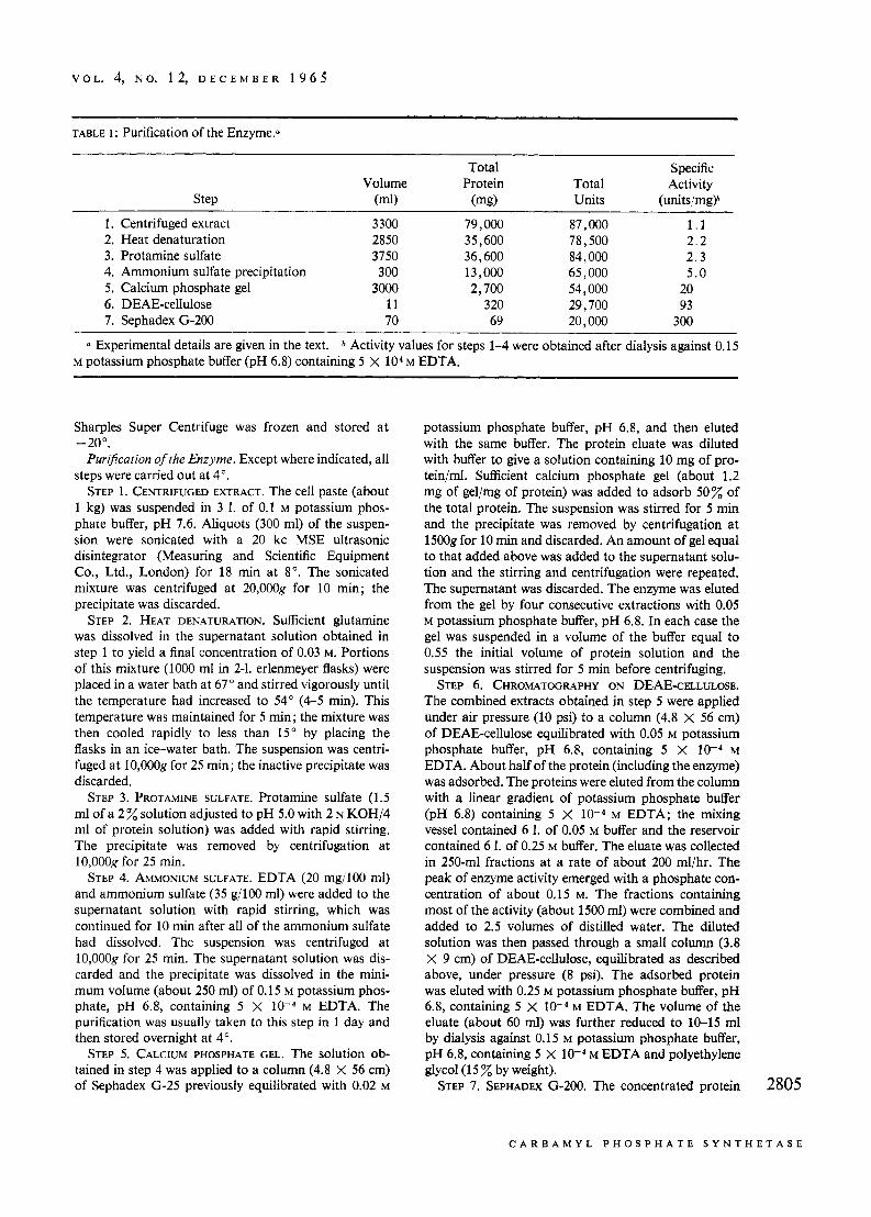
Puri3cation of the Enzyme. Except where indicated, all steps were carried out at 4".
STEP 1. CENTRIFUGED EXTRACT. The cell paste (about 1 kg) was suspended in 3 1. of 0.1 M potassium phos- phate buffer, pH 7.6. Aliquots (300 ml) of the suspen- sion were sonicated with a 20 kc MSE ultrasonic disintegrator (Measuring and Scientific Equipment Co., Ltd., London) for 18 rnin at 8". The sonicated mixture was centrifuged at 20,000g for 10 min; the precipitate was discarded.
STEP 2. HEAT DENATURATION. Sufficient glutamine was dissolved in the supernatant solution obtained in step 1 to yield a final concentration of 0.03 M. Portions of this mixture (1000 ml in 2-1. erlenmeyer flasks) were placed in a water bath at 67" and stirred vigorously until the temperature had increased to 54" (4-5 min). This temperature was maintained for 5 min; the mixture was then cooled rapidly to less than 15" by placing the flasks in an ice-water bath. The suspension was centri- fuged at 10,OOOg for 25 min; the inactive precipitate was discarded.
STEP 3. PROTAMINE SULFATE. Protamine sulfate (1.5 ml of a 2 solution adjusted to pH 5.0 with 2 N KOH/4 ml of protein solution) was added with rapid stirring. The precipitate was removed by centrifugation at 10,OOOg for 25 min.
STEP 4. AMMONIUM SULFATE. EDTA (20 mg/100 ml) and ammonium sulfate (35 g/l00 ml) were added to the supernatant solution with rapid stirring, which was continued for 10 rnin after all of the ammonium sulfate had dissolved. The suspension was centrifuged at 10,OOOg for 25 min. The supernatant solution was dis- carded and the precipitate was dissolved in the mini- mum volume (about 250 ml) of 0.15 M potassium phos- phate, pH 6.8, containing 5 X M EDTA. The purification was usually taken to this step in 1 day and then stored overnight at 4".
STEP 5. CALCIUM PHOSPHATE GEL. The solution ob- tained in step 4 was applied to a column (4.8 X 56 cm) of Sephadex G-25 previously equilibrated with 0.02 M
potassium phosphate buffer, pH 6.8, and then eluted with the same buffer. The protein eluate was diluted with buffer to give a solution containing 10 mg of pro- tein/ml. Sufficient calcium phosphate gel (about 1.2 mg of gel/mg of protein) was added to adsorb 50% of the total protein. The suspension was stirred for 5 min and the precipitate was removed by centrifugation at 15008 for 10 min and discarded. An amount of gel equal to that added above was added to the supernatant solu- tion and the stirring and centrifugation were repeated. The supernatant was discarded. The enzyme was eluted from the gel by four consecutive extractions with 0.05 M potassium phosphate buffer, pH 6.8. In each case the gel was suspended in a volume of the buffer equal to 0.55 the initial volume of protein solution and the suspension was stirred for 5 rnin before centrifuging.
STEP 6. CHROMATOGRAPHY ON DEAE-CELLULOSE. The combined extracts obtained in step 5 were applied under air pressure (10 psi) to a column (4.8 X 56 cm) of DEAE-cellulose equilibrated with 0.05 M potassium phosphate buffer, pH 6.8, containing 5 X M EDTA. About half of the protein (including the enzyme) was adsorbed. The proteins were eluted from the column with a linear gradient of potassium phosphate buffer (pH 6.8) containing 5 X M EDTA; the mixing vessel contained 6 1. of 0.05 M buffer and the reservoir contained 6 1. of 0.25 M buffer. The eluate was collected in 250-ml fractions at a rate of about 200 ml/hr. The peak of enzyme activity emerged with a phosphate con- centration of about 0.15 M. The fractions containing most of the activity (about 1500 ml) were combined and added to 2.5 volumes of distilled water. The diluted solution was then passed through a small column (3.8 X 9 cm) of DEAE-cellulose, equilibrated as described above, under pressure (8 psi). The adsorbed protein was eluted with 0.25 M potassium phosphate buffer, pH 6.8, containing 5 X M EDTA. The volume of the eluate (about 60 ml) was further reduced to 10-15 ml by dialysis against 0.15 M potassium phosphate buffer, pH 6.8, containing 5 X M EDTA and polyethylene glycol (1 5 % by weight).
STEP 7. SEPHADEX G-200. The concentrated protein 2805
C A R B A M Y L P H O S P H A T E S Y N T H E T A S E
B I O C H E M I S T R Y
solution was applied to a column (5 X 80 cm) of Sepha- dex G-200 equilibrated with 0.15 M potassium phos- phate buffer, pH 6.8, containing 5 X M EDTA and eluted with the same buffer at a flow rate of about 20 ml/hr; 5-ml fractions were collected. The peak of en- zyme activity emerged from the column when the volume of eluate was 3 5 x of the column volume. The specific activity was usually constant in the fractions that contained most of the activity. Solutions containing enzyme were frozen in a Dry Ice-acetone bath and stored at - 20".
Table I gives a summary of the purification procedure. Preliminary experiments with disk electrophoresis on polyacrylamide gel at pH 8.3 have shown that the enzyme preparation contains one minor protein con- taminant. The properties of the purified enzyme will be the subject of a later communication. All studies re- ported in this paper were carried out with enzyme processed through the final purification step.
Results
Stoichiometry. The data given in Table I1 (expt 1)
TABLE 11: Stoichiometry of the Reacti0n.a
Carbamyl ADP Glutamate Phosphate
Expt Time Formed Formed Formed No. (min) (pmoles) (pmoles) (pmoles)
a Experiment 1 : The reaction mixtures contained ATP (10 pmoles), NaHCO, (10 pmoles), MgC12 (20 pmoles), IC-L-glutamine (1.37 pmoles; 500,000 counts/min), potassium phosphate buffer (25 pmoles; pH 7.8), and enzyme (0.18 mg) in a final volume of 0.5 ml; 22". Experiment 2 : The reaction mixtures contained ATP (10 pmoles), N a H 1 F 0 3 (10 pmoles; 2 X lo5 counts/ min), MgClz (10 pmoles), NH,CI (50 pmoles), potassium phosphate buffer (25 pmoles; pH 7.8), and enzyme (0.6 mg) in a final volume of 0.5 ml; 22". At the indicated time intervals 0.1 rnl of the reaction mixtures was added to 0.3 ml of cold 95 ethanol to stop the reaction. An- alyses for ADP and lF-glutamate were carried out as described under Methods. In expt 1, carbamyl phos- phate was determined by conversion to citrulline with ornithine transcarbamylase. In expt 2,lC-carbamyl phosphate was determined as 'C-urea, as described under Methods. 2806
show that within experimental error ADP, 14C-gluta- mate, and carbamyl phosphate are formed in a ratio of 2 : 1 : 1, respectively, and that the reaction proceeds virtually to completion. Similarly, when the reaction was carried out with NHICl in place of glutamine, ADP and carbamyl phosphate were formed initially in a ratio of 2 : 1, respectively (expt 2).
Evidence for Enzyme-Bound Activured Curbon Di- oxide. When a relatively large amount of enzyme was preincubated with H 1 F 0 3 - and ATP and then mixed with a solution containing glutamine and a large excess of unlabeled HC03- the IF-carbamyl phosphate that was formed contained considerably more radioactivity than could be accounted for if it is assumed that all of the radioactive HC03- equilibrated with the unlabeled HC03-, and that all of the glutamine present was utilized (Table 111, expt 1). The possibility that the
TABLE III: Evidence for Enzymatic Activation of COZ.
a The reaction mixtures (final volume, 0.2 ml) con- tained enzyme (2.6 mg, 0.1 ml), ATP (2.13 pmoles), MgClz (2.13 pmoles), N a H l F 0 3 (0.44 pmole; 8 X lo5 counts/min), KHC03 (1 50 pmoles), L-glutamine (0.075 pmole), and potassium phosphate buffer (7.5 pmoles; pH 7.5). The components given in the first set of paren- theses were mixed with phosphate buffer and MgCL and incubated for 30 sec at 26". Then the components given in the second set of parentheses were added to- gether; after 10 sec the reaction was stopped by adding 0.2 ml of a solution containing 0.3 N ",OH and 1.3 N KOH followed by 0.2 ml of 0.001 M carbamyl phos- phate. lC-Carbamyl phosphate was determined as IC- urea as described under Methods. b The solution con- taining ",OH and KOH was added before adding the components given in the second set of parentheses.
excess radioactivity was a result of synthesis of a small amount of F-carbamyl phosphate during preincuba- tion was excluded by adding base before adding the solution containing glutamine and HC03- (expt 4). Preincubation of the enzyme with H 1 T 0 3 - in the ab- sence of ATP, or with ATP followed by HCO3- + H1FO3-, gave the amount of IF-carbamyl phosphate
P A U L M. A N D E K S O N A N D A L T O N M ~ I Y ~ E K
V O L . 4, N O . 1 2 , D E C E M B E R 196 .5
expected if the labeled HC03- equilibrated with the unlabeled HCO3- before reaction with glutamine.
These results indicate that the radioactive HC03- becomes bound to the enzyme during preincubation in a reaction that requires ATP, and that the binding is of such a nature as to permit the bound H 1 C 0 3 - to be immediately available for reaction with glutamine. According to this interpretation the l4C-carbamyl phos- phate in expt 1 contains a large excess of radioactivity because the unlabeled HC03- does not equilibrate with the radioactive enzyme-bound intermediate formed during preincubation.
Evidence for Cleuvuge of AT”P in the Absence of Glutumine. As indicated in Table IV, when a relatively large amount of enzyme was preincubated with AT32P and HC03- followed by addition of a mixture of gluta- mine and a large excess of ATP, the supernatant solu- tion obtained from the deproteinized reaction mixture after treatment with charcoal (to remove AT”P and
TABLE IV: Evidence for Cleavage of ATP in Absence of Glutamine.
The reaction mixtures (final volume, 0.4 ml) con- tained enzyme (5 mg, 0.16 mi), potassium phosphate buffer (16 pmoles, pH 7.8), NaHC03 (4 pmoles), A F P (0.8pmole, 244,00Ocounts/min), ATP(80 pmoles), L-glutamine (0.4 pmole), and MgCL in concentration equimolar with ATP and/or AT32P. The components given in the first set of parentheses were mixed with phosphate buffer and MgCh and incubated for 30 sec at 4”. Then the components given in the second set of parentheses plus MgC12 were added together; after 10 sec, the reaction was stopped by adding 0.6 ml of 0.5 N HClO, followed by 0.1 ml of a solution containing 0.1 M
carbamyl phosphate and 0.05 M potassium phosphate buffer, pH 7.8. The suspension was centrifuged immedi- ately and a 0.85-ml portion of the supernatant solution was neutralized with 0.25 ml of 0.9 N KOH. After centrifugation to remove KCI04, the nucleotides were removed by mixing with 5.15 ml of a suspension con- taining 540 mg of activated charcoal. After centrifuga- tion, the radioactivity in the supernatant was deter- mined. bPerchloric acid and carbamyl phosphate were added before adding the components given in the second set of parentheses.
-.
AD 32P) contained more radioactivity than would be expected if AT 32P and ATP had equilibrated before reaction with glutamine (expt 1). Thus, much less 32P
was found when AT32P, ATP, and glutamine were added to enzyme at the same time (expt 2). Paper elec- trophoresis of the supernatant solution obtained after charcoal treatment in expt 1 carried out as described under Methods gave a clean separation of inorganic phosphate from carbamyl phosphate; in this experiment more than 85% of the radioactivity was found in the inorganic phosphate. Control studies have shown that carbamyl 32P-phosphate can be recovered in satisfactory yield under these conditions.
The 32P found in the supernatant solution in expt 1 could conceivably result from synthesis of carbamyl phosphate during preincubation if some ammonia were present or it could arise if the enzyme preparation con- tains ATPase. The first possibility is remote since electrophoresis of the supernatant solution obtained after charcoal treatment in expt 3 showed that less than 5% of the total radioactivity was present as carbamyl 32P-phosphate; such carbamyl phosphate formation would give about equal amounts of carbamyl 32P-
phosphate and S2Pi. If the cleavage of AT32P during preincubation is simply the result of contaminating ATPase activity, the ADP concentration in the pre- incubation mixture should increase linearly with time. However, as shown in Table V the ADP formed is pro-
TABLE v: Formation of ADP in Absence of Glutamine:
Time (set)
ADP (mwnoles)
30 60
120
38 44 44
a The reaction mixture contained enzyme (5 mg), potassium phosphate buffer (16 pmoles, pH 7.8), ATP (0.8 pmole), MgC12 (0.8 pmole), and NaHC03 (4 pmoles) in a total volume of 0.2 ml; 4”. Aliquots of the reaction mixture were added to equal volumes of 0.5 M HC104, and after centrifugation of the precipitated protein, ADP was determined as described under Methods.
duced in an initial “burst,” and a significant increase in ADP does not occur after 30 sec. The finding of increased 32P in expt 1 and 3 (Table IV) and the data on ADP formation are consistent with formation of an activated enzyme-bound intermediate associated with cleavage of ATP to ADP.
Discussion
The studies on the stoichiometry of the reaction show that 2 molecules of ATP are utilized in the synthesis of 2807
C A R B A M Y L P 11 0 S P I< A T E S Y N T H E T A S E
B I O C H E M I S T R Y
1 molecule of carbamyl phosphate. The data also indicate that the reaction proceeds virtually to com- pletion. The same stoichiometry is obtained when NH&1 is substituted for glutamine. Thus, the reaction catalyzed by this enzyme is not analogous to that catalyzed by carbamate kinase, but rather to the reac- tion catalyzed by liver carbamyl phosphate synthetase. The E. coli enzyme differs from the liver enzyme in that glutamine and ammonia are both active substrates and that N-acetylglutamate does not affect the rate of the reaction (Anderson and Meister, 1965).
The data obtained in the pulse-labeling experiment with H14C03- (Table 111) provide clear evidence that the first step in the synthesis of carbamyl phosphate is formation of an enzyme-bound activated form of carbon dioxide and that this reaction requires ATP. Although the chemical nature of the activated carbon dioxide cannot be established from these experiments, the results are consistent with the following sequence of reactions.
enzyme + ATP + COz enzyme-[-OCOOP032-l f ADP (6)
enzyme-[-OCOOPO32-] + glutamine e enzyme- [”zCOO-] + Pi + glutamate (7)
enzyme-[NHzCOO-] + ATP enzyme + ADP + NHzCOOPO~~- (8)
According to this mechanism, incubation of the enzyme with ATP and HC03- in the absence of glutamine or ammonia results in cleavage of ATP to give ADP and enzyme-bound carbonate phosphate anhydride (reac- tion 6). The data show that a “burst” of ADP is pro- duced under these conditions (Table V). Although the findings do not unequivocally exclude an intermediate involving formation of free inorganic phosphate, as proposed by Metzenberg et al. (1958), it might be ex- pected that the formation of such an intermediate would be accompanied by rapid exchange between AT32P and the unlabeled phosphate buffer during preincuba- tion with enzyme and HCOI- in the absence of gluta- mine; thus, there would be extensive formation of s2Pi, much greater than actually observed. Although it is not yet possible to obtain accurate values for binding of HCO3- and phosphate to the enzyme from the present experiments, estimates of such values based on the “extra” formation of l4C and 32P products (Tables I11 and IV) and the values observed for forma- tion of ADP are of about the same order. Additional studies along these lines will be undertaken.
According to the proposed mechanism, if the mole- cule of ATP which is used for phosphorylation of carbamate is not bound to the enzyme until the inter- mediate has reacted with glutamine, i.e., it is not bound during preincubation as described in expt 1, Table IV,
4 Similar schemes have been proposed earlier (Meister, 1965; 2808 Jones, 1965).
then the radioactivity obtained in the supernatant solu- tion should be found mainly in the Pi rather than in carbamyl phosphate. The experiments described here show that there is indeed much less radioactivity in the carbamyl phosphate than in the Pi formed. It is antici- pated that additional studies and perhaps special tech- niques will be required to characterize the enzyme- bound intermediate as carbonate phosphate anhydride. However, the present findings are consistent with reac- tions 6-8, but do not unequivocally exclude other types of activated intermediates,
According to the proposal put forth here, the enzyme carries out two steps which are analogous to simple kinase reactions. Thus, carbonic acid is phosphorylated in the first step and carbamic acid is phosphorylated in the last step. In this connection the findings by Raijman and Grisolia (1964) that liver carbamyl phos- phate synthetase phosphorylates formate and acetate in reactions involving cleavage of 1 molecule of ATP are of considerable interest. It would be of importance to do studies of the type described here on the liver en- zyme; it seems probable that similar mechanisms are in~olved .~ It would also be of interest to carry out such experiments with other enzymes capable of activating carbon dioxide, e.g., the biotin-containing carboxylases.
References
Allen, C. M., and Jones, M. E. (1964), Biochemistry 3, 1238.
Anderson, E. H. (1946), Proc. Natl. Acad. Sci. U. S. 32, 120.
Anderson, P. M., and Meister, A. (1965), Abstracts of Papers, 150th National Meeting of the American Chemical Society, Atlantic City, N. J., Sept. 1965, p. 35c.
Cohen, P. P. (1962), Enzymes 6,477. Gerhart, J. C., and Pardee, A. B. (1962), J. Biol. Chem.
Jones, M. E. (1965), Ann. Rev. Biochem. 34, 381. Jones, M. E., and Lipmann, F. (1960), Proc. Natl.
Acad. Sci. U. S. 46, 1194. Kalman, S. M., Duffield, P. H., and Brzozowski, T.
(1965), Biochem. Biophys. Res. Commun. 18, 530. Layne, E. (1957), Methods Enzymol. 3, 447. Leuthardt, F., and Testa, E. (1951), Helv. Chim. Acta 24,
Levenberg, B. (1962), J. Biol. Chem. 237, 2590. Levin, R., and Brauer, R. W. (1951), J. Lab. Clin. Med.
Meister, A. (1965), Biochemistry of the Amino Acids,
Metzenberg, R. L., Marshall, M., and Cohen, P. P.
237, 891.
931.
38,474.
Vol. 11, New York, N. Y., Academic, p. 692.
(1 958), J . Biol. Chem. 233, 1560.
Although it has been reported that the liver enzyme does not utilize glutamine in place of ammonia, it is possible that theaffinity or activity of the enzyme for glutamine is altered in the course of purification. It might be of interest to reexamine the question of the utilization of glutamine by liver carbamyl phosphate synthe- tase.
P A U L M. A N D E R S O N A N D A L T O N M E I S T E R
V O L . 4, N O . 12 , D E C E M B E R 1 9 6 5
Peterson, E. A,, and Sober, H. A. (1962), Methods
PiCrard, A., and Wiame, J. M. (1964), Biochern. Biopltys.
Raijman, L., and Grisolia, S. (1964), J . B i d . Chem. 239,
1272.
Biophys. 96, 398.
29,190.
Enzymol. 5, 3.
Res. Commun. 15, 76.
Rogers, P., and Novelli, G. D. (1962), Arch. Biochem.
Singer, T. P., and Kearney, E. B. (1950), Arch. Biochem.
A Kinetic Study of Nucleotide Interactions with Pyruvate Kinase*
Kent M. Plowman? and A. R. Krallz
ABSTRACT: The apparent broad specificity of the enzyme pyruvate kinase for the nucleoside diphosphate sub- strate has been re-examined. Nucleoside diphospho- kinase activity was found to be present but was active only when exogenous adenosine triphosphate was present.
The apparent K, and V,,, values were determined at several pH values between 6.0 and 9.0 for a num- ber of nucleoside diphosphates. A doubly ionized phosphoryl group on the nucleotides andlor an imid-
A broad specificity for the nucleoside diphosphate substrate in the pyruvate kinase catalyzed reaction has been reported previously (Strominger, 1955; Tietz and Ochoa, 1958; Adam, 1961). However, doubt has been cast on this specificity by Davidson (1959), who claimed that the apparent rate obtained, using pyrimidine nucleoside diphosphates, resulted from contamination of the reaction mixture with nucleoside diphospho- kinase and adenosine phosphates. An upper limit for nucleoside diphosphokinase activity under varying con- ditions will be presented. Evidence will be given that two regions of the nucleotide are bound, that the phos- phate must be doubly ionized for binding, and that an imidazole and an a-amino group on the enzyme are involved in binding.
* From the Department of Biochemistry, University of Miami School of Medicine, Miami, Florida. Received August 2, 1965. This investigation was supported in part by the Julia Meador Palmer Memorial Institutional Grant of the American Cancer Society.
t Work done in partial fulfillment of the requirements for the Ph.D. degree and supported by a predoctoral fellowship (GM- 24602) from the U. S . Public Health Service. Present address: Department of Biochemistry, University of Wisconsin, Madison 6, Wis.
$ Supported by a career development award (MH-13947) from the National Institute of Mental Health, U. S . Public Health Service. Present address: Departments of Biochemistry and Psychiatry, University of North Carolina School of Medicine, Chapel Hill, N. C.
azole group on the enzyme appear to be essential for nucleotide binding. Both the apparent K, and V,,, values decrease between pH 7.5 and 9.0, which sug- gests the involvement of an ionized a-amino group in the reaction. The results of the determinations of kinetic parameters in the pH range of 7-8 are discussed in rela- tion to a proposed mechanism in which the nucleoside diphosphate is bound to the enzyme on two regions in an obligatory order, with the &phosphoryl group binding first and the nucleoside portion binding second.
Methods
Materials. The sodium salts of PEP,' AMP, ADP, ATP, GDP, IDP, UDP, CDP, dADP, dCDP, NADH, and N A D P ; crystalline lactic dehydrogenase, type I1 ; and crystalline yeast hexokinase were purchased from the Sigma Chemical Co. The crystalline glucose 6- phosphate dehydrogenase was prepared by C . F. Boeh- ringer und Soehn. The [32P]ATP was prepared by mito- chondrial phosphorylation of ADP by [ 32P]inorganic phosphate. The [32P]PEP was labeled by an exchange reaction using ["PIATP and pyruvate kinase by a method to be published elsewhere. Product separation was effected in both instances by chromatography on Dowex-1 chloride using the method of Khym and Cohn (1953).
Enzyme Preparation. Pyruvate kinase was prepared from rabbit muscle by the "fluorokinase" method of Tietz and Ochoa (1958). Two different preparations were used in this work. One batch had a specific ac- tivity of 60 pmoles of pyruvate formed/min/mg of protein at pH 7.0 and 25.0" and is referred to later
~
Abbreviations used in this work: PEP, phosphoenolypyru- vate; AMP, ADP, ATP, monophosphate, diphosphate, and triphosphate of adenosine; GDP, IDP, UDP, CDP, dADP, dCDP, guanosine, inosine, uridine, cytidine, deoxyadenosine, and deoxycytidine diphosphates; NADH, reduced nicotinamide- adenine dinucleotide; NADP+, oxidized nicotinamide-adenine dinucleotide phosphate. 2809
N U C L E O T I D E I N T E R A C T I O N S W I T H P Y R U V A T E K I N A S E