Evidence in Context Issue: PET/CT Programs Released: March 2009 Health research — synthesized and contextualized for use in Newfoundland and Labrador The Development of a PET/CT Program in Newfoundland and Labrador S. Demeter, S. Bornstein, J. Butler, B. Cramer, P. Hollett, L. Jones
Transcript
Evidence in Context
Issue: PET/CT Programs
Released: March 2009
Health research — synthesized and contextualized for use in Newfoundland and Labrador
The Development of a PET/CT Program
in Newfoundland and LabradorS. Demeter, S. Bornstein, J. Butler, B. Cramer, P. Hollett, L. Jones
Contextualized Health Research Synthesis: PET/CT Programs NLCAHR March 2009
2
Disclaimer
This contextualized health research synthesis report was prepared by the Newfoundland and Labrador Centre for Applied Health Research (NLCAHR), Memorial University. It was developed through the analysis, interpretation and synthesis of scientific research and/or health technology assessments conducted by other parties. It also incorporates selected information provided by experts in the subject areas and synthesis methodologies. This document may not fully reflect all the scientific evidence available at the time this report was prepared. Other relevant scientific findings may have been reported since completion of this synthesis report.
Memorial University, NLCAHR, and the CHRSP project team make no warranty, express or implied, nor assume any legal liability or responsibility for the accuracy, completeness, or usefulness of any information, data, product, or process disclosed in this report. Conclusions drawn from, or actions undertaken on the basis of, information included in this report are the sole responsibility of the user.
This report is the property of the Newfoundland and Labrador Centre for Applied Health Research (NLCAHR). Reproduction of this document for non-commercial purposes is permitted provided proper credit is given to NLCAHR.
Cite as: Demeter, S., Bornstein, S., Butler, J., Cramer, B., Hollett, P., & Jones, L. (2009). The development of a PET/CT program in Newfoundland and Labrador. St. John’s, NL: Newfoundland and Labrador Centre for Applied Health Research, Memorial University.
NLCAHR March 2009 Contextualized Health Research Synthesis: PET/CT Programs
3
About NLCAHRThe Newfoundland and Labrador Centre for Applied Health Research, established in 1999, contributes to the effectiveness of the health and community services system of the province and the physical, social, and psychological wellbeing of the population. NLCAHR accomplishes this mandate by building capacity in applied health research, supporting high-quality research, and fostering more effective use of research evidence by decision makers and policy makers in the province’s health system.
About the Contextualized Health Research Synthesis ProgramIn 2007, NLCAHR launched the Contextualized Health Research Synthesis Program (CHRSP) to provide research evidence to help guide decision makers in the provincial health system on issues of pressing interest to Newfoundland and Labrador.
CHRSP does not conduct original research, but rather analyzes the findings of high-level research (systematic reviews, meta-analyses and health technology assessments) that have already been done on the issue in question. The findings of these studies are synthesized and are subjected to a systematic process of ‘contextualization’: they are analyzed in terms of their applicability to the conditions and capacities of the unique context of Newfoundland and Labrador.
Our contextual analysis includes assessment of the specific forms that the issue takes in this province as well as the applicability of proposed solutions and methods to locally available physical and human resources, cultural conditions and financial capacities. CHRSP uses a combination of external experts and local networks to carry out and contextualize the research synthesis and to facilitate the uptake of the results by research users.
CHRSP focuses on three types of projects: health services/health policy projects; health technology assessment (HTA) projects; and projects that combine the two to examine processes for the organization or delivery of care involving a health technology.
About CADTHThe Canadian Agency for Drugs and Technologies in Health is a national body that provides Canada’s federal, provincial and territorial health system decision makers with credible, impartial advice and evidence-based information about the effectiveness and efficiency of drugs and other health technologies. Established in 1989, CADTH is one of Canada’s leading sources of health technology information and a significant, trusted contributor to the effectiveness and efficiency of Canada’s health system.
Who Should Read This Report?This report begins with the assumption that the Government of Newfoundland and Labrador will be following up on its announced decision to purchase a PET scanner. The report is intended to inform and assist those in the provincial government and the healthcare system in making decisions about the acquisition of this new technology and the organization and delivery of the new imaging services it can support. The report is specifically focused on the province of Newfoundland and Labrador, Canada, but decision makers from other jurisdictions may also find the content helpful. The full report includes explanations of terms and techniques so that a specialized medical background in the field is not needed to understand the content.
Contextualized Health Research Synthesis: PET/CT Programs NLCAHR March 2009
4
The Research Team
The Development of a PET/CT Program in Newfoundland and Labrador
The
Research Team
CHRSP Research Team: PET/CT
Dr. Sandor Demeter Section Head, Nuclear Medicine andCo-Director, Winnipeg PET/CT Program, Health Sciences CentreAssistant Professor, Department of Radiology, University of Manitoba (Team Leader)
Dr. Stephen Bornstein Director, NLCAHR Memorial University (Program Coordinator, CHRSP)
Janice Butler Research Officer, NLCAHR (Project Coordinator, CHRSP)
Dr. Benvon Cramer Chair, Discipline of RadiologyFaculty of Medicine, Memorial University(Local Scientific Expert)
Dr. Peter Hollett Clinical Associate Professor of Radiology (Nuclear Medicine)Faculty of Medicine, Memorial University (Local Scientific Expert)
Louise Jones Interim President and Chief Executive OfficerEastern Regional Health Authority, St. John’s(Health System Expert)
Project Consultants: PET/CT
Dr. David Barnes, Associate Professor, Department of Diagnostic Radiology Faculty of Medicine, Dalhousie University
Dr. David Buckley, Assistant Professor of Pediatrics Faculty of Medicine, Memorial University
Dr. Steve Burrell, Assistant Professor, Department of Diagnostic Radiology Faculty of Medicine, Dalhousie University
Dr. David Craig, Associate Professor of Psychiatry Faculty of Medicine, Memorial University
Dr. Blair Fleming, Assistant Director, Medical Services Department of Health and Community Services Government of Newfoundland and Labrador
Dr. Jack Hand, Clinical Associate Professor of Pediatrics Faculty of Medicine, Memorial University
Dr. Ed Hunt, Medical Consultant (Retired), Medical Services Branch Department of Health and Community Services Government of Newfoundland and Labrador
Dr. Don Juzwishin, Principal and Consultant, Juzwishin Consulting Inc. Former CEO, Health Council of Canada
Dr. Edward Kendall, Professor of Radiology (Medical Physics) Faculty of Medicine, Memorial University
Wanda Legge, Director, Policy Development, Policy and Planning Branch Department of Health and Community Services Government of Newfoundland and Labrador
Cindy Mosher, Liaison Officer for NL Canadian Agency for Drugs and Technologies in Health
Dr. Daria O’Reilly, Assistant Professor, Clinical Epidemiology and Biostatistics PATH, McMaster University
Dr. Andrew Ross, Associate Professor, Department of Diagnostic Radiology Faculty of Medicine, Dalhousie University
Dr. David Saltman, Chair of Oncology Faculty of Medicine, Memorial University
CHRSP Expert Advisors: PET/CT
Dr. Brendan Barrett Professor of MedicineDivision of Clinical Epidemiology, Memorial University (Special Advisor, CHRSP)
Dr. Alexander McEwan Director, Oncologic Imaging, Cross Cancer Institute Professor and Chair, Department of Oncology Faculty of Medicine and Dentistry, University of Alberta (External Reviewer)
NLCAHR March 2009 Contextualized Health Research Synthesis: PET/CT Programs
5
ContentsThe Research Team, Advisors & Consultants 4Glossary of Acronyms 6
The Research Question 8
Overview and BackgroundWhat is PET/CT Imaging? 9How does PET/CT Imaging work? 9Regulatory Issues 10PET and PET/CT Imaging in Canada 12PET/CT Imaging Research 12NL Demographics and Population Trends 14Estimating the Future Demand for FDG PET/CT Scans in NL 14
Review of the LiteratureWhat did we look for? 16What did we fi nd? 16
Clinical eff ectiveness of PET/CT scanning 16Will FDG PET/CT imaging replace conventional imaging? 18Cost-eff ectiveness of PET/CT scanning 18Other benefi ts of PET/CT scanning 18
ContextualizationComponents of a PET/CT Program 19
Where in the province should the PET/CT program be located? 1. 19Physical space and design considerations 2. 19Who will be permitted to order a PET scan? 3. 20Human resource requirements for a PET/CT program 4. 20Radiation safety issues 5. 24Service and equipment maintenance 6. 24
The Cyclotron Question The advantages of early development of a cyclotron program 1. 24The costs of a local cyclotron 2. 26Human resource requirements for a cyclotron program 3. 26
Summary of Human Resource Requirements 28Timelines and Sequencing 28
ConclusionsUses of PET/CT 30Issues for Consideration by Policy Makers 30
AppendicesF1. actors of relevance in contextualization 32Estimated number of FDG PET/CT scans per year for NL 32. 3Literature search strategy 343. Review of the literature 354.
Defi nitions of key terms a. 41Economic reviews b. 42
NCCN categories of evidence and consensus 435. Royal College of Radiologists AND Canadian Association of Radiologists categories of evidence and consensus 46. 4Baseline cost estimates for cyclotron/FDG options net present value economic analysis 47. 5Net present value analysis 468.
References 47
Contextualized Health Research Synthesis: PET/CT Programs NLCAHR March 2009
6
Glossary of AcronymsAB Alberta
ACR American College of Radiology
AD Alzheimer’s dementia
ALARA as low as reasonably achievable (occupational, patient or public radiation doses)
BC British Columbia
BCIT British Columbia Institute of Technology
BSc Bachelor of Sciences
CADTH Canadian Agency for Drugs and Technologies in Health
CAMRT Canadian Association of Medical Radiation Technologists
CANM Canadian Association of Nuclear Medicine
CAR Canadian Association of Radiologists
CFI Canada Foundation for Innovation
CHRSP Contextualized Health Research Synthesis Program
CI Confidence intervals
CME continuing medical education
CMS Centers for Medicare and Medicaid Services (USA)
CNSC Canadian Nuclear Safety Commission
COG Children’s Oncology Group
CPG Clinical Practice Guideline
CSNM Canadian Society of Nuclear Medicine
CT computed tomography
CTA clinical trials application
DHCS Department of Health and Community Services, NL Government
DNA deoxyribonucleic acid
ECD 99mTc Ethyl Cysteinate dimmer (a NM Brain imaging agent)
ECG Electrocardiogram
EEG electroencephalogram
EFT effective full time equivalent (1 EFT= full time & 0.5 EFT= half time)
FRCPC Fellow of the Royal College of Physicians and Surgeons of Canada
GA general anaesthetic
GIST gastrointestinal stromal tumour
GMP good manufacturing practice
GWL Great West Life (i.e. context of the Winnipeg GWL PET/CT program)
HC Health Canada
HL Hodgkin’s lymphoma
HSC Health Sciences Centre
HTA Health Technology Assessment
HTU Health Technology Update
NLCAHR March 2009 Contextualized Health Research Synthesis: PET/CT Programs
7
ICER incremental cost effectiveness ratio
ICES Institute of Clinical Evaluative Sciences (Ontario)
MB Manitoba
MRI magnetic resonance imaging
MSK musculoskeletal
NCCN National Comprehensive Cancer Network
NHL non-Hodgkin’s lymphoma
NHS National Health Service
NICE National Institute for Health and Clinical Excellence (UK)
NIH National Institute of Health
NL Newfoundland and Labrador
NLCAHR Newfoundland and Labrador Centre for Applied Health Research
NM Nuclear Medicine
NMTs Nuclear Medicine Technologists
NOPR National Oncological PET Registry (USA)
NPV net present value
NSCLC non-small cell lung cancer
ON Ontario
PERs positron-emitting radiopharmaceuticals
PET positron emission tomography
PLUS PET in Lung Cancer Staging
QALY quality adjusted life year
QC Quebec
RCR (UK) Royal College of Radiologists (UK)
RCT randomized controlled trial
RFP Request for proposal
RHA Regional Health Authority
RNA ribonucleic acid
RSO radiation safety officer
SAP Special Access Program (Health Canada)
SCLC small cell lung cancer
SIAM Siemens Institute for Advanced Medicine
SNM Society of Nuclear Medicine (North American)
SPECT single photon emission computed tomography
SPN solitary pulmonary nodules
TDG Transport of Dangerous Goods
UK United Kingdom
US ultrasound
USA United States of America
Contextualized Health Research Synthesis: PET/CT Programs NLCAHR March 2009
8
The Research QuestionThis report was initially designed to examine research-based evidence about whether the province of NL should acquire a Positron Emission Tomography (PET) scanner. At various stages throughout the project we were informed of developments that required us to reformulate our research question. First, we learned that the Government of NL had actually made a commitment to purchase a PET scanner. Next, it rapidly became clear that current trends in technology development have moved towards hybrid models of diagnostic imaging technologies; hence, virtually all new PET scanners come bundled with a Computed Tomography (CT) scanner in a device commonly referred to as a PET/CT scanner. Since hybrid models are the acceptable standard for clinical use and fi t the North American vendor market, they became the focus for this project.
Later in the course of our work, we were informed that the Government had determined that it would also need to purchase a cyclotron rather than depending on radioactive isotopes fl own in from elsewhere. In addition, we were told that a decision had been reached to locate these two pieces of equipment in St. John’s. Accordingly, rather than focus on our original question (PET or not?) or on the obvious related questions (should we also acquire a cyclotron? where should the new equipment be located?), we have focused on a set of ancillary, but still very important, issues:
where, within the St. John’s area, should the new equipment be located? • for what clinical indications is PET currently the best choice in terms of clinical effectiveness and cost-effectiveness? • what other indications are emerging for which it makes sense to plan for PET use? • what is the optimal method for organizing and managing access to PET scans?• what are the advantages of early development of a cyclotron program and the challenges of operating without one?• what are the requirements of a PET/CT scanning program in terms of professional competencies for physicians and • technologists, training, fi nancing and space? and, what is the optimal sequencing of the activities required for effective acquisition, installation, licensing and start-up • of a PET/CT scanning program?
All of these questions have been considered by means of a systematic reading and synthesis of the literature plus input concerning the recent experience of two other Canadian jurisdictions (Manitoba and Nova Scotia), all with a careful eye to the specifi c context of Newfoundland and Labrador.
There are currently three broad categories of accepted clinical application for PET/CT technology: (a) in oncology imaging, to help determine how extensive a cancer is, whether it has responded to therapy, and whether it has recurred; (b) in brain imaging, for select patients with seizure disorders and for the early detection of dementia; and (c) in cardiac imaging, to assess the viability of heart muscle.
At present, patients in Newfoundland and Labrador who require a PET scan must travel out of the province to either Alberta or Quebec at a substantial cost to the provincial health system as well as to the patient and his/her family. On average, on the basis of a bilateral agreement between the Department of Health and Community Services and the out-of-province provider facility, the province pays $1,250 per scan. There is no reciprocal billing for PET scanning. Patients and families pay out-of-pocket travel and accommodation expenses, a portion of which may be reimbursed through the Medical Transportation Assistance Program, depending on eligibility. While the recorded number of NL residents who received PET scans in the past has been relatively small (fewer than 35 patients per year since 2004), these numbers may not represent the true size of the population that might have benefi ted from PET, nor provide a reliable guide to future demand. This report’s lead author, who was involved in establishing the Winnipeg PET/CT program, believes that the number of patients currently being sent out of province greatly underestimates the number of scans that are likely to be performed when a PET/CT program is established in NL. The details of this analysis are explained in the report.
The purpose of this Contextualized Health Research Synthesis is to answer the following core research question:
Given the geographic, demographic, fi scal and political context of Newfoundland and Labrador, what is the most appropriate, effective, and effi cient way to operate a PET/CT program so that the population derives the maximum benefi t at the best possible cost?
NLCAHR March 2009 Contextualized Health Research Synthesis: PET/CT Programs
9
Overview and BackgroundWhat is PET/CT Imaging?PET (positron emission tomography) is a type of Nuclear Medicine imaging technology. PET technology has been in existence for decades and was originally dedicated to brain imaging. ‘Positron emission’ describes the physics behind the radioisotope component of the radiopharmaceuticals and ‘tomography’ signifi es that image ‘slices’ are produced as in CT scanning (cross-sectional imaging). PET scanners are now primarily sold as hybrid PET/CT units, which combine a PET and CT scanner into one unit.
Prior to hybrid PET/CT, physicians would ‘fuse’ PET images to CT or MRI (magnetic resonance imaging) images visually (i.e., look back and forth between the two sets of images to match the PET’s functional fi ndings to the CT or MRI anatomical fi ndings). Eventually computer software solutions evolved that ‘fuse’ different image data sets into a single image. With the advent of PET/CT units the sequential acquisition of CT and PET images allows for a high-fi delity fusion of PET and CT images by reducing patient movement between the PET and CT imaging studies. The fused images allow accurate simultaneous visualization of the function or physiology (in the PET element) and anatomy or structure (in the CT element). For example, after radiation therapy for certain types of cancer, such as lymphoma, it is not uncommon to have residual CT abnormalities that may represent either persistent active lymphoma or residual non-cancerous tissue such as scar tissue. The CT cannot always differentiate between these two conditions, but the PET scan generally can. Accurate fusion of PET and CT images enhances overall diagnostic accuracy.
Although the terms ‘PET’ and ‘PET/CT’ will be used interchangeably throughout this document, any research that was based on PET-only technology will be designated as such.
How does PET/CT Imaging work?The process of PET/CT imaging begins by injecting the patient with trace amounts of positron-emitting radiopharmaceuticals (PERs). Radiopharmaceuticals contain a radioactive isotope, which may be used by itself or as part of a synthesised target molecule. The unique feature of PERs is that they allow the radiolabeling of organic molecules (e.g. fats, sugars, proteins, hormones, parts of DNA and RNA, etc.), which are the ‘essential building blocks of life’.
PET allows true imaging of human physiological and biochemical processes. In comparison, CT and MRI primarily capture anatomy. PET exploits the fact that the human body handles radiolabeled organic molecules differently in healthy and diseased states. Because PET imaging is based on physiological processes at the molecular level it is sometimes called ‘molecular imaging’ (Valk et al., 2003).
Medical cyclotrons produce positron-emitting isotopes. These isotopes are used directly or are incorporated into other biological molecules (e.g., sugars, proteins, etc.) resulting in positron-emitting radiopharmaceuticals (PERs). This is a different process from the production of medical isotopes in nuclear reactors (e.g., Chalk River, Ontario) and the production of other so-called medical isotope parent-daughter generators. A cyclotron is a machine that accelerates charged particles in an outward spiralling pattern until the particle eventually collides with a specific target substance. The collision transforms the target substance into the desired positron-emitting medical isotope (Cherry et al., 2003).
Unlike the majority of other commonly used medical isotopes, positron-emitting isotopes generally have short half-lives ranging from minutes to a maximum of about two hours. The half-life is the time it takes an isotope to lose half of its radioactivity (i.e., a half-life can be considered as a form of shelf-life whereby at the end of one half-life only half of the original product is still usable). Radioactive oxygen, nitrogen, carbon and fluorine are the essential isotopes used to produce most PERs. They have half-lives of 2, 10, 20 and 110 minutes, respectively. Close proximity of PET scanners to a cyclotron and, in the case of the products with the shortest half-lives (20 minutes or less), an on-site cyclotron with a system for rapid delivery to the PET/CT imaging suite (e.g., a pneumatic tube transport system) is necessary to provide any clinical or basic science research utility. There are currently three broad clinical applications for PET/CT technology: oncology, brain and cardiac imaging. PET and PET/CT imaging have been used for oncology imaging in Europe for at least two decades and have been approved for various indications in the USA since the early–to mid–1990s. Radiolabeled glucose or fluorodeoxyglucose (FDG) is the primary PER currently used for oncology imaging. FDG has a half-life of 110 minutes. The basis of PET/CT imaging in
Contextualized Health Research Synthesis: PET/CT Programs NLCAHR March 2009
10
oncology is that many cancers require more energy (of which glucose is one form) than normal tissues. The body handles FDG in a very similar fashion to glucose except for a few differences that are beneficial for imaging purposes. For example, once FDG gets into a cell the normal enzyme pathways that get it out of the cell do not work as well on FDG as they do for normal glucose. Hence, FDG tends to accumulate in cells, and more so in cancer cells, which require more energy and have various mechanisms to increase glucose uptake (e.g., more glucose receptors on cell surfaces and an increased rate of glucose transport into cells) (Saha, 2004). The accumulation of FDG can be seen in the PET/CT images. Second, unlike glucose, which has to be elevated in the blood before it is passed into the urine, FDG is readily excreted into the urine without a threshold. This allows for a reduction in radiation dose to patients. It also reduces the background activity in tissues such as muscle or blood vessels that can make cancer lesions less obvious, thereby enhancing the detection of abnormal patterns of tissue uptake (Saha, 2004).
The primary role of FDG PET/CT imaging in oncology is in staging (to help determine how extensive a cancer is), determining response to therapy (e.g., surgery, chemotherapy, or radiation therapy), and assessing recurrence (whether or not a cancer has come back) (Valk, 2003).
Brain imaging with FDG PET/CT is limited to select patients with seizure disorders that are difficult to treat medically. Imaging patients’ brains between seizures, in combination and comparison to imaging their brains while they are experiencing a seizure (a separate type of Nuclear Medicine study) has been demonstrated to be relatively sensitive in anatomically localizing where in the brain the seizures start. This greatly assists surgical planning when partial brain resection is being considered as an anti-seizure therapy (Macapinlac, 2006).
FDG PET/CT imaging has also been demonstrated to be very sensitive in the early detection of dementia. There are promising new drug therapies that may slow the progression of dementia if it is detected early and, in the future, this may significantly increase the demand for PET/CT imaging for diagnosing dementia as early as possible (Macapinlac, 2006).
In cardiac care, the primary clinical role of FDG PET/CT is to assess myocardial viability (i.e., to differentiate ‘live’ or viable heart muscle from ‘dead’ or scarred/infarcted heart muscle). Heart muscle uses both fats and sugars for energy. When heart muscle is deprived of oxygen (i.e., ischemia) it preferentially uses sugars, which results in increased FDG uptake in ’ischemic’ heart muscle. In a relatively small number of cases, it is difficult to determine, with current routine imaging strategies, if a particular area of heart muscle is too damaged to benefit from therapies such as cardiac by-pass surgery or angioplasty. In these cases, determining the level of FDG uptake in the damaged heart muscle can help physicians to predict the chance of a favourable outcome after therapy and to determine whether the benefits of therapy outweigh the risks. There is also ongoing research assessing the use of PET/CT in assessing and accurately quantifying coronary blood flow (i.e., myocardial perfusion) to help detect areas that are under-perfused (i.e., myocardial ischemia) (Knuuti et al., 2008).
Regulatory IssuesThe Canadian regulatory environment for FDG and other PERs is complicated. Health Canada (HC) considers FDG, as is likely to be the case with any PERs developed in the future, as drugs rather than as general diagnostic imaging agents such as the contrast agents commonly used in CT and MRI. Hence, it requires clinical trials to prove both the safety and the clinical efficacy of PERs for specific indications. As of October 2008, HC has granted approval to five sites located in British Columbia, Alberta, Ontario and Quebec, for the use of FDG for specific oncology indications (as outlined in Table 1). FDG is also being used in a number of PET/CT centres (BC, AB, MB, ON and QC) as part of clinical trials to address its safety and efficacy for specific indications. For example, MB enrols patients as part of an Alberta Cancer Board/Edmonton Cross Cancer Institute Clinical Trials Application (CTA). In addition to the Heath Canada trials, Ontario is conducting provincially-based trials largely to determine if PET/CT scanning should be funded as a publicly insured service.
Table 1 illustrates the approved and clinical trial indications for PET/CT in Canada, USA, Europe and Australia. The specifi c indications (i.e., diagnosis, staging, response to therapy, restaging and prognostication) for each type of cancer vary considerably and are not specifi cally indicated in the table. The USA has signifi cantly broadened the number of cancer indications for FDG, essentially making it a general ‘oncology-imaging’ agent.
NLCAHR March 2009 Contextualized Health Research Synthesis: PET/CT Programs
11
Table 1: Approved International Indications for Clinical Use of FDG or Other PERs
Canada1 USA2 Europe Australia
Cancers
Brain – primary CTA YES2 YES
Breast YES YES
Cervix CTA YES2 YES
Colo-rectal YES YES YES YES
Esophagus CTA YES YES
Head and Neck CTA YES YES
Lung YES YES YES YES
Lymphoma CTA YES YES
Melanoma CTA YES YES
Ovary CTA YES2 YES
Stomach YES2 YES
Testicular CTA YES2
Thyroid CTA YES
Other Cancers
Cholangiocarcinoma
Kidney
Myeloma
Pancreas
Soft Tissue Sarcoma
Thymoma
Unknown Primary
CTA’sYES2
N/A
Non- Cancer
Cardiac – Perfusion (non-FDG tracers) CTA YES
Cardiac – Viability CTA YES
Brain – Dementia CTA YES
Brain – Seizure SAP YES
Other
Brain -Depression
Brain -Neuroreceptors
Cardiac - Syncope
Crohn’s disease
CTA’s
Adapted with modifications from Pearcey and McEwan (2006)
Notes: 1 YES » Based on approved indications for five Health Canada approved FDG products circa January, 2008.
CTA » Based on clinical trials based in Canada and registered on the NIH website (accessed March 11, 2008, http://clinicaltrials.gov/ct2/home)
SAP » e.g. seizure (inter-ictal) brain scans and a number of studies for other non-CTA indications have been done under the HC SAP in Winnipeg.2 Since 2006 under the National Oncological PET Registry (NOPR), Medicare (CMS) will cover PET scanning for almost any malignancy if the appropriate paperwork is submitted (URL accessed February 4,
2008: www.cancerpetregistry.org)
N/A= information not readily available
When HC-approved indications, and indications that are included in currently approved CTA’s, are combined, the result is quite a lengthy list. Table 1 also includes a number of non-cancer indications for FDG imaging. In addition, HC has allowed the use of FDG for indications beyond this list under its special access program (SAP). SAP is an emergency drug-release program that allows physicians to access and use drugs for indications that have not yet been specifi cally approved by HC. For example, the most common use of the SAP process in the Winnipeg PET Program is for assessing and following patients with osteomyelitis (e.g., infection of the bone) (Win et al., 2006) and a small number of non-cancerous bone growth conditions (e.g., eosinophilic granuloma, Blum et al., 2004), a use that is not included in the CTA for Winnipeg.
Contextualized Health Research Synthesis: PET/CT Programs NLCAHR March 2009
12
Although Health Canada currently recommends Good Manufacturing Practice (GMP) standards for facilities that produce PERs, including FDG, for clinical use, this will likely become a required standard in the near future. This is based on current Canadian developments (GMP certifi cation processes are underway in a number of Canadian sites) as well as on the fact that the PER-producing facilities in USA and UK require GMP standards. Final GMP standards for FDG, and for other PERs, are still pending. Although it is anticipated that the regulatory process for the production of FDG will be less complex in the future, expert advice will be required to assess the GMP regulatory situation at the time that any new PET/CT and/or cyclotron program is implemented1.
PET and PET/CT Imaging in CanadaAlthough not all provinces have PET/CT scanners, PET/CT imaging is publicly funded in all Canadian provinces. Private PET services are also available in British Columbia, Ontario and Quebec with charges to patients ranging from $2,358 to $2,850 (CADTH HTU #8, 2008). Interprovincial and territorial arrangements for patients are in place between all provinces and territories except Ontario. Ontario has a comparatively large number of PET scanners but, relative to other PET/CT centres in other provinces, it provides only limited PET/CT services. As of October, 2008, Ontario has declined to reimburse the Winnipeg PET/CT program (i.e., the Winnipeg Regional Health Authority) for PET/CT scans done in Manitoba for Ontario patients. While Ontario sites used to accept Newfoundland and Labrador residents, they stopped doing so in 2004. Their public scanners are now generally reserved for Ontario residents.
Table 2 presents the number of current and anticipated PET and PET/CT scanners and cyclotrons in Canada as of January, 2008. The majority of sites have combined PET/CT technologies. Quebec and Ontario have the highest total number of units. A limited number of cyclotrons is diffused across the country.
PET/CT Imaging ResearchPET/CT imaging research is a rapidly growing fi eld, a detailed review of which is beyond the scope of this report. If one considers that almost any organic or biochemical molecule can be radiolabeled with a positron-emitting isotope, it follows that virtually any physiologic process can be studied. Some of the broad areas of research that may be close to bedside application are described below.
Developing alternative radiopharmaceuticals (PERs) for cancers that are not imaged well with FDG: Some cancers grow 1. relatively slowly and since the metabolic rate of these cancers is not suffi ciently different from that of normal tissue, they are not well imaged by FDG. Prostate cancer is a prime example and positron-emitting radiolabeled choline, a component of cell membranes, is a promising potential imaging agent for prostate cancer (Kumar et al., 2008).
Developing alternative PERs to assist in deciding response to therapy: Two promising areas are hypoxia imaging and 2. hormone receptor imaging.
(a) Hypoxia imaging: Hypoxic tissues are starved of oxygen. Hypoxia imaging demonstrates the concentration of oxygen in various tissues, including cancer masses. Some cancers outgrow their blood supply creating relatively hypoxic zones. These hypoxic zones have been demonstrated to be more resistant to radiation therapy. There is ongoing research to use drugs that make hypoxic tissue more sensitive to radiation, in order to counteract the effect of hypoxia for patients undergoing radiation therapy (Bache et al., 2008; Krohn et al., 2008).
(b) Hormone receptor imaging: The potential utility of hormone receptor imaging has been demonstrated in breast cancer. Some women being treated with hormone chemotherapy agents (e.g., Tamoxifen™) experience disease progression despite therapy. It is important to recognize and understand why their therapy failed. Occasionally, as the breast cancer spreads, cancer cells lose their estrogen hormone receptors and subsequently become resistant to hormone therapy. PET scanning using radiolabeled estrogen as a PER can confi rm if this is the case and can make possible a more tailored approach to the management of breast cancer patients (Sundararajan et al., 2007).
1 HC’s Regulatory Requirements for Positron-Emitting Radiopharmaceuticals; accessed June 10/08 (www.hc-sc.gc.ca/dhp-mps/brgtherap/applic-demande/pol/pol_pers-prep-eng.php) & HC’s Annex to the
Good Manufacturing Practices Guidelines Good Manufacturing Practices (GMP) for Positron Emitting Radiopharmaceuticals (PERs) (GUIDE-0071) accessed June 10/08 (www.hc-sc.gc.ca/dhp-mps/compli-
NLCAHR March 2009 Contextualized Health Research Synthesis: PET/CT Programs
13
Table 2: Publicly Funded PET Scanners and Cyclotrons in Canada
Province Hospital or Centre Type (# of scanners) # of
Cyclotrons Additional Information
BC BC Cancer Agency, Vancouver PET/CT (1) 1 TRIUMF cyclotron operates principally for research
1 (anticipated)New on-site cyclotron and radiopharmaceutical lab expected to be operational in fall 2008
Alberta Cross Cancer Institute, EdmontonPET (1) (used for research only) PET/CT (1)
1
University of Alberta Hospital, Edmonton
PET/CT (1) FDG obtained from Cross Cancer Institute
Foothills Hospital, Calgary PET/CT (1) FDG obtained from Cross Cancer Institute
Manitoba Health Sciences Centre, Winnipeg PET/CT (1) 1 (anticipated) New on-site cyclotron expected to be operational in 2008
Ontario Hamilton Health Sciences, Hamilton PET (1) 1
St. Joseph’s Healthcare, Hamilton PET/CT (1)
Ottawa Hospital, Ottawa PET/CT (1)
Ottawa Heart Institute, Ottawa PET/CT (1) PET (1) 1 The PET scanner will be decommissioned in 2008
Centre for Addiction and Mental Health, Toronto
PET (1) PET/CT (1) (both scanners used for brain research only)
1
Princess Margaret Hospital, Toronto PET/CT (2) 1 (anticipated) New on-site cyclotron expected to be operational in late 2009
Sunnybrook Health Sciences Centre, Toronto
PET/CT (1)
St. Joseph’s Health Care, London PET/CT (1) 1 (anticipated) New on-site cyclotron expected to be operational in 2009
Hospital for Sick Children, Toronto PET/CT (1)
Quebec*McGill University Health Centre (Montreal General Hospital), Montreal
PET/CT (1)FDG obtained from Montreal Neurological Institute and Sherbrooke cyclotrons
Hôtel-Dieu Hospital (Centre hospitalier de l’Université de Montréal), Montreal
PET/CT (1)FDG obtained from privately owned cyclotron (Pharmalogic, Montreal)
Hôtel-Dieu Hospital (Centre hospitalier universitaire de Québec), Quebec City
PET/CT (1)
University of Sherbrooke Hospital, Sherbrooke
PET/CT (1) 1 Second on-site cyclotron planned for 2010
Jewish General Hospital, Montreal PET/CT (1) FDG obtained from Pharmalogic
Maisonneuve-Rosemont Hospital, Montreal
PET/CT (1) expected to be operational in January 2008
Sainte-Justine Hospital, MontrealPET/CT (1) expected to be operational in January 2008
New Brunswick
Saint John Regional Hospital, Saint John
PET/CT (1)FDG supplied by Sherbrooke cyclotron
Another PET/CT scanner anticipated to be operating at the Dr. Georges-L. Dumont Regional Hospital in Moncton by late 2008 or early 2009
Nova Scotia
Queen Elizabeth Health Sciences Centre, Halifax
PET/CT (1) expected to be operational by February 2008
1 (anticipated)New on-site cyclotron and radiopharmacy anticipated to be operational in late 2009
Notes: *Quebec also funds PET scanners used for research purposes at the following centres: Montreal Neurological Institute (two PET scanners), Montreal’s Notre-Dame Hospital (one PET/CT scanner), and
University of Sherbrooke Hospital (one PET/CT scanner).
This table was originally published by the Canadian Agency for Drugs and Technologies in Health (CADTH) in January 2008 (Health Technology Update, Issue 8). It is adapted and reproduced here with
permission. (http://cadth.ca/index.php/en/hta/reports-publications/health-technology-update/health-tech-update-issue8)
Contextualized Health Research Synthesis: PET/CT Programs NLCAHR March 2009
14
Customizing drugs by radiolabeling them and demonstrating where and in what concentrations they localize in the 3. body: This area of research may be especially helpful for drugs which bind to specifi c neuro-receptors in the brain, especially for drugs used in the treatment of psychiatric illness. For example, individuals with the same illness (e.g., depression or psychosis) may respond differently to the same antidepressant or antipsychotic drugs. This may be because different people have different neuro-receptor patterns or concentrations for the same drug. Knowing this in advance may improve our ability to predict treatment success or failure and reduce the need to use a serial trial and error basis for selecting the best drug (McGuire et al., 2008; Zipursky et al., 2007).
NL Demographics and Population TrendsTo support the contextualization of our primary research question for the province of NL, we have provided a brief description of the age demographics of the population, including an anticipated trend toward increasing proportions of elderly people (age 65+ years).
According to Statistics Canada, NL is getting older faster than any other province or territory in the country (www.statcan.gc.ca/daily-quotidien/071129/dq071129c-eng.htm, accessed November 25, 2008). In 2006, 13.9% of the population of NL (total population: 505,470) was over the age of 65 (Newfoundland and Labrador Statistics Agency, 2008). Despite the projected gradual decline in the province’s overall population, the proportion of individuals aged 65 years or older is expected to increase from 14.4% in 2008 to 17.4 % in the next 5 years. In fact, ten years from now, when the total population of NL is expected to have declined to 489,065, the percentage of people over the age of 65 years is expected to be 21.7%. In absolute numbers, the population of elderly will have gone from 72,436 in 2008 to 106,111 in 2018, an increase of 33,675 people over the age of 65 (Newfoundland and Labrador Statistics Agency, 2007).
The trend toward an aging population is particularly relevant since cancer incidence increases with age and the primary indication for PET/CT imaging at present is in the staging and re-staging of cancer. In the fi ve-year period from the beginning of 2003 to the end of 2007, there was a 31% increase in the incidence of all cancers in this province (Newfoundland Cancer Treatment and Research Foundation, 2008), which may be attributed to a combination of improved diagnosis, more effective reporting and recording of cancer, and population aging. The trend toward increasing cancer incidence is also seen nationally and is largely attributed to an aging population (Canadian Cancer Society, 2008).
An emerging indication for PET is for the early detection of dementia, a condition also associated with aging, which may further drive demand for PET scans in the future.
Estimating the Future Demand for FDG PET/CT Scans in NLAs the number of indications for FDG PET/CT scans can be expected to continue to grow and new PERs continue to be developed, any proposed estimate will inevitably underestimate the future demand for PET/CT. In the analysis that follows, a nominal, and admittedly arbitrary, low rate of utilization (5%) has been used for those indications where there is minimal or early evolving evidence for FDG PET/CT utility. That is, we have estimated that only 5 out of 100 cases of certain types of cancer for which there is limited evidence for the utility of FDG PET/CT scanning (e.g., thyroid, bladder and stomach cancers) would receive an FDG PET/CT scan.
The analysis is based primarily on the currently generally approved or accepted indications for PET/CT. There will always be a small number of requests for other indications (e.g., imaging for bone infection), but these are not included in the analysis. Guided by the experience of the Winnipeg PET program, such requests are usually made by physicians who face diagnostic or management dilemmas and are dealt with on an individual basis.
There are several ways to approach the challenge of estimating the future number of PET/CT scans per year in Newfoundland and Labrador. Based on the experience of the Winnipeg PET/CT program, the number of patients currently being sent out of province in NL greatly under-predicts the number of scans likely to be performed when a PET/CT program has been implemented. To demonstrate this point, Manitoba Health approved a total of 95 patients for out-of-province PET imaging between 1999 and the start of the PET/CT program in July, 2005. For the fi rst three years, from 1999 to 2002, only 1 to 3 patients were sent out per year, but this increased to a maximum of 33 patients in the fi nal year. The current volume of PET/CT scans being performed in Winnipeg is now just over 1000 per year. If one were to take the per capita yearly rate of FDG PET/CT scans in MB and transpose it to the NL population using Statistics Canada’s 2008 population estimates (NL = 514,000, MB= 1,195,000), then 1000 PET/CT scans per year currently performed in MB translates to approximately 430 scans in NL annually. This analysis is supported by Dr. Andrew Ross of the Halifax PET/CT program who adds that factors such as travel costs to patients, and whether or not patients are fi t for travel, all contribute to the low utilization rates for PET in provinces that do not have a local scanner.
NLCAHR March 2009 Contextualized Health Research Synthesis: PET/CT Programs
15
The potential demand for PET/CT scanning in NL will likely be much greater than 430 scans per year. A working paper entitled, ‘Clinical Positron Emission Tomography in Manitoba’, prepared by a provincial PET task force in 2003 estimated approximately 2,400 cases per year based on cancer incidence from 2001. The demand for PET/CT in MB once the program is fully functional (i.e., once the on-site production of radiopharmaceuticals has been implemented) is anticipated to be approximately 2000 scans per year which translates into approximately 1.7 FDG PET/CT scans per year per 1000 people. It is also interesting to note that, based on initial estimates from the MB provincial PET task force, MB Health funding is benchmarked at 2000 FDG scans per year.
This estimate for Manitoba is also in keeping with Alberta estimates. AB currently images 4,500 patients per year with a target of between 6,000 – 7,000 patients annually2. This equates to between 1.3 to 2.0 FDG PET/CT scans per year per 1000 Albertans which nicely brackets the Manitoba estimate of 1.7 scans per year per 1000 Manitobans (based on 2000 scans per year). In NL, these fi gures translate to an estimated demand for 870 PET/CT scans per year.
Members of the research team further verifi ed this estimate by consulting with the project leaders for the PET/CT program in Halifax, Nova Scotia, which became operational in July, 2008. According to Drs. Ross, Barnes and Burrell, the program had a target of 2400 scans annually, but the actual number funded by the government is between 1400 and 1500 scans per year (approximately 10 patients per day, 3 days per week). The population of Nova Scotia is almost twice that of NL, so it would again seem reasonable to expect approximately 870 PET scans per year in NL.
An alternate approach to estimating demand for PET/CT would be to take current/estimated FDG PET/CT utilization patterns established by the Winnipeg PET/CT program and apply them to the specifi c cancer epidemiology of NL. Only oncology indications are included as they are the main indication for PET/CT. A conservative and arbitrary 5% utilization rate has been applied to cancers still under investigation for use of FDG PET/CT imaging. Stage at presentation has also been taken into account, that is, the number of people who have minimal spread of the cancer versus extensive spread when they are initially diagnosed. For example, FDG PET/CT scanning would not routinely be recommended in advanced (stage IV or metastatic) lung cancer. Based on published estimates (Demeter et al., 2003), approximately 40% of lung cancer cases would be of an advanced stage at the time of presentation and would not benefi t from a FDG PET/CT scan. For other cancers, estimates for average stage at time of presentation were primarily derived from the documents of the National Comprehensive Cancer Network (www.nccn.org, accessed May, 2008). Using this approach, there would be an estimated 609 to 1,220 FDG PET/CT scans in oncology per year in NL, which again nicely brackets the estimate of 870 scans per year developed in our fi rst approach. The details of this analysis can be found in Appendix 2.
Based on data from the NL Centre for Health Information (2008), fewer patients from NL undergo brain surgeries for seizure disorders (e.g., epilepsy or other seizure disorders which can not be well controlled by medication) than in Manitoba. FDG PET/CT has demonstrated utility in helping to localize where in the brain the seizures originate and this, in turn, helps surgeons to decide on where to operate. One would anticipate a small number of FDG PET/CT scans for seizure disorders in NL (on average <10 per year).
It is anticipated that emerging drug therapies for dementias, especially Alzheimer’s dementia, may increase the demand for PET/CT imaging. This is because FDG PET/CT is a relatively sensitive tool for the early diagnosis of dementia.
Currently, the routine use of FDG PET/CT in NL for cardiac indications would be minimal unless a strong cardiac research program develops with specifi c interest in cardiac PET/CT. Even in the USA, where PET/CT technologies are well diffused, there is very limited reimbursement for clinical cardiac FDG PET/CT imaging. In Canada, the Ottawa Heart Institute has an active cardiac research PET program and should be considered a major source of information should NL want to embark on a similar program.
In summary, in the initial years of operation, NL ought to plan for suffi cient global and professional services and funding annually for 600 to 1200 PET scans for oncology indications and less than 10 scans for seizure disorders. Due to anticipated increases in demand for brain imaging (e.g., dementia imaging), the development of new PERs, and an aging population, future demands for PET scanning can be expected to increase signifi cantly.
One additional potential driver of PET imaging that is worthy of mention is the current crisis in the supply of radionuclides for general nuclear medicine studies. In the case of a catastrophic failure of Molybdenum supply, PET imaging may be called upon to ‘fi ll some of the gap’ (e.g., fl uoride [F-18] scan and FDG scans as alternates to bone scans and infl ammatory/infectious imaging studies, respectively).
2In developing the plan for Alberta, it was anticipated that approximately 5-6,000 scans would be performed in the fi rst three years or so after introduction, rising to 10-12,000 scans over ten years. To date,
the actual numbers of patient referrals have tracked these expectations remarkably closely (Dr. A. McEwan, personal communications, January, 2009).
Contextualized Health Research Synthesis: PET/CT Programs NLCAHR March 2009
16
Review of the Literature What did we look for?This review is limited to systematic reviews, health technology assessments, and a few very recent original studies for the most commonly published indications for FDG PET/CT imaging in adults. Most of the literature is oncology-related with limited discussions of cardiac and neurological PET/CT indications. The search strategy used is described in Appendix 3.
A recent comprehensive systematic review by Facey et al. (2007) of PET and PET/CT for selected cancers is, unless otherwise specifi ed, the principal source of information for this section. The Facey et al. document, which is over 300 pages in length, covers six previous systematic reviews and 158 primary studies related to both PET and PET/CT technologies, including evidence for the utility of FDG PET/CT in eight major cancers and their related economic analyses. Other pertinent systematic reviews quoted by Facey et al. will also be included in individual sections.
What did we fi nd?A detailed synthesis of the evidence on the indications for PET/CT in cancer care, neurology and cardiology can be found in Appendix 4. The salient points from this review are summarized in the following section.
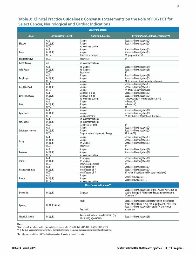
Clinical eff ectiveness of PET/CT scanningThere is good evidence that FDG PET/CT is benefi cial in the management of many types of cancer. The strongest evidence relates to the characterization of indeterminate pulmonary nodules (to assess the risk of cancer), staging of lung cancer, restaging of colorectal cancer, and the staging, assessment of response and restaging of lymphoma (cancer of the lymph nodes). There is also reasonable evidence on the limited use of FDG PET/CT for specifi c neurological (e.g., epilepsy and dementia) and cardiac (e.g., chronic ischemia) indicators.
On the basis of current best evidence and professional consensus, agencies in North America and Britain have developed clinical practice guidelines (CPG) for FDG scanning. These CPG’s for the use of FDG PET and PET/CT for select cancer, neurological, and cardiac indications are summarized in Table 3. They are derived from the Canadian Association of Radiologists, the Royal College of Radiologists (UK), and the National Comprehensive Cancer Network (USA). The criteria for the recommendations in the CPGs are found in Appendices 5 and 6.
NL will have to determine its own list of indications for PET scans. Such a list can be guided by the CPGs in Table 3 in that indications which have ‘A’ and ‘B’ levels of evidence should be strongly considered as should indications which have already garnered Health Canada approval. It is recommended that specifi c inclusion criteria be drafted up front and then a communication strategy be developed to guide referring physicians. The question of who will be authorized to order a PET scan in NL is addressed later in the report.
The following is a summary of the current clinical uses of FDG PET/CT. There are also many emerging clinical and research uses for FDG PET/CT on the horizon.
In cancer, clinical FDG PET/CT imaging is used to:Diagnose (How likely is it that cancer is present?) • Stage (Has the cancer spread beyond the original tumour?)• Assess initial treatment response, which can also be prognostic (Is the chemotherapy or radiation therapy working • and what is the probability of survival?)Assess end-of-treatment response (Is the cancer all gone?)• Re-stage (Has the cancer come back?)•
In neurology, clinical FDG PET/CT imaging is used:In select patients with epilepsy to help localize the anatomic origin of their seizures. This can be especially helpful • for patients where brain surgery is being contemplated and conventional imaging (e.g., CT and MRI) do not demonstrate any anatomic abnormalities.
NLCAHR March 2009 Contextualized Health Research Synthesis: PET/CT Programs
17
Table 3: Clinical Practice Guidelines: Consensus Statements on the Role of FDG PET for
Select Cancer, Neurological and Cardiac Indications
Cancer Indications
Cancer Consensus Statement Specific Indication Recommendation (level of evidence*)
Specialized investigation (C)Specialized investigation (C)2A (when 10 not identified by other modalities)
UterusCAR RCR (UK)NCCN
StagingStagingNo recommendation
Specific circumstances (C)Specific circumstances (C)
Non- Cancer Indications**
Dementia RCR (UK) DiagnosisSpecialized investigation (B) “Either SPECT or PET/CT can be used to distinguish Alzheimer’s disease from other forms of dementia.”
Epilepsy RCR (UK) & CAR
Adult
Paediatric
Specialized investigation (B) Seizure origin identification - When MRI negative or MRI results conflict with other tests.Specialized investigation (B) – useful for pre-surgical assessment
Notes: *Levels of evidence ratings and criteria can be found in Appendices IV and V (CAR, 2005; RCR-UK, 2007; NCCN, 2008)
** In the USA, Medicare reimburses for these three indications as a specialized investigation when specifi c criteria are met
The CAR recommendations (2005) did not comment on dementia or chronic ischemia.
Contextualized Health Research Synthesis: PET/CT Programs NLCAHR March 2009
18
In select patients with dementia where the type of dementia is uncertain. FDG PET/CT has been demonstrated to • be very sensitive in differentiating Alzheimer’s dementia, the most common cause of dementia, from other forms of dementia. Understanding the specifi c type of dementia assists physicians in understanding what to tell the patient and their family about prognosis and possible therapy.
In cardiology, clinical FDG PET/CT imaging is used:In select patients where it is diffi cult to determine if heart muscle is alive or dead. This is usually in the setting of • chronic ischemia (i.e., lack of oxygenated blood supply) where the heart muscle is not moving well but is still alive (i.e., hibernating myocardium). Patients with hibernating myocardium may benefi t from certain therapies whereas those with dead heart muscle (e.g., scarring) may not. There is a subpopulation for which conventional imaging (e.g., nuclear medicine myocardial perfusion/viability imaging, MRI, US) may not clearly differentiate scarred from hibernating myocardium and this is where FDG PET/CT may have a role.
Will FDG PET/CT imaging replace conventional imaging?PET/CT imaging, with a few exceptions described below, should be considered an adjuvant imaging modality, rather than a replacement imaging modality. The clinical benefi ts for cancer patients are more accurate diagnosis, staging and follow-up which, in turn, can result in more ideal patient management and quality of life.
One exception to this is that FDG PET/CT imaging, where available, has almost completely replaced Gallium scanning (a conventional nuclear medicine procedure) for staging and assessing response to therapy in patients with lymphoma, although some PET/CT centres may still use Gallium for paediatric patients.
Cost-eff ectiveness of PET/CT scanningAlthough PET/CT is largely an adjuvant imaging modality, cost savings can be realized in that PET/CT scanning can signifi cantly infl uence patient management. The usual clinical fi nding is that disease (e.g., cancer) has spread beyond what was demonstrated using conventional imaging. PET/CT scanning provides an opportunity to avoid futile and costly, invasive interventions (e.g., surgery or radical radiotherapy) and to provide more appropriate palliative or ‘local control only’ therapy, thus improving the patient’s quality of life despite not improving overall survival. The overall cost impact on the health system will depend on whether this alternative care ends up being less costly or more costly but, in any case what will result is a more appropriate use of resources.
The impact of PET/CT scanning on wait times for diagnostic imaging is minimal. The overall number of PET scans is so low that removing that number from the system will have little to no effect on other DI modalities, with the exception of Nuclear Medicine Gallium scanning and the associated wait times.
Other benefi ts of PET/CT scanningAnother impact of the introduction of PET/CT scanning on the NL health care system is the eligibility of patients for certain types of clinical trials. Many clinical oncology trials for both pediatric (e.g. Children’s Oncology Group) and adult patients now require FDG PET/CT imaging for recruitment and inclusion. Having a PET/CT program will enhance the access of NL patients to such trials. Having a local PET/CT scanner will also make it possible for patients who do not fi t the standard indications to enter large multi-centered trials, thus benefi ting both patients and clinicians. In addition, with a PET/CT scanner, NL will be in a better position to recruit new basic science and clinical researchers with research interest in areas such as oncology, dementia, schizophrenia, depression, and the diagnosis of FASD.
NLCAHR March 2009 Contextualized Health Research Synthesis: PET/CT Programs
19
Contextualization
Components of a PET/CT ProgramThe administrative or program advice provided in the following sections is based on the experience of the Team Leader, Dr. Demeter, in observing the formation of the Edmonton PET program and initiating and planning, as one its Co-Directors, the Winnipeg PET/CT program. Dr. Peter Hollett of Eastern Health, NL, also provided valuable information and insights on key aspects of current Nuclear Medicine services in NL, including resources and staffi ng. Although we have done our best to be context-sensitive in drawing lessons for NL from the Edmonton and Winnipeg cases, we are acutely conscious of the challenges involved. Members of the research team have also consulted with Drs. Ross, Barnes and Burrell of the Halifax PET/CT program and have used the information provided by them to verify the contextualized evidence presented below.
Where in the province should the PET/CT program be located?1. According to the 2006 Census (Newfoundland and Labrador Statistics Agency, 2008), NL has a total population of just over half a million which is roughly divided between the health regions of Eastern (293,795), Central (95,460), Western (79, 460) and Labrador-Grenfell (36,755). Residents of the three smaller health regions often use services from other regions and especially from Eastern Health. Eastern Health currently serves 58% of the NL population for primary and secondary level medical service and a considerably higher percentage for specialized tertiary care referral services, such as oncology and neurosurgery.
St. John’s is the primary site for specialty cancer services such as chemotherapy, radiation therapy and oncological surgery. All solid tumour chemotherapy planning and 61% of the approximate 14,000 annual chemotherapy treatments are administered at the Dr. H. Bliss Murphy Cancer Centre within Eastern Health with the other 39% being administered in Corner Book, Grand Falls/Windsor and Gander. All radiation therapy treatments are administered at Eastern Health. Although it is estimated that the majority of cancer surgeries is done in Eastern Health, general surgeons across the province also deliver such services depending on the site of disease (bowel, breast and prostate cancer surgeries are performed across the province whereas the majority of gynaecological cancer surgeries are done in St. John’s). Neurologists are located in St. John’s, Grand Falls-Windsor, and Corner Brook, and psychiatrists are located throughout each RHA with the smallest sites being in St. Anthony, Stephenville, Carbonear, and Clarenville (this is relevant for neuropsychiatric indications for PET/CT such as dementia) (DHCS, personal communication, January 2008).
There are only four fellowship-trained Nuclear Medicine Physicians in NL and they all practice in St. John’s. St. John’s also has the largest concentration of other Nuclear Medicine staff including technologists, physicists and radiation safety personnel as well as the highest availability of on-site imaging industry service personnel. Furthermore, to achieve any research potential, PET/CT needs to be linked to an academic institution, with graduate and postgraduate research capacity in appropriate fi elds. For NL, this means Memorial University in St. John’s.
Based on population demographics, distribution of services, availability of medical and support personnel, and research potential, St. John’s is the most appropriate option for locating the PET/CT scanner and cyclotron. It is noteworthy that there is currently no physical space available in Eastern Health for a scanner or a cyclotron. It is our understanding that the provincial Government has now authorized Eastern Health to begin planning for a full-scale redevelopment of acute care facilities in St. John’s with a 7-to-10-year timeframe. Clearly, this planning will have to include a consideration of the space and location requirements of the scanner and the cyclotron as described in the next section.
Physical space and design considerations2. Our understanding of the physical space and design requirements for the prospective PET/CT program has been guided by the experience of the lead author in Winnipeg and our discussions with leaders of the PET/CT program in Halifax. Both Winnipeg and Halifax use approximately 2000 sq. ft. for their PET/CT suite. The cyclotron suite, including the research lab, requires anywhere from 1500 to 2500 sq. ft. of space, depending upon the type of cyclotron and the type of shielding it requires for radiation protection.
The Winnipeg experience (putting the PET/CT suite initially at a distance from the main acute care campus and trying to move it later on) indicates that it is important to locate the PET/CT suite in a tertiary acute care setting and to do so from the outset. The Winnipeg PET/CT program is located off-site in a building adjacent to the main hospital campus of the Winnipeg Health Sciences Centre. As such, it does not have access to ‘stat’ emergency hospital services such as ‘code blue’
Contextualized Health Research Synthesis: PET/CT Programs NLCAHR March 2009
20
or ‘code 99’ services, or ‘stat’ laboratory, ECG or portable x-ray services. In emergencies, the program must dial ‘911’ which is far from ideal, especially when imaging in-patients who may be in poorer health than ambulatory patients. The economic and logistical costs of moving the Winnipeg PET/CT program from its ‘temporary’ location to its proposed new home, the Siemens Institute for Advanced Medicine (SIAM), have proved to be considerable if not prohibitive.
In addition, it is important to have the PET/CT experts readily available to consult with physicians who order PET/CT studies. As most of these “referring physicians” will be cancer specialists it makes the most sense to locate the services in a tertiary health care setting.
Local oncologists, with whom we consulted about the issue of location, have expressed similar concerns and are eager to provide input into the design of the PET/CT suite including the table used in scanning and other related decisions. It is important that the technology be available to patients at both adult and pediatric sites. Locating the PET/CT scanner adjacent to the Janeway Hospital would eliminate the need to transport children via ambulance to another part of the city for scanning. One pediatric oncologist noted also that the PET suite would require general anaesthetic (GA) capabilities and the requisite human resources (anaesthetists, recovery room nurses, technicians) as children under the age of 7 may need a GA/sedation to prevent movement during a PET scan.
Another consideration is that the PET/CT suite should be located as close as possible to the cyclotron facility (i.e., in the same building and as close as reasonably possible to the cyclotron facility). This would allow for rapid delivery of short-lived PET radiopharmaceuticals with half-lives as short as 2 minutes. Having the capacity to delivery short-lived PET radiopharmaceuticals rapidly is essential not only for clinical programs but also for basic science and clinical research purposes. The Winnipeg program is planning to have a pneumatic tube system installed between the cyclotron facility and the proposed PET/CT suite in the SIAM.
The cost of building or renovating a space for the PET scanner and the Cyclotron is diffi cult to estimate. In Halifax, the actual construction cost increased by 40% from the initial estimate (20% of the increase being attributable to increased labour costs).
Who will be permitted to order a PET scan?3. The Winnipeg PET program has chosen to limit referrals for PET scans to cancer specialists (medical, radiation and surgical oncologists) and other select specialists (e.g., lung specialists). Presentations at rounds and written communications have been used to inform the physician community about approved indications and procedures for ordering PET/CT imaging. All PET/CT requests are reviewed by NM PET physicians for appropriateness, potential utility and timing relative to patient treatment events. A similar approach has been taken in Halifax. All accepted indications for PET scans have been listed by the Department of Health. At present, these are all oncology indications. Only oncology specialists are permitted to order PET scans, and the Nuclear Medicine physician is the ‘gatekeeper’ who bases decisions primarily on the American Clinical Practice Guidelines. Other cases are considered on a case-by-case basis through consultation with specialists. The local NL specialists whom we have consulted concur with the approaches taken in Manitoba and Nova Scotia. They support the need for evidence-based clinical practice guidelines. They agree that the ‘gatekeeper’ for all PET scans would logically be the Nuclear Medicine physician. These specialists recommend that, as the province moves to plan a PET/CT program, a committee should be formed to establish tight controls over the ordering of scans, to examine the American CPG and to develop a clear local CPG document. A process for monitoring and approving evolving/new indications for PET would also be appropriate.
Human resource requirements for a PET/CT program4. Physiciansa.
Physician training in PET and CT: While the practice of medicine is regulated at the provincial level, most jurisdictions in Canada allow only specialists with Canadian Royal College Fellowship training in Nuclear Medicine to practice Nuclear Medicine, including PET and PET/CT scanning. Some provincial Colleges of Physicians and Surgeons have adopted additional requirements for the clinical interpretation of PET studies. For example, above and beyond a Royal College Fellowship in Nuclear Medicine, Alberta and Manitoba require three months of acceptable dedicated training and a minimum of 250 PET cases read and reviewed by a supervisor. To become a director of a PET facility requires six months of dedicated training.
NLCAHR March 2009 Contextualized Health Research Synthesis: PET/CT Programs
21
In NL, the College does not normally demand specifi c training for every physician task. That is normally done at the institutional level. However, it is the suggestion of this group that physicians involved with PET have the minimum training as documented by any other province in Canada, that being a Royal College Fellowship in Nuclear Medicine and a minimum of three additional months of dedicated PET/CT training. They should also be expected to keep up with the Royal College guidelines for continuing education especially as it applies to PET/CT.
Additional training in cross-sectional anatomy imaging (e.g., CT) is recommended if this was not part of the physician’s original training. The various professional groups both in Canada and the USA have agreements and training programs in place for this (CANM, CSNM, SNM, and ACR) (Brink et al., 2005; Wong et al., 2007).
A combination of approaches was used for the Winnipeg PET program: in-house mentoring of NM physicians, dedicated training for the Co-Directors of the PET program at a centre which had the same PET/CT equipment, dedicated PET/CT conferences for all NM physicians, and some subspecialty PET/CT training for our lead paediatric NM physician. This has served the Winnipeg PET program well, largely because there is a supportive environment for colleagues to cover clinical duties while other physicians are doing on-site or off-site PET/CT training; fi nancial resources have been available to support these endeavours. The NS program has adopted a very similar approach and this would be the recommended path for NL.
Fortunately, two of the four NM physicians in NL are also radiologists and would not require additional training in cross-sectional anatomy. For the other two NM physicians, internet-based CME resources, off-site conferences and on-site mentoring by NL radiologists would be practical ways to provide them with the additional necessary cross-sectional anatomy skills. It should be noted, however, that these two NM physicians currently interpret hybrid images in their general NM department (SPECT/CT) and already have some familiarity with these activities.
The most effi cient setting for physician PET/CT training is at an established, high-volume PET/CT centre. There appears to be variable support available for St. John’s NM physicians to take sabbaticals or time off work for specialty training. Such training is necessary and support should be provided for specialty training of NM physicians interested in participating in the PET/CT program. Once a PET/CT program is up and running in NL, it would be helpful to consider arranging for short-term on-site mentors, remote mentorship through digital communication, or additional off-site mentorship and technology familiarization for our NM physicians at a site that uses a PET/CT camera that is similar to the one that will be used in NL (i.e., the same vendor and, if possible, the same camera model). The NL site could consider partnering with established PET programs (e.g. Sherbrook, Winnipeg, Edmonton, or Vancouver) to ensure access to innovations, experience and training opportunities.
Number of physicians required for PET/CT program: It generally takes 30 to 45 minutes to review a PET/CT case, with 45 minutes being more typical for Nuclear Medicine physicians early in their learning curve. Dedicated physician time is also needed to establish the clinical PET/CT program (e.g., establish patient protocols) and to participate in regulatory processes (e.g., Health Canada clinical trials applications or special access applications). Based on experience to date in the Winnipeg PET/CT program, a dedicated day per week of physician time (or 0.2 effective full-time equivalents) is the minimum needed. This assumes that there are adequate available administrative, program planning, and regulatory/clinical trials resources. If such are not readily available this time allotment will have to increase.
It is diffi cult to recruit physicians to part-time positions; so, if the FDG PET/CT workload (an estimated 600 - 1200 cases per year) cannot be folded into the existing NM physician complement, a full-time NM physician, preferably with PET/CT experience, will probably have to be recruited. Although there are no strict criteria on the recommended case volume for PET/CT physicians to maintain competency, in Winnipeg, a dedicated day per week of PET scanning, on average, appears to be adequate. This assumes that the physician has had suffi cient formative PET/CT training.
In Newfoundland and Labrador, there are currently four Canadian Royal College qualifi ed (FRCPC) NM specialists. Two of them are at the Health Sciences Centre (HSC) and dedicate 100% of their time to clinical Nuclear Medicine. The other two have a mixed Nuclear Medicine and Radiology practice out of the St. Clare’s Hospital site. A volume of 250 cases per year per physician in NL should be suffi cient to maintain competency and is similar to the volume suggested for competency in other areas of sub-specialization in NL, such as cardiac CT. There are no FRCPC-qualifi ed Nuclear Medicine physicians outside of St. John’s. Radiologists in Corner Brook and Gander report routine Nuclear Medicine cases and send complicated cases and all cardiac studies to HSC for reporting.
Contextualized Health Research Synthesis: PET/CT Programs NLCAHR March 2009
22
Physician reimbursement for PET/CT: Physician professional reimbursement for PET/CT services varies across the country with a fee-for-service general range of $250-$350 per study. Some provinces, such as Manitoba, have elected to fund Physician PET/CT professional services through an alternate funding program (sessional fees and fi xed envelope funding for services).
The current NM physician reimbursement environment is fee-for-service and a NL PET/CT fee schedule would need to be negotiated with the DHCS. It is possible that there is adequate capacity amongst the four NM physicians in St. John’s to manage the anticipated PET/CT caseload. If not, fee-for-service groups are usually adept at determining if and when to add members to their practices. In addition, there are residents who are willing to do sub-specialty training to ensure that appropriate standards are achieved and workload is covered.
Communications with referring physicians: Referring physicians and the NL medical community at large will require imaging guidelines and CME in PET/CT scanning. While being guided by the literature and practice in other centres, NL will have to determine its own list of PET/CT indications. Some indications will be clear-cut, such as staging lung cancer and re-staging colorectal cancer. Other conditions, such as kidney cancer, have less evidence of utility and will have to be prioritized relative to available resources, demand from the higher priority indications and other tangible and intangible factors.
Once established in NL, imaging guidelines should be communicated to potential referring physicians through medical rounds or at a special local conference. A program of physician education should accompany these guidelines so that referring physicians know how to interpret the PET/CT reports and can follow up appropriately on patient management options so that the patient receives the maximum benefi t from the new imaging technology. In Winnipeg, accepted PET/CT indications have been communicated to referring physicians (primarily to medical, surgical and radiation oncologists as well as to respirologists) through mailings and selective medical rounds. The indications for PET/CT have also been communicated to other physicians (e.g., family physicians) who cannot currently directly refer their patients for a PET/CT scan so that they understand the rationale for dedicated specialist referral. It is important that non-referring physicians have up-to-date information and resources to talk to patients about PET/CT in this era of the well-informed, or at least web-informed, health consumer.
Technologistsb. Technologist training in PET and CT: Across Canada, the operation of PET/CT technologies has largely been by NM technologists. PET/CT training is part of the current training curriculum leading to NM certifi cation by the Canadian Association of Medical Radiation Technologists (CAMRT). CAMRT also offers additional distant CME training on CT specifi cally for NM technologists (www.camrt.ca). For NM technologists who trained before PET/CT was part of the standard curriculum, distant CME on PET/CT is also available (e.g., through BCIT, www.bcit.ca).
The Winnipeg PET/CT experience also suggests that it is a good idea to send NM technologists on short preceptorships of 1 to 2 weeks to a site that uses the same scanner as the one they are, or will be, working with. In Halifax, many of the technologists went away for PET and CT training, followed by one month of additional training on the local CT scanners.
Technologists’ scope of practice: Each province has its own legislation on who can operate diagnostic ‘x-ray emitting devices’, including PET/CT scanners. The default is usually an x-ray technologist, although other technologists may have a prescriptive, and usually more limited, scope of practice. For example, in Manitoba, the operation of a diagnostic x-ray emitting device by non-x-ray technologists (e.g., Nuclear Medicine Technologists) requires approval on a case-by-case basis by the Ministry of Health. This is usually granted through the Minister’s delegate, who is the Provincial Radiation Safety Offi cer. In Manitoba, a simple letter describing a NM technologist’s training and scope of practice (e.g., standardized pre-set CT acquisition parameters) has been suffi cient to date.
It should be noted that combining CT, or other x-ray emitting devices, with NM technology is not limited to PET. Routine NM cameras (e.g., gamma cameras) are also available as hybrid technologies (SPECT/CT). NM technologists at the HSC in NL currently operate two such hybrid cameras. It would be reasonable to think that they could be trained to do the same safely for a PET/CT hybrid. It is also much more cost-effective to have a single technologist operate both the CT and PET components.
Currently, all NM radiopharmaceutical production in St. John’s is done by NM technologists (mostly using commercial kits). In order to receive the FDG, technologists would require training in radiation safety, Transport Canada regulations, quality assurance testing, safe handling of PERs, drawing up individual doses (since the product would probably be shipped in a single concentrated vial), and transport regulations for shipping back the empty containers.
NLCAHR March 2009 Contextualized Health Research Synthesis: PET/CT Programs
23
Number of technologists required for PET/CT program: Once a patient is injected with a PER and has waited approximately one hour, it takes about 20 minutes, on average, to do a near whole body PET/CT scan. Based on the Winnipeg PET/CT experience, a minimum of two NM technologists would be needed on PET/CT imaging days. Human resource allocations for holiday relief and sick leave should also be considered, as should the need to minimize radiation exposure. In Halifax’s PET/CT program, there are 8 technologists rotating through 3 days per week in order to reduce radiation exposure and allow for annual leave/sick time. Depending on the number of days per week allotted for PET scanning in NL, the number of technologists needed to run the program can be calculated accordingly.
Recruitment and retention of technologists: NL has had historic problems in retaining technologists. A sponsoring program has increased the province’s recruitment success rate and currently, for the fi rst time in many years, the diagnostic imaging program in St. John’s is fully staffed, with sponsored students slated for return to St. John’s next year. In anticipation of a PET program, some technologists are already enrolled in the distance CMART PET training programs. The PET program is expected to improve the retention and future recruitment of technologists.
Physicistsc. Physics support for PET/CT is primarily related to ongoing quality assurance in technology performance. This support can be delivered in a number of ways depending on the existing pattern of service provision. NM technologists usually conduct routine daily quality assurance tests and they seek further consultation if the test results are unsatisfactory. One approach is to provide additional training for current on-site CT and NM physics support personnel. Alternatively, if physicist support is being provided through external consultants, then additional service can be negotiated. Canadian imaging physicists currently earn approximately $100,000 per annum.
In St. John’s, there is currently limited on-site NM physics support from the Cancer Clinic located in Eastern Health and Memorial University. Dedicated NM physics support would be needed for the PET/CT program. This dedicated support could be shared with the general NM program or more broadly across the Diagnostic Imaging program.
Clinical trials and regulatory personneld. The regulation of FDG in Canada is currently evolving. Five centres (CanTrace™ in BC, FluGlucoScan™ in Albeta, Glucovision™ in Ontario, and Gludef™ and 18F-Fluorodeoxyglucose in Quebec) have now obtained Health Canada approval for their specifi c FDG products. Any new sites will be required to submit clinical trial applications (CTA) for their FDG products. The idea that a centre could qualify for certifi cation simply by demonstrating ‘chemical equivalency’ between their FDG and a licensed FDG has not yet been accepted by Health Canada. The complexity of the regulatory process coupled with unknown variables in this evolving fi eld makes it diffi cult to provide an accurate prediction of the timelines required to establish a PET/CT program. Some general guidance is provided later in the report under the section entitled ‘Timelines and Sequencing.’
Whether NL produces its FDG locally or obtains it from outside the province (e.g., from Halifax), it would be prudent to create, early in the planning process, a dedicated position in NL to handle regulatory and clinical trial enrolment issues. This employee should be a health care worker with experience in clinical trials and in Health Canada’s CTA process. Some Canadian sites have hired research nurses while other sites have hired allied health professionals. In Manitoba, the ‘Clinical Trials Coordinator’ has enrolled in a Clinical Research and Ethics program through one of the local Community Colleges. If there is any thought of exploring the production or use of non-FDG PET products, whether produced on site or imported, this position will be all the more essential for working through the regulatory processes involved.
Eastern Health currently has a number of research nurses who work in the area of clinical trials, although they would need special training in Health Canada’s CTA for FDG. The cost of this position would include salary/benefi ts (approximately $77,000 at current rates for a full-time research nurse coordinator) and standard offi ce expenses.
Radiochemist/radiopharmaciste. The need for input from radiopharmaceutical or radiochemistry scientists in NL will largely depend on whether FDG is produced on site or is brought in from outside the province. Even if the province plans to purchase a cyclotron, there will be a period of time in the initial stages of the PET/CT program when FDG will still need to be imported from outside, most likely from Halifax. It will also be advisable to identify an alternate source of FDG as part of contingency planning for unexpected reductions in local supply. There may be a need to do initial validation of the stability of the FDG upon its arrival (to demonstrate that there is no product degradation related to transport). Once this has been established, and until there is on-site PER production, there would be little need for such radiochemists or radiopharmacists.
Contextualized Health Research Synthesis: PET/CT Programs NLCAHR March 2009
24
Once the production of FDG and other PERs is being done in NL, new on-site radiochemist/ radiopharmacist expertise will be required. Recruiting someone with such skills could also prove benefi cial for producing other NM materials (through on-site professional oversight and experience) and might make possible the production and use of new, non-PET NM products. Further details on the role of a radiochemist/radiopharmacist are presented below in the section on the cyclotron question.
Radiation safety issues5. There is a regulatory requirement that all NM departments have radiation safety offi cers (RSOs). Current RSO’s in NL should have the basic skills and experience to manage the new radiation safety issues. They may, however, need skills upgrading and supplementation to deal with issues specifi c to PET radiotracers. For example, compared to routine Nuclear Medicine, PET radiotracers have generally higher radiation energies and require increased diligence to minimize occupational radiation exposures. . Historical studies have demonstrated that although NM technologists who work in PET/CT programs may have higher average radiation exposures than general NM technologists (although still within regulatory limits), there are ways to reduce this exposure (Roberts et al., 2005, Guillet et al., 2005, Seierstad et al., 2007). The RSO should be adept at adapting the ‘time-distance-shielding’ mantra to maintain an occupational radiation dose that is as low as reasonably achievable. The designated NM RSO may require additional input from the CT RSO. In NL, there is currently an RSO in two sites with 0.5 days per week of dedicated time, and a corporate RSO who also has other managerial responsibilities. An assessment will need to be made as to whether this will be adequate to manage a PET/CT program in St. John’s. This assessment could be conducted by the corporate RSO and should include an examination of required dedicated time and of skill sets. Additional resources may also be needed up front for the preparation of required submissions to the Canadian Nuclear Safety Commission (CNSC). For example, the CNSC will require an analysis of anticipated environmental and occupational radiation dose levels based on the proposed PET/CT suite plans. After that, it may be possible to delegate ongoing radiation safety issues to existing staff, possibly with additional training and additional dedicated time.
Service and equipment maintenance4. To avoid disruptions in clinical services, a robust PET/CT service arrangement will be required. Since there will be only a single PET/CT camera, proper maintenance and service is crucial to minimize disruptions. Whether or not the NL program will require a full manufacturer’s warranty package including the provision of locally-based service personnel will depend on the extent to which Eastern Health’s biomedical engineering department is willing and able to develop its own specialized in-house capacity.
The Cyclotron QuestionGiven that the Government of NL has recently decided to acquire an on-site cyclotron to support the PET/CT program, this report now focuses on the optimal sequence of activities required for the acquisition of the two technologies and on the development of the programs associated with each. It usually takes longer to develop a medical cyclotron program than it does to develop a PET/CT imaging program. Experience in Winnipeg and in Halifax suggests, assuming that human and fi nancial resources are available for both programs from the start, that it takes about one year to start up a scanning program but two-to-three years to get a cyclotron program commissioned. Therefore, unless NL starts development of the cyclotron program well in advance, there will be an initial period during which PERs will have to be brought in from an out-of-province producer, such as Halifax. Other, less satisfactory options for purchasing FDG include Montreal and Sherbrooke, Quebec.
The advantages of early development of a cyclotron program 1. Having a local cyclotron has a number of important advantages including potential cost-savings, better scheduling of patients, and innovative research applications. These advantages are so substantial as to suggest that, now that the province has decided to purchase a cyclotron, it should pay serious attention to the timing so that the cyclotron program is initiated as early as possible.
Cost-savingsa. Producing isotopes locally has the major advantage of making the appropriate quantity of radioactive material available to the scanning program in a more predictable way than if the province were one client among many in a market with growing and highly variable demand and limited sources of supply. Having a local cyclotron will reduce the quantity of PERs that would be required and may nominally reduce production costs (e.g. reduced cyclotron beam time per
NLCAHR March 2009 Contextualized Health Research Synthesis: PET/CT Programs
25
production resulting in some energy savings and perhaps, some staff time costs). It would do this in two ways: fi rst the operation of the PET/CT program could be structured in the most economical way such that FDG is produced and delivered when needed versus having the program tied to external production and fl ight schedules. Secondly, it would make it unnecessary to adopt the anticipatory tactic of ordering infl ated quantities of product just to make sure that what arrives will have an adequate activity level. Even bringing in FDG from Halifax poses challenges: This approach will require at least three to four hours of travel time composed of 45 minutes by road from the cyclotron to Halifax airport, up to an hour for transport of dangerous goods clearance, 80 minutes in the air, 20 minutes by road from the St. John’s airport to the scanning clinic plus all the waiting time associated with air travel under the best of circumstances. This travel time will result in a loss of at least two half-lives which would require ordering 4 times as much activity as would be needed, to account for in-transit decay.
Schedulingb. Having a local cyclotron will not only eliminate the cost of air transport (as a trade-off, of course, against the cost of building and maintaining the cyclotron) but would have an impact on imaging schedules. With local production of FDG, the PET/CT program would have full control of the production schedules of the cyclotron and this would make for better planning of the scanning appointments and staff work schedules. Once the program is fully operational, scanning appointments could be scheduled earlier in the day than would be the case with PERs that are fl own in. Preliminary discussions with Halifax indicate that, once their cyclotron has been commissioned, they will be able to begin producing FDG as early as 4:00 am in order to have it ready for transport to NL via Air Canada on the 7:15 am fl ight, arriving St. John’s at 9:14 am (www.aircanada.com, accessed September 11, 2008). Even when all goes as planned, the earliest one could expect to start scanning appointments would likely be 10:00 am or later so that the opportunity to schedule scans in the morning is essentially lost. In Halifax, the original plan of scanning scan 10 patients per day, 3 days a week had to be cut back to 6 - 8 per day since importing FDG from Montreal in the absence of a local cyclotron meant that scanning could not begin until noon. Options such as using private air transport carriers have been explored by other jurisdictions (e.g., Manitoba and New Brunswick), but the prevailing opinion is that a commercial carrier such as Air Canada is much more economically attractive and presents fewer logistical and regulatory issues.
Winnipeg’s experience with imported PERs is also instructive. Between July 2005, and December 2007, the Winnipeg PET/CT program operated without its own cyclotron, getting its PERs fl own in daily from Edmonton. For the period between November 2005, and the end of December 2007 (ignoring the initial start-up period that saw the usual, special logistical challenges), the Winnipeg PET program planned 238 imaging days. Of these, only 31 (13%) had fully radioactive shipments delivered on time; 156 (66%) had shipments with reduced radioactivity or late delivery but with no impact on patient bookings; 12 days (5%) had shipments with reduced radioactivity requiring cancelling some appointments; and 31 days (10%) had all appointments cancelled of which 7 were for air transport or weather reasons. The weather in Atlantic Canada is at least as bad as that in the Prairies, particularly where fog is concerned. To test this supposition, we monitored all Halifax-to-St. John’s fl ights on Air Canada (the only national commercial passenger airline that will accept ‘dangerous goods’) for a trial period between February 13, 2008, and April 24, 2008. The results confi rmed our suspicions. Of the 36 scheduled fl ights, 13 (36%) arrived on time or early, 2 (6%) were cancelled, and 21 (58%) arrived late, including 4 that were over 30 minutes late, 3 that were over 45 minutes late and 2 that were over 60 minutes late. Roughly speaking, the air travel challenge was twice as great for the Halifax-St. John’s route in this late winter/early spring period than for the Edmonton-Winnipeg route (monitored over a longer timeframe). Our analysis of the impact of fl ight delays on the amount of FDG radioactivity suggests that, unless orders had been substantially infl ated in quantity (and cost) to compensate for expected delays or appointment rosters had been deliberately reduced, about 6% of all patient appointments would have had to be totally cancelled because of air carrier reasons alone. Having a cyclotron onsite would eliminate this problem.
In addition, having a cyclotron would reduce the frequency of having to re-schedule patients due to production and maintenance issues. In the Winnipeg experience, of the 238 planned patient imaging days, 14 had to be cancelled completely because of cyclotron problems in Edmonton and 6 days were cancelled because of synthesis problems. All of the patients scheduled for scans on these days had to be rebooked. When the cyclotron is located at a distance, a production problem is almost irremediable because a second batch is not likely to be ready in time to be shipped to the scanning location at a reasonable time. So, a production failure inevitably leads to complete cancellation of all appointments for the day with the associated system costs of rescheduling the patients and reallocating the staff ’s activities. With a local cyclotron, at least some of the day’s patient appointments can often be salvaged by doing a second production run. Admittedly, even with an onsite cyclotron, there may be some problems that cannot be fi xed on the same day and there will still be a need for some rescheduling of patients.
Contextualized Health Research Synthesis: PET/CT Programs NLCAHR March 2009
26
Research potentialc. A local cyclotron would permit the development in NL of a much broader range of clinical and research activities. A cyclotron program could produce other positron emitting isotopes in addition to F-18. It would also make it possible to synthesize PERs other than FDG. Scans that require isotopes with very short half-lives such as oxygen (2 minutes), nitrogen (10 minutes) and carbon (20 minutes) could then be considered. Local isotope production would make possible the development of a research program into innovative scanning in, for example, cardiology, neurology, psychiatry and basic life sciences, thus creating the potential for collaboration with research centres in other parts of Canada and in the United States. A strong research program would enhance the province’s ability to attract and retain physicians, technicians and scientists. Our research team met with clinical specialists in oncology, psychiatry, genetics/rheumatology, and radiology, and has identifi ed several potential research areas that would benefi t from a local cyclotron. There is also an opportunity for local scientists to be involved in national programs that could bring in signifi cant research investment and activity, such as the upcoming application to the Canada Foundation for Innovation that is going to be submitted by the Canadian Network for the Development of Biomarkers for Functional Imaging.
The decision to establish a research program is a key one that should be made early in the planning process. Deciding whether to run just a routine clinical cyclotron program or to also include a research arm will infl uence the direction of a number of equipment purchases, not the least of which is the type of cyclotron that would be needed. Though the level of research need not be specifi ed, it will be important to decide such things as the number of hot cells and automated synthesis units that will be installed when the program is fi rst introduced, and the number that would be installed but not operated initially. Underestimating the requirements for hot cells and GMP requirements at the time of construction is an error that should be avoided.
The costs of a local cyclotron2. We begin our analysis by examining the additional costs involved in purchasing, installing, commissioning and operating the cyclotron. Costing such projects is complicated since a large number of variables is involved and many of them (such as labour availability and construction costs) are market-driven and highly unpredictable. The following is meant to serve only as a rough guide to possible net cost differences between producing FDG on site and purchasing it from an out-of-province cyclotron.
A net present value (NPV) analysis has been used to estimate total and average case costs of operating the cyclotron over a 20-year period. An NPV converts all estimated costs for the next 20 years (the anticipated lifespan of a cyclotron) to present-day dollars. This approach allows workable, apples-to-apples, comparisons of the two options. The analysis is focused on net differences in estimated costs; costs that would be the same in the two scenarios (e.g., the cost of purchasing, installing, and operating a PET/CT scanner) are generally not included. Because exact costs are not known, a sensitivity analysis was developed using both a high and a low estimate to give a range of case cost estimates. Appendix 7 outlines baseline cost estimates and describes the sources for these estimates. The analysis is done from the perspective of the health care institution and does not include net differences in patient or other third party costs. For example, in this economic analysis the health care system is compensated for cancelled patient appointments. However, there is no accounting for patient lost work time or possible third party payer costs (e.g. travel grants). As such this economic analysis may underestimate costs related to the off-site FDG production scenario.
Using baseline assumptions, the option of in-house production of FDG is more expensive than the alternative. The in-house option has a total estimated cost of approximately $18.9 million over 20 years and an average cost per scan of $811. The imported option had a total estimated cost of $12.4 million over 20 years and an average per scan cost of $533. The net difference between the two total costs is $6.5 million or $325,000 a year over 20 years (a net difference in average case cost of $278). To account for uncertainty in the baseline cost estimates, they were adjusted (i.e., infl ated and devalued) to see if this would change the outcome relative to the baseline scenario (i.e., the sensitivity analysis). The in-house option remained more expensive in all scenarios except when all costs for imported FDG were maximized and those for in-house production were minimized. The details of this sensitivity analysis can be found in Appendix 8.
One of the more controversial components of this analysis is adopting a penalty for cancelled patients. Given the expected differences in the number of cancellations between the two options it was felt that there had to be some way to acknowledge this economically. A choice was made to use, as a surrogate, the inter-provincial transfer rate for the Winnipeg program to send one patient for a PET scan out-of-province, which is currently $1,250 (excluding transportation and accommodations). In addition, since the analysis is done from the perspective of the health institution rather than that of the society as a whole, this economic analysis does not take into account costs to patients (e.g., transportation, lost income, out-of-pocket, etc.) and does not take into account the research and alternate clinical potential of having an on-site medical
NLCAHR March 2009 Contextualized Health Research Synthesis: PET/CT Programs
27
cyclotron. Ultimately, a decision will have to be made as to whether the potential increased average case cost of local production that we have estimated is likely to be outweighed by the various intangible advantages of having local production.
Human resource requirements for a cyclotron program3. In addition to capital and operating costs, a local cyclotron would require the recruitment, remuneration and retention of a number of professional and technical employees. The availability of these professionals and the likelihood of interest among them in working in NL are both factors that would need to be examined. Staffi ng for a cyclotron program will depend on the scope of the program. Technical and professional staff numbers need to be adjusted to the size of any research program. The following is a bare minimum staff complement for a clinical PET/CT program, with some attention to a possible research program as appropriate.
Scientistsa. If the emphasis is on a clinical program, an experienced radiopharmacist with proven experience in good manufacturing practice (GMP) is optimal and should be recruited early in the planning process. If there is a desire to have a research program as well, a scientist (usually an organic chemist) with PET tracer synthesis experience may be more appropriate. A PET organic chemist can also produce product for a clinical program but may not be as adept in establishing standard operating procedures for GMP production. If both strong clinical and research programs are anticipated it would be best to have two staff members, a radiopharmacist and a radiochemist. These employees could also offer radiopharmacy and radiochemistry expertise to the regular NM program.
Cyclotron operatorb. This employee would manage routine cyclotron operations and perform preventive maintenance and troubleshooting. Although there is no single career path for this position, mechanical, electrical or biomedical technicians or technologists are suited to the job. Training is usually based on a factory preceptorship prior to the installation of the cyclotron, with on-the-job training thereafter. Cyclotron operators are internationally in very short supply, and it is diffi cult to fi nd experienced operators, especially for a specifi c cyclotron model. If demand for cyclotron time is increased (e.g., making multiple batches of product for research beyond the local FDG clinical demand) more cyclotron-operating staff will be required. Other staff (the PERs Production or Quality Assurance Technologists) may be trained to operate the cyclotron for routine production but not necessarily for preventive maintenance or troubleshooting.
Technologistsc. A GMP facility will need a minimum of two additional technologists. The person responsible for quality assurance should be different from the person responsible for production. These technologists do not necessarily have to be NM technologists. Some centres prefer Bachelor of Science graduates with an emphasis on chemistry, especially organic chemistry. If they are NM technologists, they may be able to offer some back-up support to the regular pool of NM technologists thus creating effi ciencies. The PET technologists, whether NM or BSc graduates (or both), would also have to be able to operate the cyclotron and do the chemistry for routine PER production to cover for the cyclotron operator during holidays and other absences.
Radiation safety offi cer d. Additional dedicated RSO time and expertise will be required to operate a cyclotron facility. The risk of exposure to high radiation levels is greater in this type of operation than in routine NM radiopharmacy or dispensing operations. One option is to designate one of the scientists or technologists as the RSO and provide dedicated time for these duties. It is anticipated that the CSNC will review the expertise of the RSO candidate, as well as the amount of dedicated RSO time being provided before allowing the cyclotron program to begin operation.
Regulatory staffe. Signifi cant time and resources must be dedicated to regulatory issues. Meeting the requirements of the CNSC (license for construction, service, commissioning and operations) and fulfi lling those of Health Canada (clinical trials applications, new drug notice of compliance, and GMP certifi cation) involve important and time-consuming responsibilities. This work requires special expertise and dedicated time and generally cannot be done by current full-time staff. There are two options: (a) hire an experienced project manager for a specifi ed term, or (b) hire consultants to do piece work as the project unfolds. Either way, these individuals should be hired early in the planning process. These services need to be recognized and budgeted appropriately (at least $100,000 in total as per Chuck et al., 2005).
Contextualized Health Research Synthesis: PET/CT Programs NLCAHR March 2009
28
Summary of Human Resource RequirementsTable 4 provides a summary of the minimum human resource requirements to operate both a PET/CT program and a cyclotron program. It is anticipated, especially during start up, that the PET/CT scanner and the cyclotron will not be operating full time. This would allow potential effi ciencies in sharing some staff such as clerical, technologists, (assuming all are NMTs) and RSO personnel. If additional professional staff (such as physicians or radiochemists/ radiopharmacists) is needed, we have provided for full-time positions since it may prove diffi cult to recruit on a part-time basis. When the PET/CT and the cyclotron programs are running simultaneously, there may be some effi ciencies gained by sharing some technical and clerical staff. Resources for regulatory consultants will vary with the scope of the project but, at a minimum, at least $100,000 should be budgeted to install and operate a cyclotron. Housekeeping, facility maintenance, and equipment service needs should also be considered and planned for up front.
Timelines and SequencingThe research team considered the possibility of creating a template that would specify the various key steps, and the associated timelines, for establishing both a PET/CT program and a cyclotron program. This task has proved unworkable. The timing and outcomes of a large number of steps in the process are impossible to predict. These include: the complexities of the tendering process that would render it overly ambitious to run both RFPs simultaneously; the Canadian regulatory requirements that are currently in fl ux; the availability of regulatory consultants and construction/trades services; uncertainty about the physical location of the programs (including whether the planned site involves the renovation of existing buildings or the construction of a new facility); the availability of essential staff with scarce skills and experience; and the location of the source of FDG in the interim until the local cyclotron is up and running. Thus, key steps in the process to which other steps are related are unpredictable.
It will be important to engage, early in the planning process, a project consultant with specialized expertise who can advise on regulatory issues related to both HC and CNSC, as issues related to non-compliance can be a signifi cant rate-determining step in the process. Expert advice will also be required to assess the GMP regulatory requirements at the time of implementation of each program.
The following table (Table 5) is presented as a very generic guide to the timelines and sequencing of events to establish a PET/CT program and cyclotron program in NL.
Table 4: Summary of Suggested Minimal Operational
Staffi ng Levels (EFT)1
PET/CT Scanner EFT Cyclotron EFT
Med Director/PET Physician 0.202
NM Physician 2-3
Radiochemist/radiopharmacist a Radiochemist/radiopharmacist 1.0
Management/Finance/HR support b Management/Finance/HR support b
Regulatory consultant (s) c Regulatory consultant (s) c
Notes: 1EFT=eff ective full time equivalent. 2 This is a minimum and the estimate is predicated on the fact that there are suffi cient administrative, regulatory and clinical research
support resources. If such support is lacking then this number will have to be adjusted up, accordingly.
a – may need some input early on to validate stability of imported PERs
b – allocated support relative to current infrastructure
c – there will be a signifi cant time/resource allocation to such consultants at the beginning and until full regulatory approval is realised
NLCAHR March 2009 Contextualized Health Research Synthesis: PET/CT Programs
29
Table 5: Timelines and Sequencing of PET/CT and Cyclotron Programs
Timeline for PET/CT program Timeline for Cyclotron program
Tender/RFP process for purchase of PET/CT: Timeline will be variable
depending on specific institutional policies. The tender process, if possible, can
be started before or early into the planning phase. The facility planners will
need a list of the specific equipment being purchased to accommodate vendor/
equipment specific weight, electrical, mechanical and other requirements.
PET/CT facility planning: (6 to 12 months). This includes time to submit
and receive feedback from the CNSC on detailed PET/CT facility plans and
occupational/general public radiation dose estimates.
Construction of PET/CT facility: Timeline will be variable depending on local
circumstances. The Winnipeg PET/CT suite was put into an undeveloped part
of an existing structure with some basic infrastructure in place (e.g., electrical,
mechanical, and plumbing) and it took about 6 months to produce a turn-key
facility.
Planning logistics to get FDG from an external source until NL has a fully
operational cyclotron program. Even if program planning and development of
both programs are started simultaneously, it will probably be necessary to plan
for at least two years of external FDG supply.
Human resource considerations: Radiation safety expertise to prepare the
documents for CNSC. This capacity should be involved from the beginning of
the planning and design process.
External and internal stakeholder consultation: Prior to start-up
of routine clinical FDG imaging, there should be clarity as to: what the
specific clinical indications are for PET/CT imaging and who can order it. A
communications strategy relative to the former points including guidance on
how to interpret and follow up the results of the studies should be targeted
to referring physicians as well as the physician community at large. A
communication strategy for the general public will also be helpful to inform
and manage consumer demand.
Tender/RFP process for purchase of cyclotron: Timeline will be variable
depending on specific institutional policies. This process should be as early as possible.
The facility planners will need to know what specific equipment is being purchased to
accommodate weight, electrical, mechanical and other requirements.
Cyclotron facility planning: (9 to 12 months). This is a more complicated venture
than planning for the PET/CT facility because of additional regulatory requirements
(CNSC and Health Canada). A CNSC license to construct will be required before
construction can start and it may take 6 to 9 months to get it.
Health Canada considerations: If NL is not going to produce a currently licensed FDG product (i.e., enter into an • agreement with a third party to produce an approved product in-house) a CTA will
be required and final HC approval may take up to 6 months. Based on Winnipeg’s
experience it is good to have a “pre-CTA submission meeting” with HC to expedite
the approval process.
Alternately, NL may want to produce and use their own FDG and operate under • another institution’s CTA (e.g., Winnipeg and Vancouver run a single pediatric FDG
CTA having the same inclusion criteria but using two different sources of FDG).
This would have to be approved by HC.
The issue of requiring HC GMP certification for in-house FDG production and use • is currently under consideration. This issue will have to be further explored as the
NL cyclotron program evolves. It seems that GMP certification will continue to be
required for sales of FDG and other PERs to third parties. Based on conservative
estimates, the GMP certification process takes, 9 to 12 months. It will also be
necessary to produce a new drug submission for an approved product. This takes
an additional approximately 6 months. The bottom line is that NL will have to
seek advice from those who are knowledgeable about the state of regulatory
requirements, from both HC and CNSC, throughout the cyclotron program
development.
Human resource considerations: Consultants: CNSC/HC applications for cyclotron facilities are more complicated • than applications for PET/CT facilities. Usually consultants will need to be hired
to provide in-depth input into the planning process and to help shepherd
applications through CNSC and HC.
Cyclotron facility director: Based on the experience of the Winnipeg PET/CT • program, it is also advisable to hire a Director for the cyclotron facility at the
beginning of the process to participate in planning and design activities.
Cyclotron operator: he/she should be hired and sent for factory training just prior • to commissioning and acceptance testing.
Production and quality assurance technologists: These individuals should be hired • just before cyclotron commissioning. If the task of writing standard operating
procedures falls to one of them, he/she will need to be hired earlier.
Contextualized Health Research Synthesis: PET/CT Programs NLCAHR March 2009
30
ConclusionsThe purpose of this report is to inform decision makers and policy makers about how best to implement a PET/CT imaging program in NL. Secondly, the report explores various aspects of operating a medical cyclotron in NL. The following conclusions can be drawn from our examination of the best available scientifi c evidence as it applies to the context of Newfoundland and Labrador.
Uses of PET/CTThere is good evidence that FDG PET/CT can improve the care and management of patients with specifi c cancers 1. including but not limited to lung, lymphoma and colorectal cancer. These cancers, in and of themselves, constitute a signifi cant proportion of cancer deaths with lung and colorectal cancer being the fi rst and second most common causes of cancer deaths in men and the fi rst and third most common in women.
There is growing evidence that FDG PET/CT may be benefi cial to assist in the management of patients with seizure 2. disorders and dementia. Currently, there are only very limited indications for the use of FDG PET/CT in cardiology.
Signifi cant growth is expected in both the list of standard indications for the clinical utilization of FDG PET/CT 3. imaging and in the list of indications for clinical utilization of other positron emitting radiopharmaceuticals.
PET/CT, in combination with a cyclotron, is a very powerful research tool. Essentially any biological, physiological or 4. pharmacological process can be studied in living organisms, including humans. This allows for bona fi de “bench to bedside” research.
A NL-based PET/CT and medical cyclotron program will enhance research capacity and positively affect recruitment 5. and retention of Nuclear Medicine and other specialist physicians, technologists, and academics.
Issues for Consideration by Policy MakersThe following policy-relevant suggestions can be drawn from our examination of the best available scientifi c evidence as it applies to the context of Newfoundland and Labrador:
Given that the NL Government has declared its intention to purchase PET/CT technology and since a PET/CT 1. imaging facility needs to be located in an acute tertiary care setting and have access to routine urgent/emergency hospital services, planning for a PET/CT program should begin immediately and should be integrated into the redevelopment of the acute care sector of Eastern Health.
The cyclotron facility should be located as close as possible to the PET/CT imaging facility to allow the use of 2. short half-life products.
Given the costs and logistical problems associated with importing FDG from a cyclotron outside the province 3. and given that a cyclotron program is likely to take 2 years longer to implement than a PET/CT program, the planning process for the cyclotron should begin as soon as possible and certainly no later than the process for the scanner. Both processes should utilize expertise that has developed across the country and recruitment should be started early in the planning process.
The province will need to develop a list of approved indications for FDG PET/CT scanning. This list should be 4. evidence-based and guided by accepted Clinical Practice Guidelines (e.g., using levels A and B evidence as per the Canadian and UK guidelines). The guidelines should be revisited periodically and adapted to new clinical evidence and to the development of new PERs.
A committee of specialist physicians should be struck to develop clear guidelines regarding who would be 5. permitted to order PET/CT scans. All requisitions from referring physicians should be screened by Nuclear Medicine physicians.
NLCAHR March 2009 Contextualized Health Research Synthesis: PET/CT Programs
31
A communications strategy should be developed to inform referring physicians, and the medical community at 6. large, about the approved indications for a PET/CT and who can order such studies and to provide guidance on how to interpret results with special emphasis on the implications for patient management.
The Chief of Nuclear Medicine, the Clinical Chief of Diagnostic Imaging and the Vice-President of Medicine 7. should be consulted to establish training and experiential guidelines on physician qualifi cations and continued competency to interpret PET/CT studies as well as for a Director of the PET/CT facility.
Continuing education programs for physicians, nuclear medicine technologists and other supporting technical and 8. scientifi c staff should be developed.
Human resource needs involving physics, biomedical engineering, and radiopharmaceutical support, fulfi llment 9. of regulatory requirements, clinical trials coordination, and Nuclear Medicine Technology support should be assessed (current in-house, consultant and vendor-related) and adjusted to the scope and timelines of the PET/CT and cyclotron projects. It is important to consider the paucity of radiopharmaceutical and cyclotron scientists nationally and internationally. Though there may be a need to recruit new specialist staff, it will also be possible to share such support across the Nuclear Medicine program and possibly with Diagnostic Imaging as well.
The sponsorship program in technologist training in NM, including PET/CT, should be continued so as to enhance 10. recruitment and retention of technologists in NL.
A communications program will be needed to educate the public about PET/CT imaging, the conditions that 11. benefi t from its use, and its limitations.
Given the complexities of implementing a PET/CT program and an associated cyclotron program and the fact 12. that several other Canadian jurisdictions have recently undertaken and completed similar processes, the province of NL and Eastern Health can benefi t from their experiences and should consider consulting them in a systematic way during the NL planning process.
A strategy to determine the research activities that will be undertaken by the facility should be considered 13. including determining the resources required to establish the research capability and the associated regulatory issues.
Contextualized Health Research Synthesis: PET/CT Programs NLCAHR March 2009
32
APPENDIX 1
Factors of Relevance in Contextualization
Patient-related factorsNumber of existing patients with specifi ed cancers and other approved indications for PET?a. Potential demand for PET scans in future based onb.
Changing demographics of the populationi. Incidence of cancer, dementia, seizure disorders and cardiovascular diseaseii.
Age-related considerations in relation to the design and site of service and other clinically-based decisionsc.
Factors related to design and site of PET/CT scanner and CyclotronDistribution of patients in the province with indications for PET/CTa. Distribution of specialty medical services in the provinceb. Availability of medical and support staff for the programc. Location of research expertise to maximize research potentiald. Existing facility/space for PET/CT scanner including renovations needed to receive and dispense the radiopharmaceuticalse. Operation and maintenance costsf. Radiation safety infrastructure and expertiseg. Clinical imaging capabilities to support processing h. Quality control and quality assurance systems i. Schedule of direct fl ights from Halifax for out-of-province supply of FDG in early stages of program j.
Factors related to human resources Recruitment and training ofa.
Nuclear Medicine specialistsi. Nuclear physicistsii. Radiochemists/Radiopharmacistsiii. Nuclear medicine technologistsiv. Radiation safety offi cersv. Research nursesvi. Other human resourcesvii.
Stability of workforce and likely staff turnoverb. Maintaining competency and access to staff continuing professional developmentc.
Other system factorsImpact of PET scans on other existing diagnostic imaging servicesa. Existence of controls over who will be permitted to order a PET scanb. Existence of appropriateness guidelines for referrals for PET/CTc. Eff ect of sequencing of regulatory procedures and CTAsd. Eff ect of PET/CT program on General Practitioners/Specialists in the provincee. Education of the medical community about PET, its uses and limitationsf. Other potential research uses for Cyclotron- produced radioisotopes in the university/ health systemg. Eff ect of local cyclotron on scheduling of PET scansh. Horizon scanning for new indications for PERsi. Eff ect on patients’ ability to enrol in clinical trialsj.
Economic factorsCost eff ectiveness of PET scanninga. Cost-savings of early development of a cyclotron programb. Costs of a local cyclotron programc. Impact on budget of funding increased demand for PET d. Impact of PET/CT on other health care costs (e.g., surgery, hospitalization)e. Other economic benefi ts of PET/CT (impact on mortality rates and quality of life)f.
Political Factors Raising the profi le of the hospital/university by acquiring the new technologya. Importance of public education and management of expectationsb. The infl uence of ongoing political decisions c. Appropriate use of the technologies d. Environmental implications for carbon footprint (e.g., reduction in patient travel)e.
NLCAHR March 2009 Contextualized Health Research Synthesis: PET/CT Programs
33
APPENDIX 2
Estimated Number of FDG PET/CT scans per year for NL
Number of cancer casesin 2007*
% of Patients Having
FDG PET/CT Scans1
FDG PET/CT Scans
per Patient - Average Estimate2
FDG PET/CT Scans
per Patient Range low2
FDG PET/CT Scans
per Patient Range high2
Total FDG PET/CT Scans
Average Estimate3
Total FDG PET/CT Scans
low3
Total FDG PET/CT Scans
high3
Female Male Total
Site
All cancers 1793 2279 4,072 0 0 0
Bladder 17 42 59 5 1 1 1 3 3 3
Bladder Transitional 9 20 29 5 1 1 1 1 1 1
Brain Primary 13 25 38 10 1 1 2 4 4 8
Breast 333 2 335 10 1 1 2 34 34 67
Cervical 25 N/A 25 5 1 1 1 1 1 1
Colorectal 227 299 526 25 1 1 2 132 132 263
Esophagus 6 23 29 90 1 1 1 26 26 26
Head / Neck 6 20 26 75 1 1 2 20 20 39
Liver metastasis 25 37 62 25 1 1 1 16 16 16
Lung 118 189 307 60 1 1 2 184 184 368
Lymphoma 62 64 126 95 2 1 3 239 120 359
Melanoma 34 42 76 15 1 1 1 11 11 11
MSK tumors 7 4 11 5 1 1 1 1 1 1
Myeloma 8 11 19 5 1 1 1 1 1 1
Neuro-endocrine 37 26 63 5 1 1 1 3 3 3
Ovary 27 N/A 27 5 1 1 1 1 1 1
Pancreas 15 13 28 1 1 1 1 0 0 0
Primary unknown 32 33 65 50 1 1 1 33 33 33
Renal 36 53 89 5 1 1 1 4 4 4
Stomach 37 62 99 5 1 1 1 5 5 5
Testicular N/A 11 11 20 1 1 1 2 2 2
Thyroid 49 12 61 5 1 1 1 3 3 3
Uterus 86 N/A 86 5 1 1 1 4 4 4
Total 728 609 1220
Notes: * Source: Newfoundland Cancer Treatment and Research Foundation (2008), NL Provincial Cancer/Cytology Registry, statistics for select cancers, 2007.
1An estimate, largely based on stratifi cation by stage at presentation of what proportion of cancer cases will benefi t from a PET/CT scan. For example, doing a PET/CT scan on a patient with known advanced
disease (i.e. know distant metastasis) may not add any signifi cant information relative to actual patient management. In addition, a conservative and arbitrary value of 5% is used for cancers where the
utility of PET/CT is still not fully understood.
2Based on current practice some cancer patients may, generally, only require one PET/CT scan (e.g. lung cancer staging) and others may require multiple PET/CT scans (e.g. lymphoma staging, response to
therapy and completion of therapy). “Average estimate” represents the estimated average number of PET/CT scans per patient with specifi c cancers, and the “low” and “high” columns represent a reasonable
range of scans that may be done.
3These three columns essentially are the product of the total number of cancer cases multiplied by the average, low and high point estimates.
Contextualized Health Research Synthesis: PET/CT Programs NLCAHR March 2009
34
APPENDIX 3
Literature Search Strategy*
PubMedSearch: PubMed1.
Search Termsa. i. ’PET/CT”
Limitsb. Reviews i. Published in the last 10 yearsii. English iii. Humansiv.
‘PET/CT’i. ‘Positron emission tomography/computed ii. tomography’‘Positron emission tomography’iii.
Web of ScienceCited Reference Search1.
Searched key articles from the results of the PubMed searcha. Relevant articlesi. Reviews onlyii.
The Cochrane LibrarySearch: All of the Cochrane Library1.
Search termsa. ‘PET/CT’i. ‘positron emission tomography’ and ii. ‘computed tomography’‘PET’iii.
EMBASESearch: Quick Search1.
Search Termsa. ‘PET/CT’i. ‘positron emission tomography’ and ‘computed ii. tomography’
CINAHLSearch: CINAHL with Full Text1.
Search Terms:a. ‘PET/CT’i. ‘PET’ii. ‘positron emission tomography’iii. ‘positron emission tomography’ and ‘computed iv. tomography’
InfoPoems
Search: Practice Guidelines1.
CADTH
Visual Search of published literature1.
HTA Agencies
Search: Centre for Reviews and Dissemination (CRD) Databases:1. Database of Abstracts of Reviews of Eff ects (DARE)a. NHS Economic Evaluation Database (NHS EED)b. Health Technology Assessment (HTA) Databasec. Search Termsd.
‘PET/CT’i. ‘PET’ii. ‘positron emission tomography’iii. ‘positron emission tomography’ and ‘computed iv. tomography’
Search: International Network of Agencies for Health Technology 2. Assessment (INAHTA)
PET/CTi. Provincial Health Authoritiesii. Diagnostic Imaging (Canada only)iii.
* Literature search conducted from October to December 2007, with regular updates
NLCAHR March 2009 Contextualized Health Research Synthesis: PET/CT Programs
35
APPENDIX 4
Review of the LiteratureThe following is a synthesis of the high-level research evidence on the indications for PET and PET/CT in the diagnosis and treatment of nine specifi c cancers, epilepsy (seizure disorders), dementia, and cardiovascular disease. To assist in understanding the following section, please refer to Appendix 4a for the defi nitions of key terms (e.g., sensitivity and specifi city).
CancerLung Cancer1.
Amongst the range of cancers for which FDG PET/CT may provide useful information, lung cancer is probably the one that has been the most extensively investigated and will receive the most attention in this report. It is the third most common cancer and the number one cause of cancer death in NL (Canadian Cancer Statistics, 2008).
The systematic reviews cited by Facey et al. (2007) are: Danish Centre for Evaluation and Health Technology Assessment (2001), National Collaborating Centre for Acute Care (2005), Bradbury et al., HTBS (2002), Matchar et al., AHRQ (2004), and Gould et al. (2001).
Pulmonary nodules The role of FDG PET in pulmonary nodules is to provide a metabolic assessment of pulmonary lesions. These are usually solitary pulmonary nodules (SPN) of ≥ 1 cm in diameter, which are diffi cult to classify as either low or high cancer risk by conventional CT imaging or clinical assessment. These SPN are usually termed ‘indeterminate’ pulmonary nodules.
Patients with indeterminate pulmonary nodules are usually presented with two options. The fi rst is to opt for an urgent biopsy or resection. In the second option, a ‘watch and wait’ option, the nodule is monitored for growth by CT or chest x-rays over a 2 to 5 year period. If the lesion does not grow it is considered most likely to be benign. If it does grow during the monitoring period then urgent biopsy/resection is advised.
The role of FDG PET in the clinical management of pulmonary nodules is to better stratify patients according to the risk for malignancy. Low metabolic activity in the SPN would mean a low risk for malignancy and a lesion that is ‘most likely benign’, whereas increased metabolic activity in the lung nodule is associated with a higher risk for malignancy. FDG PET has been demonstrated to have a sensitivity of 94%, a specifi city of 83%, and to effectively change management in 26% of patients.
Staging primary bronchogenic lung cancer Lung cancer is a general term that encompasses a number of specifi c types of malignancy. Non-small cell (NSCLC) and small cell (SCLC) lung cancers are the two largest groups of lung cancer. The majority of evidence for the use of FDG PET in lung cancer relates to NSCLC and to a lesser extent SCLC. Staging can be broken down into: mediastinal staging and distant metastatic staging.
The mediastinum refers to central thoracic structures from the bottom of the neck to the base of the diaphragm. It encompasses the middle of the chest including central airways and central roots of the lungs, the heart, great vessels and structures immediately adjacent to them. It is important to determine if the lung cancer has spread to the mediastinum as the presence or absence of such spread signifi cantly alters the patient’s medical and surgical management. Conventional mediastinal staging includes a chest radiograph and CT scanning. In specifi c cases, MRI or ultrasound may also be used but this is not done routinely. Depending on the current staging practices at any particular medical centre, and how far the cancer has spread (based on clinical impression and CT imaging), a mediastinoscopy (a surgical invasive procedure for sampling lymph nodes in the centre of the chest) may also be included. FDG PET has demonstrated benefi t, above and beyond conventional imaging, in the mediastinal staging of lung cancer. That is, PET/CT scanning is better than CT imaging alone at identifying how far the lung cancer has, or has not, spread. Table 6 summarizes and compares the sensitivity and specifi city of FDG PET for mediastinal staging versus conventional staging (CT scanning with or without more invasive biopsy procedures).
Overall FDG PET is superior to CT alone and comparable to relatively more invasive procedures in the detection of lung cancer that has spread to the mediastinum. A PET scan is especially useful in the presence of a normal CT for confi dently ruling in, or ruling out, the presence of disease spread to the mediastinum (Bradbury et al., 2002).
Contextualized Health Research Synthesis: PET/CT Programs NLCAHR March 2009
36
Table 6: Mediastinal Staging SummaryFDG PET and has also been demonstrated to be very sensitive (93%) and specifi c (96%) in the detection of distant metastatic disease that has spread outside the mediastinum (e.g., to bone or lymph nodes).
Various reports (Van Tinteren et al., 2002, Pieterman et al., 2000), have demonstrated that FDG PET imaging signifi cantly alters patient management, primarily due to ‘upstaging’ (detecting the spread of disease that was not evident on conventional workup), and less commonly through ‘down-staging’ (demonstrating
less disease than was found through conventional workup). The National Collaborating Centre for Acute Care (2005) noted that patient management changed in 25% of cases when FDG PET was added to conventional workup particularly by altering diagnoses to stage IIIA and above, which is considered unresectable, thus signifi cantly reducing the number of unnecessary surgeries. Van Tinteren et al. (2002), in their multicentre randomized trial (the PET in Lung Cancer Staging or PLUS study), demonstrated a 51% reduction in what they termed “futile thoracotomies”.
Other roles for PET/CT in lung cancer There is limited data on the role of FDG PET in assessing treatment response or disease recurrence in NSCLC. There is, however, growing evidence that FDG PET provides valuable information for radiation therapy planning. For example, adding FDG PET information has been reported to result in an estimated 23% change from a curative approach to a palliative approach (largely because of ‘up-staging’).
Cost-effectiveness of PET/CT in lung cancer The use of FDG PET to assess SPN and to stage NSCLC has been demonstrated to be cost-effective. A summary of the economic reviews comparing NSCLC workup/staging using conventional imaging (i.e. CT imaging) combined with FDG PET versus conventional imaging alone can be found in Appendix 4b. The economic studies cited in the table range from pure costing studies (average case cost, as in Gould et al., 1998) to true cost-effectiveness analysis (incremental cost per additional year of life saved, as in Scott et al., 1998) or cost-utility analysis (incremental cost per quality adjusted year of life saved, as in NICE studies cited in Facey et al., 2007). Although somewhat arbitrary, a cost-effectiveness, or cost-utility, threshold of less than $100,000 per additional year, or quality adjusted year, of life saved is considered reasonable and something that should be resourced. This threshold, originally proposed by Laupacis et al. (1992), has been generally accepted in the medical literature. FDG PET met this standard in many of the studies cited in Appendix 4b.
It is interesting to note that average survival is only minimally increased comparing FDG PET to conventional imaging but average case costs are signifi cantly reduced. This is largely related to averted chest surgeries. It is particularly noteworthy that one cost utility study (NICE reviewed in Facey et al., 2007) shows a relatively low incremental cost per additional quality adjusted year of life saved (£ 7,200). This refl ects the fact that although absolute changes in survival may be very small there are signifi cant changes in quality of life, largely attributable to not having to recuperate from futile invasive procedures.
Breast Cancer2. In Newfoundland and Labrador, breast cancer is the most common cancer and the second most common cause of cancer death in women (Canadian Cancer Society, 2008).
The systematic reviews cited by Facey et al. (2007) are: Agency for Healthcare Research and Quality (2001), Blue Cross and Blue Shield Technology Evaluation Centre (2003), Isasi et al. (2005), and Krak et al. (2004).
FDG PET has been investigated for breast cancer including diagnosis, staging, and assessing axillary (arm pit) lymph node status, response to therapy, and recurrence. Overall, there is little evidence to suggest that FDG PET is clinically superior to conventional imaging investigations for breast cancer. The most promising evidence relates to predicting response to therapy in locally advanced breast cancer, although impact on management and patient outcomes is yet to be determined.
InterventionSensitivity
(%)Specificity
(%)
FDG PET 84 89
CT 57 82
FDG PET CT lymph nodes abnormalFDG PET CT lymph nodes normal
9490
7193
Trans –bronchial, thoracic or endoscopic (US) Needle Biopsies 76 - 91 91 - 96
Conventional Cervical Mediastinoscopy 81 100
Source: Bradbury et al., 2002
NLCAHR March 2009 Contextualized Health Research Synthesis: PET/CT Programs
37
There are new developments in the use of PET technology in breast cancer. A relatively high-resolution dedicated PET mammography unit is commercially available in the USA (Naviscan PET System®) and evidence suggests that this technology may benefi t a subset of women, primarily those with dense breasts which are diffi cult to assess with conventional mammography (CADTH, Health Technology Update, 2007). In NL, an estimated 25% of women have dense breasts, and MRI is often recommended since the object of the investigation is to identify lesions that are less than 5 mm diameter.
Colorectal Cancer3. Colorectal cancer is the second most common cancer in NL. It is the second most common cause of cancer death in men and the third in women. In addition, NL men have the highest rate of colorectal cancer in the country (86/100,000 in NL compared to the Canadian average of 62/100,000) (Canadian Cancer Society, 2008).
The systematic reviews cited by Facey et al. (2007) are: Danish Centre for Evaluation and Health Technology Assessment (2001), Kinkel et al. (2002), and Dietlein et al. (2003).
There is evidence to support that FDG PET has a role to play in the detection of recurrent disease (i.e.,‘re-staging’), especially in the setting of rising tumour markers (usually detected by specifi c blood tests). Tumour markers are substances that are produced in the presence of, or by, specifi c cancers. For many cancers, including colorectal cancer, tumour markers offer a convenient non-invasive way to monitor patients for potential relapse or tumour recurrence by means of a simple blood test. Dietlein et al. (2003) conducted a systematic review and reported that FDG PET has a sensitivity of approximately 94% (91-96%) and specifi city of 78% (69-86%) as compared to a sensitivity of 73% (68-78%) and specifi city of 62% (52-72%) for CT. Studies have demonstrated that the addition of CT to PET improves specifi city by 20% over CT alone for liver metastasis and by 40% higher for local recurrence. One of the common uses of FDG PET is to establish whether the recurrence is limited to the liver thus infl uencing whether the patient is a candidate for partial liver resection surgery. FDG PET has a strong infl uence on such decisions and signifi cantly changes patient management, based on published estimates in different studies, 20–59% of the time.
Although the evidence for using FDG PET for primary staging of colon cancer is not as compelling as for recurrence/restaging, some studies have demonstrated that FDG PET may be more sensitive and specifi c than conventional imaging (CT) and may result in signifi cant changes in patient management, with one estimate being around 17% of cases.
In terms of the cost-effectiveness of PET/CT in colorectal cancer, economic reviews have demonstrated average case cost savings ranging from $1,785 (CAN) to $ 2,301 (Australian) with the highest cost savings being realized when PET/CT is used as a re-staging tool in patients being evaluated for colorectal cancer recurrence. (Sloka et al., 2005, Miles et al., 2001). The cost savings relate largely to averted interventions (e.g., surgeries) when the disease is discovered to have spread beyond the stage where a reasonable chance of cure exists.
Head and Neck Cancer4. The systematic reviews cited by Facey et al. (2007) are: Vermeersch et al. (2003), Blue Cross and Blue Shield Technology Evaluation Centre (2000), Medical Services Advisory Committee (2001), and Goerres et al. (2003).
Overall, FDG PET appears to be slightly more accurate than CT and MRI in identifi cation of the primary tumour (FDG PET sensitivity/specifi city 85-95% / 80-100% versus CT/MRI sensitivity/specifi city 67-88% / 45-75%). The advent of hybrid PET/CT may allow patients to have both the FDG PET scan and diagnostic CT in the same appointment.
FDG PET is superior to conventional imaging (MRI and CT) in the detection of regional lymph node and distant metastatic spread (sensitivity ~ 80% and specifi city 80-90%) with the caveat that this is for metastatic lesions over 5 mm in size. FDG PET also shows promise in guiding radiation therapy to more accurately radiate and help spare normal tissues.
Lymphoma5. The systematic reviews cited in Facey et al. (2007) are: Medical Services Advisory Committee (2001) and Bradbury et al. (2002).
For initial staging of lymphoma, FDG PET imaging is more sensitive (79-100%) and specifi c (at least 90%) than conventional NM Gallium scanning (26-72% sensitivity). FDG PET appears to be comparable to CT for staging. The real benefi t of baseline FDG PET scanning is to assess response to therapy. There is good evidence that treatment response, as assessed by FDG PET/CT, early after chemotherapy (i.e., after the fi rst 2 or 3 cycles) is strongly predictive of ultimate survival. However, except for some studies focusing on pediatric populations, there is limited evidence as to whether or not this results in
Contextualized Health Research Synthesis: PET/CT Programs NLCAHR March 2009
38
any signifi cant changes in management and, furthermore, whether or not such changes in management positively change outcomes.
There is good evidence that FDG PET is an effective tool for evaluating residual post-therapy masses that cannot be characterized by CT (e.g., residual active/recurrent lymphoma versus non-malignant soft tissue or scar).
One economic analysis (Bradbury et al., 2002) demonstrated a cost-effectiveness of £5000 per additional year of life saved for patients with advanced Hodgkin’s lymphoma. The authors demonstrated that FDG PET signifi cantly reduced the frequency of further radiotherapy in patients with CT-detected residual masses.
Melanoma6. The systematic reviews cited in Facey et al. (2007) are: Mijnhout et al. (2001) and Danish Centre for Evaluation and Health Technology Assessment (2001).
There is no convincing evidence that FDG PET/CT should be used for the initial staging of melanoma. There is some evidence that PET may have a role for detecting recurrent melanoma (sensitivity and specifi city, at least 85% and change in management, 30-34%).
Esophageal Cancer7. The systematic reviews cited in Facey et al. (2007) are: Medical Services Advisory Committee (2001), Blue Cross and Blue Shield Technology Evaluation Centre (2002), van Westreenen et al. (2004), and Westerterp (2005).
FDG PET appears to be more sensitive (53-97%) than CT (generally < 50%) for the detection of distant metastatic disease. Both CT and FDG PET have low sensitivities for the detection of local-regional spread; esophageal ultrasound is considered superior. The role of FDG PET in assessing response to therapy and guiding radiation therapy is still evolving.
Thyroid Cancer8. The systematic reviews cited in Facey et al. (2007) are: Hooft et al. (2001) and Balk et al., Agency for Healthcare Research and Quality (2002).
For the vast majority of thyroid cancer patients, conventional diagnostic imaging (NM radioactive iodine, US and CT) remains the best standard of practice. FDG PET has a role (with a sensitivity of at least 80% and a specifi city of 25-83%) in a small sub-set of patients who have biochemical evidence of an active thyroid tumour that does not take up iodine (i.e., iodine non-avid thyroglobulin positive patients).
Primary Malignancy of Unknown Origin9. The systematic reviews cited in Facey et al. (2007) are: Blue Cross and Blue Shield Technology Evaluation Centre (2000) and Medical Services Advisory Committee (2001).
There are times when cancer has spread but the original tumour can not be found (an occult primary). The detection of occult primary cancers can be very challenging. Current practice is that FDG PET imaging may be considered if conventional imaging does not demonstrate the primary tumour. FDG PET seems to have the most utility in the detection of occult head and neck squamous cell carcinomas.
Epilepsy (Seizure disorders)Epilepsy is a spectrum of neurological disorders characterized by abnormal propagation of electrical impulses in the brain leading to some form of seizure disorder. Therapy depends on the specifi c type of seizure disorder as well as the underlying cause. Most, about 70% of chronic seizure disorders can be controlled by medication. Some seizure disorders are resistant to medical management and may require brain surgery. The purpose of the surgery is to remove the part of the brain where the abnormal electrical activity starts (i.e., epileptogenic cortex) (Goffi n et al., 2008).
Various diagnostic tests and imaging investigations are currently used to help identify the ‘epileptogenic cortex’. These include electroencephalogram (EEG) which maps the brains electrical activity, MRI, and nuclear medicine imaging including both’ ictal’ imaging (whereby the radiopharmaceutical is injected within seconds of when the patient starts to seize) and inter-ictal imaging (whereby the imaging agent is injected in-between seizures when the patient is not seizing). The ictal study is done with conventional nuclear medicine imaging agents (e.g., 99mTc Ethyl Cysteinate dimmer or ECD). For ictal studies, radiopharmaceutical uptake in the brain has to be very rapid and stable relative to the instant that the patient is injected
NLCAHR March 2009 Contextualized Health Research Synthesis: PET/CT Programs
39
(i.e., within seconds of the seizure starting); FDG does not have these properties and is not used for the ictal portion of seizure imaging. The inter-ictal study can be done with either ECD or with FDG PET/CT (Goffi n et al., 2008; Saha, 2004).
During a seizure there is increased uptake of ECD in the affected brain tissue and if the injection is given right at the start of the seizure the epileptogenic cortex may be identifi ed. The epileptogenic cortex usually demonstrates decreased ECD or FDG activity when the patient is not having a seizure. The combination of increased uptake on the ictal image with corresponding decreased uptake on the inter-ictal image is fairly sensitive and specifi c at localizing the epileptogenic cortex. The sensitivity and specifi city of nuclear medicine ictal/inter-ictal imaging, in identifying the epileptogenic cortex, varies with which part of the brain is affected and the type of underlying brain abnormality (Goffi n et al., 2008).
Willmann et al. (2007) published a meta-analysis on the contribution of FDG PET in the setting of pre-operative assessment of patients with temporal lobe epilepsy. This was the only meta-analysis identifi ed in the literature search. They included 46 English language studies that met their inclusion criteria and were published between 1992 and 2006. Of these 46 studies 14 (n=153 patients) provided suffi cient patient detail to assess the utility of FDG PET in the specifi c setting of pre-surgical planning for temporal lobe epilepsy. They found that a well-localized focus of decreased FDG metabolism was a predictor of good patient outcome (i.e., 86% of those with such a fi nding had good post-surgical outcomes). They felt that FDG PET added questionable utility in cases where the EEG and MRI clearly identifi ed the abnormal part of the brain. However, in situation where the EEG or MRI were normal there was incremental benefi t by adding FDG PET which was predictive of good outcome in 72% and 80%, respectively, in such cases. There was no economic analysis in the publication.
DementiaDementia implies some degree of cognitive impairment that may be reversible, stable or progressive. The most common form of dementia is Alzheimer’s Dementia (AD), which is present in approximately two thirds of patients diagnosed with dementia. AD is a progressive form of dementia and the incidence of AD increases with age. There is promising research demonstrating that certain classes of drugs (i.e., cholinesterase inhibitors) may reduce the rate of progression of AD, especially if initiated early in the disease (Silverman et al., 2008).
Conventional anatomic imaging (e.g., MRI and CT) play a minimal role in the early detection of dementia, especially for AD (Ell et al., 2004). There is evolving literature that supports that FDG PET may be more sensitive and specifi c than conventional nuclear medicine imaging (i.e., ECD and HMPAO imaging) in the diagnosis of dementia, especially in early detection. A published review by Van Heetrum et al. (2003) reported that FDG PET had a sensitivity and specifi city of 87% to 90% and 85% to 92%, respectively, for the diagnosis of AD whereas conventional NM imaging (e.g., SPECT ECD) had a sensitivity and specifi city of 58% to 100% and 60% to 100%, respectively. Since this publication a number of higher power studies have validated the relatively high accuracy of FDG PET for diagnosing dementia.
Patwardhan et al. (2004) published a meta-analysis on sensitivity and specifi city of FDG PET to diagnose AD. They included 15 articles that met their inclusion criteria, but only 9 studies (including about 460 patients) had suffi cient data to construct 2 X 2 tables. They reported a pooled sensitivity and specifi city for the diagnosis of AD of 86% (95% CI: 76%, 93%) and 86% (95% CI: 72%, 93%), respectively. They raised concerns about the heterogeneity of results by individual studies without any justifi cation to account for such.
A more recent multicentre study (Mosconi et al., 2008) enrolled 548 patients (110 normal, 114 with mild cognitive impairment, 199 with AD, and 125 with other forms of dementia). Based on clinical endpoints they found that FDG PET correctly classifi ed 94% of normals, 95% of AD, and between 92% and 94% of other types of dementia. There was signifi cant heterogeneity in FDG patterns for patients with mild cognitive impairment. They developed standardized automated methods to analyse FDG brain scans and thought this automated consistent approach resulted in more homogeneous results than previously reported in the literature.
Cardiovascular DiseasePET radiopharmaceuticals can be used to assess both cardiac perfusion (i.e., 13N-ammonia, 15O-water, 82Rb) and heart muscle viability (FDG). This section will only address evidence related to FDG viability assessment.
Cardiac imaging tests can be roughly stratifi ed into those which assess coronary blood fl ow/cardiac muscle perfusion, systolic function (i.e., how well the heart is pumping), heart muscle viability (i.e., living, but potentially at risk, heart muscle versus dead/scarred tissue) and other less common studies which assess very specifi c metabolic processes (e.g., fatty acid metabolism) or neuromuscular function (e.g., MIBG studies). PET can be used to assess all of these areas. PET/CT, through
Contextualized Health Research Synthesis: PET/CT Programs NLCAHR March 2009
40
the CT component, can also assess coronary artery anatomy (e.g., CT angiogram) and coronary calcium scoring. These parameters can also be assessed through conventional CT or with SPECT/CT.
Relative to the assessment of cardiac viability, there is limited high-level evidence available (e.g., systematic reviews, meta-analysis or randomized controlled trials). Beanlands et al. (2007) recently published results from an RCT. They investigated the additional benefi t of FDG PET in patients with severe left ventricular dysfunction due to coronary artery disease. They found no signifi cant reduction in cardiac events (i.e., cardiac death, myocardial infarction or hospitalization for cardiac reasons within a year of entry into the study) in the FDG PET arm of the study but they did fi nd that FDG PET was benefi cial in specifi c sub-populations. These sub-populations included those with no previous cardiac angiography and groups where recommendations based on the fi ndings of the FDG PET scan were strictly adhered to.
Although Beanlands et al. (2003) discussed cost effectiveness components of their study in a previously published methodology paper, these components were not included in the RCT paper discussed above and were not readily available by publication date of this report. For reference purposes, procedure guidelines are available for the use of PET/CT in cardiology (Machac et al., 2006, America Society of Nuclear Cardiology; Hesse et al., 2005, European Association of Nuclear Medicine/European Society of Cardiology).
NLCAHR March 2009 Contextualized Health Research Synthesis: PET/CT Programs
41
APPENDIX 4a
Defi nitions of Key Terms
Accuracy Accuracy is a combination of both sensitivity and specifi city. Calculated by the following:• Accuracy (%) = (true positive + true negative result)/ (true positive + true negative + false positive + false negative • results) X100
SensitivityHow often a test is positive when the disease is present. For example a test, which is 90% sensitive for the • detection of a specifi c disease, will be positive in 9 out of 10 individuals with the disease. Calculated by the following:Sensitivity (%) = True positive results/(true positive + false negative results) X 100•
Specifi city How often a test is negative when the disease is • not present. For example a test, which is 90% specifi c for the detection of a specifi c disease, will be negative in 9 out of 10 individuals without the disease. Calculated by the following:Specifi city (%) = True negative results/ (true negative + false positive results) X 100•
Cost effectiveness
Incremental additional cost per year of life gained expressed as an incremental cost effectiveness ratio (ICER). For • example, an ICER of $30,000 means that it costs $30,000 per additional year of life gained.
Cost utilityIncremental additional cost per quality adjusted year of life gained expressed as an incremental cost effectiveness • ratio (ICER). For example, an ICER of $30,000 means that it costs $30,000 per additional quality adjusted year of life gained.
Quality adjusted life year (QALY)Compared to baseline, or as normal as possible health states, survival is adjusted for relative quality of life. For • example, an asymptomatic or healthy year of life would be counted as 1, whereas someone who is symptomatic, unhealthy or has undergone some major medical or surgical intervention (e.g., chemotherapy or surgery) would be assigned a value of less than 1.
Contextualized Health Research Synthesis: PET/CT Programs NLCAHR March 2009
42
APPENDIX 4b
Economic reviews comparing NSCLC workup/staging with
conventional imaging* + FDG PET versus conventional imaging aloneAuthor Results Comment
Gould et al. (1998)
Average cost of SPN workup (US$):$12,888 with FDG PET$21,543 fine needle aspirate$16,615 bronchoscopy
This was a cost per correct diagnosis study.
Gould et al. (2001) Cost utility (< $100,000 (US$)/QALY) in 77% of low and 99% of high pre-test probability patients with SPN. Cost effective in only 25% of intermediate pre-test probability patients.
Economic modeling demonstrated that selective use of FDG PET improved outcomes and remained cost effective for low and high pre-test probability patients who had CT negative findings.
NICE in Facey et al (2007)
Cost utility ICER £ 7,200 per QALY (<£30,000 at upper end of sensitivity analysis) for staging.
Economic model for England and Wales demonstrated cost effectiveness in a CT lymph node negative population
Verboom et al. (2003) Average case cost reduction from €9,573 to € 8,284 for staging. Reduction in costs largely attributed to averted “futile” lung cancer surgeries
Sloka et al. (2004) Average cost saving of $1,455 (CAN) per person for staging.Decision tree analysis using Canadian cost estimates. Minimal change in life expectancy (i.e., a gain of 3.1 days on average).
Nguyen et al (2005) Cost effectiveness ICER $4,689 (CAN) per additional year of life saved.
Decision tree analysis using Canadian cost estimates in Quebec demonstrated cost effectiveness. Based on expected number of new cases of lung cancer this resulted in an expected absolute cost to the Quebec heath care system of just over $8.5 million dollars per year.
Miles (2001)Average case cost savings (Australian $)$505.50 to $921.41 for SPN$34.65- $360.03 for staging
Decision tree analysis using Australian cost estimates in an Australian setting. Fairly robust sensitivity analysis which determined a threshold value where cost effectiveness became challenged when FDG PET costs were > $1,500 (baseline value $950).
Scott et al. (1998) Cost Effectiveness ICER $25,286 per additional year of life saved.Decision tree analysis using American costs. One of the early studies, which demonstrated incremental cost effectiveness for FDG PET.
Gambhir et al.(1996) Average case cost saving of $1,154 (US$) for stagingDecision tree analysis using American costs. One of the early studies, which demonstrated average case cost savings for staging.
* Conventional imaging, in this context, means CT imaging
NLCAHR March 2009 Contextualized Health Research Synthesis: PET/CT Programs
43
APPENDIX 5
NCCN Categories of Evidence and Consensus“The NCCN Guidelines are developed and updated by 44 individual panels, comprising of nearly 800 clinicians and oncology researchers from the 21 NCCN Member Institutions and their affi liates. The NCCN Guidelines are composed of recommendations based on the best evidence available at the time they are derived, but it is essential that they be continuously updated and revised to refl ect new data and new clinical information. The aim of these guidelines is to assist oncologists in making the major clinical decisions encountered in managing their patients.”
(www.nccn.org/professionals/physician_gls/about.asp. Accessed April 8, 2008)
Category 1: There is uniform NCCN consensus, based on high-level evidence, that the recommendation is appropriate.
Category 2A: There is uniform NCCN consensus, based on lower-level evidence including clinical experience, that the recommendation is appropriate.
Category 2B: There is nonuniform NCCN consensus (but no major disagreement), based on lower-level evidence, including clinical experience, that the recommendation is appropriate.
Category 3: There is major NCCN disagreement that the recommendation is appropriate.
Contextualized Health Research Synthesis: PET/CT Programs NLCAHR March 2009
44
APPENDIX 6
Royal College of Radiologists (London UK) AND Canadian Association of
Radiologists Categories of Evidence and Consensus*
Level AHigh-quality diagnostic studies in which a new test is independently and blindly compared with a reference standard in an appropriate spectrum of patientsSystematic review and meta-analysis of such high quality studies Diagnostic clinical practice guidelines/clinical decision rules validated in a test set
Level BStudies with a blind and independent comparison of the new test and reference standard in a set of non- consecutive patients or confi ned to a narrow spectrum of subjectsStudies in which the reference standard was not performed on all subjects Systematic review of such studies Diagnostic clinical practice guidelines/clinical decision rules not validated in a test set
Level CStudies in which the reference standard was not objective Studies in which the comparison between the new test and the reference standard was not blind or independent Studies in which positive and negative test results were verifi ed using different reference standards Studies performed in an inappropriate set of patients Expert opinion
* Based on the system developed by US Department of Health and Human Services, Agency for Health Care Policy and Research quoted in Royal College of Radiologists, London, ‘Making the Best Use of a
Department of Clinical Radiology – Guidelines for Doctors’, 2nd edition 2007.
NLCAHR March 2009 Contextualized Health Research Synthesis: PET/CT Programs
45
APPENDIX 7
Baseline Cost Estimates for Cyclotron/FDG Options Net Present Value
Economic Analysis Cost and other baseline variables
Baseline estimate (range for sensitivity analysis)
Comment and Source
Time period 20 years Assume life cycle of cyclotron is 20 years and that there is negligible salvage value at the end.4
Inflation – applied to all cost estimates other than salaries
1.5 % (1.0- 2.5%)Inflation rate at the time of analysis 1.4%, 1.7%, 2.2 % (Statistics Canada - Bank of Canada circa March, April, May /2008 respectively)
Discount factor 5% (0-5%)Discounting future expenditures is standard practice in costing analysis. However, an argument could be made that the decision to go forward has been made and funding is committed and that there are no lost opportunity costs (hence the range of 0 to 5%) (CADTH HTA economic evaluation guidelines 2006)
Annual salary increase 3% (1-6%)Assuming that current conservative salary increases are maintained. Salary baselines are generally based on Heath Sciences Centre middle of the scale unionized staff salaries. Salaries include a 13% surcharge for benefits. 3,4
Salary ½ time first year
full time 2nd year on
It is assumed that the technologists and regulatory position will work part time for the first year and full time thereafter. All other staff will work full time due to considerable ramping up activities (e.g., standard operating procedures, factory training and certification for cyclotron engineer etc.) 3,4
Service contract 3 years renewal period
3% (1-6%)Service contract not required for the first year (warranty) and increase every three years thereafter. Service contracts set at 10% of cost of cyclotron. 3,4
Number of patients per year500 year one, 1,000 per year thereafter
Adjusted for population growth of 2% per year. Imaging days alternating 2 and 3 days a week for the first year (average of 4 patients a day) and then 3 and 4 days a week (average of 6 – 7 patients a day) thereafter with two weeks down for cyclotron preventative maintenance (product could be shipped in for this down time but this was not factored in). 3,4
Penalties for cancelled patients. $ 1,250 ($0-1,250)$1,250 is the current inter-provincial transfer rate for PET studies. This penalty acknowledges that there is a cost to patients and the health care system to cancelling studies. 4
Cancelled patients /no delivery
Shipped in FDG
In-house production
15% (10%-25%)
5% (1% - 7.5%)
Based, conservatively, on an analysis of current delivery patterns and patient cancellations relative to Winnipeg receiving product from Edmonton and an alternate supplier. 3
Number of imaging days164 year one
214 year two +
Based on imaging 2 and 3 days per week alternating for the first year and 3 and 4 days thereafter factoring in 2 weeks down time for maintenance and 11, or a proportion thereof, stat holidays. 3,4
Cyclotron facility construction costs $2 M ($1.2 – 5M) Variable depending on use of facility (straight clinical or research) and construction cost cycle. Estimate that a minimum of 2000 ft2 required at about $600/ft2. Double this for a research facility. Also assume that the cyclotron purchased is self shielded, otherwise add another ½ to 1 million $.3,4,6
FDG cost (single run)
Shipped in FDG
In-house production
$3,200 ($2- 5K)
$757($500-1,000)
Based on data from Hamilton and Edmonton as well as publications as noted below. There are also additional costs for shipped in FDG (flight and courier ~$600, returning containers ~ 75%). 1-4
Cyclotron purchase price $2.5M (2.5-3.5M)Price dependent on functionality and desire for research capacity. Assuming a ~ 11 MeV self shielded cyclotron. An additional 10% of purchase price upgrade cost is added at year 10.2,5
Major equipment $1.5 M ($1-3M)Major equipment includes hot cells, mini cells, automatic synthesis unit (ASU) and other major production and QC equipment. An additional 10% of purchase price upgrade cost is added at year 10.3,4
Consulting costs $100,000It is anticipated that in-house expertise will not be available for CNSC and HC GMP regulatory submissions. This is a modest estimate for related consulting costs.1,3,4
Costs estimates not included as there is no foreseeable net differences between FDG options
Physician Professional Fees, all costs associated with PET/CT imaging, patient out of pocket expenses, costs associated with receiving FDG and initial stability testing considered relatively negligible. In addition no administrative costs were included as it is assumed that these duties would be folded into existing positions.
Sources of cost estimates: 1 -Chuck et al.,2005; 2 -Krug et al.,2008; 3-Winnipeg Great-West Life PET/CT program –administrative data abstracted by lead author, S. Demeter; 4-opinion of lead author;
5-Industry sources. Sources other than these are specifi cally cited.
Contextualized Health Research Synthesis: PET/CT Programs NLCAHR March 2009
46
APPENDIX 8
Net Present Value (2008) Analysis Over 20 Years Comparing
Net Diff erences in Producing FDG In-House versus Importing FDGScenario (baseline value)
NPV total cost over 20 years (millions) (Average case cost)
Comments
In-House Imported
Baseline for all variables 18.9 (811) 12.4 (533)
No discounting (5%) 26.6 (1,141) 17.7 (760)If funds have already been committed one could argue that there are no realistic alternate uses or opportunities.
Annual salary increase 6% (3%)1 21.1 (903) 12.4 (533)A realistic range in annual salary increases.
Annual inflation at 2.5% (1.5%) 19.1 (819) 13.3 (568)Canadian inflation rate April 2008 – 1.7%
Annual inflation at 1.0% (1.5%) 18.8 (807) 12.1 (518)
Service contract increase 6% (3%)1 19.4 (833) 12.4 (533)A rough estimate with a realistic range.
Service contract increase 1% (3%)2 18.7 (800) 12.4 (533)
No cancelled patient penalty ($1,250 changed to $0)17.5 (748) 8.1 (346)
$1,250 is the current MB inter-provincial transfer fee and was used as a surrogate to capture some form of penalty for cancelled patients. This is somewhat arbitrary.
Cancelled patients imported FDG 25% (15%2 18.9 (811) 15.1 (647) Wide range due to unknowns related to Halifax to St. John’s flight record.Cancelled patients imported FDG 10% (15%)1 18.9 (811) 11.1 (477)
In-house production failures 7.5% (5%)1 19.7 (842) 12.4 (533) Fairly narrow range. An in-house production failure does not necessarily result in cancelled patients as a repeat run can be attempted.In-house production failures 1% (5%)2 17.8 (761) 12.4 (533)
Cyclotron construction $5M ($2 M)1 21.9 (939) 12.4 (533)Wide range to encompass bare bones clinical and research options as well as geographical differences in construction cost.Cyclotron purchase $3.5 M ($2.5 M)1 21.2 (908) 12.4 (533)
Major equipment $3M ($1.5 M)1 20.5 (880) 12.4 (533)Wide range to encompass bare bones clinical and research options.Major equipment $1M ($1.5 M)2 18.4 (788) 12.4 (533)
FDG cost imported $5,000 ($3,200)2 18.9 (811) 16.2 (694) Cost will vary depending on supplier and maximum amount activity that can be produced.
FDG cost in-house per run $1,000 ($757)1 19.4 (832) 12.4 (533)Costs will vary depending on vendor specific components (e.g., cyclotron and automatic synthesis unit supplies).FDG cost in-house per run $500 ($757)2 18.4 (788) 12.4 (533)
Worst case scenario imported/best in-house2 15.4 (659) 18.8 (805)Cost estimates that significantly, and asymmetrically, influenced one of the strategies over the other were included.Worst case scenario in-house/best imported1 29.9 (1,281) 8.6 (369)
Notes: 1Estimates for the Worst case scenario in-house/best imported2Estimates for the Worst case scenario imported/best in-house
Sources of cost estimates - Chuck et al., 2005; Krug et al., 2007; Winnipeg Great-West Life PET/CT program
NLCAHR March 2009 Contextualized Health Research Synthesis: PET/CT Programs
47
REFERENCESAdams, E. J., Almazan, C., Morland, B., Bradbury, I., King, R., & Rheinberger, P. (2006). Joint project of the international network of agencies for health technology assessment - part 2: Managing the diff usion of
positron emission tomography with health technology assessment. International Journal of Technology Assessment in Health Care, 22(2), 149-154.
Agency for Healthcare Research and Quality (2001). FDG positron emission tomography for evaluating breast cancer – systematic review. Rockville, MD: Agency for Healthcare Research and Quality (AHRQ).
Agency for Health Technology Assessment, Poland (2007). Cost-eff ectiveness analysis of PET-CT positron emission tomography and the diagnostic technologies fi nanced from public sources in oncological
diagnostics in Poland (brief record). Agency for Health Technology Assessment in Poland (AHTAPol).
American Society of Nuclear Cardiology (2006). Imaging guidelines for nuclear cardiology Procedures.
Anderson, C., Jacobs, P., Logus, J.W., Chmielowiec, C., & McEwan, A.J.B. (2005). Marginal cost of operating a positron emission tomography centre in a regulatory environment. Int J of Technology Assessment in
Health Care, 21(4), 442-51.
Bache, M., Kappler, M., Said, H.M., Staab, A., & Vordermark, D. (2008). Detection and specifi c targeting of hypoxic regions within solid tumors: current preclinical and clinical strategies. Curr Med Chem, 15(4),
322-38.
Balk, E. & Lau, J. (2002). Systematic review of positron emission tomography for follow-up of treated thyroid cancer. Rockville, MD: Agency for Healthcare Research and Quality.
Beanlands, R.S.B., Nichol, G., Ruddy, T.D., deKemp, R.A., Hendry, P., & Humen, D., et al. (2003). Evaluation of oucome and cost-eff ectiveness using an FDG PET – guided approach to management of patients with
coronary disease and severe left ventricular dysfunction (PARR-2): rational, design and methods. Control clin trials, 24, 776-94.
Beanlands, R.S.B., Nichol, G., Huszti, E., Humen, D., Racine, N., Freeman, M., et al. (2008). F-18-Fluorodeoxyglucose positron emission tomography imaging-assisted management of patients with severe left
ventricular dysfunction and suspected coronary disease. J Am Coll Cardiol, 50, 2002-12.
Blue Cross Blue Shield Technology Evaluation Center (2000). FDG positron emission tomography in head and neck cancer. TEC Assessments, 15(4). Chicago, IL: Blue Cross Blue Shield Association.
Blue Cross Blue Shield Technology Evaluation Center (2002). FDG positron emission tomography for evaluating esophageal cancer. TEC Assessments, 16(21). Chicago, IL: Blue Cross Blue Shield Association.
Blue Cross Blue Shield Technology Evaluation Center. (2003). FDG positron emission tomography for evaluating breast cancer. TEC Assessments, 18(14). Chicago, IL: Blue Cross Blue Shield Association.
Blum, R., Seymour, J.F., & Hicks, R.J. (2004). Role of 18FDG-positron emission tomography scanning in the management of histocytosis. Leuk Lymphoma, 43(11), 2155-7.
Bradbury, I., Bonell, L., Laking, G., & Payne, E. (2002). Positron Emission Tomography (PET) imaging in cancer management. Glasgow: Health Technology Board for Scotland (HTBS).
Brink, J.A. (2005). PET/CT unplugged: The merging technologies of PET and CT imaging. American Journal of Roentgenology, 184, S135-S137.
Canadian Agency for Drugs and Technologies in Health (2006). Guidelines for the Economic Evaluation of Health Technologies. (3rd ed.). Ottawa: Canadian Agency for Drugs and Technology in Health.
Canadian Agency for Drugs and Technologies in Health (2007). Scanning the Horizon – Informing Decision Makers About Emerging Medical Technologies – PET Mammography. Health Technology Update
September 2007, 7(2).
Canadian Agency for Drugs and Technologies in Health (2008). Scanning the Horizon – Informing Decision Makers About Emerging Medical Technologies. Health Technology Update 2008, 8j(1-4), 8.
Canadian Association of Radiologists (2005). Diagnostic Imaging Referral Guidelines – A Guide for Physicians (1st ed.).
Canadian Cancer Society/National Cancer Institute of Canada (2008). Canadian Cancer Statistics 2008. Toronto, Canada: Canadian Cancer Society/National Cancer Institute of Canada.
Chuck, A., Jacobs, P., Logus, J.W., St. Hillaire, D., Chmielowiec, C., McEwan, A.J.B. (2005) Marginal cost of operating a positron emission tomography center in a regulatory environment. Int J Technol Assess Health
Care, 21(4), 442-51.
Danish Centre for Evaluation and Health Technology Assessment (2001). Positron emission tomography (PET) with 18-F-fl uorodeoxyglucose (FDG). A literature review of evidence for clinical use in the fi elds of
oncology, cardiology and neurology. Copenhagen: Danish Centre for Evaluation and Health Technology Assessment (DACEHTA).
Demeter, S., Chmielowiec, C., Logus, W., Benkovska-Angelova, P., Jacobs, P., & McEwan, A. (2003). The descriptive epidemiology of primary lung cancer in an Alberta cohort with a multivariate analysis of survival
to two years. Can Resp J, 10(8), 435-41.
Dietlein, M., Weber, W., Schwaiger, M., & Schicha, H. (2003). 18F-Fluorodeoxyglucose positron emissiontomography in restaging of colorectal cancer. Nucl Med (Stuttg), 42, 145-56.
Ell, P.J. & Gambhir, S.S. (2004). Nuclear Medicine in Clinical Diagnosis and Treatment. (3rd ed.). Edinburgh: Churchill Livingstone.
Facey, K., Bradbury, I., Laking, G., & Payne, E. (2007). Overview of the clinical eff ectiveness of positron emission Tomography imaging in selected cancers. Health Technol Assess, 11(44), iii-iv, xi-267.
Gambhir, S.S., Czernin, J., Schwimmer, J., Silverman, D.H.S., Coleman, R.E., & Phelps, M.E. (2000). A tabulated summary of the FDG PET literature. J Nuc Med, 42, 1S-93S.
Gambhir, S.S., Hoh, C.K., Phelps, M.E., Madar, I., & Maddahi, J. (1996). Decision tree sensitivity analysis for cost eff ectiveness of FDG-PET in staging and management of non-small cell carcinoma. J Nucl Med, 37,
1428-36.
Goerres, G.W., Mosna-Firlejczyk, K., Steurer, J., von Schulthess, G.K., & Bachmann, L.M. (2003). Assessment of clinical utility of 18F-FDG PET in patients with head and neck cancer: a probability analysis. Eur J
Nucl Med Mol Imaging, 30, 562–71.
Contextualized Health Research Synthesis: PET/CT Programs NLCAHR March 2009
48
Goffi n, K., Dedeurwaedere, S., van Laere, K., & van Paesschen, W. (2008). Neuronuclear assessment of patients with epilepsy. Semin nucl med, 38, 227-39.
Gould, M.K., Maclean, C.C., Kuschner, W.G., Rydzak, C.E., & Owens, D.K. (2001). Accuracy of positron emission tomography for diagnosis of pulmonary nodules and mass lesions: a meta-analysis. JAMA, 285,
914–24.
Gould, M.K. & Lillington, G.A. (1998). Strategy and cost in investigating solitary pulmonary nodules. Thorax, 53(s2), S32-S37.
Guillet, B., Quentin, P., Waultier, S., Bourrelly, M., Pisano, P., & Mundler, O. (2005). Technologist radiation exposure in routine clinical practice with 18F-FDG PET. Nuc Med Technol, 33(3), 175-9.
Health Canada (2008). Annex to the Good Manufacturing Practices Guidelines for Positron-Emitting Radiopharmaceuticals (Guide 0071). Retrieved June 10, 2008, from www.hc-sc.gc.ca/dhp-mps/compli-
Health Canada (2008). Regulatory Requirements for Positron-Emitting Radiopharmaceuticals. Retrieved June 10, 2008, from www.hc-sc.gc.ca/dhp-mps/brgtherap/applic-demande/pol/pol_pers-prep-eng.
php.
Hesse, B., Tägil, K., Cuocolo, A., Anagnostopoulos, C., Bardies, M., Bax, J., et al. (2005). EANM/ESC procedural guidelines for myocardial perfusion imaging in nuclear cardiology. Eur J Nucl Med Mol Imaging, 32,
855-97.
Hooft, L., Hoekstra, O.S., Deville, W., Lips, P., Teule, G.J., Boers, M., et al. (2001). Diagnostic accuracy of 18Ffl uorodeoxyglucose positron emission tomography in the follow-up of papillary or follicular thyroid
cancer. J Clin Endocrinol Metab, 86, 3779–86.
Isasi, C.R., Moadel, R.M., & Blaufox, M.D. (2005). A metaanalysis of FDG-PET for the evaluation of breast cancer recurrence and metastases. Breast Cancer Res Treat, 90, 105–12.
Kinkel, K., Lu, Y., Both, M., Warren, R.S., & Thoeni, R.F. (2002). Detection of hepatic metastases from cancers of the gastrointestinal tract by using noninvasive imaging methods (US, CT, MR imaging, PET): a meta-
analysis. Radiology, 224, 748–56.
Knuuti, J. & Bengel, F.M. (2008). Positron emission tomography and molecular imaging. Heart, 94, 360-7.
Krak, N.C., Hoekstra, O.S., & Lammertsma, A.A. (2004). Measuring response to chemotherapy in locally advanced breast cancer: methodological considerations. Eur J Nucl Med Mol Imaging, 31, S103–11.
Krug, B., van Zanten, A., Pierson-Ralph, A.S., Crott, R., & vander Borght, T. (2008). Activity-based costing evaluation of [18F]-fl udeoxyglucose production. Eur J Nucl Med Mol Imaging, 35(1), 80-8.
Kumar, R., Dhanpathi, H., Basu, S., Rubello, D., Fanti, S., & Alavi, A. (2008). Oncologic PET tracers beyond [(18)F]FDG and the novel quantitative approaches in PET imaging. Q J Nucl Med Mol Imaging, 52(1),
50-65.
Laupacis, A., Feeny, D., Detsky, A.S., & Tugwell, P.X. (1992). How attractive does a new technology have to be to warrant adoption and utilization? Tentative guidelines for using clinical and economic evaluations.
CMAJ, 146, 473-81.
Macapinlac, H.A. (2006). Positron Emission Tomography of the Brain. Neuroimaging Clinics, 16, 591-603.
Machac, J., Bacharach, S.L., Bateman, T.M., Bax, J.J., Beanlands, R., Bengel, F., et al. (2006). Positron emission Tomography myocardial perfusion and glucose metabolic imaging. J Nucl Cardiol, 13, e121-51.
Matchar, D.B., Kulasingam, S.L., Havrilesky, L., Mann, L.O., Myers, E.R., Patwardhan, M., et al. (2004). Positron emission testing for six cancers (brain, cervical, small cell lung, ovarian, pancreatic and testicular).
Rockville, MD: Agency for Healthcare Research and Quality.
McGuire, P., Howes, O.D., Stone, J., & Fusar-Poli, P. (2008). Functional neuroimaging in schizophrenia: diagnosis and drug discovery. Trends Pharmacol Sci, 29(2), 91-8.
Miles, K. A. (2001). An approach to demonstrating cost-eff ectiveness of diagnostic imaging modalities in Australia illustrated by positron emission tomography. Australian Radiology, 45, 9-18.
Mijnhout, G.S., Hoekstra, O.S., van Tulder, M.W., Teule, G.J., & Deville, W.L. (2001). Systematic review of the diagnostic accuracy of F-fl uorodeoxyglucose positron emission tomography in melanoma patients.
Cancer, 91, 1530–42.
Mosconi, L., Tsue, W.H., Herholz, K., Pupi, A., Drzsezga, A., Lucignani, G., et al. (2008). Multicenter standardized 18F-FDG PET diagnosis of mild cognitive impairment, Alzheimer’s disease, and other dementias. J
Nucl Med, 49, 390-8.
National Collaborating Centre for Acute Care (for NICE) (2005). Diagnosis and treatment of lung cancer. London: National Collaborating Centre for Acute Care. Retrieved April 20, 2006, from www.rcseng.ac.uk.
National Comprehensive Cancer Network® (2008). Clinical Practice Guidelines in Oncology™. Retrieved March 2008, from www.ccn.org.
National Institute of Health (2008). Clinical Trials. Retrieved on March 11, 2008, from http://clinicaltrials.gov/ct2/home
National Oncologic PET Registry (2008). Retrieved on February 4, 2008, from www.cancerpetregistry.org
Newfoundland and Labrador Centre for Health Information (2008). Hospitalizations involving seizure disorders, Newfoundland and Labrador. Clinical Database Management System, 2001/02 – 2005/06.
Newfoundland Cancer Treatment and Research Foundation (2008). NL Provincial Cancer/Cytology Registry, statistics for select cancers, 2007. St. John’s: Newfoundland Cancer Treatment and Research
Foundation.
Newfoundland and Labrador Statistics Agency (2007). Population and Demographics. www.stats.gov.nl.ca/Statistics.
Newfoundland and Labrador Statistics Agency (2008). Community Accounts. www.communityaccounts.ca/communityaccounts/onlinedata/getdata.asp
NLCAHR March 2009 Contextualized Health Research Synthesis: PET/CT Programs
49
Nguyen, V. H., Peloquin, S., & Lacasse, Y. (2005). Cost-eff ectiveness of positron emission tomography for the management of potentially operable non-small cell lung cancer in Quebec. Canadian respiratory
Journal: Journal of the Canadian Thoracic Society, 12(1), 19-25.
Patwardhan, M.B., McCrory, D.C., Matchar, D.B., Samsa, G.P., & Rutschmann, O.T. (2004). Alzheimer disease: Operating characteristics of PET – A meta-analysis. Radiology, 231, 73-80.
Pearcey, R., & McEwan, A. (2006). PET scanning in Canada. www.canceradvocacy.ca/reportcard/2006/PET%20SCANNING%20IN%20CANADA%20-%20CACC%20REPORT%20CARD%20ON%20CANCER%20
2006.pdf
PET Task Force of the Provincial Nuclear Medicine Imaging Advisory Committee (2003). Clinical Positron Emission Tomography in Manitoba – A new program submission for the Winnipeg Regional Health
Authority. Last revision January 13, 2003.
Pieterman, R.M., van Putten, J.W.G., Meuzelaar, J.J., Moovaart, E.L., Vaalburg, W., Koeter, G.H., et al. (2000). Preoperative staging of non-small cell lung cancer with positron emission tomography. NEJM, 343,
254-61.
Roberts, F.O., Gunawardana, D.H., Pathmarai, K., Wallace, A., et al. (2005). Radiation dose to PET technologists and strategies to lower occupational exposure. J Nuc Med Technol, 33(1), 44-7.
Royal College of Radiologists (2007). Making the Best Use of a Department of Clinical Radiology – Guidelines for Doctors (2nd ed.). London: Royal College of Radiologists.
Saha, G.B. (2004). Fundamentals of Nuclear Pharmacy (5th ed.). New York: Springer.
Scott, W.J., Shepherd, J., & Gambhir, S.S. (1998). Cost-eff ectiveness of FDG-PET for staging non-small cell lung cancers: A decision analysis. Ann Thorac Surg, 66, 1876-85.
Seierstad, T., Stranden, E., Bjering, K., Evensen, M., Holt, A., et al. (2007). Doses to nuclear technicians in a dedicated PET/CT centre utilizing 18F fl uorodeoxyglucose (FDG). Radiat Prot Dosimetry, 123(2), 246-9.
Silverman, D.H.S., Mosconi, L., Ercoli, L., Chen, W., & Small, G.W. (2008). Positron emission scans obtained for the evaluation of cognitive dysfunction. Semin nucl med, 38, 251-61.
Sloka, J. S., & Hollett, P. D. (2005). Cost eff ectiveness of positron emission tomography in Canada. Medical science monitor: international medical journal of experimental and clinical research, 11(10), PH1-6.
Sloka, J. S., Hollett, P. D., & Mathews, M. (2004). Cost-eff ectiveness of positron emission tomography for non-small cell lung carcinoma in Canada. Medical science monitor: international medical journal of
experimental and clinical research, 10(5), MT73-80.
Sloka, J. S., Hollett, P. D., & Mathews, M. (2005). Cost-eff ectiveness of positron emission tomography in breast cancer. Molecular imaging and biology: MIB: the offi cial publication of the Academy of Molecular
Imaging, 7(5), 351-360.
Society of Nuclear Medicine (2008). Conjoint Statement of the SNM and the American College of Nuclear Physicians and Delineation of Privileges for CT Performed in Conjunction with Body PET or SPECT. Retrieved
Van Heertum, R.L. & Tikofsky, R.S. (2003). Positron emission Tomography and single photon computed Tomography brain imaging in the evaluation of dementia. Semin nucl med, 1, 77-85.
van Tinteren, H., Hoekstra, O.S., Smit, E.F., van den Bergh, J.H.A.M., Schreurs, A.J.D., Stallaert, A.L.M., et al. (2002). Eff ectiveness of positron emission tomography in the preoperative assessment of patients with
suspected non-small cell lung cancer: the PLUS multicentre randomised trial. Lancet, 359, 1388-92.
van Westreenen, H.L., Westerterp, M., Bossuyt, P.M., Pruim, J., Sloof, G.W., van Lanschot, J.J., et al. (2004). Systematic review of the staging performance of 18F-fl uorodeoxyglucose positron emission tomography
in esophageal cancer. J Clin Oncol, 22, 3805–12.
Verboom, P., van Tinteren, H., Hoekstra, O.S., Smit, E.F., van den Bergh, A.M., et al. (2003). Cost-eff ectiveness of FDG-PET in staging non-small cell lung cancer: the PLUS study. Eur J nucl Med Mol Imaging,
30(11), 1444-49.
Vermeersch, H., Loose, D., Ham, H., Otte, A., & Van de Wiele, C. (2003). Nuclear medicine imaging for the assessment of primary and recurrent head and neck carcinoma using routinely available tracers. Eur J Nucl
Med Mol Imaging, 30, 1689–700.
von Schulthess, G.K. (2000). Cost considerations regarding an integrated CT-PET system (provisional record) No. 10.
Westerterp, M., van Westreenen, H.L., Reitsma, J.B., Hoekstra, O.S., Stoke, J., Fockens, P., et al. (2005). Esophageal cancer: CT, endoscopic US, and FDG PET for assessment of response to neoadjuvant therapy –
systematic review. Radiology, 236, 841–51.
Willmann, O., Wennberg, R., May, T., Woermann, F.G., & Pohlmann-Eden, B. (2007). The contribution of 18F-FDG PET in preoperative epilepsy surgery evaluation for patients temporal lobe epilepsy – A meta-
analysis. Seizure, 16, 509-20.
Win, Z., Flynn, E.O., O’Rourke E.J., et al. (2006). F-18 FDG PET in the Diagnosis and Monitoring of Salmonella Vertebral Osteomyelitis: A Comparison with MRI. Clinical Nuclear Medicine, 31(7), 437.
Winer-Muram, H.T. (2006). The solitary pulmonary nodule. Radiology, 239(1), 34-49.