+ All Categories
Home > Documents > Evidence of selective oxidation in surface layers of graphite-like thin sheets by mild oxidation

Evidence of selective oxidation in surface layers of graphite-like thin sheets by mild oxidation

Date post: 21-Dec-2016
Category:
Upload: masako
View: 213 times
Download: 1 times
Share this document with a friend
6
Evidence of selective oxidation in surface layers of graphite-like thin sheets by mild oxidation Maki Nakamura a, * , Takazumi Kawai b , Ryota Yuge b , Shunji Bandow c , Sumio Iijima b,c,d , Masako Yudasaka d, * a Nanosystem Research Institute, National Institute of Advanced Industrial Science and Technology (AIST), 1-1-1 Higashi, Tsukuba, Ibaraki 305-8565, Japan b Smart Energy Research Laboratories, NEC Corporation, 34 Miyukigaoka, Tsukuba, Ibaraki 305-8501, Japan c Meijo University, 1-501 Shiogamaguchi, Tenpaku-ku, Nagoya 468-8502, Japan d Nanotube Research Center, National Institute of Advanced Industrial Science and Technology (AIST), 1-1-1 Higashi, Tsukuba, Ibaraki 305-8565, Japan ARTICLE INFO Article history: Received 21 November 2013 Accepted 9 January 2014 Available online 16 January 2014 ABSTRACT Graphite-like thin sheets (GLSs) contained in globular aggregates of carbon nanohorns have few oxygenated groups; therefore, they are suitable for studying how oxidation can be finely controlled. We found that mild oxidation in GLSs with H 2 O 2 solution at room temper- ature for 7–28 days enabled GLS surface layers to be selectively oxidized, where carboxyl, quinone, carbonyl, and hydroxyl groups were created. The inner layers were little oxidized and almost no exfoliation occurred as was suggested by the lack of change in the layer– layer distances and the histogram of layer numbers in the GLS. The other evidence was that the quantity ratio for the surface and inner layers, viz., oxidized and not-oxidized layers, was estimated to be about 2:1 from thermogravimetric analysis, and this value largely coin- cided with the surface and inner ratio of layer numbers estimated from the histogram for the layer number. Ó 2014 Elsevier Ltd. All rights reserved. 1. Introduction The oxidation of graphite and graphite-based materials has been widely studied because the oxygenated groups created by oxidation change their hydrophobic properties to hydro- philic, and the oxygenated groups are useful to provide new functions by the chemical conjugation with functional mole- cules [1–4]. Strong oxidation of graphite even allows the layers to exfoliate, enabling graphene oxides to be prepared [4]. Toward the creation of required oxygenated groups at needed sites on graphitic materials by the oxidation, we stud- ied the control of oxidation of graphite-like thin sheets (GLSs). The GLSs used in this study had layer numbers mostly less than 10, which were contained in carbon nanohorns (CNHs) aggregates with 100-nm-diameter spherical forms [5–9]. As the CNH–GLSs were formed untouched and free-standing in argon gas by the laser ablation of graphite, they had unique characteristics. For example, the numbers of GLS layers were dominantly even and bi-layers were the most abundant [9]. The other unique characteristic was that as GLSs had few oxygenated groups [10], they were suitable for studying how the oxidation in GLSs could be finely controlled. Selective car- boxylation at GLS edges has been previously reported, which is possible by immersing CNH–GLSs in an aqueous solution of H 2 O 2 at room temperature (RT) for a short period of 1 h [11]. The selective carboxylation of GLS edges will provide new 0008-6223/$ - see front matter Ó 2014 Elsevier Ltd. All rights reserved. http://dx.doi.org/10.1016/j.carbon.2014.01.014 * Corresponding authors: Fax: +81 29 861 6290. E-mail addresses: [email protected] (M. Nakamura), [email protected] (M. Yudasaka). CARBON 71 (2014) 70 75 Available at www.sciencedirect.com ScienceDirect journal homepage: www.elsevier.com/locate/carbon
Transcript

C A R B O N 7 1 ( 2 0 1 4 ) 7 0 – 7 5

.sc iencedi rect .com

Avai lab le at www

ScienceDirect

journal homepage: www.elsev ier .com/ locate /carbon

Evidence of selective oxidation in surface layersof graphite-like thin sheets by mild oxidation

0008-6223/$ - see front matter � 2014 Elsevier Ltd. All rights reserved.http://dx.doi.org/10.1016/j.carbon.2014.01.014

* Corresponding authors: Fax: +81 29 861 6290.E-mail addresses: [email protected] (M. Nakamura), [email protected] (M. Yudasaka).

Maki Nakamura a,*, Takazumi Kawai b, Ryota Yuge b, Shunji Bandow c,Sumio Iijima b,c,d, Masako Yudasaka d,*

a Nanosystem Research Institute, National Institute of Advanced Industrial Science and Technology (AIST), 1-1-1 Higashi, Tsukuba,

Ibaraki 305-8565, Japanb Smart Energy Research Laboratories, NEC Corporation, 34 Miyukigaoka, Tsukuba, Ibaraki 305-8501, Japanc Meijo University, 1-501 Shiogamaguchi, Tenpaku-ku, Nagoya 468-8502, Japand Nanotube Research Center, National Institute of Advanced Industrial Science and Technology (AIST), 1-1-1 Higashi, Tsukuba,

Ibaraki 305-8565, Japan

A R T I C L E I N F O

Article history:

Received 21 November 2013

Accepted 9 January 2014

Available online 16 January 2014

A B S T R A C T

Graphite-like thin sheets (GLSs) contained in globular aggregates of carbon nanohorns have

few oxygenated groups; therefore, they are suitable for studying how oxidation can be

finely controlled. We found that mild oxidation in GLSs with H2O2 solution at room temper-

ature for 7–28 days enabled GLS surface layers to be selectively oxidized, where carboxyl,

quinone, carbonyl, and hydroxyl groups were created. The inner layers were little oxidized

and almost no exfoliation occurred as was suggested by the lack of change in the layer–

layer distances and the histogram of layer numbers in the GLS. The other evidence was that

the quantity ratio for the surface and inner layers, viz., oxidized and not-oxidized layers,

was estimated to be about 2:1 from thermogravimetric analysis, and this value largely coin-

cided with the surface and inner ratio of layer numbers estimated from the histogram for

the layer number.

� 2014 Elsevier Ltd. All rights reserved.

1. Introduction

The oxidation of graphite and graphite-based materials has

been widely studied because the oxygenated groups created

by oxidation change their hydrophobic properties to hydro-

philic, and the oxygenated groups are useful to provide new

functions by the chemical conjugation with functional mole-

cules [1–4]. Strong oxidation of graphite even allows the layers

to exfoliate, enabling graphene oxides to be prepared [4].

Toward the creation of required oxygenated groups at

needed sites on graphitic materials by the oxidation, we stud-

ied the control of oxidation of graphite-like thin sheets (GLSs).

The GLSs used in this study had layer numbers mostly less

than 10, which were contained in carbon nanohorns (CNHs)

aggregates with 100-nm-diameter spherical forms [5–9]. As

the CNH–GLSs were formed untouched and free-standing in

argon gas by the laser ablation of graphite, they had unique

characteristics. For example, the numbers of GLS layers were

dominantly even and bi-layers were the most abundant [9].

The other unique characteristic was that as GLSs had few

oxygenated groups [10], they were suitable for studying how

the oxidation in GLSs could be finely controlled. Selective car-

boxylation at GLS edges has been previously reported, which

is possible by immersing CNH–GLSs in an aqueous solution of

H2O2 at room temperature (RT) for a short period of 1 h [11].

The selective carboxylation of GLS edges will provide new

C A R B O N 7 1 ( 2 0 1 4 ) 7 0 – 7 5 71

types of multiple functionalizations of aggregates of CNH–

GLSs, thereby expanding the possibilities of applications of

aggregates for CNH–GLS composites.

We further studied how the oxidation of GLSs could be fi-

nely controlled and found that longer periods of oxidation

with an aqueous solution of H2O2 at RT from 7 to 28 days

could selectively oxidize the surface layers of GLSs as well

as the edges. The quantity of oxygenated groups corre-

sponded to the H2O2 quantity. The expansion of layer–layer

distances and exfoliation of layers that often occur when

graphite is severely oxidized [12] were not observed for the

mildly-oxidized GLSs.

2. Experimental

The CNH–GLSs used in this study were prepared by ablating

graphite in Ar (�760 Torr) with a CO2 laser without auxiliary

heating [5,13]. The CNH–GLSs (30–60 mg) were dispersed in a

30% H2O2 aqueous solution (10–20 mL and 3 mg of aggregates

per 1 mL of H2O2 solution) for oxidation with 3–5 min of son-

ication in a glass vial, which then remained still for a certain

period (from 1 h to 28 days) at RT. After oxidation, the solution

was filtered and liberally washed with water five times. The

structure was observed with high-resolution transmission

electron microscopy (TEM, Topcon 002B) at 120 kV, which re-

vealed the spherical forms of CNH–GLS aggregates and GLS

layer structures without obvious structural changes even

after oxidation (Supplementary data 1). The numbers of GLS

layers were counted and the layer–layer distances were mea-

sured [9] from the TEM micrographs. The layer–layer dis-

tances were calibrated with those of micrometer-sized

graphite particles of about 0.342 nm [9,14]. The layer–layer

distances in GLS were also evaluated by X-ray diffraction

analysis (XRD) using Rigaku Ultima IV with a position-sensi-

tive high speed detector D/teX Ultra (Rigaku Corporation) [9].

High-resolution thermogravimetric analysis (TGA) was

carried out (TA Instruments, TGA Q500) between RT and

980 �C in an oxygen or helium atmosphere to analyze the oxy-

genated groups created in CNH–GLSs. The oxygenated groups

in CNH–GLSs were identified by conducting thermogravimet-

ric–mass spectroscopic (TG–MS) measurements in helium in a

temperature range from RT to 1000 �C by using Thermo plus

TG8120 (Rigaku Corporation). The IR spectra were further

(a)

Temperature (ºC)

Der

ivat

ives

(% /

ºC)

300 400 500 600 700 800 900

0.00

−0.01

−0.02

−0.03

Wei

ght (

%)

100

60

40

20

0

80

Fig. 1 – Thermogravimetric analysis of as-grown CNH–GLS aggr

temperature curve (purple) and its derivative curve (black). (b) Re

(black) coincides with peak sum curve (magenta) of peaks corre

(blue), and graphitic particles (green). (A colour version of this fi

measured (Spectrum One, PerkinElmer Inc.) to identify the

types of oxygenated groups. The specimens were dispersed

in ethanol and sprayed on the ZnSe plate. After the ethanol

was evaporated, the IR spectra were measured.

The CNH–GLS structures were also evaluated from Raman

scattering spectrum measurements (NRS2000, JASCO) [5]. The

excitation wavelength was 488 nm.

3. Results

The TGA results of as-grown and mildly oxidized CNH–GLSs

were first compared to find whether there were GLSs with

high and low oxidation susceptibilities, respectively corre-

sponding to the surface and inner layers of GLSs. We next

confirmed these results from TEM and XRD data. The types

and quantification of oxygenated groups are discussed in

what follows.

3.1. TGA of as-grown CNH–GLSs

The results from TGA of as-grown CNH–GLSs undertaken in

oxygen gas are plotted in Fig. 1a. The deconvolution of deriv-

ative curves (Fig. 1a, black line) obtained from the weight–

temperature curve (Fig. 1a, purple line) indicated two major

components combusting at 562 and 589 �C with a weight ratio

of 55:45 (Fig. 1b). These two components correspond to CNHs

and GLSs as previously shown in Ref. [8]. The peak at 740 �C(Fig. 1) corresponds to micrometer-sized graphitic particles,

which are impurity materials [14]. The sum of the deconvolu-

tion peaks (Fig. 1b, magenta dotted line) coincide well with

the original curve (Fig. 1b, black line), indicating the validity

of the deconvolution calculations.

3.2. TGA of oxidized CNH–GLS: combustion temperature

The CNH–GLSs oxidized with H2O2 at RT for one hour to

28 days were similarly investigated with TGA. The results

from deconvolution of the derivative curves are plotted in

Fig. 2, and the combustion temperatures and weights of CNHs

and GLSs are plotted against the H2O2 oxidation periods in

Fig. 3a and b, respectively. The combustion temperatures of

CNH and GLS decreased, which seems to be reasonable be-

cause oxygenated groups could be introduced into CNH–GLSs

Peak sum Original

CNH55%

GLS45%

Gra. particle

Temperature (ºC)

Der

ivat

ives

(% /

ºC)

300 400 500 600 700 800 900

0.00

−0.01

−0.02

−0.03

(b)

egates in oxygen gas atmosphere. (a) Original data. Weight–

sults from deconvolution of derivative curve. Original curve

sponding to thin graphene sheets (red), carbon nanohorns

gure can be viewed online.)

H2O2-1h

as-grown

H2O2-1d

H2O2-7d

H2O2-28d

Temperature (ºC)

Der

ivat

ives

(% /

ºC)

300 400 500 600 700 800 900

0.00

−0.02

0.00

−0.02

0.00

−0.02

0.00

−0.02

0.00

−0.02

CNH55%

GLS45%

85% 15%

Fig. 2 – Typical deconvolution results of TGA derivative

curves for CNH–GLSs after treatment with H2O2 at RT for

various periods. Periods are indicated in panels. Original

curve (black) in each panel coincides with peak sum curve

(magenta) of peaks corresponding to thin graphene sheets

(red), carbon nanohorns (blue), and graphitic particles

(green). (A colour version of this figure can be viewed

online.)

Oxidation period (days)

Wei

ght (

%)

100

0

80

60

40

20

0.1 10 1

as-grown

Fig. 4 – Change in total weight of CNH–GLSs directly

measured with scales after treatment with H2O2 at RT for

various periods. (A colour version of this figure can be

viewed online.)

72 C A R B O N 7 1 ( 2 0 1 4 ) 7 0 – 7 5

due to H2O2 treatment and would decrease the oxidation

resistivity of CNHs and GLSs; however, this simple explana-

tion is not necessarily correct as will be discussed next.

3.3. TGA of oxidized CNH–GLSs: abnormal weightchanges

Abnormalities obviously appeared in their changes in weight:

The weight ratios of GLSs and CNHs of 45:55 changed to 15:85

(a) (GLS

CNH

Oxidation period (days)

Com

bust

ion

tem

pera

ture

(ºC

)

0.1 1 10

540

560

580

600

520

Beforeoxidation

(c)

Beforeoxidation

H

sfGLS inGLS

Fig. 3 – (a) Combustion temperatures and (b) percent quantities

arrows indicate the values of CNH and GLS in as-grown specime

on day 7 corresponds to the quantities sum of CNH and surface l

inner layers of GLS (=inGLS). Graphic display in (c) explains sfGLS

of GLSs. (A colour version of this figure can be viewed online.)

after 7 days of oxidation (Figs. 2 and 3b), and this large change

in weight did not balance with the small decrease in total

weight of �10% found by directly weighing the samples

(Fig. 4).

We accounted for this imbalance in the change in

weight by considering that the surface layers were oxidized

more easily than the inner layers (see Fig. 3c), simply be-

cause the surfaces were more exposed to H2O2. Here, we

have assumed that the surface layers of GLSs and CNHs

after H2O2 oxidation at RT combusted at similar tempera-

tures during TGA, and this would have accounted for the

apparent 30% increase in the quantity of CNHs (from 55%

to 85%) and the apparent 30% decrease in the quantity of

GLSs (from 45% to 15%). Namely, the quantity of surface

layers was ‘‘30%’’, and that of inner layers was 15% (=45–

30%).

The change in weight in Fig. 3b almost stopped on day 7.

This suggests that except for surface layer oxidation, no other

structural changes occurred, such as inner layer oxidation or

exfoliation of the layers, which is discussed below.

b)

CNH(55%) GLS(45%)

inGLS(15%)

CNH(55%)+sfGLS(30%)

Oxidation period (days)

Rel

ativ

e w

eigh

t (%

)

0.1 1 10

20

60

80

100

0

40

Afteroxidation

2O2

oxidized sfGLS

derived from deconvolution results in Fig. 2. Blue and red

ns, respectively. In (b), insets indicate that the blue diamond

ayers of GLSs (=sfGLS), and the red triangle, to the quantity of

and inGLS, and oxidation occurring mainly on surface layers

Freq

uenc

ies

(%)

10

30

50

0

Layer numbers3 4 5 10 6 7 8 9 ≥11

20

40

(a) (b)

H2O2-28d

as-grownH2O2-14d

Layer numbers3 4 5 10 6 7 8 9 ≥11

Inte

r-lay

er d

ista

nces

(nm

)

0.36

0.34

0.38

0.32 H2O2-28d

as-grownH2O2-14d

Fig. 5 – (a) Graph for numbers of layers of GLSs for as-grown and oxidized (H2O2 at RT for 14 and 28 days) GLSs and (b) that for

layer distances for same specimens. (The graphs of as-grown CNHs are reproduced from Ref. [9] by permission.) (A colour

version of this figure can be viewed online.).

C A R B O N 7 1 ( 2 0 1 4 ) 7 0 – 7 5 73

3.4. No exfoliation of GLSs and no expansion of layer–layer distances

We previously reported a histogram for the number of layers

and layer–layer distances of as-grown GLSs measured from

TEM micrographs [9]. Those after H2O2 oxidation at RT for

14 and 28 days were evaluated in this study and we found that

mild oxidation did not cause any significant changes in the

number of layers (Fig. 5a) or the layer–layer distances

(Fig. 5b). Further, the quantity ratio of the surface and inner

layers was estimated to be 1.7:1 from the layer-number histo-

gram of the as-grown GLSs (Fig. 5b) [9]. Assuming that all the

GLSs had similar layer-sizes, this ratio of 1.7:1 largely coin-

cided with the 2:1 (30%:15%) weight ratio estimated from

the TGA data (Fig. 3b).

The layer–layer distances of GLS were also checked by

XRD. The XRD revealed a broad peak at 23.7� (=distance

0.375 nm) corresponding to GLS (002) diffractions [9] (Sup-

plementary data 2). The GLS (002) peak positions did not

change after oxidation, meaning that the layer–layer dis-

tance of GLSs was not changed by H2O2 oxidation at RT.

Thus, it was apparent that mild oxidation did not exfoliate

the GLS layers. Here the slight discrepancy between the

layer–layer distances estimated from TEM micrographs and

XRD might arise from certain arbitrary property inherent

to the deconvolution of broad peaks in XRD profiles as seen

in Supplementary data 2.

Temperature (ºC)

Cou

nt(x

10−1

2 )

0 200 600 800 1000

4

8

16

20

1d

1h

0

400

as-grown

12

2

6

14

18

−2

10

22

7d

14d

28d

m/e = 44(a) (b

Fig. 6 – Thermogravimetry–mass spectroscopy analysis of CNH–

[11] with permission from the PCCP Owner Societies.) (A colour

3.5. Chemical groups on GLS surface layers

Although the H2O2 treatment did not cause the GLS layers to

exfoliate or the inner layers to oxidize, previous studies with

TG–MS analysis have revealed that oxygenated groups of car-

boxyl, carbonyl, quinone, and hydroxyl are formed by H2O2

oxidation at RT. The previously reported TG–MS data [11] are

presented in Fig. 6. The CO2 emissions below 300 �C were

due to the decomposition of carboxyl groups (Fig. 6a), and

CO emissions above 800 �C could be due to the decomposition

of quinone, carbonyl, and ether groups [2] (Fig. 6b). In the case

of 28 days oxidation, CO2 emission at 700 �C (Fig. 6a) and H2O

emission at about 200 �C (Supplementary data 3) were obvi-

ous, while the CO emission at about 700 �C was not obvious

(Fig. 6b). This suggests that the dehydration reaction occurred

between OH groups and COOH groups located nearby in CNH–

GLS and changed to the lactone group at about 200 �C during

the TG–MS measurements, and the resultant lactone group

decomposed at 700 �C [2]. The quantities of CO2 and CO emis-

sions increased with periods of H2O2 oxidation at RT. The total

quantity of oxygenated groups was about 9–11% on days 7–28

as estimated from the weight loss at 1000 �C in the previously

reported data of TGA [11]. These groups were possibly gener-

ated on both GLS and CNH surfaces.

We also measured the infrared (IR) absorption spectra of

CNH–GLSs to confirm oxygenated groups. The IR spectrum

of CNH–GLS after H2O2 oxidation at RT for 28 days had broad

)

Temperature (ºC)

Cou

nt(x

10−1

2 )

0 200 600 800 1000

10

20

40

50

1d 1h

0

400

as-grown

30

5

15

35

45

−5

25

55

7d 14d 28d

m/e = 28

GLSs. Emissions of (a) CO2 and (b) CO. (Reproduced from Ref.

version of this figure can be viewed online.)

Wavenumbers (cm-1)2000 1800 1400 1200 1000 1600

Tran

smitt

ance

(%) 70

60H2O2-28d

as-grown

C=Oaromatic ring or C=C conjugated with C=O

C-O (ether)

C-OH, O-H

Fig. 7 – Infrared absorption spectra of CNH–GLSs. As-grown

CNH–GLSs (black line) and those oxidized with H2O2 at RT

for 28 days (purple line). (Spectrum of as-grown CNHs is

reproduced from Ref. [11] with permission from the PCCP

Owner Societies.) (A colour version of this figure can be

viewed online.)

74 C A R B O N 7 1 ( 2 0 1 4 ) 7 0 – 7 5

absorption bands corresponding to the stretching of C@O

bonds (1600–1800 cm�1). The stretching of C–OH and bending

of O–H bonds overlapped from 1250 to 1500 cm�1. These

peaks did not appear in the IR spectrum of as-grown CNH–

GLSs (Fig. 7). The intensity of absorption bands of C@C bonds

at about 1550 cm�1 also increased (Fig. 7) perhaps due to the

increase in the number of C@O groups [15].

Thus, the results from IR and TG–MS indicated that the

major oxygenated groups formed on the GLS surface layers

by the longer periods of H2O2 oxidation at RT could be car-

boxyl, quinone, hydroxyl, and carbonyl groups. There were

some other sites that could have been oxidized. The GLS

edges were preferentially carboxylated by H2O2 oxidation at

RT for 1 h as has previously been reported [11], where the car-

boxyl groups and other oxygenated groups may have been in-

creased by the longer periods of oxidation from 7 to 28 days.

The CNHs were also oxidized due to the longer periods of oxi-

dation, e.g., the holes opened on the CNH walls [11] and their

edges were oxidized.

Raman spectra of as-grown CNH–GLS revealed broad G and

D bands, and D/G intensity ratio increased by �20% due to the

mild oxidation for 28 days (Supplementary data 4). This is rea-

sonable because oxidation introduced oxygenated groups into

the GLS surface layers and into the CNH walls.

4. Discussion

4.1. Sites of oxygenated groups

We believe that the carboxyl groups existed mainly at the GLS

edges or the edges of holes opened on CNH walls, because

even by the stronger oxidation with H2O2 at 100 �C for 6 h,

the carboxyl groups were found at the GLS edges but not on

the GLS planes [7]. The other oxygenated groups were likely

to exist on the basal planes of GLS or CNH surfaces, as in-

ferred from the report of Dreyer et al. [4]. However, as for

the hydroxyl groups, we infer that certain amount or many

of them exist on the GLS edges and CNH holes edges, because

they reacted with the carboxyl groups and formed lactone

groups as elucidated from TG–MS results in Fig. 6 and Supple-

mentary data 3.

4.2. Quantity estimation of oxygenated groups

The number of oxygenated groups on the GLS surface was

roughly estimated by assuming that the oxygenated groups

were C@O and their quantity was about 5.5% corresponding

to the decrease in weight between 300 and 980 �C in TGA mea-

sured in helium (data are provided in Ref. [11]). C@O groups

were further assumed to exist on the surfaces of both CNHs

and GLSs at the same percentage. The quantity of C@O groups

in the GLS surface layer was 1.94% (=5.5 · 30/(55 + 30) and

30 = 45 · 2/3) in which carbon was 0.83% (=1.94 · 12/

(12 + 16)). Thus, the percentage of carbon atoms of C@O

groups in the GLS surface carbon atoms was 2.8% (=0.83/30),

or there was one C@O group in �18 hexagonal rings (2 carbon

atoms per hexagonal ring in graphite and one C@O in 36 car-

bon atoms (35.7 = 100/2.8); therefore, there were one C@O

groups in 18 hexagonal rings).

4.3. Oxygenated groups quantity controlled by H2O2

quantity

The quantity of reactive oxygen in 1 mL of 30% H2O2 solu-

tion was 0.28 mg. After oxidation with H2O2 at RT for 7–

28 days, if all the oxygenated groups were COOH, the quan-

tity of oxygen in CNH–GLS (starting at 3 mg) was 0.24 mg

(10%: weight decrease in TGA (He) between RT and 980 �C,

10% (O (32/44): 7.27%, C (12/44): 2.72%), 3 mg/

92.72% = 3.24 mg, O: 3.24–3 = 0.24 mg). If all the oxygenated

groups were C@O, it was 0.18 mg (10%: Weight decrease in

TGA (He) between RT and 980 �C, 10% (O (16/28): 5.71%, C

(12/28): 4.29%), 3 mg/94.29% = 3.18 mg, O: 3.18–3 = 0.18 mg).

Thus, the quantity of oxygen in the starting H2O2 and that

in the oxidized CNH–GLS were comparable. When an addi-

tional 28 days of oxidation was provided by refreshing the

H2O2 solution on day 28 (=total of 56 days), the number of

oxygenated groups was almost doubled (Supplementary

data 3). It is apparent that the number of oxygenated

groups could be controlled by the quantity of H2O2 when

used at RT.

5. Conclusion

Whether oxidation could be controlled in GLSs was evalu-

ated by subjecting them to mild oxidation with H2O2 at RT

by utilizing almost oxidation free GLSs that were contained

in CNH aggregates. We have already reported that the selec-

tive carboxylation of GLS edges is achieved by H2O2 oxida-

tion at RT for 1 h. The CNH–GLSs in this study were

oxidized in the same way but for longer periods from 7 to

28 days; thereby, the surface layers of GLSs were oxidized,

and carboxyl, quinone, hydroxyl, and carbonyl groups were

created. This mild oxidation did not change the numbers

of GLS layers or the layer–layer distances, viz., no inner layer

oxidation and no exfoliation of layers occurred. The number

of oxygenated groups corresponded to the used H2O2 quan-

tity, suggesting the degree of oxidation could be controlled

by the quantity of H2O2.

C A R B O N 7 1 ( 2 0 1 4 ) 7 0 – 7 5 75

Acknowledgments

This work was supported by Grants-in-Aidfor Young Scien-

tists (B) (No. 24710133) from the Japan Society for the Promo-

tion of Science.

Appendix A. Supplementary data

Supplementary data associated with this article can be found,

in the online version, at http://dx.doi.org/10.1016/j.carbon.

2014.01.014.

R E F E R E N C E S

[1] Boehm HP. Some aspects of the surface chemistry of carbonblacks and other carbons. Carbon 1994;32:759–69.

[2] Figueiredo JL, Pereira MFR, Freitas MMA, Orfao JJM.Modification of the surface chemistry of activated carbons.Carbon 1999;37:1379–89.

[3] Boehm HP. Surface oxides in carbon and their analysis: acritical assessment. Carbon 2002;40:145–9.

[4] Dreyer DR, Park S, Bielawski CW, Ruoff RS. The chemistry ofgraphene oxide. Chem Soc Rev 2010;39:228–40.

[5] Iijima S, Yudasaka M, Yamada R, Bandow S, Suenaga K, KokaiF, et al. Nano-aggregates of single-walled graphitic carbonnano-horns. Chem Phys Lett 1999;309:165–70.

[6] Kasuya D, Yudasaka M, Takahashi K, Kokai F, Iijima S.Selective production of single-wall carbon nanohornaggregates and their formation mechanism. J Phys Chem B2002;106:4947–51.

[7] Yuge R, Zhang M, Tomonari M, Yoshitake T, Iijima S,Yudasaka M. Site identification of carboxyl groups ongraphene edges with Pt derivatives. ACSNano 2008;2:1865–70.

[8] Irie M, Nakamura M, Zhang M, Yuge R, Iijima S, Yudasaka M.Quantification of thin graphene sheets contained in sphericalaggregates of single-walled carbon nanohorns. Chem PhysLett 2010;500:96–9.

[9] Nakamura M, Kawai T, Irie M, Yuge R, Iijima S, Bandow S,et al. Graphite-like thin sheets with even-numbered layers.Carbon 2013;61:644–7.

[10] Zhang M, Yudasaka M, Ajima K, Miyawaki J, Iijima S. Light-assisted oxidation of single-wall carbon nanohorns forabundant creation of oxygenated groups that enablechemical modifications with proteins to enhancebiocompatibility. ACSNano 2007;1:265–72.

[11] Nakamura M, Irie M, Yuge R, Ichihashi T, Iijima S, YudasakaM. Carboxylation of thin graphitic sheets is faster than thatof carbon nanohorns. Phys Chem Chem Phys2013;15:16672–5.

[12] Jeong HK, Lee YP, Lahaye RJWE, Park MH, An KH, Kim IJ, et al.Evidence of graphitic AB stacking order of graphite oxides. JAm Chem Soc 2008;130:1362–6.

[13] Azami T, Kasuya D, Yuge R, Yudasaka M, Iijima S, Yoshitake T,et al. Large-scale production of single-wall carbonnanohorns with high purity. J Phys Chem C 2008;112:1330–4.

[14] Fan J, Yudasaka M, Kasuya D, Azami T, Yuge R, Imai H, et al.Micrometer-sized graphitic balls produced together withsingle-wall carbon nanohorns. J Phys Chem B2005;109:10756–9.

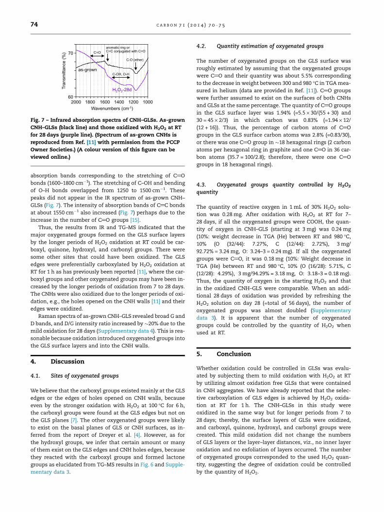
[15] Mawhinney DB, Naumenko V, Kuznetsova A, Yates Jr JT, Liu J,Smalley RE. Infrared spectral evidence for the etching ofcarbon nanotubes: ozone oxidation at 298 K. J Am Chem Soc2000;122:2383–4.


Recommended