Evolution of Conjugation and Type IV Secretion Systems Julien Guglielmini,* ,1,2 Fernando de la Cruz, 3 and Eduardo P.C. Rocha 1,2 1 De ´partement Ge ´nomes et Ge ´ne ´tique, Microbial Evolutionary Genomics, Institut Pasteur, Paris, France 2 CNRS, UMR3525, Paris, France 3 Departamento de Biologı ´a Molecular e Instituto de Biomedicina y Biotecnologı ´a de Cantabria (IBBTEC), Universidad de Cantabria-CSIC-SODERCAN, Santander, Spain *Corresponding author: E-mail: [email protected]. Associate editor: Howard Ochman Abstract Genetic exchange by conjugation is responsible for the spread of resistance, virulence, and social traits among prokaryotes. Recent works unraveled the functioning of the underlying type IV secretion systems (T4SS) and its distribution and recruitment for other biological processes (exaptation), notably pathogenesis. We analyzed the phylogeny of key conjugation proteins to infer the evolutionary history of conjugation and T4SS. We show that single-stranded DNA (ssDNA) and double-stranded DNA (dsDNA) conjugation, while both based on a key AAA + ATPase, diverged before the last common ancestor of bacteria. The two key ATPases of ssDNA conjugation are monophyletic, having diverged at an early stage from dsDNA translocases. Our data suggest that ssDNA conjugation arose first in diderm bacteria, possibly Proteobacteria, and then spread to other bacterial phyla, including bacterial monoderms and Archaea. Identifiable T4SS fall within the eight monophyletic groups, determined by both taxonomy and structure of the cell envelope. Transfer to monoderms might have occurred only once, but followed diverse adaptive paths. Remarkably, some Firmicutes developed a new conjugation system based on an atypical relaxase and an ATPase derived from a dsDNA translocase. The observed evolutionary rates and patterns of presence/absence of specific T4SS proteins show that conjugation systems are often and independently exapted for other functions. This work brings a natural basis for the classi- fication of all kinds of conjugative systems, thus tackling a problem that is growing as fast as genomic databases. Our analysis provides the first global picture of the evolution of conjugation and shows how a self-transferrable complex multiprotein system has adapted to different taxa and often been recruited by the host. As conjugation systems became specific to certain clades and cell envelopes, they may have biased the rate and direction of gene transfer by conjugation within prokaryotes. Key words: bacterial conjugation, horizontal gene transfer, type IV protein secretion, exaptation, plasmid evolution. Introduction Prokaryotic genomes adapt quickly to new environmental conditions largely because they can acquire pre-evolved traits by horizontal gene transfer (HGT) (de la Cruz and Davies 2000; Gogarten et al. 2002; Ochman et al. 2005). Conjugation is a mechanism of genetic transfer that allows single-event transfer of large DNA fragments, up to entire chromosomes. Conjugation can transfer nonhomologous genes to the recipient genome and has a broader host range than transduction or transformation (Amabile- Cuevas and Chicurel 1992; Llosa et al. 2002; Chen et al. 2005). Accordingly, recent work suggests that conjugation is the most frequent mechanism of HGT (Halary et al. 2010). Indeed, conjugative systems are major players in the spread of antibiotic resistance, metabolic pathways, symbiotic traits, and other mobile genetic elements (de la Cruz and Davies 2000; Thomas 2000; van der Meer and Sentchilo 2003; Frost et al. 2005; Ding and Hynes 2009; Allen et al. 2010). Conjugation is also involved in the establishment of social processes, promoting biofilm formation (Ghigo 2001) and spreading of cooperative traits (Nogueira et al. 2009; Rankin et al. 2011). There are two known modes of conjugation that differ both in the type of translocated DNA, single-stranded DNA (ssDNA) versus double-stranded DNA (dsDNA), and in the complexity of the transport system (de la Cruz et al. 2010; Vogelmann et al. 2011). Both types of conjugative systems are either encoded by autonomously replicating plasmids or in- serted in chromosomes as integrative conjugative elements (ICEs) (Smillie et al. 2010; Wozniak and Waldor 2010). We recently made a large-scale identification of ssDNA conjuga- tion systems, both in plasmids and ICEs, and found them to be essentially short-term variants of otherwise identical back- bone elements (Guglielmini et al. 2011). In the following, we note proteins from a given genetic element by GI MGE , where GI refers to the gene identification and mobile genetic element (MGE) to the name of the elem- ent (e.g., TraC F corresponds to the TraC protein of the F plasmid). Conjugative systems involved in ssDNA conjugation include two major protein complexes: relaxosomes and type IV secretion systems (T4SS) (reviewed in Fronzes et al. 2009; de la Cruz et al. 2010). MGE delivery through the membranes of the donor and recipient cells is done by the T4SS (fig. 1). In Proteobacteria, the T4SS are a large protein complex, includ- ing a ubiquitous ATPase (VirB4 Ti or the distant homolog TraU R64 ), mating-pair formation (MPF) proteins that form the transport channel, and a pilus that attaches to the recipient cell (Alvarez-Martinez and Christie 2009; Article ß The Author 2012. Published by Oxford University Press on behalf of the Society for Molecular Biology and Evolution. This is an Open Access article distributed under the terms of the Creative Commons Attribution Non-Commercial License (http:// creativecommons.org/licenses/by-nc/3.0/), which permits non-commercial reuse, distribution, and reproduction in any medium, provided the original work is properly cited. Open Access Mol. Biol. Evol. 30(2):315–331 doi:10.1093/molbev/mss221 Advance Access publication September 13, 2012 315 Downloaded from https://academic.oup.com/mbe/article-abstract/30/2/315/1013981 by guest on 11 April 2019
Transcript
Evolution of Conjugation and Type IV Secretion Systems
Julien Guglielmini,*,1,2 Fernando de la Cruz,3 and Eduardo P.C. Rocha1,2
1Departement Genomes et Genetique, Microbial Evolutionary Genomics, Institut Pasteur, Paris, France2CNRS, UMR3525, Paris, France3Departamento de Biologıa Molecular e Instituto de Biomedicina y Biotecnologıa de Cantabria (IBBTEC), Universidad de
Genetic exchange by conjugation is responsible for the spread of resistance, virulence, and social traits among prokaryotes. Recentworks unraveled the functioning of the underlying type IV secretion systems (T4SS) and its distribution and recruitment for otherbiological processes (exaptation), notably pathogenesis. We analyzed the phylogeny of key conjugation proteins to infer theevolutionary history of conjugation and T4SS. We show that single-stranded DNA (ssDNA) and double-stranded DNA (dsDNA)conjugation, while both based on a key AAA+ ATPase, diverged before the last common ancestor of bacteria. The two keyATPases of ssDNA conjugation are monophyletic, having diverged at an early stage from dsDNA translocases. Our data suggestthat ssDNA conjugation arose first in diderm bacteria, possibly Proteobacteria, and then spread to other bacterial phyla, includingbacterial monoderms and Archaea. Identifiable T4SS fall within the eight monophyletic groups, determined by both taxonomyand structure of the cell envelope. Transfer to monoderms might have occurred only once, but followed diverse adaptive paths.Remarkably, some Firmicutes developed a new conjugation system based on an atypical relaxase and an ATPase derived from adsDNA translocase. The observed evolutionary rates and patterns of presence/absence of specific T4SS proteins show thatconjugation systems are often and independently exapted for other functions. This work brings a natural basis for the classi-fication of all kinds of conjugative systems, thus tackling a problem that is growing as fast as genomic databases. Our analysisprovides the first global picture of the evolution of conjugation and shows how a self-transferrable complex multiprotein systemhas adapted to different taxa and often been recruited by the host. As conjugation systems became specific to certain clades andcell envelopes, they may have biased the rate and direction of gene transfer by conjugation within prokaryotes.
Key words: bacterial conjugation, horizontal gene transfer, type IV protein secretion, exaptation, plasmid evolution.
IntroductionProkaryotic genomes adapt quickly to new environmentalconditions largely because they can acquire pre-evolvedtraits by horizontal gene transfer (HGT) (de la Cruz andDavies 2000; Gogarten et al. 2002; Ochman et al. 2005).Conjugation is a mechanism of genetic transfer that allowssingle-event transfer of large DNA fragments, up to entirechromosomes. Conjugation can transfer nonhomologousgenes to the recipient genome and has a broader hostrange than transduction or transformation (Amabile-Cuevas and Chicurel 1992; Llosa et al. 2002; Chen et al.2005). Accordingly, recent work suggests that conjugation isthe most frequent mechanism of HGT (Halary et al. 2010).Indeed, conjugative systems are major players in the spread ofantibiotic resistance, metabolic pathways, symbiotic traits,and other mobile genetic elements (de la Cruz and Davies2000; Thomas 2000; van der Meer and Sentchilo 2003; Frostet al. 2005; Ding and Hynes 2009; Allen et al. 2010).Conjugation is also involved in the establishment of socialprocesses, promoting biofilm formation (Ghigo 2001) andspreading of cooperative traits (Nogueira et al. 2009; Rankinet al. 2011). There are two known modes of conjugation thatdiffer both in the type of translocated DNA, single-stranded
DNA (ssDNA) versus double-stranded DNA (dsDNA), and inthe complexity of the transport system (de la Cruz et al. 2010;Vogelmann et al. 2011). Both types of conjugative systems areeither encoded by autonomously replicating plasmids or in-serted in chromosomes as integrative conjugative elements(ICEs) (Smillie et al. 2010; Wozniak and Waldor 2010). Werecently made a large-scale identification of ssDNA conjuga-tion systems, both in plasmids and ICEs, and found them tobe essentially short-term variants of otherwise identical back-bone elements (Guglielmini et al. 2011).
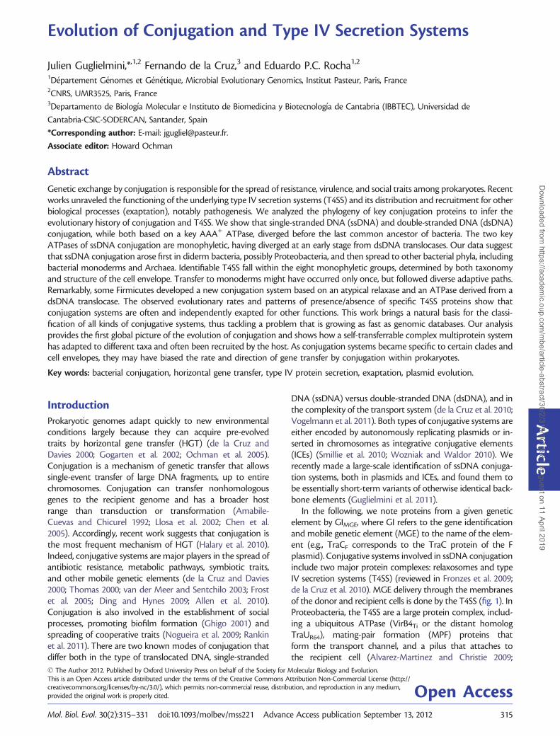
In the following, we note proteins from a given geneticelement by GIMGE, where GI refers to the gene identificationand mobile genetic element (MGE) to the name of the elem-ent (e.g., TraCF corresponds to the TraC protein of the Fplasmid). Conjugative systems involved in ssDNA conjugationinclude two major protein complexes: relaxosomes and typeIV secretion systems (T4SS) (reviewed in Fronzes et al. 2009;de la Cruz et al. 2010). MGE delivery through the membranesof the donor and recipient cells is done by the T4SS (fig. 1). InProteobacteria, the T4SS are a large protein complex, includ-ing a ubiquitous ATPase (VirB4Ti or the distant homologTraUR64), mating-pair formation (MPF) proteins thatform the transport channel, and a pilus that attaches tothe recipient cell (Alvarez-Martinez and Christie 2009;
Article
� The Author 2012. Published by Oxford University Press on behalf of the Society for Molecular Biology and Evolution.This is an Open Access article distributed under the terms of the Creative Commons Attribution Non-Commercial License (http://creativecommons.org/licenses/by-nc/3.0/), which permits non-commercial reuse, distribution, and reproduction in any medium,provided the original work is properly cited. Open AccessMol. Biol. Evol. 30(2):315–331 doi:10.1093/molbev/mss221 Advance Access publication September 13, 2012 315
Dow
nloaded from https://academ
ic.oup.com/m
be/article-abstract/30/2/315/1013981 by guest on 11 April 2019
Fronzes et al. 2009). The large (>70 kDa) VirB4 ATPase ishighly conserved in sequence and the only protein withclear-sequence homologs in all known T4SS. It is thereforethe marker of the presence of a T4SS (Alvarez-Martinez andChristie 2009). VirB4 is thought to energize the assembly oractivity of the secretion channel and is essential for pilusbiogenesis and substrate transfer (Berger and Christie 1993;Fullner et al. 1996; Wallden et al. 2012). Four MPF familieshave been described in Proteobacteria: MPFT (based on theT-DNA conjugation system of A. tumefaciens plasmid Ti),MPFF (based on plasmid F), MPFI (based on the IncI plasmidR64), and MPFG (based on ICEHIN1056) (Smillie et al. 2010).These four models describe all functionally studied and nearlyall T4SS identified by bioinformatic methods amongProteobacteria, both in plasmids and chromosomes(Guglielmini et al. 2011). The best-studied system is the viroperon (MPFT) from A. tumefaciens Ti plasmid. This smalloperon encodes 11 VirB proteins (Thompson et al. 1988;Ward et al. 1988), and we use these names as a templatefor naming the protein families of the MPFT system. T4SSfrom Cyanobacteria, Bacteroides, Firmicutes, Actinobacteria,and Archaea have homologs to VirB4 (Guglielmini et al. 2011).ssDNA-conjugative systems are very diverse, but very fewstudies have been done on the structure, function, and evo-lution of T4SS outside Proteobacteria and Firmicutes.
The two other essential components of the ssDNA conju-gation machinery are the relaxosome and the type IV cou-pling protein (T4CP). The relaxosome is composed of therelaxase (MOB) and often includes auxiliary proteins. Itnicks the dsDNA and binds the resulting ssDNA at theorigin of transfer. The diversity and evolution of the differentfamilies of relaxases has been extensively studied (Garcillan-Barcia et al. 2009). The highly conserved T4CP binds theDNA-relaxase substrate and couples it to the T4SS, possibly
using ATP to translocate the complex across the inner mem-brane (Gomis-Ruth et al. 2004; Tato et al. 2005). The majorityof T4CPs belong to the VirD4Ti family, but some T4SS wererecently found to lack VirD4 and instead use a distantlyrelated ATPase as T4CP (TcpApCW3) (Parsons et al. 2007;Steen et al. 2009). Protein secretion systems based on T4SSdo not require relaxosomes. They usually require T4CP, albeitexceptions have been found in Bordetella pertussis andBrucella spp. (Alvarez-Martinez and Christie 2009). In thesesystems, proteins are translocated across the inner membraneby other means.
Conjugation of dsDNA takes place in mycelia-producingActinobacteria (Grohmann et al. 2003; Ghinet et al. 2011). Itrelies on a single protein: TraBpSG5 that translocates dsDNAbetween neighboring cells in mycelia (Possoz et al. 2001). Thisprotein resembles, in sequence and function, the essentialprotein FtsK that segregates sister chromosomes in the laststages of chromosomal replication (Bigot et al. 2007;Vogelmann et al. 2011). They are both members of theAAA+ motor ATPase family, which also includes both typesof T4CP (VirD4 and TcpA) and both types of ATPases essen-tial for the function of T4SS (VirB4 and TraU). Hence, all keyproteins of the dsDNA and ssDNA conjugation systems areevolutionarily related. This association has not yet been clar-ified from a phylogenetic point of view.
T4SS are often recruited by bacterial pathogens to deliver ef-fectors to eukaryotic cells (Weiss et al. 1993; Vogel et al. 1998;Seubert et al. 2003; Nystedt et al. 2008). These MOBless T4SS,called so because they do not contain a relaxase gene, areclosely related to the T4SS of conjugative systems. Indeed,several T4SS can perform both conjugation between bacteriaand protein delivery (Vogel et al. 1998; Llosa et al. 2003;Schroder et al. 2011). Protein delivery by T4SS is essential forthe virulence of many plant and animal pathogens, includingLegionella pneumophila, Helicobacter pylori, Bartonella spp.,Coxiella burnetii, and A. tumefaciens (reviewed in Seubertet al. 2003; Juhas et al. 2008; Alvarez-Martinez and Christie2009). Only T4SS among MPFT and MPFI have been experi-mentally shown to be used for protein delivery. The extremeflexibility of T4SS has allowed at least two other types of ex-aptations, i.e., evolutionary events in which part of thepre-existing machinery of conjugation was recruited forother functions (Gould and Vrba 1982). H. pylori genomesencode a MOBless T4SS that is used for natural transform-ation. It is necessary to import environmental DNA (Hofreuteret al. 2001). In Neisseria gonorrhoeae, one T4SS is responsiblefor DNA export to the extracellular space, an intermediatestep in the process of natural transformation among thesebacteria (Hamilton et al. 2005). Interestingly, in the case ofNeisseria, the locus encodes a T4SS and a MOBH-type relaxasethat is necessary for DNA export (Salgado-Pabon et al. 2007).A previous analysis of MPFT systems suggests that exaptationof conjugative systems occurred several times in evolution(Frank et al. 2005). Because we recently found that MOBlessT4SS are significantly more abundant than previously thought(Guglielmini et al. 2011), this point needs to be reassessed forMPFT and developed for other MPF types.
D4
B11
B6
B8 B10
B2/B5
B7/B9
IM
OM
B3/B4
B1
MOB
FIG. 1. Scheme of the most-studied T4SS, the vir system ofA. tumefaciens Ti plasmid. The VirBX proteins are depicted as BX (e.g.,B5 refers to the VirB5 protein). The coupling protein VirD4 (D4) and themobilization complex, which includes the relaxase (MOB)-DNA com-plex are also represented. OM: outer membrane; IM: inner membrane.
316
Guglielmini et al. . doi:10.1093/molbev/mss221 MBED
ownloaded from
https://academic.oup.com
/mbe/article-abstract/30/2/315/1013981 by guest on 11 April 2019
Although studies on conjugation are as old as molecularbiology itself (Lederberg and Tatum 1946), several recentworks have significantly changed our understanding of thisprocess. These include the discovery of new conjugation sys-tems (Juhas, Crook, et al. 2007), of new key elements in knownconjugation systems, e.g., TcpA (Parsons et al. 2007) and ofthe important role of ICEs (Burrus et al. 2002; Wozniak andWaldor 2010). Recent functional studies explored the diver-sity of T4SS (Alvarez-Martinez and Christie 2009), and bio-informatics work unraveled the presence of T4SS in severalnew clades (Guglielmini et al. 2011). Finally, other works high-lighted the close structural and functional relationship be-tween T4SS used for protein secretion and conjugation(Fernandez-Gonzalez et al. 2011). This succession of worksopens the opportunity to infer a global scenario for the evo-lution of conjugative systems and T4SS, which is the goal ofthe present work. To assess the uncertainty in the phylogen-etic reconstruction, we used classical methods such as boot-strap analyses. Yet, because these large and deep phylogeneticreconstructions can be sensitive to alignment algorithms andto methods to extract informative positions (Philippe et al.2011), we also tested the robustness of our results by compar-ing them with two automatic analyses that we did in parallel.To guide the comparisons between the three sets of analyses,we made an assessment of the quality of the multiple align-ments using T-Coffee (Notredame et al. 2000). By default, weonly mention the results of our expert analysis (typically, theone with highest alignment quality), but highlight differencesbetween methods when they are relevant. The overall struc-ture of the article is the following. First, we analyze the deepbranching of the key proteins that have homologs among(nearly) all conjugative systems of a given kind. This allowsuncovering the initial split of the proteins that became key toconjugative processes. Then, we focus on the early events ofthe diversification of ssDNA conjugation, by far the mostfrequent process among prokaryotes. Finally, we detail the di-versification of the best-known conjugation families withinssDNA-based systems with a focus on the evolution of generepertoires and MOBless T4SS. This analysis provides infor-mation that naturally leads to a revision of T4SS classificationbased on evolutionary biology.
Materials and Methods
Data
Data on complete chromosomes and plasmids of prokaryoteswere taken from Genbank Refseq (ftp://ftp.ncbi.nih.gov/genomes/Bacteria/, last accessed November 2011). This included1,207 chromosomes, 891 plasmids that were sequenced alongwith these chromosomes, and 1,391 plasmids that weresequenced independently. We used the annotations of theGenbank files, having removed all pseudogenes and proteinswith inner stop codons. The information on T4SS was takenfrom Guglielmini et al. (2011).
Construction of Protein Profiles and Genome Searches
Unless mentioned explicitly, the protein profiles used arethose described in Guglielmini et al. (2011). To study the
presence/absence of the different components of the vir sys-tem, we made additional protein profiles, namely for VirB1,VirB2, VirB5, VirB7, VirB10, and VirB11. We first used PSI-BasicLocal Alignment Search Tool (BLAST) (e value< 0.1) tosearch for distant homologs, using as query each of thesegenes from the VirB locus of the A. tumefaciens plasmid pTiSAKURA (Refseq entry NC_002147) and the aforementioneddatabank of completely sequenced replicons. Given the prob-lems of convergence of PSI-BLAST when using completegenomes, and the extensive similarity of plasmid and chromo-somal conjugative systems (Guglielmini et al. 2011), we re-stricted homology searches to plasmid sequences whenbuilding protein profiles. We retrieved the proteins withhits for each protein family and built multiple alignmentsusing MUSCLE (Edgar 2004). We removed the few proteinswith sizes very different from the average. We then rebuilt themultiple alignments with MUSCLE and trimmed them toremove the sites at the edges that were poorly aligned. Weused HMMER 3.0 (Eddy 2011) to produce hidden Markovmodel (HMM) profiles and to perform searches within gen-omes. In the analysis of the evolution of the MPFT system, weonly considered the hits that colocalized with previously de-tected vir proteins (VirB3, VirB4, VirB6, VirB8, VirB9). FtsKproteins were retrieved directly by using the PFAM PF01580profile. TraB proteins, being closely related to FtsK, wereretrieved by BLASTP searches of TraB from Streptomyces plas-mid pCQ3 (YP_003280879) on the Actinomycetales proteinsfrom the Refseq database. We sampled the top results andthen built a protein profile for this protein and searched for itsoccurrences as for the other profiles. We built a web server toallow running the protein profiles. This is available at http://mobyle.pasteur.fr/cgi-bin/portal.py#forms::CONJscan-T4SSscan.
Phylogenetic Analysis
Unless explicitly stated, all phylogenetic analyses were per-formed with the following procedure. First, sequenceswere aligned using MUSCLE with default parameters asimplemented in SeaView (Gouy et al. 2010). Second, all col-umns in the multiple alignment matrix with more than 80%of gaps were removed. Third, 100 replicate trees were builtwith RAxML 7.2.7 (Stamatakis 2006) using the modelGTRGAMMA. We kept the one with the best likelihood.We calculated bootstraps with the standard implementationand used the autoMR stop criterion to obtain confidencevalues for each node. There were two exceptions to thismethod. We aligned the ATPases using MAFFT (Katoh andToh 2010) with the G-INSI algorithm and removed the sitescontaining more than 60% of gaps. We performed the phylo-genetic inference as mentioned earlier and additionally withPhyML 3.0 (Gascuel et al. 2010) under the LG model and withthe bioNJ starting tree to get aLRT support values. The align-ment of the set of VirB4 and VirD4 was built with MAFFTwith the E-INSI algorithm, since these two proteins showdifferent domain organization, and then manually edited.MAFFT was used instead of MUSCLE because it providedbetter alignments in these cases. The computation of
317
Evolution of Conjugation and T4SS . doi:10.1093/molbev/mss221 MBED
ownloaded from
https://academic.oup.com
/mbe/article-abstract/30/2/315/1013981 by guest on 11 April 2019
100 replicates plus hundreds of bootstrap trees was exces-sively time consuming, given the size of the data set in theVirB4/VirD4 analysis. Thus, we used PhyML 3.0 to build thephylogenetic tree, under the LG model and with the bioNJstarting tree. aLRT support values were also calculated foreach node.
The support tests we conducted revealed in this lasttree some weak support that conflict with the aLRT values.To further investigate this, we used a reduced data setcomposed of VirB4 proteins, excluding the distant homologTraU. Using this data set, we performed the testsdescribed later. All multiple alignments and phylogeneticreconstructions are freely available on DRYAD (http://datadryad.org/).
Tests to the Phylogenetic Analysis
To test the robustness of our conclusions based on phylo-genetic analysis, we made a number of tests. These analysesaimed at testing the robustness of the conclusions to themultiple alignments, to the identification of informativesites in multiple alignments, and to the use of a proteinmodel matrix. We therefore produced two automatic meth-ods where we make the alignment of the protein usingMAFFT and MUSCLE. Informative sites were extracted fromthe alignments using BMGE (Criscuolo and Gribaldo 2010).We fine-tuned BMGE parameters for each alignment toobtain a good compromise between the quality and thenumber of informative sites. The best model to analyze thedata was chosen with ProtTest (Darriba et al. 2011). Note thatProtTest does not analyze the GTR model for proteins, so wecannot assess whether the model chosen by ProtTest is betterthan ours. Trees were built as before using RAxML, and wegenerated 100 bootstrap trees for each analysis. To comparethe different analyses, we computed the quality of multiplealignment score using the Core component of T-Coffee(Notredame et al. 2000) for the three methods (our expertanalysis, the MAFFT and MUSCLE-based analyses). This score,ranging from 0 to 100, is computed by comparing the con-sistency of the alignment with a list of precomputed pairwisealignments called library. We used the default “Mproba_pair”library. The key results, e.g., monophyly or basal position ofcertain clades, were tested for the three methods and aredisplayed in table 1 and supplementary table S1, Supplemen-tary Material online. Each of these tests has an identificationnumber in the tables. This number is displayed in the respect-ive node in the phylogenetic trees. For example, in figure 2,the node with ID no. 3 refers to the monophyly of TraB and isindicated in table 1 as having 99% bootstrap support in ourexpert analysis, 100% in the automatic analysis using MAFFT,and 96% in the automatic analysis using MUSCLE. In supple-mentary table S1, Supplementary Material online, it is indi-cated that for this analysis the best alignment, as given byT-Coffee, is the one of the expert alignment (score 88), fol-lowed by MAFFT (76) and then MUSCLE (67). The node no. 3in figure 2 is thus indicated in a black circle (high bootstrapsupport).
Relative Decrease in Protein Similarity with DivergenceFor each pair of T4SS loci, we made pairwise alignments ofeach of the orthologous pairs of genes. Alignments were doneusing an end-gap free version of the Needleman–Wunschalgorithm (Mount 2004), with a BLOSUM60 matrix, openpenalty of 1.2, and extension penalty of 0.8. We then plottedthe percentage of similarity between VirB4 homologs andeach of the other pairs of homologs. The points for eachscatter plot were then fitted with a spline (l= 1,500), andthe curves were superimposed.
Results and Discussion
Early Evolutionary Split of the Key Conjugation ATPases
The two families of T4CPs (with prototypes given by theVirD4pTi and TcpApCW3), the two families of ATPases(based on VirB4Ti and TraUR64), the dsDNA conjugation pro-tein TraBpSG5, and FtsK are all part of the superfamily of AAA+
motor ATPases. Hence, we investigated the events at theonset of the natural history of conjugation from the analysisof the phylogeny-linking homologs for all these protein pro-files among 3,489 replicons (see Materials and Methods). Thetree was rooted using the distantly related protein familyderived from VirB11Ti (Planet et al. 2001). The monophylyof VirB11 is robust in both expert and the automatic analyses(table 1). This phylogenetic reconstruction separates a mono-phyletic VirD4/VirB4 clade (67% boostrap) from the others.This fits previous genomic and structural analysis showing thesimilarity between the dsDNA translocators FtsK and TraB onthe one hand and between the ssDNA translocators VirD4and VirB4 on the other (Iyer et al. 2004; Cabezon et al. 2011).
The previous analysis allows rooting the tree and highlightsthe early split between ssDNA and dsDNA translocases. Butthe inclusion of the distantly related VirB11 produces a mul-tiple alignment with few sufficiently conserved positions,increasing uncertainty in the process of phylogenetic infer-ence (supplementary table S1, Supplementary Materialonline). This reduced the power of this data set to robustlyresolve the more recent splits. Thus, we excluded VirB11 fromthe analysis and made a new phylogenetic reconstruction ofthe remaining five families. This tree shows the same dichot-omy at the base (fig. 2), with strong support for all five mono-phyletic groups with our expert analysis and in the bestautomatic method (table 1). These results fit our observationthat our VirB4 protein profiles often match VirD4 proteinsand vice versa, albeit with weak scores, and that none of thesematch significantly proteins from the families TraB/FtsK.T4CPs and VirB4s show clear structural similarities, under-scoring a common functional mechanism (Cabezon et al.2011). The most conspicuous structural difference betweenT4CPs and VirB4s is the existence of three alpha helices thatare conserved in the C terminus of VirB4 proteins but areabsent in T4CPs. Deletion of these helical structures in theVirB4 homolog TrwKR388 resulted in a large increase in itsATPase activity, suggesting that the C-terminal end ofVirB4 proteins functions as an autoregulatory element(Pena et al. 2011). Overall, these analyses fit structural work,suggesting that the common ancestor of the VirB4/VirD4
318
Guglielmini et al. . doi:10.1093/molbev/mss221 MBED
ownloaded from
https://academic.oup.com
/mbe/article-abstract/30/2/315/1013981 by guest on 11 April 2019
Evolution of Conjugation and T4SS . doi:10.1093/molbev/mss221 MBED
ownloaded from
https://academic.oup.com
/mbe/article-abstract/30/2/315/1013981 by guest on 11 April 2019
families consisted of a soluble protein engaged in polypeptidetransport (as it’s still the case in most studied VirB4 proteins).VirB4 later became membrane bound by association with theVirB3 component of T4SS (as in VirB4R388). This associationcan be covalent (as in VirB4R6K) (Pena et al. 2011). The proteinthat specialized in ssDNA transport (T4CP) also acquired anintegral-membrane protein domain in its N-terminus. Thiscomponent is involved in its interaction with another T4SScomponent, in this case VirB10 (Llosa et al. 2003; de Paz et al.2010).
The other basal branch in the phylogeny includes TraB,TcpA, and FtsK, all with strong to moderate evidence ofmonophyly (99%, 96%, and 62% bootstraps, respectively)(fig. 2). The relative order of the split between the threeclades is different from a previously published one, but itsbootstrap support is weak in our tree (and not documentedin Parsons et al. 2007). SpoIIIE, a protein involved in segrega-tion of chromosomes during Bacillus subtilis sporulation (Wuand Errington 1998), branches within the FtsK clade (data notshown). The elements of the TraB family are found only in
Actinobacteria and are related with FtsK, but they do notemerge from within the FtsK. Instead, they derive independ-ently from the ancestor of this protein. FtsK is an essentialprotein that, contrary to some previous suggestions (Iyer et al.2004), includes at least one member among Archaea(YP_503307.1). The latter is annotated as FtsK-like protein,and it is not closely related with HerA proteins, which branchcloser to the VirD4/B4 branches, and its study falls outside thescope of this article. FtsK phylogeny follows approximatelythe one of bacteria (Gupta 2004) and thus provides a guide-line to the timing of the diversification of these proteinfamilies. The tree in figure 2 shows that proteins havewidely diverse tip-to-root branch lengths, i.e., the proteinsdo not evolve according to a strict molecular clock. Therefore,we cannot assume a molecular clock that would allow datingthe split of these families and thus presumably that ofconjugation processes. Yet, this data does place the originof ssDNA conjugation extremely early in the history oflife. While TraB and TcpA seem to diversify after FtsK, inagreement with their presence only in Firmicutes and
AcidobacteriaProteobacteriaPlanctomycetes
FusobacteriaDeinococcus-ThermusBacteroidetes
ChlorobiFirmicutes
ActinobacteriaChloroflexiChlamydiae
ArchaeaCyanobacteria
0.5
FtsK
TraB(Actinobacteria)
TcpA(Firmicutes)
VirD4
VirB4
1
2
3
4
5
FIG. 2. Phylogenetic analysis of the AAA+ ATPases associated with conjugation. The position of the root was determined using the AAA+ ATPaseVirB11 in a separate analysis. Names along the FtsK tips correspond to the taxonomic origins of each protein, reflecting the width of sampling. Boldvertical black lines represent nodes with a high support value (bootstrap >70% and aLRT >0.7). Bold gray lines represent nodes with high aLRT score(>0.7) but a weaker bootstrap (<70%). The homologs of TcpA are found only in Firmicutes. The homologs of TraB are found only in Actinobacteria.Numbers in circles refer to the analysis of robustness in table 1 (identified in the third column of table 1); black background stands for a high support(�70% bootstrap in the best-scoring alignment) and gray background for a moderate support (�50% bootstrap in the best-scoring alignment).
320
Guglielmini et al. . doi:10.1093/molbev/mss221 MBED
ownloaded from
https://academic.oup.com
/mbe/article-abstract/30/2/315/1013981 by guest on 11 April 2019
Actinobacteria, the diversification of the pair VirB4/VirD4could be contemporaneous or shortly subsequent to thatof FtsK. These results suggest that the two conjugation mech-anisms, ssDNA and dsDNA conjugation, are based onATPases that diverged before the last common ancestor ofbacteria.
T4SS Phylogeny
We aligned the proteins matching the VirB4 and TraU profilesto infer the evolutionary history of all VirB4 homologs. Wethen used VirD4 to root this tree. Despite relatively weaksupport in the bootstrap tests (48% in the best automaticalignment and 69% in our expert analysis), this rooting showsa good aLRT support value (0.82), consistent with the litera-ture in terms of phylogeny and biochemical function (Iyeret al. 2004; de la Cruz et al. 2010; Smillie et al. 2010) and withthe previous analysis of the five ATPases (78% boostrap). Thetree shows that all VirB4 and TraU-related proteins can beclassified into eight groups, which are represented by eightwell-supported clades (fig. 3). The two basal groups in theVirB4 phylogenetic reconstruction are MPFI followed by agroup specific to Cyanobacteria (MPFC). This is in agreementwith the low similarity between TraUR64 (MPFI) and VirB4Ti
(MPFT) that had prevented previous phylogenetic recon-structions of all VirB4 homologs (Smillie et al. 2010). Withthe availability of more sequences of these proteins, notablycyanobacteria, and the inclusion of the T4CP, we could nowreconstruct a reliable phylogeny. However, the position ofMPFI at the basis of the tree must be taken with care. Ourexpert method and the two controls produce MPFI at thebasis of the phylogeny but with relatively low support (45%bootstrap in the best automatic alignment) (table 1). TheMPFC clade often arises at the basis in the bootstrap treesor as a sister clade of MPFI. In any case, this analysis places oneof these two clades at the root of the tree in more than 85% ofthe boostrap analyses.
Some mobile elements encoding an MPFI, e.g., the R64plasmid from the MOBP12 family, besides encoding a thickrigid pilus, with homology to MPFT, also encode a thin pilusthat is only required for conjugation in liquid and that ishomologous to type IV pili (Kim and Komano 1997). Thisled to the classification of MPFI as T4SSb in opposition toMPFF and MPFT, both classed as T4SSa (Christie and Vogel2000). However, other MPFI elements, e.g., plasmid CTX-M3,lack a thin pilus and are still able to mate in liquid at highfrequency (Golebiewski et al. 2007). Thus, the thin pilus ofMOBP12 plasmids is just an additional feature of some MPFI
systems, acting probably just as a facilitator of liquid matingand a selector of recipients (Kim and Komano 1997), whilethe core MPFI machinery forms the basis of this conjugationsystem. In any case, the highly divergent nature of TraUR64 is asignature for this whole family of liquid maters. Nothing isknown experimentally about MPFC. Because cyanobacteriadiverged early on from Proteobacteria, MPFC might also con-tain peculiarities relevant to the genetic or physical environ-ment of these organisms. MPFG is the next most basal groupin the tree. This system was recently discovered, was identified
only in Proteobacteria, and its features are largely unknown(Juhas, Crook, et al. 2007; Juhas, Power, et al. 2007). Interest-ingly, an MPFG encoding element, the PAPI-1 pathogenicityisland of Pseudomonas aeruginosa, has several genes homolo-gous to the thin pilus of R64 (Carter et al. 2010). Hence, theassociation between MPF and thin pili might be an ancestraltrait.
Four groups correspond to the different T4SS families ofProteobacteria (MPFF, MPFG, MPFI, MPFT) (Juhas, Crook, et al.2007; Smillie et al. 2010). These four groups are clearly sepa-rated because they all have strong bootstraps in the analysis ofmonophyly (table 1), and each contains a set of four to ninegenes that are specific, i.e., their protein profiles match loci ofa given MPF but not those of the other MPF types (Smillieet al. 2010). Interestingly, 307 out of 327 (94%) of the T4SS ofProteobacteria are classed in one of these four clades. Weinvestigated the loci of the 20 remaining VirB4 proteins.One of them does not colocalize with any of the other con-jugation protein profiles, including relaxases and T4CP. Theother 19 VirB4 are encoded near genes specific of one, andonly one, MPF type. They were not classed as a given MPF justbecause the number of these specific genes is below thequorum we set up as a minimum for a putative completeT4SS (Guglielmini et al. 2011). Many of these 20 unclassedelements are thus probably inactive, enduring a genetic deg-radation that results in incomplete loci. Alternatively, theymay correspond to highly modified versions of T4SS; theH. pylori Cag-pathogenicity island is notably found withinthese elements.
A few genomes of species not classed among Proteobac-teria encode T4SS classed within MPFF and MPFT. All thesebacteria are diderms, i.e., they have both an inner and anouter membrane. This list includes MOBless T4SS in oneAquificae (MPFF) and one Protochlamydia (MPFF), and con-jugative T4SS in one Chlorobi (MPFT), one Deferribacteres(MPFF), one Acidobacteria (MPFT), and two Fusobacteria(MPFT). These elements are scattered in the trees of MPFT
and MPFF (figs. 4 and 5), suggesting different events of hori-zontal transfer from Proteobacteria. Indeed, they do not clus-ter together in the phylogenetic trees (0% in bootstrap trees).The elements of each given bacterial clade are always mono-phyletic, suggesting one single transfer event, but the verysmall number of such elements does not allow any robustconclusions for the moment. Only one nonproteobacterialclade, Acidobacteria, is basal in the tree of MPFT (100% boot-straps in the expert analysis and the controls). Acidobacteriaare often regarded as a sister clade of Proteobacteria (Ciccar-elli et al. 2006), and therefore, we cannot discard the possi-bility of a diversification of MPFT before the split betweenAcidobacteria and Proteobacteria. However, since MPFG andMPFI are more basal in the tree of VirB4 (fig. 3), and both onlyfound in Proteobacteria, the scenario of a transfer from Pro-teobacteria to Acidobacteria remains more parsimonious.Interestingly, all T4SS predicted in these six nonproteobacter-ial clades were classed among MPFF and MPFT. Nothing isknown about conjugation in these clades, but this data sug-gest they might use mechanisms closely related to, and ori-ginating from, those of Proteobacteria.
321
Evolution of Conjugation and T4SS . doi:10.1093/molbev/mss221 MBED
ownloaded from
https://academic.oup.com
/mbe/article-abstract/30/2/315/1013981 by guest on 11 April 2019
Phylogeny of the T4CP at the Light of VirB4Phylogeny
The trees of VirD4 and VirB4 are not congruent (ELW confi-dence value: 0, and SH P value< 0.01). Yet, they share manyfeatures (fig. 3). The proteins encoded by the virD4 genescolocalizing in replicons with virB4 tend to form similarclades. Notably, the VirD4 associated with each of six of theeight VirB4 clades also clustered in nearly monophyleticclades of T4CP (MPFFA, MPFFATA, MPFB, MPFG, MPFI, and
MPFC). VirD4 of the two remaining clades (MPFT andMPFF) are scattered in a small number of clades. Most ofthe MPFFA use TcpA instead of a VirD4-like T4CP (seelater). The few VirD4 proteins found in MPFFA are also mono-phyletic (orange in the bottom of fig. 3). It was previouslyshown that plasmid T4CP are sometimes scattered in differ-ent groups corresponding to given relaxases (Smillie et al.2010). This result is still valid with the present much largerdata set. For example, the T4CP clade with a mixture of MPFT
MPFFATA
MPFFA
MPFF
MPFB
MPFT
MPFG
MPFI
MPFC
VirB4
VirD4
0.5
MPFT + MPFF
MOBP
MOBF,Q
MOBQ
MOBBMOBP,F,HMOBH
MOBC
MOBF
6
7
8
9
10101111
1212
1515
1313
FIG. 3. Joint phylogenetic reconstruction of the VirD4 and VirB4/TraU families of proteins from conjugative systems. Bold vertical black lines representnodes with a high support value (aLRT� 0.9), and black vertical gray lines represent nodes with a support value between 0.7 and 0.9. Black squarebrackets indicate the VirB4 and VirD4 clades; colored square brackets on the left delimit the different MPF clades (purple: MPFFATA, orange: MPFFA, red:MPFF, black: MPFB, blue: MPFT, yellow: MPFG, cyan: MPFC, green: MPFI); colored square brackets on the right delimit the relaxase clades within the VirD4part of the tree (blue: MOBP, green: MOBQ, red: MOBF, purple: MOBB, orange: MOBH, brown MOBC, red/green dashed brackets: clades with a mix ofMOBF and MOBQ; black: mix of MOBP, MOBF and MOBH). Numbers in circles refer to the analysis of robustness in table 1 (identified in the thirdcolumn of table 1); black background stands for a high support (�70% bootstrap in the best-scoring alignment) and gray background for a moderatesupport (�50% bootstrap in the best-scoring alignment).
322
Guglielmini et al. . doi:10.1093/molbev/mss221 MBED
ownloaded from
https://academic.oup.com
/mbe/article-abstract/30/2/315/1013981 by guest on 11 April 2019
and MPFF has one type of relaxase in common (MOBF). Onthe other hand, some relaxase types are scattered amongdifferent VirD4 clades that follow MPF types, e.g., the VirD4associated with MPFC is monophyletic and includes threedifferent relaxases, which are also found in other MPF types.Hence, evolution of conjugation is driven by two main con-straints, one acting mainly on the T4SS, represented by VirB4,and other on the relaxosome, represented by the relaxases.T4CP tends to coevolve with both components.
Cell Envelope Adaptation in Monoderms
The most basal clades in both VirB4 and VirD4 phylogeniescorrespond to bacteria with both inner and outer
membranes, i.e., diderms (98–100% of the bootstrap treesin all three analyses). This strongly suggests that ssDNA con-jugation was invented among diderms. In this scenario,ssDNA conjugation would have been acquired by monodermprokaryotes, i.e., organisms devoid of an outer membrane, byHGT. This also fits the observation that all monoderm con-jugation systems are in two sister clades: MPFFA and MPFFATA
(monophyletic in 67–55% of the bootstrap trees).MPFFATA includes six distinct groups of Firmicutes (mono-
phyly of all Firmicutes supported by 0% of the bootstrap trees,table 1), two of Actinobacteria (monophyly of all Actinobac-teria supported by 0% bootstrap trees), one of Tenericutes(monophyly of the clade supported by 96–99% bootstrap
R721
R388pKM101
RP4
R6K
pRA3
pCRY
pTi SAKURA (MOBP)
pMOL28
ICE Tn4371
0.6
Chlorobi
Fusobacteria
AcidobacteriaOutgroup
ICE MlSymR7A
pTi SAKURA (MOBQ)
Bartonella Trw T4SS
Brucella VirB T4SS
Helicobacter ComB T4SS
L. pneumophila LvhB system
1 2 3 4 5 6 7 8 9 10 11
1 2 3 4 6 5 7 8 9 10 11
1 2 3 4 5 67 8 9 10 11
11 2 3 4 5 6 7 8 9 10 1
1 2 3 4 5 67 8 9 10111 2 3 4 5 6 78 9 10 11
1818
1919
FIG. 4. Phylogenetic analysis of MPFT VirB4 proteins. Bold vertical black lines represent nodes with a high support value (bootstrap> 90%), and boldvertical gray lines represent nodes with a support value between 70% and 90%. Green branches correspond to taxa that are not within Proteobacteria(or the outgroup). Red branches represent VirB4 not associated to a relaxase (MOBless T4SS). The leftmost vertical bar on the right stands forchromosomal (black) or plasmidic (white) proteins. The colored bar represents the different gene order patterns found; the patterns and theircorresponding color are depicted at the bottom (the numbers represent the corresponding virB gene); a pattern is attributed to a system if, consideringthe possibly missing vir genes, the gene order is preserved. For example, a system composed of the genes virB1, virB4, virB6, virB5, virB8, virB9, and virB10in this order will be assigned to the orange pattern. Unique or atypical patterns are depicted in black. Known representative systems are labeled.Numbers in circles refer to the analysis of robustness in table 1 (identified in the third column of table 1); black background stands for a high support(�70% bootstrap in the best-scoring alignment) and gray background for a moderate support (�50% bootstrap in the best-scoring alignment).
323
Evolution of Conjugation and T4SS . doi:10.1093/molbev/mss221 MBED
ownloaded from
https://academic.oup.com
/mbe/article-abstract/30/2/315/1013981 by guest on 11 April 2019
trees), and a group of Archaea unlikely to be monophyletic(bootstrap of only 17–29%) with a clear separation betweenEuryarchaeota and Crenarchaeota (91–96%, respectively, and100% bootstrap support for each clade) (fig. 6). The deeperrelations between these clades are difficult to disentangle,given the low bootstrap supports of the basal nodes.Within the Firmicutes clades, we find the main divisions,i.e., Bacillales, Lactobacillales, and Clostridia, scattered in thetree. This suggests that, once a conjugative system arose inthis phylum, it spreads early among the main divisions, andtransfers between divergent clades were maintained througha certain moment in evolution. The monophyly of mono-derms in the VirB4 tree suggests that monoderms acquiredconjugative systems by transfer from diderms. This earlyacquisition was followed by the adaptation of the T4SS tomonoderms. Finally, frequent conjugation between diderms
contributed to the scattered distribution of taxa in the phylo-genetic tree of MPFFATA and MPFFA.
The MPFFA clade includes two groups of Actinobacteriaintermingled with three groups of Firmicutes (<5% bootstrapsupport for a net separation of the two clades) (fig. 7). Themost basal group (Firmicutes III in fig. 7) is constituted by afew elements from Firmicutes (bootstrap support for thisbasal position of 52–100%, table 1). This suggests that theancestral conjugative system might have arisen withinFirmicutes from which it was transferred to Actinobacteria.This is consistent with the observation of a basal group,including only Firmicutes and Tenericutes in the sisterMPFFATA tree (fig. 6). The subsequent split in the MPFFA
group separates a clade with Actinobacteria and FirmicutesII from Firmicutes I (fig. 7). The latter encodes TcpA as aputative T4CP, which further supports the monophyly of
Chlamydiae
DeferribacteresAquificae
Outgroup
0.6
F plasmid
ICE SXT
GGI DNA release system
plasmid R100
L. pneumophila pLPL
R. bellii tra system
2020
FIG. 5. Phylogenetic analysis of MPFF VirB4 proteins. Bold vertical black lines represent nodes with a high support value (bootstrap >90%), and boldvertical gray lines represent nodes with a support value between 70% and 90%. Green branches correspond to taxa that are not from Proteobacteria(plus the outgroup). Red branches represent the VirB4 not associated to a relaxase (MOBless T4SS). Green and red dotted branches represent MOBlessT4SS that are not from Proteobacteria. The bar on the right stands for the chromosomal (black) or plasmidic (white) proteins. Known representativesystems are labeled. The GGI DNA release system corresponds to the N. gonorrhoeae gonococcal genetic island (Hamilton et al. 2005). Number in circlesrefers to the analysis of robustness in table 1 (identified in the third column of table 1); black background stands for a high support (�70% bootstrap inthe best-scoring alignment) and gray background for a moderate support (�50% bootstrap in the best-scoring alignment).
324
Guglielmini et al. . doi:10.1093/molbev/mss221 MBED
ownloaded from
https://academic.oup.com
/mbe/article-abstract/30/2/315/1013981 by guest on 11 April 2019
Firmicutes
Actinobacteria
Firmicutes
Euryarchaeota
Firmicutes
Tenericutes
Firmicutes
Crenarchaeota
Actinobacteria
Firmicutes
0.5
Firmicutes
Outgroup
pGO1
TnGBS22121
2222
2323
FIG. 6. Phylogenetic analysis of MPFFATA VirB4 proteins. Bold vertical black lines represent nodes with a high support value (bootstrap>90%), and boldvertical gray lines represent nodes with a support value between 70% and 90%. Squared brackets delimit the different taxonomic clades (plus theoutgroup). Red branches represent the VirB4 not associated to a relaxase (MOBless T4SS). The bar on the right stands for the chromosomal (black) orplasmidic (white) proteins. Numbers in circles refer to the analysis of robustness in table 1 (identified in the third column of table 1); black backgroundstands for a high support (�70% bootstrap in the best-scoring alignment) and gray background for a moderate support (�50% bootstrap in thebest-scoring alignment).
0.5Firmicutes I
Actinobacteria I
Firmicutes II
Actinobacteria II
Outgroup
ICEBs1
Tn916
Firmicutes IIIICE SpCGSP14
2424
2626
2727
2828
FIG. 7. Phylogenetic analysis of MPFFA VirB4 proteins. Bold vertical black lines represent nodes with a high support value (bootstrap >90%), and boldvertical gray lines represent nodes with a support value between 70% and 90%. Squared brackets delimit the different taxonomic clades (plus theoutgroup). Red branches represent the VirB4 not associated to a relaxase (MOBless T4SS). The bar on the right stands for the chromosomal (black) orplasmidic (white) proteins. Numbers in circles refer to the analysis of robustness in table 1 (identified in the third column of table 1); black backgroundstands for a high support (�70% bootstrap in the best-scoring alignment) and gray background for a moderate support (�50% bootstrap in thebest-scoring alignment).
325
Evolution of Conjugation and T4SS . doi:10.1093/molbev/mss221 MBED
ownloaded from
https://academic.oup.com
/mbe/article-abstract/30/2/315/1013981 by guest on 11 April 2019
Firmicutes I based on VirB4 sequences (52–99% of bootstrapsupport). Homologs of TcpA were found in the plasmidpCW3 of Clostridium perfringens, in ICEBs1 of B. subtilis, andin Tn916 of Enterococcus faecalis (Teng et al. 2008). We foundthat 63% of the TcpApCW3 hits were colocalized with VirB4 inMPFFA systems of Firmicutes, and all 47 of these regionslacked a VirD4-like protein. This gives further credit to thehypothesis that TcpA is an alternative T4CP (Parsons et al.2007; Steen et al. 2009). TcpA-associated systems are, withone single exception, also associated with MOBT. The MOBT
relaxase of Tn916 (Orf20), when assisted by the accessoryprotein Int, produces strand- and sequence-specific cleavagegenerating a 30-OH (Rocco, Churchward 2006). Thus, al-though phylogenetically different, TcpA and VirD4 T4CPsseem to be both alternatives for ssDNA conjugation, suggest-ing the recruitment of a new dsDNA translocase to makessDNA conjugation in this subclade of MPFFA. This processwas concomitant with the acquisition of a very atypical relax-ase, which has no similarity with other relaxases, and insteadresembles replication initiator factors of phages and plasmids(Garcillan-Barcia et al. 2009). Interestingly, ICEBs1 transfersextremely fast within chains of bacteria (Babic et al. 2011).It is currently unknown if this behavior reminiscent of TraB,which as we showed earlier is a closer homolog of TcpA thanVirD4, has associated mechanistic analogies, e.g., if TcpAmight have maintained a dsDNA translocase activity.
Evolution of MPFT
Except for VirB4, which has homologs in every T4SS, most ofour protein profiles for a given MPF type allow identifyinghomologs only within the respective MPF system. Several ofthese are nearly ubiquitous within a given MPF type, and wehave previously used them to class MPF types in plasmids andchromosomes (Smillie et al. 2010; Guglielmini et al. 2011). Toanalyze in detail the patterns of presence and absence of MPFspecific genes, we analyzed the MPFT system, the best studiedand most frequently found in sequenced genomes. Its proto-type is the vir system of the A. tumefaciens plasmid Ti, whichencodes 11 genes: virB1 to virB11. We built HMM profiles foreach protein and used them to scan plasmids for homologs.We excluded chromosomes from this particular analysisbecause these are more likely to contain inactivated T4SSongoing genetic degradation, and this would lead to the intro-duction of false positives in the analysis. Most systems includebetween 8 and 11 out of the 11 genes, but not always thesame genes are missing (supplementary fig. S1, Supplemen-tary Material online). The only gene nonessential for conju-gation in this system, the lytic transglycosylase virB1 (Bergerand Christie 1994), is often missing or not identified (absent in48% of the MPFT). The small VirB7 lipoprotein interacts withVirB9 and performs some sort of stabilizing function (Spudichet al. 1996) and is also often missed in the search (67%). Themost basal branches within the MPFT tree show an increasingnumber of proteins that we fail to detect, most notably theminor component of the pilus VirB5 (missing in 25%). VirB5and VirB7 are the most exposed proteins at the cell outermembrane (Christie and Vogel 2000; Fronzes et al. 2009) and
are cell receptors for phages and the immune system (Haaseet al. 1995; Harris and Silverman 2002; Alvarez-Martinez andChristie 2009). They are therefore likely to evolve rapidly be-cause of these two types of selection pressure. Accordingly,both VirB5 and VirB7 show evidence of positive selection inthe T4SST of Bartonella (Engel et al. 2011). Hence, the patternsof gene absence are probably caused by both gene absenceand rapid evolution of some T4SS components.
The names of the different vir genes correspond to theirorder within the prototype VirBTi system. This prototype geneorder pattern (from 1 to 11 in ascending order) is conservedin a large fraction of the MPFT (fig. 4). For almost all MPFT loci,the order is strictly conserved for a core composed of virB2,virB3, virB4, virB8, virB9, and virB10. As mentioned earlier,virB7 is often missed by our scan. The gene virB11 can befound before virB2, and virB1 after virB10; this defines thegene order depicted in green in figure 4. Importantly, thenode separating the two large clades of MPFT relative togene order is also highly supported by the analysis of theVirB4 phylogeny (98% bootstrap). The genes virB5 andvirB6 are sometimes placed after virB10 (fig. 4, in dark blue),which seems a derivation from the previous pattern. Thesethree patterns of gene order represent more than 80% of allthe MPFT. Interestingly, the prototype pattern is less oftenfound on chromosomes, the “green” pattern being more rep-resented. It is difficult to say for the moment if this differenceis a simple consequence of the higher frequency of chromo-somal T4SS in this part of the tree or if this gene order isadaptive in chromosomal loci. Importantly, the clusters ofgene order in the tree accurately reflect the phylogeny ofVirB4. This is further evidence that recombination of distantVirB4 variants rarely occurs, even within MPF types.
Considering the number of possible permutations and therelatively low number of different patterns, these data suggestthat the gene order within vir systems is highly constrained inmost genes, with four genes often being found in differentpositions (virB1, virB5, virB6, and virB11). The gene successionis also preserved; indeed, the vast majority of virB genes aredirectly adjacent, suggesting strong counterselection forinsertions in the loci (data not shown). Highly conservedgene order at a locus is a sign of selection for a given organ-ization of transcription (Rocha 2006). In the case of largeprotein complexes, such organization can give rise to anordered assembly of the complex, as it has been shown forthe flagellum (Kutsukake et al. 1990). Gene order conserva-tion thus suggests conservation of a developmental plan. Thevariants we see, outlined in figure 4, could reflect innovationsin this plan.
T4SS Exaptation
We recently uncovered that a large fraction of T4SS lackneighboring relaxases (Guglielmini et al. 2011). A few obser-vations suggest that most of these are not genetic elementsongoing degradation. First, these MOBless T4SS are moreoften chromosomal than plasmidic. Second, many of thesechromosomal elements lack neighboring integrases. Third,the T4SS known to deliver proteins were classed as
326
Guglielmini et al. . doi:10.1093/molbev/mss221 MBED
ownloaded from
https://academic.oup.com
/mbe/article-abstract/30/2/315/1013981 by guest on 11 April 2019
MOBless T4SS. These observations suggest that manyMOBless T4SS are not undergoing degradation but that, in-stead, they result from recruitment of conjugation systems forother functions (exaptation). The VirB4 phylogeny confirmsthat, within MPFT, the loss of the relaxase occurred manytimes and that this pattern is also found among the otherMPF types (figs. 4–7). Just like conjugative systems of ICEs andplasmids are interspersed in the phylogenetic trees (Gugliel-mini et al. 2011), MOBless T4SS are interspersed with con-jugative systems. This shows that MOBless T4SS arosefrequently and in independent instances. The only exceptionconcerns the Archaea and the Actinobacteria, for which thelack of known relaxases has been pointed out before(Garcillan-Barcia et al. 2009). In these clades, it is likely thatthe abundance of MOBless T4SS predominantly reflects thepresence of unknown relaxases. Importantly, the T4SS thatare experimentally known to have nonconjugation-relatedfunctions are interspersed in the trees of MPFT and MPFF(figs. 4 and 5). This suggests that conjugative T4SS have beenfrequently recruited for other functions.
An Evolution-Based Classification System for MPF
The lack of an all-encompassing classification scheme forconjugative systems and the extreme diverse gene nomencla-ture for homologous conjugation genes greatly and unneces-sarily complicates the analysis of the literature of the domain.We suggest that the phylogeny of VirB4, the only ubiquitouslyrecognizable protein of T4SS, could be used to class ssDNAconjugative systems and other T4SS. This could be the foun-dation for the much-needed gene name standardization inthe literature and databases. The model systems of the viroperon of A. tumefaciens Ti plasmid (MPFT), F plasmid(MPFF), R64 plasmid (MPFI), and ICEHin1056 (MPFG) couldbe used for all Proteobacteria and possibly for other didermclades such as Acidobacteria. Four other MPF types for nowcover the diversity of all the other systems in so far as theVirB4 phylogeny is concerned. These would include a typethat for the moment only includes Bacteroides (MPFB) andanother that includes only Cyanobacteria (MPFC). The classi-fication would also include the two types that are specific tomonoderms, the MPFFA and MPFFATA. The MPFFA type, givenits heterogeneity in the use of T4CP, might be split into twogroups when more is known about the differences in thebiochemistry of conjugation in the group. The advantage ofthis classification is that it is based on evolutionary biology,tends to reflect similarity between elements, and can be doneeven when one knows yet relatively little of the biochemistryof the elements being classed.
We believe there is little risk of an excessive inflation in thenumber classes of MPF with the uncovering of new unculti-vated bacterial clades. First, all monoderms seem to cluster inonly two sister clades. Second, MPF of a string of poorlysampled clades of diderms are classed along with the fourcommon MPF types of Proteobacteria. Some previous classi-fications of conjugation systems have been based on the typeof replicon or on the secretion substrate. The former, separ-ating conjugative plasmids from ICEs, are pertinent to class
mobile elements but are inadequate to separate conjugativesystems because MPF cannot be discriminated based on thetype of the host replicon (Guglielmini et al. 2011).Classifications regarding the secretion substrate, i.e., proteinsor DNA–protein complexes, pertain to the role of the T4SSand its impact on genetic mobility. They are extremelyimportant to understand the adaptive role of T4SS in a bac-terium. However, as shown in this work, they carry littleinformation allowing classification of the T4SS.
T4SS were divided on structural grounds in two classes:T4SSa-including elements from MPFT and MPFF and T4SSbincluding elements from MPFI (Christie and Vogel 2000).These two classes can easily be mapped into the VirB4 phyl-ogeny in these three different MPF types. Although thisclassification reflects important differences in terms of con-jugative pili among Proteobacteria, it no longer represents thediversity of T4SS. It is unclear how MPFG or any MPF type notpresent in Proteobacteria should be classed in this scheme(fig. 3). Our analysis provides a natural classification schemefor T4SS and may also help highlight the commonalities anddifferences between systems. Together with the classificationof relaxases (Garcillan-Barcia et al. 2009; Guglielmini et al.2011), it can be easily extended to class ssDNA conjugativesystems. Furthermore, this classification system can beapplied to partial data, e.g., from metagenomics, because itrequires the identification of a single gene.
ConclusionOur work provides a scenario for the evolution of conjugationand T4SS from their origin to recent exaptations (fig. 8). Theseresults suggest that conjugation is a very ancient process thatarose in two independent ways for ssDNA and dsDNA mech-anisms, starting from ancestrally related AAA+ ATPasesinvolved in DNA translocation. Conjugation of ssDNA is byfar the best studied and also the mechanism most frequentlyfound in prokaryotes. It probably appeared very early amongbacteria with two cell envelopes, possibly ancient Proteobac-teria, and from there it spread to all clades of prokaryotes. TheT4SS of monoderms seem less complex, in that they involvefewer genes (Grohmann et al. 2003), and could initially evolveby gene deletion from the larger T4SS of diderms. Our evo-lutionary scenario links together all known ssDNA conjuga-tive systems, and their T4SS, by the common ancestry ofVirB4. Several observations show the validity of the use ofthis protein for the classification of T4SS. First, it is the onlyubiquitous protein in T4SS. Second, its phylogeny closelymatches those of other conserved proteins, notably theVirD4. Third, patterns of the presence/absence of MPF spe-cific genes match the VirB4 phylogeny. Fourth, the order ofMPF-specific genes, at least in MPFT, also matches the VirB4phylogeny.
The structure of the VirB4 tree, with its robust separationin eight large clades, reflects in part an effect of the cell en-velope. Indeed, once systems arose within a clade with a pe-culiar membrane structure, they tended to adapt to this cellstructure and were not further passed on to other clades. Thisresulted in large clades of VirB4, including monoderms—suchas Archaea or Firmicutes—or diderms with peculiar
327
Evolution of Conjugation and T4SS . doi:10.1093/molbev/mss221 MBED
ownloaded from
https://academic.oup.com
/mbe/article-abstract/30/2/315/1013981 by guest on 11 April 2019
membrane compositions such as Cyanobacteria (Wada andMurata 1998) or Bacteroides (An et al. 2011). Adaptation ofthe T4SS to such cell envelopes is likely to increase the effi-ciency of conjugation within taxa but at the cost of reducingits efficiency between taxa, effectively leading to T4SS special-ization. This process has the potential to bias the rate and dir-ection of genetic transfer between prokaryotes and thusshape the networks of gene sharing (Halary et al. 2010;Dagan 2011). Notably, it might contribute to the observedcoherence between high bacterial taxonomic ranks (Philippotet al. 2010).
Surprisingly, one group of ssDNA T4SS has radically chan-ged into a system with a new T4CP (TcpA) and relaxase(MOBT). Although the cognate VirB4 protein fits clearly inour T4SS classification and is presumably representative ofthe evolutionary history of the remaining proteins of theMPFFA T4SS, the replacement of the T4CP suggests that theevolution of the coupling protein can in certain cases differradically from the one of the T4SS. In several cases (fig. 3), thisseems to reflect the double evolutionary constraint of T4CPin adapting to both the T4SS and to the relaxase.
Our work also shows that exaptations of T4SS can occurfrequently in the evolutionary history. Conjugation consists inthe secretion of a nucleoprotein complex. Passing from thisfunction to a protein secretion system can probably occur infew evolutionary steps. Accordingly, several systems areknown to transfer both proteins and relaxosomes (Vogelet al. 1998; Fernandez-Gonzalez et al. 2011; Schroder et al.2011). Furthermore, conjugation systems and MOBless T4SScan interchange components without loss of function(de Paz et al. 2005). The exaptation of H. pylori comB
system is more surprising because this system has evolvedinto a DNA import mechanism (Hofreuter et al. 2001).Several other protein secretion systems are thought to beexaptations, e.g., nonflagellum T3SS are related with the bac-terium flagellum and T6SS show structural homologies withphages (Ginocchio et al. 1994; Pell et al. 2009). Yet, T4SS pre-sent an uncommon case in that exaptations occurred mul-tiple times in the evolutionary history. Given the presentresults, it is not unlikely that novel exaptations, e.g., proteintransfer among bacteria, are present among the poorly stu-died MOBless T4SS of free-living bacteria.
Supplementary MaterialSupplementary table S1 and figure S1 are available atMolecular Biology and Evolution online (http://www.mbe.oxfordjournals.org/).
Acknowledgments
The authors thank Maria Pilar Garcillan-Barcia for commentsand suggestions on the manuscript. They also thank BertrandNeron and the MOBYLE team for their help in building theweb server. Work in the group of EPCR was supported by aEuropean Research Council starting grant (EVOMOBILOME281605). Work in the FdlC group was supported by grantBFU2011-26608 from Ministerio de Ciencia e Innovacion(Spain), RETICS research network RD06/0008/1012 from Insti-tuto de Salud Carlos III (Spain), and grants 248919/FP7-ICT-2009-4 and 282004/FP7-HEALTH.2011.2.3.1-2 from theEuropean VII Framework Program.
1. Origin of ssDNA/dsDNA ATPase translocases.
VirB4
VirD4
TcpA
TraB
FtsK
Preponderance of Proteobacteria
MPFI
MPFC
MPFG
MPFT
MPFF
MPFB
MPFFATA
MPFFA
2. Diversification within diderms and transfer to monoderms.
ICEs
ICEs and plasmids
Conjugation and exaptations
3. Replicons diversification, exaptations.
Archaea
TcpA
FIG. 8. Model for the evolution of conjugation. First, DNA translocases diversify into a number of families that are involved in conjugation (ssDNA forVirB4, VirD4, and TcpA, and dsDNA for TraB). Second, ssDNA conjugation diversified in a series of clades that are the basis of MPF classes. Several ofthese show a preponderance of Proteobacteria. Transfer of a conjugative system to monoderms led to the diversification and further spread withinFirmicutes, Actinobacteria, Archaea, and Tenericutes. Among MPFFA, some elements engaged in a dramatically different system, including TcpA and therelaxase MOBT. Finally, at much shorter evolutionary distances, we observe diversification of conjugative systems among integrative (ICEs) andextrachromosomal (plasmids) elements. Exaptation of the conjugative systems for protein delivery, DNA uptake and other, also arise relatively latein the evolutionary scale.
328
Guglielmini et al. . doi:10.1093/molbev/mss221 MBED
ownloaded from
https://academic.oup.com
/mbe/article-abstract/30/2/315/1013981 by guest on 11 April 2019
DJ, Hood DW. 2007. Novel type IV secretion system involved in
propagation of genomic islands. J Bacteriol. 189:761–771.
Juhas M, Crook DW, Hood DW. 2008. Type IV secretion systems: tools of
bacterial horizontal gene transfer and virulence. Cell Microbiol. 10:
2377–2386.
Juhas M, Power PM, Harding RM, et al. (16 co-authors). 2007. Sequence
and functional analyses of Haemophilus spp. genomic islands.
Genome Biol. 8:R237.
Katoh K, Toh H. 2010. Parallelization of the MAFFT multiple sequence
alignment program. Bioinformatics 26:1899–1900.
Kim SR, Komano T. 1997. The plasmid R64 thin pilus identified as a
type IV pilus. J Bacteriol. 179:3594–3603.
Kutsukake K, Ohya Y, Iino T. 1990. Transcriptional analysis of the fla-
gellar regulon of Salmonella typhimurium. J Bacteriol. 172:741–747.
Lederberg J, Tatum EL. 1946. Gene recombination in E. coli. Nature
158:558.
Llosa M, Gomis-Ruth FX, Coll M, de la Cruz Fd F. 2002. Bacterial con-
jugation: a two-step mechanism for DNA transport. Mol Microbiol.
45:1–8.
Llosa M, Zunzunegui S, de la Cruz F. 2003. Conjugative coupling proteins
interact with cognate and heterologous VirB10-like proteins while
exhibiting specificity for cognate relaxosomes. Proc Natl Acad Sci
U S A. 100:10465–10470.
Mount DW. 2004. Bioinformatics: sequence and genome analysis. Cold
Spring Harbor (NY): Cold Spring Harbor Laboratory Press.
Nogueira T, Rankin DJ, Touchon M, Taddei F, Brown SP, Rocha EP. 2009.
Horizontal gene transfer of the secretome drives the evolution of
bacterial cooperation and virulence. Curr Biol. 19:1683–1691.
Notredame C, Higgins DG, Heringa J. 2000. T-Coffee: a novel method for
fast and accurate multiple sequence alignment. J Mol Biol. 302:
205–217.
Nystedt B, Frank AC, Thollesson M, Andersson SG. 2008. Diversifying
selection and concerted evolution of a type IV secretion system in
Bartonella. Mol Biol Evol. 25:287–300.
Ochman H, Lerat E, Daubin V. 2005. Examining bacterial species under
the specter of gene transfer and exchange. Proc Natl Acad Sci U S A.
102(Suppl. 1):6595–6599.
Parsons JA, Bannam TL, Devenish RJ, Rood JI. 2007. TcpA, an FtsK/SpoIIIE homolog, is essential for transfer of the conjugative plasmidpCW3 in Clostridium perfringens. J Bacteriol. 189:7782–7790.
Pell LG, Kanelis V, Donaldson LW, Howell PL, Davidson AR. 2009. Thephage lambda major tail protein structure reveals a common evo-lution for long-tailed phages and the type VI bacterial secretionsystem. Proc Natl Acad Sci U S A. 106:4160–4165.
Pena A, Ripoll-Rozada J, Zunzunegui S, Cabezon E, de la Cruz F, ArechagaI. 2011. Autoinhibitory regulation of TrwK, an essential VirB4 ATPasein type IV secretion systems. J Biol Chem. 286:17376–17382.
Philippe H, Brinkmann H, Lavrov DV, Littlewood DT, Manuel M,Worheide G, Baurain D. 2011. Resolving difficult phylogeneticquestions: why more sequences are not enough. PLoS Biol. 9:e1000602.
Philippot L, Andersson SG, Battin TJ, Prosser JI, Schimel JP, Whitman WB,Hallin S. 2010. The ecological coherence of high bacterial taxonomicranks. Nat Rev Microbiol. 8:523–529.
Planet PJ, Kachlany SC, DeSalle R, Figurski DH. 2001. Phylogeny of genesfor secretion NTPases: identification of the widespread tadA sub-family and development of a diagnostic key for gene classification.Proc Natl Acad Sci U S A. 98:2503–2508.
Possoz C, Ribard C, Gagnat J, Pernodet JL, Guerineau M. 2001. Theintegrative element pSAM2 from Streptomyces: kinetics and modeof conjugal transfer. Mol Microbiol. 42:159–166.
Rankin DJ, Rocha EPC, Brown SP. 2011. What traits are carried on mobilegenetic elements, and why? Heredity 104:1–10.
Rocco JM, Churchward G. 2006. The integrase of the conjugative trans-poson Tn916 directs strand- and sequence-specific cleavage of theorigin of conjugal transfer, oriT, by the endonuclease Orf20.J Bacteriol. 188:2207–2213.
Rocha EPC. 2006. Inference and analysis of the relative stability of bac-terial chromosomes. Mol Biol Evol. 23:513–522.
Salgado-Pabon W, Jain S, Turner N, van der Does C, Dillard JP. 2007.A novel relaxase homologue is involved in chromosomal DNA pro-cessing for type IV secretion in Neisseria gonorrhoeae. Mol Microbiol.66:930–947.
Schroder G, Schuelein R, Quebatte M, Dehio C. 2011. Conjugative DNAtransfer into human cells by the VirB/VirD4 type IV secretion systemof the bacterial pathogen Bartonella henselae. Proc Natl Acad SciU S A. 108:14643–14648.
Seubert A, Hiestand R, de la Cruz F, Dehio C. 2003. A bacterial conju-gation machinery recruited for pathogenesis. Mol Microbiol. 49:1253–1266.
Smillie C, Garcillan-Barcia MP, Francia MV, Rocha EP, de la Cruz F. 2010.Mobility of plasmids. Microbiol Mol Biol Rev. 74:434–452.
Spudich GM, Fernandez D, Zhou XR, Christie PJ. 1996. Intermoleculardisulfide bonds stabilize VirB7 homodimers and VirB7/VirB9 hetero-dimers during biogenesis of the Agrobacterium tumefaciensT-complex transport apparatus. Proc Natl Acad Sci U S A. 93:7512–7517.
Stamatakis A. 2006. RAxML-VI-HPC: maximum likelihood-based phylo-genetic analyses with thousands of taxa and mixed models.Bioinformatics 22:2688–2690.
Steen JA, Bannam TL, Teng WL, Devenish RJ, Rood JI. 2009. The putativecoupling protein TcpA interacts with other pCW3-encoded pro-teins to form an essential part of the conjugation complex.J Bacteriol. 191:2926–2933.
Tato I, Zunzunegui S, de la Cruz F, Cabezon E. 2005. TrwB, the couplingprotein involved in DNA transport during bacterial conjugation, is aDNA-dependent ATPase. Proc Natl Acad Sci U S A. 102:8156–8161.
330
Guglielmini et al. . doi:10.1093/molbev/mss221 MBED
ownloaded from
https://academic.oup.com
/mbe/article-abstract/30/2/315/1013981 by guest on 11 April 2019
Teng WL, Bannam TL, Parsons JA, Rood JI. 2008. Functional character-ization and localization of the TcpH conjugation protein fromClostridium perfringens. J Bacteriol. 190:5075–5086.
Thomas CM. 2000. Horizontal gene pool: bacterial plasmids and genespread. Amsterdam: CRC.
Thompson DV, Melchers LS, Idler KB, Schilperoort RA, Hooykaas PJ.1988. Analysis of the complete nucleotide sequence of the Agrobac-terium tumefaciens virB operon. Nucleic Acids Res. 16:4621–4636.
van der Meer JR, Sentchilo V. 2003. Genomic islands and the evolutionof catabolic pathways in bacteria. Curr Opin Biotechnol. 14:248–254.
Vogel J, Andrews H, Wong S, Isberg R. 1998. Conjugative transfer by thevirulence system of Legionella pneumophila. Science 279:873–876.
Vogelmann J, Ammelburg M, Finger C, Guezguez J, Linke D, FlotenmeyerM, Stierhof YD, Wohlleben W, Muth G. 2011. Conjugal plasmidtransfer in Streptomyces resembles bacterial chromosome segrega-tion by FtsK/SpoIIIE. EMBO J. 30:2246–2254.
Wada H, Murata N. 1998. Membrane lipids in cyanobacteria. In:Siegenthaler P-A, Murata N, editors. Lipids in photosynthesis:
structure, function and genetics. Dordrecht (The Netherlands):
Springer. p. 65–81.
Wallden K, Williams R, Yan J, Lian PW, Wang L, Thalassinos K, Orlova EV,
Waksman G. 2012. Structure of the VirB4 ATPase, alone and bound
to the core complex of a type IV secretion system. Proc Natl Acad Sci
U S A. 109:11348–11353.
Ward JE, Akiyoshi DE, Regier D, Datta A, Gordon MP, Nester EW. 1988.
Characterization of the virB operon from an Agrobacterium tume-
faciens Ti plasmid. J Biol Chem. 263:5804–5814.
Weiss AA, Johnson FD, Burns DL. 1993. Molecular characterization of an
operon required for pertussis toxin secretion. Proc Natl Acad Sci
U S A. 90:2970–2974.
Wozniak RA, Waldor MK. 2010. Integrative and conjugative elements:
mosaic mobile genetic elements enabling dynamic lateral gene flow.
Nat Rev Microbiol. 8:552–563.
Wu LJ, Errington J. 1998. Use of asymmetric cell division and spoIIIE
mutants to probe chromosome orientation and organization in
Bacillus subtilis. Mol Microbiol. 27:777–786.
331
Evolution of Conjugation and T4SS . doi:10.1093/molbev/mss221 MBED
ownloaded from
https://academic.oup.com
/mbe/article-abstract/30/2/315/1013981 by guest on 11 April 2019