Examination Skills of the Musculoskeletal System Self-study Program Author: Robert Sallis, MD, FAAFP, FACSM Department of Family Medicine Kaiser Permanente Medical Center Fontana, California
Transcript
Examination Skills of the Musculoskeletal SystemSelf-study ProgramAuthor:
Robert Sallis, MD, FAAFP, FACSMDepartment of Family MedicineKaiser Permanente Medical CenterFontana, California
The material presented in this program is being made available by the American Academy of Family Physicians for educational purposes only. This material is not intended to represent the only, nor necessarily best, methods or procedures appropriate for the medical situations discussed, but rather is intended to present an approach, view, statement or opinion of the faculty that may be helpful to others who face similar situations.
Physicians may choose to check specific details such as drug doses and contraindications, etc., in standard sources prior to clinical application. Every effort has been made to assure theaccuracy of the data presented in this program.
This program should be used as an educational tool to help learn the procedures involved inthe examination of the musculoskeletal system. In addition to this program, attendance at a for-mal course related to these skills and supervised clinical experience is highly recommended.
AUTHOR DISCLOSURE
The AAFP has selected and provides funding for all authors of this syllabus. According to AAFPpolicy, all relationships between speakers and proprietary entities will be disclosed.
The following author has returned a disclosure form indicating the following:
Robert Sallis, M.D. – Member, Sports Medicine Review Board, Gatorade Sports Science Institute
The above author has declared that the content of his presentation will not includediscussion of unapproved or investigational uses of products or devices.
ABOUT THE AUTHOR
Robert Sallis, MD, FAAFP, FACSM serves as Co-director of the Sports Medicine Fellowship at Kaiser Permanente Medical Center in Fontana, Calif., and as head Team Physician forPomona College. He is an Assistant Clinical Professor of Family Medicine at the UCR/UCLABiomedical Sciences Program in Riverside, Calif., and is a Fellow of both the AmericanAcademy of Family Physicians and the American College of Sports Medicine. He currentlyserves as Vice President of the American College of Sports Medicine, and is the Editor-in-Chiefof Current Sports Medicine Reports and Associate Editor-in-Chief of Medicine & Science inSports & Exercise.
Examination Skills of the Musculoskeletal System
– 3 –
CONTRIBUTORS
Author: Robert E. Sallis, M.D., FAAFP, Co-director of Sports Medicine Fellowship, Kaiser PermanenteMedical Center, Fontana, California; Assistant Clinical Professor of Family Medicine;UCR/UCLA Biomedical Sciences Program; Team Physician, Pomona College, RanchoCucamonga, California
AAFP Project Manager: Sandy Shelton, CME Production Manager, Division of Continuing Medical Education
AAFP Staff Editor: Cara Sloan, Senior CME Projects Editor, Division of Continuing Medical Education
AAFP CME Projects Coordinator: Nina Carnoali, Senior CME Program Coordinator, Division of Continuing Medical Education
AAFP Editorial Assistant: Julie McNamara, Manager, CME Marketing, Division of Continuing Medical Education
Illustrator:Stephen Beebe, Kaiser Permanente, Los Angeles, California
Videographer:Rick Tilley, Kaiser Permanente, Los Angeles, California
CME CREDIT INFORMATION
See insert for CME credit information and credit statement.
AAFP members wishing to obtain CME credit should complete and return the enclosed post-test to the CME Production Department, American Academy of Family Physicians, 11400Tomahawk Creek Parkway, Leawood, Kansas 66211-2672. These credits will automatically beadded to your continuing medical education records.
Physicians who are nonmembers should complete the post-test and return it to the CMEProduction Department, American Academy of Family Physicians, 11400 Tomahawk CreekParkway, Leawood, Kansas 66211-2672. A letter documenting program completion will be sentautomatically upon receipt of the post-test.
– 4 –
Examination Skills of the Musculoskeletal System
OBJECTIVES
Upon completion of this program, you should be able to:
1. State important history questions used to evaluate patients presenting with problems involving the shoulder, neck, elbow, wrist, hand, back, knee, foot and ankle.
2. Locate important anatomic landmarks in each of these areas and understand their clinical significance.
3. Perform essential exam maneuvers needed to effectively diagnose problems involving the shoulder, neck, elbow, wrist, hand, back, knee, foot and ankle.
Examination Skills of the Musculoskeletal System
– 5 –
INTRODUCTION
This video presentation is designed to give primary care physicians a practical review of essentialexamination techniques necessary to diagnose patients presenting with common musculoskeletalcomplaints. A systematic approach is presented, which includes a focused history followed by athorough physical exam. The techniques reviewed in this presentation should enable the primarycare physician to make confident evaluations and diagnoses.
This self-study program is divided into 7 modules:
1. Neck/shoulder
2. Elbow
3. Wrist/hand
4. Lower back
5. Hip
6. Knee
7. Ankle
To maximize learning, it is advised you watch the video presentation with a partner while carefullyreviewing this syllabus material. At the end of each module, locate each of the anatomic landmarkson your partner (it may be helpful to mark them with an erasable pen). Next, review the list of“exam essentials” for each module and practice them on your partner.
You may consider viewing the companion “Joint Injection and Aspiration” self-study program aswell. Together, these educational offerings can help the practicing family physician becomemore confident with diagnosing and managing patients with common musculoskeletal disorders.
TerminologyBefore you begin this module, it may be helpful to review terms that will be used to describevarious exam maneuvers and findings:
1. Valgus — describes the position of a joint when the distal segment is angled away from the midline of the body (eg, genu valgum is the knock-kneed position).
2. Varus — the opposite of valgus, in which the distal segment is angled toward the midline ofthe body (eg, genu varum is the bow-legged position).
3. Abduction — refers to motion away from the midline of the body.
4. Adduction — refers to motion toward the midline of the body.
5. Proximal — means closer to any point of reference; opposed to distal.
6. Distal — means further from any point of reference; opposed to proximal.
7. Volar — the palmar surface of the hand.
8. Dorsal — the posterior surface (or back) of the hand.
– 6 –
Examination Skills of the Musculoskeletal System
Shoulder and NeckEvaluation
Shoulder and Neck Exam Landmarks
Shoulder and Neck Exam Essentials
Examination Skills of the Musculoskeletal System
– 7 –
– 8 –
Examination Skills of the Musculoskeletal System
SHOULDER AND NECK EVALUATION
HISTORY
A thorough history is critical in evaluating patients with shoulder pain. Important questionsinclude:
What was the mechanism of injury or overuse?It is important to determine if this is a chronic injury related to overuse, or an acute injury relatedto trauma. Specifically ask what activities cause the pain. Most commonly, pain from an overuseinjury will be related to repetitive overhead activity and will tend to worsen with activity andimprove with rest. Keep in mind also that pain in the shoulder can radiate from a variety ofsources, including the chest, abdomen and the cervical spine.
Are there symptoms of instability?Ask the patient if they have ever had a dislocated shoulder. This injury will generally result inloosening of the static restraints of the shoulder (capsule and glenohumeral ligaments) andchronic problems of shoulder instability. Inquire if the shoulder “slips out of place” with throwingor other overhead motions. This is an obvious sign of glenohumeral instability. Instability is commonly seen in young, active patients with recurrent shoulder pain.
What is the location and character of pain?Asking about the location of pain can be helpful in pinpointing its source, and can be confirmedby palpation. The character of the pain can be helpful in diagnosing rotator cuff problems. Withrotator cuff tendinitis, the pain tends to worsen with activity, improve with rest and is typicallylocated in the subacromial area. Pain from impingement syndrome is worse with overheadmotions (such as washing hair or reaching for an overhead cupboard). Patients will often wakeat night when rolling over onto an extended arm. Finally, pain from a rotator cuff tear will pres-ent as a dull, unrelenting ache (toothache-type pain). It often leads to severe night pain that pre-vents sleep and makes it hard to lie on the shoulder.
Are there mechanical symptoms (locking or popping)?Popping or snapping in the shoulder with overhead motion is common but rarely of clinical significance. However, when it is painful or leads to a true blocking of motion, a labrum tearshould be suspected.
What is the relationship of pain to the throwing motion?Repetitive throwing commonly causes shoulder pain. The throwing motion can be simply divid-ed into three phases: (1) cocking, (2) acceleration and (3) release/deceleration (Figure 1).Where in the throwing motion the pain occurs can be a clue to its cause. Pain during the cock-ing phase suggests anterior cuff tendinitis or anterior instability/subluxation. Pain during theacceleration phase suggests rotator cuff tendinitis or impingement. Pain during release/decelera-tion suggests posterior cuff tendinitis or posterior instability/subluxation (rare).
Examination Skills of the Musculoskeletal System
– 9 –
Figure 1. The throwing motion can be simply divided into three phases: (1) cocking, (2) acceleration and (3) release/deceleration.
EXAMINATION
When examining the shoulder, it is important to have the patient remove enough clothing so thatboth shoulders can be viewed and compared. Essential components of the shoulder exam include:
Inspection
Look at both exposed shoulders and compare for asymmetry. Muscle atrophy may suggest rotator cuff tear with disuse or nerve injury. Keep in mind that you may see asymmetry due toadaptive hypertrophy of the throwing shoulder in an athlete. Venous distension may suggesteffort thrombosis (often only with exertion). Ecchymosis or swelling around the shoulder maysuggest trauma or muscle tear.
PalpationPalpate the shoulder for areas of tenderness (Figures 2 and 2a). Important areas to palpateinclude:1. Sternoclavicular joint — tenderness suggests traumatic dislocation or osteoarthritis (OA).
2. Clavicle — tenderness suggests fracture or contusion.
3. Acromioclavicular (AC) joint — tenderness suggests AC separation, OA, or osteolysis. Three grades of AC separation are seen:
A. Grade I — tender, no bump.
B. Grade II — tender bump as distal clavicle elevates, but maintains contact with acromion.
C. Grade III — larger bump at distal clavicle that elevates above its articulation with the acromion.
4. Bicipital groove — tenderness suggests long head of biceps tendinitis or tear.
5. Glenohumeral joint line (anterior/posterior) — tenderness may suggest OA or labrum tear.
6. Subacromial space (anterior/lateral/posterior) — tenderness suggests rotator cuff tendinitis,impingement or tear.
7. Spine of the scapula — with supraspinatus muscle above the spine, and the infraspinatusand teres minor muscles below.
– 10 –
Examination Skills of the Musculoskeletal System
Examination Skills of the Musculoskeletal System
– 11 –
Figure 2. Locations of commoncauses of shoulder pain.
Figure 2a. Muscles of the rotator cuff.
Range of Motion (ROM)Range of motion in the shoulder should be assessed both actively and passively. A loss ofactive motion alone suggests rotator cuff (RC) tear or nerve injury. A loss of both active andpassive motion suggests a mechanical block (such as a labrum tear, adhesive capsulitis orsevere impingement). The following motions should be assessed when checking ROM:
1. Forward flexion (180°)
2. Extension (45°)
3. Abduction (150°)
4. External rotation (90°)
5. Internal rotation (90°)
6. Horizontal adduction (130°)
The “Drop Arm Test” is the inability to lift or hold the arm in the 90° abducted position.When positive, a large rotator cuff tear or nerve injury is suggested.
Supraspinatus m.
Subscapularis m.Biceps m.
Rotator cuff
ANTERIOR POSTERIOR
Supraspinatus m.
Deltoid m.
Teres minor m.
Infraspinatus m.
Teres major m.
Anterior Posterior
Rotator cuff tear
Impingement syndrome
Frozen shoulder
Cervical radiculopathy (spine)
Acromioclavicular separation
Osteolysis of distal clavicle
Osteoarthritis
Clavicle fracture
Biceps tendinitis
Impingement syndrome
Rotator cuff tear
Frozen shoulder
Glenohumeral arthritis
Labrum injury
Humeral shaft fracture
Ruptures biceps tendon(proximal)
Cervical radiculopathy (spine)
Rotator cuff tear
Scapular fracture
Strength TestingStrength testing of the rotator cuff is performed using resisted motion. Pain during resistedmotions suggests tendinitis; weakness suggests a RC tear. It is essential to differentiate trueweakness from a painful inhibition of strength that may be seen with severe tendinitis. The following resisted motions should be tested:1. Internal rotation — subscapularis
2. External rotation — infraspinatus, teres minor (Figure 3)
3. Abduction — supraspinatus and deltoid
4. Abduction with thumbs down and 30° horizontal adduction (“empty can test”) — isolatessupraspinatus (Figure 4)
5. Palms up with elbows bent to 15° flexion and resisted upward motion (Speed’s test) —biceps (Figure 5)
Impingement Signs/Impingement TestImpingement signs are evaluated to diagnose the impingement syndrome. Pain or lack ofmotion with these maneuvers suggests impingement of the RC tendons in the subacromialspace. Three impingement signs are commonly used:1. Neer’s sign — extreme forward flexion with the forearm pronated (Figure 6)
2. Hawkin’s sign — 90° forward flexion of the shoulder with the elbow flexed to 90° then inter-nal and external rotation movements of the shoulder (Figure 7)
3. Crossover sign — extreme horizontal adduction (this maneuver also worsens AC joint pain)(Figure 8)
– 12 –
Examination Skills of the Musculoskeletal System
Figure 3. External rotation strength Figure 4. “Empty can test” for Figure 5. Speed’s test for bicepstest (infraspinatus and teres minor). supraspinatus. strength.
The impingement test involves injecting Lidocaine into the subacromial space. The above signsare repeated and relief of pain confirms impingement syndrome. RC strength testing should bere-tested after injection to relieve painful inhibition of strength and more accurately assess forRC weakness (tear).
Instability TestsSeveral tests can be performed to assess for glenohumeral joint instability:1. Apprehension tests — these tests are positive when they provoke an unpleasant sensation
of the shoulder coming out of joint. Simple pain with these tests may be from rotator cuff orlabrum injury rather than instability. Apprehension tests can be performed in both the anteri-or and posterior direction, although the vast majority of shoulder instability is anterior.
A. Anterior apprehension — performed with shoulder and elbow at 90°; apply an anteriorforce to the posterior shoulder pushing the humeral head anteriorly (Figure 9)
B. Posterior apprehension — performed with shoulder and elbow at 90°; apply a similarposterior force to the anterior shoulder pushing the humeral head posteriorly
2. Relocation test — this test is performed supine with shoulder and elbow bent to 90° andhanging off the edge of the exam table. The shoulder is then cranked into external rotation untildiscomfort is noted. Posterior pressure on the humeral head relieves discomfort in those withanterior instability. No change or worsening of pain suggests impingement (Figure 10).
3. Sulcus sign — performed with arms hanging at side.Downward pull on arm causes “sulcus” to formbetween acromion and humeral head with inferiorinstability (often suggests multi-directional instability). (Figure 11)
Labrum TestsInjury to the glenoid labrum can be difficult to detect clinically. The tests used to evaluate the labrum are analogous to tests used in the knee to detect meniscal injury:1. Clunk test — performed with the patient supine or
erect and the shoulder rotated through a full over-head ROM. A prominent clunk or pop may indicate a labrum tear.
2. Labrum grind test — performed sitting or supine withthe elbow bent to 90° and shoulder abducted to 120°.The humeral head is compressed into glenoid whileinternally and externally rotating the humerus.Significant pain or clunking may suggest labrum injury(Figure 12).
Figure 10. Relocation test — first place shoulder into maximal external rotation. Next, applyposterior pressure to humeral head.
Figure 11. Sulcus sign.
Location of“sulcus.”
Figure 12. Labrum grind test.
3. O’Brien’s test — the patient forward flexes both arms to 90° with 10° horizontal adductionand elbows extended. Apply a downward force to both arms, first with the thumbs up andagain with the thumbs down. Increased pain in the thumbs-down position (compared to thethumbs-up) is suggestive of superior labrum, anterior/posterior (SLAP) injury to the labrum.Keep in mind that this maneuver will also aggravate AC joint pain (Figure 13).
Cervical SpineThe cervical spine is a common source of radicular pain to the shoulder. For this reason, theneck should be evaluated as a routine part of every shoulder exam: 1. Palpate over the spinous processes for bony tenderness or a step-off. Also palpate over the
paraspinous muscles for tenderness or spasm.
2. Check neck range of motion (active, passive and resisted), including forward flexion (normallyabout 45°), extension (55°), twisting (70° each way) and side bending (40° each way). Ask ifthis reproduces shoulder pain.
3. Atlanto-axial compression test (Spurling’s test) — performed by applying an axial load to thetop of the head while the neck is twisted. Radicular pain to the shoulder and arm suggestscervical nerve root irritation (Figure 14).
4. Forward flexion test — forward flex the neck with the head turned toward the side. Radicularpain to ipsilateral arm suggests disc impingement on a cervical nerve root.
Examination Skills of the Musculoskeletal System
– 15 –
Figure 13. O’Brien’s test. Check resisted upward motion, first with thumbs up and then withthumbs down.
Figure 14. Spurling’s test.
Shoulder And Neck Exam Landmarks
The following anatomic landmarks should be located:
Sternoclavicular (SC) Joint
Clavicle
Acromioclavicular (AC) Joint
Glenohumeral Joint Line (anterior and posterior)
Acromion Process
Coracoid Process
Biceps Tendon, Long Head and Bicipital Groove
Subacromial Space (anterior/lateral/posterior)
Spine of Scapula
Supraspinatus Muscle
Infraspinatus and Teres Minor Muscles
Medial Border Scapula
Trapezius Muscle
Spinous Processes of Cervical Vertebrae C5, C6, C7
Paraspinous Muscles
– 16 –
Examination Skills of the Musculoskeletal System
Shoulder and Neck Exam Essentials
__ 1. Inspect both exposed shoulders from front and back. Look for asymmetry from atrophy,swelling, ecchymosis, or venous distension.
__ 2. Palpate the shoulder for areas of tenderness.
A. Sternoclavicular joint
B. Clavicle
C. AC joint
D. Bicipital groove
E. Glenohumeral joint — anterior and posterior
F. Subacromial space and rotator cuff tendons
__ 3. Range of motion should be performed first actively and then passively (if necessary) comparing both shoulders.
A. Forward flexion (180°)
B. Extension (45°)
C. Abduction (150°)
D. External rotation (90°)
E. Internal rotation (90°)
F. Horizontal adduction (130°)
__ 4. Strength testing to look for muscle weakness and/or pain, performed as resisted movements.
A. Resisted internal rotation (subscapularis)
B. Resisted external rotation (infraspinatus, teres minor)
C. Resisted abduction (supraspinatus) — performed with thumbs down and
arms forward 30º
D. Speed’s test (biceps) — palms up with 15° bend in elbow
E. Yergason’s test (biceps) — simultaneously resist wrist supination and elbow flexion
__ 5. Impingement signs cause pain and/or decreased motion when positive.
A. Extreme forward flexion (Neer’s sign)
B. Extreme horizontal adduction (Crossover test) — also hurts with AC joint pathology
C. Internal and external rotation with 90° forward flexion at shoulder and 90° flexion atelbow (Hawkin’s test)
Examination Skills of the Musculoskeletal System
– 17 –
Shoulder and Neck Exam Essentials (continued)
__ 6. Instability is assessed by checking for an apprehension sign.
A. Anterior apprehension (performed with shoulder at 90° abduction and elbow flexed at 90°; patient can be supine or erect)
B. Posterior apprehension (same position as anterior)
C. Sulcus sign (suggests inferior or multidirectional instability)
D. Relocation test (performed with shoulder at 90° abduction and external rotation,decreased pain with posterior pressure on humeral head indicates instability)
__ 7. Labrum tests are performed to look for tear.
A. Clunk test — performed with elbow at 90° flexion and shoulder brought through full overhead motion. Check for obvious clunk or pop
B. Labrum grind test — compress glenoid humerus (GH) joint while rotating arm, lookingfor pop or pain
C. O’Brien’s test — resisted forward flexion at 90° with elbow extended, hurts more withthumb down than with thumb up if labrum pathology
__ 8. Cervical spine should be assessed as a possible etiology for shoulder pain.
A. Range of motion (flexion, extension, twisting, side bending)
B. Tenderness (spinous processes, paraspinous muscles)
C. Spurling’s test — extend neck while twisting head to the side and apply axial load
D. Forward flexion test — forward flex neck with head turned toward side
– 18 –
Examination Skills of the Musculoskeletal System
Elbow EvaluationElbow Exam Landmarks
Elbow Exam Essentials
Examination Skills of the Musculoskeletal System
– 19 –
– 20 –
Examination Skills of the Musculoskeletal System
ELBOW EVALUATION
HISTORY
The evaluation of elbow problems begins with a thorough history. Important questions include:
What was the mechanism of injury or overuse?Ask if there was an acute injury (trauma), or if this is a chronic problem (overuse)? Determine thespecific activities that cause pain and by what mechanism (ie, throwing, tennis, lifting, etc.). Ask ifthe symptoms improve with rest and worsen with activity. This is typical of overuse injuries.
What is the character and location of the pain?The character of the pain can be helpful diagnostically. A painful pop at the medial elbow whilethrowing may indicate an ulnar collateral ligament tear. Recurrent popping at the medial elbowassociated with tingling to 4th and 5th fingers suggests ulnar nerve subluxation. Delayed pain (afteractivity or trauma) suggests an effusion or OA. Finally, pinpointing the exact location of the pain(front, back, medial, lateral) is extremely helpful and can be confirmed with palpation (Figure 15).
Figure 15. Common locations of elbow pain.
Are there mechanical symptoms?Catching or locking in the elbow during motion may indicate a loose body in the joint. An elbowthat is stiff or hard to bend may suggest effusion in the bursa or joint, or OA.
Is there swelling?The most common area for swelling in the elbow is posterior at the olecranon bursa. This ismost often seen after trauma (either acute or repetitive minor trauma). Swelling in the elbowjoint is seen anterior, at the antecubital fossa.
What is the relationship of pain to the throwing motion?Throwing creates a valgus stress at the elbow. This leads to compression forces at the lateralelbow (radial-capitellar joint) and tensile forces at the medial elbow (ulnar collateral ligament).These forces are greatest during the acceleration phase of the throwing motion. Pain can alsooccur at the posterior elbow at the end of the throwing motion as the elbow snaps into extension,causing the olecranon to jam into the olecranon fossa.
Examination Skills of the Musculoskeletal System
– 21 –
Triceps tendonitis
Olecranon fossitis
Olecranon process fracture
Humeral shaft fracture
Ulnar nerve entrapment Olecranon bursitis
Lateral epicondylitis
Posterior interossecus
nerve entrapment
Lateral epicondylitis
OCD of capitellum
Radial head fracture
Distal bicepstendonitis
Olecranon fracture
Medial epicondylitis
Flexor-Pronator Strain
Ulnar collateral ligament injury
Ulnar nerve entrapment (neuritis)
Posterior
Anterior
Ulnar nerve entrapment
Are there past problems with the elbow?Elbow pain at a young age (such as Little League elbow) is often a precursor to later problems.Classic Little League elbow causes medial pain at the epicondyle. Lateral elbow pain in a littleleaguer is more ominous and can lead to osteochondritis dessicans (OCD) involving the radial-capitellar joint. Also keep in mind that imperfect healing after trauma can relatively weaken tissuesand predispose to later injury.
EXAMINATION
When examining the elbow, it is important to compare to the uninvolved side. Essential elements of the elbow exam include:
InspectionWhen inspecting the elbow, look for swelling, redness, warmth and carrying angle. 1. Compare the size of the elbows, looking for asymmetry. Keep in mind it is common to see
adaptive hypertrophy in the dominant elbow of a thrower.
2. With elbow swelling, determine if in the bursa or joint. The most common site for swelling inthe elbow is posterior, in the olecranon bursa. Swelling in the elbow joint will appear anteriorlyand laterally.
3. Redness or warmth at the back of the elbow suggests olecranon bursitis or infection.
4. The carrying angle is formed by the upper and lower arm in the anatomic position. It is normally 5 to 10° in males and 10 to 15° in females. This angle can be altered by prior supracondylar fracture or infection.
Range of Motion (ROM)ROM at the elbow should be evaluated by comparing to the uninvolved side. A lack of motionsuggests stiffness (due to injury or arthritis) or a mechanical block within the joint (due to aloose body). Check the following motions:1. Extension (0°) — slight flexion contracture in a thrower is common (Figure 16)
2. Flexion (150°) (Figure 16)
3. Pronation (70°) — palm down
4. Supination (90°) — palm up
– 22 –
Examination Skills of the Musculoskeletal System
Figure 16. Testing elbow flexion and extension.
Strength TestingStrength testing is performed as resisted movements. Pain with these resisted motions is com-monly due to tendinitis or epicondylitis. Strength should be evaluated in the following motions:1. Supination of wrist — resistance will aggravate lateral epicondylitis (supinators attach at lat-
eral epicondyle). (Figure 17)
2. Pronation of wrist — resistance will aggravate medial epicondylitis (pronators attach at medi-al epicondyle). (Figure 17)
3. Extension of wrist — resistance will aggravate pain of lateral epicondylitis (wrist extensorsattach at lateral epicondyle).
4. Flexion of wrist — resistance will aggravate pain of medial epicondylitis (wrist flexors attachat medial epicondyle).
5. Resisted long finger extension — pulls at the lateral epicondyle and will aggrevate pain ofepicondylitis. (Figure 18)
Stretch TestsThese tests will aggravate pain caused by medial or lateral epicondylitis by pulling at the epicondyle.1. Stretch the wrist into flexion or pronation, pulling at the lateral epicondyle and
aggravating the pain of lateral epicondylitis.
2. Stretch the wrist into extension or supination, pulling at the medial epicondyle and aggravating the pain of medial epicondylitis.
Examination Skills of the Musculoskeletal System
– 23 –
Figure 17. Testing supination and Figure 18. Resisted long finger extensionpronation strength. will aggravate the pain of lateral epicondylitis.
Collateral Ligament TestingThe collateral ligaments of the elbow should be evaluated for pain and/or laxity. The medial collateral ligament is injured much more commonly than the lateral. Two tests are used to evaluate these ligaments:1. Valgus/varus stress — performed with the shoulder in full external rotation and the elbow
in 20° to 30° flexion (to unlock the olecranon from the olecranon fossa). Place your palmover the lateral elbow and using the opposite hand create a valgus stress (Figure 19) toassess the medial collateral ligament. A varus stress is used in an opposite fashion toassess the lateral collateral ligament. Check for pain and/or laxity to grade the severity ofligament injury.
A. Grade I (ligament stretched) — pain, with no laxity.
B. Grade II (partial tear) — pain with minimal laxity (soft end-point).
C. Grade III (complete tear) — pain with no good end-point.
2. Milking maneuver — bend affected elbow to 90° and full supination with thumb extended(Figure 20). Reach opposite arm under involved elbow and grasp thumb. Pulling laterally onthumb creates valgus stress at the medial collateral ligament of the affected elbow, and willresult in pain if injured.
– 24 –
Examination Skills of the Musculoskeletal System
Figure 19. Medial collateral ligament testing Figure 20. Milking maneuver for testingwith valgus stress. medial collateral ligament.
Examination Skills of the Musculoskeletal System
PalpationPalpation is extremely helpful in pinpointing the source of elbow pain. It is helpful to generallylocalize elbow pain to anterior, posterior, medial or lateral. Important areas to palpate that causepain in each of these areas include:
1. Anterior elbow
A. Distal biceps tendon
B. Anterior joint capsule
2. Posterior elbow
A. Distal triceps tendon
B. Olecranon process
C. Olecranon fossa
D. Olecranon bursae
3. Medial elbow
A. Medial epicondyle
B. Wrist flexor and pronator muscles
C. Medial collateral ligament
D. Ulnar nerve
4. Lateral elbow
A. Lateral epicondyle
B. Radial head
C. Radialcapitellar joint
D. Posterior interosseous nerve (entrapment can mimic lateral epicondylitis)
– 25 –
Elbow Exam Landmarks
The following anatomic landmarks should be located:
Distal Biceps Tendon
Olecranon Process and Fossa
Olecranon Bursa
Medial Epicondyle
Medial Collateral Ligament
Ulnar Groove and Nerve
Lateral Epicondyle
Radial Head
Radial – Capitellar Joint
– 26 –
Examination Skills of the Musculoskeletal System
Examination Skills of the Musculoskeletal System
– 27 –
Elbow Exam Essentials
__ 1. Inspect and compare both fully exposed elbows for swelling, redness, size, and carrying angle.
__ 2. Range of motion should be performed first actively and then passively (if necessary)while comparing both elbows.
A. Extension (0°)
B. Flexion (150°)
C. Pronation (70°)
D. Supination (90°)
__ 3. Strength testing is performed as resisted movements. Look for weakness and/or pain.
A. Flexion and extension of elbow
B. Flexion and extension of wrist
C. Pronation of wrist
D. Supination of wrist
__ 4. Stability testing is performed to assess the integrity of the elbow’s collateral ligaments.Check for evidence of laxity and/or pain.
A. Valgus stress (elbow flexed at 20° to 30°) — stresses the medial collateral ligament
B. Varus stress (elbow flexed at 20° to 30°) — stresses the lateral collateral ligament
C. Milking maneuver — stresses the medial collateral ligament
__ 5. Palpation should identify areas of tenderness anterior, posterior, medial and lateral.
A. Anterior
a. Biceps tendon
b. Anterior joint capsule
B. Posterior
a. Triceps
b. Olecranon and olecranon fossa
c. Olecranon bursae
C. Medial
a. Medial epicondyle
b. Medial collateral ligament
c. Ulnar nerve
D. Lateral
a. Lateral epicondyle
b. Radiocapitellar joint
– 28 –
Examination Skills of the Musculoskeletal System
Wrist and Hand EvaluationWrist and Hand Exam Landmarks
Wrist and Hand Exam Essentials
Examination Skills of the Musculoskeletal System
– 29 –
– 30 –
Examination Skills of the Musculoskeletal System
WRIST AND HAND EVALUATION
HISTORY
Evaluation of the wrist and hand begins with a detailed history. Important questions include:
What was the mechanism of injury or overuse?Common mechanisms of injury in the wrist include impact, weight bearing, twisting and throwing.Any of these mechanisms can cause an acute injury, or an overuse injury when repetitive. With an acute injury, ask about wrist position – was it flexed or extended? Did rotation occur?Throwing or racquet activities, as well as weight bearing events like gymnastics often causechronic injury to the wrist.
What is the location and character of the pain?Ask where the pain is felt (Figure 21). The exact location is extremely helpful (dorsal, volar, radial, ulnar) and can be confirmed with palpation (Figure 22). Ask what movements cause thepain and how frequently it occurs. Pain that improves with rest and worsens with activity is typi-cal of an overuse injury. Asking how function is impaired can help gauge the severity of the pain.
Are there abnormal sounds or sensations felt with movement?A grinding sensation in the wrist often represents synovitis. Clunking may signal carpal instability,while snapping usually indicates subluxing tendons.
Is there swelling or stiffness?This is commonly seen with various types of arthritis. OA is common in the distal interphalangeal(DIP) and proximal interphalangeal (PIP) joints, while rheumatoid arthritis (RA) is seen in themetacarpophalangeal (MCP) and PIP joints. Ganglion cysts commonly cause lumps in the wristand hand.
Examination Skills of the Musculoskeletal System
– 31 –
Felon (fingertip infection)
Flexor tendonsheath infection
Dupuytren's disease
Wrist arthritisUlnar nerve entrapment
Boutonnieredeformity
Flexor sheathganglia
Trigger finger
Thumb (CMC) arthritis
Ulnarcollateralligament tear
Carpal tunnel syndrome Scaphoid fracture
Wrist arthritis
Scaphoid fracture
Ganglion
Kienboch's disease
Scapholunate dissociation
Osteoarthritis (wrist)
Metacarpalfracture
Rheumatoidarthritis
ParonychiaMallet finger
Mucoid cyst
Osteoarthritis (finger)
Phalangealfracture
Sprained collateral ligament
Figure 21. Locations of common causes of wrist and hand pain.
– 32 –
Examination Skills of the Musculoskeletal System
Is there burning, tingling, numbness or weakness?Various nerve entrapments can cause these symptoms to radiate to the wrist and hand. Thelocation of symptoms can suggest which nerve is entrapped. Keep in mind the neck may be asource of nerve entrapment and radicular symptoms as well. Ask if neck movements aggravatepain in the wrist and hand.
Previous injury or medical problems?Ask about previous wrist injury or surgery. Also ask about any orthopedic or rheumatologic disorders, as well as a history of diabetes or thyroid disorder.
EXAMINATION
Key aspects of the wrist and hand exam include:
InspectionShould compare side to side, looking for:1. Swelling or masses in the joints or soft tissue, commonly from arthritis or a ganglion.
2. Redness or warmth, which may suggest inflammation or infection.
3. Atrophy of muscles (can be seen with severe nerve injury or entrapment).
Range of MotionThis should be assessed first actively and then passively, generally following the rule of 90s. Compare side to side looking for deficits in range of motion.1. With elbows at sides, palms should turn directly upward (90° supination) and downward
(90° pronation) without pain.
2. With hands pressed together, wrists should extend (dorsiflex) and flex (palmarflex) approximately 90° (Figure 23).
Phalanges
Distal
Middle
Proximal
Smooth for fingernail
I
IIIIIIVV
Metacarpals
Distal end
Shaft or body
Base
Capitate
Hamate
TriquetrumLunate
Scaphoid
Trapezium
TrapezoidCarpals
Pisaform
Figure 22. Bony anatomy of the wrist and hand.
Examination Skills of the Musculoskeletal System
– 33 –
3. Ask the patient to make a fist with all fingertips touching the palmar crease. In this position, each MCP and IP joint is flexed to 90°.
4. Ask the patient to touch the tip of thumb to base of the pinkie.
Strength TestingStrength should be tested as follows, looking for pain or weakness:1. Fully flex and then extend wrist against resistance. This should be painless (Figure 24).
2. Ask the patient to grip your finger. This should be painless and you should not be able topull your finger free (Figure 25).
3. Ask the patient to pinch a piece of paper between thumb and index finger, and againbetween thumb and long finger. It should take a significant tug to get the paper free.
Figure 23. Testing wrist extension and flexion.
Figure 24 . Testing wrist flexion and extension strength.
Figure 25. Testing hand grip and pinch strength.
– 34 –
Examination Skills of the Musculoskeletal System
Motor ExaminationAssess motor function of the hand using the following tests:1. Flex and extend thumb — checks median and radial nerves.
2. “Scissor” fingers together and apart — checks ulnar nerve.
3. With hand on flat surface palm up, raise thumb against resistance — checks median nerve.
CirculationEvaluate circulation to the hand by palpating for radial and ulnar pulse (Figure 26). Occasionally, the ulnar pulse may not be easily palpable. Also, check capillary refill. After pressure is applied to the finger pad, color should return within 2 seconds.
SensationEvaluate sensations by checking for light touch, pinprick, and 2-point discrimination (separated7 mm or more) on finger pads. Specifically check the tip of thumb (median nerve), tip of 5th fin-ger (ulnar nerve) and dorsum of hand (radial nerve).
PalpationPalpate the following areas of the wrist for tenderness or deformity:1. Radial dorsal side
A. Radial styloid
B. Scaphoid (anatomic snuff box)
C. 1st CMC joint (arthritis)
D. Abductor pollicis longus (APL) and extensor pollicis brevis (EPB) tendons (deQuervain’s)
2. Central dorsal side
A. Lunate (Kienbock’s disease or scapholunate dissociation) and capitate
B. Extensor carpi radialis longus and brevis (intersection syndrome can occur where theycross APL and EPB tendons)
C. Ganglion cysts common (may be occult)
3. Ulnar dorsal side
A. Ulnar styloid
B. Triquetrum and hamate bones
C. Triangular fibrocartilage complex (TFCC)
Figure 26. Palpating the radial pulse.
Examination Skills of the Musculoskeletal System
4. Radial volar side
A. Scaphoid tubercle
B. Long finger flexors and palmaris longus
C. Median nerve (carpal tunnel) — at middle volar wrist
5. Ulnar volar side
A. Hook of hamate and pisiform bones
B. Ulnar nerve and artery (Guyon’s canal)
Palpate the following areas of the hand for tenderness or deformity:1. Dorsal side
A. Extensor tendons
B. Metacarpals and phalanges
2. Volar side
A. Flexor tendons (both flexor digitorum profundus and superficialis)
B. Palmar aponeurosis (thickened with Dupuytren’s contracture)
3. Joints
A. MCP, PIP and DIP
B. Collateral ligaments
Ligament and Tendon TestingIt is important to stress the ligaments in injured areas to evaluate for possible tear. Commonlyinjured ligaments include:1. Collateral ligaments of the fingers — assess these ligaments applying a varus and valgus
stress to the injured joint. Laxity is indicative of ligament tear (Figure 27).
2. Ulnar collateral ligament of the thumb — this is tested by applying an abduction stress to the1st MCP joint with the thumb both flexed and extended. Pain during this test suggests astrain of the ligament, while laxity suggests a tear (Figure 28).
3. DIP extensor and flexor tendons — evaluate the extensor and flexor tendons of the fingersby stabilizing the PIP joint and asking the patient to both flex and extend the DIP joint. Theinability to extend suggests rupture of the extensor tendon (Mallet finger), while the inabilityto flex suggests rupture of the flexor tendon (Jersey finger).
Figure 27. Testing the PIP collateral Figure 28. Testing the ulnar collateral ligament. ligament of the thumb for pain and/or laxity.
– 35 –
Special TestsThere are several important diagnostic-specific tests that are commonly performed in evaluationof the wrist and hand. These include:1. Carpal tunnel syndrome tests — these tests will typically aggravate symptoms associated
with carpal tunnel syndrome into the 1st – 3rd fingers.
A. Tinel’s Test — performed by tapping on the volar side of the wrist over the median nerve(Figure 29).
B. Phalen’s Test — performed by having the patient hold the wrist in a maximally flexed position (Figure 30).
C. Carpal tunnel compression test — performed by pressing firmly over the carpal tunnel forup to 30 seconds (Figure 31).
2. Finkelstein’s test — performed by having the patient first flex their thumbacross the palm and then flex the other 4 fingers around it(Figure 32). Next ask the patient to ulnar deviate the wrist.Significant pain with this maneuver is suggestive ofdeQuervain’s tendinitis.
3. Arthritis of the thumb (1st CMC joint) tests — these testswill aggravate the pain associated with this condition.
A. Watson stress test — with the hand resting palm upand all fingers extended, the thumb is pushed down(Figure 33).
B. Grind test — grasp the affected thumb and apply axialpressure moving the joint in a circular motion at thesame time.
The following anatomic landmarks should be located:
Proximal Wrist Crease
Carpal Tunnel (median nerve)
Thumb Crease
Palmaris Longus Tendon
Radial Styloid
Snuff Box
Radial Artery
Hook of Hamate
Triangular Fibrocartilage Complex
Ulnar Styloid
Examination Skills of the Musculoskeletal System
– 37 –
Wrist And Hand Exam Essentials
__ 1. Inspect both wrists and hands for any redness, warmth, swelling or atrophy.
__ 2. Palpate for areas of tenderness. Should specifically palpate these areas:
A. Anatomic “snuff box” for suspected scaphoid fractureB. Radial styloid (tender with deQuervain’s tendinitis) and CMC joint of thumbC. Carpal tunnel (median nerve)D. Canal of Guyon (between pisiform and hook of hamate — ulnar nerve runs here)E. Triangular fibrocartilage complex (TFCC) at distal ulnaF. Dorsum of wrist and hand (extensor tendons)G. Palmar aponeurosisH. All MCP, PIP and DIP joints of fingers
__ 3. Range of motion testing at wrist and hand.
A. Wrist flexion (80°) – ie, palm turned toward forearmB. Wrist extension (70°)C. Wrist ulnar deviation (30°)D. Wrist radial deviation (20°)E. Finger flexion and extension (palpate for volar popping)
__ 4. Strength testing with manually resisted motions listed above, as well as:
A. Grip strengthB. Opponens strength – resist thumb opposition
__ 5. Sensory testing
A. Radial nerve — dorsum of hand from 3rd digit to thumb (most at 1st and 2nd web space)B. Median nerve — palmar aspect of hand from 3rd digit to thumb (most at tip of index finger)C. Ulnar nerve — palmar and dorsal aspects of 4th and 5th digits (most at tip of little finger)
__ 6. Special tests
A. CMC stress test and grind testB. Finkelstein’s test for deQuervain’s tendinitisC. Tinel’s and Phalen’s tests for carpal tunnel syndrome
__ 7. Ligament and tendon testing
A. Ulnar collateral ligament of the thumb (skier’s thumb)B. Collateral ligaments of the interphalangeal jointsC. DIP extensor tendons (mallet finger) and flexor tendons (jersey finger)
__ 8. Miscellaneous
A. Ganglion cysts
B. Paronychia
C. Felon
– 38 –
Examination Skills of the Musculoskeletal System
Lower Back EvaluationLower Back Exam Landmarks
Lower Back Exam Essentials
Examination Skills of the Musculoskeletal System
– 39 –
– 40 –
Examination Skills of the Musculoskeletal System
Examination Skills of the Musculoskeletal System
– 41 –
LOWER BACK EVALUATION
HISTORY
A complete history is essential in the evaluation of patients with lower back problems. Importantquestions include:
What was the mechanism of injury or overuse?Ask if there has been acute trauma or injury. Also ask if there has been a recent history ofexcessive lifting or bending.
How severe is the pain?Gauge the severity of patients’ pain by asking if they are able to work and what activities thepain prevents them from doing. Also ask what they have used to relieve the pain, and if itworked.
Where is the pain located?Location of the patient’s back pain can be suggestive of its etiology (Figure 34).
Evaluate for “Red Flags” Ask about symptoms which could indicate a more serious etiology for back pain:1. Cancer (primary or metastatic) — ask about a history of cancer, as well as recent weight
loss, rest pain or pain lasting more than 4 to 6 weeks despite therapy. Cancer is more common in patients older than 50 years.
2. Spinal Infection — ask about recent infection (urinary tract or skin), fever or rest pain.Infection is more common in those with immune-compromised states (diabetes, steroid use,human immunodeficiency virus, organ transplant) or intravenous drug use.
3. Fracture (usually compression fracture) — ask about recent trauma or use of corticosteroids.Fracture risk is increased in patients older than 70 years or with a history of osteoporosis.
Low back strain
Degenerative disk disease
OA of spinespondylolysis
spondylolisthesis
Lumbar radiculopathy
Herniated lumbar disc
spinal stenosis}
Figure 34. Locations of common causes of back pain.
– 42 –
Examination Skills of the Musculoskeletal System
4. Sciatica — ask about pain radiating down the posterior or lateral aspect of the leg to belowthe knee, as well as numbness, paresthesia or motor loss in legs. Sciatica pain tends toworsen with cough, sneeze or Valsalva.
5. Cauda Equina Syndrome — ask about bilateral lower extremity weakness, numbness, progressive neurological deficit or saddle anesthesia. Also ask about recent urinary incontinence/retention or fecal incontinence.
6. Ankylosing Spondylitis (AS) — ask about morning stiffness. Pain from AS usually beginsslowly, persists at least 3 months and improves with exercise. The age of onset is usuallyyounger than 40 years.
EXAMINATION
When examining a patient with back pain, make sure to remove clothing to expose the entireback and sacral area. Key components of the back exam include:
InspectionInspect the entire back for redness, asymmetry, deformity, scoliosis or abnormal hair growth.
PalpationPalpate for areas of tenderness (Figure 35). Importantareas to check include:1. Spinous processes (look for a step-off at L4-S1
suggestive of spondylolisthesis)
2. Paraspinous muscles
3. Sacroiliac joints
4. Tip of coccyx
5. Firm percussion over the posterior spine may aggravate pain associated with infection, tumor or nerve impingement.
6. Place hands on both iliac crests and compare height to assess for leg length inequality.
Range of MotionThe range of motion of the back should be evaluated.Look for deficits or excessive pain. Key motions include:1. Forward flexion (normally 80 to 90°) can measure
distance of fingertips from floor – this loads the discsand is thus more likely to increase disc pain. Be sureto observe from behind when bent forward to look forasymmetry of the back suggestive of scoliosis.
2. Extension (20 to 30°) – loads the facets and thus ismore likely to increase facet pain (Figure 36).
3. Lateral bending (20 to 30° in each direction) – stretches muscle and is more likely to aggravate pain from muscle strain.
4. Twisting (30 to 40° in each direction) – also stretches muscle and increases pain from this source.
Figure 35. Palpating the lumbar spinousprocesses.
Figure 36. Assessing back extension.
Strength TestingThe strength of muscles innervated by key nerve roots exiting the lumbar-sacral spine shouldbe evaluated. Weakness suggests irritation of these nerve roots from disc or bony pathology.These include:1. Heel walking (anterior tibial muscles; L4). (Figure 37)
2. Toe walking (gastroc-soleus muscles; S1). (Figure 37)
3. Resisted great toe dorsiflexion (L5). (Figure 38)
Neurologic ExamA focused neurologic exam should be performed in patients with lower back pain to include:1. Deep tendon reflexes (knee jerk – L4 nerve root; ankle jerk – S1 nerve root).
2. Straight-leg raise – this test is performed by lifting the leg, with the knee extended, in the sitting (or supine) position (Figure 39). Pain radiating past the knee suggests sciatica, oftencaused by disc herniation in the lumbar-sacral area (L5 and S1 nerve roots). Dorsiflexion ofthe ankle during the straight-leg raise test increases sciatic tension and pain, while plantarflexion relieves sciatic tension and pain.
3. Ankle clonus may be elicited by sudden passive ankle dorsiflexion and result in repetitiveuncontrolled ankle twitches. This suggests an upper motor neuron lesion, such as proximalspinal cord compression.
4. Crossed straight-leg raise test is performed by doing a straight-leg raise test on the opposite(uninvolved leg). If this maneuver aggravates the sciatica pain in the opposite leg, it is highlysuggestive of sciatica.
5. Consider rectal exam (to check for decreased sphincter tone and perianal sensation) whencauda equina syndrome is suspected.
Special TestsSpecial tests of the back include:1. Stork test (one-leg standing hyperextension test) – performed by having the patient
hyper-extend the back while standing on one leg. This position will aggravate pain associatedwith spondylolysis, spondylolisthesis or sacroiliac (SI) joint dysfunction.
2. Patrick’s or FABER test – performed by placing the hip and leg into the figure-4 position(flexion, abduction and external rotation). This position will aggravate SI joint pain (Figure 40).
– 44 –
Examination Skills of the Musculoskeletal System
Figure 39. Straight-leg raise. Pain from sciatica should worsen with dorsiflexion of the ankle.
Figure 40. FABER (Flexion, Abduction and External Rotation) test.
Lower Back Exam Landmarks
The following anatomic landmarks should be located:
Iliac Crests (Level of L4)
Paraspinous Muscles
Spinous Process L4, L5, S1
Tip of Coccyx
Sacroiliac Joints
Ischeal Tuberosity
Examination Skills of the Musculoskeletal System
– 45 –
Lower Back Exam Essentials
__ 1. Inspect entire back for redness, asymmetry, deformity, scoliosis or hair growth.
__ 2. Palpate for areas of tenderness.
A. Spinous processus (look for a step-off at L4-S1)
B. Paraspinous muscles
C. Sacroiliac joints
D. Tip of coccyx
E. Percussion over spine (may elicit pain with infection, tumor or nerve impingement)
__ 3. Range of motion testing should be observed from behind.
A. Forward flexion — worsens disc pain (observe for asymmetry seen with scoliosis)
B. Extension — worsens facet pain
C. Lateral bending — worsens muscle pain
D. Twisting — worsens muscle pain
__ 4. Strength testing
A. Heel walking (anterior tibial muscles; L4)
B. Toe walking (gastroc-soleus muscles; S1)
C. Resisted great toe dorsiflexion (L5)
__ 5. Neurologic exam
A. Deep tendon reflexes (knee jerk – L4, ankle jerk – S1)
B. Straight-leg raise (pain radiating past knee indicates sciatic pain)
C. Dorsiflexion of ankle during straight-leg raise test increases sciatic tension and pain
D. Plantar flexion at ankle during straight-leg raise relieves sciatic tension and pain
E. Ankle clonus — may occur with sudden ankle dorsiflexion (indicates long tract spinalcord involvement)
F. Consider rectal exam (for tone) and check for perianal sensation (cauda equina syndrome)
__ 6. Special tests
A. Patrick’s or FABER test — flexion, abduction and external rotation at the hip will elicit SI joint pain
B. Stork test (one–leg standing hyperextension test) — elicits lower back pain
associated with spondylolysis, spondylolisthesis or SI dysfunction
– 46 –
Examination Skills of the Musculoskeletal System
Hip EvaluationHip Exam Landmarks
Hip Exam Essentials
Examination Skills of the Musculoskeletal System
– 47 –
– 48 –
Examination Skills of the Musculoskeletal System
Examination Skills of the Musculoskeletal System
– 49 –
HIP EVALUATION
HISTORY
Evaluation of a patient with hip pain should begin with a thorough history. Important questionsinclude:
What was the mechanism of injury?
Ask if there was acute trauma or if this chronic pain is due to overuse.
What is the duration and location of the pain?
Ask how long the pain has been present. Also ask the general location of the pain – is it in thefront, back or side (Figure 41). Suspect the following based on location of the pain:1. Front — suspect hip joint (OA, fracture, osteochondritis dissecans [OCD]).
3. Back — suspect hip joint, sciatica, SI joint, hamstring pull, ischeal bursitis.
Is there pain in the back or down the leg?Pain from sciatica may start at the posterior hip (sciatic notch) and then radiate down the backor side of the leg. Also keep in mind that hip pathology may refer pain to the inner thigh or knee(via obturator nerve irritation).
Is there snapping or clicking with movement?When this occurs at the lateral hip it is usually due to the IT band or gluteus maximus snappingover the greater trochanter. If it occurs on the medial side it is usually due to the iliopsoas tendonpopping over the lesser trochanter or hip subluxation.
Does the problem affect gait or activity?The presence of a limp, limitation of activity or the inability to sit and remove footwear can indicate the significance of a hip problem.
Figure 41. Locations of common causes of hip pain.
– 50 –
Examination Skills of the Musculoskeletal System
Is there a history of prior hip problems?Childhood problems (avascular necrosis of the femoral head [Legg-Perthe’s disease], slipped capital femoral epiphysis [SCFE], hip dislocation) frequently lead to significant problems later in life.
What is the age of the patient?The most common conditions affecting the hip vary, depending on the patient’s age:1. Newborn — congenital hip dislocation, synovitis
2. 2 to 8 yrs. — Legg-Perthe’s, synovitis
3. 10 to 14 yrs. — slipped capital femoral epiphysis (SCFE)
4. 14 to 25 yrs. — stress fracture, synovitis
5. 20 to 60 yrs. — avascular necrosis, synovitis, RA
6. 45 to 60 yrs. — OA, synovitis
7. 65+ yrs. — OA, fracture, stress fracture
EXAMINATION
Clothing should be removed to expose and compare both hips. Essential aspects of the hipexam include:
InspectionInspect both hips from the front, back and sides. Note asymmetry due to muscle wasting orswelling. Observe gait up and down the hall checking for limp.
PalpationPalpate the hip in the following areas for tenderness:1. Anterior hip joint — pain from OA, fracture or avascular necrosis (AVN)
2. Anterior superior iliac spine — sartorius attachment
5. Iliotibial band — can rub over greater trochanter with hip flexion
6. Posterior superior iliac spine (PSIS) — posterior tip of iliac bone
7. SI joint — lies just under the PSIS, common source of pain
8. Sciatic notch — located slightly below the SI joint — tender with sciatica
9. Gluteus muscle — main extensor of the hip
10. Ischial tuberosity — hamstrings attach here
11. Proximal hamstring muscles
Range of Motion (ROM)Hip ROM should be tested looking for pain or limitation. Check the following motions:1. Internal rotation (30°) — stabilize knee at 90° flexion with patient seated and move foot
away from midline (Figure 42).
Examination Skills of the Musculoskeletal System
– 51 –
2. External rotation (60°) — in the same position, move foot toward midline (lost early with hip OA)(Figure 42).
3. Flexion (120°) — with patient supine, grasp bent knee and pull to chest (stop when back flattens) (Figure 43).
4. Extension (15°) — while prone, lift leg off table (Figure 43).
5. Abduction (45°) — with patient supine, hold ankle and pull leg away from midline (Figure 44).
6. Adduction (30°) — with patient supine, pull leg toward midline (until pelvis tilts) (Figure 44).
Figure 42. Hip internal and external rotation.
Figure 43. Hip flexion and extension.
Figure 44. Hip abduction and adduction.
– 52 –
Examination Skills of the Musculoskeletal System
Strength Testing Strength should be evaluated by resisting range of motion:1. Extension strength — while prone, raise
entire leg from table (gluteus maximus andhamstrings).
2. Flexion strength — while seated, flex hipupward against resistance (iliopsoas, rectusfemoris and sartorius). (Figure 45)
3. Adduction strength — while supine, resistattempts to push feet together (gluteus mediusand minimus).
4. Abduction strength — while supine, resistattempts to pull feet apart (adductorslongus/brevis/magnus and gracilis). (Figure 45)
SensoryEvaluate sensation about the hip in the followingareas:1. Distal lateral thigh — hypesthesia here may
indicate meralgia paresthetica, caused by compression of the lateral femoral cutaneousnerve.
2. Obturator nerve — innervates hip as well asmedial thigh and knee (may cause pain fromhip pathology to be felt in knee).
Special TestsEvaluate the hip using the following special tests:1. Trendelenburg test — while standing on one
foot, look for pelvic tilt toward raised foot.Indicates weak hip abductor muscles (Figure 46).
2. Hop test — stand or hop unsupported on oneleg. Look for reproduced pain at groin area.This test is usually positive with a femoral neckstress fracture.
3. Leg length — should be measured from theanterior superior iliac spine (ASIS) to the medial malleolus and compared to oppositeside. X-ray to confirm a suspected discrepancy.
Figure 46. Trendelenburg test for hip abductor weakness.
Figure 45. Checking for hip flexion andabduction strength.
4. Log roll test — severe pain with gentle to-and-fro motion of pelvis may indicate fracture,infection or synovitis (Figure 47).
5. FABER test — performed while supine, with ankle placed on top of the opposite knee in thefigure-4 position. Discomfort is often seen with SI joint pathology (see Figure 40, p.44).
6. Ober’s test — lie on side with upper knee flexed to 90° (Figure 48). Measure distance offlexed knee from table. Inability to bring knee down to table suggests iliotibial (IT) band tightness, which can predispose to the IT band friction syndrome.
Examination Skills of the Musculoskeletal System
– 53 –
Figure 47. Log roll test. Figure 48. Ober’s test for iliotibial band tightness.
Hip Exam Landmarks
The following anatomic landmarks should be located:
Anterior Hip Joint
Iliac Crests (L4 spinal level)
Anterior Superior Iliac Spine
Anterior Inferior Iliac Spine
Greater Trochanter
Iliotibial Band
Gluteus Muscle
Posterior Superior Iliac Spine
Sacroiliac Joints
Sciatic Notch
Ischeal Tuberosity
Hamstring Muscles
– 54 –
Examination Skills of the Musculoskeletal System
Hip Exam Essentials
__ 1. Inspect both hips from front and back. Observe gait up and down the hall.
__ 2. Palpate the hip for areas of tenderness.
A. Anterior hip joint
B. Anterior superior/inferior iliac spine
C. Greater trochanter
D. Iliotibial band
E. Gluteus muscle
F. SI joint
__ 3. Range of motion is tested while looking for pain or limitation.
A. Internal rotation (30°) — stabilize knee at 90° flexion with patient seated and movefoot away from midline
B. External rotation (60°) — move foot toward midline
C. Flexion (120°) — with patient supine, grasp bent knee and pull to chest (stop whenback flattens)
D. Hyperextension (15°) — while prone, lift leg off table
E. Abduction (45°) — with patient supine, hold ankle and pull leg away from midline
F. Adduction (30°) — with patient supine, pull leg toward midline (until pelvis tilts)
__ 4. Strength testing is performed by resisted range of motion.
A. Flexion strength — while seated, flex hip upward against resistance
B. Adductor strength — while supine, resist attempts to push feet together
C. Abductor strength — while supine, resist attempts to pull feet apart
D. Extension — while prone, raise entire leg from table
__ 5. Sensory
A. Distal lateral thigh — hypesthesia here may indicate meralgia paresthetica
B. Obturator nerve — innervates hip as well as medial thigh and knee
__ 6. Special tests
A. Trendelenburg test — while standing on one foot, look for pelvic tilt toward raised foot
B. Ober’s test — lie on side with upper knee flexed to 90°, measure distance of flexedknee from table for IT band tightness
C. Hop test — hop unsupported on one leg. Look for reproduced pain at groin area (withfemoral neck stress fracture)
D. Log roll test — severe pain with gentle to-and-fro motion of pelvis (may indicate fracture, infection or synovitis)
E. Leg length — should be measured from the ASIS to the medial malleolus
Examination Skills of the Musculoskeletal System
– 55 –
– 56 –
Examination Skills of the Musculoskeletal System
Knee EvaluationKnee Exam Landmarks
Knee Exam Essentials
Examination Skills of the Musculoskeletal System
– 57 –
– 58 –
Examination Skills of the Musculoskeletal System
KNEE EVALUATION
HISTORY
The evaluation of the patient with a knee problem begins with a detailed history. Essential questions include:
What was the mechanism of injury? Picturing forces applied to the knee during the injury can be helpful in determining what structureswere injured (Table 1, page 65). The “two fist sign,” in which the patient describes injury by oppos-ing both fists with a twisting motion, is highly predictive of anterior cruciate ligament (ACL) tear.
Did you hear or feel a pop?The presence of a pop in the knee, when associated with a twisting injury, is a very significantsymptom. This is most suggestive of an ACL tear (~80% chance), meniscal tear (~15%) or possibly a fracture.
Where was the pain located?The location of pain at the time of injury can also suggest which structures might be injured(Figure 49).1. Medial — medial collateral ligament (MCL), meniscus, pes anserine.
Figure 49. Locations of common causes of knee pain.
Were you able to continue the sport or other activity?It is unlikely a patient will continue playing a sport or other physical activity if they have suffereda serious ligament or cartilage injury. Patients with an ACL injury may try, but invariably stopbecause the knee feels unstable. Generally, an MCL injury feels fine when walking or runningstraight ahead, but hurts when moving laterally.
How long until swelling occurred?The length of time between when an injury occurs and when swelling is first noticed can behelpful diagnostically. Swelling occurring within 24 hours of injury is usually blood (aspirationmay be used to confirm this or to relieve associated discomfort).1. 0 – 12 hours — suspect ACL tear or patella dislocation.
2. 12 – 24 hours — suspect meniscal tear.
3. Recurring — suspect chronic or degenerative meniscal tear or OA.
What treatments were used after the injury?Asking if proper treatment was used immediately after the injury can be a helpful gauge ofinjury severity. If RICE (rest, ice, compression, elevation) was used and the knee is still swollenand sore, significant injury is more likely. Likewise, if range of motion and therapy exerciseshave been performed since the injury and the knee remains stiff or the quads appear atrophied, significant injury is more likely.
Are there any mechanical symptoms since the injury?Mechanical symptoms in the knee refer to swelling, locking or giving way. The presence ofthese symptoms suggests a meniscal tear. Locking is the inability to fully extend the knee.
Is there a history of prior knee problems?Prior knee injury can predispose to future problems. The natural history of an ACL-deficientknee is to develop a meniscal tear and eventually osteoarthritis. A healed or repaired meniscaltear is at risk to re-tear and a patella dislocation at risk to recur.
EXAMINATION
Both legs should be exposed from the thigh down when examining the knee to compare theuninvolved knee. Important aspects of the knee exam include:
InspectionCarefully compare both knees looking for:1. Swelling or effusion — see below.
Palpate for EffusionSwelling in front of the kneecap suggests pre-patellar bursitis or infection, while swelling behindthe kneecap suggests knee joint effusion and likely significant internal derangement. Theamount of swelling has some diagnostic significance and can be roughly graded from 0 to 3+:1. 0 (no effusion) — a normal knee joint has no effusion.
– 60 –
Examination Skills of the Musculoskeletal System
2. 1+ (trace effusion) — think OA or old meniscal tear.
Palpate for TendernessImportant areas to palpate include (Figure 50):1. Tibial tubercle (Osgood Schlatter).
2. Patella tendon (tendinitis).
3. Medial and lateral divots — palpable divots can be felt on either side of the patella tendon (represent the anterior most part of the joint line).
4. Patella — around (patellofemoral pain), over (pre-patella bursitis) and under (chondral injury). Pain at the inferior pole of the patella suggestsSynding-Larsen disease (patella apophysitis).
5. Joint line — medial/lateral and anterior/posterior(meniscal tear). Start at medial and lateral divots and work your way to the back of the knee.
6. Medial side (MCL, pes anserine bursa).
7. Lateral side (LCL, IT band).
Range of MotionThe range of motion of the knee joint is generallyassessed passively, with the patient lying supine. Theknee should be checked for the following motions:1. Extension (0° normal) — a lack of knee extension
suggests mechanical block (ie, torn meniscus, loosebody or large effusion). This is most accuratelyassessed by lying the patient prone and measuringthe difference in heal height to detect a more subtlelack of extension (Figure 51).
2. Flexion (130° normal) — can be limited by joint effusionor quadriceps spasm.
3. Extensor mechanism — it is essential to check foractive knee extension to assess the integrity of theextensor mechanism (quadriceps muscle and tendon, patella, patella tendon and tibial tubercle).(Figure 52)
4. Crepitus — is rarely significant unless associated with significant pain, effusion or limitation of motion.
Examination Skills of the Musculoskeletal System
– 61 –
Figure 50. Palpating the knee for tenderness.
Figure 51. Measuring heel height difference, with patient prone, to detect a lack of extension.
Figure 52. Checking for active extension toevaluate the extensor mechanism.
– 62 –
Examination Skills of the Musculoskeletal System
Ligament TestingThe following knee ligaments should be evaluated:1. MCL — test by exerting a valgus stress on the knee
with it extended and then flexed to 20°. Threegrades of injury are described:
A. Grade I — pain without laxity.
B. Grade II — pain with slight laxity (weak end point).
C. Grade III — less pain with significant laxity (no good end point).
D. Laxity to valgus stress applied with the knee in full extension suggests injury to the ACL and/or PCL, as well as the MCL (Figure 53). The ACL and PCL are taut when the knee is in full extension and should act to prevent valgus laxity in this position. Bending the knee to 20° loosens these ligaments and allows isolated testing of the MCL.
2. LCL — test by exerting a varus stress, also with the knee extended and then flexed to 20°.The LCL is much less commonly injured than the MCL. The same grading that is used forMCL injury can be used to describe injury to the LCL. If significant laxity to varus stress isnoted, suspect more serious injury to the posterolateral corner of the knee. Such an injurymay accompany a knee dislocation and warrants urgent orthopedic referral.
3. ACL — three tests are commonly used to evaluate the ACL:
A. Lachman test — performed with knee in 20° of flexion and applying an anteriorly directedforce on the tibia while stabilizing the thigh (Figure 54). This is the definitive exam to evaluate for ACL tear.
B. Anterior drawer test — performed with knee in 90° of flexion and foot flat on table by pulling the tibia anterior (Figure 55). The sensitivity of this exam is limited because with the knee flexed to 90° the collateral ligaments are taut and restrain anterior motion.
C. Pivot shift — performed with the leg internally rotated by flexing the knee past 20° while applying a slight valgus stress and looking for the tibia to shift backward. This test is help-ful in assessing the secondary restraints of the knee joint, and will usually only be positive when the knee is very lax after an ACL tear. This test is often painful, which limits its useful-ness (not shown on video).
Figure 53. Assessing the MCL with theknee in full extension by applying a valgusstress.
Figure 54. Lachman test for ACL laxity. Figure 55. Anterior drawer test for ACL.
Examination Skills of the Musculoskeletal System
– 63 –
4. PCL – two tests are commonly performed to evaluate the PCL:
A. Posterior drawer test – also performed with knee in 90° of flexion by pushing the tibia posterior (Figure 56). Significant posterior displacement of the tibia suggests a PCL injury.
B. Sag sign – performed with both knees flexed to 90° and feet flat on exam table (Figure 57).A posterior directed tibial sag on the involved knee suggests a PCL tear with significant posterior laxity.
Meniscal Tests The tests performed to evaluate for meniscal injury are often nonspecific with a high rate offalse positives. The most common meniscal tests described include:1. Bounce test — performed with the patient lying supine by bouncing the knee into full extension.
An injured meniscus will cause significant pain as it gets pinched with knee extension.
2. Joint line tenderness — palpable tenderness over the meniscus at the medial or lateral jointline suggests injury to this structure.
3. Prone knee extension — while lying the patient prone with both knees hanging just off theend of the exam table, look for a difference in heel height. This may indicate a mechanicalblock to knee extension caused by a torn and displaced meniscus (Figure 51, p. 61).
4. Duck walk — performed by getting into a full squatand walking in the squatted position (Figure 58). Apatient is unlikely to have a significant cartilage orligament injury if they are able to do this.
5. McMurray’s test — performed by flexing and extending the knee, combined with internal and external rotation. A significant clunk with thismaneuver may indicate a displaced meniscal tear.This test should be performed with caution as onemay cause a torn meniscus to displace and lock theknee joint. The usefulness of this test is limited by its high rate of false-positives (not shown on video).
6. Apley compression test — performed with the knee bent to 90° while lying prone on theexam table. Watch for a significant clunk during knee flexion and extension while applyingan axial load in both internal and external rotation. This test is also limited by a high false-positive rate (not shown on video).
Figure 58. Duck walk test.
Figure 56. Posterior drawer test for PCL. Figure 57. Checking for the sag sign indicating PCL laxity.
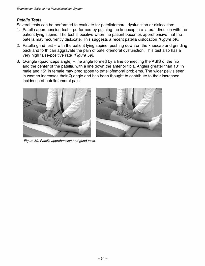
Patella TestsSeveral tests can be performed to evaluate for patellofemoral dysfunction or dislocation:1. Patella apprehension test – performed by pushing the kneecap in a lateral direction with the
patient lying supine. The test is positive when the patient becomes apprehensive that thepatella may recurrently dislocate. This suggests a recent patella dislocation (Figure 59).
2. Patella grind test – with the patient lying supine, pushing down on the kneecap and grindingback and forth can aggravate the pain of patellofemoral dysfunction. This test also has avery high false-positive rate (Figure 59).
3. Q-angle (quadriceps angle) – the angle formed by a line connecting the ASIS of the hip and the center of the patella, with a line down the anterior tibia. Angles greater than 10° inmale and 15° in female may predispose to patellofemoral problems. The wider pelvis seen in women increases their Q-angle and has been thought to contribute to their increased incidence of patellofemoral pain.
– 64 –
Examination Skills of the Musculoskeletal System
Figure 59. Patella apprehension and grind tests.
TABLE 1FORCES THAT INJURE THE KNEE
(Summary)
1. Valgus Force
A. MCL (or physeal fracture)
B. ACL
C. Medial Meniscus
2. Varus Force
A. LCL (or physeal fracture)
B. ACL
C. Lateral Meniscus
3. Anterior Drawer (anterior directed force with knee bent)
A. Isolated Anterior Cruciate Ligament
4. Posterior Drawer (posterior directed force with knee bent)
A. Isolated Posterior Cruciate Ligament
5. Internal Rotation Force
A. ACL
B. LCL
6. External Rotation Force
A. ACL
B. MCL
C. Patellar Dislocation
7. Hyperextension
A. ACL
B. PCL
C. Knee dislocation with potential for tearing all ligaments, cartilage and even neurovascular injury
Examination Skills of the Musculoskeletal System
– 65 –
– 66 –
Examination Skills of the Musculoskeletal System
Knee Exam Landmarks
The following anatomic landmarks should be located:
Palpate with knee flexed to 90° and foot flat on table
Tibial Tubercle
Patella Tendon
Patella
Medial & Lateral Divots (either side of patella tendon)
Medial Joint Line
Lateral Joint Line
Head of Fibula
Medial Femoral Condyle
Lateral Femoral Condyle
Medial Collateral Ligament
Lateral Collateral Ligament
Iliotibial Band
Pes Anserinus
Knee Exam Essentials
__ 1. Inspect the knees for evidence of swelling, ecchymosis or atrophy.
__ 2. Palpate for swelling or joint effusion as well as warmth.
__ 3. Palpate for areas of tenderness. Should specifically palpate these areas:
A. Tibial tubercle
B. Medial and lateral divots
C. Patella (medial, lateral, tendon)
D. Quadriceps tendons
E. Joint line (medial/lateral and anterior/posterior)
F. Ligaments (MCL/LCL)
__ 4. Range of motion testing should include both active and passive.
A. Extension (0°) – also check heel height difference while lying prone
B. Flexion (130°)
C. Palpation over knee for crepitus
D. Assess extensor mechanism (by checking for active extension of knee)
__ 5. Ligament testing should be performed for evidence of pain or laxity.
A. MCL — Valgus stress at 0° and 20° to 30° flexion
B. LCL — Varus stress at 0° and 20° to 30° flexion
C. ACL — Lachman’s test (20° flexion); Anterior drawer test (90° flexion); Pivot shift (optional)
D. PCL — Sag sign (90° flexion); Posterior drawer test (90° flexion)
__ 6. Meniscal tests — look for pain, locking or a decreased range of motion.
*Note: All these tests have high rate of false-positives.
A. Prone knee extension (observe for lack of extension)
B. Bounce test
C. Apley compression test (optional)
D. McMurray’s test (optional)
E. Duck walk
__ 7. Patella tests reveal evidence of patellofemoral dysfunction.
A. Apprehension test
B. Patellar grind test
C. Q-angle
Examination Skills of the Musculoskeletal System
– 67 –
– 68 –
Examination Skills of the Musculoskeletal System
Ankle EvaluationAnkle Exam Landmarks
Ankle Exam Essentials
Examination Skills of the Musculoskeletal System
– 69 –
– 70 –
Examination Skills of the Musculoskeletal System
ANKLE EVALUATION
HISTORY
Evaluation of the patient with an ankle problem should begin with a good history. Importantquestions include:
What was the mechanism of injury or overuse?The ankle can be injured by repetitive overuse (such as with running) or an acute injury. Themost common mechanisms for ankle injury include:1. Inversion — this is the most common way to injure the ankle and typically occurs after
stepping wrong and rolling the ankle into inversion. This will injure the lateral ankle ligaments,and when combined with plantar-flexion can injure the peroneal tendon.
2. Eversion — the opposite mechanism to inversion can injure the medial ankle ligament (deltoidligament). Combined eversion and external rotation can dislocate the peroneal tendon.
3. External rotation — when applied forcefully can injure the ankle syndesmosis (tibiofibular ligament).
4. Dorsiflexion — a sudden dorsiflexion caused by landing after a jump can injure the achillestendon. Very forceful dorsiflexion at the ankle can also injure the ankle syndesmosis.
5. Plantar-flexion — against resistance, caused by forceful gastro-soleus contraction, can tearthe achilles tendon. Forcing the ankle into extreme plantar flexion can dislocate the ankle joint.
Did you hear or feel a pop?A pop at the lateral or medial ankle suggests a ligament tear, while a pop at the back of theankle suggests achilles tendon rupture. Recurrent lateral ankle popping is suggestive of peroneal tendon subluxation.
Were you able to bear weight after the injury? Continue playing?The answer to these questions are helpful in assessing the severity of injury to the ankle. Theinability to bear weight is a sign of increased fracture risk and need for x-ray. The Ottawa anklerules (Figure 60) can be useful in deciding when to order an x-ray in a patient with an ankleinjury. In general, an ankle x-ray should be ordered if:1. There is tenderness to palpation along the lower 6 cm of the posterior border of either the
medial or lateral malleolus.
2. The patient is unable to bear weight both immediately following the injury and on presentationdue to pain in the ankle.
3. Foot x-rays should be obtained if there is significant tenderness at the proximal 5thmetatarsal or over the tarsal navicular bone.
Examination Skills of the Musculoskeletal System
– 71 –
– 72 –
Examination Skills of the Musculoskeletal System
Was there associated swelling or ecchymosis?Swelling and bruising suggest ligament or tendon rupture. The initial location of swelling orbruising can be helpful in localizing injured structures. Over time, swelling and bruising associatedwith ankle injury sinks to the foot and is reabsorbed.
Where was the pain located?The location of pain in the ankle can be helpful diagnostically (Figure 61). Localizing the pain to medial, lateral, anterior or posterior can suggest the injured structure.1. Lateral — anterior talofibular ligament, calcaneofibular ligament, posterior talofibular ligament,
Figure 60. Locations of tenderness with Ottawa ankle rules.
Figure 61. Location of common causes of ankle and foot pain.
Examination Skills of the Musculoskeletal System
– 73 –
What treatments were used after the injury?If RICE (rest, ice, compression, elevation) was used after the injury and the ankle is still veryswollen, a significant injury is more likely.
Has there been persistent pain and/or swelling since the initial injury?The most common reason for persistent symptoms after an ankle injury is inadequate or incorrecttreatment. This could also indicate an underlying fracture, cartilage injury or ligament injury.
EXAMINATION
The ankle, along with the lower leg and foot should be exposed on both sides to compare fordifferences. Important parts of the ankle exam include:
InspectionInspect and compare both fully exposed ankles from the front, the side and from behind. Lookfor asymmetry as well as arch deformities, swelling or ecchymosis. You should also look atshoes for abnormal wear patterns.
PalpationPalpate the lower leg, ankle and foot in the followingareas of tenderness:1. Lower leg: fibula, interosseous membrane, anterior
Range of Motion Ankle motion should first be assessed actively and thenpassively, comparing both ankles to look for asymmetry.Check for limited motion and/or pain.1. Dorsiflexion normally about 20° (Figure 63)
2. Plantar flexion about 50°
3. Inversion about 30°
4. Eversion about 10°
Figure 62. Palpating the lateral ankle for tenderness.
Figure 63. Checking ankle dorsiflexionmotion.
Examination Skills of the Musculoskeletal System
Strength Testing Check for muscle weakness and/or pain. This is performed by resisted ankle movements.1. Resisted dorsiflexion (tibialis anterior). (Figure 64)
Special Tests A variety of tests can be performed on the ankle to assess integrity of the ankle ligaments andtendons. When doing these tests, check for evidence of laxity and/or pain.1. Anterior drawer test — performed by stabilizing the lower leg with one hand while cupping
the heel with the other, then pulling forward on the calcaneus/talus complex(Figure 65). Laxity compared to the uninvolved side suggests lateral ligament rupture.
2. Talar tilt test — performed by similarly stabilizing the lower leg with one hand while cuppingthe heel with the other, then inverting the ankle joint. Inversion laxity compared to the unin-volved side suggests tearing of the lateral ligaments, while pain with this maneuver suggestsligament injury.
3. Squeeze test — performed by squeezing the proximal tibia and fibula together while askingabout pain distally at the ankle (Figure 66). Pain in the ankle suggests injury to the tibiofibularligament (syndesmosis sprain). Passsively rotating the ankle into external rotation will alsoaggravate pain from a syndesmosis injury.
Figure 64. Testing ankle dorsiflexion and inversion strength.
Figure 65. Anterior drawer test for Figure 66. Squeeze test for lateral ankle ligament laxity. syndesmosis injury.
– 74 –
Examination Skills of the Musculoskeletal System
– 75 –
4. Thompson test — performed by squeezing at the base of the calf muscle and looking forankle plantar-flexion (Figure 67). A lack of plantar flexion suggests a complete achilles tendon rupture.
Functional Tests These sequential activities are performed to see if they cause pain or other symptoms. This canbe helpful in advising return to play or activity.1. Walking
2. Standing/walking on toes/heels
3. Squatting
4. Jogging
5. Running straight ahead
6. Running and cutting
Figure 67. Thompson test for complete achilles tendon rupture.
– 76 –
Examination Skills of the Musculoskeletal System
Ankle Exam Landmarks
The following anatomic landmarks should be located:
Ankle Joint Line
Anterior Talofibular Ligament
Calcaneofibular Ligament
Posterior Talofibular Ligament
Deltoid Ligament
Tip and Posterior Edge of Medial Malleolus
Tibialis Posterior Tendon
Tip and Posterior Edge of Lateral Malleolus
Peroneal Tendon
Achilles Tendon
Base of the 5th Metatarsal
Navicular bone of foot
Examination Skills of the Musculoskeletal System
Ankle Exam Essentials
__ 1. Inspect and compare both fully exposed ankles from the front, the side and from behind.Look for arch deformities, swelling or ecchymosis. Also note appearance of shoes.
__ 2. Palpate the lower leg, ankle and foot for areas of tenderness.
A. Lower leg: fibula, interosseous membrane, anterior compartment
B. Medial ankle: medial malleolus, deltoid ligament, tibialis posterior tendon
C. Lateral ankle: lateral malleolus, ATFL, CFL, PTFL, peroneal tendon
D. Posterior ankle: achilles tendon
E. Foot: proximal 5th metatarsal, navicular
__ 3. Range of motion should be performed first actively and then passively if needed, while comparing both ankles.
A. Dorsiflexion 20°
B. Plantar flexion 50°
C. Inversion 30°
D. Eversion 10°
__ 4. Strength testing should look for muscle weakness and/or pain. It is performed as resisted ankle movements.
A. Resisted dorsiflexion (tibialis anterior)
B. Resisted plantar-flexion (gastroc, peroneal longus, tibialis posterior)
C. Resisted inversion (tibialis posterior, tibialis anterior)
D. Resisted eversion (peroneal longus brevis)
__ 5. Stability testing is performed to assess the integrity of the ankle ligaments. Check for evidence of laxity and/or pain.
A. Talar tilt test
B. Anterior drawer test
C. Squeeze test (for syndesmotic sprains)
D. Thompson test (for achilles tendon ruptures)
__ 6. Functional tests are performed to see whether these sequential activities produce pain or other symptoms.
A. Squatting
B. Standing/walking on toes/heels
C. Jogging
D. Running straight ahead
E. Running and cutting
– 77 –
– 78 –
Examination Skills of the Musculoskeletal System
References
Acknowledgements
Self-assessment Questions
Examination Skills of the Musculoskeletal System
– 79 –
REFERENCES
1. Adkins SB III, Figler RA. Hip pain in athletes. Am Fam Physician. 2000;61:2109-18.
2. Bach AW. Finger joint injuries in active patients: pointers for acute and late-phase management. Physician Sportsmed. 1999;27:89.
3. Birnbaum, JS. The Musculoskeletal Manual. 2d ed. Orlando, Fl.: Grune and Stratton; 1986.
4. Chumbley EM, O’Conner FG, Nirschl RP. Evaluation of overuse elbow injuries. Am FamPhysician. 2000;61:691-700.
5. Greene WB, American Academy of Orthopaedic Surgeons, American Academy of Pediatrics.Essentials of Musculoskeletal Care. 2d ed. Rosemont, Ill.: American Academy of Orthopaedic Surgeons; 2001.
6. Hockenbury TR, Sammarco GJ. Evaluation and treatment of ankle sprains: clinical recommendations for a positive outcome. Physician Sportsmed. 2001;29:57.
8. Patel AT, Ogle AA. Diagnosis and management of acute low back pain. Am Fam Physician.2000;61:1779-86,1789-90.
9. Reider B. The Orthopedic Physical Exam. Philadelphia, Pa.: WB Saunders: 1999.
10.Rubin AL, Sallis RE. Evaluation and diagnosis of ankle injuries. Am Fam Physician.1996;54:1609-18.
11. Smith BW, Green GA. Acute knee injuries: Part II. Diagnosis and management. Am FamPhysician. 1995;51:799-806.
12.Woodward TW, Best TM. The painful shoulder: part I. Clinical evaluation. Am FamPhysician. 2000;61:3079-88.
– 80 –
Examination Skills of the Musculoskeletal System
Examination Skills of the Musculoskeletal System
– 81 –
Acknowledgements
I would like to thank the sports medicine fellows at Kaiser Permanente in Fontana: Cher Cohler,MD, Eric Hoeper, MD, and Jared Williams for their help with question writing and review of themanuscript.
I would also like to thank Ana Lara, PA for serving as the model in the videotapes. Her inputduring filming was invaluable.
Finally, I would like to thank Rick Tilley and the Kaiser Permanente Multimedia Communicationsdepartment for their help with filming and editing of the videotapes.
– 82 –
Examination Skills of the Musculoskeletal System
1. The “drop arm test”, when positive, suggests?
a. Clavicle fracture
b. AC joint separation
c. Large rotator cuff tear or nerve injury
d. Biceps tendinitis
2. The cross-over test, when positive, maysuggest?
a. AC joint pain
b. Glenohumeral joint instability
c. Impingement syndrome
d. Both a and c
3. Glenohumeral instability is commonlyseen in?
a Young, active patients
b. Elderly
c. Young, sedentary patients
d. Pregnancy
4. Characteristics of rotator cuff tendinitisinclude?
a. Pain that improves with activity andworsens with rest
b. Pain located in the bicipital groove
c. Pain that improves with rest and worsens with activity
d. Pain related to trauma
5. Abduction with thumbs down and 30°horizontal adduction (“empty can test”)isolates which rotator cuff muscle?
a. Subscapularis
b. Teres minor
c. Supraspinatous
d. Infraspinatous
6. Loss of both active and passive shouldermotion may suggest which of the follow-ing?
a. Labrum tear
b. Adhesive capsulitis
c. Severe impingement
d. All of the above
7. When performing Neer’s test, pain or lackof motion is commonly seen with?
a. Impingement syndrome
b. AC separation
c. Glenohumeral instability
d. Labrum tear
8. A positive Spurling’s test (radicular pain to the shoulder and arm during axial loading to the top of the head with the neck twisted) may represent?
a. Cervical nerve root irritation
b. Deltoid atrophy
c. Impingement syndrome
d. Muscle spasm
Examination of the Musculoskeletal SystemSelf-study Program
Self-assessment Questions
Examination Skills of the Musculoskeletal System
– 83 –
9. A common source of radicular pain to theshoulder is?
a. Wrist
b. Hip
c. Thoracic spine
d. Cervical spine
10.Resisted extension and supination of thewrist tends to aggravate?
a. Medial epicondylitis
b. Ulnar nerve subluxation
c. Lateral epicondylitis
d. Olecranon bursitis
11. A sudden painful pop at the medial elbowwhile throwing may indicate?
a. Osteoarthritis of the elbow
b. Epicondylitis
c. Ulnar collateral ligament tear
d. Carpal tunnel syndrome
12.Recurrent popping at the medial elbowassociated with tingling to the 4th and 5th finger may indicate?
a. Ulnar nerve subluxation
b. Ulnar collateral ligament tear
c. Arthritis
d. Carpal tunnel syndrome
13.Severe night pain in the shoulder thatprevents sleep suggests?
a. Rotator cuff tear
b. Biceps tendinitis
c. Glenohumeral instability
d. AC separation
14.Shoulder muscle asymmetry may indicatewhich of the following?
a. Rotator cuff tear
b. Nerve injury
c. Adaptive hypertrophy
d. All of the above
15.A snapping sound in the wrist usually represents?
a. Synovitis
b. Carpal tunnel syndrome
c. Subluxing tendons
d. Infection
16.Which nerve is being tested when the handis placed on a flat surface, palm up and thethumb is raised against resistance?
a. Radial
b. Median
c. Ulnar
d. None of the above
17.The inability to flex the DIP joint is suggestive of?
a. Ulnar collateral ligament tear
b. Mallet finger
c. Jersey finger
d. deQuervain’s tendinitis
18.When testing range of motion of the back,forward flexion most increases?
a. Facet pain
b. Muscular pain
c. Tendon pain
d. Disc pain
– 84 –
Examination Skills of the Musculoskeletal System
19.Weakness with resisted great toe dorsiflexion is suggestive of irritation of which nerve root?
a. S1
b. L5
c. L4
d. L3
20.The Stork test will aggravate pain associated with?
a. Spondylolysis
b. Spondylolisthesis
c. SI joint dysfunction
d. All of the above
21.Pain with the FABER test is suggestive of?
a. SI joint pathology
b. Sciatica
c. IT band tightness
d. Synovitis
22.Which test is usually positive with afemoral neck stress fracture?
a. Hop test
b. Log roll test
c. Ober’s test
d. Trendelenburg test
23.Pain from sciatica worsens with whichmaneuver?
a. Extension of back
b. Flexing knee
c. Twisting of back
d. Valsalva
24.Which test when positive is suggestive ofdeQuervain’s tendinitis?
a. Watson stress test
b. Grind test
c. Tinel’s test
d. Finkelstein’s test
25.The presence of locking of the knee suggests what injury?
a. Patellofemoral syndrome
b. MCL tear
c. Quadriceps strain
d. Meniscal tear
26.This is the most common injury to theankle:
a. Inversion injury
b. Eversion injury
c. Dorsiflexion
d. External rotation
27.A large (3+) effusion of the knee occurring within 12 hours of an injury suggests which of the following?
a. ACL tear
b. Osgood-Schlatter’s disease
c. Patellofemoral syndrome
d. LCL sprain
28.Pain over the tibia tubercle is an importantexam finding in which of the following?
a. Meniscal tears
b. MCL sprain
c. Bipartate Patella
d. Osgood-Schlatter’s disease
Examination Skills of the Musculoskeletal System
– 85 –
29.What is the typical mechanism for injuryto the deltoid ligament of the ankle?
a. Ankle inversion
b. Ankle dorsiflexion
c. Ankle plantarflexion
d. Ankle eversion
30.What is the most common reason for persistent symptoms after an ankleinjury?
a. Underlying fracture
b. Cartilage injury
c. Fat pad necrosis
d. Inadequate or incorrect treatment
31.The definitive exam used to evaluate forACL tear is?
a. Pivot shift
b. Anterior drawer test
c. Sag sign
d. Lachman’s test
32.A positive Thompson's test is suggestiveof which injury?
a. High ankle sprain
b. Achilles tendon rupture
c. Patellofemoral syndrome
d. Lateral ankle sprain
33.A Q-angle greater than 10° in malesor 15° in females predisposes to what problem?
a. ACL tears
b. LCL tears
c. Baker's cysts
d. Patellofemoral syndrome
34.What is the likelihood of having an ACLtear if a "pop" was felt or heard during atwisting knee injury?
a. ~10%
b. ~25%
c. ~60%
d. ~80%
35.Weakness with resisted dorsiflexion of theankle suggests injury to which muscle?
a. Tibialis posterior
b. Gastrocnemius
c. Tibialis anterior
d. Peroneal longus
– 86 –
Examination Skills of the Musculoskeletal System
Examination of the Musculoskeletal SystemSelf-study Program