32
| Date post: | 24-Oct-2015 |
| Category: |
Documents |
| Upload: | diana-phiri |
| View: | 32 times |
| Download: | 4 times |
2 Examining Children
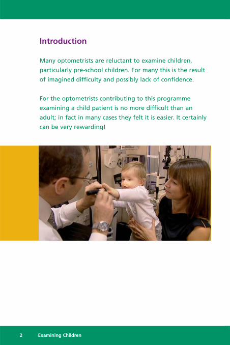
Introduction
Many optometrists are reluctant to examine children,
particularly pre-school children. For many this is the result
of imagined difficulty and possibly lack of confidence.
For the optometrists contributing to this programme
examining a child patient is no more difficult than an
adult; in fact in many cases they felt it is easier. It certainly
can be very rewarding!
Key Points
Examining Children is NOT more difficult than examining adults� Create a child friendly environment:
� A low table and chairs for the waiting room� Have a supply of toys, colouring materials etc.
Take a pragmatic approach� You may not be able to do every test at one visit:
� You may not be able to do retinoscopy� You may not be able to do a subjective� You may not be able to do a cover test
� Abandon tests quickly if they are not successful and move on� A series of short visits may be more productive than one long one� Do not be afraid to re-book
You must learn to trust your observations� Rely on objective not subjective findings� Results will vary – especially retinoscopy and cover test findings
Absolute accuracy is not the key goal� 0.50 D sphere errors or a small cyl not corrected is not important
The structure of your routine needs to be fluid and adaptable� Base it on the presenting history� Begin with something achievable, i.e. cover test or stereopsis
Managing the parental expectations may be the hardest part� Address parents concerns:
� Why have you come?� Are you worried?� What are you worried about?
� Don’t be dismissive
Communicate with the child at their level� Talk to child NOT parents� Address child by name frequently� Praise the child, don’t criticise
Above all:• MAKE IT FUN!
Examining Children 3
4 Examining Children
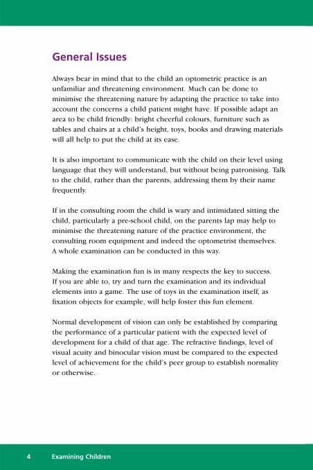
General Issues
Always bear in mind that to the child an optometric practice is anunfamiliar and threatening environment. Much can be done tominimise the threatening nature by adapting the practice to take intoaccount the concerns a child patient might have. If possible adapt anarea to be child friendly: bright cheerful colours, furniture such astables and chairs at a child’s height, toys, books and drawing materialswill all help to put the child at its ease.
It is also important to communicate with the child on their level usinglanguage that they will understand, but without being patronising. Talkto the child, rather than the parents, addressing them by their namefrequently.
If in the consulting room the child is wary and intimidated sitting thechild, particularly a pre-school child, on the parents lap may help tominimise the threatening nature of the practice environment, theconsulting room equipment and indeed the optometrist themselves. A whole examination can be conducted in this way.
Making the examination fun is in many respects the key to success. If you are able to, try and turn the examination and its individualelements into a game. The use of toys in the examination itself, asfixation objects for example, will help foster this fun element.
Normal development of vision can only be established by comparingthe performance of a particular patient with the expected level ofdevelopment for a child of that age. The refractive findings, level ofvisual acuity and binocular vision must be compared to the expectedlevel of achievement for the child’s peer group to establish normalityor otherwise.
Examining Children 5
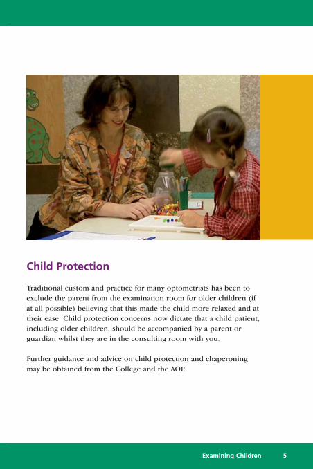
Child Protection
Traditional custom and practice for many optometrists has been toexclude the parent from the examination room for older children (ifat all possible) believing that this made the child more relaxed and attheir ease. Child protection concerns now dictate that a child patient,including older children, should be accompanied by a parent orguardian whilst they are in the consulting room with you.
Further guidance and advice on child protection and chaperoningmay be obtained from the College and the AOP.
6 Examining Children
Examination Routine
Don’t expect your examination of a child patient to mimic the routineyou would use on an adult. Your routine needs to be flexible andadaptable and you should be prepared to abandon a particular test ifit is not successful and move on to something else, or use analternative technique. Indeed the order in which tests are conductedmight seem quite illogical. The only important rule is that you do thetests that you believe are necessary; the order in which they are donedoes not matter.
Base the structure of your routine on the presentation and the historyof each individual patient.
It is important not to be frustrated or concerned about minorvariations or perceived lack of accuracy in the results of clinicalinvestigations. It is important, though, to have confidence and to beable to trust your own observations. Absolute accuracy in theexamination of a child patient is not critical. A sphere which is 0.50 Dout, or failure to correct a small cylinder, is not going to affect thevisual development of the child. It should be expected that findingswill fluctuate, particularly retinoscopy and cover test results.
Don’t expect to necessarily get all tests completed on one single visit.Depending on the level of cooperation and alertness of the child thismay not be possible. Split the routine into smaller, more manageableelements that the child can cope with and re-book for another day tocomplete your examination.
It will help the routine to run smoothly if the child has a successrather than a failure early on. For that reason it may be better to picksomething like the cover test to begin rather than checking vision orvisual acuity first. Any child over 36 months should have stereopsis, sothat may be a good place to start.
It is particularly important to be positive and encouraging to the childeven if they do not perform a test well, praising rather than criticisingthem. However it is not necessary to pretend that every test has asuccessful result and showing failure is acceptable. Do end theexamination on a positive note, however, and finish with a success,even if it is necessary to return to a test completed earlier. Finishingon a line of letters on the chart that you know the child can see andpraising the result works well and is a good standby.
History and Symptoms
History and symptoms will inevitably come from the parents who will be expressing their own concerns about their child’s vision. The “symptoms” will be largely based on parental observation of the child’s behaviour to give clues to the state of the vision. So it isimportant to establish a few basic questions:
• Why have you brought your child in to be examined?• Are you worried about your child’s vision?• What are you worried specifically about?
Never underestimate or dismiss a parents concerns or observations;they are rarely wrong.
It is helpful to ask questions relevant to a child patient and thesymptoms they are likely to experience. For example children rarelycomplain of symptoms such as headaches and won’t experiencediplopia.
Always bear in mind any risk factors that might apply and in particularrelevant family history, such as high refractive error and squint. Alwaysobtain as much information about an absent parent as diplomaticallyas possible, bearing in mind there might be a relationship with thechild but not the other parent.
Examining Children 7

Specific elements of the child’s history and the mother’s pregnancymay be of great importance. Maternal illness during pregnancy,prematurity at birth, a difficult or assisted delivery may haverelevance for the development of the child’s visual system.
Visual Acuity testing
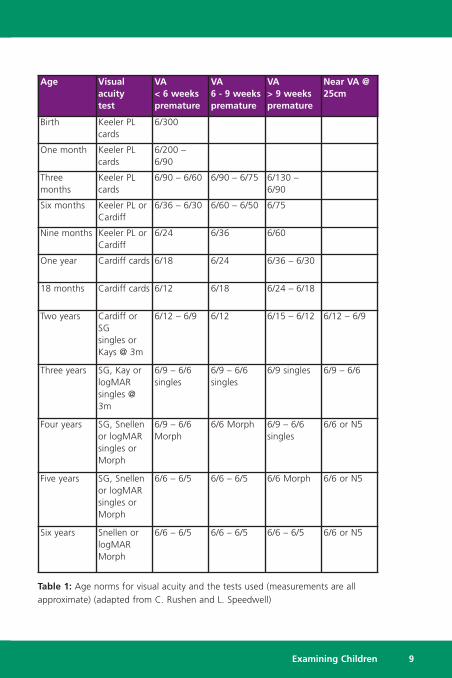
NormsTable 1 gives average expected visual acuity measures for childrenfrom birth to six years of age. In young children visual acuity valuestend to vary depending which type of test is used.
Birth to 12 monthsPreferential Looking Cards (Keeler Acuity Cards) – the bestOcclusion behaviourHundreds and ThousandsVisually directed reaching
12 months to 2.5 yearsCardiff CardsHundreds and Thousands
2.5 years onwardsLea symbolsKay picturesSheridan-GardinerSonksen-SilverCambridge acuity cardsCrowded Kay picturesLogMAR acuity cardsSnellen Letters
8 Examining Children
Examining Children 9
Age Visualacuitytest
VA< 6 weekspremature
VA6 - 9 weekspremature
VA> 9 weekspremature
Near VA @25cm
Birth Keeler PLcards
6/300
One month Keeler PLcards
6/200 –6/90
Threemonths
Keeler PLcards
6/90 – 6/60 6/90 – 6/75 6/130 –6/90
Six months Keeler PL orCardiff
6/36 – 6/30 6/60 – 6/50 6/75
Nine months Keeler PL orCardiff
6/24 6/36 6/60
One year Cardiff cards 6/18 6/24 6/36 – 6/30
18 months Cardiff cards 6/12 6/18 6/24 – 6/18
Two years Cardiff orSGsingles orKays @ 3m
6/12 – 6/9 6/12 6/15 – 6/12 6/12 – 6/9
Three years SG, Kay orlogMARsingles @3m
6/9 – 6/6singles
6/9 – 6/6singles
6/9 singles 6/9 – 6/6
Four years SG, Snellenor logMARsingles orMorph
6/9 – 6/6Morph
6/6 Morph 6/9 – 6/6singles
6/6 or N5
Five years SG, Snellenor logMARsingles orMorph
6/6 – 6/5 6/6 – 6/5 6/6 Morph 6/6 or N5
Six years Snellen orlogMARMorph
6/6 – 6/5 6/6 – 6/5 6/6 – 6/5 6/6 or N5
Table 1: Age norms for visual acuity and the tests used (measurements are allapproximate) (adapted from C. Rushen and L. Speedwell)

Preferential Looking Visual Acuity (PL) CardsAlthough cooperation is known to decline noticeably around the ageof 12 months, forced-choice preferential looking (PL) is regarded as aquick and sensitive indicator of monocular visual deficit in childrenunder 1 year.
This test uses a series of rectangular cards with a patch of square-wave gratings of various spatial frequencies on one side and an equal-sized patch of equal luminance in plain grey on the other. The cardsare presented unseen by the optometrist and there is a peepholehalfway between the two patches through which the optometristwatches the infant. A judgement must be made if it is fixating to theright or left (the forced-choice).
The test works on the basis that a child will prefer to look at anobject with visual interest, i.e. the grating, rather than a plain field ofthe same luminance. When the grating is too narrow to bedifferentiated the child will gaze randomly at one side or the other.The card with the highest spatial frequency expected to be seen inaccordance with the infant's age is, presented to each eye, preferablytwice, for a definite response. The visual acuity is estimated as thehighest spatial frequency the child is believed to be able to see. Thetest seems to be equally effective if the patches are presentedvertically so a variation of horizontal and vertical presentations helpsto maintain the infant's interest.
Occlusion BehaviourFrom about three months of age, a child will object if an eye withbetter vision is covered while he is looking at something of interest.Gross loss of vision in one eye is unlikely if the baby appears equallyhappy and able with either eye covered.
10 Examining Children
Hundreds and Thousands/Visually Directed ReachingSmall cake decorations (100s & 1000s) held in the palm of the hand(or mother's hand) can be used to gain attention in the over sixmonth olds. At nine months the baby may prod the decorations andat one year old attempt to pick them up. The cake decorationsthemselves should not however be regarded as a reliable measure ofvisual acuity: the average decoration held at 33 cm is roughlyequivalent to 6/60 and when held at 25 cm, equivalent to 6/150.
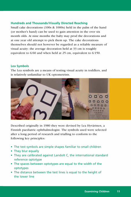
Lea SymbolsThe Lea symbols are a means of testing visual acuity in toddlers, andis relatively unfamiliar to UK optometrists.
Described originally in 1980 they were devised by Lea Hyvärinen, aFinnish paediatric ophthalmologist. The symbols used were selectedafter a long period of research and trialling to conform to thefollowing key principles:
• The test symbols are simple shapes familiar to small children• They blur equally• They are calibrated against Landolt C, the international standard
reference optotype• The spaces between optotypes are equal to the width of the
optotypes• The distance between the test lines is equal to the height of
the lower line
Examining Children 11

The test is made up of a combination of the Lea shapes, a square, acircle, a house, and an apple (heart), and may be applied as a linetest or single symbols at near as well as far distances.
The test is easy to apply and well accepted by children. The near testis contained on a reading card with large reference symbols printedat the bottom so the child can match the shape of the test symbol ifthey are unable or unwilling to name it. Visual acuity for distance ismeasured with the chart held at three metres. If that distance is toogreat for a young child, testing can be performed at two metres. Inolder children measurements can be made at distances up to sixmetres if required.
Kay Picture TestThe test comprises of a series of symbols, the component parts ofwhich conform to the Snellen principle of subtending 1 minute ofarc, but the overall size of the picture subtends 10 minutes of arc.The pictures are of common objects that should be known to a childand the test is based on the child recognising and naming the object,although there are matching cards available for very shy children. It iseffective and useful for children aged 2-3 years.
The test is available as either single pictures or crowding in LogMARformat for use at 3 metres to measure acuity from 1.0 – 0.1, or astandard Snellen format with single pictures for use at 3 or 6 metresto measure acuity from 3/3 (6/6) – 3/30 (6/60).
Recognition booklets can also be supplied and add to the usefulnessof the test, as they can be loaned to a parent for practice in namingthe pictures and having one eye occluded at home.
12 Examining Children
Sheridan-Gardiner TestIn the Sheridan-Gardiner test single letters are displayed singly ateither 3 or 6 metres and the child points to the matching letter on akey card. The test’s main drawback is that it measures single lettervisual acuity giving a misleadingly high result for children withamblyopia.
Sonksen-Silver Acuity SystemThe Sonksen-Silver Acuity System uses the Sheridan-Gardiner letterselection but presented in a line with standardised spacing tointroduce crowding and remove the single letter advantage foramblyopes. The letters are matched on a key card in the same way asthe Sheridan-Gardiner test.
Cambridge Crowding CardsThis is another test for use at 3m or 6m, designed to elicit thecrowding phenomenon. The Sheridan Gardiner selection of letters isused and the child has to identify the letter which is surrounded byfour others. The matching letters can be arranged so that there is noconfusing resemblance to the letters displayed on the test card; thismethod is said to be more accurate than linear testing.
Although both the Sonksen-Silver Acuity System and the CambridgeCrowding Cards arc better than single letter presentation, they aredifficult to administer without pointing, thus to some extent isolatingthe required letter.
Examining Children 13

Binocular Status
Cover TestThe cover test is one of the simplest objective tests to perform andyet potentially the most informative. It has the potential to giveinformation about the type, size and control of a deviation, the likelybinocular function present and the probable involvement of extra-ocular muscle anomalies. In order to maximise this potential though,it is vital that the child can be encouraged to fixate a suitable objectappropriate for their age.
Suitable fixation targets can be many and varied. For a younger childa pen torch, retinoscope or ophthalmoscope light may be suitable, or a small, brightly coloured, interesting toy may be used. Asaccommodation must be suitably stimulated any fixation targetshould preferably have some visual interest. For an older child asmall graphical object recognisable to the patient, such as a cartooncharacter, “budgie stick” etc. might be more suitable.
14 Examining Children
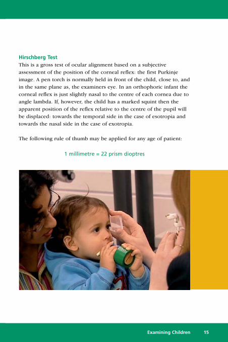
Hirschberg TestThis is a gross test of ocular alignment based on a subjectiveassessment of the position of the corneal reflex: the first Purkinjeimage. A pen torch is normally held in front of the child, close to, andin the same plane as, the examiners eye. In an orthophoric infant thecorneal reflex is just slightly nasal to the centre of each cornea due toangle lambda. If, however, the child has a marked squint then theapparent position of the reflex relative to the centre of the pupil willbe displaced: towards the temporal side in the case of esotropia andtowards the nasal side in the case of exotropia.
The following rule of thumb may be applied for any age of patient:
1 millimetre = 22 prism dioptres
Examining Children 15
16 Examining Children
20Δ Base-out TestThe 20Δ base-out test is a basic test of fusion for children up to 5years of age. The presence of fusion is a sign that some degree ofbinocular vision has developed. A base-out prism is held before oneeye while the child looks at a suitable fixation target. The eye behindthe prism should adduct rapidly to restore normal fusion and quicklyabduct again on removal of the prism. The speed of the fusionmovements is a simple guide to the quality of the binocularity.
The following prisms are suitable:
Other possible responses are:
• No movement – implies no fusion or lack of attention• Slow to overcome the prism or slow to recover after the prism
has been removed – implies poor fusion
By the age of 5 years, a child should cooperate with the measurementof the prism fusion range using a prism bar. The normal range fornear is 35 Δ – 45 Δ base-out to 12 Δ – 8 Δ base-in.
6 months of age 10Δ base-out
12 – 18 months of age 15Δ base-out
Over 18 months of age 20Δ base out
Motility TestTogether with the cover test, the motility test is a key assessment whenexamining a child patient. It will give information about the range ofocular movements and whether they are concomitant or incomitant.
As with the cover test success in motility testing for a younger childpatient is heavily dependent on them being offered an interesting andstimulating fixation target. A wide variety of toys can be adapted foruse as a motility target if they are reasonably small, have an interestingand stimulating appearance and/or are brightly coloured. It may helpto attract or maintain the child’s attention to have a toy that flashes,lights up, squeaks or has some other form of auditory stimulus.
In very young children it may not be possible to explain the nature ofthe test and what you expect of them to the child. In these situationmove the child rather than the stimulus, the so-called “swinginginfant” procedure. In this technique the target is kept stationary andthe child is rotated. It will be necessary for the parent, by gentleholding, to prevent the child from turning their head as they rotatethem. If they have been successfully encouraged to direct theirattention to the stimulus they will maintain their fixation as they arerotated and their ocular motility assessed.
Examining Children 17
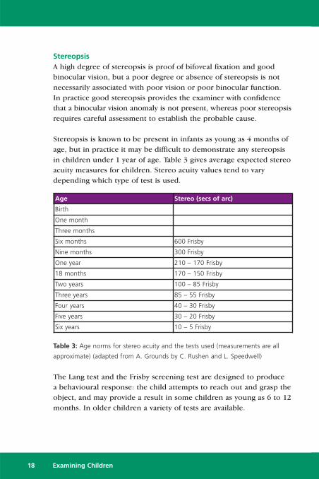
StereopsisA high degree of stereopsis is proof of bifoveal fixation and goodbinocular vision, but a poor degree or absence of stereopsis is notnecessarily associated with poor vision or poor binocular function. In practice good stereopsis provides the examiner with confidencethat a binocular vision anomaly is not present, whereas poor stereopsisrequires careful assessment to establish the probable cause.
Stereopsis is known to be present in infants as young as 4 months ofage, but in practice it may be difficult to demonstrate any stereopsisin children under 1 year of age. Table 3 gives average expected stereoacuity measures for children. Stereo acuity values tend to varydepending which type of test is used.
Table 3: Age norms for stereo acuity and the tests used (measurements are all
approximate) (adapted from A. Grounds by C. Rushen and L. Speedwell)
The Lang test and the Frisby screening test are designed to produce a behavioural response: the child attempts to reach out and grasp theobject, and may provide a result in some children as young as 6 to 12months. In older children a variety of tests are available.
18 Examining Children
Age Stereo (secs of arc)
Birth
One month
Three months
Six months 600 Frisby
Nine months 300 Frisby
One year 210 – 170 Frisby
18 months 170 – 150 Frisby
Two years 100 – 85 Frisby
Three years 85 – 55 Frisby
Four years 40 – 30 Frisby
Five years 30 – 20 Frisby
Six years 10 – 5 Frisby
Stereopsis tests are available in a variety of designs and produce a 3-dimensional object in a variety of different ways. Most tests usesimple geometrical shapes as test objects presented against a randompatterned background. It seems to be unavoidable that stereo testsproduce monocular clues of depth to some degree; precautions mayneed to be taken in the application of the tests to ensure that theseclues are minimised. The following tests are in common use:
The Lang test.The Lang Stereotest is a screening test for young children and uses twoimages, reproduced in fine strips, which are separately seen by eacheye when focused through a series of fine cylindrical lens elements. If binocularity exists in some part, then the images are fused and seenin depth. Its big advantage, particularly for younger children, is thatno filters are required so no spectacles need to be worn.
The test is available in two forms. The Lang I test measures stereopsisat 550, 600 and 1200 seconds if arc and the Lang II test, which isfiner, measures 200, 400 600 seconds of arc.
Answers can be checked by holding the test vertically, where stereoclues disappear, or by holding the test upside down when the objectpattern is inverted.
Examining Children 19

The Frisby testThe Frisby stereo test presents objects viewed with “real” depth, thatis, they do not use stereograms to create three-dimensional effects. As with the Lang test children avoid having to wear red/green orpolaroid spectacles. Care must be taken to avoid monocular cluesthrough parallax movements when using the test. The test canmeasure stereo acuity in the range 600 – 15 seconds of arc.
The Frisby test is available in a screening version designed foryounger children and infants. It presents a three-dimensional objectfield, together with a flat image side by side in a preferential lookingformat. In this case a spontaneous pointing or looking responses canbe observed to establish that stereopsis is present.
20 Examining Children
TNOThe TNO test is a random-dot type of stereotest requiring the wearingof red and green glasses. It comprises seven test plates and comes inan adult and children’s version. It can assess stereo acuity down to 15seconds of arc.
The Titmus testThis is perhaps the most familiar stereo test because of it use of alarge 3 dimensional fly to elicit a response in the vast majority ofpatients. It uses a crossed Polaroid visor to achieve stereopsis and inpart cartoon characters to make the test more appealing to youngerchildren. It suffers because of noticeable monocular clues which maybe difficult to disguise. The Titmus test will assess stereo acuity froma gross 3552 – 700 for the fly and 800 – 40 seconds of arc for thegraded tests.
The Randot testThe Randot test is in effect a modified Titmus test and is based on the same polarised design. It uses a random pattern background toremove many of the monocular clues present in that test.
Examining Children 21
Refraction
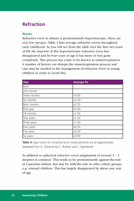
NormsRefractive error in infants is predominantly hypermetropic; there arevery few myopes. Table 4 lists average refractive errors throughoutearly childhood. As you will see from the table over the first two yearsof life the majority of this hypermetropic refractive error hasdisappeared and by four years of age it has more or less gonecompletely. This process has come to be known as emmetropisation.A number of factors can disrupt the emmetropisation process andcare may be needed in the management of refractive error in youngchildren in order to avoid this.
Table 4: Age norms for refractive error (measurements are all approximate)
(adapted from A. Grounds by C. Rushen and L. Speedwell)
In addition to spherical refractive error astigmatism of around 1 – 2dioptres is common. This tends to be predominantly against-the-rulein Caucasian infants, but may be with-the-rule in other ethnic groups,e.g. oriental children. This has largely disappeared by about one yearof age.
22 Examining Children
Age Average Rx
Birth
One month
Three months +3.00
Six months +2.50
Nine months +2.25
One year +2.00
18 months +1.50
Two years +1.25
Three years +1.00
Four years +0.50
Five years +0.50
Six years +0.50
As with other areas of examining children described in this text, theassessment of refractive error will by necessity have to rely heavily onvarious objective retinoscopy techniques and the use of measures tocontrol accommodation.
Static RetinoscopyThis is the term used to describe distance fixation retinoscopy wherethe patient’s accommodation is relaxed. This may be difficult toachieve in very young children, and the use of an assistant toencourage the child to maintain fixation can be very valuable. As with the motility test flashing or squeaking toys can be very useful.
It is likely that for many children it is the retinoscope light itselfwhich is the most interesting object and it may be very difficult tostop them looking at it rather than your intended target. If you arerefracting an infant then this may be used to your advantage byadopting the Mohindra technique.
The Mohindra technique uses the retinoscope as the fixation target.Providing all other light sources in the examination room areextinguished the child will fixate the retinoscope light but theiraccommodation will be relaxed. -1.25 D is added to the final ret.result.
With infants and very young children a trial frame is impractical andretinoscopy should be done by holding trial lenses in front of thepatient’s eyes. It may be easier to refract using spherical lenses onlyrather than attempt to hold a sphere and a cyl together.
Examining Children 23

Cycloplegic RefractionCycloplegic refraction is an essential tool in the examination ofchildren, although the application of the drops can be traumatic forthe child, the parents and the optometrist. Many practitioners wouldargue that it is essential for every child to be refracted using acycloplegic, and for infants and young children this is probably true,certainly on the first occasion they are examined. It might be arguedthough that for a child with good acuity, normal oculomotor balance,good stereopsis, no relevant family history and normal accommodationthat a cycloplegic examination is unnecessary. Certain groups ofchildren, however, should always be considered for a cycloplegicrefraction. These are:
• Children with unexplained poor VA• Children with poor stereopsis• Children with an esophoria or manifest esotropia• Children with over or underactive accommodation• Children of parents with high hypermetropia or squint
Various techniques have been proposed to minimise the problemsassociated with the application of cycloplegic drops – for instance the pharmacy at Moorfields Eye Hospital produce a Cyclopentolateatomiser.
Various steps can be taken to minimise the discomfort and ensurethat the cycloplegic can be instilled effectively:
• Proxymetacaine – Proxymetacaine 0.5% is a topical localanaesthetic which causes minimal stinging; it may be instilledbefore the cycloplegic to prevent discomfort
• Instilling on the Eyelids – It is possible to instil a cycloplegiceffectively by dropping it onto the closed eyelids, where enoughof the drug will find its way through the closed lids for it to be effective
24 Examining Children
For most children confidence and decisiveness are all that isnecessary in order to effectively instil a cycloplegic.
For the vast majority of child patients Cyclopentolate 1.0% is thecycloplegic of choice. It is generally well tolerated, produces effectivecycloplegia in most cases, requires no tonus allowance and isavailable in single dose preservative free modality.
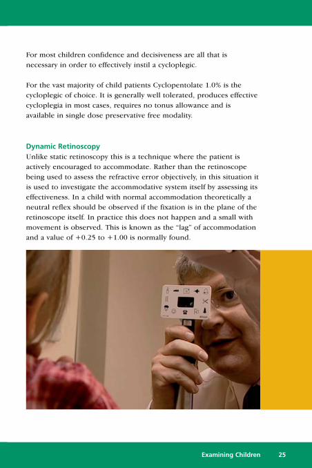
Dynamic RetinoscopyUnlike static retinoscopy this is a technique where the patient isactively encouraged to accommodate. Rather than the retinoscopebeing used to assess the refractive error objectively, in this situation itis used to investigate the accommodative system itself by assessing itseffectiveness. In a child with normal accommodation theoretically aneutral reflex should be observed if the fixation is in the plane of theretinoscope itself. In practice this does not happen and a small withmovement is observed. This is known as the “lag” of accommodationand a value of +0.25 to +1.00 is normally found.
Examining Children 25
If the fixation target is kept still and the retinoscope moved awayfrom the patient a point will be found where the reflex is neutral; thedistance between the fixation target and the retinoscope equates tothe lag. A lag greater than 1 D suggests a degree of hypermetropiathat the child cannot manage. An unequal lag between the two eyessuggests a degree of anisometropia that is poorly compensated for.
In recent years this technique has been adapted for the measurementof accommodation by Margaret Woodhouse and others for childrenand the learning disabled. In this technique an internally illuminateddetailed target is used and they have shown a good correlationbetween the results obtained using dynamic retinoscopy and normalpush-up techniques.
Colour Vision
Congenital colour vision defects are present in around 8% of boys andaround 0.5% of girls. In most cases the defect causes no appreciablehandicap in normal life. It is of value for a child patient and its parentto know if a defect is present so that certain decisions – particularlycareer related decisions – can be approached in an informed way.
Ishihara TestThis pseudoisochromatic plate test is the most commonly used of all colour vision tests. It is intended to be used in northern daylightat a viewing distance of 75 centimetres. The test will identify protanand deutan defects, but is very sensitive and can only very crudelyquantify a defect. Although the majority of the plates rely on numeracyskills there are a number where finger tracing of a winding line canbe utilised.
26 Examining Children
Examining Children 27
Color Vision Testing Made Easy“Color Vision Testing Made Easy” is a 14 plate pseudoisochromatic testthat uses common objects such as a dog, balloon, car and boat andsymbols such as squares, circles and stars, rather than numbers orletters. This makes the test simple to use on young children as well as people with learning disabilities. It was designed by US optometristTerrace Waggoner and is intended for children aged 3 to 5 years,although it can be successfully used on older and younger age groups.
The test is quick and easy to administer and is divided into two parts.Part I uses two of the symbols, and each of these cards is designed sothat a colour deficient person can always see at least one of them andtherefore does not get discouraged or self-conscious. This device alsoallows the tester to verify that the child understands the test and iscooperating. Part II uses the objects for matching or tracing with veryyoung children.
The test strategy allows a child to be scored and a quantifiable result is obtained. The response patterns of the normal and colour deficientchild are clear-cut so that a diagnosis can be made with a high degreeof confidence.
Management
Key PointsThe hardest part of managing the child patient may be managingparental expectations.
Parents are often told to do something but are not given an explanationof why. Parents find this is very frustrating and it has an adverse effecton compliance.
The joint Guidelines for Childrens Eye Care issued by the College ofOptometrists, the Royal College of Ophthalmologists and the BritishOrthoptic Society contain the following principles:
� All members of the ophthalmic team should be confident thatthey have the appropriate skills and expertise before managingany child.
� Refractive error plays a significant part in the aetiology andmanagement of strabismus and/or amblyopia. Children with theseconditions should have regular refractions (normally this will beapproximately at least once a year) with fundus examinationsundertaken as appropriate.
� Strabismus may be indicative of wider ocular or neurologicalpathology and in best practice should be referred for ophthalmicand orthoptic assessment.
� Many children with anisometropic amblyopia can be managed byoptometrists in the community. The improvement of vision in theamblyopic eye with the use of spectacles alone should bemonitored regularly over a six-month period. The child willrequire referral to an ophthalmologist if:� there is no improvement on two consecutive visits during
this period, and� the vision is still below normal
or� vision improvement is not sustained
28 Examining Children
Refractive CorrectionThe following Guidelines for Prescribing have been drawn up by Dr Margaret Woodhouse of the Department of Optometry & VisionSciences at Cardiff University and are widely thought to representcurrent good practice.
NB Although evidence based, these represent Margaret’s personalopinion and are published for guidance only. Other practitionersmay adopt different criteria.
Children of any ageConsider prescribing in cases of:
� Extreme refractive errors for age� Strabismus and/or amblyopia� Persistent anisometropia (over 1.00D seen on at least two visits 3
months apart). Depending on other factors, such as the level ofrefractive error in the “better” eye, prescribing the inter-oculardifference only may be acceptable
Children under 2 years of age� Monitor refractive error only
Children over 2 years of ageConsider prescribing in cases of:
� Significant refractive error that is not decreasing
What is a significant refractive error (in children over 2 years)?� Hypermetropia of +3.00D or greater
� Prescribe reduced by 1.00D if no BV anomalies� Myopia of –0.75 or greater
� Since very young children are interested mainly in near,prescription not needed
� Prescribe full amount when child begins to need cleardistance vision
� Astigmatism (in the absence of hypermetropia/myopia) of 2.50Dor greater
Examining Children 29
30 Examining Children
Children with Down Syndrome and Cerebral Palsy (and otherdisabilities?)
� Are less likely to emmetropise, so consider prescribing forrefractive errors earlier
� Are likely to have poor accommodative responses, so DO NOTreduce hypermetropic prescription
� Are likely benefit from bifocals or other style of near prescription
Squint and AmblyopiaSeveral risk factors can be analysed to predict the possible developmentof squint and/or amblyopia and help plan a management strategy.
Family History of squint, amblyopia and high refractive error,prematurity and low birth weight are significant risk factors.
David Stidwell offers the following guidance:� The onset of strabismus (and therefore strabismic amblyopia) has
an age distribution very similar to the critical period, i.e. mostchildren develop tropias between 6 months and 5 years, with apeak at 21, 30 and 30 months for non-accommodative, partiallyand fully accommodative esotropia respectively
� Refraction over +2.00 in the better eye, anisometropia over+1.50, and a pre-existing marked heterophoria (or anycombination of these) are predictable risk factors
� Bilateral ametropia over +6.00 will produce bilateral amblyopiarather than strabismus
� Monocular ametropia with the better eye under one dioptre willproduce straight anisometropic amblyopia
� The higher the refractive error and the higher the anisometropiathe earlier the strabismus and/or amblyopia will occur, so:� R +2.00 DS L +6.00 DS will produce both anisometropic
amblyopia starting from 2 months oldand
� An accommodative strabismus starting from 3 or 4 years old
Examining Children 31
OcclusionThe visual acuity at first examination and compliance with occlusionare now thought to be the main predictors of the visual outcome inchildren prescribed occlusion for amblyopia. Hours of occlusion andage at first visit do not seem to be associated with better outcomes.
In general occlusion therapy should be part-time, to avoid the risk ofinducing deprivational amblyopia in the “good” eye, and should beeffective in all cases. Patching for 2 – 3 hours per day, preferablycombined with detailed visual tasking such as drawing or computerand video games, has been shown to be as effective as longer periodsand avoids many of the pitfalls of occlusion therapy and helps toimprove compliance.
Detailed instruction, together with the reasoning for the therapy,should be given to the child and their parents.
Referral Criteria� If you do not feel you have the skills and expertise to manage the
child appropriately then refer.� Large angle squints may require surgery and should be referred.� If referring a child with squint, consider the length of time to first
appointment and the effect this may have on the degree ofamblyopia. Consider interim occlusion therapy.
� Refer anisometropic amblyopia when, on two consecutive visitsduring the first six months of refractive correction:� No VA improvement is shown� The VA remains below an acceptable level� An early VA improvement is not sustained

Further information
The College of Optometristshttp://www.college-optometrists.org
The Association of Optometristshttp://www.assoc-optometrists.org
Lea Symbolshttp://www.lea-test.fi
Kay Picture Testhttp://www.kaypictures.co.uk/
Color Vision Testing Made Easyhttp://colorvisiontesting.com/color5.htm
CET
Earn CET credits by visiting the Distance Learning section of theDOCET website – www.docet.info
Acknowledgements
DOCET would like to thank the following for their help andassistance in the production of this Distance Learning Project:
Paul Adler
Lynne Weddell


![[AIESEC LC HCMC] ALL FOR CHILDREN 's FUTURE Project booklet](https://static.documents.pub/doc/80x56/568c57061a28ab4916c8e122/aiesec-lc-hcmc-all-for-children-s-future-project-booklet.jpg)


