44
DR. REBECCA WONG FACULTY OF MEDICINE SEGI UNIVERSITY . . .
| Date post: | 09-Dec-2015 |
| Category: |
Documents |
| Upload: | leeminhoangrybird |
| View: | 224 times |
| Download: | 0 times |
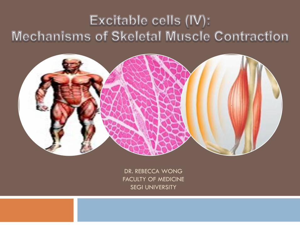
DR. REBECCA WONG
FACULTY OF MEDICINE
SEGI UNIVERSITY
. . .
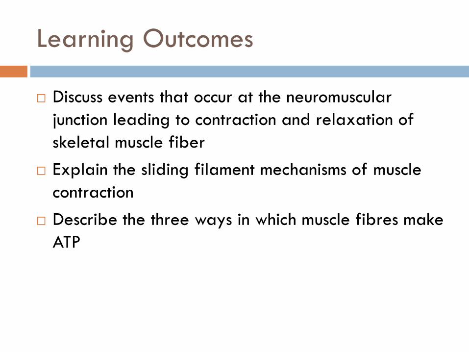
Learning Outcomes
Discuss events that occur at the neuromuscular
junction leading to contraction and relaxation of
skeletal muscle fiber
Explain the sliding filament mechanisms of muscle
contraction
Describe the three ways in which muscle fibres make
ATP

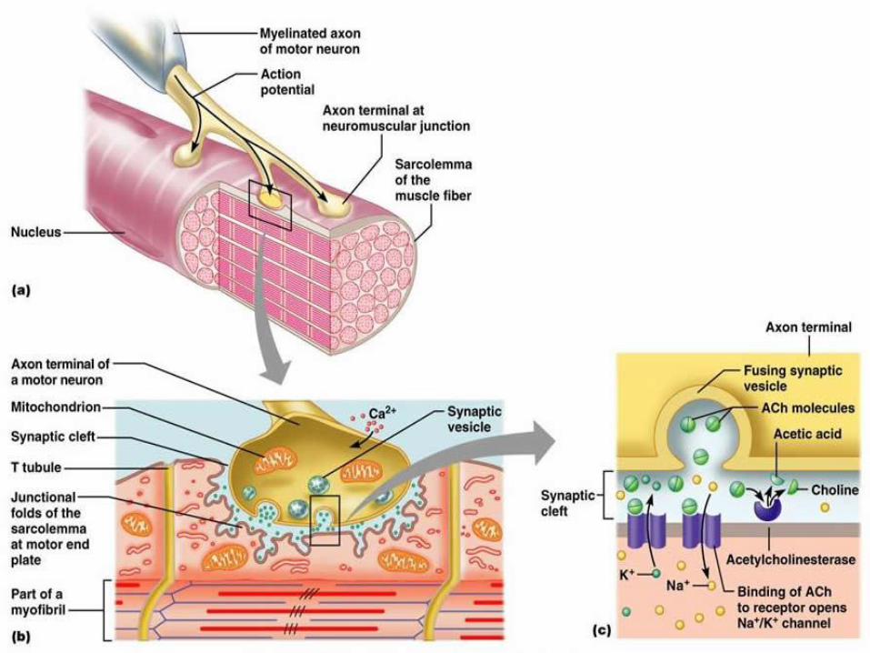
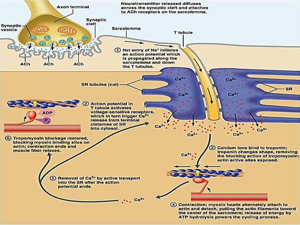
Neuromuscular Junction (NMJ)
The neuromuscular junction is a connection between an axon terminal and a muscle fiber where stimulation of the muscle cell to contract occurs.
The neuromuscular junction consists of the plasma membrane of the motor neuron axon terminal, the synaptic cleft, and the motor endplate.
The motor endplate is part of the sarcolemma where chemically regulated ion channels that respond to neural stimulation are found.
Junctional folds increase the surface area at the motor endplate.

Components of the NMJ
Synapse Where communication occurs
between a somatic motor neuron and a muscle fiber
Synaptic cleft Gap that separates the two cells
Neurotransmitter Chemical released by the initial cell
communicating with the second cell
Synaptic vesicles Sacs suspended within the synaptic
end bulb containing molecules of the neurotransmitter acetylcholine (Ach)
Motor end plate The region of the muscle cell
membrane opposite the synaptic end bulbs. Contain acetylcholine receptors
http://users.bergen.org/dondew/bio/AnP/AnP1/An
P1Tri2/FIGS/AnPT2Fig06.html
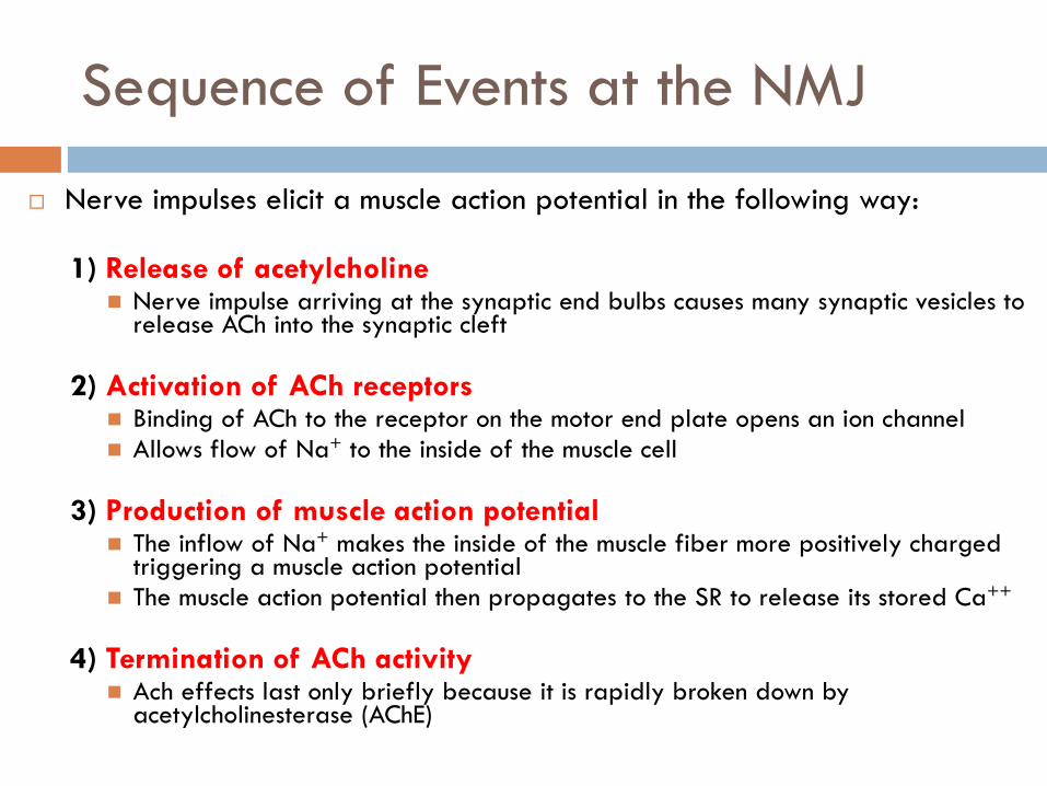
Sequence of Events at the NMJ
Nerve impulses elicit a muscle action potential in the following way:
1) Release of acetylcholine Nerve impulse arriving at the synaptic end bulbs causes many synaptic vesicles to
release ACh into the synaptic cleft
2) Activation of ACh receptors Binding of ACh to the receptor on the motor end plate opens an ion channel
Allows flow of Na+ to the inside of the muscle cell
3) Production of muscle action potential The inflow of Na+ makes the inside of the muscle fiber more positively charged
triggering a muscle action potential
The muscle action potential then propagates to the SR to release its stored Ca++
4) Termination of ACh activity Ach effects last only briefly because it is rapidly broken down by
acetylcholinesterase (AChE)

Copyright 2009, John Wiley & Sons, Inc.

Some interesting facts
Botulinum toxin
Produced by Clostridium Botulinum
Blocks release of ACh from synaptic vesicles
Results in muscle paralysis
May be found in improperly canned foods
A tiny amount can cause death by paralyzing respiratory muscles
Used as a medicine (Botox®)
Strabismus (crossed eyes)
Blepharospasm (uncontrollable blinking)
Spasms of the vocal cords that interfere with speech
Cosmetic treatment to relax muscles that cause facial wrinkles
Alleviate chronic back pain due to muscle spasms in the lumbar region

Some interesting facts
Curare A plant poison used by South American
Indians on arrows and blowgun darts
Causes muscle paralysis by blocking
ACh receptors inhibiting Na+ ion channels
Derivatives of curare are used during
surgery to relax skeletal muscles
Anticholinesterase Slow actions of acetylcholinesterase and
removal of ACh
Can strengthen weak muscle contractions E.g. : Neostigmine
Treatment for myasthenia gravis Antidote for curare poisoning Terminate the effects of curare after surgery
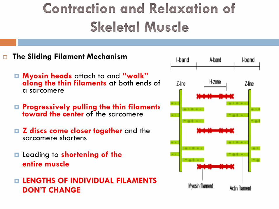
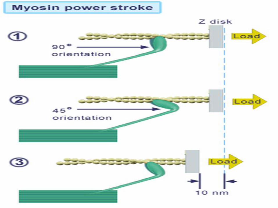
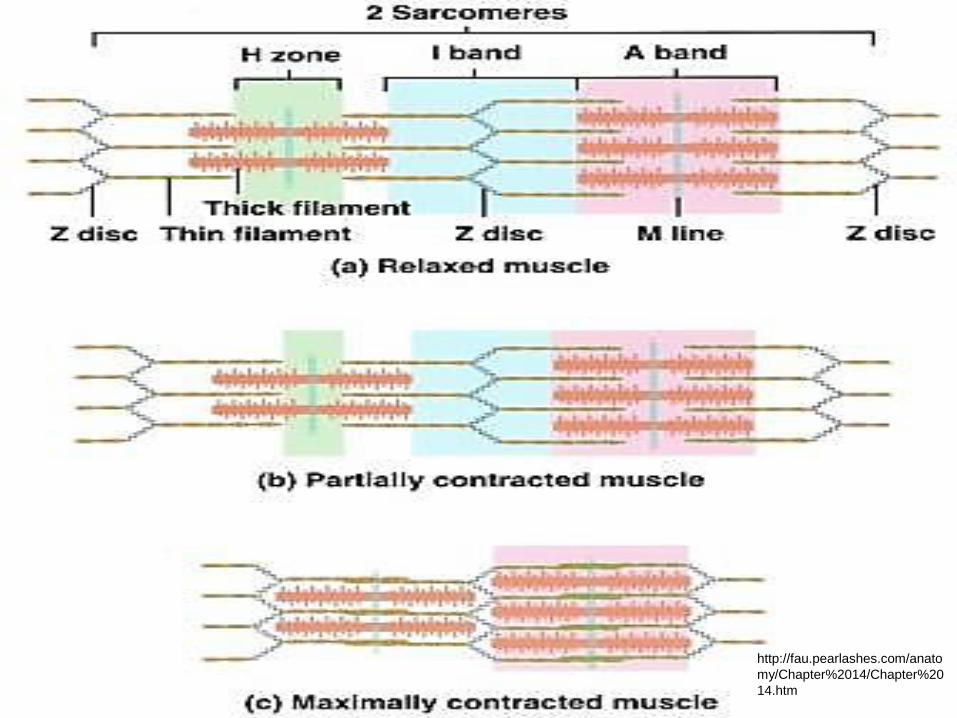
The Sliding Filament Mechanism
Myosin heads attach to and “walk” along the thin filaments at both ends of a sarcomere
Progressively pulling the thin filaments toward the center of the sarcomere
Z discs come closer together and the sarcomere shortens
Leading to shortening of the
entire muscle
LENGTHS OF INDIVIDUAL FILAMENTS
DON’T CHANGE

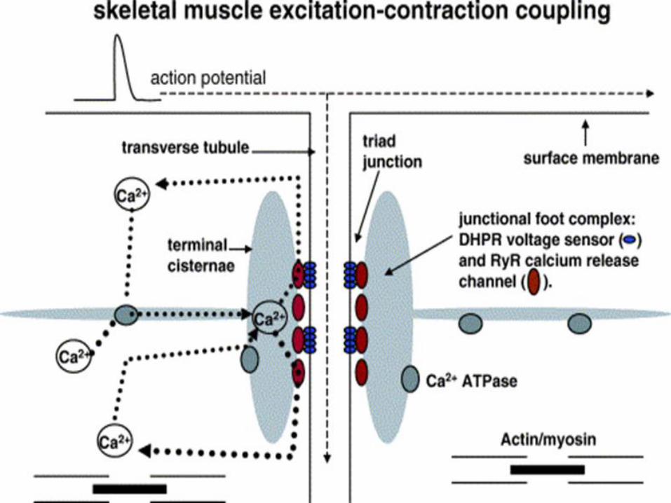
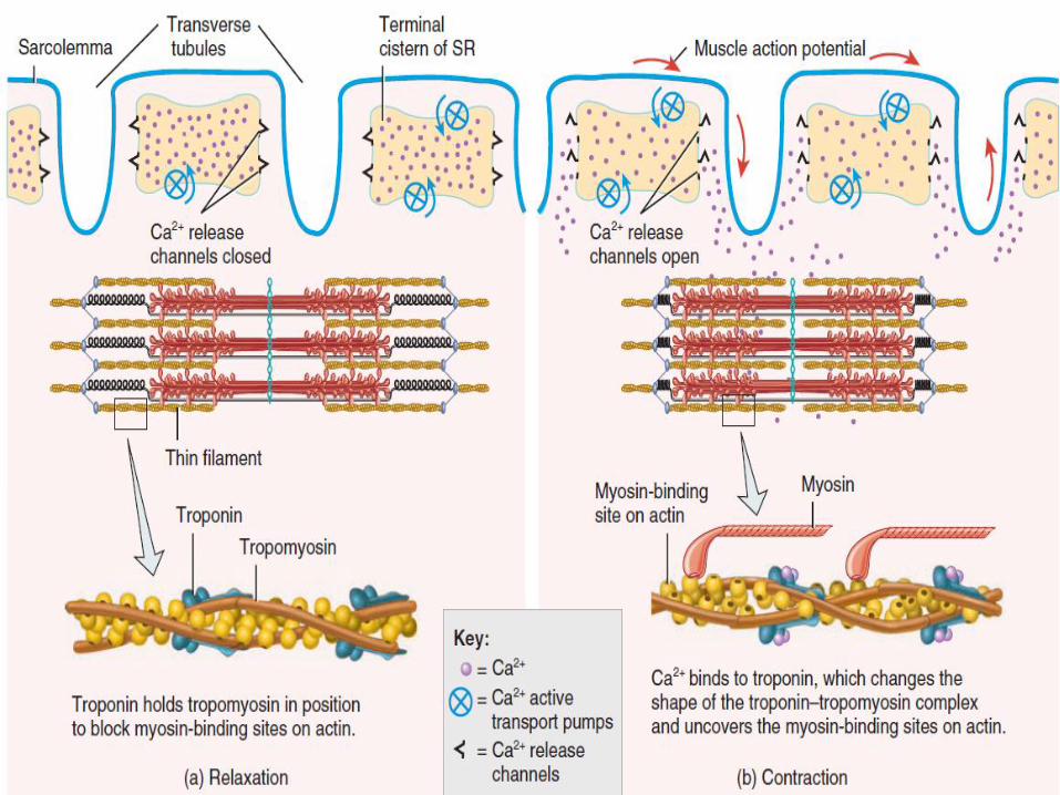
Excitation-contraction coupling
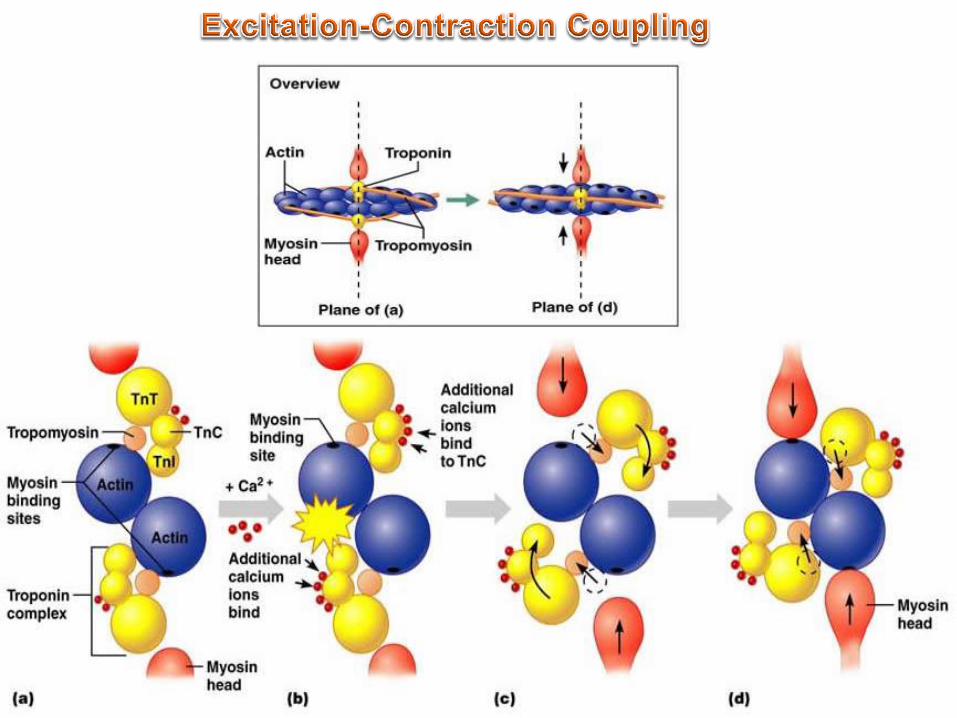
An increase in Ca2+ concentration in the muscle starts contraction
A decrease in Ca2+ stops it
Action potentials causes Ca2+ to be released from the SR into the muscle cell
Ca2+ binding of troponin moves tropomyosin away from the myosin-binding sites on actin allowing cross-bridges to form
The muscle cell membrane contains Ca2+ pumps to return Ca2+ back to the SR quickly
Decreasing calcium ion levels
As the Ca2+ level in the cell drops, myosin-binding sites are covered and the muscle relaxes
Copyright 2009, John Wiley & Sons, Inc.
Copyright 2009, John Wiley & Sons, Inc.

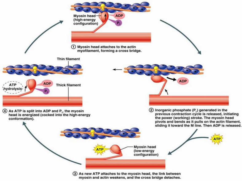
The Contraction Cycle
The contraction cycle consists of 4 steps 1) ATP hydrolysis
Hydrolysis of ATP reorients and energizes the myosin head
2) Formation of cross-bridges Myosin head attaches to the myosin-binding site on actin
3) Power stroke During the power stroke the cross-bridge rotates, sliding the filaments
4) Detachment of myosin from actin As the next ATP binds to the myosin head, the myosin head detaches from
actin
The contraction cycle repeats as long as ATP is available and the Ca++ level is sufficiently high
Continuing cycles applies the force that shortens the sarcomere
Copyright 2009, John Wiley & Sons, Inc.
Copyright 2009, John Wiley & Sons, Inc.
Copyright 2009, John Wiley & Sons, Inc.

Length-Tension relationship
The forcefulness of muscle contraction depends on the length of the sarcomeres
When a muscle fiber is stretched there is less overlap between the thick and thin filaments and tension (forcefulness) is diminished
When a muscle fiber is shortened the filaments are compressed and fewer myosin heads make contact with thin filaments and tension is diminished
Copyright 2009, John Wiley & Sons, Inc.
http://fau.pearlashes.com/anato
my/Chapter%2014/Chapter%20
14.htm
Muscle Metabolism
Production of ATP in Muscle Fibers
A huge amount of ATP is needed to: Power the contraction cycle Pump Ca2+ into the SR
The ATP inside muscle fibers will power contraction for only a few seconds
ATP must be produced by the muscle fiber after reserves are used up
Muscle fibers have three ways to produce ATP 1) From creatine phosphate 2) By anaerobic cellular respiration 3) By aerobic cellular respiration

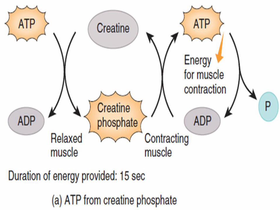
Creatine Phosphate stores
Excess ATP is used to synthesize creatine phosphate
Energy-rich molecule
Creatine phosphate transfers its high energy phosphate group to ADP regenerating new ATP
Creatine phosphate and ATP provide enough energy for contraction for about 15 seconds
Copyright 2009, John Wiley & Sons, Inc.

Anaerobic Respiration
Glucose is derived from the blood and from glycogen stored in muscle fibers
Glycolysis breaks down glucose into molecules of pyruvic acid and produces two molecules of ATP
If sufficient oxygen is present, pyruvic acid formed by glycolysis enters aerobic respiration pathways producing a large amount of ATP
If oxygen levels are low, anaerobic reactions convert pyruvic acid to lactic acid which is carried away by the blood
Anaerobic respiration can provide enough energy for about 30 to 40 seconds of muscle activity
No oxygen required When Creatine phosphate levels depleted

Copyright 2009, John Wiley & Sons, Inc.
Aerobic Respiration
Activity that lasts longer than half a minute depends on aerobic respiration
Pyruvic acid entering the mitochondria is completely oxidized generating:
ATP, carbon dioxide, water & heat
Each molecule of glucose yields about 36 molecules of ATP
Muscle tissue has two sources of oxygen
1) Oxygen from hemoglobin in the blood
2) Oxygen released by myoglobin in the muscle cell
Aerobic respiration provides more than 90% of the needed ATP in activities lasting more than 10 minutes

Copyright 2009, John Wiley & Sons, Inc.

Types of Fibers
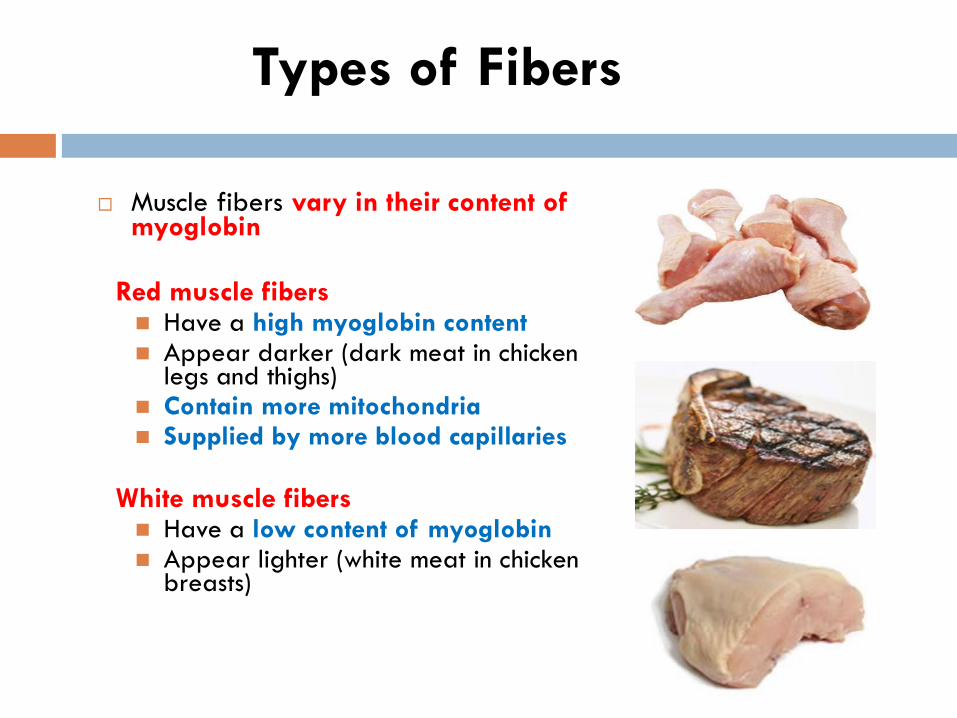
Muscle fibers vary in their content of myoglobin
Red muscle fibers Have a high myoglobin content Appear darker (dark meat in chicken
legs and thighs) Contain more mitochondria Supplied by more blood capillaries
White muscle fibers Have a low content of myoglobin Appear lighter (white meat in chicken
breasts)
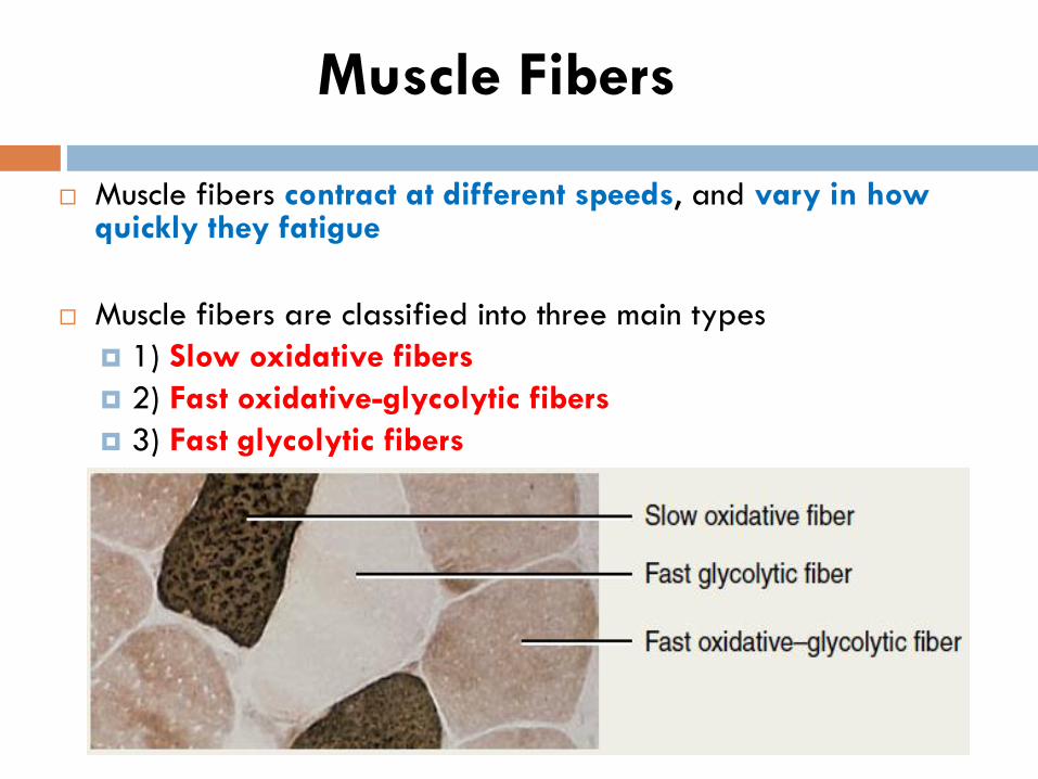
Muscle fibers contract at different speeds, and vary in how quickly they fatigue
Muscle fibers are classified into three main types
1) Slow oxidative fibers
2) Fast oxidative-glycolytic fibers
3) Fast glycolytic fibers
Muscle Fibers

Smallest in diameter Least powerful type of muscle fibers Appear dark red (more myoglobin) Generate ATP mainly by aerobic cellular respiration Have a slow speed of contraction
Twitch contractions last from 100 to 200 msec
Very resistant to fatigue Capable of prolonged, sustained
contractions for many hours Adapted for maintaining posture
and for aerobic, endurance-type activities such as running a marathon
Slow Oxidative (SO) Fibers
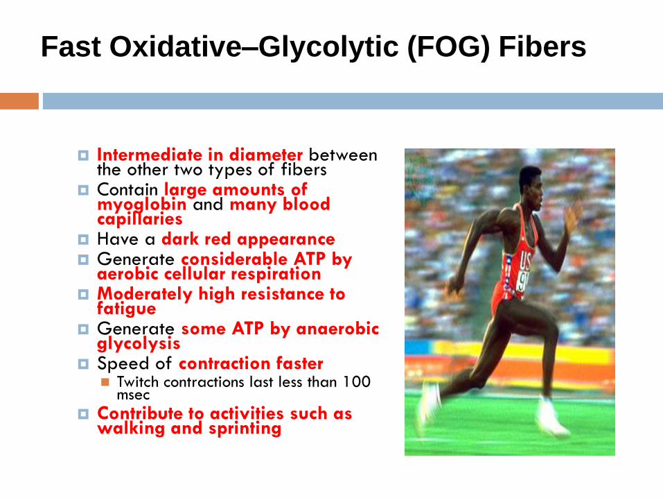
Intermediate in diameter between
the other two types of fibers Contain large amounts of
myoglobin and many blood capillaries
Have a dark red appearance Generate considerable ATP by
aerobic cellular respiration Moderately high resistance to
fatigue Generate some ATP by anaerobic
glycolysis Speed of contraction faster
Twitch contractions last less than 100 msec
Contribute to activities such as walking and sprinting
Fast Oxidative–Glycolytic (FOG) Fibers

Largest in diameter
Generate the most powerful
contractions
Have low myoglobin content
Relatively few blood capillaries
Few mitochondria
Appear white in color
Generate ATP mainly by glycolysis
Fibers contract strongly and quickly
Fatigue quickly
Adapted for intense anaerobic movements of short duration Weight lifting or throwing a ball
Fast Glycolytic (FG)Fibers

Characteristics of Three Types of Skeletal Muscle Fibres
Type I
Slow-oxidative fibres
Type IIA
Fast-oxidative glycolytic
fibres
Type IIB
Fast-glycolytic fibres
Primary source of ATP
production
Oxidative
phosphorylation
Oxidative
phosphorylation
Glycolysis
Mitochondria Many Many Few
Capillaries Many Many Few
Myoglobin content High (red muscle) High (red muscle) Low (white muscle)
Glycolytic enzyme activity Low Intermediate High
Glycogen content Low Intermediate High
Rate of fatigue Slow Intermediate Fast
Myosin-ATPase activity Low High High
Contraction velocity Slow Fast Fast
Fibre diameter Small Intermediate Large
Motor unit size Small Intermediate Large
Size of motor neuron
innervating fibre
Small Intermediate Large
Ca++ sequestration Slow Fast Fast
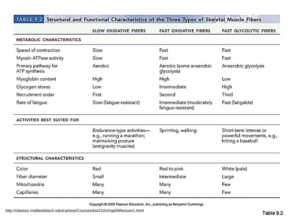
http://classes.midlandstech.edu/carterp/Courses/bio210/chap09/lecture1.html
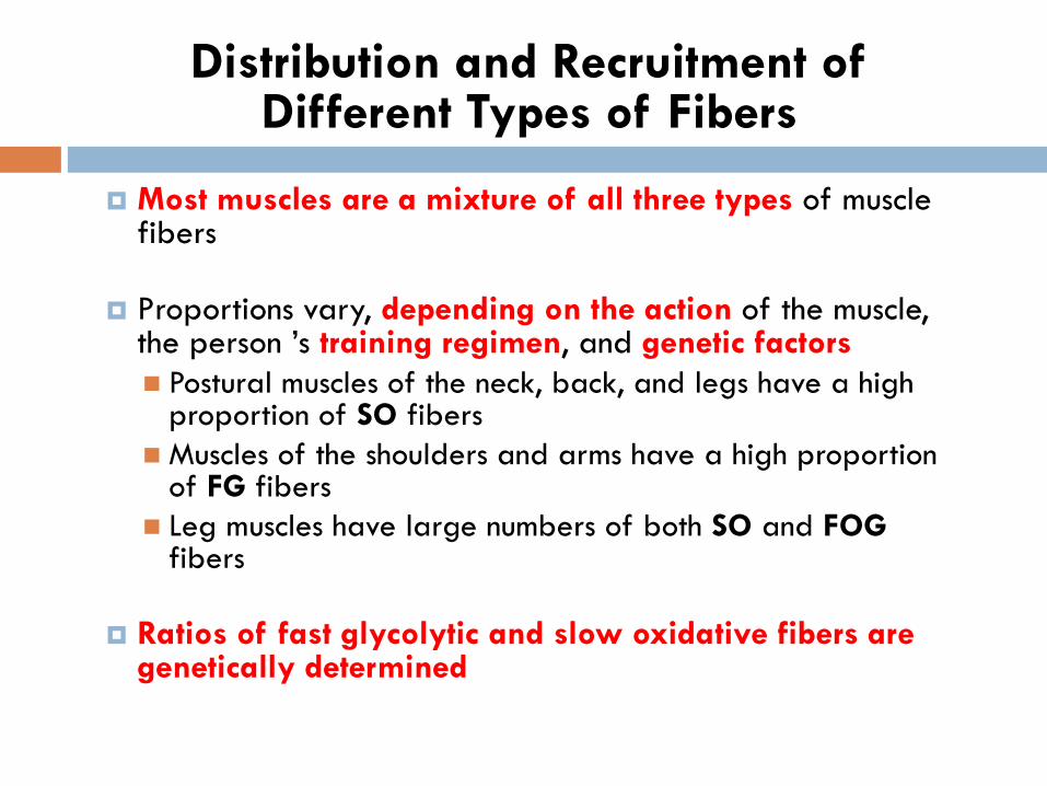
Distribution and Recruitment of Different Types of Fibers
Most muscles are a mixture of all three types of muscle fibers
Proportions vary, depending on the action of the muscle, the person ’s training regimen, and genetic factors
Postural muscles of the neck, back, and legs have a high proportion of SO fibers
Muscles of the shoulders and arms have a high proportion of FG fibers
Leg muscles have large numbers of both SO and FOG fibers
Ratios of fast glycolytic and slow oxidative fibers are genetically determined
Exercise and Skeletal Muscle Tissue
Various types of exercises can induce changes in muscle fibers
Aerobic exercise transforms some FG fibers into FOG
fibers
Endurance exercises do not increase muscle mass
Exercises that require short bursts of strength produce an increase in the size of FG fibers
Muscle enlargement (hypertrophy) due to increased synthesis of thick and thin filaments

Muscles during & after exercise
Muscle Fatigue
Inability of muscle to maintain force of contraction in prolonged activity
Factors that contribute to muscle fatigue
Inadequate release of calcium ions from the SR
Depletion of creatine phosphate
Insufficient oxygen
Depletion of glycogen and other nutrients
Buildup of lactic acid and ADP
Failure of the motor neuron to release enough acetylcholine

Oxygen debt (recovery O2 uptake)
↑ muscle activity → ↑ breathing rate & blood flow to muscle
After exercise, heavy breathing continues and oxygen
consumption remains above the resting level
The added oxygen that is taken into the body after exercise is used
to restore muscle cells to the resting level in three ways
1) to convert lactic acid into glycogen
2) to synthesize creatine phosphate and ATP
3) to replace the oxygen removed from myoglobin
Muscles during & after exercise

Further reading
Fundamentals of Anatomy & Physiology 7th Ed.
2006. Frederic H Martini. Pearson/Benjamin
Cummings.
Seeley's principles of anatomy & physiology 2009.
Philip Tate McGraw-Hill Higher Education
Principles of Anatomy and Physiology, 12th Ed.
2008. Gerard J. Tortora. Wiley.
Human anatomy & physiology 8th ed. 2006, Elaine
N. Marieb. Pearson/Benjamin Cummings.


