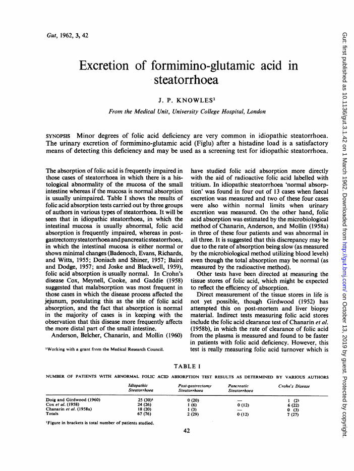
Gut, 1962, 3, 42 Excretion of formimino-glutamic acid in steatorrhoea J. P. KNOWLES1 From the Medical Unit, University College Hospital, London SYNOPSIS Minor degrees of folic acid deficiency are very common in idiopathic steatorrhoea. The urinary excretion of formimino-glutamic acid (Figlu) after a histadine load is a satisfactory means of detecting this deficiency and may be used as a screening test for idiopathic steatorrhoea. The absorption of folic acid is frequently impaired in those cases of steatorrhoea in which there is a his- tological abnormality of the mucosa of the small intestine whereas if the mucosa is normal absorption is usually unimpaired. Table I shows the results of folic acid absorption tests carried out by three groups of authors in various types of steatorrhoea. It will be seen that in idiopathic steatorrhoea, in which the intestinal mucosa is usually abnormal, folic acid absorption is frequently impaired, whereas in post- gastrectomy steatorrhoea and pancreatic steatorrhoea, in which the intestinal mucosa is either normal or shows minimal changes (Badenoch, Evans, Richards, and Witts, 1955; Doniach and Shiner, 1957; Baird and Dodge, 1957; and Joske and Blackwell, 1959), folic acid absorption is usually normal. In Crohn's disease Cox, Meynell, Cooke, and Gaddie (1958) suggested that malabsorption was most frequent in those cases in which the disease process affected the jejunum, postulating this as the site of folic acid absorption, and the fact that absorption is normal in the majority of cases is in keeping with the observation that this disease more frequently affects the more distal part of the small intestine. Anderson, Belcher, Chanarin, and Mollin (1960) 'Working with a grant from the Medical Research Council. have studied folic acid absorption more directly with the aid of radioactive folic acid labelled with tritium. In idiopathic steatorrhoea 'normal absorp- tion' was found in four out of 13 cases when faecal excretion was measured and two of these four cases were also within normal limits when urinary excretion was measured. On the other hand, folic acid absorption was estimated by the microbiological method of Chanarin, Anderson, and Mollin (1958a) in three of these four patients and was abnormal in all three. It is suggested that this discrepancy may be due to the rate of absorption being slow (as measured by the microbiological method utilizing blood levels) even though the total absorption may be normal (as measured by the radioactive method). Other tests have been directed at measuring the tissue stores of folic acid, which might be expected to reflect the efficiency of absorption. Direct measurement of the tissue stores in life is not yet possible, though Girdwood (1952) has attempted this on post-mortem and liver biopsy material. Indirect tests measuring folic acid stores include the folic acid clearance test of Chanarin et al. (1958b), in which the rate of clearance of folic acid from the plasma is measured and found to be faster in patients with folic acid deficiency. However, this test is really measuring folic acid turnover which is BLE I NUMBER OF PATIENTS WITH ABNORMAL FOLIC ACID ABSORPTION TEST RESULTS AS DETERMINED BY VARIOUS AUTHORS Idiopathic Steatorrhoea Doig and Girdwood (1960) Cox et al. (1958) Chanarin et al. (1958a) Totals 25 (30)' 24 (26) 18 (20) 67 (76) Post-gastrectomy Steatorrhoea 0 (20) 1 (6) 1 (3) 2 (29) Pancreatic Steatorrhoea 0 (12) 0 (12) Crohn's Disease 1 (2) 6 (22) 0 (3) 7 (27) 'Figure in brackets is total number of patients studied. 42 on October 13, 2019 by guest. Protected by copyright. http://gut.bmj.com/ Gut: first published as 10.1136/gut.3.1.42 on 1 March 1962. Downloaded from
Transcript
Gut, 1962, 3, 42
Excretion of formimino-glutamic acid insteatorrhoeaJ. P. KNOWLES1
From the Medical Unit, University College Hospital, London
SYNOPSIS Minor degrees of folic acid deficiency are very common in idiopathic steatorrhoea.The urinary excretion of formimino-glutamic acid (Figlu) after a histadine load is a satisfactorymeans of detecting this deficiency and may be used as a screening test for idiopathic steatorrhoea.
The absorption of folic acid is frequently impaired inthose cases of steatorrhoea in which there is a his-tological abnormality of the mucosa of the smallintestine whereas if the mucosa is normal absorptionis usually unimpaired. Table I shows the results offolic acid absorption tests carried out by three groupsof authors in various types of steatorrhoea. It will beseen that in idiopathic steatorrhoea, in which theintestinal mucosa is usually abnormal, folic acidabsorption is frequently impaired, whereas in post-gastrectomy steatorrhoea and pancreaticsteatorrhoea,in which the intestinal mucosa is either normal orshows minimal changes (Badenoch, Evans, Richards,and Witts, 1955; Doniach and Shiner, 1957; Bairdand Dodge, 1957; and Joske and Blackwell, 1959),folic acid absorption is usually normal. In Crohn'sdisease Cox, Meynell, Cooke, and Gaddie (1958)suggested that malabsorption was most frequent inthose cases in which the disease process affected thejejunum, postulating this as the site of folic acidabsorption, and the fact that absorption is normalin the majority of cases is in keeping with theobservation that this disease more frequently affectsthe more distal part of the small intestine.Anderson, Belcher, Chanarin, and Mollin (1960)
'Working with a grant from the Medical Research Council.
have studied folic acid absorption more directlywith the aid of radioactive folic acid labelled withtritium. In idiopathic steatorrhoea 'normal absorp-tion' was found in four out of 13 cases when faecalexcretion was measured and two of these four caseswere also within normal limits when urinaryexcretion was measured. On the other hand, folicacid absorption was estimated by the microbiologicalmethod of Chanarin, Anderson, and Mollin (1958a)in three of these four patients and was abnormal inall three. It is suggested that this discrepancy may bedue to the rate of absorption being slow (as measuredby the microbiological method utilizing blood levels)even though the total absorption may be normal (asmeasured by the radioactive method).
Other tests have been directed at measuring thetissue stores of folic acid, which might be expectedto reflect the efficiency of absorption.
Direct measurement of the tissue stores in life isnot yet possible, though Girdwood (1952) hasattempted this on post-mortem and liver biopsymaterial. Indirect tests measuring folic acid storesinclude the folic acid clearance test of Chanarin et al.(1958b), in which the rate of clearance of folic acidfrom the plasma is measured and found to be fasterin patients with folic acid deficiency. However, thistest is really measuring folic acid turnover which is
BLE INUMBER OF PATIENTS WITH ABNORMAL FOLIC ACID ABSORPTION TEST RESULTS AS DETERMINED BY VARIOUS AUTHORS
IdiopathicSteatorrhoea
Doig and Girdwood (1960)Cox et al. (1958)Chanarin et al. (1958a)Totals
25 (30)'24 (26)18 (20)67 (76)
Post-gastrectomySteatorrhoea
0 (20)1 (6)1 (3)2 (29)
PancreaticSteatorrhoea
0 (12)
0 (12)
Crohn's Disease
1 (2)6 (22)0 (3)7 (27)
'Figure in brackets is total number of patients studied.
Excretion offormimino-glutamic acid in steatorrhoea
likely to be abnormal when the demand for folic acidis increased by hyperactivity of the bone marrow, andthere need not necessarily be any deficiency of folicacid. There is little information available on clearanceof folic acid in steatorrhoea; Chanarin et al. (1958b)found normal clearances in two out of six patients.A more promising test is the measurement of the
fasting serum folic acid level analogous to measuringthe serum vitamin B12 level. Until recently methodswere insufficiently sensitive to detect folic acid in thesera of all normal subjects but Herbert (1959) andBaker, Herbert, Frank, Pasher, Hutner, Wasserman,and Sobotka (1959) have recently evolved a moresensitive method. To date this work has not beenconfirmed by other workers (Luhby and Cooperman,1960).The detection of an abnormal excretion of for-
mimino-glutamic acid (Figlu) after a histidine testload seems to be a reliable, though indirect, measure-ment of the folic acid stores (Bakerman, Silverman,and Daft, 1951; Tabor, Silverman, Mehler, Daft,and Bauer, 1953) and we have recently summarizedthe theoretical basis of the test and introduced asimplified method for the detection of Figlu(Knowles, Prankerd, and Westall, 1960). Briefly, thetest depends on the inability of the body to metabolisehistidine fully in the absence of folic acid. Figlu is adegradation product of histidine metabolism whichis normally broken down to glutamic acid, but iffolic acid is deficient this reaction is impeded andFiglu accumulates in the body and is excreted in theurine.The excretion of Figlu in 44 cases of steatorrhoea
has been studied and the results are reported here,using and comparing several different methods ofdetecting Figlu.
MATERIAL
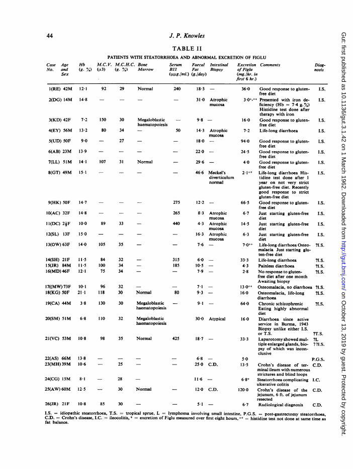
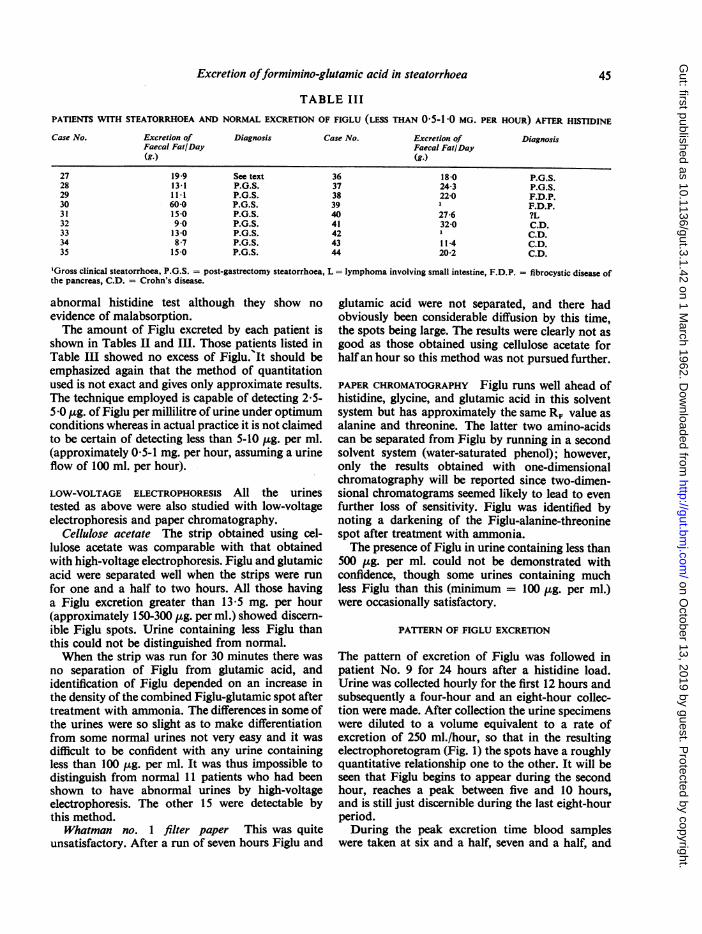
Forty-four patients were studied. Clinical details andlaboratory investigations are listed in Tables Il and III.
An attempt was made to do histidine tests on all thosepatients admitted to University College Hospital betweenMay 1960 and January 1961 who were being investigatedfor steatorrhoea. All patients who had an abnormal fatbalance were tested except one. Five patients (Cases 5 and30 to 33) under the care of Dr. J. R. Nassim at the RoyalNational Orthopaedic Hospital, Stanmore, three patientsunder the care of Dr. F. Avery Jones at the CentralMiddlesex Hospital (Cases 23 to 25), and another patient(Case 20) under the care of Dr. J. H. Walters at QueenMary's Hospital, Roehampton, were also studied.
METHODS
DETECTION OF FIGLU BY HIGH-VOLTAGE ELECTROPHORESIS(KNOWLES et al., 1960) We have modified the method ofperforming the histidine test in several details. A single
urine collection is now made for six hours after a 20 g.histidine load, and we have increased the strength of ouracetone acetic acid mixture from 23/2 to 20/5 (v/v).
DETECTION OF FIGLU BY PAPER CHROMATOGRAPHY One-dimensional ascending runs using 25-200 gl. of urinewere made on Whatman No. 1 paper as suggested byLuhby, Cooperman, and Teller (1959a) using t-butanol75%, formic acid 15%, and water 15% (Block, 1950) asa solvent. A few runs were made in two dimensions usingwater-saturated phenol as the second solvent.
DETECTION OF FIGLU USING LOW-VOLTAGE ELECTROPHOR-ESIS 1 Using Whatman No. 1 paper in the apparatusdescribed by Flynn and de Mayo (1951), 10 pl. of urinewas placed on the strips which were then run at 120v.and 2 milliamp. for seven hours.
2 Using cellulose-acetate paper as described by Kohn(1960), 5-10 pl. of urine was placed on the strips whichwere then run at 200v. and 6-8 milliamp. for 30 minutesor two hours.The buffer used was the same as that previously de-
scribed for the high-voltage technique (Knowles et al.,1960), and the staining procedure was also the same,except that, as acetone cannot be used in treating celluloseacetate, alcoholic solvents were used.
In all the above methods duplicate strips were run of allurines, and one of these strips was treated with ammoniabefore reacting with ninhydrin. Ninhydrin reacts with allcompounds with a free amino group, but Figlu has nofree amino group and treatment with alkali is required torelease the formimino group and free the amino group,thus allowing it to react. Markers were always run witheach strip.
Vitamin B12 was assayed by Dr. Peter Sewell usingLactobacillus leishmannii as test organism. Dr. MargotShiner kindly undertook biopsy of the small intestine inCases 11, 12, 20, and 27 and Dr. David Edwards did biop-sies in Cases 4 and 10. Biopsies in Cases 2 and 8 weredone at laparotomy, and in Case 40 material was obtainedat necropsy.
COMPARISON OF METHODS
HIGH-VOLTAGE ELECTROPHORESIS Apart from thetests reported here experience has been gained of over400 tests in approximately 200 patients, and themethod is satisfactory in differentiating patients intotwo groups: (1) normal, with no excess of Figludetected, and (2) abnormal, with a definite excess ofFiglu. Almost all patients who have had an abnor-mal test have had a disease in which folic aciddeficiency might be expected to occur. There havebeen two exceptions: two elderly women left hospitalbefore investigations could be completed. It seemedpossible that both might be suffering from dietarydeficiency of folic acid but steatorrhoea was not ex-cluded in either. When folic acid has been admin-istered the test has been normal always. Five to 10%of patients with iron-deficiency anaemia have an
- 12.0 C.D. 120.0 Crohn's disease of the C.D.jejunum, 6 ft. of jejunumresected
_ 5-1 6-7 Radiological diagnosis C.D.
I.S. = idiopathic steatorrhoea, T.S. = tropical sprue, L = lymphoma involving small intestine, P.G.S. = post-gastrectomy steatorrhoea,C.D. = Crohn's disease, I.C. = ileocolitis, + = excretion of Figlu measured over first eight hours, ++ = histidine test not done at same time asfat balance.
abnormal histidine test although they show noevidence of malabsorption.The amount of Figlu excreted by each patient is
shown in Tables II and Ill. Those patients listed inTable III showed no excess of Figlu. It should beemphasized again that the method of quantitationused is not exact and gives only approximate results.The technique employed is capable of detecting 2.5-5.0 gg. of Figlu per millilitre of urine under optimumconditions whereas in actual practice it is not claimedto be certain of detecting less than 5-10 ,.g. per ml.(approximately 05-1 mg. per hour, assuming a urineflow of 100 ml. per hour).
LOW-VOLTAGE ELECTROPHORESIS All the urinestested as above were also studied with low-voltageelectrophoresis and paper chromatography.
Cellulose acetate The strip obtained using cel-lulose acetate was comparable with that obtainedwith high-voltage electrophoresis. Figlu and glutamicacid were separated well when the strips were runfor one and a half to two hours. All those havinga Figlu excretion greater than 13.5 mg. per hour(approximately 150-300 ,ug. per ml.) showed discern-ible Figlu spots. Urine containing less Figlu thanthis could not be distinguished from normal.When the strip was run for 30 minutes there was
no separation of Figlu from glutamic acid, andidentification of Figlu depended on an increase inthe density of the combined Figlu-glutamic spot aftertreatment with ammonia. The differences in some ofthe urines were so slight as to make differentiationfrom some normal urines not very easy and it wasdifficult to be confident with any urine containingless than 100 gg. per ml. It was thus impossible todistinguish from normal 11 patients who had beenshown to have abnormal urines by high-voltageelectrophoresis. The other 15 were detectable bythis method.
Whatman no. 1 filter paper This was quiteunsatisfactory. After a run of seven hours Figlu and
glutamic acid were not separated, and there hadobviously been considerable diffusion by this time,the spots being large. The results were clearly not asgood as those obtained using cellulose acetate forhalfan hour so this method was not pursued further.
PAPER CHROMATOGRAPHY Figlu runs well ahead ofhistidine, glycine, and glutamic acid in this solventsystem but has approximately the same R, value asalanine and threonine. The latter two amino-acidscan be separated from Figlu by running in a secondsolvent system (water-saturated phenol); however,only the results obtained with one-dimensionalchromatography will be reported since two-dimen-sional chromatograms seemed likely to lead to evenfurther loss of sensitivity. Figlu was identified bynoting a darkening of the Figlu-alanine-threoninespot after treatment with ammonia.The presence of Figlu in urine containing less than
500 ,ug. per ml. could not be demonstrated withconfidence, though some urines containing muchless Figlu than this (minimum = 100 ,ug. per ml.)were occasionally satisfactory.
PATTERN OF FIGLU EXCRETION
The pattern of excretion of Figlu was followed inpatient No. 9 for 24 hours after a histidine load.Urine was collected hourly for the first 12 hours andsubsequently a four-hour and an eight-hour collec-tion were made. After collection the urine specimenswere diluted to a volume equivalent to a rate ofexcretion of 250 ml./hour, so that in the resultingelectrophoretogram (Fig. 1) the spots have a roughlyquantitative relationship one to the other. It will beseen that Figlu begins to appear during the secondhour, reaches a peak between five and 10 hours,and is still just discernible during the last eight-hourperiod.During the peak excretion time blood samples
were taken at six and a half, seven and a half, and
FIG. 1. Electrophoretogram after treatment with ammoniaand ninhydrin (Case 9). An oral load of J-histidine wasgiven and urine collected over 24 hours. This was dividedinto aliquots as indicated. After collection all urine speci-mens were diluted to give a rate of urine flow equivalentto 250 ml. per minute. (Glutamic acid is seen as a smallspot just to the right of the Figlu spot.) + = cathode;- = anode.
The diagnosis of lymphoma in patients 21 and 40has not been confirmed histologically, although bothhad macroscopically abnormal lymph glands.Biopsy of the small intestine was not obtained inpatient 21 and in patient 40 it was unsatisfactory dueto post-mortem changes. It is possible that both hadidiopathic steatorrhoea.
Patients 27 and 40 were the only two patients whomight have been suffering from idiopathic steator-rhoea who had normal tests. Both showed manyatypical features.The finding that four out of eight patients with
Crohn's disease had normal test results is in keepingwith the results of the absorption tests of Cox et al.(1958) and is compatible with the scattered nature ofthe intestinal lesions in this disease.Both patients with fibrocystic disease had normal
test results which again is in agreement with thenormal absorption in all 12 cases of pancreaticsteatorrhoea investigated by Cox et al. (1958).
DISCUSSION
eight and a half hours, and plasma was separated andultrafiltered. Figlu could then be detected in theplasma and the concentration was approximatelythe same in all three specimens of plasma. In the six-,seven-, and eight-hour urine specimens, the con-centrations were also approximately equal. It wasfound that the urine spots had to be diluted 24-32times to give a spot about equal to that in the plasma.The urine flow during this time was 250 ml./minutethus giving an approximate plasma clearance of96 to 128 ml./minute. There was no evidence ofrenal failure; blood urea was 24 mg./l00 ml. Usingthe same methods of quantitation, clearances ofhistidine and glutamic acid were 4-8 ml./minute and1-2 ml./minute respectively.
ANALYSIS OF RESULTS IN MALABSORPTION
Although only a few biopsies have been done it seemsreasonable to suggest that the histidine test correlatesfairly well with the state of the intestinal mucosain a similar way to the folic acid absorption testsdiscussed in the introduction. However, there wereexceptions. Patient 22 was the only one of 11 casesof post-gastrectomy steatorrhoea to have an abnor-mal test result, but unfortunately the state of hisintestinal mucosa is not known, whereas patient 27,who had a normal test, had almost complete villousatrophy and the few villi remaining were short andsquat. For various reasons he is not thought to besuffering from coeliac disease. He has previouslybeen reported as a case of congenital hypoprotein-aemia with aminoaciduria (Bound and Hackett,1953).
The present work has confirmed that the diagnosisof folic acid deficiency by histidine loading and thedetection of Figlu by high-voltage electrophoresiscompares well with the more elaborate folic acidabsorption tests of other workers shown in Table I,and in particular an equally high proportion ofpatients had abnormal test results in idiopathicstreatorrhoea.Of the various other methods for detecting Figlu
by physical separation, low-voltage electrophoresisusing cellulose acetate seems to be the most promis-ing. Using pure Figlu in normal urine the minimumamount detectable is 40-80 ,g. per ml. of urine, butin actual practice it is often difficult to be confidentwith urine containing less than 100 gg. per ml. Itseems possible that without prior concentrationFiglu will not be detected in the urine from somecases which would not be missed by high-voltageelectrophoresis. Unfortunately, if urine is too con-centrated, streaking occurs both with celluloseacetate and with high-voltage electrophoresis.From the point of view of detecting Figlu by
electrophoresis it is very fortunate that the renalclearance of Figlu is so high and that of glutamic acidis so low. Glutamic acid, like most commonlyoccurring amino-acids, has a low clearance of lessthan 2 ml. per minute (Cusworth and Dent, 1960).Normally, the urinary concentration of glutamicacid is low and little or none can be detected afterelectrophoresis of 25 ml. of urine. However, afterhistidine loading demonstrable quantities are foundin the urine and presumably come from the cata-bolism of histidine and Figlu. The separation of
Excretion offormimino-glutamic acid in steatorrhoea 47
amino-acids by electrophoresis depends on theiriso-electric point. That of Figlu is only slightlydifferent from that of glutamic acid, and to obtainseparation of the two usually requires a run of 10 to15 minutes using high-voltage electrophoresis (6,000volts) or 90 to 120 minutes using cellulose acetate atlow voltage (200 volts). When the excretion of Figluis high the proportion of Figlu to glutamic acid ishigh and separation of the spots is not necessary toobtain satisfactory results. However, when theexcretion is low this ratio is very much reduced andseparation of the spots is to be preferred for accurateresults.
Paper chromatography has proved less rewardingthan cellulose acetate. Luhby et al. (1959a) do notgive detailed information about their method whichthey claim can detect 20-50 ,ug. per ml. of Figlu underfavourable conditions. If this were so it would bemore sensitive than cellulose acetate (40-80 ,ug. perml.), but in the present investigation it has not beenpossible to reproduce this degree of sensitivity.
Figlu can also be determined microbiologicallyand enzymatically. The advantages and disadvan-tages of these methods have been discussed pre-viously (Knowles et al., 1960). Using the enzymemethod of Tabor and Wyngarden (.1958), Luhby etal. (1959b) found the excretion of Figlu after his-tidine loading to be increased in all 10 cases theystudied of 'macrocytic anaemia associated withsprue or malabsorption syndrome'.The fact that there is a poor correlation between
the amount of Figlu excreted and the severity of thesteatorrhoea as judged by the M.C.V., haemoglobin,and the daily faecal fat excretion is not necessarilysurprising. The blood picture in steatorrhoea is oftendimorphic, due to microcytosis secondary to irondeficiency and macrocytosis due to vitamin B12and folic acid deficiency. The M.C.V. and haemo-globin clearly depend on at least two factors otherthan folic acid. It is rather more surprising that twoof the four heaviest excretors of Figlu should havehaemoglobin concentrations greater than 12 g. per100 ml. The daily faecal fat excretion is a measure ofthe state of the intestine at the time the test is done,whereas the excretion of Figlu is an indirect test ofthe body stores of folic acid. In general there will bea rough correlation between tests measuring excre-tion and absorption; it will not necessarily beclose.
Malabsorption of folic acid seems to be one of thecommonest disorders in idiopathic steatorrhoea. Wehave recently shown that the body stores of folicacid can be depleted sufficiently to give an abnormalhistidine test in four to six weeks (Knowles et al.,1961). It is not surprising, therefore, that this test isfrequently abnormal in idiopathic steatorrhoea. For
these reasons it offers a simple and effective screeningtest for idiopathic steatorrhoea, with the addedadvantage that it can be performed easily on out-patients.At the moment there is no satisfactory explanation
for the occurrence of abnormal histidine tests in asmall proportion of patients with iron-deficiencyanaemia and post-gastrectomy steatorrhoea, and theproblem is under investigation.
SUMMARY
Minor degrees of folic acid deficiency are very com-mon in idiopathic steatorrhoea. The urinary excre-tion of Figlu after a histidine load is a satisfactorymeans of detecting this deficiency and it is suggestedthat this is the best screening test for idiopathicsteatorrhoea. Experience with this test is described.The currently available methods of detecting Figlu
which do not involve enzyme or microbiologicalassays are described and compared. It is concludedthat the method involving high-voltage electro-phoresis is the best of these simpler methods, butthat low-voltage electrophoresis using celluloseacetate offers a promising alternative. Paper chro-matography is laborious by comparison and hasnot yet been demonstrated to be sufficiently sensitivefor practical purposes.
ADDENDUM
Since submitting this paper for publication experi-ence has been gained on 121 cases of steatorrhoea.Of these, 47 are thought to be suffering from idio-pathic steatorrhoea. No further cases of possibleidiopathic steatorrhoea with normal histidine testshave been seen since the two mentioned in the text.
I should like to express my thanks to the many physicianswho have allowed me to study their cases, and I amespecially grateful to Drs. T. A. J. Prankerd and R. G.Westall for their encouragement and advice.
REFERENCES
Anderson, B., Belcher E. H., Chanarin, 1., and Mollin, D. L. (1960).The urinary and faecal excretion of radioactivity after oraldoses of 'H-folic acid. Brit. J. Haemat., 6, 439.
Badenoch, J., Evans, J. R.. Richards, W. C. D., and Witts, L. J. (1955).Megaloblastic anaemia following partial gastrectomy andgastro-enterostomy. Ibid., 1, 339.
Baird, I. McL., and Dodge, 0. G. (1957). Jejunal biopsy after partialgastrectomy. Quart. J. Med., 26, 393.
Baker, H., Herbert, V., Frank, 0., Pasher, L., Hutner, S. H., Wasser-man, L. R. and Sobotka, H. (1959). A microbiologic methodfor detecting folic acid deficiency in man. Clin. Chem., 5, 275.
Bakerman, H. A., Silverman, M., and Daft, F. S. (1951). Influence ofsuccinylsufathiazole and folic acid on glutamic acid excretion.J. biol. Chem., 188, 117.
Block, R. J. (1950). Estimation of amino acids and amines on paperchromatograms. Analyt. Chem., 22, 1327.
Bound, J. P., andHackett, W. R. (1953). Idiopathic hypoproteinaemicoedema and amino-aciduria in an infant. Arch. Dis. Childh.,28, 104.
Chanarin, I., Anderson, B. B., and Mollin, D. L. (1958a). The absorp-tion of folic acid. Brit. J. Haemat., 4, 156.
, Mollin, D. L., and Anderson, B. B. (1958b). The clearance fromthe plasma of folic acid injected intravenously in normalsubjects and patients with megaloblastic anaemia. Ibid., 4, 435.
Cox, E. V., Meynell, M. J., Cooke, W. T., and Gaddie, R. (1958).The folic acid excretion test in the steatorrhea syndrome.Gastroenterology, 35, 390.
Cusworth, D. C., and Dent, C. E. (1960). Renal clearance of aminoacids in normal adults and in patients with aminoaciduria.Biochem. J., 74, 550.
Doig, A., and Girdwood, R. H. (1960). The absorption of folic acidand labeiled cyanocobalamin in intestinal malabsorption.Quart. J. Med. 29, 333.
Doniach, I., and Shiner, M. (1957). Duodenal and jejunal biopsies.II. Histology. Gastroenterology, 33, 71.
Flynn, F. V., and de Mayo, P. (1951). Micro-electrophoresis ofproteinon filter-paper. Lancet, 2, 235.
Girdwood, R. H. (1952). The occurrence of growth factors for Lacto-bacillus lelchmannii, Streptococcus faecalis and Leuconostoccitrovorum in the tissues of pernicious anaemia patients andcontrols. Biochem. J., 52, 58.
Herbert, V. (1959). The Megaloblastic Anemias, p.85. Grune &Stratton, New York and London.
Joske, R. A., and Blackwell, J. B. (1959). Alimentary histology in themalabsorption syndrome following partial gastrectomy.Lancet, 2, 379.
Knowles, J. P., Prankerd, T. A. J., and Westall, R. G. (1960). Simpli-fied method for detecting formiminoglutamic acid in urine asa test of folic-acid deficiency. Ibid., 2, 347.
Kohn, J. (1960). Cellulose acetate electrophoresis and immunodiffusion techniques. In Chromatographic and ElectrophoreticTechniques, ed. Smith, 1., Vol. 2, p.56 Heinemann, London.
Luhby, A. L., and Cooperman, J. M. (1960). Formiminoglutamic acidversus serum "folic acid" as an index of folic acid deficiency.J. clin. Invest., 39, 1008.
- ,andTeller, D. N. (1959a). Urinary excretion of formimino-glutamic acid: Application in diagnosis of clinical folic aciddeficiency. Amer. J. clin. Nutr., 7, 397.,-, (1959b). Histidine metabolic loading test to dis-
tinguish folic acid deficiency from vit. B,, in megaloblasticanemias. Proc. Soc. exp. Biol. (N. Y.), 101, 350.
Tabor, H., Silverman, M., Mehler, A. H., Daft, F. S., and Bauer, H.(1953). 1-Histidine conversion to a urinary glutamicacid derivative in folic-deficient rats. J. Amer. chem. Soc.,75, 756.
, and Wyngarden, L. (1958). A method for the determinationof formiminoglutamic acid in urine. J. clin. Invest., 37,824.