74
Exercise 41 Digestive System 1
| Date post: | 25-Dec-2015 |
| Category: |
Documents |
| Upload: | felicity-mcbride |
| View: | 218 times |
| Download: | 2 times |
Exercise 41
Digestive System
1

Digestion and absorption
It is the physical and chemical break down of food
AbsorptionIt is the passing of the digested food
through the epithelial cells into the blood stream
2

Digestive system
3

Gastrointestinal tract
It is the alimentary canal Mouth Pharynx Esophagus Stomach Small intestine Large instestine
4

Accessory digestive organs
Salivary glands Gallbladder Liver Pancreas Teeth
5

General histology of the gastrointestinal tract
It has 4 tunicsMucosa
• Epithelium – simple columnar• Lamina propria – areolar tissue• Muscularis mucosa
• Smooth muscle that enables movement of the mucosa
6
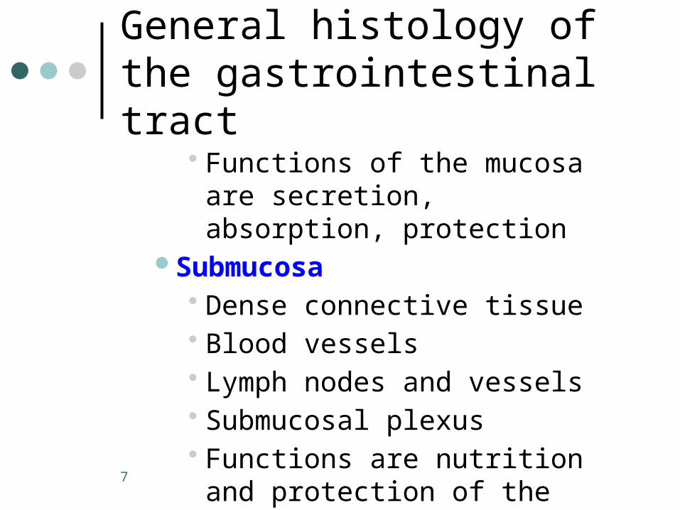
General histology of the gastrointestinal tract
• Functions of the mucosa are secretion, absorption, protection
Submucosa• Dense connective tissue• Blood vessels• Lymph nodes and vessels• Submucosal plexus • Functions are nutrition and
protection of the mucosa7

General histology of the gastrointestinal tract
Muscularis externa• Inner circular layer of smooth
muscle• Outer longitudinal layer of smooth
muscle• Myenteric plexus• Allows GI movements
8

General histology of the gastrointestinal tract
Serosa (abdominal organs)• Most outer layer• Mesothelium – areolar tissue• Functions is to reduce friction
between GI organsAdventitia
• Coarse fibrous tissue that binds the GI organs to the surrounding tissues. Anchors and protects them9
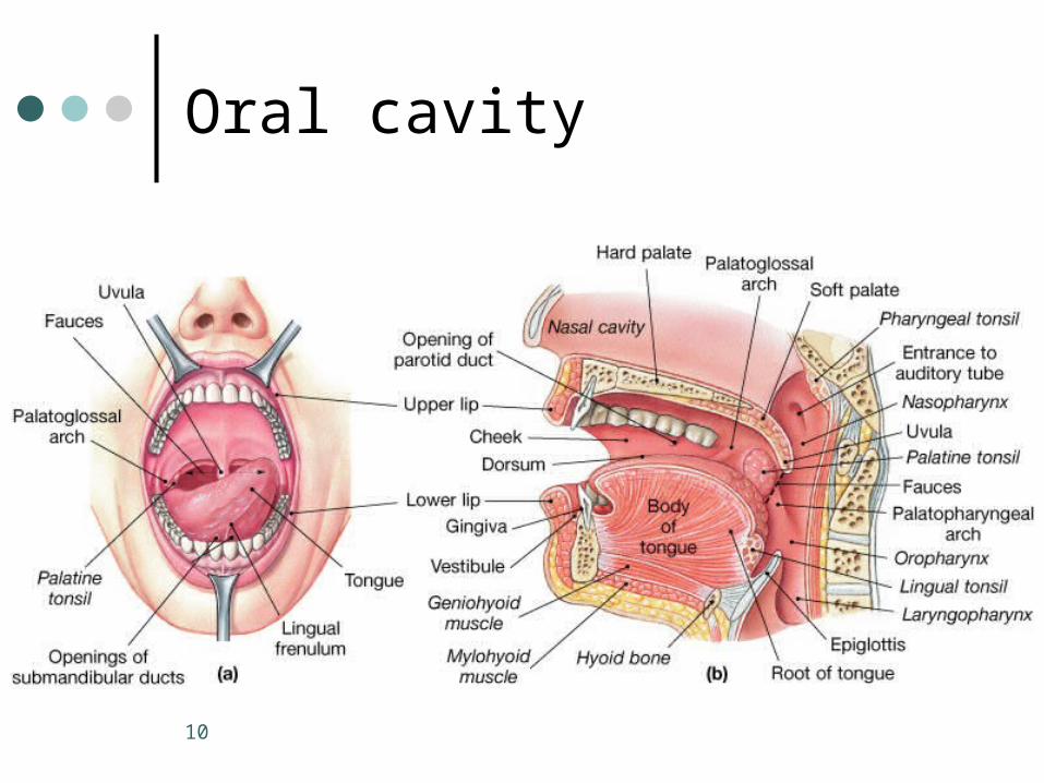
Oral cavity
10

Macroscopy of the digestive tract
Oral cavity or mouthOral cavityLips or labia
• Superior and inferior labial frenulumCheeksPalate
• Soft with uvula• Hard• Palatine raphe
11
Macroscopy of the digestive tractTongue
• Lingual frenulumVestibulePalatine tonsil
• Palatoglossal arch• Palatopharyngeal arch
12
Macroscopy of the digestive tract
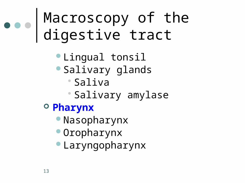
Lingual tonsilSalivary glands
• Saliva• Salivary amylase
PharynxNasopharynxOropharynxLaryngopharynx
13

Macroscopy of the digestive tract Esophagus
PeristalsisGastroesophageal sphincterAdventitia and not serosa
StomachCardiac regionFundusBody
14
Macroscopy of the digestive tract
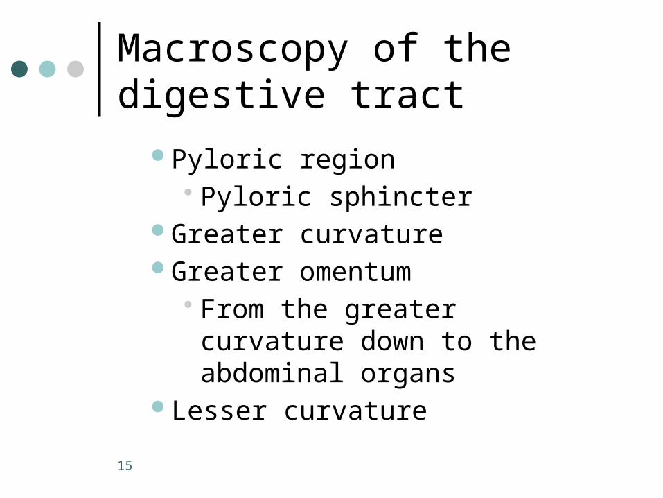
Pyloric region• Pyloric sphincter
Greater curvatureGreater omentum
• From the greater curvature down to the abdominal organs
Lesser curvature
15

Macroscopy of the digestive tract
Lesser omentumFrom the lesser curvature to the liverGastric pitGastric rugaeFunction of the stomach is to
process the food forming the chyme
16

Histology of the stomach
17
Histology of the stomach
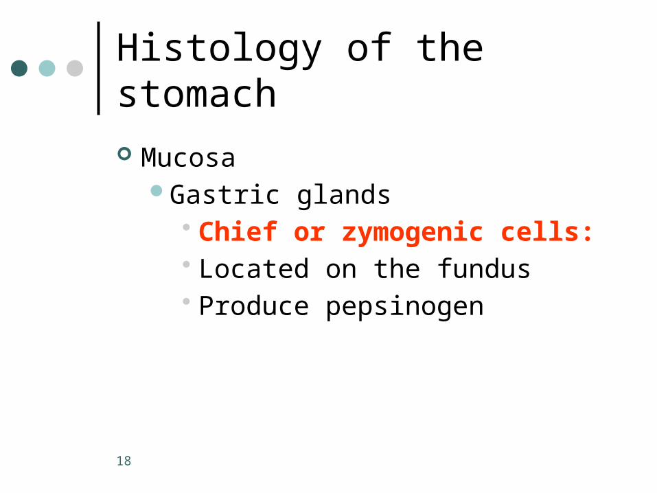
MucosaGastric glands
• Chief or zymogenic cells: • Located on the fundus• Produce pepsinogen
18
Histology of the stomach
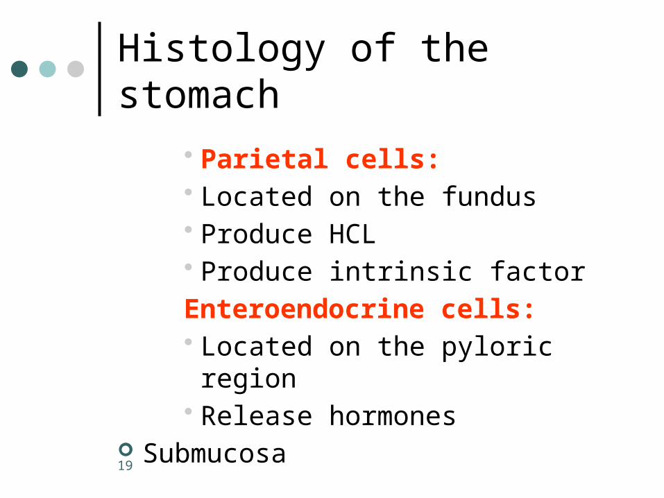
• Parietal cells:• Located on the fundus• Produce HCL• Produce intrinsic factor
Enteroendocrine cells:• Located on the pyloric region• Release hormones
Submucosa
19

Histology of the stomach
Muscularis externaOblique layerCircular layerLongitudinal layer
Gastroesphageal junction (Cardioesophageal)Stratified squamous epithelium on
the esophagusSimple columnar on the stomach
20
Small intestine
From the pyloric sphincter to the ileocecal valve
Mesentery Proper• Double layer of peritoneum that
attaches the small intestine to the posterior body wall
21
Small intestine
PlicaeDeep folds of the mucosa and
submucosaThey cause the chyme to spiral
through the intestine slowing and mixing it
Intestinal crypts of crypts of LieberkuhnIt is the invaginated area of the
mucosa between the villi
22

Small intestine
LactealIt is the lymphatic capillary present in
each villus Function of the small intestine
Nutrients absorption
23
PART B
24
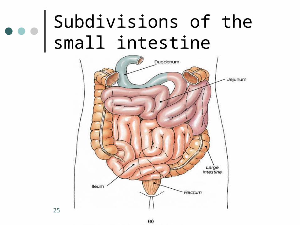
Subdivisions of the small intestine
25

Small intestine
DuodenumPancreatic ductBile ductHepatopancreatic ampullaMajor duodenal papillaHepatopancreatic sphincter or
sphincter of OddiDuodenal glands or Brunner’s
glands – located in the submucosal layer
26

Small intestine
JejunumWhere the food is most absorbed
IleumIleocecal valvePeyer’s patches
• Aggregation of lymphoid tissue more prominent in the ileum
27

Small intestine
Superficial structures of the small intestine that increases the absorptive area of the mucosaVilli
• Fingerlike projections of the mucosa
28
Small intestine
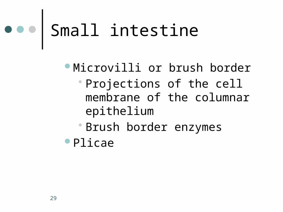
Microvilli or brush border• Projections of the cell membrane
of the columnar epithelium• Brush border enzymes
Plicae
29

Histology of the small intestine
Identify these structures on the slide:PlicaCriptsVilliBrush borderLayers of the intestine
30

Histology of the small intestine
DuodenumSubmucosa with Brunner’s glands
JejunumLongest, leafy villi
IleumSubmucosa with Peyer’s patches
31
The large intestine
32
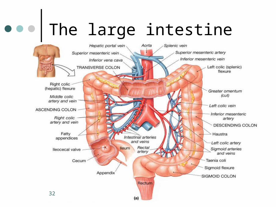
Large intestine
From the ileocecal valve to the anus Mesocolon
Attaches the large intestine to the body wall
CecumIt is the first part
AppendixA blind tube like structure connected
to the cecum33

Large intestine
Colon: Ascending
Right side of the abdominal cavityRight colic (hepatic) flexureIt is retroperitoneal
TransverseCross the abdominal cavityLeft colic (splenic) flexure
34
Large intestine
DescendingIt is retroperitoneal
SigmoidS-shaped Located in the pelvis
Rectum
35

Large intestine
AnusExternal sphincter - skeletal muscle
• VoluntaryInternal sphincter – smooth muscle
• involuntary
36
Large intestine - structures
Tenia coliIt is the longitudinal muscle layer of
muscularis externaIt is in the shape of a muscle band
HaustraPocket like sacs of the large intestineIt is caused by the tenia coli
37

Large intestine - structures
Epiploic appendagesFat-filled pouches of visceral
peritoneum hanging for the colon’s surface
38
Large intestine
Functions of the large intestineConsolidate and propel the fecal
matter to the anusSite for intestinal bacteria to
synthesize vitamins B and KSite for water absorption
39
Histology of the large intestine
Lumen Crypts Layers of the digestive tract Mucosa with the maximum amount of
goblet cells No villi
40
Accessory digestive organs
Teeth:Deciduous (milk teeth)
• They appear between 6 month and 2 ½ years of age
• They begin to shed at 6 years of age
• They are completely shed by the age of 12
41

Accessory digestive organs
Permanent• They begin to appear at 6 years of
age• They last for a lifetime
42

Types of teeth
43

Accessory digestive organs
Classification of the teethIncisors
• Chisel shaped• Shearing action when biting• 4 superiors and 4 inferiors (2
centrals and 2 laterals)• Single-rooted
44
Accessory digestive organs
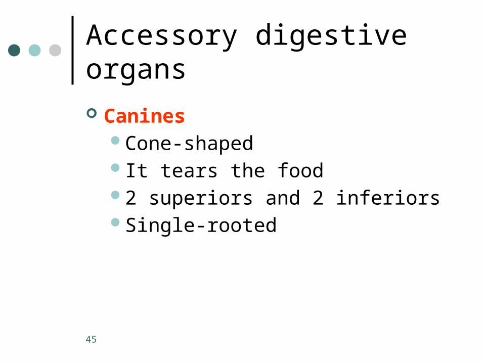
CaninesCone-shapedIt tears the food2 superiors and 2 inferiorsSingle-rooted
45
Accessory digestive organs
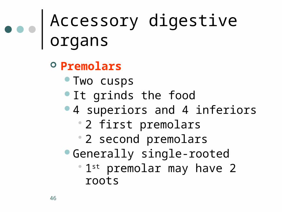
PremolarsTwo cuspsIt grinds the food4 superiors and 4 inferiors
• 2 first premolars• 2 second premolars
Generally single-rooted• 1st premolar may have 2 roots
46
Accessory digestive organs
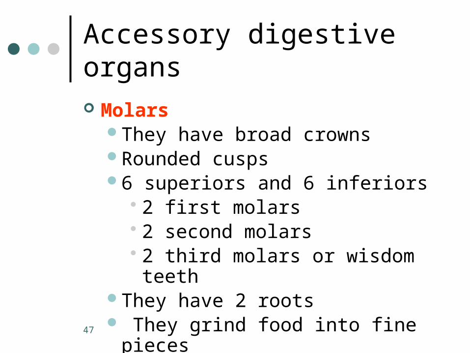
MolarsThey have broad crownsRounded cusps6 superiors and 6 inferiors
• 2 first molars• 2 second molars • 2 third molars or wisdom teeth
They have 2 roots They grind food into fine pieces
47
Accessory digestive organs
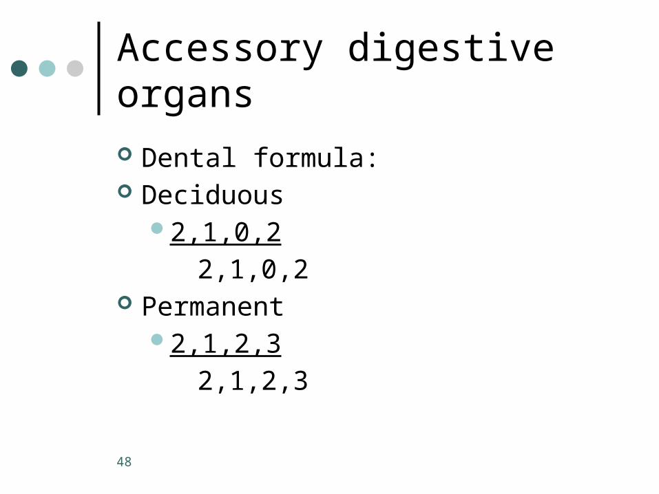
Dental formula: Deciduous
2,1,0,2
2,1,0,2 Permanent
2,1,2,3
2,1,2,3
48
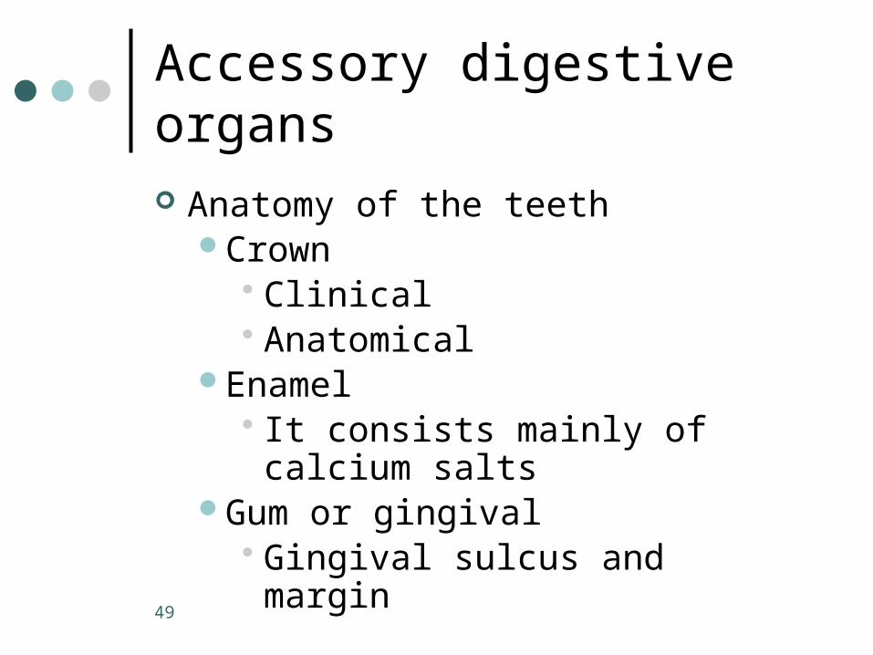
Accessory digestive organs
Anatomy of the teethCrown
• Clinical• Anatomical
Enamel• It consists mainly of calcium salts
Gum or gingival• Gingival sulcus and margin
49

Accessory digestive organs
Neck Root Cementum Periodontal ligament Dentin Pulp
Contain blood vessels and nerves Pulp cavity
50

Accessory digestive organs
Odontoblasts Root canal Apical foramen
51
PART C
52

Accessory digestive organs
Salivary glandsParotid glands
• Anterior to the ear• He parotid duct open at the level
of the second superior molar• Mainly a serous gland
53
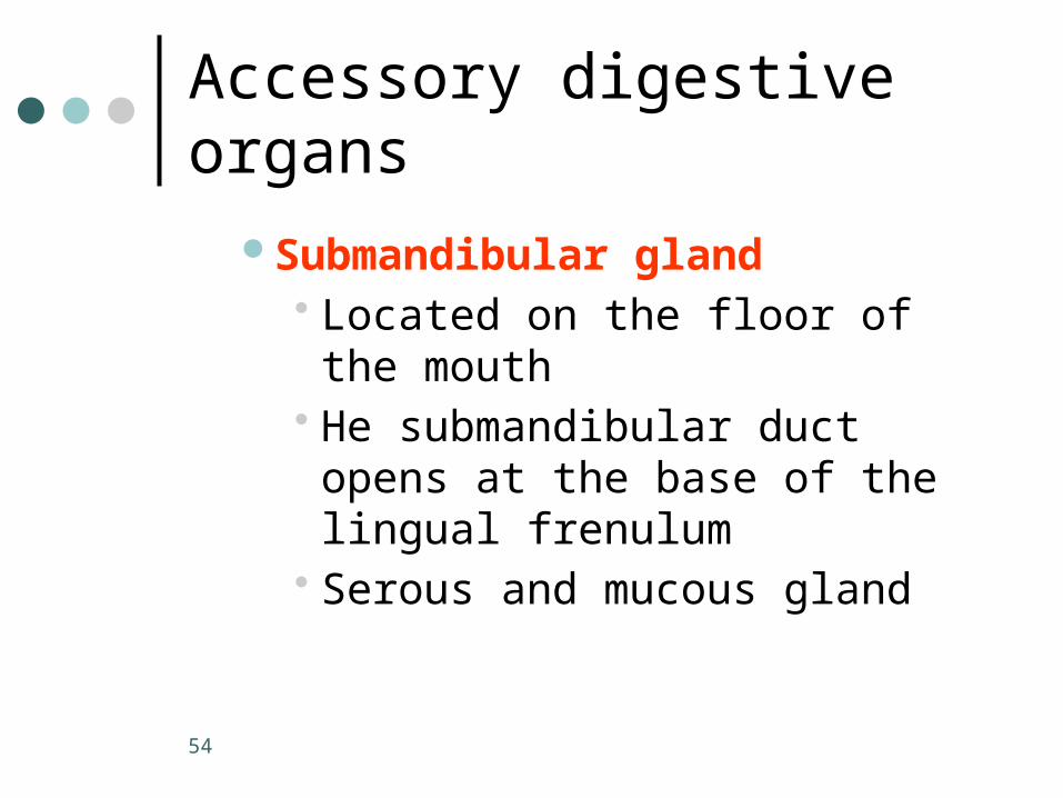
Accessory digestive organs
Submandibular gland• Located on the floor of the mouth• He submandibular duct opens at
the base of the lingual frenulum• Serous and mucous gland
54
Accessory digestive organs
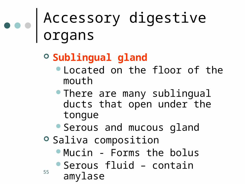
Sublingual glandLocated on the floor of the mouthThere are many sublingual ducts that
open under the tongueSerous and mucous gland
Saliva compositionMucin - Forms the bolusSerous fluid – contain amylase
55

Accessory digestive organs
Histology of the salivary glandsMucous cells forming the aciniSerous cells forming demilunes
around the mucous cellsDucts with cuboidal epithelium
56
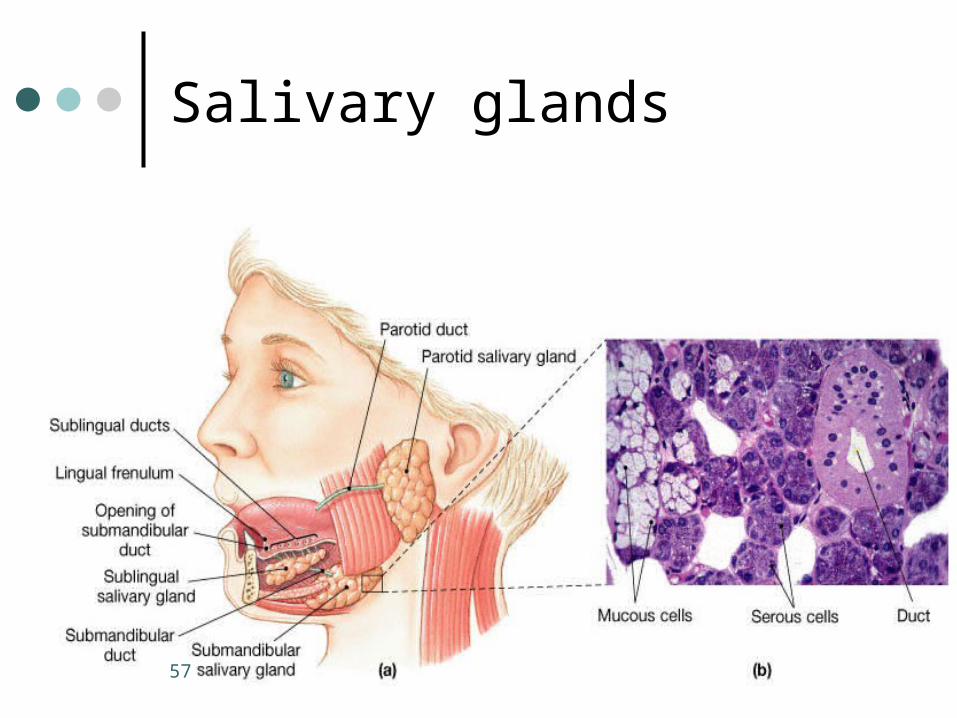
Salivary glands
57

Accessory digestive organs
LiverLocated mainly in the right
hypochondriac region4 lobes
• Right, left, caudate, quadrateFalciform ligament
• Suspend the liver from the diaphragm and anterior abdominal wall
58
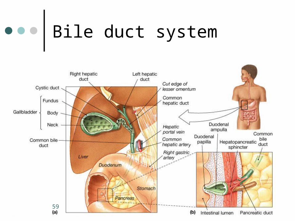
Bile duct system
59

Accessory digestive organs
BileProduced by the liver Responsible for emulsification of the
lipid from the diet Bile duct system
Bile canaliculus• Carries the bile to the duct of the
nearest portal area
60
Accessory digestive organs
Bile ducts carry bile to the:Right and left hepatic ducts Common hepatic duct
61
Accessory digestive organs
Histology Lobules
• Structural and functional units of the liver
• They have cords of hepatocytes running away from the central vein
• Hexagonal shapeCentral vein
62

Histology of the liver
63
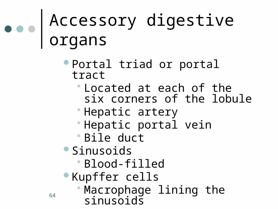
Accessory digestive organs
Portal triad or portal tract• Located at each of the six corners
of the lobule• Hepatic artery• Hepatic portal vein• Bile duct
Sinusoids• Blood-filled
Kupffer cells• Macrophage lining the sinusoids
64

Accessory digestive organs
GallbladderStores the bile not being usedConcentrates the stored bileCystic duct
65

Accessory digestive organs
PancreasIt is a retroperitoneal organEndocrine and exocrine organSecretes the pancreatic juice into the
duodenumIt alkalinizes the chyme coming from
the stomachPancreatic duct or duct of WirsungAccessory pancreatic duct or duct of
Santorini66

Accessory digestive organs
Histology of the pancreasAcinar or exocrine pancreasIslets or endocrine pancreasSepta
• Connective tissue
67

Microscopic structures to be identified
Identify the organ and its layers:Mucosa, submucosa, muscularis
externa, adventitia or serosa Esophagus
Stratified squamous epitheliumGastroesophageal junction
68

Microscopic structures to be identified Stomach
Simple columnar epitheliumGastric pit
DuodenumVilli
• Brush border• Goblet cells
Intestinal criptsBrunner’s glands
69
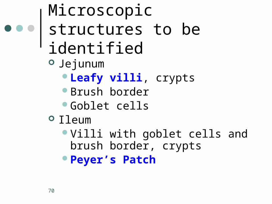
Microscopic structures to be identified Jejunum
Leafy villi, cryptsBrush borderGoblet cells
IleumVilli with goblet cells and brush
border, cryptsPeyer’s Patch
70

Large intestineCripts, abundant goblet cells
Salivary glandsSerous acini (demilunes)Mucous aciniDucts
PancreasAcinar exocrine vs. endocrine
pancreatic islets
Microscopic structures to be identified
71

Microscopic structures to be identified Liver
Hexagonal lobulesTriad
• Hepatic portal vein• Hepatic artery• Bile duct
Central veinSinusoids vs. plates of hepatocytes
72

Cat structures to be identified
Esophagus Stomach
Lesser and greater curvaturesLesser and greater omentum
Small intestineMesentery properIleocecal valve
73
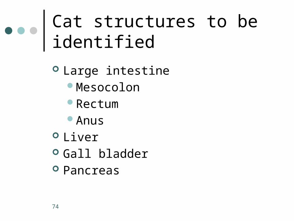
Cat structures to be identified
Large intestineMesocolonRectumAnus
Liver Gall bladder Pancreas
74


