Exercise Intolerance Dalane W. Kitzman, MD a, *, Leanne Groban, MD b Exercise intolerance is the primary symptom of chronic diastolic heart failure (DHF). It is part of the definition of heart failure and is intimately linked to its pathophysiology. Further, exercise intoler- ance affects the diagnosis and prognosis of heart failure. In addition, understanding the mechanisms of exercise intolerance can lead to developing and testing rational treatments for heart failure. This article focuses on the fundamental principles of exercise physiology and on the assessment, path- ophysiology, and potential treatment of exercise intolerance in DHF. IMPORTANCE OF EXERCISE INTOLERANCE Heart failure is defined as a syndrome in which cardiac output is insufficient to meet metabolic demands. This definition implies that insufficient cardiac output will be expressed symptomatically. Heart failure often may manifest by occasional epi- sodes of acute decompensation with overt sys- temic volume overload and pulmonary edema. 1,2 Exertional fatigue and dyspnea, however, are the primary chronic symptoms in outpatients, even when well compensated and non-edematous, and whether associated with reduced or normal ejection fraction (EF). 3 In addition, these symptoms and other consequences of exercise intolerance are potent determinants of health-related quality of life in patients who have heart failure. Several investigators have reported that objective mea- sures and even subjective estimates of exercise tolerance are predictors of survival. 4,5 Exercise intolerance can be quantified objec- tively using semiquantitative assessments, such as interview (New York Heart Association [NYHA] classification) and surveys (Minnesota Living with Heart Failure or Kansas City Cardiomyopathy questionnaires), and quantitative methods, includ- ing timed walking tests (6-minute walk distance) and graded exercise treadmill or bicycle exercise tests. Cardiopulmonary exercise testing on a tread- mill or a bicycle ergometer provides the most accu- rate, reliable, and reproducible assessments of exercise tolerance and yields multiple important outcomes, including metabolic equivalents, exer- cise time, exercise workload, blood pressure and heart rate responses, and rate–pressure product. Using commercially available instruments that perform automated concentration and volume analyses of expired gas, one can assess simulta- neously measures of oxygen consumption (VO 2 ), carbon dioxide generation, and ventilatory re- sponse both at rest and during exercise. Patient effort is an important modifier of data quality and can itself be assessed simultaneously, objectively by expired gas analysis (as the respiratory exchange ratio) and by the somewhat subjective but more easily obtained measures of perceived effort by the Borg scale and percent age- predicted maximal heart rate. Submaximal exercise is in some ways a more important outcome variable than peak exercise capacity because it is more applicable to everyday life and is relatively independent of effort. Submax- imal exercise capacity can be assessed as the This article originally appeared in Heart Failure Clinics, volume 4, number 1. This work was supported in part by National Institute on Aging Grants, R37-AG18915 (MERIT), Dennis Jahnigen Career Development and Paul Beeson Award (K08-AG026764) and Claude D. Pepper Older Americans Indepen- dence Center (P30 AG21332). a Wake Forest University Health Sciences, Winston-Salem, NC, USA b Wake Forest University Health Sciences Center, Winston-Salem, NC, USA * Corresponding author. Section of Cardiology, Wake Forest University School of Medicine, Medical Center Boulevard, Winston-Salem, NC 27157. E-mail address: [email protected]KEYWORDS Diastolic heart failure Heart rate Hypertrophic cardiomyopathy Systolic heart failure Cardiol Clin 29 (2011) 461–477 doi:10.1016/j.ccl.2011.06.002 0733-8651/11/$ – see front matter Ó 2011 Elsevier Inc. All rights reserved. cardiology.theclinics.com
Exercise intolerance is the primary symptom ofchronic diastolic heart failure (DHF). It is part ofthe definition of heart failure and is intimately linkedto its pathophysiology. Further, exercise intoler-ance affects the diagnosis and prognosis of heartfailure. In addition, understanding the mechanismsof exercise intolerance can lead to developing andtesting rational treatments for heart failure. Thisarticle focuses on the fundamental principles ofexercise physiology and on the assessment, path-ophysiology, and potential treatment of exerciseintolerance in DHF.
IMPORTANCE OF EXERCISE INTOLERANCE
Heart failure is defined as a syndrome in whichcardiac output is insufficient to meet metabolicdemands. This definition implies that insufficientcardiac output will be expressed symptomatically.Heart failure often maymanifest by occasional epi-sodes of acute decompensation with overt sys-temic volume overload and pulmonary edema.1,2
Exertional fatigue and dyspnea, however, are theprimary chronic symptoms in outpatients, evenwhen well compensated and non-edematous,and whether associated with reduced or normalejection fraction (EF).3 In addition, these symptomsand other consequences of exercise intoleranceare potent determinants of health-related qualityof life in patients who have heart failure. Severalinvestigators have reported that objective mea-sures and even subjective estimates of exercisetolerance are predictors of survival.4,5
This article originally appeared in Heart Failure Clinics, vThis work was supported in part by National Institute on ACareer Development and Paul Beeson Award (K08-AG026dence Center (P30 AG21332).a Wake Forest University Health Sciences, Winston-Salemb Wake Forest University Health Sciences Center, Winsto* Corresponding author. Section of Cardiology, Wake FBoulevard, Winston-Salem, NC 27157.E-mail address: [email protected]
Cardiol Clin 29 (2011) 461–477doi:10.1016/j.ccl.2011.06.0020733-8651/11/$ – see front matter � 2011 Elsevier Inc. All
Exercise intolerance can be quantified objec-tively using semiquantitative assessments, suchas interview (New York Heart Association [NYHA]classification) and surveys (Minnesota Living withHeart Failure or Kansas City Cardiomyopathyquestionnaires), and quantitative methods, includ-ing timed walking tests (6-minute walk distance)and graded exercise treadmill or bicycle exercisetests. Cardiopulmonary exercise testing ona tread-mill or a bicycle ergometer provides themost accu-rate, reliable, and reproducible assessments ofexercise tolerance and yields multiple importantoutcomes, including metabolic equivalents, exer-cise time, exercise workload, blood pressure andheart rate responses, and rate–pressure product.Using commercially available instruments thatperform automated concentration and volumeanalyses of expired gas, one can assess simulta-neously measures of oxygen consumption (VO2),carbon dioxide generation, and ventilatory re-sponse both at rest and during exercise. Patienteffort is an important modifier of data quality andcan itself be assessed simultaneously, objectivelyby expired gas analysis (as the respiratoryexchange ratio) and by the somewhat subjectivebut more easily obtained measures of perceivedeffort by the Borg scale and percent age-predicted maximal heart rate.
Submaximal exercise is in some ways a moreimportant outcome variable than peak exercisecapacity because it is more applicable to everydaylife and is relatively independent of effort. Submax-imal exercise capacity can be assessed as the
olume 4, number 1.ging Grants, R37-AG18915 (MERIT), Dennis Jahnigen764) and Claude D. Pepper Older Americans Indepen-
, NC, USAn-Salem, NC, USAorest University School of Medicine, Medical Center
ventilatory anaerobic threshold by expired gasanalysis, using either the Wasserman-Whipp orthe V-slope method. Cardiopulmonary exercisetesting measurements and expired gas analysiswith automated, commercially available instru-ments provides measures of both peak oxygenconsumption and of ventilatory anaerobic thresh-old that are valid and highly reproducible in elderlypatients who have DHF as well as in those whohave systolic heart failure (SHF) (Fig. 1). Anothervariable provided by these methods, the ventila-tion/carbon dioxide production (VE/VCO2) slope,is a strong predictor of survival, independent ofVO2.
6 The VE/VCO2 slope is abnormal in patientswho have DHF, although it is not as abnormal asit is in those who have SHF.7
Submaximal exercise performance also can beassessed by timed and distance walk tests. Thesetests are simple to perform and are widely avail-able. The authors have shown that the 6-minutewalk distance is decreased considerably in elderlypatients who have DHF. In group data, the reduc-tion is in proportion to both peak exercise oxygenconsumption and ventilatory anaerobic threshold.The authors’ published studies, however, suggestthat 6-minute walk testing has only modest ac-curacy for predicting peak exercise capacity in
400
600
800
1000
1200
1400
1600
1800
2000
400 600 800 1000 1Test 1 (VO
2
Test 2 (V
O2m
ax, m
l/m
in
)
0
500
1000
1500
0 0.083
0.167
0.25
Tim
VO
2 (m
l/m
in
)
Start of exercise
Fig. 1. Excellent reproducibility of peak exercise VO2 in owho have LVEF. (Top panel) Group data. (Bottom panel) 15(From Marburger CT, Brubaker PH, Pollock WE, et al. Reelderly heart failure patients. Am J Cardiol 1998;82:905–9
individual patients compared with direct mea-surement with cardiopulmonary exercise testingwith expired gas analyses and also is not asreproducible.8
PATHOPHYSIOLOGY OF EXERCISEINTOLERANCE
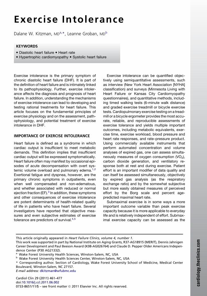
To understand the pathophysiology of exerciseintolerance in DHF, the authors performed a com-parative study of maximal exercise testing withexpired gas in 119 older subjects in three distinct,well-defined groups: persons who had heart failurewith severe left ventricular (LV) systolic dysfunc-tion (mean EF, 30%); persons who had isolatedDHF (EF �50% and no significant coronary,valvular, pericardial, or pulmonary disease andno anemia); and age-matched controls.3 In com-parison with the controls, peak exercise VO2 wasseverely reduced in the patients who had DHF,to a degree similar to the reduction in those withSHF (Fig. 2).3 Submaximal exercise capacity, asmeasured by the ventilatory anaerobic threshold,was reduced in patients who had DHF versusthose who had SHF, and this reduced exercisecapacity was accompanied by reduced health-related quality of life.3
200 1400 1600 1800 2000max, ml/min)
r = 0.92
p < 0.0004
0.333
0.417
0.5 0.583
e (min)
Test 1
Test 2
lder patients who have heart failure, including those-Second averaged data from a representative patient.producibility of cardiopulmonary exercise testing in; with permission.)
0
5
10
15
20
25
NO SD DD
Peak V
O2 (m
l/kg
/m
in
)
‡‡
0
2
4
6
8
10
12
14
16
NO SD DD
VO
2 (m
l/m
in
/kg
) at A
T
‡ ‡
Peak
Submaximal
Fig. 2. VO2 during peak exhaustive exercise (left panel) and during submaximal exercise at the ventilatory anaer-obic threshold (right panel) in age-matched normal subjects (NO), elderly patients who have heart failure causedby systolic dysfunction (SD), and elderly patients who have heart failure with normal systolic function, that is,presumed diastolic dysfunction (DD). Exercise capacity is severely reduced in patients who have DHF comparedwith normal controls (P<.001), to a degree similar to the reduction in patients who have SHF. Overall, peak exer-cise VO2 was 33% lower in the women than in the men (not shown). (Data from Kitzman DW, Little WC, BrubakerPH, et al. Pathophysiological characterization of isolated diastolic heart failure in comparison to systolic heartfailure. JAMA 2002;288(17):2144–50.)
Exercise Intolerance 463
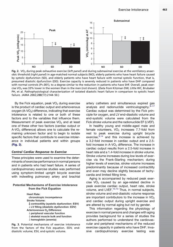
By the Fick equation, peak VO2 during exerciseis the product of cardiac output and arteriovenousoxygen (A-VO2) difference, indicating that exerciseintolerance is related to one or both of thesefactors and to the variables that influence them.Measurement of peak exercise VO2 and at leastone of these other two factors (cardiac output orA-VO2 difference) allows one to calculate the re-maining unknown factor and to begin to isolatespecific factors that contribute to exercise intoler-ance in individual patients and within groups(Fig. 3).
Central Cardiac Response to Exercise
These principles were used to examine the deter-minants of exerciseperformance in normal personsand in patients who had heart failure. A series ofcardiopulmonary exercise studies was performedusing symptom-limited upright bicycle exercisewith indwelling pulmonary artery and brachial
Potential Mechanisms of Exercise Intolerance
from the Fick Equation
Heart Rate:
chronotropic incompetence
Stroke Volume:
↓↓ contractility (systolic dysfunction: ESV)
↓ LV filling (diastolic dysfunction: EDV)
↓ peripheral vascular function
↓ skeletal muscle bulk and function
↓ hemoglobin (anemia)
Fig. 3. Potential mechanisms of exercise intolerancefrom the factors of the Fick equation. EDV, end-diastolic volume; ESV, end-systolic volume.
artery catheters and simultaneous expired gasanalysis and radionuclide ventriculography.9–17
Cardiac output was determined by the Fick prin-ciple for oxygen, and LV end-diastolic volume andend-systolic volume were calculated from theFick stroke volume and the radionuclide EF (LVEF).
In healthy young and middle-aged male andfemale volunteers, VO2 increases 7.7-fold fromrest to peak exercise during upright bicycleexercise,9,14 and this increase is achieved bya 3.2-fold increase in cardiac output and a 2.5-fold increase in A-VO2 difference. The increase incardiac output results from a 2.5-fold increase inheart rate and a 1.4-fold increase in stroke volume.Stroke volume increases during low levels of exer-cise via the Frank-Starling mechanism; duringhigher levels of exercise, stroke volume increasespredominantly because of increased contractilityand even may decline slightly because of tachy-cardia and limited filling time.
Aging is accompanied by reduced peak exer-cise VO2 caused by an age-related decline inpeak exercise cardiac output, heart rate, strokevolume, and LVEF.10,13 Thus, in normal subjects,stroke volume and end-diastolic volume responseare important contributors to the increase in VO2
and cardiac output during upright exercise andare altered by normal aging but not by gender.
This information regarding the physiology ofexercise in normal persons and changeswith agingprovides background for a series of studies theauthors performed to understand the cardiovas-cular and peripheral mechanisms of the reducedexercise capacity in patients who have DHF. Inva-sive cardiopulmonary exercise testing was
Kitzman & Groban464
performed in seven patients who had severe butstable chronic heart failure, six of whom hadhad at least one episode of clinically and radio-graphically documented pulmonary edema.15
Patients had no significant coronary artery diseasedetected by angiography, normal LVEF (�50%), nowall motion abnormalities, and no evidence ofvalvular or pericardial disease. Most, but not all,patientshadahistoryofhypertensionand increasedLV mass. Ten age-matched and gender-matchedhealthy volunteers served as normal controls.Patients who had DHF had marked exercise
intolerance and a 48% reduction in peak oxygenconsumption. In patients and normal subjects,exercise was limited primarily by leg fatigue, anddyspnea also was reported frequently.15 The peakrespiratory exchange ratio was greater than 1.10and was similar in patients who had DHF andnormal subjects, suggesting good exercise effortin both groups. In both groups, arterial lactateconcentration increased several fold from rest topeak exercise. During submaximal exercise at 50watts, where oxygen consumption was similar inpatients andcontrols, lactate concentration tendedto be increased in the patients compared with thenormal subjects (2.2 � 1.1 vs 1.4 � 0.7 mmol/L).At rest, there were no intergroup differences
between the two groups in cardiac output, centralA-VO2 difference, stroke volume, or heart rate.Cardiac output was significantly reduced in thepatients at submaximal workloads, however, andwas severely reduced by 41% at peak exercise(Fig. 4A). Central A-VO2 difference was increasedby approximately 10% in the patients during thesubmaximal exercise, partially compensating forthe reduced cardiac index (Fig. 4B). At peak exer-cise, however, this mechanism was outstripped,and the A-VO2 difference was reduced by 13%.In the patients, the change in cardiac output fromrest to peak exercise correlated closely with theincrease in VO2 during exercise (r 5 0.81; P<.03).Stroke volume was reduced in the patients
during submaximal exercise and was markedlyreduced (�26%) at peak exercise (Fig. 4C).15 Like-wise, heart rate was reduced by 18% in patientscompared with controls at peak exercise(Fig. 4D). The change in stroke volume correlatedwell with the increase in cardiac output duringexercise, suggesting that reduced stroke volumewas the primary factor for reduced cardiac outputand for the 48% reduction in peak VO2 in thepatients who had DHF.A number of factors might contribute to the
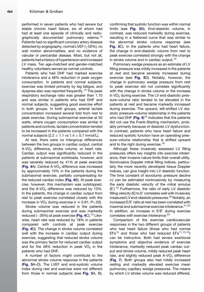
abnormal stroke volume response in the patients(Fig. 5A–D). The LVEF and end-systolic volumeindex during rest and exercise were not differentfrom those in normal subjects (see Fig. 5A, B),
confirming that systolic function was within normallimits (see Fig. 5B). End-diastolic volume, incontrast, was reduced markedly during exercise,resulting in a flattened curve that was similar tothe abnormal stroke volume response (seeFig. 5C). In the patients who had heart failure,the change in end-diastolic volume from rest topeak exercise correlated strongly with the changein stroke volume and in cardiac output.15
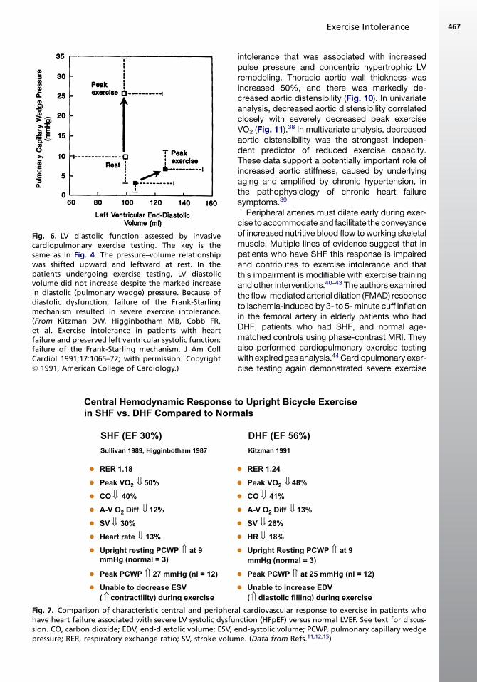
Pulmonary wedge pressure as an estimate of LVfilling pressure was mildly increased in the patientsat rest and became severely increased duringexercise (see Fig. 5D). Notably, however, thechange in pulmonary wedge pressure from restto peak exercise did not correlate significantlywith the change in stroke volume or the increasein VO2 during exercise. The LV end-diastolic pres-sure–volume ratio tended to be elevated in thepatients at rest and became markedly increasedduring exercise. The upward, left-shifted LV dia-stolic pressure–volume relationship in the patientswho had DHF (Fig. 6)15 indicates that the patientsdid not use the Frank-Starling mechanism, prob-ably primarily because of diastolic LV dysfunction.In contrast, patients who have heart failure andreduced systolic function have an operating pres-sure–volume relationship that is shifted upwardand to the right during exercise.18
Although these invasively assessed LV fillingpressures offers key insights into exercise intoler-ance, their invasive nature limits their overall utility.Noninvasive Doppler mitral filling indices, particu-larly the more recently developed tissue Dopplerindices, can give insight into LV diastolic function.The time constant of isovolumic pressure decline(t) can be estimated noninvasively by measuringthe early diastolic velocity of the mitral annulus(E0).19 Furthermore, the ratio of early LV diastolicfilling velocity (E) to E0 correlates well with invasivelymeasuredLVend-diastolicpressures.20Notably, anincreased E/E0 ratio at rest has been correlated withmaximal and submaximal exercise intolerance.21,22
In addition, an increase in E/E0 during exercisecorrelates with exercise intolerance.23
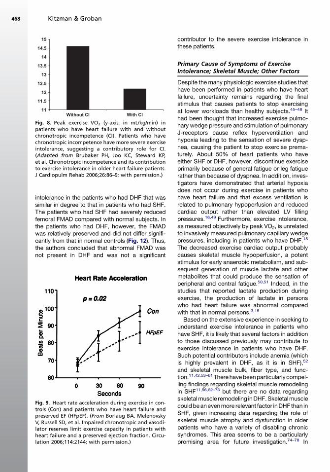
Comparison of the exercise cardiovascularresponses in the two different groups of patientswho had heart failure (those who had normalEFs15 and those who had reduced EFs11,13,15)can be instructive. Both had severe exertionalsymptoms and objective evidence of exerciseintolerance, markedly reduced peak cardiac out-put and stroke volume, mildly reduced peak heartrate, and slightly reduced peak A-VO2 difference(Fig. 7). Both groups also had mildly increasedresting and markedly elevated exercise meanpulmonary capillary wedge pressures. The meansby which LV stroke volume was reduced differed,
Fig. 4. Cardiovascular function assessed by invasive cardiopulmonary exercise testing in patients who have heartfailure and normal systolic function (open boxes) and age-matched normal controls (closed boxes). The primarycomponents of the Fick equation for VO2, cardiac output, and A-VO2 difference are shown in panels A and B,respectively. The components of cardiac output, stroke volume, and heart rate are shown in panels C and D.The X-axis is exercise workload in kilopounds/min (kpm); 150 kpm is equivalent to 25 W. (From Kitzman DW,HigginbothamMB, Cobb FR, et al. Exercise intolerance in patients with heart failure and preserved left ventricularsystolic function: failure of the Frank-Starling mechanism. J Am Coll Cardiol 1991;17:1065–72; with permission.Copyright � 1991, American College of Cardiology.)
Exercise Intolerance 465
however. In one group patients had profoundsystolic contractile dysfunction and were able touse markedly increased LV filling pressure toproduce greater-than-normal use of the Frank-Starling mechanism to compensate partially andmaintain an increase in exercise stroke vol-ume.11,13 In the other group, despite normal sys-tolic contractile function and markedly increasedLV filling pressure,15 patients were unable to usethe Frank-Starling mechanism to increase strokevolume during exercise (see Fig. 7).24
Heart Rate Response to Exercise
Decreased heart rate response also contributes tothe reduced cardiac output at peak exercise and
thus to reduced peak exercise VO2. Indeed, chro-notropic incompetence has been a frequentfinding during cardiopulmonary exercise studiesin SHF, but data were lacking for older patientsand particularly for those who had normal EFs.Therefore, the authors compared heart rate andexpired gas analyses responses in elderly patientswho had DHF with those in a group of age- andgender-matched patients who had SHF and inhealthy normal controls. Using the most standarddefinition of chronotropic incompetence, theauthors found that chronotropic incompetencewas present in 20% to 25% of older patientswho had heart failure, that the prevalence wassimilar in patients who had DHF and those whohad SHF, that the presence of chronotropic
Fig. 5. The components of the LV stroke volume response during exercise, LVEF, end-systolic volume, end-diastolicvolume, and LV filling pressure are shown in panels A–D. Not shown are systolic and mean arterial pressures,which were not different between groups. The key is the same as in Fig. 4. (From Kitzman DW, HigginbothamMB, Cobb FR, et al. Exercise intolerance in patients with heart failure and preserved left ventricular systolic func-tion: failure of the Frank-Starling mechanism. J Am Coll Cardiol 1991;17:1065–72; with permission. Copyright �1991, American College of Cardiology.)
Kitzman & Groban466
incompetence contributed significantly to thedegree of exercise intolerance, measured asmaximal VO2, and that this contribution was inde-pendent of medications, including beta-adrenergic antagonists (Fig. 8).25 The importantcontribution of chronotropic incompetence toexercise intolerance in patients who had DHFwas confirmed by Borlaug and colleagues,26 whostudied a cohort of primarily elderly, African Amer-ican women who had hypertension and heartfailure with a preserved EF. They reported thatsignificant reductions in the rate of heart rateincrease during exercise were primary contributorsto reduced peak cardiac index and maximal exer-cise VO2 (Fig. 9). The implication of this findingmerits further investigation and has therapeuticimplications.
Central and Peripheral Vascular Contributionsto Exercise Intolerance
Abnormal afterload and abnormal ventricular–vascular coupling may contribute to the abnormalFrank-Starling response seen in the patients who
have DHF. Nearly all such patients (88%) havea history of chronic systemic hypertension.27–29
In animal models, diastolic dysfunction developsearly in systemic hypertension, and LV diastolicrelaxation is sensitive to increased afterload,30–35
which can impair relaxation, leading to increasedLV filling pressures and decreased stroke volumeand could lead to symptoms of dyspnea andcongestion.1,2,33
In animal models and in humans, chronicsystolic hypertension accelerates and magnifiesthe age-related increase in fibrotic thickening ofthe aortic wall and resultant increase in aortic stiff-ness, which is a major determinant of LV after-load and ventricular–vascular coupling.36,37 Todetermine whether abnormally decreased aorticdistensibility contributes to the severe exerciseintolerance in heart failure with normal EF, theauthors performed MRI and maximal exercisetesting with expired gas analysis in a group ofelderly patients who had isolated DHF, as definedearlier, with young healthy subjects and age-matched healthy subjects as normal controls.The patients who had DHF had severe exercise
Fig. 6. LV diastolic function assessed by invasivecardiopulmonary exercise testing. The key is thesame as in Fig. 4. The pressure–volume relationshipwas shifted upward and leftward at rest. In thepatients undergoing exercise testing, LV diastolicvolume did not increase despite the marked increasein diastolic (pulmonary wedge) pressure. Because ofdiastolic dysfunction, failure of the Frank-Starlingmechanism resulted in severe exercise intolerance.(From Kitzman DW, Higginbotham MB, Cobb FR,et al. Exercise intolerance in patients with heartfailure and preserved left ventricular systolic function:failure of the Frank-Starling mechanism. J Am CollCardiol 1991;17:1065–72; with permission. Copyright� 1991, American College of Cardiology.)
Central Hemodynamic Response t
in SHF vs. DHF Compared to Norm
RER 1.18
Peak VO2 50%
CO 40%
A-V O2 Diff 12%
SV 30%
Heart rate 13%
Upright resting PCWP at 9
mmHg (normal = 3)
Peak PCWP 27 mmHg (nl = 12)
Unable to decrease ESV
( contractility) during exercise
SHF (EF 30%)
Sullivan 1989, Higginbotham 1987
Fig. 7. Comparison of characteristic central and peripherahave heart failure associated with severe LV systolic dysfusion. CO, carbon dioxide; EDV, end-diastolic volume; ESV, epressure; RER, respiratory exchange ratio; SV, stroke volum
Exercise Intolerance 467
intolerance that was associated with increasedpulse pressure and concentric hypertrophic LVremodeling. Thoracic aortic wall thickness wasincreased 50%, and there was markedly de-creased aortic distensibility (Fig. 10). In univariateanalysis, decreased aortic distensibility correlatedclosely with severely decreased peak exerciseVO2 (Fig. 11).
38 In multivariate analysis, decreasedaortic distensibility was the strongest indepen-dent predictor of reduced exercise capacity.These data support a potentially important role ofincreased aortic stiffness, caused by underlyingaging and amplified by chronic hypertension, inthe pathophysiology of chronic heart failuresymptoms.39
Peripheral arteries must dilate early during exer-cise to accommodate and facilitate the conveyanceof increased nutritive blood flow to working skeletalmuscle. Multiple lines of evidence suggest that inpatients who have SHF this response is impairedand contributes to exercise intolerance and thatthis impairment is modifiable with exercise trainingand other interventions.40–43 The authors examinedthe flow-mediatedarterial dilation (FMAD) responseto ischemia-induced by 3- to 5-minute cuff inflationin the femoral artery in elderly patients who hadDHF, patients who had SHF, and normal age-matched controls using phase-contrast MRI. Theyalso performed cardiopulmonary exercise testingwith expired gas analysis.44 Cardiopulmonary exer-cise testing again demonstrated severe exercise
o Upright Bicycle Exercise
als
RER 1.24
Peak VO2 48%
CO 41%
A-V O2 Diff 13%
SV 26%
HR 18%
Upright Resting PCWP at 9
mmHg (normal = 3)
Peak PCWP at 25 mmHg (nl = 12)
Unable to increase EDV
( diastolic filling) during exercise
DHF (EF 56%)
Kitzman 1991
l cardiovascular response to exercise in patients whonction (HFpEF) versus normal LVEF. See text for discus-nd-systolic volume; PCWP, pulmonary capillary wedgee. (Data from Refs.11,12,15)
11
11.5
12
12.5
13
13.5
14
14.5
15
Without CI With CI
Fig. 8. Peak exercise VO2 (y-axis, in mL/kg/min) inpatients who have heart failure with and withoutchronotropic incompetence (CI). Patients who havechronotropic incompetence have more severe exerciseintolerance, suggesting a contributory role for CI.(Adapted from Brubaker PH, Joo KC, Steward KP,et al. Chronotropic incompetence and its contributionto exercise intolerance in older heart failure patients.J Cardiopulm Rehab 2006;26:86–9; with permission.)
Kitzman & Groban468
intolerance in the patients who had DHF that wassimilar in degree to that in patients who had SHF.The patients who had SHF had severely reducedfemoral FMAD compared with normal subjects. Inthe patients who had DHF, however, the FMADwas relatively preserved and did not differ signifi-cantly from that in normal controls (Fig. 12). Thus,the authors concluded that abnormal FMAD wasnot present in DHF and was not a significant
Fig. 9. Heart rate acceleration during exercise in con-trols (Con) and patients who have heart failure andpreserved EF (HFpEF). (From Borlaug BA, MelenovskyV, Russell SD, et al. Impaired chronotropic and vasodi-lator reserves limit exercise capacity in patients withheart failure and a preserved ejection fraction. Circu-lation 2006;114:2144; with permission.)
contributor to the severe exercise intolerance inthese patients.
Primary Cause of Symptoms of ExerciseIntolerance; Skeletal Muscle; Other Factors
Despite the many physiologic exercise studies thathave been performed in patients who have heartfailure, uncertainty remains regarding the finalstimulus that causes patients to stop exercisingat lower workloads than healthy subjects.45–48 Ithad been thought that increased exercise pulmo-nary wedge pressure and stimulation of pulmonaryJ-receptors cause reflex hyperventilation andhypoxia leading to the sensation of severe dysp-nea, causing the patient to stop exercise prema-turely. About 50% of heart patients who haveeither SHF or DHF, however, discontinue exerciseprimarily because of general fatigue or leg fatiguerather than because of dyspnea. In addition, inves-tigators have demonstrated that arterial hypoxiadoes not occur during exercise in patients whohave heart failure and that excess ventilation isrelated to pulmonary hypoperfusion and reducedcardiac output rather than elevated LV fillingpressures.16,49 Furthermore, exercise intolerance,as measured objectively by peak VO2, is unrelatedto invasively measured pulmonary capillary wedgepressures, including in patients who have DHF.15
The decreased exercise cardiac output probablycauses skeletal muscle hypoperfusion, a potentstimulus for early anaerobic metabolism, and sub-sequent generation of muscle lactate and othermetabolites that could produce the sensation ofperipheral and central fatigue.50,51 Indeed, in thestudies that reported lactate production duringexercise, the production of lactate in personswho had heart failure was abnormal comparedwith that in normal persons.3,15
Based on the extensive experience in seeking tounderstand exercise intolerance in patients whohave SHF, it is likely that several factors in additionto those discussed previously may contribute toexercise intolerance in patients who have DHF.Such potential contributors include anemia (whichis highly prevalent in DHF, as it is in SHF),52
and skeletal muscle bulk, fiber type, and func-tion.11,42,53–61 Therehavebeenparticularlycompel-ling findings regarding skeletal muscle remodelingin SHF11,56,62–73 but there are no data regardingskeletalmuscle remodeling inDHF.Skeletalmusclecouldbeanevenmore relevant factor inDHF than inSHF, given increasing data regarding the role ofskeletal muscle atrophy and dysfunction in olderpatients who have a variety of disabling chronicsyndromes. This area seems to be a particularlypromising area for future investigation.74–78 In
Fig. 10. Data and images from representative subjects from healthy young persons, healthy elderly persons, andelderly patients who have diastolic heart failure (HF). Maximal exercise oxygen consumption (VO2max), aorticdistensibility at rest, and the LV mass:volume ratio are shown. Patients who have DHF have severely reduced exer-cise tolerance (VO2max) and aortic distensibility and increased aortic wall thickness (arrows). (Adapted fromHundley WG, Kitzman DW, Morgan TM, et al. Cardiac cycle dependent changes in aortic area and aortic disten-sibility are reduced in older patients with isolated diastolic heart failure and correlate with exercise intolerance.J Am Coll Cardiol 2001;38(3):796–802; with permission. Copyright � 2001, American College of Cardiology.)
Exercise Intolerance 469
addition, the amount of adipose tissue betweenskeletal muscle bundles seems to be a potentialmodifier of skeletal muscle function and of exercisecapacity as well. This area probably will also bea fruitful area for future research, particularlybecause skeletal muscle bulk and function seemto be potentially modifiable through nutrition andexercise interventions.79
INTERVENTIONS TO IMPROVE EXERCISETOLERANCE
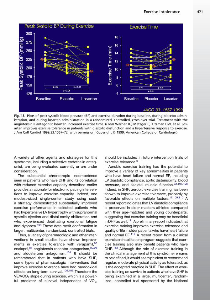
During exercise in normal subjects, systolic andpulse pressure increase substantially, and thisresponse is magnified by increased arterial stiff-ness. Data from animal models suggest that theexercise-related increase in systolic blood pres-sure is mediated, in part, by exercise-induced in-creases in circulating angiotensin II. Indeed, ina randomized, double-blind, placebo-controlled
cross-over trial, angiotensin receptor blockadereduced the exaggerated exercise increase insystolic and pulse pressures, resulting in signifi-cantly improved exercise treadmill time and qualityof life (Fig. 13).80
In a group of patients who had NYHA class IIIheart failure and presumed diastolic dysfunction(EF >50%), Aronow and Kronzon81 showed thatthe angiotensin-converting enzyme inhibitor ena-lapril significantly improved functional class, exer-cise duration, EF, diastolic filling, and LV mass.
In hypertrophic cardiomyopathy, a disorder inwhich diastolic dysfunction is common, verapamilseems to improve symptoms and objectivelymeasured exercise capacity.82–85 This agent alsoimproves ventricular vascular coupling and exer-cise performance in aged individuals who havehypertension.86 In laboratory animal modelscalcium antagonists, particularly dihydropyridines,prevent ischemia-induced increases in LV diastolic
y= 0.0002x - 0.0021r = 0.665
00.5
11.5
22.5
33.5
44.5
55.5
66.5
77.5
88.5
99.510
0 10 20 30 40O
Fig. 11. There is a close relationship between peakexercise VO2 (horizontal axis) and proximal aorticdistensibility (vertical axis) in a group of 30 subjects(10 healthy young persons, 10 healthy elderly persons,and 10 elderly patients who have DHF). Each symbolrepresents the data from one participant. (FromHundley WG, Kitzman DW, Morgan TM, et al. Cardiaccycle dependent changes in aortic area and aorticdistensibility are reduced in older patients with iso-lated diastolic heart failure and correlate with exer-cise intolerance. J Am Coll Cardiol 2001;38(3):796–802;with permission. Copyright � 2001, American Collegeof Cardiology.)
0
2
4
6
8
10
12
14
FMAD %
Normal
HFNEF
HFREF
Fig. 12. Flow-mediated arterial dilation (FMAD) of thefemoral artery by phase-contrast MRI in normalsubjects, elderly patients who have heart failure andnormal EF (HFNEF), and patients who have heartfailure and reduced EF (HFREF). FMAD is severelyreduced in HFREF but is relatively preserved in HFNEFcompared with age-matched healthy normal subjects.(From Hundley WG, Bayram E, Hamilton CA, et al. Legflow-mediated arterial dilation in elderly patientswith heart failure and normal left ventricular EF. AmJ Physiol Heart Circ Physiol 2007;292(3):H1427–34;with permission.)
Kitzman & Groban470
stiffness87 and improve diastolic performancein pacing-induced heart failure.88–90 Negativeinotropic calcium antagonists significantly impairearly relaxation,90–94 however, and in generalhave shown a tendency toward adverse outcomein patients who have SHF.90 Nonetheless, Setaroand colleagues95 examined 22 men (mean age,65 years) who had clinical heart failure despite anEF greater than 45% in a randomized, double-blind, placebo-controlled cross-over trial of verap-amil. There was a 33% improvement in exercisetime, and there also were significant improvementsin clinicoradiographic heart failure scoring andpeak filling rate.In a randomized, cross-over, blinded trial, Little
and colleagues96 compared the calcium-channelantagonist verapamil with the angiotensin receptorantagonist candesartan using the outcomes ofpeak exercise blood pressure, exercise time, andquality of life. Although both agents blunted thepeak systolic blood pressure response to exer-cise, only candesartan, but not verapamil, im-proved exercise time and quality of life.96
In a subsequent trial with a similar randomized,cross-over, blinded design, the diuretic hydrochlo-rothiazide was compared with the angiotensinreceptor antagonist losartan using the outcomesof peak exercise blood pressure, exercise time,and quality of life.97 Although both agents bluntedthe peak systolic blood pressure response to exer-cise, only losartan, but not hydrochlorothiazide,improved exercise time and quality of life.97
The addition of low-dose spironolactone (12.5–50 mg daily) to standard therapy has been shownto improve exercise tolerance in patients who havesevere SHF. Aldosterone antagonism has num-erous potential benefits in patients who haveDHF, including LV remodeling, reversal of myocar-dial fibrosis, and improved LV diastolic functionand vascular function.98–100 Few data, however,are presently available regarding aldosterone an-tagonism in DHF. In one small study, low-dose spi-ronolactone was well tolerated and seemed toimprove exercise capacity and quality of life in old-er women who had isolated DHF.101 In another,spironolactone improved measures of myocardialfunction in hypertensive patients who had DHF.102
Glucose cross-links increase with aging anddiabetes and cause increased vascular andmyocardial stiffness. Alagebrium, a novel cross-link breaker, improved vascular and LV stiffnessin dogs. In a small, open-label, 4-month trial ofthis agent in elderly patients, LV mass, quality oflife, and tissue Doppler diastolic function indexesimproved (Fig. 14), but there were no significantimprovements in exercise capacity or aorticdistensibility, the primary outcomes of the trial.103
Fig. 13. Plots of peak systolic blood pressure (BP) and exercise duration during baseline, during placebo admin-istration, and during losartan administration in a randomized, controlled, cross-over trial. Treatment with theangiotensin II antagonist losartan increased exercise time. (From Warner JG, Metzger C, Kitzman DW, et al. Los-artan improves exercise tolerance in patients with diastolic dysfunction and a hypertensive response to exercise.J Am Coll Cardiol 1999;33:1567–72; with permission. Copyright � 1999, American College of Cardiology.)
Exercise Intolerance 471
A variety of other agents and strategies for thissyndrome, including a selective endothelin antag-onist, are being evaluated currently or are underconsideration.
The substantial chronotropic incompetenceseen in patients who have DHF and its correlationwith reduced exercise capacity described earlierprovides a rationale for electronic pacing interven-tions to improve exercise capacity. Indeed, onemodest-sized single-center study using sucha strategy demonstrated substantially improvedexercise performance in selected patients whohad hypertensive LV hypertrophywith supranormalsystolic ejection and distal cavity obliteration andwho experienced debilitating exertional fatigueand dyspnea.104 These data merit confirmation inlarger, multicenter, randomized, controlled trials.
Thus, a variety of pharmacologic and other inter-ventions in small studies have shown improve-ments in exercise tolerance with verapamil,95
and aldosterone antagonism.101 It should beremembered that in patients who have SHF,some types of pharmacologic interventions thatimprove exercise tolerance have had paradoxicaleffects on long-term survival.105,106 Therefore theVE/VCO2 slope during exercise, which is a power-ful predictor of survival independent of VO2,
should be included in future intervention trials ofexercise tolerance.6
Aerobic exercise training has the potential toimprove a variety of key abnormalities in patientswho have heart failure and normal EF, includingLV diastolic compliance, aortic distensibility, bloodpressure, and skeletal muscle function.72,107,108
Indeed, in SHF, aerobic exercise training has beenshown to improve exercise tolerance, probably byfavorable effects on multiple factors.17,109,110 Arecent report indicates that LVdiastolic complianceis preserved in older masters athletes comparedwith their age-matched and young counterparts,suggesting that exercise training may be beneficialinDHFaswell.111Apreliminary report indicates thatexercise training improves exercise tolerance andquality of life in older patientswho have heart failureand normal EF.112 A recent report from a clinicalexercise rehabilitation program suggests that exer-cise training also may benefit patients who haveDHF.113 Although the role of exercise training inthe clinical management of this syndrome remainstobedefined, itwould seemprudent to recommendregular, moderate physical activity as tolerated, asis the accepted practice in SHF. The effect of exer-cise training on survival in patients who have SHF isbeing examined in a large, multicenter, random-ized, controlled trial sponsored by the National
Fig. 14. Effect of alagebrium on LV mass seen by MRI (top panel) and E0 seen by tissue Doppler imaging (TDI) inolder patients who have DHF. (From Little WC, Zile MR, Kitzman DW, et al. The effect of alagebrium chloride (ALT-711), a novel glucose cross-link breaker, in the treatment of elderly patients with diastolic heart failure. J Card Fail2005;11(3):191–5; with permission.)
Kitzman & Groban472
InstitutesofHealth (HF-ACTION).Presently, there isno trial examining mortality and exercise training inpatients who have heart failure and normal EF.
SUMMARY
Even when stable and non-edematous, patientswho have heart failure and normal EF have severe,chronic exercise intolerance. The pathophysiologyof exercise intolerance in this syndrome is incom-pletely understood but probably is multifactorial.Presently available data suggest that importantcontributors include decreased LV diastolic comp-liance, decreased aortic distensibility, exagger-ated exercise systolic blood pressure, relativechronotropic incompetence, and possibly anemiaand skeletal muscle remodeling. Because it isa primary determinant of quality of life, can be
quantified objectively, is reproducible, and ismodifiable, exercise intolerance is an attractivetherapeutic target. A number of pharmacologicand other interventions seem to improve exerciseintolerance in DHF. Although it is unknown whe-ther these interventions will be accompanied byimproved survival, the parallel outcome of im-proved quality of life supports the clinical rele-vance of exercise performance outcomes.
REFERENCES
1. Gandhi SK, Powers JE, Fowle KM, et al. The patho-
genesis of acute pulmonary edema associated with
hypertension. N Engl J Med 2000;344(1):17–22.
2. Powers JE, Gandhi SK, Kramer RK, et al. Predic-
tors of poor outcome in patients with hypertensive
Exercise Intolerance 473
pulmonary edema [abstract]. J Am Coll Cardiol
2004;43(5A):227A.
3. Kitzman DW, Little WC, Brubaker PH, et al. Patho-
physiological characterization of isolated diastolic
heart failure in comparison to systolic heart failure.
JAMA 2002;288(17):2144–50.
4. Bol E, de Vries WR, Mosterd WL, et al. Cardiopul-
monary exercise parameters in relation to all-
cause mortality in patients with chronic heart
failure. Int J Cardiol 2000;72:255–63.
5. Jones RC, Francis GS, Lauer MS. Predictors of
mortality in patients with heart failure and preserved
systolic function in the Digitalis Investigation Group
trial. J Am Coll Cardiol 2004;44(5):1025–9.
6. Francis DP, Shamin W, Davies LC, et al. Cardiopul-
monary exercise testing for prognosis in chronic
heart failure: continuous and independent prog-
nostic value from VE/VCO2 slope and peak VO2.
Eur Heart J 2000;21:154–61.
7. Moore B, Brubaker PH, Stewart KP, et al. VE/VCO2
slope in older heart failure patients with normal
versus reduced ejection fraction compared with
age-matched healthy controls. J Card Fail 2007;
13(4):259–62.
8. Maldonado-Martin S, Brubaker PH, Kaminsky LA,
et al. The relationship of a 6-min walk to VO(2
peak) and VT in older heart failure patients. Med
Sci Sports Exerc 2006;38(6):1047–53.
9. Higginbotham MB, Morris KG, Williams RS, et al.
Regulation of stroke volume during submaximal
and maximal upright exercise in normal man. Circ
Res 1986;58:281–91.
10. Higginbotham MB, Morris KG, Williams RS, et al.
Physiologic basis for the age-related decline in
aerobic work capacity. Am J Cardiol 1986;57:
1374–9.
11. Sullivan M, Knight JD, Higginbotham MB, et al.
Relation between central and peripheral hemody-
namics during exercise in patients with chronic
heart failure: muscle blood flow is reduced with
maintenance of arterial perfusion pressure. Circu-
lation 1989;80:769–81.
12. Higginbotham MB, Sullivan M, Coleman RE, et al.
Regulation of stroke volume during exercise in
patients with severe left ventricular dysfunction:
importance of Starling mechanism. J Am Coll Car-
diol 1987;9:58A.
13. Kitzman DW, Sullivan M, Cobb FR, et al. Exercise
cardiac output declines with advancing age in
normal subjects. J Am Coll Cardiol 1989;13(2):
241A.
14. Sullivan M, Cobb FR, Knight JD, et al. Stroke
volume increases by similar mechanisms in men
and women. Am J Cardiol 1991;67:1405–12.
15. Kitzman DW, Higginbotham MB, Cobb FR, et al.
Exercise intolerance in patients with heart failure
and preserved left ventricular systolic function:
failure of the Frank-Starling mechanism. J Am Coll
Cardiol 1991;17:1065–72.
16. Sullivan M, Higginbotham MB, Cobb FR. Increased
exercise ventilation in patients with chronic heart
failure: intact ventilatory control despite hemody-
namic and pulmonary abnormalities. Circulation
1988;77:552–9.
17. Sullivan M, Higginbotham MB, Cobb FR. Exercise
training in patients with chronic heart failure delays
ventilatory anaerobic threshold and improves
submaximal exercise performance. Circulation
1989;79:324–9.
18. Sullivan M, Cobb FR. Central hemodynamic
response to exercise in patients with chronic heart
failure. Chest 1992;101:340S–6S.
19. Nagueh SF, Middleton KJ, Kopelen HA, et al.
Doppler tissue imaging: a noninvasive technique
for evaluation of left ventricular relaxation and esti-
mation of filling pressures. J Am Coll Cardiol 1997;
30(6):1527–33.
20. Ommen SR, Nishimura RA, Appleton CP, et al. Clin-
ical utility of Doppler echocardiography and tissue
Doppler imaging in the estimation of left ventricular
filling pressures: a comparative simultaneous
Doppler-catheterization study. Circulation 2000;
102(15):1788–94.
21. Hadano Y, Murata K, Yamamoto T, et al. Usefulness
of mitral annular velocity in predicting exercise
tolerance in patients with impaired left ventricular
systolic function. Am J Cardiol 2006;97(7):1025–8.
22. Skaluba SJ, Litwin SE. Mechanisms of exercise
intolerance: insights from tissue Doppler imaging.
Circulation 2004;109(8):972–7.
23. Ha JW, Oh JK, Pellikka PA, et al. Diastolic stress
echocardiography: a novel noninvasive diagnostic
test for diastolic dysfunction using supine bicycle
exercise Doppler echocardiography. J Am Soc
Echocardiogr 2005;18(1):63–8.
24. Kitzman DW, Sullivan M. Exercise intolerance in
patients with heart failure: role of diastolic dysfunc-
tion. In: Grossman W, editor. Diastolic relaxation of
the heart. Boston: Kluwer Academic Publishers;
1994. p. 295–302.
25. Brubaker PH, Joo KC, Stewart KP, et al. Chrono-
tropic incompetence and its contribution to exer-
cise intolerance in older heart failure patients.
J Cardiopulm Rehabil 2006;26(2):86–9.
26. Borlaug BA, Melenovsky V, Russell SD, et al.
Impaired chronotropic and vasodilator reserves
limit exercise capacity in patients with heart failure
and a preserved ejection fraction. Circulation 2006;
114(20):2138–47.
27. Kitzman DW, Gardin JM, Gottdiener JS, et al.
Importance of heart failure with preserved systolic
function in patients > or 5 65 years of age. Cardio-
vascular Health Study Research Group. Am J Car-
diol 2001;87(4):413–9.
Kitzman & Groban474
28. Iriarte M, Murga N, Morillas M, et al. Congestive
heart failure from left ventricular diastolic dysfunc-
tion in systemic hypertension. Am J Cardiol 1993;
71:308–12.
29. Iriarte MM, Perez OJ, Sagastagoitia D, et al.
Congestive heart failure due to hypertensive
ventricular diastolic dysfunction. Am J Cardiol
1995;76(13):43D–7D.
30. Little WC. Enhanced load dependence of relaxa-
tion in heart failure: clinical implications. Circulation
1992;85(6):2326–8.
31. Gelpi RJ. Changes in diastolic cardiac function in
developing and stable perinephritic hypertension
in conscious dogs. Circ Res 1991;68:555–67.
32. Shannon RP, Komamura K, Gelpi RJ, et al. Altered
load: an important component of impaired diastolic