Persons with spinal cord injury (SCI) exhibit deficits in volitional motorAbstractcontrol and sensation that limit not only the performance of daily tasks but also theoverall activity level of these persons. This population has been characterised asextremely sedentary with an increased incidence of secondary complicationsincluding diabetes mellitus, hypertension and atherogenic lipid profiles. As thedaily lifestyle of the average person with SCI is without adequate stress forconditioning purposes, structured exercise activities must be added to the regularschedule if the individual is to reduce the likelihood of secondary complicationsand/or to enhance their physical capacity. The acute exercise responses and thecapacity for exercise conditioning are directly related to the level and complete-ness of the spinal lesion. Appropriate exercise testing and training of persons withSCI should be based on the individual’s exercise capacity as determined byaccurate assessment of the spinal lesion. The standard means of classification ofSCI is by application of the International Standards for Classification of SpinalCord Injury, written by the Neurological Standards Committee of the AmericanSpinal Injury Association. Individuals with complete spinal injuries at or abovethe fourth thoracic level generally exhibit dramatically diminished cardiac accel-eration with maximal heart rates less than 130 beats/min. The work capacity ofthese persons will be limited by reductions in cardiac output and circulation to theexercising musculature.
Persons with complete spinal lesions below the T10 level will generally displayinjuries to the lower motor neurons within the lower extremities and, therefore,will not retain the capacity for neuromuscular activation by means of electricalstimulation. Persons with paraplegia also exhibit reduced exercise capacity andincreased heart rate responses (compared with the non-disabled), which have beenassociated with circulatory limitations within the paralysed tissues. The recom-mendations for endurance and strength training in persons with SCI do not varydramatically from the advice offered to the general population. Systems offunctional electrical stimulation activate muscular contractions within theparalysed muscles of some persons with SCI. Coordinated patterns of stimulationallows purposeful exercise movements including recumbent cycling, rowing andupright ambulation. Exercise activity in persons with SCI is not without risks,with increased risks related to systemic dysfunction following the spinal injury.These individuals may exhibit an autonomic dysreflexia, significantly reducedbone density below the spinal lesion, joint contractures and/or thermal dysregula-tion. Persons with SCI can benefit greatly by participation in exercise activities,but those benefits can be enhanced and the relative risks may be reduced withaccurate classification of the spinal injury.
The human spinal cord is a complex association or disease of spinal tracts results in varying typesand degrees of dysfunction depending upon the spe-of upper and lower motor neurons that functions as acific neural structures affected.bidirectional conduit between the brain and its mo-
tor, sensory and autonomic targets. It also serves as a The interruption of spinal cord functions by trau-site for reflex integration between body sensors and ma affects 10 000 Americans annually, with an esti-their motor and autonomic effectors. Because spinal mated 179 000 persons having survived their initialcord functions differ by level and structure, injury to injury.[1-4] Thereafter, these individuals experience
unique physical, social and psychological changes cise conditioning, contemporary methods of injuryclassification, exercise options and established ben-throughout their lives, including diminished abilityefits of training, and risks imposed by inappropriateto perform and benefit from exercise condition-exercise recommendations for those with SCI.ing.[5,6] The latter limitation is cause for concern as:
(i) individuals with spinal cord injury (SCI) areusually young and physically active at the time of 1. Neurological Classification of Personsinjury;[3] (ii) profound physical deconditioning is with Spinal Cord Injury (SCI)common after injury;[7-9] and (iii) physical decondi-
If trauma to, or disease of, the spinal cord alwaystioning contributes to multisystem medical compli-resulted in its anatomical or physiological transec-cations,[10-19] activity limitations[7,20,21] and acceler-tion, classification of spinal cord dysfunctions fol-ated aging.[6,7,22-26]
lowing SCI would be relatively easy. However, theMany reviews and chapters published over the
spinal cord is rarely severed unless penetrated by apast decades have addressed the need for persons
bullet or severed by its bony covering during verywith SCI to adopt habitual exercise as part of a
high velocity impact. More often the cord remainshealthy lifestyle.[5,6,9,17,18,27-33] Exercise options con-
anatomically intact but suffers contusion, infarctiontained in these reports have focused on use of mus-
or mechanical deformation that interrupts its local orcle activities still under voluntary nervous system
relay circuitry. It also can undergo secondary dam-control, as well as sequenced contractions of mus-
age from inflammatory autodestruction, which ex-cles stimulated by electrical current. Outcomes of
plains the routine administration of high-dose corti-studies cited in these monographs provide credible costeroids within hours of an SCI.[37] Notwithstand-evidence that exercise performed by persons with ing the cause of spinal damage, more than half of theSCI enhances physical conditioning and reduces survivors will experience varying degrees of motor,multisystem disease susceptibility. They further sensory or autonomic sparing at different spinal cordsuggest that habitual exercise might reduce fatigue, levels,[2,38,39] making classification of persons withpain, weakness, joint deterioration and incipient SCI a challenging, yet important, science.[4]
neurological deficits that appear as persons age with To assist with accurate and consistent classifica-disability. As these deficits challenge the ability of tion, uniform standards have been developed thatthose with SCI to perform essential daily activities allow persons with SCI to be systematically ex-first mastered after injury, their prevention will like- amined and classified, and to document changes inly foster fullest health and life satisfaction for those sensorimotor function that accompany the passageaging with a disability. of time, clinical treatments or research interven-
Use of exercise as a healthy activity and lifestyle tions. The benchmark system used to classify per-elective for those with SCI might appear fairly sons with SCI is the International Standards forstraightforward. To the contrary, exercise options Classification of Spinal Cord Injury (revised 2000),available to those with SCI are more limited than which is written by the Neurological Standardsthose without disability,[33] their acute exercise and Committee of the American Spinal Injury Associa-training responses less robust than those of persons tion (ASIA), and endorsed as the recommendedwithout SCI,[28,34,35] and the risks of impudent activi- international standards by the International Spinalty greater and longer lasting.[33,36] This makes an Cord Society (ISCoS; formerly the Internationalunderstanding of neurological classification, exer- Medical Society for Paraplegia, and hereinafter thecise opportunities and risks of activity important if ‘ASIA Guidelines’) [figure 1].[40] While a completethose with SCI are to benefit and not be harmed description of spinal column and spinal cord anato-from exercise conditioning. This paper will review my is beyond the scope of this review, the accuratecommon medical problems experienced by persons description of an injury to a given spinal cord seg-with SCI that lend themselves to benefit from exer- ment involves designation of a spinal column region
Hip flexorsKnee extensorsAnkle dorsiflexorsLong toe extensorsAnkle plantar flexors
R L R L R L
0 = total paralysis1 = palpable or visible contraction2 = active movement, gravity eliminated3 = active movement, against gravity4 = active movement, against some resistance5 = active movement, against full resistanceNT = not testable
TOTALS(maximum) (50) (50) (100)
0 = absent1 = impaired2 = normalNT = not testable
Motorkey muscles
Lighttouch
Pinprick
Sensorykey sensory points
TOTALS(maximum) (56) (56) (56) (56)
+ = Motor score
Voluntary anal contraction (yes/no)
+ = Light touch score
+ = Pin prick score
(max: 112)
(max: 112)
Any anal sensation (yes/no)
Neurological levelThe most caudal segment
with normal function
R LComplete or incomplete?
Incomplete = any sensory or motor funtion in S4-S5
ASIA impairment scale
Zone of partialpreservation
Caudal extent of partiallyinnervated segments
R L
Sensory
A = Complete: No motor or sensoryfunction is preserved in thesacral segments S4-S5
B = Incomplete: Sensory but notmotor function is preservedbelow the neurological level andincludes the sacral segments S4-S5
C = Incomplete: Motor function ispreserved below the neurologicallevel, and more than half of keymuscles below the neurological levelhave a muscle grade less than 3
D = Incomplete: Motor function ispreserved below the neurologicallevel, and at least half of keymuscles below the neurological levelhave a muscle grade of 3 or more
E = Normal: motor and sensoryfunction are normal
Central cord
Brown-Sequard
Anterior cord
Conus medullaris
Cauda equina
Clinical syndromesASIA impairment scale
Motor
Sensory
Motor
Fig. 1. American Spinal Injury Association (ASIA) classification of spinal cord injury (reproduced from the American Spinal Injury Associa-tion,[41] with permission).
(e.g. cervical, thoracic, lumber or sacral), a spinal trauma or disease. The long tracts of the spinal cordnerve within that region, and the degree to which the are upper motor neurons. By convention, the lowerinjury is neurologically complete or incomplete. motor neuron system begins in the anterior horn ofShould the injury result in a specific spinal cord the grey matter and exits the intervertebral foramensyndrome this should also be designated. to join the returning sensory axons as a mixed nerve.
The motor portion of this mixed nerve then travels toits target muscle(s). The returning sensory nerve1.1 Upper and Lower Motor Neuron Lesionsfibres travel in this mixed nerve but, upon entry tothe foramen, form a dorsal root that penetrates theIt is clinically important for those working withcord in the dorsal root entry zone and synapses withsubjects having SCI to differentiate between upperthe anterior horn cells of the anterior grey matter.and lower motor neuron injuries. The spinal cord isThis completes the motor-sensory reflex arc. Le-organised in such a way that either upper or lower
motor neuron function, or both, can be disrupted by sions of upper motor neurons result in ‘decentralisa-
tion’ of the nervous system, spastic paralysis and the level of injury, the trunk, legs and pelvic organsexaggerated sensorimotor reflexes below the injury. may be involved. The term is used when referring toThis means that motor, sensory and autonomic re- cauda equina and conus medullaris injuries, but notflex activities are preserved, but no longer under to lumbosacral plexus lesions or injury to peripheralcommand of the brain. The contrasting injury is a nerves outside the neural canal.”[42,43]
lower motor neuron lesion, which often accompa-1.3 Neurological, Sensory and Motor Levelsnies SCI at the T10 level or lower and almost always
occurs at, or caudal to, T12. Persons with theseNeurological level refers to the most caudal seg-
lesions lose central nervous system control of sen-ment of the spinal cord with normal sensory and
sorimotor functions, as well as sensorimotor reflexmotor function on both sides of the body. Because
activity, which causes flaccid paralysis and areflexiathe results of sensorimotor testing often differ by
(‘denervation’). This explains the greater loss ofside, up to four different segments may be identified
lower extremity muscle mass in individuals within determining the neurological level, i.e. R-sensory,
flaccid rather than spastic paralysis. Otherwise, inju-L-sensory, R-motor and L-motor. In such cases it is
ry to the cord at any site resulting in damage to thestrongly recommended that each of these segments
reflex arc can leave a denervated (areflexic) segmentbe separately recorded and that a single ‘level’ not
among neighbouring segments that remain spastic.be used, as this can be misleading. When the term
It is important to note that while spastic muscles ‘sensory level’ is used, it refers to the most caudalare capable of contracting upon stimulation with segment of the spinal cord with normal sensorytranscutaneous electrical alternating currents, gener- function on both sides of the body; the ‘motor level’ally, flaccid muscles are not. This provides a means is similarly defined with respect to motor function.of discriminating upper from lower motor neuron These ‘levels’ are determined by neurological ex-lesions and also determining subject or patient can- amination of: (i) a key sensory point within each ofdidacy for use of electrical prostheses that provide 28 dermatomes on the right and 28 dermatomes onexercise or ambulation in persons with SCI. the left side of the body;[42] and (ii) a key muscle
within each of ten myotomes on the right and ten1.2 Defining Spinal Cord Lesions: Plegias myotomes on the left side of the body. Assessment
of sensory and motor level is key in those withIndividuals with SCI are described as having
paraplegia having injuries from T1 through T8,lesions affecting either sensorimotor function of all
whereas motor function is the determining factor inlimbs or the upper extremities alone. The former are
classification of tetraplegia.said to have tetraplegia (preferred to ‘quadriplegia’),which is defined as: “A term referring to impairment 1.4 Complete and Incomplete Spinalor loss of motor and/or sensory function in the Cord Lesionscervical segments of the spinal cord due to damageof neural elements within the spinal canal. Te- Confusion and imprecise language surround thetraplegia results in impairment of function in the definitions of neurological completeness and incom-arms as well as in the trunk, legs and pelvic organs. pleteness. Historically, the Functional ClassificationIt does not include brachial plexus lesions or injury scale of Frankel et al.[44] was used to assign a gradeto peripheral nerves outside the neural canal.”[42,43] of SCI from A to E. These grades were assigned onParaplegia is defined as: “A term referring to im- the basis of sensorimotor sparing below the level ofpairment or loss of motor and/or sensory function in SCI. In contrast, the ASIA scale uses ‘sacral spar-the thoracic, lumbar or sacral (but not cervical) ing’ as the criterion for determining neurologicalsegments of the spinal cord, secondary to damage of completeness.[39,42,45] This requires a test of motorneural elements within the spinal canal. With para- function examining the presence of voluntary con-plegia, arm functioning is spared, but, depending on traction of the external anal sphincter upon digital
examination. The ASIA definitions of complete and planations for resolution of ‘spinal shock’ and re-incomplete lesions are: covery of sensorimotor functions, the designation of
‘neurological completeness’ within the first year of• ‘Complete injury’: a term describing absence ofinjury leaves open the possibility for erroneous clas-sensory and motor function in the lowest sacralsification of a subject thereafter. This explains whysegment.[42]
many investigators use the first anniversary of injury• ‘Incomplete injury’: a term describing partial
as both a benchmark for neurological stability and apreservation of sensory and/or motor functions
criterion for enrolment of subjects with SCI in re-below the neurological level and including the
search studies, especially when sensorimotor func-lowest sacral segment. Sacral sensation includes
tion is under study. Clearly, however, the use of thesensation at the anal mucocutaneous junction as
term ‘complete’ written on an initial trauma exami-well as deep anal sensation.[42]
nation might ultimately have little bearing on wheth-Both ASIA and Frankel methods are useful for er recovery of reflexes or sensorimotor function
describing completeness of injuries, as an individual might result. Thus, examination and classification atwith infralesional sensory sparing (i.e. incomplete the beginning of exercise testing and training issensory loss; Frankel Grade B) can still be neuro- needed.logically complete when using the ASIA guidelines.Also, the ASIA C classification is so broad as to 1.5 SCI Syndromessometimes limit useful classification of motor andfunctional abilities of patients with SCI that may be As the various spinal columns transmit differentbetter understood when using Frankel grades. types of information, and because most persons
surviving SCI do not experience complete severanceAccurate descriptions of complete and incom-of their cords, many different patterns of sparedplete lesions are often confounded by recovery ofsensorimotor function are possible. The syndromessensorimotor function within weeks or months afterthat ensue are usually described when defining theinjury, which is not uncommon for some individualslevels and completeness of injury. The major syn-initially presenting without preserved reflex or sen-dromes, their definitions and causations are listed insorimotor functions.[46] This recovery, however, willthe following sections.more likely occur in individuals with very low ve-
locity injuries in which cord bleeding or anatomic 1.5.1 Anterior Cord Syndromedisruption of neural elements are limited. Several An anterior cord syndrome is “a lesion that pro-mechanisms have been postulated to explain ‘return duces variable loss of motor function and of sensi-of function’ after SCI. The first involves a belief that tivity to pain and temperature, while preservinga ‘spinal shock’ occurs immediately upon SCI in proprioception”.[42] An anterior cord syndrome re-some individuals – a state of areflexia thought to sults primarily in a profound motor loss below theresult from hyperpolarisation of cord neurons ac- level of injury. Destruction of the anterior portionscompanying injury-related loss of descending facili- of the white and grey matter of the spinal cordtation.[47] Thereafter, those with incomplete lesions affects the corticospinal motor tracts and, to a lesseroften recover reflexes in a caudal to rostral se- extent, sensory tracts that mediate light touch andquence, although the extent of reflex recovery has pressure.questionable value in predicting eventual patterns or
1.5.2 Central Cord Syndromeextent of motor recovery.[47] Otherwise, post-synap-tic receptor up-regulation and synaptic growth have A central cord syndrome is “a lesion occurringbeen postulated as explanations for neuronal recov- almost exclusively in the cervical region, that pro-ery following SCI,[48,49] although the precise mecha- duces sacral sensory sparing and greater weaknessnisms promoting these changes remain incompletely in the upper limbs than in the lower limbs”.[42] Aunderstood. Notwithstanding the controversial ex- person with a central cord syndrome often presents
with greater motor impairment of the upper than bosacral nerve roots within the neural canal result-lower extremities if the damage or disease is prima- ing in areflexic bladder, bowel and lower limbs”.[42]
rily limited to the central grey and white spinal cord2. Medical and Health Consequencesmatter. Because upper extremity motor function isof SCIprimarily activated by the medial corticospinal
tracts, sensorimotor functions of the lower extremi-Persons with SCI face unique health challenges
ties are usually affected to a lessor degree. Centralthroughout their lives, and their injuries dissociate
cord injury is common among individuals havingthe normally well integrated homoeostatic responses
congenitally narrow spinal canals and in those, espe-of body systems known to accompany physical ac-
cially aging persons, whose osteoarthritic pathologytivity. Nervous system damage disrupts to varying
results in spinal canal stenosis.degrees the necessary signal integration among mo-tor, sensory and autonomic targets, and thus has a
1.5.3 Brown-Sequard Syndrome profound effect on fitness, exercise capacities andA Brown-Sequard syndrome is “a lesion that health. Depending on the level and type of cord
produces relatively greater ipsilateral proprioceptive lesion, persons with SCI are among the most physi-and motor loss and contralateral loss of sensitivity to cally deconditioned of all humans.[7,50] Not surpris-pain and temperature”.[42] The Brown-Sequard syn- ingly, young persons with chronic SCI experiencedrome results from an anterior-posterior hemisec- accelerated pathological states and conditions nor-tion of the spinal cord often accompanying a pene- mally associated with physical deconditioning andtrating wound that severs the neural elements. When premature aging, including: dyslipidaemias andthe elements of only one lateral portion of the spinal heart disease;[23,24,51,52] arterial circulatory insuffi-cord are destroyed, the injury results in an unusual ciency[53-57] and clotting disorders;[58,59] bone andpattern of sensorimotor function with a loss of in- joint diseases;[60-62] and pain of musculoskeletal andfralesional motor function, proprioception, fine neuropathic origins.[63-68]
touch and vibration discrimination on the same side Notwithstanding their physical limitations, manyof injury, but loss of pain, temperature, crude touch persons with SCI can still undergo exercise recondi-and deep pressure on the opposite side of injury. The tioning. Those who retain upper extremity functionsyndrome is explained by decussation of motor can participate in a wide variety of exercise activi-tracts in the brain stem before they descend in the ties and sports,[6,69] and ambulate with the assistancecord. In contrast, ascending sensory tracts of the of orthoses and computer-controlled electricalanterolateral systems cross to the opposite side at (or neuroprostheses.[70-76] Individuals with upper motornear) the level in which the dorsal roots enter the neuron lesions can pedal cycle ergometers by sur-cord. This explains why ‘same side’ motor deficits face electrical stimulation of selected lower extremi-are common in patients with Brown-Sequard syn- ty muscle groups under computer control.[30,31,33]
drome, but sensory preservation is greater on the Many body organs and tissues acutely respond tocontralateral side of injury. exercise despite their decentralised or denervated
states, and because many survivors of SCI experi-1.5.4 Conus Medullaris and Cauda ence complete sensory loss or significantly dimin-Equina Syndrome
muscle contractions can often be utilised withoutof the sacral cord (conus) and lumbar nerve roots
pain.within the spinal canal, which usually results in anareflexic bladder, bowel and lower limbs”.[42] Sacral 2.1 Physical Deconditioningsegments may occasionally show preserved reflexes(e.g. bulbocavernosus and micturition reflexes). A Persons with SCI usually live sedentarycauda equina syndrome involves “injury to the lum- lives,[7,9,68,77] which explains their poor physical fit-
ness and heightened risk of cardiovascular morbidi- a number of ‘noxious’ stimuli unrelated to musclety and mortality.[9,22,51,78] Nearly one in four healthy stretch, including urinary voiding, venous thrombo-young persons with paraplegia fails to achieve levels sis, thermal dysregulation, occult fracture or infec-of oxygen consumption (V̇O2) on an arm exercise tion.[96] In contrast, damage to lower motor neuronstest sufficient to perform many essential activities of involving injury below T10 usually results in flacciddaily living.[8] While those with paraplegia have far paralysis and loss of neuromuscular response togreater capacities for activity and more extensive administration of alternating electrical currents.choices for exercise participation than persons with Rapid bone demineralisation is expected duringtetraplegia, they are only marginally more fit.[7,11] the first year after SCI, after which bone density
levels continue to slowly decay. Increased urinary2.2 Musculoskeletal Decline excretion of calcium and hydroxyproline,[97]and pro-
gressive rarefying of bone on radiographs are evi-Altered structural and contractile properties of dent throughout this period. About one-third to one-
muscle after SCI limit the ability of totally paralysed half of bone mineral density is lost by 1 year afterand weakened muscle to sustained intense contrac- injury, with primary losses occurring in thetions for extended durations. Most studies of suble- supracondylar femur. The inevitable course of SCIsional muscle after SCI in humans report fibres that: leads to underhydroxylated and hypocalcific• are smaller than those above the lesion and those bone[98,99] with permanently heightened susceptibili-
of persons without SCI;[79-83]ty to fracture, even following trivial or impercepti-
• have less contractile protein;[84]ble trauma.[100] Joints experience similar deteriora-
• produce lower peak contractile forces;[85,86]tion[60] and heightened injury susceptibility brought
• transform toward fast phenotypic protein expres- on by cartilage atrophy[101] and joint space deformi-sion;[81,87-90]
ties.[102-104]
• increase myosin heavy chain isoforms;[90,91]
• decrease their resistance to fatigue.[19,79,84,92,93]2.3 Cardiovascular Disease, Dyslipidaemia
Muscle fibre cross-sectional area declines within and Comorbidities1 month of SCI,[80,89] while electrical stimulation of
Epidemiological studies conducted in the earlymuscle paralysed for more than 1 year evokes forces1980s, and thereafter, reported emergence of cardio-only one-seventh to one-third those of subjects with-vascular disease as a major cause of death in personsout SCI.[85,86,94] Physiological and contractilewith SCI.[1,3,24,105] While genitourinary complica-properties adopted after SCI further compromise thetions accounted for 43% of deaths in the 1940s andability of muscle without motor sparing to optimally1950s, mortality from these causes were reduced toincrease in size and metabolic activity after exercise10% of cases in the 1980s and 1990s.[24,106,107] Car-reconditioning,[95] which has adverse consequencesdiovascular diseases currently represent the mostfor cardiovascular, musculoskeletal and comorbidfrequent cause of death among persons survivingdiseases to which those with SCI are highly suscep->30 years after injury (46% of deaths) and amongtible.persons >60 years of age (35% of deaths)[107,108]Persons sustaining SCI develop muscles below
the level of the lesion that are either hypertonic or Decline of cardiovascular function in personsatonic depending on the level and type of SCI. aging with SCI mirrors that experienced by personsHypertonia is the more common condition in which who age without SCI, although at an acceleratedexaggerated rate-dependent stretch results in spastic rate.[23] Asymptomatic cardiovascular disease alsocontraction. This results from damage to the upper occurs at earlier ages after SCI[108,109] and its symp-motor neurons alone, in which descending inhibition toms may be masked by interruption of ascendingof reflex response in interrupted by cord injury. afferent pain fibres conveying warnings of impend-Hypertonia in these individuals can by worsened by ing heart damage or death.[108-110] Several major risk
factors commonly reported in persons with SCI have been firmly identified, although physical inactivi-ty,[127] truncal obesity[113,114] and sympathetic dys-been linked with their accelerated course of cardio-function[111,112] have been suggested as causes. Anvascular disease; these include dyslipidaemia[7,22,78]
association may also exist between their abnormaland a sedentary lifestyle imposed by muscle paraly-lipid profiles and insulin resistance. Such an associ-sis and limited exercise options.[8,9] The cardiovas-ation has been identified in persons without SCIcular disease risks of individuals without SCI arehaving depressed serum HDL-C, as they are alsoworsened by hyperinsulinaemia[22,111,112] and elevat-especially prone to insulin resistance.[128-130] Thised percentages of body fat,[113,114] which are com-risk profile closely matches that of persons withmon among persons with SCI.[24]
SCI, in whom isolated low HDL-C and insulin resis-An atherogenic lipid profile is commonly report-tance are often comorbid.[51]
ed in persons with chronic SCI.[22,78,112-115] This lipidprofile satisfies criteria for designation as ‘dys-
2.4 Cardiac Structure and Functionlipidaemic’, i.e. elevated total cholesterol (TC), trig-lyceridaemia and plasma low density lipoprotein- Individuals with chronic SCI experience variouscholesterol (LDL-C) or depressed high density lipo- types of circulatory dysregulation depending on theprotein-cholesterol (HDL-C).[116] All have all been level of their cord lesion.[96] When injury occursreported in sedentary persons with SCI, although above the level of sympathetic outflow at the T1elevated TC is not observed in all of these stud- spinal level, resting hypotension with mean arterialies,[115,117] and LDL-C concentrations, while some- pressures of 70mm Hg are common.[131] In additiontimes elevated, show patterns of elevation typical of to challenging effective orthostatic pressure regula-
tion, low pressures also instigate altered heart struc-those reported in the general population.[115] Theture and function observed after SCI.[132] As size andmost consistent finding of reports examining thearchitecture of the human heart are known to belipid profiles of persons with SCI is a depressedinfluenced by peripheral circulatory volume andblood plasma concentration of the cardioprotectivesystemic pressures, withdrawal from normal activityHDL-C[22,78,114,115,118,119] whose levels are inverselylevels and altered circulatory dynamics transformassociated with cardiovascular risk.[120] More thanthe structure of the heart and alter its pumping40% of persons with SCI have HDL-C levels belowefficiency.[133] For those with tetraplegia, a chronicthe earlier criterion score for high cardiovascularreduction of cardiac preload and myocardial volumerisk (HDL >35 mg/dL). When combined with othercoupled with pressure underloading causes the leftknown risks factors for vascular disease in personsventricle to atrophy.[132,134] In contrast, long-termwith SCI (e.g. prevalent truncal obesity,[114] elevatedsurvivors of paraplegia are normotensive and havebody mass indices,[113] physical inactivity[8,9] re-normal left ventricular mass and resting cardiacduced lean body mass,[95,113,121-123] diabetes,[124,125]
output, but experience a cardiac output comprised ofmetabolic syndrome X[126] and advancing age),[18]
elevated resting heart rate (HR) and depressed rest-the risks of abnormal lipid profile on disease pro-ing stroke volume (SV).[28,134] This lowered SV isgression become magnified.attributed to decreased venous return from the im-Insulin resistance occurring in a high percentagemobile lower extremities or frank venous insuffi-of persons with SCI was first reported more than twociency of the paralysed limbs.[135,136]
decades ago.[125] Since that time, others have con-firmed this finding and included insulin resistance 2.5 Vascular Structure and Functionamong the cardiovascular risks sustained by personsaging with SCI.[22,111,112,114,127] Almost half the per- The volume and velocity of lower extremity arte-sons with SCI live in a state of carbohydrate intoler- rial circulation is significantly diminished after SCI,ance or insulin resistance.[22,125] A reason for preva- with volume flow of about half to two-thirds thatlent insulin resistance in persons with SCI have not reported in matched subjects without paraly-
sis.[56,137] This circulatory ‘hypokinesis’[138] results sence of, or meagre catecholamine responses to,from loss of autonomic control of blood flow as well exercise in these individuals have been reported[161]
as diminished regulation of local blood flow by and explains their suppressed HR responses to exer-vascular endothelium.[56] The lowering of volume cise. Thus, cardiac acceleration during exercise im-and velocity contribute to heightened thrombosis plies withdrawal of vagal tone and not increasedsusceptibility commonly reported in those with sympathetic drive.acute and subacute SCI.[58] A contributing factor to Few direct clinical comparisons can be madethrombosis disposition appears to be a markedly between persons having SCI above and below thehypofibrinolytic response to venous occlusion of the highest level of sympathetic outflow at T1 (i.e. be-paralysed lower extremities,[139] a poor response tween those persons whose motor status results inlikely attributable to low blood flow conditions in tetraplegia and paraplegia). Testing of subjects hav-the paralysed lower extremities[54,56] or interruption ing inhomogeneous lesion levels usually leads toof adrenergic pathways that regulate fibrinolysis in widely variable chronotropic, pressor, fuel, periph-the intact neuraxis.[59] eral circulatory, thermal and work capacity re-
sponses. For those with paraplegia from T2 to T5 (orT6), sparing of sympathetic efferents to the heart2.6 Autonomic Dysregulationwith resulting noradrenergic-mediated cardiac ac-
Like those of the motor system, sympathetic celeration will be observed. A relatively normalnerve tracts descend in the spinal cord, albeit within response is observed below T5 or T6,[162] as centralthe intermediolateral columns and not the corticos- inhibitory control of the adrenals (normally inner-pinal tracts, and exit with motor nerves in the vated from T6–T9) is regained below thesethoracolumbar segments. As no sympathetic auto- levels.[158]
nomic tracts exit the cord above the T1 spinal level,individuals with cervical injuries often sustain 3. Volitional Upper Extremity Exercise indecentralisation of their sympathetic nervous sys- Persons with SCItem. Loss of autonomic outflow to the adrenals isalso observed in persons with paraplegia above theT6 spinal level. Injury above the sacral cord seg- 3.1 Acute Physiological Responses toments abolishes central parasympathetic regulation Exercise Stressof genitourinary organs (S2-4), which explains thecommon occurrences of neurogenic bowel and blad- SCI blunt the expected physiological responsesder after SCI, as well as erectile dysfunction in to exercise and diminish peak exercise capacity.[163]
males.[140] Autonomic dysfunction that results from These limitations are associated with the level ofinjury above the thoracolumbar levels of sympathet- SCI, and are explained by various factors. First,ic nerve outflow is associated with cardiac and cir- ascending levels of injury cause greater loss of mus-culatory dysfunction,[34,132] clotting disorders,[58] al- cle mass necessary to serve as prime movers andtered insulin metabolism,[112,125] resting and exercise stabilisers of trunk position. This requires that theimmunodysfunction,[65,141-151] orthostatic incompe- arms simultaneously generate propulsive forces andtence,[152] osteoporosis and joint deterioration,[60,153] steady the trunk during exercise. Secondly, increas-and thermal dysregulation at rest and during exer- ing levels of injury are associated with increasingcise.[154-157] A blunted chronotropic response to ex- adrenergic dysfunction, and at key spinal levelsercise in persons with tetraplegia is well document- dissociate adrenal, cardiac and total sympatheticed,[158-160] and usually yields peak HRs in the nervous system control from the brain. As the adren-mid-120 beats/min range, similar in magnitude to ergic and noradrenergic systems play key roles inpersons without SCI who exercise under conditions cardiovascular regulation and fuel homeostasis dur-of pharmacological adrenergic blockade. The ab- ing exercise, their loss attenuates the cardiovascular
and metabolic efficiencies achieved in persons with injuries below T5 may be due to adrenergic overac-tivity accompanying their paraplegia.[158,161,169]an intact neuraxis.
Many studies have observed a direct association3.2 Cardiorespiratory Testingamong level of injury, peak workload attained and
peak oxygen uptake (V̇O2peak) reached during armAssessment of cardiorespiratory fitness in per-
crank testing. This association serves as the basis forsons with SCI requires specialised knowledge of
sports classification used during paralympic and both exercise testing procedures and the uniqueother sports events for persons competing with physiology that ensues SCI. Exercise testing modal-physical disability. Exercise performance after SCI ities for these individuals commonly employ over-is limited by circulatory dysregulation accompany- ground wheelchair propulsion on a treadmill oring thoracic injuries.[138,156,159,164-166] Individuals wheelchair rollers, cyclical arm ergometry and vari-with injuries below the level of sympathetic outflow ations of electrically stimulated exercise. Exerciseat T6 have a significantly lower resting SV and testing by wheelchair propulsion offers the advan-higher resting HR than persons without paraple- tage of comparison to many wheelchair-based dailygia.[156,159,167] The significant elevation of resting tasks. However, wheelchair propulsion testing gen-and exercise HR is thought to compensate for a erally requires specialised equipment. Also, it is notlower cardiac SV imposed by pooling of blood in the known whether testing is best when using a singlelower extremity venous circuits, diminished venous standardised wheelchair, in which the usual wheel-return and cardiac end-diastolic volumes, or frank chair-used interface is compromised, or having sub-circulatory insufficiency.[138,168] Adjustments to the jects use their habitual chair, in which some individ-adrenergic systems after SCI may also regulate the uals may be advantaged by better equipment. Theseexcessive chronotropic response to work, as higher factors limit use of wheelchair ergometry as a re-resting catecholamine levels and exaggerated cat- search and clinical testing tool.echolamine responses to physical work have been The exercise test mode most commonly used inreported in individuals with paraplegia with middle the cardiorespiratory testing of persons with SCI isthoracic (T5) cord injuries. These exceed resting and the arm crank ergometer. The lack of task specificityexercise levels of both high level paraplegics and to wheelchair mobility has been considered a limita-healthy persons without SCI.[155,169] Hypersensitivi- tion to their usefulness. However, as these devicesty of the supralesional spinal cord is believed to are generally found in the rehabilitation environ-regulate this atypical adrenergic state and dynamic, ment and allow standardised application of testingwhich contrasts the downregulation of adrenergic stresses, it represents the most frequently usedfunctions observed in persons with high thoracic and means for clinical or fitness assessment of wheel-cervical cord lesions.[155,161]
chair users. Reliable test results require consistentAn exaggerated HR response is experienced dur- positioning of the test subject and the ergometer.
ing physical activity by persons with paraplegia,[158] The test subject and the cranking ergometer shouldwhich may be performance delimiting. This belief is be positioned so that the axis of the crank arm is
horizontally aligned with the subject’s shoulderconsistent with reports in which subjects with para-joint and the elbows are slightly flexed at the pointplegia require higher levels of V̇O2 to performof furthest reach. To ensure enhanced reliability ofthe same work intensity as subjects withouttest results, care should be taken to replicate subjectSCI[32,138,156,170] and may represent a limiting factorpositioning and to use the same wheelchair andin the performance of activities of daily liv-cushion. The latter may affect seating stability anding.[21,156,171] As the sympathetic nervous systemexercise efficiency.regulates haemodynamic and metabolic changes ac-
companying exercise, the elevated V̇O2 and HR Cardiorespiratory exercise testing of personsresponse to work in individuals with paraplegia with with SCI follows the same general protocols as
testing in the general population. In most cases, the cardiac output [Q̇]) adaptations or increases in pe-graded multi-stage tests proceed in 2- to 3-minute ripheral oxygen extraction (a-V̇O2 difference), orstages until volitional exhaustion. As V̇O2peak is whether central adaptations take place at all. Onlydependent on level of SCI, lower intensity loads are one endurance training study examining 16 weeks ofapplied than those used in standard exercise testing. intense arm exercise observed clear evidence ofThese workloads have been detailed in several pub- central cardiovascular benefit.[176] Notwithstandinglications,[172-175] although no particular advantage the mechanisms contributing to increased V̇O2peakfor a single testing strategy has been tested or estab- after arm exercise training, many studies report indi-lished. However, accurate and valid results require viduals with paraplegia improve their V̇O2peak bythat testing utilise metabolic monitoring to establish 10–20% following 8–12 weeks of training. Thesethe V̇O2peak, as prediction equations normally used gains are inversely proportional to the level of SCI,to estimate maximal responses in persons without as those with higher levels of injury attain lowerdisability have not been validated in those with SCI. peak exercise capacities on arm exercise test than
those with lower-level SCI, and both groups lowerthan persons without disability tested by arm3.3 Endurance Trainingergometry.
In most cases, injury to the spinal cord renders Endurance training recommendations for thosethe lower extremities without sufficient strength, with SCI do not vary dramatically from advice di-endurance or motor control to support safe and rected to the general population.[192-194] The Ameri-effective physical training. This explains why most can College of Sports Medicine recommended train-exercise training after SCI employs upper extremity ing frequencies, durations and intensities are con-exercise modes including arm crank ergometry, tained in Exercise Management for Persons withwheelchair ergometry and swimming. All of these Chronic Diseases and Disabilities.[192] Generally,training modes improve V̇O2peak in those with three to five weekly exercise sessions of 20–60SCI,[27,32,172,176-186] with the magnitude of improve- minutes in duration and at an intensity of 50–80%ment inversely proportional to level of spinal lesion. V̇O2peak is the recommended exercise prescriptionWhile it is possible for persons with low tetraplegia for persons with paraplegia using the followingto train on an arm ergometer, special measures must modes of exercise:be taken to affix the hands to an ergometer and their • arm crankinggains in V̇O2peak fail to match those of their SCI • wheelchair propulsioncounterparts with paraplegia.[187] Therefore, the lev-
• swimmingel of injury is a key to predicting outcome from arm
• wheelchair sportsendurance training.[188,189] Guidelines for training• circuit resistance training (CRT)after SCI have been published by several authori-• electrically stimulated cyclingties,[32,52,172] although testing for superiority of train-• electrically stimulated walking.ing algorithms has not been performed. However,
given that most persons with SCI are sedentary long This reference suggests that target exercise inten-after injury and the accepted dictum that training sities should produce HR responses equivalent tobest benefits the most deconditioned individuals, 50–80% of the individual’s peak HR. As in thethese methods are nonetheless successful in achiev- general population, excessive exercise volume hasing higher levels of fitness. been associated with increase pain and injury. Thus,
Benefits of arm conditioning are widely reported exercise frequency and duration should be carefullyfor persons with SCI, with many studies reporting monitored and gradually increased with the input ofsignificant increases in V̇O2peak after train- certified exercise professionals familiar with theing.[27,185-187,190,191] It is less clear whether the unique physiological responses of persons with SCIchanges in V̇O2peak result from central (HR, SV, to exercise. As long-term compliance and injury
avoidance are major goals of training, erring on the were limited to subjects assigned high-intensityconservative side of selected exercise durations and training, and occurred only in the shoulder jointintensities are prudent and more important for per- extensor and elbow flexor muscles. Otherwise, nosons training with a disability than those without. changes in shoulder joint abduction or adduction
strengths were reported, and none of the musclesthat move or stabilise the scapulothoracic articula-3.4 Resistance Training for Persons with SCItion or chest were stronger following training. These
Far less is known about effects of resistance than results suggest that arm crank exercise is ineffectiveendurance training in persons with SCI. This might as a training mode for upper extremity strengtheningseem counterintuitive, as muscle weakness has been because it fails to target the muscles most involvedreported to precipitate pain and dysfunction as per- in performance of daily activities.sons with SCI age with their disability. Moreover,
Conditioning of five paraplegic and five te-the most commonly reported symptom of upper
traplegic subjects three times weekly for 9 weeksextremity physical dysfunction among persons with
was reported using a hydraulic fitness machine.[205]
SCI is pain of the shoulder joint and girdle.[66,195-200]
Exercises were limited to two manoeuvres: (i) chestWhile a single cause for shoulder pain has not been
press/chest row; and (ii) shoulder press/latisimusidentified, deterioration and injury resulting from
pull.[205] Significant increases in peak values of V̇O2insufficient shoulder strength is one commonly citedand power output measured by arm ergometry test-
source.[63,199-202] Pain that accompanies wheelchairing were observed at the conclusion of the study,
locomotion and other wheelchair activities reported-although no testing was conducted that directly mea-ly interferes with functional activities including up-sured strength gain in any muscle groups undergo-per extremity weight bearing for transfers, high re-ing training. Another study focused their training onsistance muscular activity in extremes of range ofstrengthening of the scapular muscles, although thismotion, wheelchair propulsion up inclines and fre-study focused solely on the scapular retractor mus-quent overhead activity.[196,198-200] Onset of pain iscles when comparing seated rowing and acommon during body transfer activities and severitystandardised scapular retraction exercise, and did soincreases as time following injury lengthens.[196]
only for concentric actions.[206] The authors foundGiven evidence that wheelchair locomotion is athat higher levels of retractor activation were ob-major source of pain and dysfunction for personstained during backward compared with forwardwith SCI, incorporation of resistance training intowheelchair locomotion and suggested that rowingthe healthcare plan of those with SCI appears bothwas effective for improving scapular retractor activ-justified and essential.ity and cardiorespiratory fitness. A recent studySeveral investigators have studied effects of re-observed reduced shoulder pain following a series ofsistance exercise in persons with paraplegia. In ashoulder resistance exercises using elastic bands.[63]
study of Scandinavian men (most of whom hadAs both endurance and resistance exercise bene-incomplete low thoracic lesions) a weight-training
fit those without SCI, the effects of CRT[207] onprogramme emphasising triceps strengthening forvarious attributes of fitness, dyslipidaemia andcrutch walking was undertaken for 7 weeks withshoulder pain have been studied in young and mid-modest but significant increases in V̇O2peak ob-dle-aged subjects with paraplegia. This exercise pro-served following training. These endurance gainsgramme incorporates periods of low-intensity high-were accompanied by increased strength of the tri-paced movements interposed within activities per-ceps brachii.[203] Another study[204] examined theformed at a series of resistance training stations. Theeffects of arm ergometry in subjects assigned toCRT exercise programme adapted for persons withhigh-intensity (70% of their V̇O2peak) or low-inten-paraplegia consisted of three circuits of six resis-sity training (40% of their V̇O2peak) for 20 or 40
minutes per session, respectively. Strength gains tance stations encompassing three pairs of agonist/
antagonist movements (e.g. overhead press and pull) include site-specific stimulation of the lower ex-and three 2-minute periods of free wheeling arm tremities[82,209-212] and upper extremities,[65,213-218]
cranking performed between resistance ma- leg cycling,[30,34,134,219-222] leg exercise with uppernoeuvres. No true rest periods were allowed during extremity assist,[222-225] lower body rowing,[50] elec-the performance of CRT, with active recovery limit- trically assisted arm ergometry,[226,227] electricallyed to the time necessary for the subject to propel the stimulated standing[76,217] and electrically stimulatedwheelchair to the next exercise station. Three week- bipedal ambulation either with[74-76,228,229]or withoutly sessions were completed with each session lasting an orthosis.[55,70,72,173,230-232] Most forms of exerciseapproximately 45 minutes. Young subjects undergo- require that the lower motor neuron system remaining 16 weeks of mixed resistance and endurance intact following injury, as direct activation of theexercise increased their arm V̇O2peak by an average peripheral nerve and not the muscle by electricalof 29%, with accompanying strength gains of current ultimately results in muscle activation.[233]
13–40%, depending on the upper extremity site test- This reality excludes most individuals having caudaed.[174] Subjects undergoing CRT lowered their total equina or conus medullaris syndromes from electri-and LDL-C while increasing their HDL-C by nearly cally stimulated exercise. It may also compromise10%.[175] Subjects aged over 40 years undergoing the efficiency of a muscle having a denervated seg-the same treatment for 12 weeks experienced signif- ment from injury to its anterior horn cells or spinalicant gains in all of endurance, strength and anaerob- (Wallnerian) degeneration from injured adjacentic power, even though the latter was not specifically spinal areas. Many applications of SCI target muscletargeted by training (Nash MS, unpublished data). strengthening of limb segments whose motor func-Shoulder pain present in these subjects before train- tion is partially spared by injury,[76] while others useing was assessed by a pain instrument validated in electrical current as a neuroprosthesis for the lowerthe population and significantly reduced. Pain was extremities[71,229] and upper extremities.[234-238]
eliminated in four of ten subjects. Most electrical stimulation devices currently ap-In a separate study, the effects of circuit training proved for use by the US FDA employ surface, not
performed on an adapted multi-exercise system implanted, electrical stimulation. The most commoncould be replicated by use of elastic bands.[208] Evi- uses of surface electrical stimulation for systemdence therefore supports health and fitness advan- exercise in those with SCI include electrically stim-tages of CRT over either endurance or resistance ulated cycling, either with or without upper extremi-exercises alone for persons with paraplegia. ty assistive propulsion and electrically stimulated
ambulation. Qualifications necessary to safely par-General recommendations for strength trainingticipate in exercise programmes have been de-exercise prescription include three sets of 8–12 repe-scribed[71,72,233,239,240] and risks of participation aretitions per exercise movement for two sessions perincluded in the section of this monograph addressingweek at moderate to high intensity using the follow-adverse events associated with exercise by thoseing modes of exercise:with SCI.• free weights
• weight machines4.2 Leg Cycling Exercise• elastic tubing and bands.
The simplest form of multi-limb segmental exer-4. Electrically Stimulated Exercise incise for persons with SCI employs a cycling motionPersons with SCIperformed in a slightly recumbent seated position.This electrically stimulated cycling uses electrically4.1 Basis of Electrically Stimulated Movementactivated contractions of the bilateral quadriceps,
Many forms of electrically stimulated exercise hamstrings and gluteus muscles under computerare available for use by persons with SCI. These microprocessor command.[31] Control of pedal ca-
dence and muscle stimulation intensity is exerted by creased rate of bone turnover by another,[10] al-feedback provided from position sensors placed in though the sites benefiting from training are thethe pedal gear.[241] lumbar spine and proximal tibia, not the proximal
femur.[249] Not all studies have found a post-trainingTraining with electrically stimulated cycling isincrease in mineral density for bones located belowoften preceded with electrically stimulated strength-the level of the lesion.[250] Those that fail to do soening of the quadriceps muscles, which is necessaryhave usually studied subjects with longstanding pa-in cases of severe muscle atrophy or diminishedralysis, in which attenuation or reversal of oste-muscle endurance.[209,221] Responses to electricallyopaenia by any treatment has yet to be reported.stimulated exercise are variable, ranging from poorNotwithstanding, a study examining the appearanceto robust. As noted, poor muscle strength and endur-of lower extremity joints and joint surfaces usingance and altered muscle contractile properties nor-magnetic resonance imaging reported no degenera-mally accompany SCI. In general, these factors willtive changes induced by cycling, and less joint sur-slow success in training, especially in those individ-face necrosis than previously reported in sedentaryuals with longstanding paralysis, absent spasticitypersons with SCI.[60] Improved body compositionand flexor patters of spasticity. Electrically stimulat-favouring increased lean mass and decreased fated strength training programmes are capable of al-mass[251] and an enhancement of whole-body insulintering the strength, resistance to fatigue and contrac-uptake, insulin-stimulated 3-0-methyl glucose trans-tile properties of the muscles undergoing the train-port and increased expression of GLUT4 transporting. Dudley and associates,[209] demonstrated theprotein in the quadriceps muscle have been report-effectiveness of a simple programme of electricallyed.[252] In a recent report, this training has resulted ininduced knee extensions, performed twice weeklyimproved profiles of insulin resistance.[253] Whenover an 8-week period, dramatically reversed mus-combined with simultaneous upper extremity armcular atrophy of the quadriceps muscles.ergometry, the acute cardiovascular metabolic re-Notwithstanding poor levels of muscle strengthsponses to electrically stimulated cycling are moreand endurance early in most training programmesintense and the gains in fitness greater than observedand despite limited ability to exercise against intensewith lower extremity cycling alone.[224,253]workloads, enhanced levels of fitness,[224,242,243] im-
proved gas exchange kinetics[244,245] and increasedmuscle mass[218] have been reported following exer- 4.3 Bipedal Ambulationcise training using electrically stimulated cycling.For those with neurologically incomplete injuries, Complex electrical stimulation to achieve bipe-gains in lower extremity mass, as well as isometric dal ambulation has been used as a neuroprosthesisstrength and endurance under conditions of volunta- for those with motor-complete injuries[71,72,228,239,254]
ry and electrically stimulated cycling have been and an assistive neuroprosthesis for persons withreported.[218] Reversal of the adaptive left ventricu- incomplete SCI who lack necessary strength to sup-lar atrophy reported in persons with tetraplegia has port independent ambulation.[255-263] Surface and im-also been observed, with near normalisation of pre- plantable neuroprostheses for those without sparedtraining cardiac mass.[134] This change may be asso- motor function have both been fabricated,[76,264] al-ciated with significantly improved lower extremity though the sole method currently approved by thecirculation following training,[246,247] which is also FDA (Parastep-1®)1 uses surface electrical stimula-accompanied by a more robust hyperaemic response tion of the quadriceps and gluteus muscles.[72,230]
to experimental occlusion ischaemia.[55,56] Muscle activation is sequenced by a microprocessorAttenuation of paralytic osteopaenia has been worn on the belt, with activation of step initiated by
observed by several investigators[248,249] and an in- a finger-sensitive control switch located on a rolling
1 The use of trade names is for product identification purposes only and does not imply endorsement.
walker used by ambulating subjects. When pressed, augmented hyperaemic response to an experimentalischaemic stimulus.[55] No change in lower extremi-the electrical stimulator sends a current to the stancety bone mineralisation following has been report-limb that initiates contraction of the quadriceps anded,[232] although most subjects participating in re-gluteus muscles. Contralateral hip flexion is thensearch trials of electrically stimulated exercise areachieved by exploiting an ipsilateral flexor with-beyond the duration of injury at which reversal ofdrawal reflex obtained by introducing a nociceptivetheir neurogenic osteoporosis is expected.[232]electrical stimulus over the common peroneal nerve
at the head of the fibula. This allows the hip, knee5. Risks of Exercise When Performed byand ankle to move into flexion followed by exten-Persons with SCIsion of the knee joint by electrical stimulation to the
quadriceps. As muscle fatigue occurs increasing Special attention is required when designing, in-levels of stimulation can be provided by a switch stituting or performing exercise programmes formounted on the handle of the rolling walker. persons with SCI. Some of the risks encountered
As in the case of electrically stimulated cycling, will be similar to those experienced by personsfunctional use of ambulation neuroprostheses by without paralysis, although complications such asthose with paraplegia are compromised by post- general overuse may be exaggerated in persons withinjury muscle weakness and poor endurance. These SCI, and their occurrence will likely compromiseneed to be addressed in subject preparation before daily activities to a far greater extent than similarambulation training is initiated. Electrically activat- injuries arising in persons without SCI.ed knee extensions against progressively increased
5.1 Autonomic Dysreflexiaresistance (sandbag at ankle) is the standard trainingexercise. While not as commonly addressed, condi-
Individuals having cord injuries at or above thetioning of the gluteal muscles with electrically pro-
T6 spinal level are prone to episodes of autonomicduced hip bridging (in supine position) prior to
hyperreflexia when exposed to noxious stimuli.[266]
bodyweight-supported gait training will enhance the The neurological basis for these episodes involvesresponse of the hip extensors to electrical stimula- loss of supralesional sympathetic inhibition that nor-tion. A single most effective protocol to satisfy mally suppresses the unrestricted autonomic reflexrequisite strength and endurance needs for upright accompanying such exposure. This allows the ad-ambulation training has not been reported. Howev- renals to release high concentrations of epinephrineer, it is inadvisable to undertake electrically stimu- (adrenaline) under reflex control and infralesionallated ambulation training until the ability to stand adrenergic targets to experience the full measure ofwith electrical stimulation for at least 3–5 minutes reflex noradrenergic stimulation.[267] The most com-has been demonstrated. mon stimuli evoking autonomic dysreflexia are
Once training is initiated, rates of ambulation are bladder and bowl distention before their emptying.relatively slow and distances of ambulation relative- Other stimuli include venous thromboembolism,ly limited when using the system,[229] and the com- bone fracture, sudden temperature change, febrilemunity use of these devices remains limited to a episodes and exercise. The disposition to autonomicsmall percentage of training subjects. Despite the dysreflexia during exercise is especially heightenedlimitations of ambulation velocity and distance, am- when an electrical current is used to generate musclebulation distances of up to 1.6km (1 mile) have been movement, or when exercising while febrile or dur-reported after training in some subjects[229] and up- ing bladder emptying. Episodes of autonomic dys-per extremity fitness is enhanced following ambula- reflexia are characterised by hypertension andtion training.[71,173] Other adaptations to training in- bradycardia, supralesional erythema, piloerectionclude significantly increased lower extremity mus- and headache. In some cases, hypertension can risecle mass,[265] improved resting blood flow[55] and an to the point where crisis headache results, and cere-
bral haemorrhage and death might ensue. Recogni- vironment controlled for temperature and humidi-tion of these episodes, withdrawal of the offending ty.[278,282] Thus, attention should be paid to hydrationstimulus, and the possible administration of a fast- and, if possible, limiting the duration and intensityacting peripheral vasodilator may be critical in of activities performed in intemperate environ-preventing serious medical complications. Prophy- ments.laxis with a slow calcium channel antagonist orα1-selective adrenergic antagonist may be needed 6. Conclusionsprior to exercise.[268-270] It is known that wheelchair
Multisystem organ dysfunction is common afterracers have intentionally induced dysreflexia as anSCI. Depending on the level of injury, alterations ofergogenic aid by restricting urine outflow through acardiac function, peripheral circulation, autonomicFoley catheter,[271] which represents a dangerousfunction, skeletal integrity, muscle composition andand possibly life-threatening practice.genitourinary functions all accompany an SCI. The
5.2 Musculoskeletal Injury fact that many of these pathological system changesare reversible permits better understanding of theFracture and joint dislocation of the lower ex-roles played by central innervation and physicaltremities is a risk of participation in exercise byactivity on organ system function in persons withthose with SCI, and may be caused by asynergisticintact neuraxes. As levels of injury, types of injurymovement of limbs against the force imposed byand the extent of organ system dysfunction varyeither electrical stimulation or the device used foramong persons with SCI, careful attention must beexercise.[36] This explains why these activities arepaid to accurate classification of individuals beforecontraindicated for individuals with severe spastici-entry into clinical treatment or study. The use ofty or spastic response to the introduction of electri-homogeneous subject populations based on similarcal current. Precautions to prevent overuse injurieslevels, types and durations of injury is especiallyof the arms and shoulders must be taken for thoseimportant if conclusions concerning deconditioningparticipating in upper extremity exercise.[201,206,272]
and reconditioning in these persons are to beAs the shoulder joints are mechanically ill-suited toreached.perform locomotor activities, but must do so in
Despite these special needs and warnings, manyindividuals using a manual wheelchair for transpor-persons with SCI benefit from sustained therapeutictation, these injuries may ultimately compromiseand recreational exercise. Individuals with higherperformance of essential daily activities includinglevels of cord injury may require electrical stimula-wheelchair propulsion, weight relief and depressiontion to perform exercise, which poses special restric-transfers.[273,274]
tions on use and unique risks from participation.Qualified individuals safely improved cardiovascu-5.3 Hypotensionlar and musculoskeletal functions. Positive benefits
Small risks of post-exercise hypotension[131,275]of training on bone density, regulation of orthostatic
are associated with lost vasomotor responses to or- tolerance and affect have been reported in studiesthostatic repositioning,[276] although these episodes with limited numbers of subjects and require wellcan abate after upper limb training. controlled investigations for confirmation. Individu-
als with spared motor control of the upper extremity5.4 Thermal Dysregulation
can perform arm or wheelchair exercises and partici-Individuals with SCI often lack sudomotor re- pate in recreational sports. Greater emphasis needs
sponses below their level of injury and are thus to be placed on strengthening of the upper extremitychallenged to maintain thermal stability.[154,277-279] to preserve shoulder and arm functions for perform-These responses are less pronounced as the level of ance of daily living as these individuals age withSCI descends,[280,281] and when exercising in an en- their paralysis. Risks of injury or illnesses associat-
14. Kocina P. Body composition of spinal cord injured adults.ed with imprudent exercise must be managed toSports Med 1997; 23: 48-60
ensure that the desirable benefits of physical activity 15. Naftchi NE, Viau AT, Sell GH, et al. Mineral metabolism inspinal cord injury. Arch Phys Med Rehabil 1980; 61: 139-42can be sustained. If carefully prescribed, exercise
16. Nash MS. Immune responses to nervous system decentralizationhas the demonstrated ability to enhance the activity,and exercise in quadriplegia. Med Sci Sports Exerc 1994; 26:
life satisfaction and health of those with disability 164-7117. Nash MS. Exercise reconditioning of the heart and peripheralfrom SCI.
circulation after spinal cord injury. Top Spinal Cord InjRehabil 1997; 3: 1-15
Acknowledgements 18. Ragnarsson KT. The cardiovascular system. In: Whiteneck GG,Carlifue SW, Gerhart KA, et al., editors. Aging with spinalcord injury. New York: Demos Publications, 1993: 73-92The preparation of this manuscript was made possible by
19. Shields RK. Muscular, skeletal, and neural adaptations follow-funding from the Miami Project to Cure Paralysis, Universitying spinal cord injury. J Orthop Sports Phys Ther 2002; 32:of Miami, Florida, USA, and the Rehabilitation Research and 65-74
Development Service, Department of Veterans Affairs, USA. 20. Dallmeijer AJ. Spinal cord injury and physical activity: wheel-The authors have no conflicts of interest directly relevant to chair performance in rehabilitation and sports [dissertation].the content of this review. Amsterdam: Vrijr Universiteit (Free University), Faculty of
Human Movement Sciences, 199821. Janssen TW, van Oers CA, Veeger HE, et al. Relationship
between physical strain during standardised ADL tasks andReferencesphysical capacity in men with spinal cord injuries. Paraplegia1. DeVivo MJ, Black KJ, Stover SL. Causes of death during the1994; 32: 844-59first 12 years after spinal cord injury. Arch Phys Med Rehabil
22. Bauman WA, Spungen AM. Disorders of carbohydrate and lipid1993; 74: 248-54metabolism in veterans with paraplegia or quadriplegia: a2. DeVivo MJ, Go BK, Jackson AB. Overview of the nationalmodel of premature aging. Metabolism 1994; 43: 749-56spinal cord injury statistical center database. J Spinal Cord
23. Bauman WA, Spungen AM, Adkins RH, et al. Metabolic andMed 2002; 25: 335-8endocrine changes in persons aging with spinal cord injury.3. DeVivo MJ, Shewchuk RM, Stover SL, et al. A cross-sectionalAssist Technol 1999; 11: 88-96study of the relationship between age and current health status
24. Gerhart KA, Bergstrom E, Charlifue SW, et al. Long-termfor persons with spinal cord injuries. Paraplegia 1992; 30:spinal cord injury: functional changes over time. Arch Phys820-7Med Rehabil 1993; 74: 1030-44. Ergas Z. Spinal cord injury in the United States: a statistical
25. Ohry A, Shemesh Y, Rozin R. Are chronic spinal cord injuredupdate. Cent Nerv Syst Trauma 1985; 2: 19-32patients (SCIP) prone to premature aging? Med Hypotheses5. Nash MS. Cardiovascular fitness after spinal cord injuries. In:1983; 11: 467-9Lin V, editor. Spinal cord medicine: principles and practice.
26. Whiteneck G, Menter RR. Where do we go from here? In:New York (NY): Demos Medical Publications, 2002: 637-46Whiteneck GG, Cahrlifue SW, Gerhart KA, et al., editors.6. Nash MS, Horton JA. Recreational and therapeutic exerciseAging with spinal cord injury. New York: Demos, 1993: 361-9after SCI. In: Kirshbaum S, Campagnolo DI, DeLisa JS, edi-
27. Cowell LL, Squires WG, Raven PB. Benefits of aerobic exer-tors. Spinal cord medicine. Philadelphia (PA): Lippincott,cise for the paraplegic: a brief review. Med Sci Sports ExercWilliams and Wilkins, 2002: 331-71986; 18: 501-87. Dearwater SR, LaPorte RE, Robertson RJ, et al. Activity in the
28. Davis GM. Exercise capacity of individuals with paraplegia.spinal cord-injured patient: an epidemiologic analysis of meta-Med Sci Sports Exerc 1993; 25: 423-32bolic parameters. Med Sci Sports Exerc 1986; 18: 541-4
29. Figoni SF. Perspectives on cardiovascular fitness and SCI. J Am8. Noreau L, Shephard RJ, Simard C, et al. Relationship of impair-Paraplegia Soc 1990; 13: 63-71ment and functional ability to habitual activity and fitness
following spinal cord injury. Int J Rehabil Res 1993; 16: 30. Glaser RM. Exercise and locomotion for the spinal cord injured.265-75 Exerc Sport Sci Rev 1985; 13: 263-303
9. Washburn RA, Figoni SF. Physical activity and chronic cardio- 31. Glaser RM. Functional neuromuscular stimulation: exercisevascular disease prevention in spinal cord injury: a comprehen- conditioning of spinal cord injured patients. Int J Sports Medsive literature review. Top Spinal Cord Injury Rehabil 1998; 3: 1994; 15: 142-816-32 32. Hoffman MD. Cardiorespiratory fitness and training in
10. Bloomfield SA, Mysiw WJ, Jackson RD. Bone mass and endo- quadriplegics and paraplegics. Sports Med 1986; 3: 312-30crine adaptations to training in spinal cord injured individuals. 33. Jacobs PL, Nash MS. Modes, benefits, and risks of voluntaryBone 1996; 19: 61-8 and electrically induced exercise in persons with spinal cord
11. Bostom AG, Toner MM, McArdle WD, et al. Lipid and lipopro- injury. J Spinal Cord Med 2001; 24: 10-8tein profiles relate to peak aerobic power in spinal cord injured 34. Nash MS, Bilsker MS, Kearney HM, et al. Effects of electrical-men. Med Sci Sports Exerc 1991; 23: 409-14 ly-stimulated exercise and passive motion on echocardi-
12. Demirel G, Yilmaz H, Paker N, et al. Osteoporosis after spinal ographically-derived wall motion and cardiodynamic functioncord injury. Spinal Cord 1998; 36: 822-5 in tetraplegic persons. Paraplegia 1995; 33: 80-9
13. Hjeltnes N, Jansen T. Physical endurance capacity, functional 35. Thomas AJ, Davis GM, Sutton JR. Cardiovascular and metabol-status and medical complications in spinal cord injured sub- ic responses to electrical stimulation-induced leg exercise injects with long-standing lesions. Paraplegia 1990; 28: 428-32 spinal cord injury. Methods Inf Med 1997; 36: 372-5
36. Hartkopp A, Murphy RJ, Mohr T, et al. Bone fracture during flow and hyperemic responses to occlusion are augmented byelectrical stimulation of the quadriceps in a spinal cord injured ambulation training. Arch Phys Med Rehabil 1997; 78: 808-14subject. Arch Phys Med Rehabil 1998; 79: 1133-6 56. Nash MS, Montalvo BM, Applegate B. Lower extremity blood
37. Bracken MB, Shepard MJ, Collins WF, et al. A randomized, flow and responses to occlusion ischemia differ in exercise-controlled trial of methylprednisolone or naloxone in the treat- trained and sedentary tetraplegic persons. Arch Phys Medment of acute spinal-cord injury. Results of the Second Nation- Rehabil 1996; 77: 1260-5al Acute Spinal Cord Injury Study. N Engl J Med 1990; 322: 57. Webb P, Saris WH, Schoffelen PF, et al. The work of walking: a1405-11 calorimetric study. Med Sci Sports Exerc 1988; 20: 331-7
38. Ditunno Jr JF. The John Stanley Coulter Lecture. Predicting 58. Green D, Hull RD, Mammen EF, et al. Deep vein thrombosis inrecovery after spinal cord injury: a rehabilitation imperative. spinal cord injury: summary and recommendations. ChestArch Phys Med Rehabil 1999; 80: 361-4 1992; 102: 633S-5S
39. Marino RJ, Ditunno Jr JF, Donovan WH, et al. Neurologic 59. Winther K, Gleerup G, Snorrason K, et al. Platelet function andrecovery after traumatic spinal cord injury: data from the fibrinolytic activity in cervical spinal cord injured patients.Model Spinal Cord Injury Systems. Arch Phys Med Rehabil Thromb Res 1992; 65: 469-741999; 80: 1391-6 60. Nash MS, Tehranzadeh J, Green BA, et al. Magnetic resonance
40. Ditunno Jr JF, Graziani V, Tessler A. Neurological assessment imaging of osteonecrosis and osteoarthrosis in exercisingin spinal cord injury. Adv Neurol 1997; 72: 325-33 quadriplegics and paraplegics. Am J Phys Med Rehabil 1994;
41. American Spinal Injury Association. International standards for 73: 184-92neurological classification of spinal cord injury, revised 2002. 61. Segatore M. The skeleton after spinal cord injury, part 1: theo-Chicago (IL): American Spinal Injury Association, 2002 retical aspects. SCI Nurs 1995; 12: 82-6
42. Marino RJ, Ditunno Jr JF, Donovan WH, et al. Neurologic 62. Segatore M. The skeleton after spinal cord injury, part 2: man-recovery after traumatic spinal cord injury: data from the agement of sublesional osteoporosis. SCI Nurs 1995; 12:Model Spinal Cord Injury Systems. Arch Phys Med Rehabil 115-201999; 80: 1391-6 63. Curtis KA, Tyner TM, Zachary L, et al. Effect of a standard
43. Maynard Jr FM, Bracken MB, Creasey G, et al. International exercise protocol on shoulder pain in long-term wheelchairstandards for neurological and functional classification of spi- users. Spinal Cord 1999; 37: 421-9nal cord injury. American Spinal Injury Association. Spinal 64. Levi R, Hultling C, Nash MS, et al. The Stockholm spinal cordCord 1997; 35: 266-74 injury study, 1. medical problems in a regional SCI population.
44. Frankel HL, Hancock DO, Hyslop G, et al. The value of postural Paraplegia 1995; 33: 308-15reduction in the initial management of closed injuries of the 65. Nash MS. The immune system. In: Whiteneck G, Charlifue SW,spine with paraplegia and tetraplegia, I. Paraplegia 1969; 7: Gerhart KA, et al., editors. Aging with spinal cord injury. New179-92 York: Demos Publications, 1993: 159-82
45. Waters RL, Adkins RH, Yakura JS. Definition of complete 66. Sie IH, Waters RL, Adkins RH, et al. Upper extremity pain inspinal cord injury. Paraplegia 1991; 29: 573-81 the postrehabilitation spinal cord injured patient. Arch Phys
46. Ko HY, Ditunno Jr JF, Graziani V, et al. The pattern of reflex Med Rehabil 1992; 73: 44-8recovery during spinal shock. Spinal Cord 1999; 37: 402-9 67. Waring WP, Maynard FM. Shoulder pain in acute traumatic
47. Little JW, Ditunno Jr JF, Stiens SA, et al. Incomplete spinal cord quadriplegia. Paraplegia 1991; 29: 37-42injury: neuronal mechanisms of motor recovery and hyper- 68. LaPorte RE, Adams LL, Savage DD, et al. The spectrum ofreflexia. Arch Phys Med Rehabil 1999; 80: 587-99 physical activity, cardiovascular disease and health: an epide-
48. Nacimiento W, Noth J. What, if anything, is spinal shock? Arch miologic perspective. Am J Epidemiol 1984; 120: 507-17Neurol 1999; 56: 1033-5 69. Curtis KA, McClanahan S, Hall KM, et al. Health, vocational,
49. White RJ, Likavec MJ. Spinal shock: spinal man. J Trauma and functional status in spinal cord injured athletes and1999; 46: 979-80 nonathletes. Arch Phys Med Rehabil 1986; 67: 862-5
50. Laskin JJ, Ashley EA, Olenik LM, et al. Electrical stimulation- 70. Davis R, Houdayer T, Andrews B, et al. Paraplegia: prolongedassisted rowing exercise in spinal cord injured people: a pilot closed-loop standing with implanted nucleus FES- 22 stimula-study. Paraplegia 1993; 31: 534-41 tor and Andrews’ foot-ankle orthosis. Stereotact Funct
51. Bauman WA, Spungen AM. Metabolic changes in persons after Neurosurg 1997; 69: 281-7spinal cord injury. Phys Med Rehabil Clin N Am 2000; 11: 71. Graupe D, Kohn KH. Functional neuromuscular stimulator for109-40 short-distance ambulation by certain thoracic-level spinal-
52. Phillips WT, Kiratli BJ, Sarkarati M, et al. Effect of spinal cord cord-injured paraplegics. Surg Neurol 1998; 50: 202-7injury on the heart and cardiovascular fitness. Curr Probl 72. Klose KJ, Jacobs PL, Broton JG, et al. Evaluation of a trainingCardiol 1998; 23: 641-716 program for persons with SCI paraplegia using the Parastep 1
53. Hopman MT, Monroe M, Dueck C, et al. Blood redistribution ambulation system, part 1: ambulation performance and an-and circulatory responses to submaximal arm exercise in per- thropometric measures. Arch Phys Med Rehabil 1997; 78:sons with spinal cord injury. Scand J Rehabil Med 1998; 30: 789-93167-74 73. Marsolais EB, Edwards BG. Energy costs of walking and stand-
54. Jacobs PL, Mahoney ET, Robbins A, et al. Hypokinetic circula- ing with functional neuromuscular stimulation and long legtion in persons with paraplegia. Med Sci Sports Exerc 2002; braces. Arch Phys Med Rehabil 1988; 69: 243-934: 1401-7 74. Solomonow M, Reisin E, Aguilar E, et al. Reciprocating gait
55. Nash MS, Jacobs PL, Montalvo BM, et al. Evaluation of a orthosis powered with electrical muscle stimulation (RGO II),training program for persons with SCI paraplegia using the part II: medical evaluation of 70 paraplegic patients. Orthope-Parastep 1 ambulation system: part 5: lower extremity blood dics 1997; 20: 411-8
75. Thoumie P, Le Claire G, Beillot J, et al. Restoration of function- 93. Shields RK, Law LF, Reiling B, et al. Effects of electricallyal gait in paraplegic patients with the RGO-II hybrid orthosis: a induced fatigue on the twitch and tetanus of paralyzed soleusmulticenter controlled study, II. physiological evaluation. Par- muscle in humans. J Appl Physiol 1997; 82: 1499-507aplegia 1995; 33: 654-9 94. Rochester L, Barron MJ, Chandler CS, et al. Influence of
76. Triolo RJ, Bieri C, Uhlir J, et al. Implanted functional neuro- electrical stimulation of the tibialis anterior muscle in paraple-muscular stimulation systems for individuals with cervical gic subjects, 2: morphological and histochemical properties.spinal cord injuries: clinical case reports. Arch Phys Med Paraplegia 1995; 33: 514-22Rehabil 1996; 77: 1119-28 95. Spungen AM, Bauman WA, Wang J, et al. Measurement of
77. Bernard PL, Mercier J, Varray A, et al. Influence of lesion level body fat in individuals with tetraplegia: a comparison of eighton the cardioventilatory adaptations in paraplegic wheelchair clinical methods. Paraplegia 1995; 33: 402-8athletes during muscular exercise. Spinal Cord 2000; 38: 16-25 96. McKinley WO, Jackson AB, Cardenas DD, et al. Long-term
78. Bauman WA, Spungen AM, Zhong YG, et al. Depressed serum medical complications after traumatic spinal cord injury: ahigh density lipoprotein cholesterol levels in veterans with regional model systems analysis. Arch Phys Med Rehabilspinal cord injury. Paraplegia 1992; 30: 697-703 1999; 80: 1402-10
79. Burnham R, Martin T, Stein R, et al. Skeletal muscle fibre type 97. Claus-Walker J, Carter RE, Compos RJ, et al. Hypercalcemia intransformation following spinal cord injury. Spinal Cord 1997; early traumatic quadriplegia. J Chronic Dis 1975; 28: 81-9035: 86-91 98. Chantraine A, Nusgens B, Lapiere CM. Bone remodeling during
80. Castro MJ, Apple Jr DF, Hillegass EA, et al. Influence of the development of osteoporosis in paraplegia. Calcif Tissuecomplete spinal cord injury on skeletal muscle cross-sectional Int 1986; 38: 323-7area within the first 6 months of injury. Eur J Appl Physiol 99. Uebelhart D, Demiaux-Domenech B, Roth M, et al. Bone me-Occup Physiol 1999; 80: 373-8 tabolism in spinal cord injured individuals and in others who
81. Castro MJ, Apple Jr DF, Rogers S, et al. Influence of complete have prolonged immobilisation: a review. Paraplegia 1995; 33:spinal cord injury on skeletal muscle mechanics within the first 669-736 months of injury. Eur J Appl Physiol 2000; 81: 128-31 100. Ragnarsson KT, Sell GH. Lower extremity fractures after spinal
82. Greve JM, Muszkat R, Schmidt B, et al. Functional electrical cord injury: a retrospective study. Arch Phys Med Rehabilstimulation (FES): muscle histochemical analysis. Paraplegia 1981; 62: 418-231993; 31: 764-70 101. Anderson J, Breidahl P. Cartilage atrophy following spinal cord
83. Grimby G, Broberg C, Krotkiewska I, et al. Muscle fiber com- damage. Australas Radiol 1981; 25: 98-103position in patients with traumatic cord lesion. Scand J Rehabil 102. Khan MA, Kushner I, Freehafer AA. Sacroiliac joint abnormali-Med 1976; 8: 37-42 ties in paraplegics. Ann Rheum Dis 1979; 38: 317-9
84. Castro MJ, Apple Jr DF, Staron RS, et al. Influence of complete 103. Wright V, Catterall RD, Cook JB. Bone and joint changes inspinal cord injury on skeletal muscle within 6 mo of injury. J paraplegic men. Ann Rheum Dis 1965; 24: 419-31Appl Physiol 1999; 86: 350-8
104. Wylie EJ, Chakera TM. Degenerative joint abnormalities in85. Levy M, Mizrahi J, Susak Z. Recruitment, force and fatigue
patients with paraplegia of duration greater than 20 years.characteristics of quadriceps muscles of paraplegics isometri-
105. Le CT. Survival from spinal cord injury. J Chronic Dis 1982; 35:Biomed Eng 1990; 12: 150-6487-9286. Rochester L, Chandler CS, Johnson MA, et al. Influence of
106. Kennedy EJ, Stover SL. Spinal cord injury: the facts and figures.electrical stimulation of the tibialis anterior muscle in paraple-Birmingham (AL): US National Spinal Cord Injury Staisticalgic subjects, 1: contractile properties. Paraplegia 1995; 33:Center, University of Alabama, 1986437-49
107. Whiteneck G. Learning from emprical investigations. In: Men-87. Andersen JL, Mohr T, Biering-Sorensen F, et al. Myosin heavyter R, Whiteneck G, editor. Perspectives on aging with spinalchain isoform transformation in single fibres from m. vastuscord injury. New York: Demos Publishers, 1992lateralis in spinal cord injured individuals: effects of long-term
108. Bauman WA, Spungen AM, Raza M, et al. Coronary arteryfunctional electrical stimulation (FES). Pflugers Arch 1996;disease: metabolic risk factors and latent disease in individuals431: 513-8with paraplegia. Mt Sinai J Med 1992; 59: 163-888. Lotta S, Scelsi R, Alfonsi E, et al. Morphometric and
109. Bauman WA, Raza M, Spungen AM, et al. Cardiac stress testingneurophysiological analysis of skeletal muscle in paraplegicwith thallium-201 imaging reveals silent ischemia in individu-patients with traumatic cord lesion. Paraplegia 1991; 29:als with paraplegia. Arch Phys Med Rehabil 1994; 75: 946-50247-52
110. Bauman WA, Raza M, Chayes Z, et al. Tomographic thalli-89. Scelsi R, Marchetti C, Poggi P, et al. Muscle fiber type morphol-um-201 myocardial perfusion imaging after intravenousogy and distribution in paraplegic patients with traumatic corddipyridamole in asymptomatic subjects with quadriplegia.lesion: histochemical and ultrastructural aspects of rectusArch Phys Med Rehabil 1993; 74: 740-4femoris muscle. Acta Neuropathol (Berl) 1982; 57: 243-8
111. Karlsson AK. Insulin resistance and sympathetic function in90. Talmadge RJ, Castro MJ, Apple Jr DF, et al. Phenotypic adapta-high spinal cord injury. Spinal Cord 1999; 37: 494-500tions in human muscle fibers 6 and 24 wk after spinal cord
injury. J Appl Physiol 2002; 92: 147-54 112. Karlsson AK, Attvall S, Jansson PA, et al. Influence of thesympathetic nervous system on insulin sensitivity and adipose91. Talmadge RJ, Roy RR, Edgerton VR. Prominence of myosintissue metabolism: a study in spinal cord-injured subjects.heavy chain hybrid fibers in soleus muscle of spinal cord-Metabolism 1995; 44: 52-8transected rats. J Appl Physiol 1995; 78: 1256-65
92. Shields RK. Fatigability, relaxation properties, and electromy- 113. Maki KC, Briones ER, Langbein WE, et al. Associations be-ographic responses of the human paralyzed soleus muscle. J tween serum lipids and indicators of adiposity in men withNeurophysiol 1995; 73: 2195-206 spinal cord injury. Paraplegia 1995; 33: 102-9
114. Zlotolow SP, Levy E, Bauman WA. The serum lipoprotein 133. Cooper IV G, Tomanek RJ. Load regulation of the structure,profile in veterans with paraplegia: the relationship to nutri- composition, and function of mammalian myocardium. Circtional factors and body mass index. J Am Paraplegia Soc 1992; Res 1982; 50: 788-9815: 158-62 134. Nash MS, Bilsker S, Marcillo AE, et al. Reversal of adaptive left
115. Bauman WA, Adkins RH, Spungen AM, et al. Is immobilization ventricular atrophy following electrically-stimulated exerciseassociated with an abnormal lipoprotein profile? Observations training in human tetraplegics. Paraplegia 1991; 29: 590-9from a diverse cohort. Spinal Cord 1999; 37: 485-93 135. Hopman MT. Circulatory responses during arm exercise in
116. Denke MA, Sempos CT, Grundy SM. Excess body weight: an individuals with paraplegia. Int J Sports Med 1994; 15: 126-31underrecognized contributor to high blood cholesterol levels in 136. Hopman MT, van Asten WN, Oeseburg B. Changes in bloodwhite American men. Arch Intern Med 1993; 153: 1093-103 flow in the common femoral artery related to inactivity and
117. Spungen AM, Bauman WA, Wang J, et al. Measurement of muscle atrophy in individuals with long-standing paraplegia.body fat in individuals with tetraplegia: a comparison of eight Adv Exp Med Biol 1996; 388: 379-83clinical methods. Paraplegia 1995; 33: 402-8 137. Taylor PN, Ewins DJ, Fox B, et al. Limb blood flow, cardiac
118. Washburn RA, Figoni SF. High density lipoprotein cholesterol output and quadriceps muscle bulk following spinal cord inju-in individuals with spinal cord injury: the potential role of ry and the effect of training for the Odstock functional electri-physical activity. Spinal Cord 1999; 37: 685-95 cal stimulation standing system. Paraplegia 1993; 31: 303-10
119. Zhong YG, Levy E, Bauman WA. The relationships among 138. Hjeltnes N, Vokac Z. Circulatory strain in everyday life ofserum uric acid, plasma insulin, and serum lipoprotein levels in paraplegics. Scand J Rehabil Med 1979; 11: 67-73subjects with spinal cord injury. Horm Metab Res 1995; 27: 139. Boudaoud L, Roussi J, Lortat-Jacob S, et al. Endothelial fibrino-283-6 lytic reactivity and the risk of deep venous thrombosis after
120. Grundy SM. Atherogenic dyslipidemia: lipoprotein abnormali- spinal cord injury. Spinal Cord 1997; 35: 151-7ties and implications for therapy. Am J Cardiol 1995; 75: 140. Brackett NL, Nash MS, Lynne CM. Male fertility following45B-52B spinal cord injury: facts and fiction. Phys Ther 1996; 76:
121. Aksnes AK, Hjeltnes N, Wahlstrom EO, et al. Intact glucose 1221-31transport in morphologically altered denervated skeletal mus- 141. Campagnolo DI, Bartlett JA, Keller SE. Influence of neurologi-cle from quadriplegic patients. Am J Physiol 1996; 271: cal level on immune function following spinal cord injury: aE593-600 review. J Spinal Cord Med 2000; 23: 121-8
122. Bauman WA, Kahn NN, Grimm DR, et al. Risk factors for142. Campagnolo DI, Keller SE, DeLisa JA, et al. Alteration ofatherogenesis and cardiovascular autonomic function in per-
immune system function in tetraplegics. A pilot study. Am Jsons with spinal cord injury. Spinal Cord 1999; 37: 601-16Phys Med Rehabil 1994; 73: 387-93
123. Jones LM, Goulding A, Gerrard DF. DEXA: a practical and143. Cruse JM, Keith JC, Bryant Jr ML, et al. Immune system-accurate tool to demonstrate total and regional bone loss, lean
neuroendocrine dysregulation in spinal cord injury. Immunoltissue loss and fat mass gain in paraplegia. Spinal Cord 1998;Res 1996; 15: 306-1436: 637-40
144. Cruse JM, Lewis RE, Bishop GR, et al. Neuroendocrine-im-124. Bauman WA, Adkins RH, Spungen AM, et al. The effect ofmune interactions associated with loss and restoration of im-residual neurological deficit on oral glucose tolerance in per-mune system function in spinal cord injury and stroke patients.sons with chronic spinal cord injury. Spinal Cord 1999; 37:Immunol Res 1992; 11: 104-16765-71
145. Cruse JM, Lewis RE, Dilioglou S, et al. Review of immune125. Duckworth WC, Solomon SS, Jallepalli P, et al. Glucose intoler-function, healing of pressure ulcers, and nutritional status inance due to insulin resistance in patients with spinal cordpatients with spinal cord injury. J Spinal Cord Med 2000; 23:injuries. Diabetes 1980; 29: 906-10129-35126. Kuhne S, Hammon HM, Bruckmaier RM, et al. Growth per-
146. Cruse JM, Lewis RE, Roe DL, et al. Facilitation of immuneformance, metabolic and endocrine traits, and absorptive ca-function, healing of pressure ulcers, and nutritional status inpacity in neonatal calves fed either colostrum or milk replacerspinal cord injury patients. Exp Mol Pathol 2000; 68: 38-54at two levels. J Anim Sci 2000; 78: 609-20
147. Cruse JM, Lewis Jr RE, Bishop GR, et al. Decreased immune127. Burstein R, Zeilig G, Royburt M, et al. Insulin resistance inreactivity and neuroendocrine alterations related to chronicparaplegics: effect of one bout of acute exercise. Int J Sportsstress in spinal cord injury and stroke patients. PathobiologyMed 1996; 17: 272-61993; 61: 183-92128. Cominacini L, Zocca I, Garbin U, et al. High-density lipoprotein
148. Kliesch WF, Cruse JM, Lewis RE, et al. Restoration of de-composition in obesity: interrelationships with plasma insulinpressed immune function in spinal cord injury patients receiv-levels and body weight. Int J Obes 1988; 12: 343-52ing rehabilitation therapy. Paraplegia 1996; 34: 82-90129. Hashimoto R, Adachi H, Tsuruta M, et al. Association of
149. Nash MS. Exercise and immunology. Med Sci Sports Exerchyperinsulinemia and serum free fatty acids with serum high1994; 26: 125-7density lipoprotein-cholesterol. J Atheroscler Thromb 1995; 2:
53-9 150. Nash MS. Known and plausible modulators of depressed im-mune functions following spinal cord injuries. J Spinal Cord130. Jeppesen J, Facchini FS, Reaven GM. Individuals with highMed 2000; 23: 111-20total cholesterol/HDL cholesterol ratios are insulin resistant. J
Intern Med 1998; 243: 293-8 151. Qian T, Campagnolo D, Kirshblum S. High-dose methyl-prednisolone may do more harm for spinal cord injury. Med131. King ML, Lichtman SW, Pellicone JT, et al. Exertional hypo-Hypotheses 2000; 55: 452-3tension in spinal cord injury. Chest 1994; 106: 1166-71
132. Kessler KM, Pina I, Green B, et al. Cardiovascular findings in 152. Figoni SF. Cardiovascular and haemodynamic responses toquadriplegic and paraplegic patients and in normal subjects. tilting and to standing in tetraplegic patients: a review. Para-Am J Cardiol 1986; 58: 525-30 plegia 1984; 22: 99-109
153. Minaire P. Immobilization osteoporosis: a review. Clin Rheu- ambulation system, part 2: effects on physiological responsesmatol 1989; 8 Suppl. 2: 95-103 to peak arm ergometry. Arch Phys Med Rehabil 1997; 78:
794-8154. Gass GC, Camp EM, Nadel ER, et al. Rectal and rectal vs174. Jacobs PL, Nash MS, Rusinowski JW. Circuit training providesesophageal temperatures in paraplegic men during prolonged
cardiorespiratory and strength benefits in persons with para-exercise. J Appl Physiol 1988; 64: 2265-71plegia. Med Sci Sports Exerc 2001; 33: 711-7155. Hopman MT, Oeseburg B, Binkhorst RA. Cardiovascular re-
175. Nash MS, Jacobs PL, Mendez AJ, et al. Circuit resistancesponses in paraplegic subjects during arm exercise. Eur J Appltraining improves the atherogenic lipid profiles of persons withPhysiol 1992; 65: 73-8chronic paraplegia. J Spinal Cord Med 2001; 24: 2-9156. Hopman MT, Oeseburg B, Binkhorst RA. The effect of an anti-
176. Davis GM, Kofsky PR, Kelsey JC, et al. CardiorespiratoryG suit on cardiovascular responses to exercise in persons withfitness and muscular strength of wheelchair users. CMAJparaplegia. Med Sci Sports Exerc 1992; 24: 984-901981; 125: 1317-23157. Menard MR, Hahn G. Acute and chronic hypothermia in a man
177. Davis GM, Shephard RJ, Leenen FH. Cardiac effects of shortwith spinal cord injury: environmental and pharmacologicterm arm crank training in paraplegics: echocardiographiccauses. Arch Phys Med Rehabil 1991; 72: 421-4evidence. Eur J Appl Physiol 1987; 56: 90-6158. Bloomfield SA, Jackson RD, Mysiw WJ. Catecholamine re-
178. DiCarlo SE, Supp MD, Taylor HC. Effect of arm ergometrysponse to exercise and training in individuals with spinal cordtraining on physical work capacity of individuals with spinalinjury. Med Sci Sports Exerc 1994; 26: 1213-9cord injuries. Phys Ther 1983; 63: 1104-7159. Hooker SP, Greenwood JD, Hatae DT, et al. Oxygen uptake and
179. Gass GC, Watson J, Camp EM, et al. The effects of physicalheart rate relationship in persons with spinal cord injury. Medtraining on high level spinal lesion patients. Scand J RehabilSci Sports Exerc 1993; 25: 1115-9Med 1980; 12: 61-5160. Irizawa M, Yamasaki M, Muraki S, et al. Relationship between
180. Hooker SP, Wells CL. Effects of low- and moderate-intensityheart rate and oxygen uptake during submaximal arm crankingtraining in spinal cord-injured persons. Med Sci Sports Exercin paraplegics and quadriplegics. Ann Physiol Anthropol 1994;1989; 21: 18-2213: 275-80
181. Knutsson E, Lewenhaupt-Olsson E, Thorsen M. Physical work161. Schmid A, Huonker M, Stahl F, et al. Free plasma catecho-capacity and physical conditioning in paraplegic patients. Par-lamines in spinal cord injured persons with different injuryaplegia 1973; 11: 205-16levels at rest and during exercise. J Auton Nerv Syst 1998; 68:
96-100 182. Miles DS, Sawka MN, Wilde SW, et al. Pulmonary functionchanges in wheelchair athletes subsequent to exercise training.162. Gass GC, Camp EM. The maximum physiological responsesErgonomics 1982; 25: 239-46during incremental wheelchair and arm cranking exercise in
male paraplegics. Med Sci Sports Exerc 1984; 16: 355-9 183. Pachalski A, Mekarski T. Effect of swimming on increasing ofcardio-respiratory capacity in paraplegics. Paraplegia 1980;163. Schneider DA, Sedlock DA, Gass E, et al. VO2peak and the gas-18: 190-6exchange anaerobic threshold during incremental arm crank-
ing in able-bodied and paraplegic men. Eur J Appl Physiol 184. Pollock ML, Miller HS, Linnerud AC, et al. Arm pedaling as an1999; 80: 292-7 endurance training regimen for the disabled. Arch Phys Med
Rehabil 1974; 55: 418-24164. Hjeltnes N. Oxygen uptake and cardiac output in graded armexercise in paraplegics with low level spinal lesions. Scand J 185. Sedlock DA, Knowlton RG, Fitzgerald PI. The effects of armRehabil Med 1977; 9: 107-13 crank training on the physiological responses to submaximal
wheelchair ergometry. Eur J Appl Physiol 1988; 57: 55-9165. Hjeltnes N. Cardiorespiratory capacity in tetra- and paraplegiashortly after injury. Scand J Rehabil Med 1986; 18: 65-70 186. Taylor AW, McDonell E, Brassard L. The effects of an arm
ergometer training programme on wheelchair subjects. Para-166. Hopman MT, Pistorius M, Kamerbeek IC, et al. Cardiac outputplegia 1986; 24: 105-14in paraplegic subjects at high exercise intensities. Eur J Appl
Physiol 1993; 66: 531-5 187. Yim SY, Cho KJ, Park CI, et al. Effect of wheelchair ergometertraining on spinal cord-injured paraplegics. Yonsei Med J167. Van Loan MD, McCluer S, Loftin JM, et al. Comparison of1993; 34: 278-86physiological responses to maximal arm exercise among able-
bodied, paraplegics and quadriplegics. Paraplegia 1987; 25: 188. DiCarlo SE. Effect of arm ergometry training on wheelchair397-405 propulsion endurance of individuals with quadriplegia. Phys
Ther 1988; 68: 40-4168. Houtman S, Thielen JJ, Binkhorst RA, et al. Effect of a pulsatinganti-gravity suit on peak exercise performance in individual 189. Drory Y, Ohry A, Brooks ME, et al. Arm crank ergometry inwith spinal cord injuries. Eur J Appl Physiol 1999; 79: 202-4 chronic spinal cord injured patients. Arch Phys Med Rehabil
1990; 71: 389-92169. Schmid A, Huonker M, Barturen JM, et al. Catecholamines,heart rate, and oxygen uptake during exercise in persons with 190. Pollock ML, Wilmore JH. Exercise in health and disease. Phila-spinal cord injury. J Appl Physiol 1998; 85: 635-41 delphia (PA): WB Saunders, 1990: 670-1
170. Davis GM, Shephard RJ. Cardiorespiratory fitness in highly 191. Wicks JR, Oldridge NB, Cameron BJ, et al. Arm cranking andactive versus inactive paraplegics. Med Sci Sports Exerc 1988; wheelchair ergometry in elite spinal cord-injured athletes. Med20: 463-8 Sci Sports Exerc 1983; 15: 224-31
171. Janssen TW, van Oers CA, van der Woude LH, et al. Physical 192. Figoni SF. Spinal cord injury. In: Durstine JL, editor. Exercisestrain in daily life of wheelchair users with spinal cord injuries. management for persons with chronic diseases and disabilities.Med Sci Sports Exerc 1994; 26: 661-70 Champaign (IL): Human Kinetics, 1997: 175-9
172. Franklin BA. Exercise testing, training and arm ergometry. 193. Lockette KF, Keyes AM. Conditioning with physical dis-Sports Med 1985; 2: 100-19 abiliites. Champaign (IL): Human Kinetics, 1994
173. Jacobs PL, Nash MS, Klose KJ, et al. Evaluation of a training 194. Miller PD. Fitness programming and physical disability. Cham-program for persons with SCI paraplegia using the Parastep 1 paign (IL): Human Kinetics, 1995
195. Escobedo EM, Hunter JC, Hollister MC, et al. MR imaging of al and physiological evaluation. Arch Phys Med Rehabil 2000;rotator cuff tears in individuals with paraplegia. AJR Am J 81: 80-8Roentgenol 1997; 168: 919-23 215. Klefbeck B, Mattsson E, Weinberg J. The effect of trunk support
196. Gellman H, Sie I, Waters RL. Late complications of the weight- on performance during arm ergometry in patients with cervicalbearing upper extremity in the paraplegic patient. Clin Orthop cord injuries. Paraplegia 1996; 34: 167-721988; 233: 132-5 216. Li GL, Farooque M, Holtz A, et al. Expression of the ubiquitin
197. Goldstein B, Young J, Escobedo EM. Rotator cuff repairs in carboxyl-terminal hydrolase PGP 9.5 in axons following spinalindividuals with paraplegia. Am J Phys Med Rehabil 1997; 76: cord compression trauma. An immunohistochemical study in316-22 the rat. APMIS 1997; 105: 384-90
198. Nichols PJ, Norman PA, Ennis JR. Wheelchair user’s shoulder? 217. Mulcahey MJ, Betz RR, Smith BT, et al. Implanted functionalShoulder pain in patients with spinal cord lesions. Scand J electrical stimulation hand system in adolescents with spinalRehabil Med 1979; 11: 29-32 injuries: an evaluation. Arch Phys Med Rehabil 1997; 78:
199. Pentland WE, Twomey LT. The weight-bearing upper extremity 597-607in women with long term paraplegia. Paraplegia 1991; 29: 218. Scremin AM, Kurta L, Gentili A, et al. Increasing muscle mass521-30 in spinal cord injured persons with a functional electrical
200. Pentland WE, Twomey LT. Upper limb function in persons with stimulation exercise program. Arch Phys Med Rehabil 1999;long term paraplegia and implications for independence: part I. 80: 1531-6Paraplegia 1994; 32: 211-8 219. Glaser RM. Physiologic aspects of spinal cord injury and func-
201. Burnham RS, May L, Nelson E, et al. Shoulder pain in wheel- tional neuromuscular stimulation. Cent Nerv Syst Traumachair athletes: the role of muscle imbalance. Am J Sports Med 1986; 3: 49-621993; 21: 238-42 220. Hooker SP, Figoni SF, Glaser RM, et al. Physiologic responses
202. Curtis KA, Roach KE, Applegate EB, et al. Reliability and to prolonged electrically stimulated leg-cycle exercise in thevalidity of the Wheelchair User’s Shoulder Pain Index (WUS- spinal cord injured. Arch Phys Med Rehabil 1990; 71: 863-9PI). Paraplegia 1995; 33: 595-601
221. Ragnarsson KT, Pollack S, O’Daniel Jr W, et al. Clinical203. Nilsson S, Staff PH, Pruett ED. Physical work capacity and the evaluation of computerized functional electrical stimulation
effect of training on subjects with long-standing paraplegia. after spinal cord injury: a multicenter pilot study. Arch PhysScand J Rehabil Med 1975; 7: 51-6 Med Rehabil 1988; 69: 672-7
204. Davis GM, Shephard RJ. Strength training for wheelchair users. 222. Raymond J, Davis GM, Climstein M, et al. CardiorespiratoryBr J Sports Med 1990; 24: 25-30 responses to arm cranking and electrical stimulation leg cy-
205. Cooney MM, Walker JB. Hydraulic resistance exercise benefits cling in people with paraplegia. Med Sci Sports Exerc 1999;cardiovascular fitness of spinal cord injured. Med Sci Sports 31: 822-8Exerc 1986; 18: 522-5
223. Krauss JC, Robergs RA, Depaepe JL, et al. Effects of electrical206. Olenik LM, Laskin JJ, Burnham R, et al. Efficacy of rowing, stimulation and upper body training after spinal cord injury.
backward wheeling and isolated scapular retractor exercise as Med Sci Sports Exerc 1993; 25: 1054-61remedial strength activities for wheelchair users: application of
224. Mutton DL, Scremin AM, Barstow TJ, et al. Physiologic re-electromyography. Paraplegia 1995; 33: 148-52sponses during functional electrical stimulation leg cycling
207. Gettman LR, Ayres JJ, Pollock ML, et al. The effect of circuitand hybrid exercise in spinal cord injured subjects. Arch Phys
weight training on strength, cardiorespiratory function, andMed Rehabil 1997; 78: 712-8
body composition of adult men. Med Sci Sports 1978; 10:225. Phillips W, Burkett LN. Arm crank exercise with static leg FNS171-6
in persons with spinal cord injury. Med Sci Sports Exerc 1995;208. Nash MS, Jacobs PL, Woods JM, et al. A comparison of 227: 530-5circuit exercise training techniques for eliciting matched meta-
226. Cameron T, Broton JG, Needham-Shropshire B, et al. An upperbolic responses in persons with paraplegia. Arch Phys Medbody exercise system incorporating resistive exercise and neu-Rehabil 2002; 83: 201-9romuscular electrical stimulation (NMS). J Spinal Cord Med209. Dudley GA, Castro MJ, Rogers S, et al. A simple means of1998; 21: 1-6increasing muscle size after spinal cord injury: a pilot study.
227. Needham-Shropshire BM, Broton JG, Cameron TL, et al. Im-Eur J Appl Physiol Occup Physiol 1999; 80: 394-6proved motor function in tetraplegics following neuromuscular210. Figoni SF, Glaser RM, Rodgers MM, et al. Acute hemodynamicstimulation-assisted arm ergometry. J Spinal Cord Med 1997;responses of spinal cord injured individuals to functional neu-20: 49-55romuscular stimulation-induced knee extension exercise. J
228. Phillips CA. Functional electrical stimulation and lower extrem-Rehabil Res Dev 1991; 28: 9-18ity bracing for ambulation exercise of the spinal cord injured211. Gruner JA, Glaser RM, Feinberg SD, et al. A system forindividual: a medically prescribed system. Phys Ther 1989; 69:evaluation and exercise-conditioning of paralyzed leg muscles.842-9J Rehabil R D 1983; 20: 21-30
229. Phillips CA, Gallimore JJ, Hendershot DM. Walking when212. Rodgers MM, Glaser RM, Figoni SF, et al. Musculoskeletalutilizing a sensory feedback system and an electrical muscleresponses of spinal cord injured individuals to functional neu-stimulation gait orthosis. Med Eng Phys 1995; 17: 507-13romuscular stimulation-induced knee extension exercise train-
ing. J Rehabil Res Dev 1991; 28: 19-26 230. Brissot R, Gallien P, Le Bot MP, et al. Clinical experience withfunctional electrical stimulation-assisted gait with Parastep in213. Billian C, Gorman PH. Upper extremity applications of func-spinal cord-injured patients. Spine 2000; 25: 501-8tional neuromuscular stimulation. Assist Technol 1992; 4:
31-9 231. Guest RS, Klose KJ, Needham-Shropshire BM, et al. Evaluation214. Bryden AM, Memberg WD, Crago PE. Electrically stimulated of a training program for persons with SCI paraplegia using the
elbow extension in persons with C5/C6 tetraplegia: a function- Parastep 1 ambulation system, part 4: effect on physical self-
concept and depression. Arch Phys Med Rehabil 1997; 78: training of paralyzed limbs in spinal cord injured man. Calcif804-7 Tissue Int 1997; 61: 22-5
232. Needham-Shropshire BM, Broton JG, Klose KJ, et al. Evalua- 250. Leeds EM, Klose KJ, Ganz W, et al. Bone mineral density aftertion of a training program for persons with SCI paraplegia bicycle ergometry training. Arch Phys Med Rehabil 1990; 71:using the Parastep 1 ambulation system, part 3: lack of effect 207-9on bone mineral density. Arch Phys Med Rehabil 1997; 78: 251. Hjeltnes N, Aksnes AK, Birkeland KI, et al. Improved body799-803 composition after 8 wk of electrically stimulated leg cycling in
233. Phillips CA. Medical criteria for active physical therapy: physi- tetraplegic patients. Am J Physiol 1997; 273: R1072-9cian guidelines for patient participation in a program of func- 252. Hjeltnes N, Galuska D, Bjornholm M, et al. Exercise-inducedtional electrical rehabilitation. Am J Phys Med 1987; 66: overexpression of key regulatory proteins involved in glucose269-86 uptake and metabolism in tetraplegic persons: molecular
234. Dimitrijevic MM. Mesh-glove: 1. A method for whole-hand mechanism for improved glucose homeostasis. FASEB J 1998;electrical stimulation in upper motor neuron dysfunction. 12: 1701-12Scand J Rehabil Med 1994; 26: 183-6
253. Mohr T, Dela F, Handberg A, et al. Insulin action and long-term235. Kameyama J, Handa Y, Hoshimiya N, et al. Restoration of electrically induced training in individuals with spinal cord
shoulder movement in quadriplegic and hemiplegic patients by injuries. Med Sci Sports Exerc 2001; 33: 1247-52functional electrical stimulation using percutaneous multiple
254. Gallien P, Brissot R, Eyssette M, et al. Restoration of gait byelectrodes. Tohoku J Exp Med 1999; 187: 329-37functional electrical stimulation for spinal cord injured pa-236. Mulcahey MJ, Smith BT, Betz RR. Evaluation of the lowertients. Paraplegia 1995; 33: 660-4motor neuron integrity of upper extremity muscles in high
255. Bajd T, Andrews BJ, Kralj A, et al. Restoration of walking inlevel spinal cord injury. Spinal Cord 1999; 37: 585-91patients with incomplete spinal cord injuries by use of surface237. Peckham PH, Creasey GH. Neural prostheses: clinical applica-electrical stimulation: preliminary results. Prosthet Orthot Inttions of functional electrical stimulation in spinal cord injury.1985; 9: 109-11Paraplegia 1992; 30: 96-101
256. Bajd T, Kralj A, Stefancic M, et al. Use of functional electrical238. Scott TR, Peckham PH, Keith MW. Upper extremity neuropros-stimulation in the lower extremities of incomplete spinal cordtheses using functional electrical stimulation. Baillieres Clininjured patients. Artif Organs 1999; 23: 403-9Neurol 1995; 4: 57-75
257. Bajd T, Munih M, Savrin R, et al. Dermatome electrical stimula-239. Graupe D. An overview of the state of the art of noninvasivetion as a therapeutic ambulatory aid for incomplete spinal cordFES for independent ambulation by thoracic level paraplegics.injured patients. Artif Organs 2002; 26: 260-2Neurol Res 2002; 24: 431-42
240. Ragnarsson KT. Physiologic effects of functional electrical 258. Barbeau H, Ladouceur M, Mirbagheri MM, et al. The effect ofstimulation-induced exercises in spinal cord-injured individu- locomotor training combined with functional electrical stimu-als. Clin Orthop 1988; 233: 53-63 lation in chronic spinal cord injured subjects: walking and
reflex studies. Brain Res Brain Res Rev 2002; 40: 274-91241. Petrofsky JS, Phillips CA. The use of functional electricalstimulation for rehabilitation of spinal cord injured patients. 259. Field-Fote E. Spinal cord stimulation facilitates functional walk-Cent Nerv Syst Trauma 1984; 1: 57-74 ing in a chronic, incomplete spinal cord injured subject. Spinal
242. Hooker SP, Figoni SF, Rodgers MM, et al. Physiologic effects Cord 2002; 40: 428of electrical stimulation leg cycle exercise training in spinal 260. Granat M, Keating JF, Smith AC, et al. The use of functionalcord injured persons. Arch Phys Med Rehabil 1992; 73: 470-6 electrical stimulation to assist gait in patients with incomplete
243. Hooker SP, Scremin AM, Mutton DL, et al. Peak and submax- spinal cord injury. Disabil Rehabil 1992; 14: 93-7imal physiologic responses following electrical stimulation leg 261. Ladouceur M, Barbeau H. Functional electrical stimulation-cycle ergometer training. J Rehabil Res Dev 1995; 32: 361-6 assisted walking for persons with incomplete spinal injuries:
244. Barstow TJ, Scremin AM, Mutton DL, et al. Gas exchange longitudinal changes in maximal overground walking speed.kinetics during functional electrical stimulation in subjects Scand J Rehabil Med 2000; 32: 28-36with spinal cord injury. Med Sci Sports Exerc 1995; 27: 262. Ladouceur M, Barbeau H. Functional electrical stimulation-1284-91 assisted walking for persons with incomplete spinal injuries:
245. Barstow TJ, Scremin AM, Mutton DL, et al. Changes in gas changes in the kinematics and physiological cost of over-exchange kinetics with training in patients with spinal cord ground walking. Scand J Rehabil Med 2000; 32: 72-9injury. Med Sci Sports Exerc 1996; 28: 1221-8
263. Stein RB, Belanger M, Wheeler G, et al. Electrical systems for246. Gerrits HL, de Haan A, Sargeant AJ, et al. Peripheral vascular improving locomotion after incomplete spinal cord injury: an
changes after electrically stimulated cycle training in people assessment. Arch Phys Med Rehabil 1993; 74: 954-9with spinal cord injury. Arch Phys Med Rehabil 2001; 82:
264. Graupe D, Kohn KH. Transcutaneous functional neuromuscular832-9stimulation of certain traumatic complete thoracic paraplegics247. Schmidt-Trucksass A, Schmid A, Brunner C, et al. Arterialfor independent short-distance ambulation. Neurol Res 1997;properties of the carotid and femoral artery in endurance-19: 323-33trained and paraplegic subjects. J Appl Physiol 2000; 89:
265. Jaeger RJ. Lower extremity applications of functional neuro-1956-63muscular stimulation. Assist Technol 1992; 4: 19-30248. BeDell KK, Scremin AM, Perell KL, et al. Effects of functional
266. Erickson RP. Autonomic hyperreflexia: pathophysiology andelectrical stimulation-induced lower extremity cycling on bonemedical management. Arch Phys Med Rehabil 1980; 61:density of spinal cord-injured patients. Am J Phys Med431-40Rehabil 1996; 75: 29-34
249. Mohr T, Podenphant J, Biering-Sorensen F, et al. Increased 267. Comarr AE, Eltorai I. Autonomic dysreflexia/hyperreflexia. Jbone mineral density after prolonged electrically induced cycle Spinal Cord Med 1997; 20: 345-54
268. Chancellor MB, Erhard MJ, Hirsch IH, et al. Prospective evalu- 277. Gerner HJ, Engel P, Gass GC, et al. The effects of sauna onation of terazosin for the treatment of autonomic dysreflexia. J tetraplegic and paraplegic subjects. Paraplegia 1992; 30: 410-9Urol 1994; 151: 111-3
278. Ishii K, Yamasaki M, Muraki S, et al. Effects of upper limb269. Steinberger RE, Ohl DA, Bennett CJ, et al. Nifedipine pretreat- exercise on thermoregulatory responses in patients with spinal
ment for autonomic dysreflexia during electroejaculation.cord injury. Appl Human Sci 1995; 14: 149-54Urology 1990; 36: 228-31
279. Sawka MN, Latzka WA, Pandolf KB. Temperature regulation270. Vaidyanathan S, Soni BM, Sett P, et al. Pathophysiology of
during upper body exercise: able-bodied and spinal cord in-autonomic dysreflexia: long-term treatment with terazosin injured. Med Sci Sports Exerc 1989; 21: S132-40adult and paediatric spinal cord injury patients manifesting
recurrent dysreflexic episodes. Spinal Cord 1998; 36: 761-70 280. Muraki S, Yamasaki M, Ishii K, et al. Effect of arm cranking
271. Webborn AD. ‘Boosting’ performance in disability sport. Br J exercise on skin blood flow of lower limb in people withSports Med 1999; 33: 74-5 injuries to the spinal cord. Eur J Appl Physiol Occup Physiol
1995; 71: 28-32272. Curtis KA, Drysdale GA, Lanza RD, et al. Shoulder pain inwheelchair users with tetraplegia and paraplegia. Arch Phys 281. Muraki S, Yamasaki M, Ishii K, et al. Relationship between coreMed Rehabil 1999; 80: 453-7
temperature and skin blood flux in lower limbs during pro-273. Ballinger DA, Rintala DH, Hart KA. The relation of shoulder longed arm exercise in persons with spinal cord injury. Eur J
pain and range-of-motion problems to functional limitations,Appl Physiol 1996; 72: 330-4disability, and perceived health of men with spinal cord injury:
a multifaceted longitudinal study. Arch Phys Med Rehabil 282. Price MJ, Campbell IG. Thermoregulatory responses of spinal2000; 81: 1575-81 cord injured and able-bodied athletes to prolonged upper body
exercise and recovery. Spinal Cord 1999; 37: 772-9274. Bayley JC, Cochran TP, Sledge CB. The weight-bearing shoul-der: the impingement syndrome in paraplegics. J Bone JointSurg Am 1987; 69: 676-8
Correspondence and offprints: Dr Patrick L. Jacobs, The275. King ML, Freeman DM, Pellicone JT, et al. Exertional hypoten-sion in thoracic spinal cord injury: case report. Paraplegia Miami Project to Cure Paralysis, University of Miami1992; 30: 261-6
School of Medicine, 1095 Northwest 14th Terrace, Miami,276. Lopes P, Figoni SF, Perkash I. Upper limb exercise effect on tilt
R-48, FL 33136, USA.tolerance during orthostatic training of patients with spinalcord injury. Arch Phys Med Rehabil 1984; 65: 251-3 E-mail: [email protected]

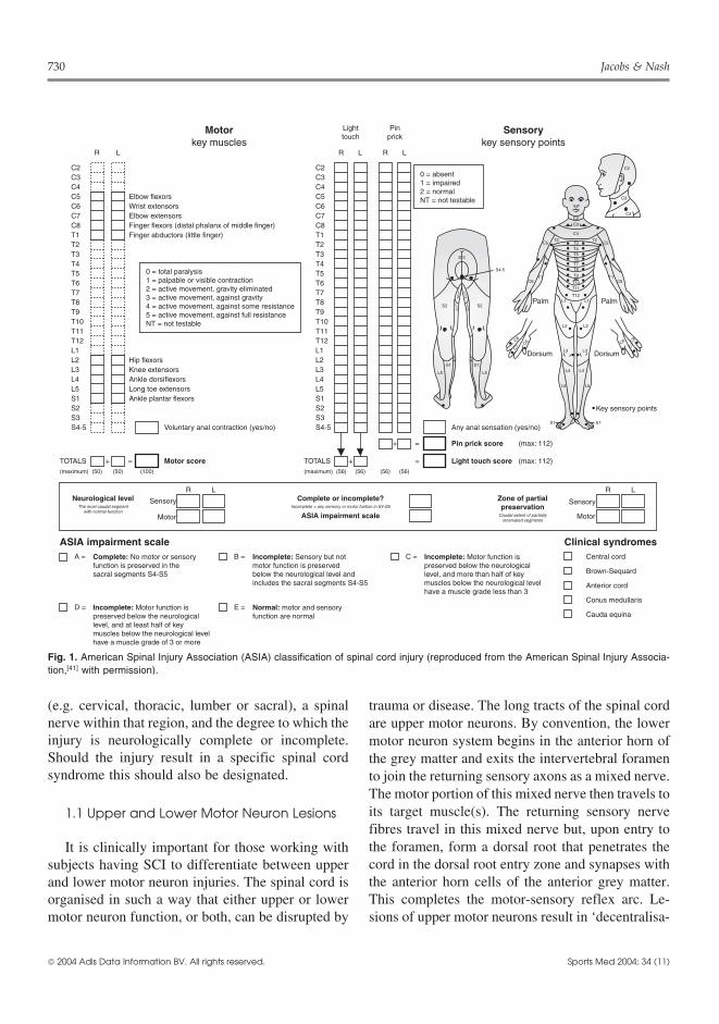
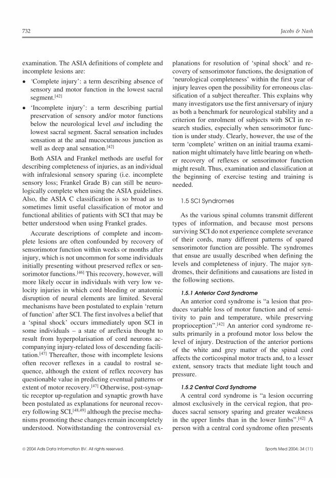
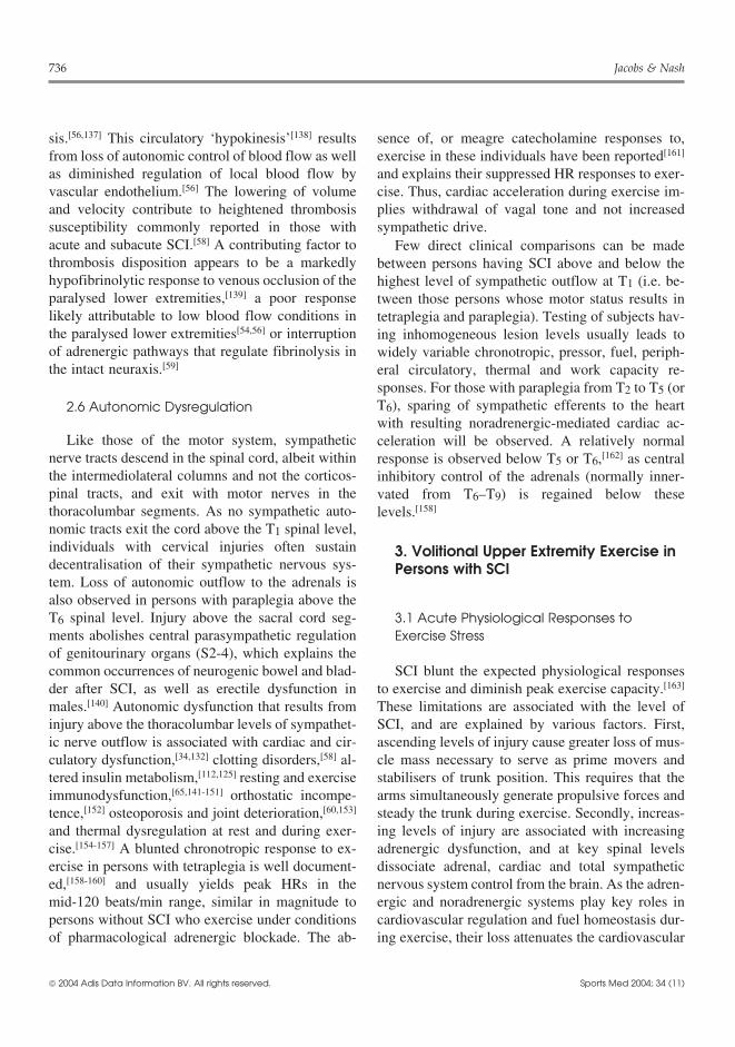
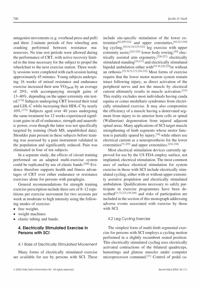

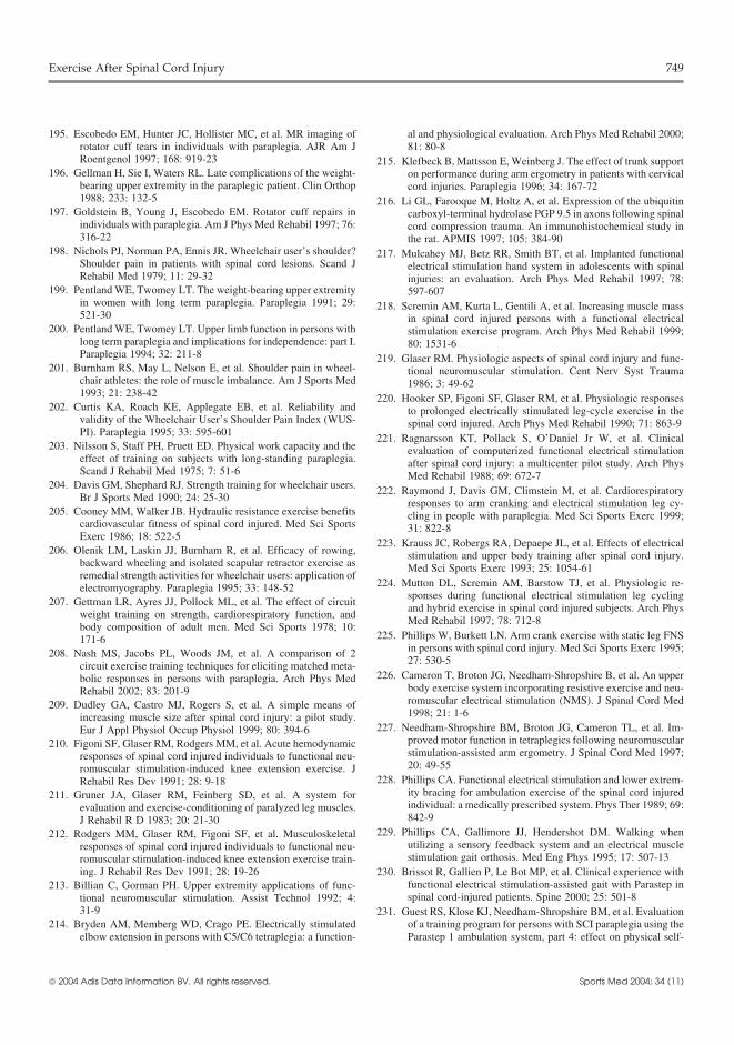


![SCI Standing and Walking Assessment - Spinal Cord Injury … … · assessment for individuals with spinal cord injury. July 2017 | Version 4.0 [ Toolkit For ] SCI Standing and Walking](https://static.documents.pub/doc/80x56/5f03e88b7e708231d40b5d36/sci-standing-and-walking-assessment-spinal-cord-injury-assessment-for-individuals.jpg)


