HAL Id: inserm-02439419 https://www.hal.inserm.fr/inserm-02439419 Submitted on 14 Jan 2020 HAL is a multi-disciplinary open access archive for the deposit and dissemination of sci- entific research documents, whether they are pub- lished or not. The documents may come from teaching and research institutions in France or abroad, or from public or private research centers. L’archive ouverte pluridisciplinaire HAL, est destinée au dépôt et à la diffusion de documents scientifiques de niveau recherche, publiés ou non, émanant des établissements d’enseignement et de recherche français ou étrangers, des laboratoires publics ou privés. Exosomes, an Unmasked Culprit in Neurodegenerative Diseases Federico Soria, Olatz Pampliega, Mathieu Bourdenx, Wassilios Meissner, Erwan Bézard, Benjamin Dehay To cite this version: Federico Soria, Olatz Pampliega, Mathieu Bourdenx, Wassilios Meissner, Erwan Bézard, et al.. Ex- osomes, an Unmasked Culprit in Neurodegenerative Diseases. Frontiers in Neuroscience, Frontiers, 2017, 11, pp.26. 10.3389/fnins.2017.00026. inserm-02439419
Transcript
HAL Id: inserm-02439419https://www.hal.inserm.fr/inserm-02439419
Submitted on 14 Jan 2020
HAL is a multi-disciplinary open accessarchive for the deposit and dissemination of sci-entific research documents, whether they are pub-lished or not. The documents may come fromteaching and research institutions in France orabroad, or from public or private research centers.
L’archive ouverte pluridisciplinaire HAL, estdestinée au dépôt et à la diffusion de documentsscientifiques de niveau recherche, publiés ou non,émanant des établissements d’enseignement et derecherche français ou étrangers, des laboratoirespublics ou privés.
Exosomes, an Unmasked Culprit in NeurodegenerativeDiseases
Federico Soria, Olatz Pampliega, Mathieu Bourdenx, Wassilios Meissner,Erwan Bézard, Benjamin Dehay
To cite this version:Federico Soria, Olatz Pampliega, Mathieu Bourdenx, Wassilios Meissner, Erwan Bézard, et al.. Ex-osomes, an Unmasked Culprit in Neurodegenerative Diseases. Frontiers in Neuroscience, Frontiers,2017, 11, pp.26. �10.3389/fnins.2017.00026�. �inserm-02439419�
Among intracellular organelles, eukaryotic cells also contain organelles that are released into themicroenvironment. These membranous extracellular vesicles can be classified according to theirsize into exosomes, ectosomes, and apoptotic blebs (Mathivanan et al., 2010), many of which exhibitpleiotropic biological functions. In particular, exosomes were initially thought to act as cellularwaste disposal compartments because of their extrusion from the cell, but it was only recentlydiscovered that exosomes contain not only cellular proteins but also act as natural carriers of nucleicacid material in the form of DNA, mRNA transcripts, miRNA, and noncoding RNA (Schorey andBhatnagar, 2008). They are released under normal as well as pathological conditions and since, havebeen recognized as a critical messenger for short and long-distance cell-to-cell communication.As such, cells use exosomes in the central nervous system (CNS) as a major route of secretion inorder to get rid of waste membranes, harmful RNA or proteins or, more importantly, to act asmessengers, and signal carriers to other neural cells, modifying their function in normal physiologyas well as in states of disease (Zappulli et al., 2016). In this regard, given their ability to mediateintercellular communication, exosomes are increasingly being considered as one of the actors in
transporting pathological misfolded proteins that are involvedin neurodegenerative diseases and in disseminating disease intoand within the brain. Nevertheless, since exosomes would becapable of transporting drugs across the blood brain barrier, theirtherapeutic potential has generated great interest in the field.
EXOSOMES AND INTERCELLULARCOMMUNICATION
Studies in a variety of model systems have provided insightsabout origin, composition, and functions of exosomes. Thephysiological role of these vesicles is to transport biomoleculesbetween different cells, being considered as a mechanismof paracrine (and possibly endocrine) communication.However, most of the experimental investigations are related topathophysiological conditions and disease models, from whichthe physiological function has been inferred. Therefore, it hasbeen assumed that the function of exosomes is maintainedduring physiological and pathological conditions, althoughfurther basic studies about their role would be key to corroboratethis assumption.
Exosome BiogenesisThree main types of vesicles have been described so far:exosomes, ectosomes and apoptotic blebs. Exosomes range from30 to 100 nm in diameter, whereas ectosomes range between50 and 1000 nm and apoptotic blebs, released by dying cells,between 50 and 5000 nm. Among them, the best characterizedones are exosomes. Exosomes are membrane-bound sphericalnanovesicles of endocytic origin that are released by almostall cell types, and which can be found in abundance in bodyfluids (Simpson et al., 2008). The biogenesis of exosomesbegins with the invagination of endosomal membranes to formmultivesicular bodies (MVBs) containing intraluminal vesicles(ILVs) that range from 30 to 100 nm in diameter (Simonsand Raposo, 2009). MVBs can either fuse with lysosomes forcargo degradation, or with the plasma membrane, which leadsto the release of the ILVs to the extracellular space as exosomes(Mathivanan et al., 2010).
Exosome Membrane MarkersGeneral markers for exosome include enrichment in CD9,CD63, CD81, and Heat Shock Cognate 70 (HSC70) proteins,as well as the embryonic stem cell marker Nanog (Shelleret al., 2016). As described in this study, a combination ofdifferentmethods is used to accurately characterize the exosomes;electron microscopy for shape and morphology, particle sizing,western blot and flow cytometry for detection of specificmarkers and cargo contents, and UV absorbance for DNAquantification. Altogether, a high-quality isolation method forexosomes, followed by their characterization and identification,is mandatory to distinguish exosomes from other microvesicles.
Exosome Cargo MarkersThe characterization of exosomal cargo will vary depending onthe cell type that releases the exosome, and although the diversityin the cargo profile suggests different mechanisms of selection,
there is still no study about the molecular details nor whetherit is a random event. The myriad of results obtained from thestudies that focus on the characterization of exosome contentare gathered in different databases such as Exocarta, EVpedia, orVesiclepedia (Mathivanan and Simpson, 2009; Kalra et al., 2012;Mathivanan et al., 2012; Simpson et al., 2012; Kim et al., 2013).They provide information about the protein, lipid or acid nucleiccontent of the different types of exosomes. Despite the varietyin isolation techniques and different cell types used in thesestudies, it seems that a significant number of the proteins foundin exosomes come from the biogenesis process, as it happensfor components of the endosomal sorting complex for transport(ESCRT) pathway, Rab proteins needed for the formation andrelease of vesicles (Rab27A, Rab11B, ARF6), or the tetraspaninsCD9, CD63, and CD81.
Cargo loading into the exosomes can be achieved by differentmechanisms, although protein binding to the plasma membraneand its subsequent oligomerization seems to be the main processfor cargo acquisition. However, any protein that binds to anexosome cargo could potentially be loaded into the nascentexosomes, as it is the case for RNAs, which typically exist incomplex with proteins. Therefore, the selective trafficking ofthese macromolecules is linked to the delivery of their bindingpartners. Alternatively, cargo proteins can be covalently linked toexosomes (Yang and Gould, 2013).
Lipids are also loaded into exosomes as cargoes, in whichcase they share many characteristics with the lipid membranesof their cells of origin. Their main differential characteristicis the enrichment in phosphatidylserine in the outer face ofthe exosomal membrane, which facilitates the internalization byrecipient cells (Subra et al., 2007; Fitzner et al., 2011).
Regarding nucleic acids, exosomes are enriched in small RNAsas compared with DNA, although almost any type of RNAhas been found in these vesicles (Abels and Breakefield, 2016).However, the profile of the RNA content in exosomes differssubstantially from that of the total cell (Guduric-Fuchs et al.,2012; Jenjaroenpun et al., 2013; Yagi et al., 2016). Similarly,the analysis of different secretomes led to the conclusion thatexosomes derived from mesenchymal stromal cells (MSCs)in the bone marrow enhance neurite growth, as opposed tomicrovesicles or total secretome. The differential profiles inexosomal cargo content found in this study suggest that selectivemechanisms underlie the processes of cargo selection and/orreception, which are still unexplored, and could open the door tothe development of new therapeutic tools (Lopez-Verrilli et al.,2016).
Exosome Release and TransmissionAlthough poorly investigated, exosome release is thought to beaffected by other cellular events, and it has been described thatsynaptic glutamatergic AMPA and NMDA receptors modulateexosome release in differentiated neurons (Lachenal et al., 2011).Indeed, neuronal depolarization and calcium influx events intothe neuron after ionomycin treatment enhanced exosome release(Lachenal et al., 2011), suggesting that this is an event that can bemodulated and therefore suitable to be developed as a therapeutictool. Several other works have confirmed the role of exosomes
Frontiers in Neuroscience | www.frontiersin.org 2 January 2017 | Volume 11 | Article 26
in modulating synaptic activity, as for example the polarizeddelivery of exosomes at synapses (Mittelbrunn et al., 2015).Also, endocannabinoids, which are secreted through extracellularvesicles produced by microglia, are able to inhibit presynaptictransmission (Gabrielli et al., 2015). Exosomes can indeed act asnovel mechanisms of trans-synaptic communication, in whichsynaptogenic factors such as the presynaptic protein Evi arereleased into the synaptic cleft via these vesicles (Korkut et al.,2009).
Another rising issue in exosome research is that mostof the studies use scenarios where a certain biomolecule isoverexpressed, due to technical requirements or to the specificpathology that is being studied. It has to be pointed out thatoverexpression techniques can lead to a forced transmissionevent that otherwise would not occur in physiological conditions.This concern, together with the fact that exosome release isseldom the primary route of cell-to-cell transmission, questionwhether exosomes are a complementary but not necessarymechanism of paracrine/endocrine communication.
Once delivered, exosomes are taken up by the recipient cell,in a process that involves fusion with the plasma membrane,and different mechanisms of endocytosis that depend on thecell type (Abels and Breakefield, 2016). For example, in theCNS, neurons internalize exosomes through clathrin-dependentendocytosis or phagocytosis (Feng et al., 2010; Fruhbeis et al.,2013), while microglia internalizes these membrane vesiclesthrough macropinocytosis (Fitzner et al., 2011). The uptakemay depend on the cellular state of the recipient cell, sincesurface ligands recognize receptors on the exosome. However, animportant question that remains unanswered about extracellularvesicles is whether the recipient cell has any role in the selectionof the receiving cargo, or if this only depends on the emissarycell. Other factors that modulate the uptake of exosomes is thepresence of heparin sulfate proteoglycans (HSPGs) on the plasmamembrane (Atai et al., 2013; Christianson et al., 2013), or theblockage of specific receptors on the plasma membrane, as it wasdescribed for SR-B1 receptor (Plebanek et al., 2015).
EXOSOMES IN NEURODEGENERATIVEDISEASES
Intercellular communication is a cornerstone in the researchof neurodegenerative diseases. Beyond the complex molecularevents that take place within the cell, the effects that thesecan have on neighboring or distant cells are a matter ofintense investigation. This is mostly because communicationoften happens through the extracellular space, the compartmentwhere it is easier to intervene, either by using a therapeutictool to modify the disease, or by probing and sampling todiagnose or assess disease progression. During the last decade,among the several mechanisms for cell communication studied inneurodegenerative diseases, exosomes and extracellular vesicleshave emerged as common players. Clearance and disposal ofbyproducts or toxic molecules, as well as cell-to-cell transmissionof nucleic acids, cytokines and enzymes, but also aggregated ormisfolded proteins, are among the several cargos being studied
in brain pathophysiology (Figure 1). At this time, most of theadvances have been achieved in vitro, but the results are vastlyinteresting, particularly for the prion-like spreading hypothesisapplied in the understanding of neurodegenerative diseases.
Prion DiseasesPrions are infectious agents consisting of proteins misfolded intoaberrant conformations that can transmit their misfolded state to“naïve” proteins of the same type and aggregate with themselvesinto oligomers and fibrils. According to the prion hypothesis(Prusiner, 1982), these infectious particles operate, thus, asproteopathic seeds that trigger a chain-reaction of refoldingand self-aggregation, spreading the pathophysiological statusto other cells, and ultimately causing neurodegeneration andloss of gray matter. Transmissible spongiform encephalopathiessuch as Creutzfeldt-Jacob disease in humans or scrapie inanimals, are known to originate from this mechanism (Collinge,2001). Similarly, in other neurodegenerative diseases such asAlzheimer’s or Parkinson’s Disease there is also a progressiveaccumulation of misfolded proteins such as β-amyloid, tau,and α-syn, among others, which occurs concomitantly withthe spreading of the pathology between neuroanatomicallyconnected regions. These characteristics have led to thehypothesis that these and other proteinopathies share a commonoriginating mechanism, which has given rise to the “prion-like”hypothesis (Jucker and Walker, 2013).
The idea of exosome-mediated propagation in prion diseaseswas introduced in the last decade, when Fevrier et al. (2004)found that the prion protein (PrP)-expressing cells couldrelease PrPC (normal) and PrPSc (abnormal) in association withexosomes. The first report that linked in vivo the prion diseasepathogenesis and exosomes came 4 years later, when Vella et al.(2008) found PrPC associated to extracellular vesicles in the CSFof sheep. The interplay between prions and exosomes has justbegun to be unveiled in recent years: The biogenesis of exosomesappears to be implicated both in the conversion of PrPSc (Yimet al., 2015) as well as in its packaging (Vella et al., 2007).Moreover, PrPSc can apparently alter the structure (Colemanet al., 2012) and cargo (Bellingham et al., 2012b) of exosomes,and upregulating the exosomal secretion pathway increase theinfectiousness of PrPSc (Guo et al., 2016).
Alzheimer’s Disease: β-Amyloid and TauRajendran et al. (2006) first described in vitro the intracellularβ-cleavage of the amyloid precursor protein (APP) in earlyendosomes, and the subsequent secretion of β-amyloid (Aβ) tothe extracellular space via exosomes, suggesting a role of thesevesicles in Aβ propagation. There have been no further reportsassociating exosomal secretion to Aβ spreading, but APP-C-terminal fragments have been found, indeed, in exosomes in vitro(Sharples et al., 2008) and in vivo (Perez-Gonzalez et al., 2012),which at least suggests a role of the exosomal secretory pathwayin the generation of Aβ. Nonetheless, consensus within the fieldseems to be that direct cell-to-cell transmission is more prevalentthan exosome-mediated transfer for the pathological spreadingof Aβ (Bellingham et al., 2012b).
Frontiers in Neuroscience | www.frontiersin.org 3 January 2017 | Volume 11 | Article 26
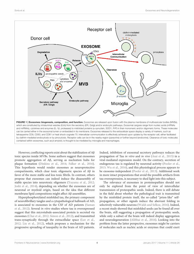
FIGURE 1 | Exosomes: biogenesis, composition, and function. Exosomes are released upon fusion with the plasma membrane of multivesicular bodies (MVBs),
which are constituted by intraluminal vesicles (ILVs) from the secretory (ER, Golgi) and/or endocytic pathways. Exosomal cargoes range from nucleic acids (mRNAs
and miRNAs), cytokines and enzymes (IL-1β, proteases) to misfolded proteins (α-synuclein, SOD1, PrP) in their monomeric and/or oligomeric forms. These molecules
can be carried either in the exosomal lumen or embedded in its membrane. Exosomes released to the extracellular space display a variety of markers, such as
tetraspanins CD9, CD63, and CD81 or heat shock cognate 70. Intercellular communication is effectively achieved upon uptake by the receptor cell, either facilitated
by clathrin-mediated endocytosis or by pinocytosis. Receptor cells can be in the nearby region (paracrine) or further beyond (endocrine). Clearance of toxic molecules
contained within exosomes, such as β-amyloid, is thought to be mediated by microglia and macrophages.
However, conflicting reports exist about the stabilization of Aβ
toxic species inside MVBs. Some authors suggest that exosomespromote aggregation of Aβ, serving as nucleation hubs forplaque formation (Dinkins et al., 2014; Falker et al., 2016).This hypothesis would render exosomes as neuroprotectivecompartments, which clear toxic oligomeric species of Aβ infavor of the more stable and less toxic fibrils. In contrast, otherspropose that exosomes can indeed induce the disassembly ofstable species into neurotoxic oligomers (Yuyama et al., 2012;Joshi et al., 2014), depending on whether the exosomes are ofneuronal or myeloid origin, based on the idea that differentmembrane lipid compositions might affect Aβ assemblies.
Hyperphosphorylated misfolded tau, the primary componentof neurofibrillary tangles and a cytopathological hallmark of AD,is associated to exosomes in the CSF of AD patients (Samanet al., 2012). Several in vitro studies where tau is overexpressedsuggest that this microtubule-associated protein is secreted viaexosomes (Chai et al., 2012; Simon et al., 2012), and transmittedtrans-synaptically through the extracellular space (Lee et al.,2012; Liu et al., 2012), which proposes a mechanism for theprogressive spreading of tauopathy in the brain of AD patients.
Indeed, inhibition of exosomal secretory pathways reduces thepropagation of Tau in vitro and in vivo (Asai et al., 2015) in aviral-mediated expression model. On the contrary, secretion ofendogenous tau is regulated by neuronal activity (Pooler et al.,2013; Wu et al., 2016), and this physiological process appears tobe exosome-independent (Pooler et al., 2013). Additional workin more intact preparations that avoid the possible artifacts fromtau overexpression, is necessary to shed light into this subject.
The relevance of exosomes in proteinopathies should notonly be explored from the point of view of intercellulartransmission of proteopathic seeds. Indeed, there is still debatein the field about whether the pathological trait is transmittedby the misfolded protein itself, the so-called “prion-like” self-propagation, or other signals induce the aberrant folding inselectively vulnerable neurons (Walsh and Selkoe, 2016). Indeed,a recent study showed that misfolded seeds can be found all overthe brain, still suggesting a propagation of proteopathic seeds,while only a subset of the brain will indeed display aggregationand neurodegeneration (Alibhai et al., 2016). Looking into theproblem from the latter perspective, exosomes might be carriersof molecules such as nucleic acids or enzymes that could exert
Frontiers in Neuroscience | www.frontiersin.org 4 January 2017 | Volume 11 | Article 26
a pathophysiological modification in an endocrine manner. Forinstance, secretion of enzymes that degrade Aβ are associated toexosomes (Bulloj et al., 2010; Tamboli et al., 2010), and miRNAstransported within neuron-derived exosomes can activate glialcell functions, such as activation of microglial phagocytosis forthe clearance of neurodegenerating dendrites (Bahrini et al.,2015). Furthermore, microglial and neuronal exosomes havebeen proposed to participate in the disposal of Aβ in the brain(Yuyama et al., 2012, 2015), a concept that becomes furtherrelevant given that impaired Aβ clearance is a characteristic ofAD (Mawuenyega et al., 2010). Altogether, the current state-of-the-art in the field shows that not only cell-to-cell transmission,but also exosome-mediated communication and clearance mightbe important players in AD.
Parkinson’s Disease and OtherSynucleinopathiesAlpha-synuclein (α-syn) is a major component of Lewy bodies(Spillantini et al., 1997), the neuropathological hallmark ofParkinson’s Disease (PD) and Dementia with Lewy bodies (DLB).α-Syn is a pre-synaptic protein that exists in an equilibriumbetween monomeric and oligomeric states. Mutations and otherdisease-related factors affect the aggregation dynamics, favoringthe balance toward fibrillization (Dehay et al., 2015). Theneurodegenerative process observed in PD spreads throughanatomically interconnected regions in the CNS (Braak et al.,2003), and the pathogenic trait of misfolded α-syn is thought topropagate through a prion-like process (Olanow and Prusiner,2009; Lee et al., 2010). Very recently, a team demonstrated thatexosomal α-syn species isolated from the CSF of PD and DLBpatients is able to promote aggregation of α-syn (Stuendl et al.,2016). However, whether exosomes play a role in α-syn spreadingis still a matter of debate.
Among all proteopathic seeds, most reports of secretionassociated to exosomes and related vesicles belongs to the α-syn field (Emmanouilidou et al., 2010; Alvarez-Erviti et al.,2011a; Danzer et al., 2012; Kong et al., 2014; Shi et al., 2014;Tsunemi et al., 2014). The first report that identified α-synshowed that this is a protein present in synaptic vesicles.Due to its cellular function, a link to exosomes might berelated to its intrinsic function within the exocytic machineryat the pre-synaptic terminal (Bendor et al., 2013). Despite thefact that the physiological role of α-syn is not completelyunderstood, it has been suggested that α-syn is involved insynaptic vesicular processing (Vargas et al., 2014), where it bindsto the membrane and undergoes conformational changes thatfacilitate the generation of the vesicle curvature (Varkey et al.,2010; Westphal and Chandra, 2013). It has also been reportedthat α-syn prefers membranes with high curvature (Jensen et al.,2011), like the ones found in the 50–100 nm exosomes. It isworth to note that non-monomeric, i.e., aggregated α-syn, isunable to perform this task, and it is not clear whether membraneassociation induces or prevents oligomerization (Bendor et al.,2013). In the first study that characterized the secretion of α-synby unconventional secretory pathways, Lee et al. (2005) alreadydiscussed that its compartmentalization into vesicles couldpromote the misfolding of α-syn. A recent study demonstratedthat exosomes accelerate α-syn aggregation (Grey et al., 2015),
and that the lipid composition of the vesicles is critical forthis process. The mechanism by which α-syn is incorporatedinto exosomes is still unknown, but given the vast number ofgenes linked to PD that have a role in endocytic and autophagypathways such as PARK2, GBA, LRRK2, or ATP13A2 (Gan-Or et al., 2015), the rapid advancement of research in vesicletrafficking might give some clues in the next years.
In multiple system atrophy (MSA), oligodendrocytesforming α-syn-containing aggregates named glial cytoplasmicinclusions (GCIs) accumulate α-syn, which is accompaniedby demyelination and extensive dopaminergic neuronal lossthroughout the brain. Although transgenic MSA modelsoverexpressing α-syn under different oligodendrocyte-specificpromoters have been studied for a decade (Fernagut and Tison,2012), the link between oligodendroglial α-syn aggregationand neuronal death remains elusive. Moreover, as α-syn isa predominantly neuronal protein, it is also unknown ifoligodendrocytes in a disease state can synthesize α-syn denovo, or if α-syn is readily transferred from neurons. In thisregard, there are several reports indicating that oligodendrocytesand neurons communicate via exosomes, which supportsthe hypothesis of α-syn transfer between these two cell types(Kramer-Albers et al., 2007; Bakhti et al., 2011). This exosomalcommunication between neurons and oligodendrocytes includesthe transfer of genetic information by miRNAs (Fruhbeis et al.,2013), which in fact are altered in MSA (Ubhi et al., 2014;Schafferer et al., 2016). Therefore, a potential involvement of theexosomal secretory pathway in the pathogenesis of MSA shouldnot be overlooked.
Amyotrophic Lateral Sclerosis andHuntington’s DiseaseAmyotrophic lateral sclerosis (ALS) is characterized by themisfolding of Cu/Zn superoxide dismutase (SOD1) into aberrantconformations, which has been pinpointed as a central eventin familiar and sporadic forms of the disease (Bosco et al.,2010). In vitro, neurons secrete exosomes loaded with mutantSOD1 to the extracellular space (Gomes et al., 2007), wherethese exosomes are able to transmit the pathogenic trait toother neurons, and propagate the misfolding of SOD1 (Gradet al., 2014). Furthermore, astrocytes also secrete SOD1-loadedexosomes that are selectively toxic for motor neurons (Bassoet al., 2013), showing that exosome-mediated transfer of mutantSOD1 is pathogenic.
TDP-43 is a nuclear protein that mislocalizes to the cytoplasm,aggregates and forms inclusions in ALS and in fronto-temporaldementia (FTD). Dipeptide repeat proteins (DPRs) are producedby the unconventional translation of the C9orf72, the mostcommon mutated gene in ALS and FTD as well. Recently,both molecules have been associated with exosomal release andtransport (Nonaka et al., 2013; Ding et al., 2015; Westergardet al., 2016), further suggesting a link between exosome-mediatedcommunication and the disease.
Exosomes can also propagate toxic molecules in an inter-species manner. An interesting work showed recently a cell-to-cell propagation of mutant Huntingtin (mtHtt) in exosomesusing 143 CAG repeats fibroblasts and SH-SY5Y neuroblastomacells. Furthermore, injecting newborn wild-type mice with
Frontiers in Neuroscience | www.frontiersin.org 5 January 2017 | Volume 11 | Article 26
human exosomes carrying mtHtt triggered Huntington disease-like (HD) symptoms and pathology in these animals (Jeon et al.,2016), this being the first report of human-to-mouse exosome-mediated propagation of toxic molecules.
Multiple Sclerosis and InflammationInflammation is a central player in most neurodegenerativediseases. Neuroinflammatory processes exert profound changesnot only in the vicinity but beyond the local environment ofcells. In addition to the propagation of toxic proteins, exosomesalso carry important inflammatory modulators, such as mRNAs,miRNAs, and cytokines (Gupta and Pulliam, 2014). Microglia,the resident macrophages of the CNS, can shed exosomes loadedwith pro-inflammatory molecules such as IL-1β (Bianco et al.,2005) and other cytokines. Their scavenging functions are alsocrucial in the clearance of toxic seeds such as Aβ (Yuyamaet al., 2012). Furthermore, endocrine signals from hematopoieticcells directed to the brain can be transported by exosomes, aphenomenon that is augmented in a context of inflammation(Ridder et al., 2014). It is interesting to note that extracellularvesicles can readily cross the blood brain barrier, adding acommunication channel by which systemic inflammation canmodulate physiological processes in the CNS.
Multiple sclerosis (MS) is a demyelinating diseasecharacterized by the loss of oligodendrocytes, whereinflammation plays a prominent role (Compston and Coles,2008). Increased levels of circulating exosomes are present inthe inflammation-driven disease model of MS, experimentalautoimmune encephalomyelitis (EAE). In this model, pro-inflammatory cytokines promote exosome release by immunecells, which in turn carry more pro-inflammatory molecules,spreading inflammation. Animals where exosome-releaseactivity had been hampered showed reduced inflammation andEAE symptoms (Verderio et al., 2012). Another component inthe pathogenesis of EAE is glutamate excitotoxicity (Pitt et al.,2000), which is characterized by pathological accumulation ofthis neurotransmitter and amino acids, among other features.Glutamate can indeed trigger the release of exosomes byoligodendrocytes (Fruhbeis et al., 2013), cells whose homeostasisis known to be altered in demyelinating diseases (Matuteet al., 2001). These oligodendrocyte-derived exosomes regulateaxon myelination (Bakhti et al., 2011). Nevertheless, despiteaccumulating evidence suggesting a role of exosomes ininflammatory processes during pathology, whether theseextracellular vesicles participate in the interplay betweeninflammation and excitotoxicity in demyelinating diseasesremains unanswered.
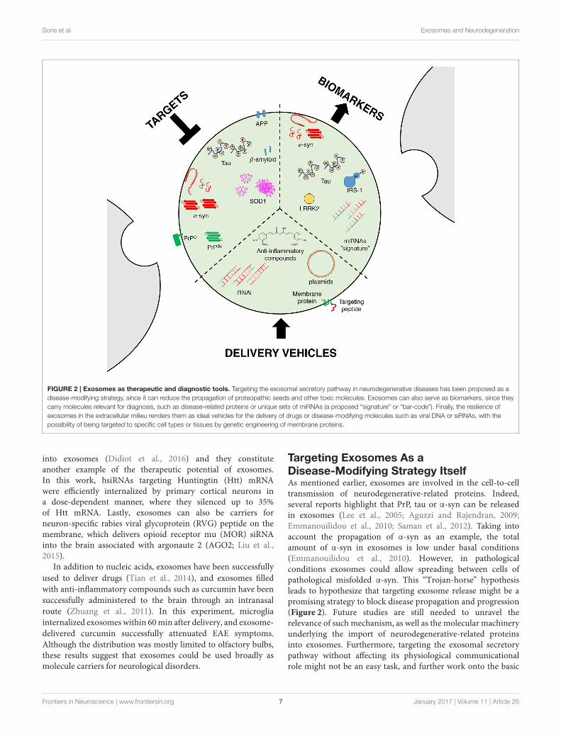
EXOSOMES AS THERAPEUTIC ANDDIAGNOSTIC TOOLS
Potential of Exosomes As Carriers forDisease-Modifying StrategiesAlthough the exact mechanism of exosomal entry into thebrain is not fully understood, it should be noted that exosomesare able to cross the blood brain barrier (Record et al., 2011;
Tominaga et al., 2015), rendering them as ideal vehicles todelivery molecules into the brain (Figure 2). Exosomes areable to transfer mRNAs, microRNAs or proteins, and theyare thought to be an exchange mechanism between cells(Valadi et al., 2007; Simpson et al., 2008), suggesting thatthese vesicles could be a new delivery machinery especiallyfor RNA interference (RNAi; Valadi et al., 2007; Simons andRaposo, 2009). Indeed, RNAi-based technologies need vectors,since siRNAs can be degraded by extracellular endonucleasespresent in the serum, cells or extracellular space, and are alsoimmunogenic (Whitehead et al., 2011; Kalani et al., 2014).Regarding delivery, exosomes harbor interesting advantagesover other delivery systems such as liposomes, since they havea prolonged half-life and no immunogenicity (Kalani et al.,2014).
The pioneer work of Alvarez-Erviti and coworkers hasshown that exosomes can be engineered to target specifictissues (Alvarez-Erviti et al., 2011b). They inserted brain- ormuscle-specific peptides in one of the most abundant exosomalmembrane proteins: the lysosomal-associated membrane protein2b (LAMP-2B). In the brain, the proof-of-concept was obtainedby using exosomes that contain siRNA for brain-specificknockdown of BACE1, encoding the beta-secretase 1 enzyme, inwild type mice (Alvarez-Erviti et al., 2011b). In a more recentstudy, the same strategy was successfully used to knockdownα-syn in transgenic mouse brain (Cooper et al., 2014). Altogether,these results suggest that exosomes might be interesting vehiclesfor RNAi delivery. As exosomes are able to target mostbrain cell types, these results pave the way to applicabilityin other diseases associated to α-syn accumulation, such asMSA.
microRNAs are highly abundant and largely studied in thebiology of exosomes, in part due to their importance in theregulation of gene expression. Therefore, delivery of miRNAs-containing exosomes can constitute a precious therapeutic tool(Ohno et al., 2013). In this sense, MSCs release exosomesthat contain overexpressed miRNA-133b, which was found toimprove the recovery of ischemic (MCAO) rats, including neuriteremodeling in the infarcted area (Xin et al., 2016). The authorsused in vitro exosomes from MSCs along with overexpressionof miR-133b+, which increased the release of exosomes fromoxygen and glucose-deprived astrocytes. These experimentsresulted in increased neurite branching, which suggests thatsecondary astrocyte release may contribute to neuronal plasticityand recovery after stroke. It should be noted that whereas in vivoMSCs are in charge of miRNA-containing exosome release, Xinet al. used astrocytes for the in vitro studies, a different cell typewhere the role and function of miRNAs might be substantiallydifferent. Interestingly, five miRNAs from the exosome-releasedpool showed differential expression levels during traumaticbrain injury (TBI), for which miR-21 showed the larger change(Harrison et al., 2016). The authors hypothesize that the increasein miR-21 is due to its release in neuronal exosomes close tothe injury boundary, in which vicinity reactive microglia wasfound.
Engineered nucleic acids, such as hydrophobically modifiedsmall interfering RNAs (hsiRNAs) have been shown to load
Frontiers in Neuroscience | www.frontiersin.org 6 January 2017 | Volume 11 | Article 26
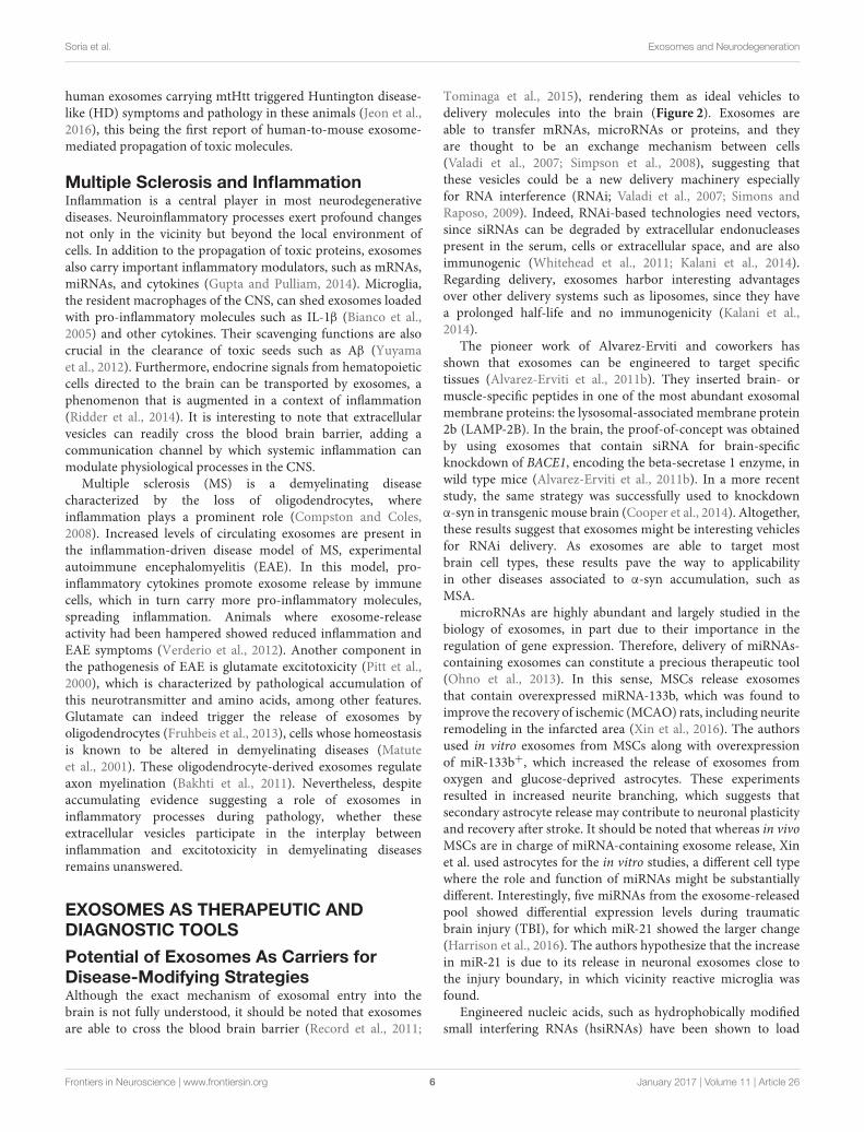
FIGURE 2 | Exosomes as therapeutic and diagnostic tools. Targeting the exosomal secretory pathway in neurodegenerative diseases has been proposed as a
disease-modifying strategy, since it can reduce the propagation of proteopathic seeds and other toxic molecules. Exosomes can also serve as biomarkers, since they
carry molecules relevant for diagnosis, such as disease-related proteins or unique sets of miRNAs (a proposed “signature” or “bar-code”). Finally, the resilience of
exosomes in the extracellular milieu renders them as ideal vehicles for the delivery of drugs or disease-modifying molecules such as viral DNA or siRNAs, with the
possibility of being targeted to specific cell types or tissues by genetic engineering of membrane proteins.
into exosomes (Didiot et al., 2016) and they constituteanother example of the therapeutic potential of exosomes.In this work, hsiRNAs targeting Huntingtin (Htt) mRNAwere efficiently internalized by primary cortical neurons ina dose-dependent manner, where they silenced up to 35%of Htt mRNA. Lastly, exosomes can also be carriers forneuron-specific rabies viral glycoprotein (RVG) peptide on themembrane, which delivers opioid receptor mu (MOR) siRNAinto the brain associated with argonaute 2 (AGO2; Liu et al.,2015).
In addition to nucleic acids, exosomes have been successfully
used to deliver drugs (Tian et al., 2014), and exosomes filled
with anti-inflammatory compounds such as curcumin have been
successfully administered to the brain through an intranasal
route (Zhuang et al., 2011). In this experiment, microglia
internalized exosomes within 60min after delivery, and exosome-
Although the distribution was mostly limited to olfactory bulbs,these results suggest that exosomes could be used broadly asmolecule carriers for neurological disorders.
Targeting Exosomes As aDisease-Modifying Strategy ItselfAs mentioned earlier, exosomes are involved in the cell-to-celltransmission of neurodegenerative-related proteins. Indeed,several reports highlight that PrP, tau or α-syn can be releasedin exosomes (Lee et al., 2005; Aguzzi and Rajendran, 2009;Emmanouilidou et al., 2010; Saman et al., 2012). Taking intoaccount the propagation of α-syn as an example, the totalamount of α-syn in exosomes is low under basal conditions(Emmanouilidou et al., 2010). However, in pathologicalconditions exosomes could allow spreading between cells ofpathological misfolded α-syn. This “Trojan-horse” hypothesisleads to hypothesize that targeting exosome release might be apromising strategy to block disease propagation and progression(Figure 2). Future studies are still needed to unravel therelevance of such mechanism, as well as the molecular machineryunderlying the import of neurodegenerative-related proteinsinto exosomes. Furthermore, targeting the exosomal secretorypathway without affecting its physiological communicationalrole might not be an easy task, and further work onto the basic
Frontiers in Neuroscience | www.frontiersin.org 7 January 2017 | Volume 11 | Article 26
physiological mechanisms of exosome release and cargo selectionwill be crucial in this matter.
Exosomes As Biomarkers for DiseaseSince the identification of neurodegenerative disease relatedproteins in exosomes, these vesicles have been proposed asputative biomarkers to monitor disease progression (Figure 2).Exosomes can be isolated from multiple fluids including urine,blood or cerebrospinal fluid (CSF), which facilitates their clinicaluse as biomarkers (Street et al., 2012; Cheng et al., 2014a,b).Quantifications of neurodegenerative disease related proteinsin total CSF or in exosomes purified from CSF have led tocontradictory results in both AD and PD (Parnetti et al., 2013;Kim et al., 2016; Vella et al., 2016). Interestingly, a reporthas shown that hyperphosphorylated-tau (often considered aspathological tau) in exosomes is significantly increased inpatients with mild forms of AD compared to controls (Samanet al., 2012). Although such result suggests that measuring tauphosphorylation in exosomes could be used as a biomarkerfor AD, the fact that moderate or severe forms of the diseasedo not show similar phosphorylation levels dampens thathypothesis. One possible explanation could be that high levelsof phosphorylated tau in exosomes is specific of a disease-stage,and not the entire pathological process. Regarding PD, similardivergences are also present in the literature. Indeed, a reportshowed that α-syn is increased in microvesicles for PD patientswhile another did not observed variation in the α-syn content(Shi et al., 2014; Tomlinson et al., 2015). This discrepancy couldbe explained by technical differences as well as differences in thepatient population.
As many other proteins are present in exosomes, recentinitiatives have used large-scale methodologies to analyzeexosome content. Usingmass-spectrometry to analyze circulatingmicrovesicles, it was identified that PD patient fibroblasts areenriched in syntenin 1, a regulator of exosome biogenesis(Tomlinson et al., 2015). Similarly, a set of nine miRNAs havebeen shown to be distinct in exosomes purified from controland prion-infected cells (Bellingham et al., 2012a), defining amolecular “signature” that can identify a pathological process.Although the relevance of such hits in the disease is notestablished yet, these approaches suggest that establishing a “bar-code” from the exosomal profile of patients might be a morevaluable diagnostic tool than quantifying specific disease-relatedproteins, which are often mixed at early disease stages.
Exosomes can originate from most cell types and have beenidentified in most body fluids. A study identified the Leucine-Rich Repeat Kinase 2 (LRRK2), a protein involved in somehereditary forms of PD, in urinary exosomes (Fraser et al.,2013). The strong variability observed in clinical populationsargued against its use as a biomarker for PD. However, onecan hypothesize that exosomes originating from brain mightbe diluted in body fluids containing exosomes originating fromother organs. To test this hypothesis, a recent study usedan immunochemical method to purify brain exosomes fromperipheral blood plasma (Shi et al., 2014). The authors reportedan increase of α-syn specifically in exosomes from plasma ofPD patients (i.e., no overall modification was observed in total
plasma). Although this approach was developed to be brain-specific based on the expression of the L1-cell adhesion molecule(L1-CAM), this protein is also expressed in the renal system(Allory et al., 2008; Vella et al., 2016), therefore these resultshave to be taken carefully. However, and more importantly,the immunochemical capture method provides an innovativeapproach for quantifying neurodegenerative disease relatedproteins in peripheral fluids to be used as biomarkers, and whichhold the promise of a better specificity (Vella et al., 2016).
Interestingly, insulin resistance (i.e., decreased insulin/insulinlike growth factor IGF-1 signaling) has been established in bothMSA patients and in a well-characterized transgenic model, thePLP-SYN mice (Bassil et al., 2017). Insulin resistance can bemeasured by analyzing the amount of the downstreammessengerinsulin receptor substrate-1 phosphorylated at serine residues312 (IRS-1pS312) or 616 (IRS-1pS616; Moloney et al., 2010;Talbot et al., 2012). Since this biomarker can be traced inperipheral exosomes (Kapogiannis et al., 2015), we evaluatedthe murine plasma neural derived exosomal IRS-1pS307 levels(corresponding to human IRS-1pS312) in transgenic MSA micethat were treated with the glucagon-like peptide-1 (GLP-1)agonist exenatide. We found a correlation with the number ofnigral dopaminergic neurons and with striatal oligomeric α-synload in transgenic MSA mice, indicating that PLP-SYN micewith highest plasma exosomal IRS-1pS307 concentrations hada lower number of nigral TH and Nissl-stained neurons as wellas higher striatal oligomeric α-syn levels (Bassil et al., 2017).Altogether, these data suggest that peripheral neural derivedexosomal IRS-1pS312 is an exosomal biomarker candidatethat may serve as an objective outcome measure of targetengagement for preclinical studies as well as clinical trials withGLP-1 analogs and other compounds modulating insulin/IGF-1signaling.
CONCLUDING REMARKS
The last decade has witnessed a dramatic increase in publicationsin the field of exosomes and related extracellular vesicles.Initially considered waste disposal material, recent evidence hasprogressively changed this view, and exosomes are currentlyconsidered, in fact, as a broad intercellular communicationsystem that can internalize, transport and transfer all types ofbiomolecules, from nucleic acids to peptides and proteins.
Most of the research in exosomes and extracellular vesicleshas been carried out in cell cultures. This in vitro approachhas been very useful to screen cell types, cargos, and geneticbackgrounds, providing useful data to understand the biogenesisand physiological role of exosomes. However, their rolein neurodegenerative diseases, either as carriers of disease-modifying molecules, or as an effective clearance system, isyet to be completely understood. Several questions remainunanswered, mostly because of the lack of in vivo studies, as wellas experiments in more intact preparations where the relevantmolecules are expressed in endogenous levels. Among severalopen questions in the field, of particular interest, are (i) whetherexosomes from diseased organisms can effectively propagate thepathogenic trait in vivo, (ii) whether targeting the exosomal
Frontiers in Neuroscience | www.frontiersin.org 8 January 2017 | Volume 11 | Article 26
secretory pathway can be beneficial for the pathology, and (iii)what is the molecular profile or “signature” that can be readfrom exosomes isolated from patients to be used as biomarkersin different brain disorders. Future studies will hopefully fulfillthe high expectations raised among neurobiologists regarding theuse of exosomes as therapeutic targets and/or tools.
AUTHOR CONTRIBUTIONS
FS, OP, MB, and BD were responsible for the conception of thearticle. FS, OP, MB, and BD did the scientific literature review. FScreated the figures. FS, OP, MB, and BDwrote the first draft of the
article. FS, OP, MB, WM, EB, and BD critically revised the entiremanuscript and approved the final version.
ACKNOWLEDGMENTS
The University of Bordeaux and the Centre National de laRecherche Scientifique provided infrastructural support. Thiswork was supported by Fondation de France (BD and EB),Fondation Simone and Cino Del Duca (EB), LABEX BRAINANR10-LABX-43 (EB), IdEx Université de Bordeaux (FS andOP), and LECMA-Vaincre Alzheimer (OP). MB is recipient ofFrance Parkinson and LABEX BRAIN fellowships.
REFERENCES
Abels, E. R., and Breakefield, X. O. (2016). Introduction to extracellular vesicles:
biogenesis, RNA cargo selection, content, release, and uptake. Cell Mol.


![[Shinobi] Bleach - Ulquiorra UNMASKED](https://static.documents.pub/doc/80x56/568c51f01a28ab4916b4b8ab/shinobi-bleach-ulquiorra-unmasked.jpg)


