Experience of the use of motor-evoked potentials in patients with tumors of the cervical spinal cord. PhD Klimov V.S., PhD Evsukov A.V., Chishina N.V., Kelmakov V.V. FSBI “Federal Neurosurgikal Center” of Ministry of Public Health RF, Novosibirsk e-POSTER 1316
Transcript
Experience of the use of motor-evoked potentials in patients with tumors of the
cervical spinal cord.
PhD Klimov V.S., PhD Evsukov A.V., Chishina N.V., Kelmakov V.V.FSBI “Federal Neurosurgikal Center” of Ministry of Public Health RF, Novosibirsk
e-POSTER 1316
Neurophysiological intraoperative monitoring, which is actively developing the last two decades, combines different methods of evaluating the functional state of the central and peripheral nervous system during surgery (Khilko VA et al., 1998; 1999; Ostreiko LM et al., 1998; 1999; crucibles GS et al., 1999; Dawson EG, 1991; Owen JH, 1993; Ashkenaze D., 1993; Padberg AM, 1997). Methods of intraoperative assessment of motor function of the spinal cord - monitoring motor evoked potentials (Shields SV, 1988). Applying these techniques during surgery, it is possible to determine the beginning of a surgically induced damage and carry out relevant measures to prevent or reduce postoperative neurological complications.
Objective: To study the dependence of the dynamics of neurological status and changes of intraoperative motor-evoked potentials in patients with tumors of the cervical spinal cord in the early postoperative period.
Introduction
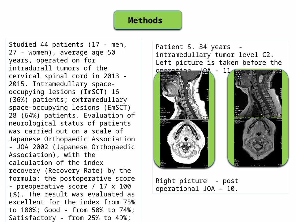
Studied 44 patients (17 - men, 27 - women), average age 50 years, operated on for intradurall tumors of the cervical spinal cord in 2013 - 2015. Intramedullary space-occupying lesions (ImSCT) 16 (36%) patients; extramedullary space-occupying lesions (EmSCT) 28 (64%) patients. Evaluation of neurological status of patients was carried out on a scale of Japanese Orthopaedic Association - JOA 2002 (Japanese Orthopaedic Association), with the calculation of the index recovery (Recovery Rate) by the formula: the postoperative score - preoperative score / 17 x 100 (%). The result was evaluated as excellent for the index from 75% to 100%; Good - from 50% to 74%; Satisfactory - from 25% to 49%; unchanged - from 0% to 24%; bad - less than 0%.
Methods
Patient S. 34 years - intramedullary tumor level C2. Left picture is taken before the operation. JOA – 11
Right picture - post operational JOA – 10.
Insert Pictures
Insert Pictures
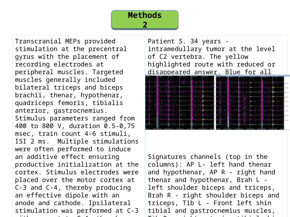
Transcranial MEPs provided stimulation at the precentral gyrus with the placement of recording electrodes at peripheral muscles. Targeted muscles generally included bilateral triceps and biceps brachii, thenar, hypothenar, quadriceps femoris, tibialis anterior, gastrocnemius. Stimulus parameters ranged from 400 to 800 V, duration 0.5-0,75 msec, train count 4-6 stimuli, ISI 2 ms. Multiple stimulations were often performed to induce an additive effect ensuring productive initialization at the cortex. Stimulus electrodes were placed over the motor cortex at C-3 and C-4, thereby producing an effective dipole with an anode and cathode. Ipsilateral stimulation was performed at C-3 with respect to C-4, thereby providing data for the patient’s left-sided corticospinal pathway. The dipole was then reversed to stimulate at C-4 with respect to C-3, providing motor responses from the patient’s right side.
Methods 2
Patient S. 34 years - intramedullary tumor at the level of C2 vertebra. The yellow highlighted route with reduced or disappeared answer. Blue for all the routes allocated Baseline initial amplitude response measures before tumor removal.
Signatures channels (top in the columns): AP L- left hand thenar and hypothenar, AP R - right hand thenar and hypothenar, Brah L - left shoulder biceps and triceps, Brah R - right shoulder biceps and triceps, Tib L - Front left shin tibial and gastrocnemius muscles, Tib R - right anterior tibial shin and calf muscles.Each side of the track indicated the registration answer.
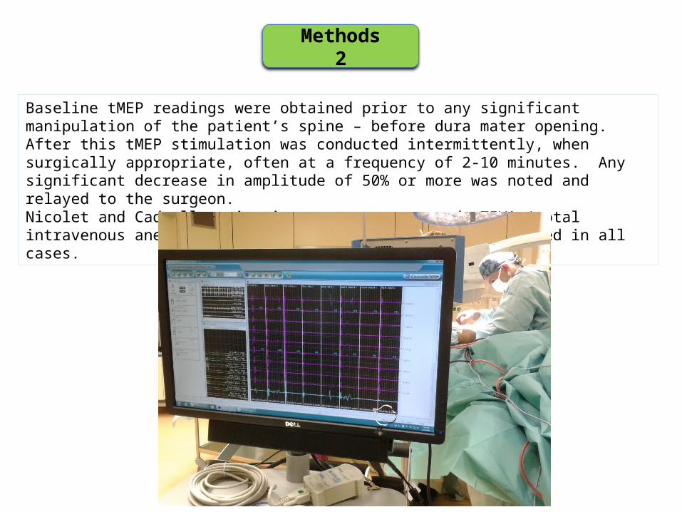
Methods 2
Baseline tMEP readings were obtained prior to any significant manipulation of the patient’s spine – before dura mater opening. After this tMEP stimulation was conducted intermittently, when surgically appropriate, often at a frequency of 2-10 minutes. Any significant decrease in amplitude of 50% or more was noted and relayed to the surgeon. Nicolet and Cadwell monitoring systems were used. TIVA (total intravenous anesthesia) with propofol and fentanyl was used in all cases.
Results
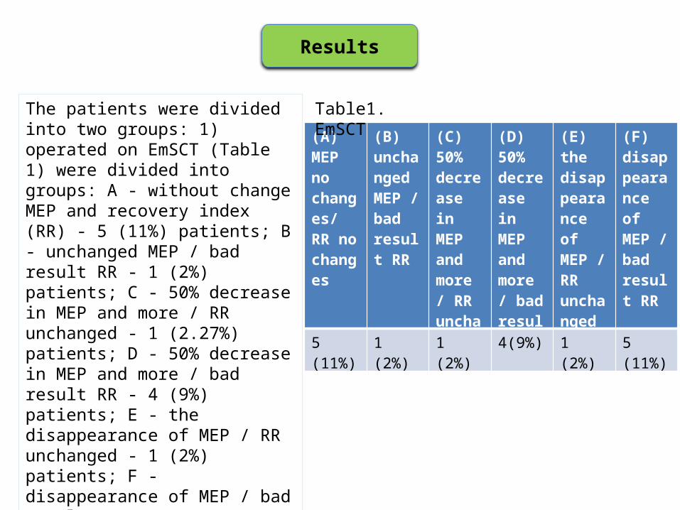
The patients were divided into two groups: 1) operated on EmSCT (Table 1) were divided into groups: A - without change MEP and recovery index (RR) - 5 (11%) patients; B - unchanged MEP / bad result RR - 1 (2%) patients; C - 50% decrease in MEP and more / RR unchanged - 1 (2.27%) patients; D - 50% decrease in MEP and more / bad result RR - 4 (9%) patients; E - the disappearance of MEP / RR unchanged - 1 (2%) patients; F - disappearance of MEP / bad result RR - 5 (11%) patients. Satisfactory results in this group of patients were observed.
(А) МЕР no changes/ RR no changes
(В) unchanged MEP / bad result RR
(С) 50% decrease in MEP and more / RR unchanged
(D) 50% decrease in MEP and more / bad result RR
(E) the disappearance of MEP / RR unchanged
(F) disappearance of MEP / bad result RR
5 (11%) 1 (2%) 1 (2%) 4(9%) 1 (2%) 5 (11%)
Table1. EmSCT
Results
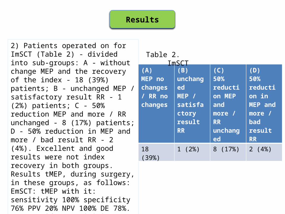
2) Patients operated on for ImSCT (Table 2) - divided into sub-groups: A - without change MEP and the recovery of the index - 18 (39%) patients; B - unchanged MEP / satisfactory result RR - 1 (2%) patients; C - 50% reduction MEP and more / RR unchanged - 8 (17%) patients; D - 50% reduction in MEP and more / bad result RR - 2 (4%). Excellent and good results were not index recovery in both groups. Results tMEP, during surgery, in these groups, as follows: EmSCT: tMEP with it: sensitivity 100% specificity 76% PPV 20% NPV 100% DE 78%. tMEP upside: sensitivity 0% specificity 72% PPV% NPV 0 100% DE 72%. ImSCT: tMEP with it: sensitivity 100% specificity 67% PPV 86% NPV 100% DE 80%. tMEP upside: sensitivity 83% specificity 0% PPV 56% NPV 0% DE 50%.
(А) МЕР no changes / RR no changes
(В) unchanged MEP / satisfactory result RR
(C) 50% reduction MEP and more / RR unchanged
(D) 50% reduction in MEP and more / bad result RR
18 (39%) 1 (2%) 8 (17%) 2 (4%)
Table 2. ImSCT
Conclusions
1. tMEP responses obtained from upper extremity muscles are quite reliable, highly sensitive and specific methods and they can be used for motor function control in intraoperative monitoring during neurosurgical operations for intramedullary and extramedullary tumors of cervical level of spinal cord. In group of patients with extramedullary tumors PPV (positive predictive value) of this method is much lower. The diagnostic efficiency (accuracy) of this method in these two groups of patients is approximately the same.
2. tMEP responses obtained from lower extremity muscles. There is an extremely high probability of false-positive results of tMEP from lower extremity muscles in the group of patients with intramedullary tumors (specificity = 0%, it means that there are too frequent cases of reducing the amplitude of the tMEP responses in the absence of postoperative neurological deficit worsening). And this method is absolutely uninformative in the group of patients with extramedullary tumors and there were no one true positive result in that group.