[ 177 ] EXPERIMENTAL STUDIES ON THE STAINING OF NERVOUS TISSUE WITH SILVER PROTEINATES BY ALAN PETERS Department of Anatomy, University of Edinburgh INTRODUCTION Soluble silver proteinates were first synthesized by Eichengrun in 1898 (Porter & Davenport, 1951) and Regaud & Dubreuil (1903) used them, in particular Protargol, to stain epithelia in fresh tissue. In the following year, Lugaro (1904) employed silver proteinates to stain neurofibrillae and in 1933, Bartelmez & Hoerr substituted Protargol for the silver nitrate used in Rogers' (1931) method of staining nerves in paraffin sections. It was not until 1936 that Protargol gained wide popularity as an histological reagent for nerve staining by the introduction of Bodian's method, in which the addition of metallic copper was used to improve the selectivity of the stain for nerves. Between 1936 and the beginning of the last war, various methods of using Pro- targol were suggested (see Davenport & Kline, 1938; Davenport, MacArthur & Bruesch, 1939; and Davenport, Porter & Thomas, 1947), and although many pro- teinates were available commercially, only Protargol manufactured by Bayer in Germany and Winthrop in America was satisfactory for staining nerves. After 1939, when Bayer Protargol was no longer available, a number of methods em- ploying simple silver salts were published, but despite attempts to simplify and investigate these latter methods of staining (see Silver, 1942; Holmes, 1943; Palmgren, 1948; Romanes, 1950; Samuel, 1953; Peters, 1955; and Wolman, 1955) they proved to be less satisfactory and frequently less consistent than the Bodian technique. More recently Protargol-S has been produced by Winthrop-Stearns Inc. of America, and, in 1956, Polley described a new silver compound from France (Laboratoire Roque); both of these compounds have been widely used to stain nerves. Some of the factors involved in staining with silver proteinates were considered by Bodian (1936), Holmes (1943), Romanes (1950), and Davenport and his co- workers. Bodian (1936) found that when sections were impregnated in Protargol solutions which contained metallic copper, copper went into solution and some was deposited, along with silver, on nerve fibres. He also suggested that since silver was plated-out on to the copper, it was likely that the ionic silver content of the Protargol solution decreased as impregnation proceeded. Later, Holmes (1943) found that while the pH value of a Protargol solution only fell from pH 8-2 to 8-0 during an impregnation period of 24 hr., if copper was present, the pH value fell to 6-6. Thus, there was some information available about the changes which occurred during impregnation, but their significance was unknown. In the present series of experiments an attempt was made to measure these various changes quantitatively, with the ultimate aim of analysing the impregnation process. At the same time 12-2
Transcript
[ 177 ]
EXPERIMENTAL STUDIES ON THE STAINING OFNERVOUS TISSUE WITH SILVER PROTEINATES
BY ALAN PETERSDepartment of Anatomy, University of Edinburgh
INTRODUCTION
Soluble silver proteinates were first synthesized by Eichengrun in 1898 (Porter &Davenport, 1951) and Regaud & Dubreuil (1903) used them, in particular Protargol,to stain epithelia in fresh tissue. In the following year, Lugaro (1904) employedsilver proteinates to stain neurofibrillae and in 1933, Bartelmez & Hoerr substitutedProtargol for the silver nitrate used in Rogers' (1931) method of staining nerves inparaffin sections. It was not until 1936 that Protargol gained wide popularity as anhistological reagent for nerve staining by the introduction of Bodian's method, inwhich the addition of metallic copper was used to improve the selectivity of thestain for nerves.Between 1936 and the beginning of the last war, various methods of using Pro-
targol were suggested (see Davenport & Kline, 1938; Davenport, MacArthur &Bruesch, 1939; and Davenport, Porter & Thomas, 1947), and although many pro-teinates were available commercially, only Protargol manufactured by Bayer inGermany and Winthrop in America was satisfactory for staining nerves. After1939, when Bayer Protargol was no longer available, a number of methods em-ploying simple silver salts were published, but despite attempts to simplify andinvestigate these latter methods of staining (see Silver, 1942; Holmes, 1943;Palmgren, 1948; Romanes, 1950; Samuel, 1953; Peters, 1955; and Wolman, 1955)they proved to be less satisfactory and frequently less consistent than the Bodiantechnique. More recently Protargol-S has been produced by Winthrop-StearnsInc. of America, and, in 1956, Polley described a new silver compound from France(Laboratoire Roque); both of these compounds have been widely used to stainnerves.Some of the factors involved in staining with silver proteinates were considered
by Bodian (1936), Holmes (1943), Romanes (1950), and Davenport and his co-workers. Bodian (1936) found that when sections were impregnated in Protargolsolutions which contained metallic copper, copper went into solution and some wasdeposited, along with silver, on nerve fibres. He also suggested that since silverwas plated-out on to the copper, it was likely that the ionic silver content of theProtargol solution decreased as impregnation proceeded. Later, Holmes (1943)found that while the pH value of a Protargol solution only fell from pH 8-2 to 8-0during an impregnation period of 24 hr., if copper was present, the pH value fell to6-6. Thus, there was some information available about the changes which occurredduring impregnation, but their significance was unknown. In the present series ofexperiments an attempt was made to measure these various changes quantitatively,with the ultimate aim of analysing the impregnation process. At the same time
12-2
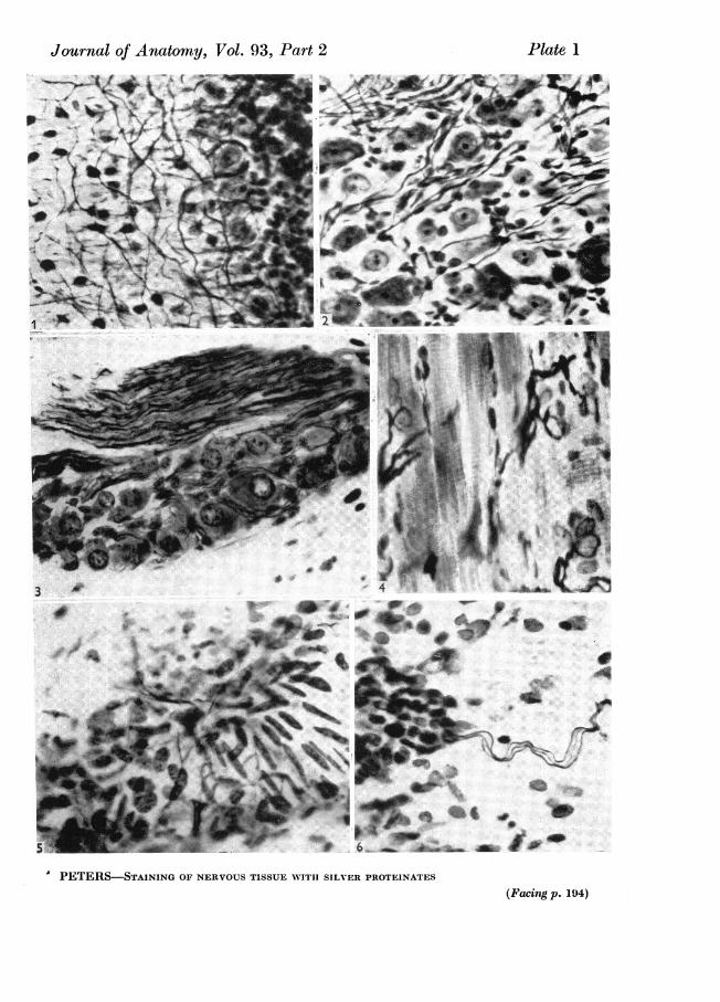
factors such as pH, temperature of impregnation and the amount of metallic copperin the solution were varied and correlated with the type of staining produced. Pro-targol-S and pre-1939 Bayer Protargol were used in the initial experiments andlater other types of silver proteinates and protein-silver nitrate mixtures wereemployed to determine which properties of silver proteinates effected the productionof a specific nerve stain.The criteria used in deciding whether a given section was well stained were:(a) The contrast, by either colour or intensity, between nerves and other tissues
(PI. 1, figs. 4-6).(b) The absence of connective tissue staining, particularly reticulin (PI. 1).(c) The extent which peripheral, including autonomic nerves, were stained (PI. 1,
figs. 4-6).(d) The extent to which the central nervous system and ganglia were stained
(PI. 1, figs. 1-3).
MATERIALS AND METHODS
The standard tissues used in these experiments were rat and mouse heads, takenfrom animals fixed by intra-arterial injection. 10% formol was used in the initialexperiments, but since formol was not the ideal or most suitable fixative for alltissues, the results of these experiments were confirmed with heads fixed withBouin's fixative and alcohol-formol-acetic (50% alcohol 90 ml.: formol 5 ml.: glacialacetic acid 5 ml.). The heads were decalcified by Bensley's decalcifier (equal volumesof 50% formic acid and 20% sodium acetate) for 72 hr., after which they were wellwashed in running tap water. Sections of rat brain and pieces of muscle which didnot require decalcification were also used. It was appreciated that the method ofdehydration and embedding could alter the stain, but since the mechanism of thestaining process was under consideration and not the study ofan individual structure,it was felt necessary to standardize the method for the preparation of sections, sothat the tissues were dehydrated in a graded series of ethyl alcohol, cleared inbenzene and embedded in 560 C. paraffin wax. It was found that although changesin procedure altered the extent to which various nervous elements were stained,they did not affect the overall staining of one section in relation to another sectionfrom the same block of tissue.
Bodian's method of impregnation (1936) was used as a standard technique andexcept in one experiment, where a physical developer was used to replace thehydroquinone-sodium sulphite developer, steps 2 to 6 in the following scheme werekept constant, although numerous variations were introduced during impregnation(step 1). The standard technique was as follows:
(1) After dewaxing and taking through graded alcohols to water, sections wereimpregnated for 16-24 hr. at 370 C. in a 1% solution of silver proteinate to whichapproximately 4 g. of clean copper wire (1.6 mm. in diameter) had been added per100 ml. of solution.
(2) Sections were rinsed in distilled water and developed for 5 min. in a solu-tion containing: hydroquinone, 1 g.; sodium sulphite (anhydrous), 10 g.; distilledwater, 100 ml.; this developer contained double the quantity of sodium sulphiteused by Bodian (Peters, 1955 b).
Alan Peters178
Staining of nervous tissue with silver proteinates 179(3) The developer was removed by washing the sections in running tap water for
10 mi.(4) Sections were taken through distilled water and immersed in 0-2% gold
chloride, acidified with 1 drop of glacial acetic acid per 100 ml. of solution, for10 min.
(5) The excess gold was removed by rinsing in distilled water and the sectionstransferred to 2% oxalic acid for 2 min. They were then washed in running tapwater for 5 min. and the residual silver removed by immersing in 5% sodium thio-sulphate for 10 min.
(6) Finally, sections were washed in running tap water for 10 min., rinsed indistilled water, taken through graded alcohols to xylene and mounted in CanadaBalsam.The hydrogen ion concentrations in the impregnating solutions were measured
with a Pye Universal pH meter, and the same instrument was employed as a milli-volt meter to measure the potential of the concentration cell used in determining thesilver ion concentration in the proteinate solutions (0.IM silver nitrate was used inthe standard half cell and the activity coefficient of this solution was taken to be0.72).The silver deposited on to the copper wire during impregnation was determined
as silver chloride, the silver being dissolved from the surface of the wire with 4%nitric acid. The protein was precipitated from the resulting solution with phospho-tungstic acid and the silver then precipitated with sodium chloride. The silverchloride was filtered off after the solution had stood for 12 hr. and was then dried andweighed.
Copper from the wire went into solution during impregnation and after removalof protein from solution by phosphotungstic acid, this copper was determined byadding sodium diethyldithiocarbamate to the protein-free filtrate. The resultinggolden-brown colour was checked visually against standard copper sulphate solu-tions. This method was not considered to be very accurate.
PROPERTIES OF SILVER PROTEINATES
The silver proteinates listed in Table 1 were used in the experiments. Of theseeleven compounds, pre-1939 Bayer Protargol, Winthrop batches N-051 and N346BJ,Heyden batch 8214, Bayer Albargin and the American Pharmaceutical Companyproteinate were relatively old samples that had been kept in the laboratory for sometime. The other proteinates were obtained recently.
According to the classification of Martindale (1952) and Osol & Farrer (1955)these proteinates were classified as 'strong' (containing 7 5-S 5% silver) and 'mild'proteinates 19-0-23% silver). While most of the proteinates fitted into this classifica-tion, Albargin (15 % silver) fell between the two groups. 1 % solutions of the strongproteinates showed a greater degree of ionization than those of mild proteinates,and determinations with a silver electrode (Table 1) showed that 1% solutions ofstrong proteinates had silver concentrations of the order of 10-4M, while for mildproteinates the value was between 10-6 and 1o-8M. The one exception was the pro-teinate manufactured by the American Pharmaceutical Company, for this sample,
180 Alan Peterswhich was old and dark in colour like the mild proteinates, gave a value of 6-7 x1O-7M. When the concentration of the ionized silver was measured in solutions ofBayer Protargol, Protargol-S and the Heyden proteinate of different strength, itwas found that the silver ion concentration was not proportional to the concentrationof proteinate in solution. Ionization was suppressed as the concentration of proteinateincreased, so that these compounds were behaving like weakly ionized salts, aswould be expected of metallo-organic compounds.As seen in Table 1, most of the strong proteinates were light or medium brown
in colour, while the mild proteinates were in the form of shiny black granules. Thisprobably gave some indication of the colloidal particle sizes of the compounds, so
Type content (%) DescriptionStrong About-8* Light brown
powderLight brown
Strong 7 5-8-5t powderStrong 7 *5-8$5 Light brown
powderStrong 7*5-8*51 Light brown
powderStrong 7.5-85§ Medium brown
powderStrong About 811 Medium brown
powderStrong 7-5-8-5 Medium brown
powderStrong 7-5-8.5§ Black tablets
- 1511 Shiny blackgranules
Mild About 2011 Shiny blackgranules
Mild 19-23§ Shiny blackgranules
Properties of 1% solu-tion (mean values)
Ionizedsilver concn.
pH (M)
8-0 7-5 x 10⁻⁴
8-3 7-1x10⁻⁸-1 9-6x 10⁻⁴
8*1 9.0x10⁻⁴
9.1 1-4 x 10-'
9*1 14x10-4
7-6 32x 10-4
8-7 6-7 x 10⁻⁷
7*6
9.3
10.0
1*6 x 10-8
3-3 x 10⁻⁶
7 9x 10⁻⁸
References to silver contents* Holmes (1943), Romanes (1950).$ Osol & Farrer (1955).11 Information supplied by the manufacturer.
t Information supplied by Bayer Products Ltd.§ Martindale (1952).
that as pointed out by Romanes (1950), the lighter coloured compounds would havea smaller particle size than the other compounds. The small size of the colloidalparticles in Protargol was emphasized by Neegaard (1928), who gave their size as138,s/ and found that one-third of the silver was in the colloidal state and two-thirdsin true solution in a 065% solution of Protargol.
Little information was available about the chemical composition of silver pro-teinates. Neegaard (1923) recorded that the original Protargol was an albumose-silver compound and this was supported by Romanes (1950), who stated that it wasa combination of silver with partially hydrolysed egg albumen. Of the other pro-teinates, Davenport, Porter & Myhre (1952) reported that pharmaceutical peptonetogether with gelatin had been used by Winthrop-Stearns in the preparation of
Staining of nervous tissue with silver proteinatesProtargol, and gelatin was also used in the manufacture of Albargin, which waslabelled as a gelatose-silver compound. Thus, the different preparations varied con-siderably in their compositions. Even different batches of the same product variedand the fact that only certain batches of Protargol were suitable for staining nerveswas well known.Mean values for the pH value of 1 % solutions of different proteinates were as
given in Table 1, although individual solutions varied by as much as + 0*2pH units.Furthermore, proteinate solutions were not stable, and at room temperature boththe pH values and silver ion concentrations in the solutions changed slowly with time.These changes were accelerated by heat, so that at 370 C. the pH value of a Pro-targol solution fell from 8-3 to 7-6 in 24 hr.
8
807 23
70
6
0~~~~~~~~~~~~~56
0:
Text-fig. 1. The silver ion concentration in 1 % solutions of proteinates at different pH values. ThepH values were attained by the addition of 1 M nitric acid and for the sake of clarity experi-mental points have been omitted from the graph. The numbers on the curves correspond tothe following compounds. (1) Protargol-S; (2) Boots Argentoproteinium B.P. (strong);(8) Bayer Protargol; (4) British Drug Houses Strong Protein (8 % Ag); (5) Winthropbatch N846BJ; (6) Winthrop batch N-051; (7) Heyden Chemical Corporation compound;(8) British Drug Houses Mild Protein (20% Ag); (9) Bayer Albargin; (10) Boots Argentopro-teinium B.P.C. (Mild) and American Pharmaceutical Co. Proteinate. The scale on the rightgives the weight of ionized silver in mg./g. of proteinate and shows that Protargol, Protargol-Sand Boots strong proteinate are almost completely ionized at pH 3.
Holmes (1948) recorded that there was a marked decrease in the pH value of acopper-containing Protargol solution during impregnation. Clearly, such a changein the pHvalue may affect the degree of ionization of the proteinate and to determinewhether or not this was the case, 1M nitric acid was added drop by drop to 1%proteinate solutions, while the pH values and silver ion concentrations in the
181
182 Alan Peterssolutions were measured. In all cases (Text-fig. 1) a decrease in the pH value wasaccompanied by an increase in the ionic silver concentration. A maximum valuewas reached at pH 2-5-85 (in the region of the isoelectric points of the proteins) andbeyond this the silver ion concentration decreased. The addition of acid also produceda heavy precipitate on the alkaline side of the maximum silver concentration, butthis began to dissolve again once the maximum was passed. In solutions of Pro-targol, Protargol-S and Boots strong Argentoproteinium, practically all of the silverwas in the ionic state atpH 3 and, with the exception of the American PharmaceuticalCompany compound, the strong silver proteinates were more highly ionized, at allpH values, than the mild proteinates. The importance of this observation becameapparent when the staining properties of the compounds were considered.
bO
C 8028-
U 0.0a
24 - C
T 0 10 6030 407
16-~~~onn ofcperi olto x1
UC~~~~~~~~~~~~0~~~~~~~~~~~~~~~~~~~~~~~0
7.0
Text-fig. 2. The addition of 0 1 M cupric nitrate to 50 ml. portions of 1 % Bayer Protargol and 1 %Boots Argentoproteinium H.P. (strong), showing the changes in silver ion concentration (fulllines) and pH value (broken lines). *-*, Bayer Protargol silverion concentration; *---*,Bayer Protargol pH value; x -x, Boots Argentoproteinium silver ion concentration;x--x, Boots Argentoproteinium pH value. The range of copper concentrations, determinedat the end of impregnation, in solutions of Bayer Protargoboontaining4 g. of copperper 100 ml.Of solution is given (see Table 2).
Since Bodian (1936) found that copper went into solution during impregnation,the effect of adding o01 mcupric nitrate to 1% solutions of proteinates was de-termined. In all cases this led to a decrease in the pH value, together with anincrease in the silver ion concentration; the changes which occurred in 1 solutionsof Protargol and Boots Argentoproteinium are shown in Text-fig. 2. No doubt theaddition of cupric nitrate resulted in a replacement, by copper, of some of the boundsilver of the proteinate, so that the silver ion concentration increased. A combinationbetween copper and th acidic groups of the protein could account for the decreasein the pH value.
Staining of nervous tissue with silver proteinates 183
IMPREGNATION IN PROTARGOL AND PROTARGOL-S
In the following experiments, impregnating solutions were removed from the incu-bator at intervals during impregnation and the pH values and hydrogen ion con-centrations measured.
(a) The role of copper and the effect of temperature andproteinate concentration on staining
Both Protargol and Protargol-S gave good staining when sections were im-pregnated at 370 C. by the modified Bodian technique, although Protargol-S gavethe better overall staining of nervous elements and especially of the autonomic
Table 2. Impregnation in 1% solutions of Bayer Protargol, showing the changes inpH value, silver concentration and copper concentration
of Dura- Wt. of released (b) Conen. of Ag+ releasedVol. im- tion of copper by pH (a) Molarity as copper in by p1 change
Expt. of soln. pregn. impregn. pH wire change (mg./g. solution solution Ag/Cu Ag on wireno. (ml.) (0C.) (hr.) change (g.) (as) wire) (X) (X) (8(b)/9) (7/8(b))8 100 18 15-75 7.9- 7X3 4-1 1P5 x 10-8 6-8 2-6 x 10-8 O*96 x 10-8 2*7 0-582 50 37 16 8-1-* 6-1 1-9 2-8 x 10⁻⁸ 10-0 3.5 x 10⁻⁸ 1*2 x 10-8 2-9 0.808 100 37 15-75 7.9- 6-3 4.4 2-8 x 10⁻³ 11-0 3*8 x 10-3 1-0 x 10-3 3-8 0 74
28 50 37 47 8-1-- 6-3 2.4 2-9 x 10⁻⁸ 9*8 4.3 x 10⁻⁸ 2-3 x 10⁻³ 1 9 0-694 50 56 50 7.9- 6-0 2.1 2.9 x 10⁻⁸ 11-0 4.2 x 10-' 1.9 x 10⁻³ 2-2 0-69
21 100 56 24 8.2-+ 6.2 4-7 2-9 x 10⁻⁸ 9-1 40 x 10-8 1-5 x 10⁻⁸ 2-7 0-7228 50 56 47 8-1--*5.9 2-4 3{0 x 10-8 100 4.3 x 10⁻⁸ 1.9 x 10⁻⁸ 2-2 0*7026 100 37 11-5 8-1-+ 6.6 12-0 2*1 x 10-8 6-8 4-1 x 10⁻⁸ 2-7 x 10⁻⁸ 1-5 0*5126 100 37 11-5 8-1-+ 6-75 8-0 1-8 x 10⁻⁸ 9-2 3.7 x 10⁻⁸ 1-4 x 10⁻⁸ 2-6 0-4826 100 37 11-5 8-1-4 7-2 4-0 1-4 x 10⁻³ 11-4 2.2 x 10⁻⁸ 0-45 x 10-3 2-3 0-6426 100 37 11-5 8*1-).7*7 0 0-8 x 10- - - - -
* The value for the concentration of ionic silver released by the pH change was determined on the basis of Text-fig. 1.
system. During impregnation by the standard method with Protargol, the pHvalue and silver ion concentration in the impregnation solution both decreased withtime as shown in Text-fig. 3. In this particular case two slides of sections were inthe impregnating solution, but it was shown subsequently that the presence orabsence of sections had no effect on the changes in the solution. The initial fall inthe pH value and silver ion concentration was rapid, but the rate of fall of thesedecreased with time; similar results were obtained with Protargol-S . Visible changeswere also observed during impregnation, in that silver plated-out on to the copperwire, which simultaneously became coated with a protein precipitate, while at thesame time the brown colour of the impregnating solution assumed a green tint dueto copper going into solution from the copper wire. About 10 mg. of silver/g. ofcopper wire were plated-out during impregnation and the final concentration ofcopper in the solution was about 1-2 x 10⁻³M; some values are given in Table 2.The effects of temperature of impregnation, proteinate concentration and the
amount of copper wire in the solution were as shown in Table 3, which gives examplesof the types of results obtained in the experiments. In recording the values of pHand silver ion concentration during impregnation, it was found that the rate of fallof these values was accelerated when either the amount of copper added to the
184 Alan Peterssolution or the temperature of impregnation was increased. Thus, the concentrationof ionic silver and the pH value of the solution were less, at any given time, in asolution containing 12 g. of copper wire per 100 ml. than in a solution containingonly 4 g. of copper. Such changes were also related to the type of staining obtained,for an increase in either the amount of copper or the temperature of impregnationled to a decrease in the overall intensity of staining together with an increasedcontrast between nerve fibres and other tissues.The correlation between final staining intensity and the concentration of silver
ions in the solution was also confirmed by experiments in which the proteinateconcentration was varied (Table 3), while the amount of copper in the solution andthe temperature of impregnation were kept constant.
Periodofim-
pregn.(hr.)
Temp.Strength ofofsolu- im-tion pregn.(%) (° C.)
24 111
183756
24 1 3705 370-25 37
Ili 1111
37373737
Table 3. The effect of temperature, proteinate concentrationand copper on impregnation
(a) Effect of temperature (Bayer Protargol)4 g./100 ml. 8-1 7-55 7 x 10⁻⁴ 5 x 1O-4 Increase in contrast between nerve4 g./100 ml. 8-1 6-85 7 x 10⁻⁴ 08 x 10-' fibres and othertissues as tempera-4 g./100 ml. 8-1 6-40 7 x 10⁻⁴ 0-4 x 10-' ture was raised. Little contrast
180 C. Non-nervous tissues onlyfaintly visible at 560C. Order ofintensity of general staining18 > 37 > 560 C. Best result at370 C.
(b) Effect of proteinate concentration (Protargol-S)4 g./100 ml. 8-2 6-80 7 x 10⁻⁴ 1 X 10⁻⁴ Decrease in intensity of staining of4 g./100 ml. 8-2 6-95 3.5 x 10⁻⁴ 0-9 X 10⁻⁴ all tissues with decrease in con-4 g./100 ml. 8*2 7*05 2*5 x 10-4 0-2 x 10⁻⁴ centration of proteinate. In
0-25% solution, staining verylight
(c) Effect of amount of copper (Bayer Protargol) (see Table 2)0 g./100 ml. 8-1 7 70 8-5 x 10⁻⁴ 4*0 x 10-4 Increase in contrast between nerv s4 g./100 ml. 8-1 7-20 8-5 x 10⁻⁴ 2-0 x 10-4 and other tissues as amount of S8 g./100 ml. 8-1 6-75 8*5 x 10-4 0-8 x 10⁻⁴ copper in solution increased. With12 g./100 ml. 8-1 6-60 8-5 x 10⁻⁴ 0.3 x 10⁻⁴ no copper nerves difficult to -M
distinguish against equallyintense background. Best resultwith 4 g. of copper. With 12 g!of copper, nerve staining patch,and pale with muscle almost gre nin colour
The initial pH value of the impregnating solution also played an important partin determining the type of staining produced. When the initial pH values of 1%solutions of Protargol and Protargol-S, containing 4 g. of copper per 100 ml. ofsolution, were changed by adding boric acid-borax buffers and the sections impreg-nated at 370 C.; the best results were obtained at pH 8-3. At pH 8-7 there was littlecontrast between nerves and other tissues and at pH 7-6 and 7-8 the staining wascoarse and more connective tissue was visible than at higher pH values. As wouldbe expected, during impregnation in a buffered solution there was little change inthe pH value, but when the pH value was adjusted with ammonia there was a
Staining of nervous tissue with silver proteinates 185decrease, so that, for example, when the initial pH value was 9 3 it fell to 7-6 during16 hr. Since the nerve staining in the latter solution was better than that in a solu-tion buffered to pH 9 3, it appeared that the fall in pH during impregnation playedan important role in the impregnation process.The function of the metallic copper in the impregnating solution could not be
simulated by adding a copper salt to the solution. Sections impregnated in a 1 %solution of Protargol containing approximately the same concentration of copper(1 X 10⁻³M cupric nitrate) as that found in a solution at the end of impregnationwhen metallic copper was present (Table 2), showed a similar type of staining to acopper-free solution. This suggested that copper ions themselves played little partin determining the type of staining produced by a solution, and the main functionof the metallic copper appeared to be the removal of silver ions from the impreg-nating solutions. This point will be referred to later.
(b) The effect oftime on stainingPeters (1955a) showed that during impregnation two different reactions took
place between the sections and silver ions. Some of the silver combined with thesections and remained in the unreduced state, reducible silver, while a smallerfraction was reduced to form silver nuclei, which during development in hydro-quinone-sulphite acted as centres for the reduction and deposition of the reduciblesilver. The formation of silver nuclei appeared to be irreversible and the relativeamount of silver in the form of nuclei was determined by immersing sections insodium sulphite, which removed the reducible silver, and then developing in a glycinphysical developer for a constant period oftime. Under these conditions the intensityof the stain was dependent on the concentration of silver nuclei in the sections(Peters, 1955a, c).A comparison of the amount of reducible silver plus silver nuclei in different
sections was indicated by the intensity of the stain obtained after development inthe hydroquinone-sodium sulphite. Thus, since the formation of silver nuclei wasirreversible, when sections were put into an impregnating solution simultaneously,removed after different lengths of time and developed in hydroquinone-sodiumsulphite, any decrease in the intensity of staining associated with a longer impregna-tion was due to a decrease in the reducible silver fraction in the section.When sections were impregnated at either 37 or 560 C. in copper-containing
Protargol-S solutions, and removed at intervals over a period of 24 hr., hydro-quinone-sodium sulphite development showed that after the first hour of im-pregnation, the amount of reducible silver in combination with the section decreasedwith time. Conversely, there was an increase in the amount of silver in the formof nuclei.
In a further experiment, sections of rat head were impregnated at 560 C. insolutions of 1% Protargol containing 4 g. of copper per 100 ml. of solution. Oneset of sections was impregnated in sequence, i.e. only one section was in the solutionat any given time and was removed from the solution and replaced by another, atintervals over 24 hr. A second set of sections, which acted as controls, was put intothe solution at the beginning of impregnation and one section was removed at thesame time as each section of the first set. The results of this experiment were shown
186 Alan Petersin Text-fig 8. Sections were removed from the solution at times shown by eachvertical arrow in Text-fig. 3, and the results obtained by comparing the changingnature of the stain in the sections impregnated in sequence are shown between thevertical arrows. The horizontal arrows refer to the control sections and indicatetrends which were visible in these sections impregnated from zero time. This ex-periment showed that after the first hour of incubation there was a decrease in theoverall staining intensity and since reducible silver was removed most readily fromthe background elements, the contrast between the nerve fibres and other tissuesimproved as impregnation continued, so that the stain was self differentiating. Adecrease in nuclear staining occurred after the 6th hour of incubation and there wasalmost no further axon staining in the second half of the period of impregnation,when the only new nervous elements to stain were the Purkinje dendrites. Thefollowing examples illustrate these points.
0 hr. 2 hr. 31 hr. 6 hr. 8 hr. 11 hr. 24 hr.Overall dark Lighter overall Lighter Lighter overall stain.stain. N ervestain. overall Very faint axon staining. No nuclear staining.staining Selectivity for stain. Only cell bodies and Purkinje cell dendritesunselective nerves Peripheral visible in the C.N.S.imrvd ,nerve
~ stainingfainter +Improvement in contrast between nerves and other tissues
Decrease in overall staining intensityDecrease in nuclear staining _
Text-fig. 3. The effect of time on staining formol fixed sections of rat head impregnated at 560 C.in 100 ml. of 1 % Bayer Pretargol containing 4 g. of copper wire. The graph shows the changesin silver ion concentration (full line) and pH value (broken line). Above the graph, thehorizontal arrows indicate continuous trends in the staining of sections impregnated from zerotime, while the vertical lines indicate the removal of sections from the impregnating solution.The intervals between the vertical arrows indicate the times of impregnation of individualsections stained in sequence, and the type of staining produced during these times is indicatedbetween the lines. See text for full explanation.
(1) A section put in at zero time and incubated for 24 hr. showed a selective stainfor axons, cell nuclei and Purkinje cell dendrites. In a section impregnated from the11th to the 24th hour, the only nervous elements to be well stained were thePurkinje cell dendrites; the overall staining was light and there was almost nostaining of axons and cell nuclei. Thus, the only feature shared by these two sectionswas the staining of Purkinje cell dendrites, and as can be seen from Text-fig. 3 theseonly stained in sections removed from the solution after the 11th hour.
Staining of nervous tissue with silver proteinates 187(2) A section put in at 8 hr. and removed at 11 hr. was much lighter in overall
staining intensity, including axons and cell nuclei, which were only faintly visible,than a section put in at 31 hr. and removed at 6 hr.Under the same conditions, Protargol-S showed a slightly different sequence of
staining, for satisfactory nerve staining persisted over the 24 hr., although theintensity decreased and there was some staining of neuro-keratin in the later stages.Purkinje cell dendrites were never stained well in Protargol-S solutions (P1. 1, fig. 1).The correlation between the trends in staining and the changes in pH value and
silver ion concentration during impregnation will be discussed later.
IMPREGNATION IN OTHER SILVER PROTEINATE SOLUTIONS
As a basic test of their staining potentials, solutions of the other silver proteinates,listed in Table 1, were used to replace Protargol in the modified Bodian technique.In addition to the initial pH values, temperatures of impregnation and the con-centrations of the solutions were varied. The only compounds which producedstaining like that obtained with Protargol and Protargol-S were the Winthropcompounds batches N-051 and N346BJ, but these gave a somewhat lighter andless specific stain. In the case of the Boots and British Drug Houses strong silverproteinates the stain was granular and showed little specificity for nerves, even when
10
T
CE X~~~~~~~~~~~~~~~~~~~~I4 3x 3 7.5
o+~~~ ~ ~ ~ ~~~-7.7< ~ ~ -2 3 48.0
0 4 1 6 420.24.0..121620924
0 4 8 12 16 20 24 0 4 8 12 16 20 24
souton>fpoentscnann .o oprpr10m.o ouina 7 .Tegah
126 10 7/X.8
V ~~ ~ ~ ~ /7.400 X
< 2 x5 x680
9.00 4 812 162024 0 4 812 162024
Time (hr.)Text-fig. 4. A comparison of silver ion concentration and pH value changes, with time, in 1 %
solutions of proteinates containing 4 g. of copper per 100 ml. of solution at 370 C. The graphson the left show the changes in silver ion concentration and those on the right the correspondingchanges in pH value. The number on the curves refer to the following proteinates: (1) WinthropBatch N346BJ; (2) Bayer Protargol; (3) Heyden Chemical Corporation strong proteinate;(4) Boots Argentoproteinium B.P. (strong); (5) Bayer Albargin; and (6) American Pharma-ceutical Co. strong proteinate.
188 Alan Petersthe initial pH value of the solutions was adjusted to pH 8-1-8-3 with acid. Both ofthe mild proteinates, together with Albargin, produced a light stain which was notspecific for nerves. The obvious reason for this light stain was the low concentrationof ionic silver present in the solutions (Table 1).
Interesting results were obtained with the Heyden strong silver proteinate.Although a normal solution of this gave a light stain which showed little specificityfor nerves, the type of staining was completely transformed when the initial pHvalue of the solution was changed from 7*6 to 8-2-8-4 by the addition of ammonia,for the results were then as good as those produced by Protargol (P1. 1, fig. 2). Fromthis it appeared that the initial pH value of the normal solution was too low, for thestaining produced by such a solution was similar to that obtained if the pH valueof a Protargol solution was changed to pH 7-6 by the addition of acid.The changes in pH value and silver ion concentration that took place when sections
were impregnated in 1% copper-containing solutions of different proteinates werecompared directly (Text-fig. 4). It was clear that in all solutions similar changes inpH value and silver ion concentration took place during impregnation. The impor-tance of the initial pH value of the solution was emphasized by the experimentswith Heyden compound, but since no such great change in the specificity of thestain was obtained when the initial pH values of the Boots and British Drug Housesstrong proteinates were adjusted to pH 8*3, it seemed likely that the protein usedin the preparation of the proteinate also played an important part in determiningthe staining properties of the preparations. This point was investigated by the useof various protein-silver nitrate mixtures.
IMPREGNATION IN PROTEIN-SILVER NITRATE MIXTURES
Solutions of 2, 1 and 05% of dried egg albumen, blood albumen, casein, gelatin andbacteriological peptone (all obtained from British Drug Houses Ltd.); 2% freshcitrated human blood plasma and 3*5% fresh egg albumen were used as the proteinfraction in these experiments. Enough 2% silver nitrate was added to the proteinsolution to give a silver ion concentration of 2 0-5O x 10-4M when the pH valueof the solution was adjusted to pH 8-2-8-5 by the addition of ammonia, sodiumhydroxide or 0 1 M boric acid-borax buffer. The volume of silver nitrate necessaryto produce this silver ion concentration varied with the protein. Sections wereimpregnated for 16 hr. at 370 C. in the absence of copper, developed in thehydroquinone-sodium sulphite developer and gold toned.The solutions containing either gelatin, blood albumen, fresh citrated blood
plasma or bacteriological peptone produced some nerve staining, but the resultswere poor and relatively unspecific for nerves. On the other hand, dried eggalbumen-silver nitrate mixtures gave an excellent stain which was specific for nerves(P1. 1, fig. 3). Almost no connective tissue was visible and the results were as goodas those produced by Protargol (P1. 1, fig. 4) and Protargol-S (P1. 1, figs. 1, 5 and 6).Fresh egg albumen gave inconsistent results, although the staining was good in somecases. Casein-silver nitrate mixtures also stained nerve selectively, but the resultsdiffered from those produced by the dried egg albumen mixtures in that the stainwas rather coarse and somewhat uneven, particularly in muscle. The addition of
Staining of nervous tissue with silver proteinates 189metallic copper to either the dried egg albumen or casein mixtures had little effecton the result beyond promoting some connective tissue staining. It was clear fromthese experiments that the protein fraction of the proteinate did play a part indetermining the staining properties of the solution.
Table 4. Impregnation in egg albumen-silver nitrate mixtures of differentcompositions and at different temperatures
Composition of impregnating solution: 50 ml. of 0*5% dried egg albumen, x ml. of 2 % silvernitrate; pH value adjusted with ammonia
Control
Solution 1
(1) Pro(a) Silver nitrate added (ml.) 1.8(b) Initial pH value 8-3(c) Initial silver ion concentra-
tion(xlO-4M) 2-3(d) Temperature of impregna-
tion (' C.) 37(e) Final silver ion
concentration ( x 10-9M) 0-6(f) Final pH value 7.7
(a) Staining intensityOverallCNSConnective tissue
(b) Selectivity of nerve stainingIn muscleIn CNSIn subepithelial tissue
2 3 4perties of impregnating solution
5 6 7
1-8 1-8 1.8 1-8 1-4 2-27.7 9-0 8-3 8-3 8-3 8-3
7-7 0-4 2-3 2-3 1-2 6-3
37 37 18 56 37 37
0-4 0-16-7 8-3
(2) Staining results
+++ +++++ +++ +++
000000000
00000
+
000000
1-2 0-3 0-2 0-38-1 7-3 7.7 7-8
++
+++
00000
+++++
+
000 0000 0000 0
++
0000000
Key to symbols. Staining intensity: + faint; through + + andSelectivity of nerve staining: 0 very poor; 00 poor; 000 good.
+++,to ++++ intense.
7-6
7 8-3
0 2 4 6 8 10 12 14 16 18 20 22 24Time (hr.)
Text-fig. 5. The change in the pH value (broken line) and silver ion concentration (full line) duringimpregnation in a solution containing 50 ml. of 0.5% dried egg albumen and 1-8 ml. of 2%silver nitrate. The initial pH value was adjusted to pH 8-3 by the addition of ammonia. Notethe similarity between the shapes of these curves and those obtained with silver proteinates.
The best results were obtained with dried egg albumen-silver nitrate mixtureswhen sections were impregnated at 370 C. in a solution consisting of 50 ml. of 0-5 %/dried egg albumen solution with 1-8 ml. of 2%°/ silver nitrate, the pH value of whichwas adjusted to pH 883 with ammonia (P1. 1, fig. 8). A typical example of the
190 Alan Peterschanges occurring in the pH value and silver ion concentration during the courseof impregnation in such a solution is shown in Text-fig. 5, and it will be seen thatthese changes were very similar to those which took place in copper-containingProtargol solutions (Text-fig. 3). Full details of the staining procedure employingdried egg albumen-silver nitrate solutions have been given elsewhere (Peters, 1958).The egg albumen-silver nitrate mixtures proved to be very useful experimentally,
since the different factors involved in impregnation could be easily studied byaltering the composition of the mixtures. The composition, properties and thestaining results obtained in such experimental solutions are given in Table 4, inwhich it will be seen that:
(1) In impregnating solutions at the same temperature and initial pH value(solutions 1, 6 and 7), the intensity of staining depended upon the silver ionconcentration.
(2) The intensity of the stain and the specificity for nerves were influenced bythe pH value of the impregnating solution (solutions 1-3), the best result beingobtained at an initial pH value of 8-3 (solution 1).
(3) Good results were obtained when sections were impregnated at pH 8&3 ateither 370 C. (solution 1), or 560 C. (solution 5). At 180 C. (solution 4) the resultswere patchy and coarse.
Similar results were obtained with casein-silver nitrate mixtures.
DISCUSSION
From these experiments it was possible to form a picture of some of the chemicaland physical processes that took place during normal impregnation with Protargoland Protargol-S. Due to the complexity of the system it was obviously necessaryto simplify the reactions, but nevertheless the following formed an adequate workinghypothesis. As impregnation proceeded, silver was lost from the impregnatingsolution (Text-figs. 3, 4) and this appeared to be effected mainly by a plating-outof silver on to the copper wire. Silver was also taken from the solution by the section,but that this was insignificant was shown in previous experiments (Peters, 1955c),where a 151% section 1 cm.2 contained only about 2 x 10⁻⁵ g. of silver compared tothe amount of silver plated out on to the copper wire (approximately 1 x 10⁻² g./g.of copper wire).On the basis of their chemical equivalents, for every two atoms of silver plated
out one copper ion would enter solution. Some values for the concentration ofcopper going into solution, together with the corresponding concentration of silverplated-out, were given in Table 2, but it was found that in almost every case thevalue of the ratio Ag/Cu was more than 2; the mean value being about 2-5. Never-theless, the results were in some agreement with the theoretical considerations andsome part of the discrepancies were attributable to (a) the protein which precipitatedon to the surface of the copper during impregnation; this could not be removed, sothat it was included in the figures for the amount of silver plated-out; and (b) themethod for determining the copper in solution was not considered to be very accurate,while copper bound with the proteinate could not be taken into account.The loss of ionic silver was accompanied by a decrease in the pH value of the
Staining of nervous tissue with silver proteinates 191solution and these changes appeared to be related, for both were accelerated byeither heat or an increase in the amount of copper wire added to the solution (Table3). Since the addition of a copper salt to a Protargol solution also decreased thepH value of that solution, it was concluded that the decrease in the pH value of acopper-containing solution was the resultant, primarily, of the decrease resultingfrom copper going into solution (Text-fig. 1), and secondarily, of the decrease dueto the instability of the Protargol solution at the temperature of impregnation.On the basis of Text-fig. 1 a calculation was made of the amount of ionic silver
that would be released from the Protargol as a result of the decrease in pH valueduring impregnation. The results (column 7, Table 2) showed that this accountedfor only 50-80% of the silver plated-out on to the copper (column 11, Table 2).Thus, it appeared that there was no simple correlation between the release of ionicsilver and the fall in pH value. It was impossible to take into account the dis-placement of silver from the proteinate by copper ions, and in the impregnatingsystem the proteinate would probably release more than the calculated amountof silver to counteract the continual withdrawal of silver by the plating-out process.
These considerations, together with the results of staining sections in differentsolutions, suggested the following theory of chemical reactions taking place duringimpregnation. In the impregnating solution there was an equilibrium system be-tween the solution, the reducible silver in the section and the colloidal fraction of theproteinate. Silver ions were removed from this system by (a) plating-out on to thecopper, and (b) the formation of silver nuclei in the section. It was found that theamount of silver removed by both sites increased as impregnation proceeded. Tocounteract this loss, the system tended towards equilibrium again by the with-drawal of silver ions both from the proteinate, which may be regarded as thereservoir for silver, and the reducible silver fraction of the section. The withdrawalfrom the reducible silver fraction accounted for the observed decrease in the overallstaining intensity of sections as impregnation continued (Text-fig. 3), and this lossof silver appeared to take place most readily from the non-nervous elements in thesection, as suggested by Glassner, Breslau & Agress (1954) to account for theimprovement in contrast between nervous and non-nervous elements when sectionswere impregnated for long periods of time (Text-fig. 3). Such a theory would alsoexplain the decrease in overall staining intensity, together with the improvementin differentiation at 560 C. as opposed to 370 C., for as the temperature of impregna-tion was increased the loss of ionic silver from the impregnating solution wasaccelerated (Table 3).
Clearly, the metallic copper took part in the impregnation process by removingsilver ions from the solution. Its presence led to an improvement in the differentia-tion of the stain, but whether the copper ions released in exchange for the silverplayed any part in staining could not be determined, although it was found thatin the absence of the metal, the presence of copper ions had no effect on the result,beyond tending to make the connective tissue stain. Copper ions would certainlyenter into competition with silver ions for the binding sites in the proteinate and asBodian (1936) showed, during impregnation, copper was deposited, together withsilver, in the nerve fibres.From the experiments, it was shown that the pH value, the silver ion concentra-13 Anat. 93
192 Alan Peterstion in the impregnating solution and the protein used in the formation of theproteinate were all important factors in determining the staining properties of acompound. Thus, only those solutions of proteinates and protein silver nitratemixtures which initially contained more than about 1-3 x 10⁻⁴M ionic silver gave asufficiently intense stain. The best results were produced when the initial pH valueof the system was between 8-0 and 8-4, and the importance of this was demonstratedby the Heyden proteinate which gave excellent nerve staining when the initial pHvalue was raised from pH 7*6 to 8*3. Further, the changes in pH value that occurredduring impregnation also appeared to play an essential part in staining. Duringimpregnation by Bodian's method, sections were subjected to a continually fallingrange of pH values, and the type of staining obtained at different phases of theprocess was shown in the experiments where sections were stained in sequence(Text-fig. 3). Optimum conditions for nerves obtained at an early phase of impreg-nation and they were only lightly stained during the final stages. Changing theinitial pH value of a solution showed that connective tissue stained most readilyat low pH values (see Table 4), but in normal impregnation in Protargol, such valueswere only attained at a late stage when the silver ion concentration was also at alow level. Consequently, connective tissue was only lightly stained, if at all, by theBodian and egg albumen-silver nitrate methods.With the exception of the Heyden proteinate, other proteinates which had a
sufficiently high silver ion concentration, i.e. the Boots and British Drug Housesstrong Proteinates, still did not produce a good nerve stain even when the initial pHvalue of the solution was changed to pH 8*3. It seemed that this lack of nervestaining could be attributed to the protein fraction in these proteinates, for it wasshown by the use of protein-silver nitrate mixtures that the protein played a definitepart in determining the type of staining produced. Only the mixtures containingdried egg albumen or casein gave good results, and this confirmed the results ofRomanes (1950), who obtained somewhat similar results when he impregnatedsections in solutions produced by the addition of silver nitrate to hydrolysates ofegg albumen, casein and gelatin; the former two hydrolysates produced goodstaining which was never obtained with the gelatin hydrolysate. It was also in-teresting that egg albumen appeared to have been used in the manufacture ofProtargol (Neegaard, 1923; Holmes, 1943; and Romanes, 1950). While this indicatedthe importance of the protein, Porter & Davenport (1951) concluded, as a resultof the study of a series of silver proteinates prepared from split proteins, that themanner in which a protein was degraded was more important in determining theselectivity of the stain than was the source of the protein itself.
Thus, it has been shown that the staining properties of a silver proteinate dependedupon the pH value and silver ion concentration of its solution, as well as the proteinused in the manufacture of the proteinate. The type of staining could also be variedby altering the amount of metallic copper added to the solution and the temperatureof impregnation, but even then, the method of fixation, dehydration and embeddingaffected the result. The most satisfactory method of staining which emerged fromthis study was that in which silver nitrate was added to solutions of either driedegg albumen or casein, for in such mixtures the pH value and silver ion concen tra-tion could be controlled in a way that was not possible with silver proteinates.
Staining of nervous tissue with silver proteinates 193
SUMMARY
During impregnation, by Bodian's method, in solutions of Protargol and Protargol-S, there was an equilibrium between the silver combined with the section, the silverin the silver proteinate and the silver ions in solution. Silver ions were removedfrom the solution by (a) combination with the section to form reducible silver andsilver nuclei, and (b) plating-out of silver on to the metallic copper; compared to theamount of silver plated-out on to the copper (about 1 x 10⁻² g./g. of copper wire,1*6 mm. diameter) the silver combined with the section (about 1-3 x 10⁻⁵ g. in a15,u section 1 cm.2) was negligible. This removal of silver led to a decrease in thesilver ion concentration in the solution and, as a result, the equilibrium system wasreadjusted by the withdrawal of silver from the proteinate and the reducible silverfraction of the section. As silver plated-out, copper ions went into solution, so thatat the end of impregnation, the concentration of copper in the solution was 1-2 x10⁻³M. In part, the copper ions were responsible for the observed fall in the pHvalue of the solution during impregnation, but there was no evidence that thecopper ions played any part in the actual impregnation process in the section itself.An increase in the temperature of impregnation or in the amount of metallic copperadded to the solution, led to an increased rate of plating-out of silver and hence anincrease in the rate of fall of both the silver ion concentration and pH value.The conditions necessary to produce a specific stain for nerves were investigated
using eleven different silver proteinates and various protein-silver nitrate mixtures.It was found that the initial pH value of the solution must be between 7-8-8-4, andthat for the stain to be sufficiently intense, the initial silver ion concentration shouldbe greater than 13 x 10-4M. The protein in the silver proteinate played a part indetermining the specificity of the stain for nerves.
This work was carried out during the tenure of a Post-graduate Fellowship in theUniversity of Edinburgh. I wish to express sincere thanks to Prof. G. J. Romanesfor his constant interest and advice during the course of this work, to Mr H. Tullyfor his able technical assistance and to Dr J. W. Minnis of the Biochemistry Depart-ment for carrying out silver chloride determinations on my behalf. Bayer ProductsLtd. gave the Protargol-S and Boots Pure Drug Co. Ltd. gave samples of Argento-protein; I am grateful to both of them for their co-operation.
REFERENCESBARTELMEZ, G. W. & HOERR, N. L. (1933). The vestibular club endings in Ameiurus. J. comp.
Neurol. 57, 401-428.BODIAN, 1). (1936). A new method of staining nerve fibres and nerve endings in mounted paraffin
sections. Anat. Rec. 65, 89-97.DAVENPORT, H. A. & KLINE, C. L. (1938). Staining paraffin sections with protargol. Parts 1 and 2.
Stain Tech. 13, 147-160.DAVENPORT, H. A., MACARTHUR, J. & BRUESCH, S. R. (1939). Staining paraffin sections with
protargol. Parts 3 and 4. Stain Tech. 14, 21-26.DAVENPORT, H. A., PORTER, R. W. & MYHRE, B. A. (1952). Preparation and testing of silver-
protein compounds. Stain Tech. 27, 243-248.DAVENPORT, H. A., PORTER, R. W. & THOMAS, R. W. (1947). Stainability of nerve fibres by pro-
targol with various fixatives and staining techniques. Stain Tech. 22, 41-50.EICHENGRUN, A. (1898). United States Patent No. 615970, Dec. 13th.
13-2
194 Alan PetersGLASSNER, H. F., BRESLAU, A. M. & AGRESS, C. M. (1954). Silver staining of nerve fibres in cardiac
muscle. Stain Tech. 29, 189-195.HOLMES, W. (1943). Silver staining of nerve fibres in paraffin sections. Anat. Rec. 86, 157-187.LUGARO, E. (1904). Un metodo di colorzaione delle neurofibrille mediante l'argento colloidale.
Monit. Zool. ital. 15, 350-353.MARTINDALE, W. (1952). The Extra Pharmacopoeia, 23rd ed. London.NEEGAARD, v. K. (1923). Bestimmung des molekular gelosten Silbers und seines Ionisationsgrades
in Gegenwart von Kolloidem Silbers bei einigen therapeutischen Silberpraparaten mit Angabeeiner potentiometrischen Methodes. Arch. exp. Path. Pharmakol. 100, 162-189.
OSOL, A. & FARRAR, G. E. (1955). United States Dispensatory, 25th ed. J. B. Lippincott Co. Phil.PALMGREN, A. (1948). A rapid method for selective silver staining of nerve endings in mounted
paraffin sections. Acta zool., Stockh., 29, 377-392.PETERS, A. (1955 a). Experiments on the mechanism of silver staining. I. Impregnation. Quart. J.
micr. Sci. 96,'84-102.PETERS, A. (1955 b). Experiments on the mechanism of silver staining. II. Development. Quart. J.
micr. Sci. 96, 103-115.PETERS, A. (1955c). Experiments on the mechanism of silver staining. III. Quantitative studies.
Quart. J. micr. Sci. 96, 301-315.PETERS, A. (1955d). Experiments on the mechanism of silver staining. IV. Electron microscope
studies. Quart. J. micr. Sci. 96, 317-322.PETERS, A. (1955 e). A general-purpose method of silver staining. Quart. J. micr. Sci. 96, 323-328.PETERS, A. (1958). Staining of nervous tissue by protein-silver mixtures. Stain Tech. 33, 47-53.POLLEY, E. A. (1956). Silver staining of nerve tissue with a new silver proteinate. Anat. Rec. 125,
509-520.PORTER, R. W. & DAVENPORT, H. A. (1951). Synthesis of silver proteinates for neurological
staining. Stain Tech. 26, 1-4.REGAUD, C. & DUBREUIL, C. (1903). Sur un nouveau procede d'argentation des epitheliums au
moyen du protargol. C. R. Ass. Anat. v. session. Suppl. 121.ROGERS, W. M. (1931). New silver methods for paraffin sections. Anat. Rec. 49, 81-87.ROMANES, G. J. (1950). The staining of nerve fibres in paraffin sections with silver. J. Anat., Lond.,
84, 104-115.SAMUEL, E. P. (1953). Impregnation and development in silver staining. J. Anat., Lond., 87,
268-277.SILVER, M. L. (1942). Collodial factors controlling silver staining. Anat. Rec. 82, 507-529.WOLMAN, M. (1955). Studies on the impregnation of nervous tissue elements. I. Impregnation
of axons and myelin. Quart. J. micr. Sci. 96, 329-336.
EXPLANATION OF PLATEAll photomicrographs were taken from rat and mouse heads fixed by intra-arterial injection and
decalcified in Bensley's decalcifier. After impregnation, sections were developed in hydroquinone-sodium sulphite and gold toned.Fig.1. Formol fixed rat cerebellum. Section impregnated for 16 hr. at 370 C. in 1 % Protargol-S
containing 4 g. of copper wire per 100 ml. of solution ( x 510).Fig. 2. Trigeminal ganglion from mouse fixed with formol. Section impregnated for 16 hr. at
370 C. in a 1 % solution of Heyden proteinate adjusted to an initial pH value of 8-3 withsodium hydroxide. The solution contained 4 g. of copper wire per 100 ml. ( x 470).
Fig. 3. Autonomic ganglion from the tongue of a mouse fixed with alcohol-formol-acetic. Sectionimpregnated for 16 hr. at 370 C. in a solution containing 100 ml. of dried egg albumen and3-6 ml. of 2 % silver nitrate, the initial pH value of which was adjusted to 8-3 with ammonia( x 500).
Fig. 4. End plates from the head of a rat fixed in formol. Section impregnated for 16 hr. at 370 C.in 1 % pre-1939 Bayer Protargol containing 4 g. of copper wire per 100 ml. ( x 350).
Fig. 5. Arteriole from the head of a mouse fixed in Bouin's fixative. The section was impregnatedfor 16 hr. at 370 C. in a 1 % solution of Protargol-S containing 4 g. of copper wire per 100 ml.Nerve fibres can be seen in association with the vessel.
Fig. 6. Nerve fibres in the epiglottis of a mouse fixed in Bouin's fixative. The section was impreg-nated for 16 hr. at 370 C. in a 1 % solution of Protargol-S containing 4 g. of copper per 100 ml.( x 600).
Journal of Anatomy, Vol. 93, Part 2
__I~~~~~~~~~~~~~.~~~~~ ~ ~ ~~-OVI' 4
.fIr ^t 1,b 1# *pqjtI- __ I~of
PETERS-STMNIG OF NERVOUS TISSUE WIh SILX_ ER_RTlNTES