www.insights.bio 489 CELL & GENE THERAPY INSIGHTS EXPERT INSIGHT Advances and challenges in umbilical cord blood and tissue bioprocessing: procurement and storage Lindsey Parker, Shaun Mansfield, Kate Sneddon, Ben Charles & Qasim A Rafiq Umbilical cord tissue and blood is banked to complement the rapidly ad- vancing field of tissue engineering and regenerative medicine for both autologous and allogeneic therapeutic applications. Whilst many prob- lems concerning the use of the hematopoietic and multipotential mes- enchymal stromal cells contained therein may be addressed through the future development of GMP-compliant manufacturing strategies, collec- tion and bioprocessing of these tissues can be optimised in the present to maximise clinical outcomes. In this review, we describe current pro- curement, processing and storage approaches for umbilical cord blood and tissue; current challenges and how these may be met to augment translation and use of therapeutics harnessing their derivatives. Submitted: May 18 2017 u Published: Aug 3 2017 OVERCOMING DOWNSTREAM BOTTLENECKS IN CELL AND GENE THERAPY MANUFACTURING Cord blood and tissue banking is increasingly popular on account of the stem cells they contain and the clinical research associated with such material advances. Umbilical cord blood (UCB) is a source of hematopoietic stem cells (HSCs; hereafter referred to as UCB-HSC) and is clinically applied for the reconstitution of hematopoiesis [1,2]. Umbilical cord tissue (UCT) contains mesenchymal stem cells (hereafter referred to as UCT-MSC) that have been widely investigated for applications in tissue restoration and repair, and the treatment of immune-mediated disorders [3,4]. UCB is defined as the blood that
Transcript
www.insights.bio
489
CELL & GENE THERAPY INSIGHTS
EXPERT INSIGHT
Advances and challenges in umbilical cord blood and tissue bioprocessing: procurement and storage
Lindsey Parker, Shaun Mansfield, Kate Sneddon, Ben Charles & Qasim A Rafiq
Umbilical cord tissue and blood is banked to complement the rapidly ad-vancing field of tissue engineering and regenerative medicine for both autologous and allogeneic therapeutic applications. Whilst many prob-lems concerning the use of the hematopoietic and multipotential mes-enchymal stromal cells contained therein may be addressed through the future development of GMP-compliant manufacturing strategies, collec-tion and bioprocessing of these tissues can be optimised in the present to maximise clinical outcomes. In this review, we describe current pro-curement, processing and storage approaches for umbilical cord blood and tissue; current challenges and how these may be met to augment translation and use of therapeutics harnessing their derivatives.
Submitted: May 18 2017 u Published: Aug 3 2017
OVERCOMING DOWNSTREAM BOTTLENECKS IN CELL AND GENE THERAPY MANUFACTURING
Cord blood and tissue banking is increasingly popular on account of the stem cells they contain and the clinical research associated with such material advances. Umbilical cord blood (UCB) is a source of
hematopoietic stem cells (HSCs; hereafter referred to as UCB-HSC) and is clinically applied for the reconstitution of hematopoiesis [1,2]. Umbilical cord tissue (UCT) contains mesenchymal stem cells
(hereafter referred to as UCT-MSC) that have been widely investigated for applications in tissue restoration and repair, and the treatment of immune-mediated disorders [3,4]. UCB is defined as the blood that
CELL & GENE THERAPY INSIGHTS
490 DOI: 10.18609/cgti.2017.042
remains in the umbilical cord and placenta following neonatal de-livery, and UCT as the cord itself. Both tissues are procured immedi-ately after birth in a process not del-eterious to mother or child. In fact, banking of these tissues may confer future health benefits to the corre-sponding child or immediate family as they potentially comprise an ex-act or partial human leukocyte an-tigen (HLA) match, reducing com-plications such as graft-versus-host disease (GvHD) upon transplanta-tion. Additionally, both tissues may be applied in allogeneic models; UCT-hMSC are immune evasive and donor-recipient mismatches at some HLA loci are well tolerated in UCB transplantation [5].
The potential applications of UCB-HSC and UCT-MSC in both autologous and allogeneic therapeu-tics has encouraged the coexistence, sometimes within the same organi-zation, of two tissue storage models [6]. Parents may choose to publicly bank their UCB for use for any pa-tient, or store privately in the event of disease in the immediate family for which UCB transplantation is indicated. Over 750,000 UCB units are thought to be banked pub-licly worldwide, with over 4 million banked in private family storage ar-rangements [7]. No storage data is available for UCT, in part because UCT-MSC has no current clinical application, and consequently is not publicly banked. There are, howev-er, a considerable number of clini-cal trials concerning the application of UCT-MSC, creating demand for UCT processing strategies that main the viability, efficacy and safe-ty of any derived therapeutic (Table
1). In this review, we offer insight into how the tissue bioprocessing of today may be tailored to ensure the
safety and efficacy of the UCB- and UCT-derived cell therapies of the future.
UMBILICAL CORD BLOOD BIOPROCESSINGTransplantation of HSC is an es-tablished treatment for reconstitu-tion of hematopoiesis in numerous disease states [2]. These include myelodysplastic syndromes [8,9], leukemias [10,11], lymphomas [12], multiple sclerosis [13], metabolic diseases [14] and numerous disor-ders of blood cell proliferation, in-cluding sickle cell disease and Fan-coni’s anemia (Table 2) [1,15]. UCB provides a viable alternative to other source tissues, including peripheral blood (PB) and bone marrow (BM) [16]. Although most HSC trans-plantations are currently sourced from PB, challenges persist that may be resolved through the use of UCB. For example, HSC extraction from PB by apheresis after admin-istration of hematopoietic growth factors has known side effects in pediatric donors including perivas-cular pain, emesis, hypotension, urticaria, numbness, chest pain, facial flushing and hypocalcemia. The lower intracorporeal blood vol-ume of children is believed to result in this reduced safety profile, with a complication rate of 6% recently reported in such patients [17,18]. Significant immune hemolysis upon transplantation has also been reported as a result of minor ABO blood group system incompatibility between donor and recipient [19]. Additionally, the long duration of apheresis (~4 hours), which must be supervised by a health care profes-sional (HCP), increases healthcare costs.
EXPERT INSIGHT
491Cell & Gene Therapy Insights - ISSN:2059-7800
f
TAB
LE 1
Sum
mar
y o
f th
e fi
rst
15
op
en c
linic
al t
rial
s, s
ort
ed b
y re
leva
nce
, fo
un
d b
y se
arch
ing
‘um
bili
cal m
esen
chym
al s
tem
cel
ls’ a
s an
in
terv
enti
on
at
ww
w.c
linic
altr
ials
.gov
at
the
tim
e o
f wri
tin
g (n
= 4
0).
Clin
ical
in
dic
atio
n
Do
no
r ty
pe
Ph
ase
Ove
rvie
wId
enti
fier
Ast
hm
aA
lloge
nei
cP
has
e I/
IISa
fety
an
d e
arly
effi
cacy
stu
dy
of i
ntr
a-n
asal
ad
min
istr
atio
n o
f MSC
tro
ph
ic
fact
ors
NC
T0
21
92
73
6
Au
tism
sp
ec-
tru
m d
iso
rder
(A
SD)
Allo
gen
eic
Ph
ase
ISa
fety
stu
dy
of a
dm
inis
trat
ion
of U
CT-
MSC
eve
ry 2
mo
nth
s in
ch
ildre
n w
ith
ASD
NC
T0
30
99
23
9
Allo
gen
eic
Ph
ase
I/II
Safe
ty a
nd
ear
ly e
ffica
cy s
tud
y o
f ad
min
istr
atio
n o
f UC
T-M
SC e
very
3 m
on
ths
for
1 y
ear
in c
hild
ren
wit
h A
SDN
CT
02
19
27
49
Mu
ltip
le
scle
rosi
sA
lloge
nei
cP
has
e I/
IISa
fety
an
d e
arly
effi
cacy
stu
dy
of e
ffec
t o
f ad
min
istr
atio
n o
f UC
T-M
SC d
aily
for
7
day
s in
MS
pat
ien
tsN
CT
02
03
41
88
Rh
eum
ato
id
arth
riti
sA
lloge
nei
cP
has
e I/
IISa
fety
an
d e
arly
effi
cacy
stu
dy
on
th
e ef
fect
of t
reat
men
t w
ith
UC
T-M
SC d
aily
fo
r 5
day
s to
ind
uce
rem
issi
on
N
CT
01
98
54
64
Allo
gen
eic
Ph
ase
ISa
fety
an
d e
arly
effi
cacy
stu
dy
of a
dm
inis
trat
ion
of U
CT-
MSC
wee
kly
for
4 w
eeks
NC
T0
26
43
82
3
Ap
last
ic
anem
ia
Allo
gen
eic
Ph
ase
ISa
fety
an
d e
arly
effi
cacy
stu
dy
of U
CT-
MSC
ad
min
istr
atio
n
NC
T0
30
55
07
8
Hep
atic
ci
rrh
osi
sA
lloge
nei
cP
has
e I
Safe
ty a
nd
ear
ly e
ffica
cy s
tud
y o
f wee
kly
UC
T-M
SC a
dm
inis
trat
ion
for
4 w
eeks
N
CT
02
65
23
51
Spin
al c
ord
in
jury
A
lloge
nei
cP
has
e II
ISa
fety
an
d e
ffica
cy s
tud
y o
f in
trat
hec
al a
dm
inis
trat
ion
of U
CT-
MSC
N
CT
02
48
14
40
Cer
ebra
l h
emo
rrh
age
Allo
gen
eic
Ph
ase
ISa
fety
an
d e
arly
effi
cacy
stu
dy
of w
eekl
y U
CT-
MSC
ad
min
istr
atio
n fo
r 4
wee
ks
NC
T0
22
83
87
9
Typ
e 1
d
iab
etes
Allo
gen
eic
Ph
ase
III
Effi
cacy
stu
dy
of U
CT-
MSC
tre
atm
ent
of p
atie
nts
wit
h T
ype
1 d
iab
etes
NC
T0
27
63
42
3
Syst
emic
lup
us
eryt
hem
ato
sus
Allo
gen
eic
Ph
ase
IIE
ffica
cy s
tud
y o
f ad
min
istr
atio
n o
f UC
T-M
SC
NC
T0
26
33
16
3
No
nu
nio
n
frac
ture
Allo
gen
eic
Ear
ly P
has
e I
Co
mp
arat
ive
stu
dy
of t
reat
men
t o
f no
n-u
nio
n fr
actu
res
wit
h U
CT-
, bo
ne
mar
-ro
w- o
r ad
ipo
se t
issu
e d
eriv
ed M
SC
NC
T0
23
07
43
5
CELL & GENE THERAPY INSIGHTS
492 DOI: 10.18609/cgti.2017.042
f
TAB
LE 2
Sum
mar
y o
f th
e fi
rst 1
5 o
pen
clin
ical
tria
ls, s
ort
ed b
y re
leva
nce
, fo
un
d b
y se
arch
ing
for
‘um
bili
cal c
ord
blo
od
’ as
an in
terv
enti
on
at
ww
w.c
linic
altr
ials
.gov
at
the
tim
e o
f wri
tin
g (n
=1
39
).C
linic
al in
dic
atio
nD
on
or
typ
eP
has
eO
verv
iew
Iden
tifi
er
Cer
ebra
l pal
syA
lloge
nei
cN
ot
pro
vid
edE
ffica
cy s
tud
y o
f UC
B a
dm
inis
trat
ion
in c
hild
ren
wit
h C
P u
nd
er n
on
-mye
lo-
bla
tive
imm
un
osu
pp
ress
ion
NC
T0
16
39
40
4
Au
tolo
gou
sP
has
e II
Safe
ty s
tud
y o
f UC
B a
dm
inis
trat
ion
in c
hild
ren
wit
h C
PN
CT
01
14
76
53
Allo
gen
eic
Ph
ase
ISa
fety
stu
dy
of s
iblin
g U
CB
tra
nsp
lan
t in
ch
ildre
n w
ith
CP
NC
T0
25
99
20
7A
lloge
nei
cP
has
e II
Effi
cacy
stu
dy
of U
CB
ad
min
istr
atio
n in
ch
ildre
n w
ith
CP
un
der
no
n-m
yelo
-b
lati
ve im
mu
no
sup
pre
ssio
nN
CT
01
52
84
36
Glo
bal
dev
elo
pm
ent
del
ayA
lloge
nei
cN
ot
pro
vid
edE
ffica
cy s
tud
y o
f UC
B a
dm
inis
trat
ion
in c
hild
ren
wit
h G
lob
al D
evel
op
men
t D
elay
un
der
no
n-m
yelo
bla
tive
imm
un
osu
pp
ress
ion
NC
T0
16
01
15
8
Isch
emic
str
oke
Allo
gen
eic
Ph
ase
ISa
fety
stu
dy
of s
ingl
e U
CB
un
it t
ran
spla
nta
tio
n, m
atch
ed b
y A
BO
/Rh
, wit
h-
in 3
-10
day
s o
f acu
te s
tro
keN
CT
02
39
70
18
Allo
gen
eic
Ph
ase
ISa
fety
stu
dy
of U
CB
tra
nsp
lan
tati
on
an
d m
ann
ito
l wit
hin
72
ho
urs
of s
tro
keN
CT
02
43
35
09
Allo
gen
eic
Ph
ase
IIE
ffica
cy s
tud
y o
f sin
gle
UC
B u
nit
tra
nsp
lan
tati
on
, mat
ched
by
AB
O/R
h a
nd
et
hn
icit
y, w
ith
in 3
-10
day
s o
f acu
te s
tro
keN
CT
03
00
49
76
Au
tism
sp
ectr
um
d
iso
rder
Au
tolo
gou
sP
has
e I
Safe
ty s
tud
y o
f sin
gle
UC
B u
nit
tra
nsp
lan
tati
on
in c
hild
ren
wit
h A
SDN
CT
02
17
63
17
Infe
rtili
tyA
lloge
nei
cN
ot
pro
vid
edE
ffica
cy s
tud
y o
f th
e tr
eatm
ent
of i
nfe
rtili
ty c
ause
d b
y se
ver
intr
aute
rin
e ad
hes
ion
s o
r en
do
met
rial
dys
pla
sia
by U
CB
der
ived
MSC
s o
n a
co
llage
n
scaf
fold
NC
T0
23
13
41
5
Co
nge
nit
al p
edia
tric
d
iso
rder
sA
lloge
nei
cN
ot
pro
vid
edSa
fety
stu
dy
of U
CB
tra
nsp
lan
tati
on
for
con
gen
ital
pae
dia
tric
dis
ord
ers,
esp
. w
rt in
cid
ence
of G
vHD
NC
T0
09
50
84
6
Imm
un
od
efici
ency
Allo
gen
eic
Ph
ase
IIE
ffica
cy o
f in
trab
on
e in
fusi
on
of U
CB
to
imp
rove
hem
ato
po
ieti
c re
con
stit
uti
on
NC
T0
17
11
78
8
Hyp
op
last
ic le
ft h
eart
sy
nd
rom
e (H
LHS)
Au
tolo
gou
sP
has
e I
Safe
ty a
nd
feas
ibili
ty s
tud
y o
f in
trav
entr
icu
lar
adm
inis
trat
ion
of U
CB
in
child
ren
wit
h H
LHS
NC
T0
18
83
07
6
Neo
nat
al
ence
ph
alo
pat
hyA
uto
logo
us
Ph
ase
ISa
fety
an
d fe
asib
ility
of U
CB
tra
nsp
lan
tati
on
wit
hin
72
ho
urs
po
stp
artu
mN
CT
02
25
66
18
Typ
e 1
dia
bet
esA
lloge
nei
cP
has
e I/
IIIs
ola
tio
n, e
x vi
vo e
xpan
sio
n a
nd
tra
nsp
lan
tati
on
of r
egu
lato
ry T
cel
ls fr
om
U
CB
NC
T0
29
32
82
6
EXPERT INSIGHT
493Cell & Gene Therapy Insights - ISSN:2059-7800
Bone marrow, obtained in an in-vasive and painful procedure, has commonly been used as a source of HSC [20]. The functional char-acteristics of HSC are not constant throughout life; they exhibit re-duced proliferative potential and myeloid-biased differentiation out-put with aging. This reduced capac-ity for lymphoid differentiation is believed to account for the decline in immune function in the elderly, and consequently HSC from aged donors administered for hemato-poietic reconstitution may result in poorer patient outcomes than HSC from younger donors, such as the neonate population from which UCB-HSC are derived [21,22].
UCB offers an allogeneic source of HSC in lieu of PB and BM, and has been employed in clinical trials for the treatment of a range of indi-cations including blood cancers, di-abetes, stroke and Fanconi’s anemia (Figure 1 & Table 2). In fact, since the first successful transplant in 1988 for the treatment of Fanconi’s anae-mia [1], it is estimated that more than 35,000 UCB transplants have been performed worldwide [7].
Procurement, processing & storage of UCB
UCB procurement
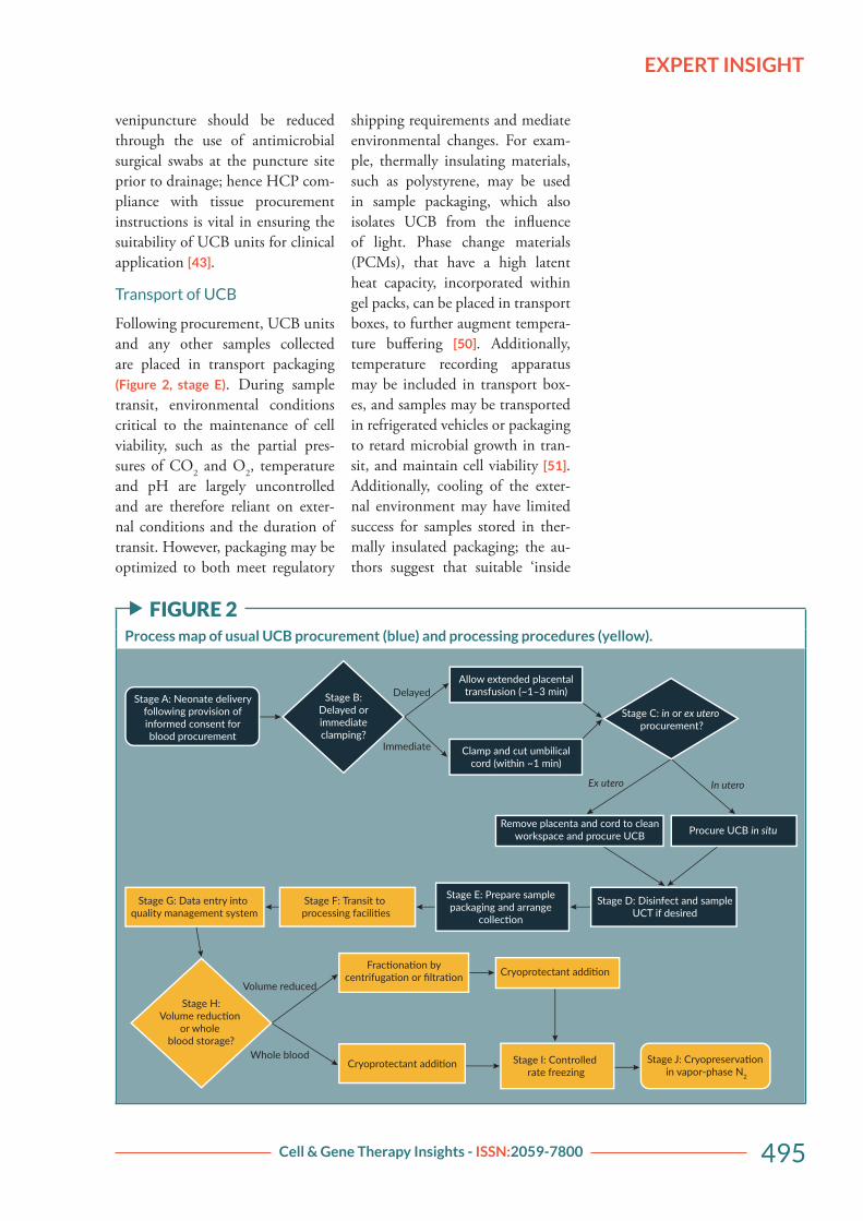
As for most tissue donation, donors of UCB, regardless of storage mod-el, must provide informed consent prior to collection, and undergo in-fectious disease screening between 7 days before and 7 days after delivery (Figure 2, stage A). Units are always procured postpartum, but may be collected before or after separation of the placenta from the uterus. These methods are termed in and ex utero procurement respectively (Figure 2, stage C) [23]. Additionally,
UCB procurement is not limited by birth type; units are routinely col-lected following both induced and non-induced vaginal and cesarean births. For all approaches, the blood supply from the umbilical cord to the neonate is interrupted by post-partum clamping following birth, and the residual cord blood extract-ed by venipuncture. In the UK, the Human Tissue Authority (HTA) recommends ex utero procurement of UCB [24]; in contrast, in ute-ro collection is widely favoured elsewhere, including the USA and Western Europe, due to the signifi-cantly greater sample volumes and total nucleated cell counts (TNCs) achieved [25]. This may be due to the fact that, during the third stage of labor, the uterus continues to contract, aiding the expulsion of blood [26].
Impact of obstetric practices on UCB quality
The final volume and total nucleated cell count (TNC) of UCB collected is partially dependent on uncon-trollable obstetric factors, such as maternal age [27], gestational pe-riod [28,29], placental weight [30], number of prior live births [31,32], delivery type [33–35], maternal smoking [36], cord length [28,37], ethnicity [38], maternal preeclamp-sia [39,40], fetal distress [34], birth weight [28,41], and possibly neonate sex [27]. However, birth and pro-curement approaches, under clini-cian and maternal control, may also impact UCB unit quality. For ex-ample, the timing of cord clamping directly impacts the volume of UCB collected [42] and the pre-venipunc-ture preparation of the cord impacts UCB unit contamination [43,44].
Delayed cord clamping, classed as interruption of the blood supply
CELL & GENE THERAPY INSIGHTS
494 DOI: 10.18609/cgti.2017.042
between cord and infant after more than one minute postpartum, is recommended by the World Health Organisation (WHO) [45]; extend-ed placental transfusion following delivery improves term infant iron status for up to 6 months and re-duces the risk of maternal postpar-tum hemorrhage (Figure 2, stage B)
[26,46]. Additionally, delayed cord clamping reduces the risk of intra-ventricular hemorrhage, late-onset sepsis and necrotizing enterocolitis in pre-term neonates [47]. The loss of blood flow as a result of delayed cord clamping, temperature chang-es and other factors cause vascular occlusion, which in turn triggers co-agulation of entrapped blood [48]. The volume of UCB units collected following delayed cord clamping are significantly reduced as a result, although some tissue banks advise that UCB collection is still viable if the cord is clamped after no more than 3 minutes. Similarly, those procured following expectant versus
actively managed labors (whereby prophylactic uterotonics are ad-ministered to hasten delivery of the extraembryonic tissues) are also of smaller volume [49].
UCB is contained within the aseptic, closed system comprising baby, placenta and cord, and is drained into a sterile container for subsequent processing. Microbial contamination is therefore not pres-ent in either UCB (in the absence of vertically transmitted infections) or storage materials; in fact, contami-nants may only be introduced at the point of procurement during which UCB is briefly exposed to the cord exterior. The birth environment is inherently non-sterile because, par-ticularly during vaginal births, the epithelial amniotic membrane is in contact with vaginal and colon-de-rived fluids. Microbes transferred via these fluids then access the cord interior through the procurement venipuncture. The risk of microbi-al contamination of UCB through
f FIGURE 1Representation of the clinical target of all open trials investigating UCB as a clinical intervention at the time of writing (n = 139).
58%
16%
9%
6%
3%3%
3% 2%
Blood cancers
Disorders of blood cell prolifera�on
Other
Other brain injuries
Cerebral palsy
Stroke
Diabetes
Fanconi’s anemia
Clinical targets included in the ‘other’ category include HIV infection, low birth weight and various types of hemorrhage.
EXPERT INSIGHT
495Cell & Gene Therapy Insights - ISSN:2059-7800
venipuncture should be reduced through the use of antimicrobial surgical swabs at the puncture site prior to drainage; hence HCP com-pliance with tissue procurement instructions is vital in ensuring the suitability of UCB units for clinical application [43].
Transport of UCB
Following procurement, UCB units and any other samples collected are placed in transport packaging (Figure 2, stage E). During sample transit, environmental conditions critical to the maintenance of cell viability, such as the partial pres-sures of CO2 and O2, temperature and pH are largely uncontrolled and are therefore reliant on exter-nal conditions and the duration of transit. However, packaging may be optimized to both meet regulatory
shipping requirements and mediate environmental changes. For exam-ple, thermally insulating materials, such as polystyrene, may be used in sample packaging, which also isolates UCB from the influence of light. Phase change materials (PCMs), that have a high latent heat capacity, incorporated within gel packs, can be placed in transport boxes, to further augment tempera-ture buffering [50]. Additionally, temperature recording apparatus may be included in transport box-es, and samples may be transported in refrigerated vehicles or packaging to retard microbial growth in tran-sit, and maintain cell viability [51]. Additionally, cooling of the exter-nal environment may have limited success for samples stored in ther-mally insulated packaging; the au-thors suggest that suitable ‘inside
f FIGURE 2Process map of usual UCB procurement (blue) and processing procedures (yellow).
Delayed
Immediate
Ex utero In utero
Stage E: Prepare samplepackaging and arrange
collec�on
Volume reduced
Whole blood Stage J: Cryopreserva�on in vapor-phase N2
Stage G: Data entry into quality management system
Stage H:Volume reduc�on
or whole blood storage?
Stage F: Transit to processing facili�es
Stage D: Disinfect and sampleUCT if desired
Frac�ona�on bycentrifuga�on or filtra�on Cryoprotectant addi�on
Stage I: Controlled rate freezing
Cryoprotectant addi�on
Procure UCB in situRemove placenta and cord to cleanworkspace and procure UCB
out’ cooling technologies that may be enclosed in transport boxes are lacking and should be developed.
In the authors’ experience, lo-gistical factors between the site of procurement and storage can impinge on the quality of a cord blood unit. Interestingly, private cord blood banking is prohibited in some countries in favor of pub-lic banking models, but export of samples for overseas processing and storage is sometimes permitted. On average, the procurement volume of private units originating from such countries is smaller than from other regions [Unpublished data]; we spec-ulate that this may reflect the reser-vations of HCPs regarding the eth-ics of private cord blood banking. Furthermore, extended transit, as may be caused by geographical and local customs/legal requirements, is associated with lower white blood cell counts and HSC viability [Un-
published data].
Bioprocessing of UCB for cryopreservation
UCB processing may incorporate automated or operator-based, and whole blood or volume-reduced ap-proaches (Figure 2, stage G onwards). Economic, safety and quality con-siderations inform the technologies employed in a given paradigm. A balance must be sought between the comparatively reduced storage costs and the greater processing time re-quired for volume reduced versus whole blood cord units [52]. Ad-ditionally, volume-reduced trans-plants confer improved patient out-comes, such as a reduction in ABO incompatibility alloreactivity and dimethyl sulfoxide (DMSO) toxic-ity (Figure 2, stage H) [53].
In volume reduction proto-cols, the red blood cell and plasma
fractions of units are largely deplet-ed. This approach has been adopted by many banks worldwide, includ-ing public organisations such as the NHS Cord Blood Bank (UK). In summary, incoming units are separated by centrifugation or fil-tration and plasma, red blood cell and HSC-containing ‘buffy coat’ fractions are separated using either semi-automated devices such as the Macopress Smart Evo (Macoph-arma, France), Optipress II (Fen-wal, USA) and CellEffic (Kaneka, Japan) products, or fully automat-ed, enclosed systems (such as the Sepax™ (Biosafe, USA) or AutoX-press® (Cesca Therapeutics, USA) [49,54]. Cryoprotectant is added to the buffy coat fraction, which is stored in vapor phase N2. Sedimen-tation agents such as pentastarch [55] and hydroxyethyl starch (HES) [56] may also be used to enhance red blood cell removal. Fully or semi-automated systems may offer advantages with respect to stan-dardization, necessary to ensure the safety and efficacy of UCB-derived cell therapies, and allow UCB pro-cessing to take place in ungraded environments. In whole blood stor-age protocols, the complete unit is cryopreserved following cryopro-tectant addition with no additional processing steps [57].
Challenges in the clinical application of UCB
Use of UCB for haematopoietic reconstitution was first reported by Ende et al., for the treatment of acute lymphoblastic leukemia, in 1972 [58]. Unfortunately, clinical use of UCB is limited by the avail-ability of human leucocyte antigen (HLA)-matched donors, although mismatches at one or two loci are well tolerated [5]. Approximately
EXPERT INSIGHT
497Cell & Gene Therapy Insights - ISSN:2059-7800
30% of all recipients are matched with an HLA-identical donor through worldwide registries; around 75% of Caucasians [59], but only 17% of other ethnicities find a suitable donor [60].
Low hematopoietic progenitor yields per unit present a yet more significant challenge for the clinical use of UCB, and mean that only low weight patients, such as chil-dren, are eligible for transplant of single units [61]. Low cell doses are also associated with increased en-graftment times, prolonged immu-nodeficiency and greater healthcare burdens [62]. Double UCB trans-plantation, an alternative treatment regimen that increases the number of HSC transplanted, has been widely reported to improve clini-cal outcomes [63], but is associated with greater acquisition costs. How-ever, one notable study by Labopin et al., in which Markov analysis was used to evaluate the costs and out-comes of single and double UCB transplantation in France, reported the latter as more cost-effective [64]. Alternatively, ex vivo expansion of UCB-HSC may increase available cell doses, and manufacturing strat-egies to produce advanced therapy medicinal products (ATMP) are under investigation [65]. Current UCB therapies are considered mini-mally manipulated products by reg-ulators [66,67].
UMBILICAL CORD TISSUE BIOPROCESSINGUCT is banked to preserve UCT-MSC, which offer different clini-cal potentials to UCB-HSC. The identification of clonogenic fibro-blast-like cells in bone marrow by Friedenstein et al. in 1970 [68] is
oft viewed as the dawn of modern MSC research. MSC have subse-quently been widely investigated for applications in wound healing and various pathologies, utilizing their multilineage plasticity and immunomodulatory functions [5,69]; at the time of writing this review, there are approximately 2600 clinical trials involving MSC, of which 40 utilized UCT-de-rived MSC (www.clinicaltrials.gov; search terms: “mesen*” AND “cell”, “MSC”, “umb*” AND “mes-en*”). Although MSC reside in vir-tually all postnatal tissues [70], they have been frequently sourced from bone marrow. Major drawbacks as-sociated with bone marrow sources include the invasiveness of aspira-tion, influence of donor age on the functional characteristics of MSC and their paucity within this tissue. In comparison, UCT is considered clinical waste, is relatively MSC dense, and is sourced from neo-nates [71]. Other clinically useful properties of MSC include the lack of expression of MHC-II (major histocompatibility complex II) and co-stimulatory molecules CD80 and CD86, conferring hypoimmu-nogenicity. Hence, MSC derived from suitable sources and subjected to appropriate bioprocessing may also be harnessed for ‘off-the-shelf ’ allogeneic therapeutics [72].
Challenges in procurement & storage of UCT
UCT is procured following neo-nate delivery and transported for processing in transport solution. Following neonate delivery and UCB procurement the cord is sev-ered from the placenta, disinfected, and placed in a sterile container (Figure 3, stages A–E). Tissue via-bility and sterility are impacted by
CELL & GENE THERAPY INSIGHTS
498 DOI: 10.18609/cgti.2017.042
the same transit, packaging and environmental factors as described previously for UCB (Figure 3, stage
F). Upon arrival at a processing laboratory, cord tissue processing is typically performed using the ‘slice and dice’ method (Figure 3,
stage J) [73]; briefly, UCT is disin-fected, rinsed, minced and added to cryoprotectant before controlled rate freezing and long-term storage in vapor-phase N2 (Figure 3, stages
G–M). Although tissue procure-ment is relatively uncontrolled due to the clinical demands of the birth environment, the authors recognize that key aspects of industry practice may be improved to optimize UCT collection and storage [74].
Microbial contamination of UCT
The quality of banked cord tis-sue may be considered in terms
of sterility, cell yield, functional-ity and anticipated safety for clin-ical application. Since UCT is maintained in aseptic conditions throughout transit and processing, microbial contamination may only be introduced at the point of pro-curement as a result of contact with the ex utero environment. The col-lection method used may influence the probability of microbial con-tamination; we have found that in utero procurement can be associat-ed with UCT contamination rates approximately three times greater than ex utero counterparts [Unpub-
lished data]. Additionally, microbial species found on in utero procured samples are overwhelmingly of fae-cal origin; this is likely due to ex-tended proximity to excreta in the birth environment [Unpublished
data]. The course, type, and du-ration of birth may be reasonably
f FIGURE 3Process map of usual UCT procurement (blue) and processing procedures (yellow).
Stage A: Neonate deliveryfollowing provision of
informed consent for �ssueprocurement
Stage B:Delayed or immediateclamping?
Delayed
Immediate
Allow extended placentaltransfusion (~1–3 min)
Clamp and cut umbilicalcord (within ~1 min)
Procure UCB in situ
In utero Ex utero
Stage C: In or ex utero�ssue procurement?
Remove placenta and cord toclean workspace and procure UCB
Stage H: Tissue transfer tograde A environment and
disinfec�on
Stage G: Data entry intoquality management system
EXPERT INSIGHT
499Cell & Gene Therapy Insights - ISSN:2059-7800
expected to affect the probability of microbial contamination but are not controllable to the UCT pro-curer. However, the impact of these factors and hence contact with bodily fluids may be mitigated by thorough disinfection of all sam-ples using surgical swabs (Figure 3,
stage D).Additionally, we have also found
that there may be a significant re-lationship between sample tran-sit duration and the incidence of contaminated cord tissue (Figure 3,
stage F) [Unpublished data]. It is pos-tulated that this may be explained by increased microbial growth with increasing transit times. Longer in-cubation times may also encourage the formation of biofilm communi-ties that are not easily subsequently eliminated; biofilms are often recal-citrant to the antibiotic and biocidal measures fatal to planktonic bacte-ria [75].
Yield & functionality of MSC content
The yield and functionality of MSC subsequently isolated from UCT is affected by tissue bioprocessing methods. For example, intra- and inter- cord variability in MSC con-tent should be considered with re-spect to tissue procurement. An optimal cell donor cannot be se-lected in autologous paradigms, but strategic tissue sampling is possible. Lim et al. investigated the yield and functionality of UCT-hMSC iso-lated from tissue segments excised from distinct cord regions. Fetal and maternal segment-derived MSC had greater viability, lower doubling times and higher differentiation po-tentials than middle cord-derived MSC [76]. Hence, selective tissue resection from the ends of umbili-cal cords may improve UCT-hMSC
characteristics, but further evidence is necessary to support any change in collection practices.
In allogeneic models, donor MSC are expanded for off-the-shelf sup-ply, and both inter- and intra-cord sampling comprise controllable variables. Allogeneic donor criteria must incorporate infectious disease screening and demographic factors; studies concerning the impact of donor characteristics are limited, but reports indicate that gestational diabetes mellitus and maternal age of ˃29 years impact the functional characteristics of UCT-hMSC, and hence should form exclusion crite-ria [77,78].
Safety considerations in bio-processing of UCT
The cryoprotectants and choice of transport/wash solutions used for UCT bioprocessing directly con-tribute to patient outcomes. For example, UCT is often transported in penicillin-containing solutions despite the well-documented aller-genicity of β-lactam antibiotics. In fact, penicillin allergy affects up to 8% of patients, and complications can be fatal [79]. In the opinion of the authors, the use of antibi-otic-treated tissues in cell therapy manufacture will present increased challenges in gaining regulatory approval.
Additionally, both cryoprotectant and cryopreservation media choice pose safety concerns. The most com-monly used cryoprotectant, dimeth-yl sulfoxide (DMSO), is toxic at the molar concentrations used and at temperatures greater than 4°C [71,80]. In fact, a plethora of adverse reactions have been reported from DMSO exposure in humans and other mammals, including some following post-thaw infusion of
CELL & GENE THERAPY INSIGHTS
500 DOI: 10.18609/cgti.2017.042
washed cells in clinical trials, where up to 50% of patients were affect-ed [81,82]. The allo- or xeno-geneic components of serum-containing cryopreservation media, believed to supply nutrients in the post-thaw period, are notoriously difficult to remove and may trigger immuno-genic responses [83].
Challenges in isolation of MSC from UCT
UCT banking has the potential to meet clinical and commercial MSC demands with the maturation of bi-oprocessing and expansion technol-ogies. Unlike the UCB-HSC, UCT-MSC must always be expanded ex vivo to supply clinically relevant cell doses. Although a critical area for research and development activities, the scope of this review comprises current progress and challenges in the procurement and processing of UCT for cryopreservation. Readers are referred to the following reviews for insight regarding the ex vivo ex-pansion of MSC [84–86].
Isolation of MSC from UCT has been described in a number of publications [87–89], but there is no consensus regarding an optimal approach that maximizes cell yield, viability and population homoge-neity. UCT-MSC are isolated either by enzymatic disaggregation of cord tissue or the selective migration of MSC from cultured explants. Col-lagenase, hyaluronidase and trypsin are commonly used in digestion protocols [90,91], either alone or in combination, and different explant sizes and explant-substrate adhesion modes have been investigated [92]. Only one report has emerged in which a combination of both meth-ods was investigated [93].
MSC are found throughout UCT, which may be stored as
whole minced tissue, with a lack of consensus amongst investigators as to whether UCT-MSC isolated from different anatomical regions are equivalent [94,95]. However, MSC are most commonly isolated from Wharton’s jelly or the peri-vascular region [96,97]. Although these anatomical regions contain approximately 75% of UCT-MSC and this approach may reduce the number of contaminating epithelial and endothelial cells in early passage UCT-MSC cultures, incorporating dissection of an inherently variable tissue into a closed, automated process according to best practice, remains problematic [98,99]. Since the macroscale dimensions of in-dividual cords differ, separation of distinct tissues must be conducted by a skilled operator, and to meet cGMP standards, manual biopro-cessing must be housed in a class A biosafety cabinet installed in a Class B room [66,100].
PRESERVATION, SHIPPING & CONTAMINATION MAN-AGEMENT OF UCB & UCTContamination management strat-egies in UCB and UCT banking must be devised with care to max-imize the clinical utility of these tissues. We propose that two main approaches should be employed to reduce the incidence of con-tamination: management of HCP practices to avoid initial microbial inoculation of tissue, and ablation of microbial growth during transit.
Owing to the inflated contamina-tion rates observed in in utero pro-cured UCT, ex utero procurement of these tissues is recommended to en-sure the safety of derived UCT-MSC. Alternatively, if HCPs strongly favor
EXPERT INSIGHT
501Cell & Gene Therapy Insights - ISSN:2059-7800
the in utero collection method, the sample should be taken to a des-ignated clean area after UCB pro-curement for thorough disinfection according to the processing facility’s protocol, which should incorporate a combined disinfection approach such as treatment with both chlor-hexidine and alcohol, rather than al-cohol alone, since this combination results in greater microbial kill [101]. Whilst the formation of biofilms on both in and ex vivo soft tissues is widely acknowledged in microbiol-ogy, to the authors’ knowledge, no investigators have attempted to de-tect biofilm matrix on contaminated UCT to date. Such results could be instrumental in refining current in-dustry practices.
Penicillin-containing solutions have frequently been added to tis-sue transport solutions to ablate bacterial growth during transit. Whilst the impact of growth during transit is acknowledged to be large, penicillin is a common allergen and treated tissue is unlikely to find al-logeneic uses. The most frequently encountered contaminant species on UCT and UCB are mesophilic, and moderate cooling during tran-sit is suggested to extend population doubling times as an alternative [102,103]. The increased expense of courier services using refrigerated versus ambient vehicles means that refrigeration inside sample pack-aging is more economically viable. This may be achieved through ad-dition of cooling devices to sample packaging in conjunction with ther-mally insulating packaging.
Other safety concerns impact-ing the utility of UCB and UCT in the clinic include cryopreservation strategies, and whole versus volume -reduced bioprocessing of UCB. The success of cryopreservation
is dependent on cryopreservation medium and the method of cool-ing. Controlled rate freezing of DMSO-infused tissues is preferred throughout the tissue banking in-dustry; vitrification of UCT has previously been attempted to the detriment of the UCT-MSC con-tained within [104]. The toxicity of DMSO to both cells and man means that the lowest effective con-centration should be used and post-thaw recovery optimized. Concen-trations of 10% (v/v) DMSO are often used, but increasing evidence suggests that this can be reduced to 2.5% (v/v), with benefits for the functional characteristics of UCB-HSC and UCT-MSC, and reduc-ing the risk of infusional toxicity of DMSO [105,106]. Current practic-es achieve DMSO removal by wash-ing with isotonic salt solutions and centrifugation, or using automated cell washing devices [107], but these introduce mechanical and osmotic stresses that can result in significant cell loss [71].
Slow-cooling cell injury can be further precluded through inclusion of polysaccharides such as trelahose [108], sugar alcohols, hydroxyeth-yl starch (HES) [109] and dextran in cryopreservation media [110]. DMSO readily permeates cell mem-branes and impedes extracellular ice formation by depressing the freezing point of water, and polysaccharides are thought to assist by confound-ing the formation of intracellular ice [111]. The addition of polysaccharide solutions also reduces the concentra-tion of DMSO necessary for suc-cessful cryopreservation [106,112]. Hence, we recommend that the minimum concentration of DMSO required, validated with respect to optimal post-thaw cell recovery, in conjunction with the nontoxic
CELL & GENE THERAPY INSIGHTS
502 DOI: 10.18609/cgti.2017.042
additives previously listed, be used to cryopreserve UCT and UCB for translation/clinical application.
Although UCT may be stored in a saline/DMSO solution, se-rum-containing culture media are also widely used in cryopreservation media. Although the Federal Drug Administration (FDA) (USA) per-mits cell therapy manufacture with cGMP-compliant sera [113], xeno- and allogeneic antigens present may be hyperimmunogenic to patients, and zoonotic, viral and prion trans-mission is possible. Chemically de-fined, serum-free medium (SFM) minimizes lot-to-lot variability in cell products, but culture substrates coated with exogenous proteins are frequently needed to aid adherence. Fortunately, successful xeno- and serum-free MSC culture has now been reported, and should be har-nessed in cryopreservation media for tissue banking [114].
Volume reduction of UCB is com-monplace because it reduces per unit storage costs and improves transplan-tation outcomes. At present, there are no known clinical uses for the RBC fraction of cord blood, and RBC ly-sate generated during cryopreserva-tion and thawing can impact kidney function in recipients [61]. Allore-activity of whole blood UCB units may also present where donor and recipient have differing blood groups, regardless of HLA type [115]. Conse-quently, volume reduction is recom-mended in UCB processing for maxi-mal transplant success.
TRANSLATIONAL INSIGHTUltimately, effective bioprocess de-velopment is dependent upon an intimate knowledge of critical qual-ity attributes (CQAs) of the final
product and intended clinical ap-plication. Transplantation of UCB-MSC is a standard therapy for nu-merous diseases, but no UCT-MSC therapeutics currently exist. This is reflected in the fact that, at the time of writing, there are approxi-mately 139 active clinical trials of UCB interventions, but just 40 em-ploying UCT-MSC (Figures 1 & 4). Despite this, UCT-MSCs are being investigated as a therapy in clinical indications such as GvHD, hepatic cirrhosis, myocardial infarction and myopathies, spinal cord injury, ar-thritis, multiple sclerosis, and stroke [71,116]. A ‘chicken and egg’ para-digm exists in cell therapy research – bioprocess strategies are required to facilitate ATMP development, but clinical direction is needed to define CQAs of such bioprocesses. Clinical trials are suggested as the best indicators of clinically import-ant parameters in tissue banking of UCB and UCT (Tables 1 & 2).
In the absence of scalable man-ufacturing technology, the demand for high UCT-MSC and UCB-HSC doses can be tackled through the adoption of optimal bioprocess and clinical approaches that seek to preserve both maximal cell numbers and engraftment potentials. The number of HSC administered has been increased using the scale out approach of double UCB transplan-tation, maintaining the ‘minimally manipulated’ regulatory status of the treatment [117]. Reduced in-tensity patient conditioning prior to transplantation [118,119], di-rect intra-bone injection [120,121] and co-transplantation of UCB with MSC for their immunopro-tective function [122,123], have all been utilized to maximize engraft-ment. We suggest that the number of UCT-hMSC sampled during
EXPERT INSIGHT
503Cell & Gene Therapy Insights - ISSN:2059-7800
UCT collection may be optimized through selective procurement, where practicable, of equal sized tissue segments from the fetal and placental ends of the cord.
A consensus regarding an op-timal method for the isolation of UCT-MSC is yet to emerge, with scant data available to assess the merits of explant and enzymatic digestion methods. Thus, compar-ison studies of UCT-MSC extract-ed by each method are contrasted, according to their kinetics and the defining criteria outlined by the International Society for Cellular Therapy (ISCT) are required for ef-fective translation [124].
CONCLUSIONTo complement the body of work that aims to elucidate clinical uses of UCB and UCT-MSC, biopro-cessing strategies that are optimal with respect to cell functionality, patient outcomes and cost must be
developed. In this review, we ar-gue that UCB volume reduction, antibiotic-free bioprocesses, low DMSO in cryopreservation media, and improved antimicrobial strate-gies may aid the readiness of UCB and UCT banked in the present for incorporation into the cell thera-peutics of the future.
FINANCIAL & COMPETING
INTERESTS DISCLOSURE
LP, SM, KS & BC wish to acknowledge their involvement with Biovault Technical Ltd. The authors have no other relevant financial involvement with an organization or entity with a financial interest in or financial conflict with the subject matter or materials discussed in the manuscript. No writing assistance was utilized in the production of this manuscript.
f FIGURE 4Representation of the clinical target of all open trials investigating UCT-MSC as an intervention at the time of writing (n = 40).
26%
17%
14%
11%
11%
9%
6%6%
Other
Lung disease
Liver cirrhosis/failure
Diabetes related
Cardiovascular disease
Arthri�s
Nonunion fracture
Psoriasis
This work is licensed under
a Creative Commons Attri-
bution – NonCommercial – NoDerivatives 4.0
International License
CELL & GENE THERAPY INSIGHTS
504 DOI: 10.18609/cgti.2017.042
REFERENCES1. Gluckman E, Broxmeyer HE, Auer-
bach AD et al. Hematopoietic recon-stitution in a patient with Fanconi’s anemia by means of umbilical-cord blood from an HLA-identical sibling. N. Engl. J. Med. 1989; 321: 1174–8.
2. Laughlin MJ, Barker J, Bambach B et al. Hematopoietic engraftment and survival in adult recipients of um-bilical-cord blood from unrelated donors. N. Engl. J. Med. 2001; 344: 1815–1822.
3. Hudson KD, Bonassar LJ. Hypoxic Expansion of Human Mesenchymal Stem Cells Enhances 3D Maturation of Tissue Engineered Intervertebral Discs. Tissue Eng. Part A 2016; 23: 293-300.
4. Li WJ, Tuli R, Okafor C et al. A three-dimensional nanofibrous scaffold for cartilage tissue engineering using human mesenchymal stem cells. Bio-materials 2005; 26: 599–609.
5. Ankrum J, Ong JF, Karp JM. Mesen-chymal stem cells: immune evasive, not immune privileged. Nat. Biotechnol. 2014; 32: 252–60.
6. Hauskeller C, Beltrame L. The hybrid bioeconomy of umbilical cord blood banking: Re-examining the narrative of opposition between public and pri-vate services. Biosocieties 2015; 11: 1745–8552.
7. Ballen KK, Verter F, Kurtzberg J. Um-bilical cord blood donation: public or private? Bone Marrow Transplant. 2015; 50: 1271–8.
8. Eapen M, Klein JP, Sanz GF et al. Effect of donor-recipient HLA matching at HLA A, B, C, and DRB1 on outcomes after umbilical-cord blood transplanta-tion for leukaemia and myelodysplas-tic syndrome: A retrospective analysis. Lancet Oncol. 2011; 12: 1214–21.
9. Ooi J, Iseki T, Takahashi S et al. Un-related cord blood transplantation for
adult patients with advanced myelo-dysplastic syndrome. Blood 2003; 101: 4711–3.
10. Rocha V, Labopin M, Sanz G et al. Transplants of umbilical-cord blood or bone marrow from unrelated donors in adults with acute leukemia. N. Engl. J. Med. 2004; 351: 2276–85.
11. Eapen M, Rubinstein P, Zhang M-J et al. Outcomes of transplantation of un-related donor umbilical cord blood and bone marrow in children with acute leukaemia: a comparison study. Lancet 2007; 369: 1947–54.
12. Majhail NS, Weisdorf DJ, Wagner JE et al. Comparable results of umbilical cord blood and HLA-matched sibling donor hematopoietic stem cell transplantation after reduced-intensity preparative reg-imen for advanced Hodgkin lympho-ma. Blood 2006; 107: 3804–7.
13. Burman J, Fransson M, Tötterman TH et al. T-cell responses after haematopoi-etic stem cell transplantation for aggres-sive relapsing-remitting multiple sclero-sis. Immunology 2013; 140: 211–9.
14. Prasad VK, Mendizabal A, Parikh SH et al. Unrelated donor umbilical cord blood transplantation for inherited metabolic disorders in 159 pediatric patients from a single center: influence of cellular composition of the graft on transplantation outcomes. Blood 2008; 112: 2979–89.
15. Bizzetto R, Bonfim C, Rocha V et al. Outcomes after related and unrelated umbilical cord blood transplantation for hereditary bone marrow failure syndromes other than fanconi anemia. Haematologica 2011; 96: 134–41.
16. Gluckman E, Anderson V, Ocha R et al. Outcome of cord-blood transplanta-tion from related and unrelated donors. N. Engl. J. Med. 1997; 337: 373–81.
17. Ohara Y, Ohto H, Tasaki T et al. Com-prehensive technical and patient-care
optimization in the management of pediatric apheresis for peripheral blood stem cell harvesting. Transfus. Apher. Sci. 2016; 55. 338-343.
18. Lane BTA, Law P, Maruyama M et al. Harvesting and Enrichment of Hema-topoietic Progenitor Cells Mobilized Into the Peripheral Blood. Blood 1995; 85: 275–82.
19. Bolan CD, Childs RW, Procter JL et al. Massive immune haemolysis after allogeneic peripheral blood stem cell transplantation with minor ABO in-compatibility. Br. J. Haematol. 2001; 112: 787–95.
20. Auquier P, Macquart-Moulin G, Moat-ti JP et al. Comparison of anxiety, pain and discomfort in two procedures of hematopoietic stem cell collection: leukacytapheresis and bone marrow harvest. Bone Marrow Transplant. 1995; 16: 541–7.
21. Rossi DJ, Bryder D, Zahn JM et al. Cell Intrinsic Alterations Underlie Hemato-poietic Stem Cell Aging. Proc. Natl. Acad. Sci. 2005; 102: 9194–9.
22. Morrison SJ, Wandycz M, Akashi K et al. The aging of hematopoietic stem cells. Nat. Med. 1996; 2: 1011–6.
23. Ballen KK, Barker JN, Stewart SK et al. Collection and Preservation of Cord Blood for Personal Use. Biol. Blood Marrow Transplant. 2008; 14: 356–63.
24. HTA. Guidance document for estab-lishments working with Umbilical cord blood. 2010.
25. Hosing C, Munsell M, Armitage S et al. Ex-Utero Plus in Utero Collection of Umbilical Cord Blood (CB) for Bank-ing Yields Higher Total Nucleated Cell Counts (TNC) Compared to Either Procedure Alone. Biol. Blood Marrow Transplant. 2015; 21: S154–55.
26. Begley C, Gyte G, Devane D et al. Ac-tive versus expectant management for women in the third stage of labour (
27. McGuckin CP, Basford C, Hanger K et al. Cord blood revelations-The im-portance of being a first born girl, big, on time and to a young mother! Early Hum. Dev. 2007; 83: 733–41.
28. Nakagawa R, Watanabe T, Kawano Y et al. Analysis of maternal and neonatal factors that influence the nucleated and CD34+ cell yield for cord blood bank-ing. Transfusion 2004; 44: 262–7.
29. Surbek D, Visca E, Steinmann C. Umbilical cord blood collection be-fore placental delivery during cesarean delivery increases cord blood volume and nucleated cell number available for transplantation. Am. J. Obstet. Gynecol. 2000; 183: 218–21.
30. Askari S, Miller J, Chrysler G et al. Im-pact of donor- and collection-related variables on product quality in ex utero cord blood banking. Transfusion 2005; 45: 189–94.
31. Jones J, Stevens CE, Rubinstein P et al. Obstetric predictors of placental/um-bilical cord blood volume for transplan-tation. Am. J. Obstet. Gynecol. 2003; 188: 503–9.
32. Jan RH, Wen SH, Shyr MH et al. Im-pact of maternal and neonatal factors on CD34+ cell count, total nucleated cells, and volume of cord blood. Pedi-atr. Transplant. 2008; 12: 868–73.
33. Mousavi SH, Abroun S, Zarrabi M et al. The effect of maternal and infant factors on cord blood yield. Pediatr. Blood Cancer 2017; 64: 1–8.
34. Manegold G, Meyer-Monard S, Tichelli A et al. Cesarean section due to fetal distress increases the number of stem cells in umbilical cord blood. Transfusion 2008; 48: 871–6.
35. Omori A, Takahashi K, Hazawa M et al. Maternal and neonatal factors as-sociated with the high yield of mono-nuclear low-density/CD34+ cells from
placental/umbilical cord blood. Tohoku J. Exp. Med. 2008; 215: 23–32.
36. Ballen KK, Klein JP, Pedersen TL et al. Relationship of race/ethnicity and sur-vival after single umbilical cord blood transplantation for adults and children with leukemia and myelodysplastic syn-dromes. Biol. Blood Marrow Transplant. 2012; 18: 903–12.
37. Wen SH, Zhao WL, Lin PY et al. Asso-ciations among birth weight, placental weight, gestational period and product quality indicators of umbilical cord blood units. Transfus. Apher. Sci. 2012; 46: 39–45.
38. Yang H, Loutfy MR, Mayerhofer S et al. Factors affecting banking quality of umbilical cord blood for transplanta-tion. Transfusion 2011; 51: 284–92.
39. Keersmaekers CL, Mason BA, Keers-maekers J et al. Factors affecting umbil-ical cord blood stem cell suitability for transplantation in an in utero collection program. Transfusion 2014; 54: 545–9.
40. Wahid FSA, Nasaruddin MZ, Idris MRM et al. Effects of preeclampsia on the yield of hematopoietic stem cells obtained from umbilical cord blood at delivery. J. Obstet. Gynaecol. Res. 2012; 38: 490–7.
41. Kurtzberg J, Wagner EL, Fraser JK et al. Results of the Cord Blood Transplant Study (COBLT) Unrelated Donor Banking Program from Donor Screen-ing to Characterization of Banked Units. Blood 2003; 102: 461a.
42. Allan DS, Scrivens N, Lawless T et al. Delayed clamping of the umbilical cord after delivery and implications for public cord blood banking. Transfusion 2016; 56: 662–5.
43. Roura S, Pujal J-M, Gálvez-Montón C et al. The role and potential of umbilical cord blood in an era of new therapies: a review. Stem Cell Res. Ther. 2015; 6: 123.
44. Clark P, Trickett A, Stark D et al. Fac-tors affecting microbial contamina-tion rate of cord blood collected for transplantation. Transfusion 2012; 52: 1770–7.
45. World Health Organization. Guide-line: Delayed umbilical cord clamp-ing for improved maternal and infant health and nutrition outcomes. World Heal. Organ. 2014.
46. Hutton EK, Hassan ES. Late vs early clamping of the umbilical cord in full-term neonates: systematic review and meta-analysis of controlled trials. JAMA 2007; 297: 1241–52.
47. Robertson J. Beyond Survival – 2nd Edition. Pan Am. Heal. Organ. 2013; 73.
48. Yao AC, Lind J, Lu T. Closure of the human umbilical artery: a physiological demonstration of Burton’s theory. Eur. J. Obstet. Gynecol. Reprod. Biol. 1977; 7: 365–8.
49. Lapierre V, Pellegrini N, Bardey I et al. Cord blood volume reduction us-ing an automated system (Sepax) vs. a semi-automated system (Optipress II) and a manual method (hydroxyethyl starch sedimentation) for routine cord blood banking: a comparative study. Cytotherapy 2007; 9: 165–9.
50. Farid MM, Khudhair AM, Razack SAK et al. A review on phase change energy storage: Materials and applica-tions. Energy Convers. Manag. 2004; 45: 1597–615.
51. Pal R, Hanwate M, Totey SM. Effect of holding time, temperature and differ-ent parenteral solutions on viability and functionality of adult bone marrow-de-rived mesenchymal stem cells before transplantation. J. Tissue Eng. Regen. Med. 2008; 2: 436–44.
52. Sousa T, de Sousa ME, Godinho MI et al. Umbilical cord blood process-ing: volume reduction and recovery
CELL & GENE THERAPY INSIGHTS
506 DOI: 10.18609/cgti.2017.042
of CD34+ cells. Bone Marrow Transpl. 1997; 19: 311–3.
54. Batista A, Lamontagne D, Chang-Fong S et al. Processing the 5-mL umbilical cord blood unit with the use of an au-tomated wash procedure. Cytotherapy 2015; 17: 336–7.
55. Yang H, Acker JP, Abley D et al. High-efficiency volume reduction of cord blood using pentastarch. Bone Marrow Transplant. 2001; 27: 457–61.
56. Schwandt S, Korschgen L, Peters S et al. Cord blood collection and processing with hydroxyethyl starch or non-hy-droxyethyl starch. Cytotherapy 2016; 18: 642–52.
57. Schindler S, Asmus S, von Aulock S et al. Cryopreservation of human whole blood for pyrogenicity testing. J. Immu-nol. Methods 2004; 294: 89–100.
58. Ende M, Ende N. Hematopoietic transplantation by means of fetal (cord) blood. Virginia Med. J. 1972; 99: 276–80.
59. Tse WW, Zang SL, Bunting KD et al. Umbilical cord blood transplantation in adult myeloid leukemia. Bone Mar-row Transplant. 2008; 41: 465–72.
60. Solh M. Haploidentical vs cord blood transplantation for adults with acute myelogenous leukemia. World J. Stem Cells 2014; 6: 371–9.
61. HTA. umbilical cord blood banking A guide for parents. 2016.
62. Rubinstein P, Carrier C, Scaradavou A et al. Outcomes Among 562 Recipients of Placental-Blood Transplants from Unrelated Donors. N. Engl. J. Med. 1998; 339: 1565–77.
63. Brunstein CG, Fuchs EJ, Carter SL et al. Alternative donor transplantation after reduced intensity conditioning: Results of parallel phase 2 trials using partially HLA-mismatched related
bone marrow or unrelated double um-bilical cord blood grafts. Blood 2011; 118: 282–8.
64. Labopin M, Ruggeri A, Gorin NC et al. Cost–effectiveness and clinical outcomes of double versus single cord blood transplantation in adults with acute leukemia in France. Haematologi-ca 2014; 99: 535–40.
65. Cabral JMS. Ex vivo expansion of he-matopoietic stem cells. Biotechnol. Lett. 2001; 23: 741–51.
66. United States Food and Drug Adminis-tration. Minimal Manipulation of Hu-man Cells, Tissues, and Cellular and Tissue-Based Products. 2014.
67. The European Parliament and the Council of the European Union. Di-rective 2004/23/EC of the European Parliament and of The Council of 31 March 2004: On setting standards of quality and safety for the donation, procurement, testing, processing, pres-ervation, storage and distribution of human tissues and cells. Off. J. Eur. Union 2004; L 102.
68. Friedestein AJ, Chailakhjan RK, Laly-kina KS. The development of fibroblast colonies in monolayer cultures of guin-ea-pig bone marrow and spleen cells. Cell Tissue Kinet. 1970; 3: 393–403.
69. Baiguera S, Jungebluth P, Mazzan-ti B et al. Mesenchymal stromal cells for tissue-engineered tissue and organ replacements. Transpl. Int. 2012; 25: 369–82.
70. da Silva Meirelles L, Chagastelles PC, Nardi NB. Mesenchymal stem cells reside in virtually all post-natal organs and tissues. J. Cell Sci. 2006; 119: 2204–13.
72. Gornostaeva A, Andreeva E, Bu-ravkova L. Factors governing the
immunosuppressive effects of multipo-tent mesenchymal stromal cells in vitro. Cytotechnology 2015; 68: 1515–27.
73. Fazzina R, Mariotti A, Procoli A et al. A new standardized clinical-grade pro-tocol for banking human umbilical cord tissue cells. Transfusion 2015; 55: 2864–73.
74. Iftimia-Mander A, Hourd P, Dainty R et al. Mesenchymal stem cell isolation from human umbilical cord tissue: un-derstanding and minimizing variability in cell yield for process optimization. Biopreserv. Biobank. 2013; 11: 291–8.
75. Stewart PS, Costerton JW. Antibiotic resistance of bacteria in biofilms. Lancet 2001; 358: 135–8.
76. Lim J, Razi ZRM, Law J et al. MSCs can be differentially isolated from ma-ternal, middle and fetal segments of the human umbilical cord. Cytotherapy 2016; 18: 1493–502.
77. Kim J, Piao Y, Pak YK et al. Umbilical cord mesenchymal stromal cells af-fected by gestational diabetes mellitus display premature aging and mitochon-drial dysfunction. Stem Cells Dev. 2015; 24: 575–86.
78. Huang S, Feng C, Wu Y et al. Dissim-ilar characteristics of umbilical cord mesenchymal stem cells from donors of different ages. Cell Tissue Bank. 2013; 14: 707–13.
79. Pichichero ME, Pichichero DM. Di-agnosis of penicillin, amoxicillin, and cephalosporin allergy: Reliability of ex-amination assessed by skin testing and oral challenge. J. Pediatr. 1998; 132: 137–43.
80. Hak AM, Offerijns FGJ, Verheul CC. Toxic effects of DMSO on cultured beating heart cells at temperatures above zero. Cryobiology 1973; 10: 244–50.
81. Rowley SD, Feng Z, Yadock D et al. Post-thaw removal of DMSO does not completely abrogate infusional toxicity
EXPERT INSIGHT
507Cell & Gene Therapy Insights - ISSN:2059-7800
or the need for pre-infusion histamine blockade. Cytotherapy 1999; 1: 439–46.
82. Galvao J, Davis B, Tilley M et al. Un-expected low-dose toxicity of the uni-versal solvent DMSO. FASEB J. 2014; 28: 1317–30.
83. Jung S, Panchalingam KM, Rosenberg L et al. Ex vivo expansion of human mesenchymal stem cells in defined se-rum-free media. Stem Cells Int. 2012; 2012: 123030.
84. Panchalingam KM, Jung S, Rosenberg L et al. Bioprocessing strategies for the large-scale production of human mes-enchymal stem cells: a review. Stem Cell Res. Ther. 2015; 6: 225.
85. Torre ML, Lucarelli E, Guidi S et al. Ex Vivo Expanded Mesenchymal Stromal Cell Minimal Quality Requirements for Clinical Application. Stem Cells Dev. 2015; 24: 677–85.
86. Dong-rui L, Jian-hui C. Methods of isolation, expansion, differentiating induction and preservation of human umbilical cord mesenchymal stem cells. Chin. Med. J. (Engl.) 2012; 125: 4504–10.
87. de Soure AM, Fernandes-Platzgummer A, Moreira F et al. Integrated culture platform based on a human platelet lysate supplement for the isolation and scalable manufacturing of umbil-ical cord matrix-derived mesenchymal stem/stromal cells. J. Tissue Eng. Regen. Med. 2017; 11: 1630-1640.
88. La Rocca G, Anzalone R, Corrao S et al. Isolation and characterization of Oct-4+/HLA-G+ mesenchymal stem cells from human umbilical cord ma-trix: Differentiation potential and de-tection of new markers. Histochem. Cell Biol. 2009; 131: 267–82.
89. Lu L, Liu Y, Yang S et al. Isolation and characterization of human umbilical cord mesenchymal stem cells with he-matopoiesis-supportive function and
other potentials. Haematologica 2006; 91: 1017–26.
90. Zhang H, Zhang B, Tao Y et al. Isola-tion and characterization of mesenchy-mal stem cells from whole human um-bilical cord applying a single enzyme approach. Cell Biochem. Funct. 2012; 30: 643–9.
91. Han Y-F, Tao R, Sun T-J et al. Op-timization of human umbilical cord mesenchymal stem cell isolation and culture methods. Cytotechnology 2013; 65: 819–27.
92. Mori Y, Ohshimo J, Shimazu T et al. Improved Explant Method To Isolate Umbilical Cord-derived Mesenchymal Stem Cells And Their Immunosup-pressive Properties. Tissue Eng. Part C. Methods 2014; 21: 5–9.
93. Romanov YA, Balashova EE, Volgina NE et al. Optimized Protocol for Iso-lation of Multipotent Mesenchymal Stromal Cells from Human Umbilical Cord. Bull. Exp. Biol. Med. 2015; 160: 148–54.
94. Davies JE, Walker JT, Keating A. Con-cise Review: Wharton’s Jelly: The Rich, but Enigmatic, Source of Mesenchymal Stromal Cells. Stem Cells Transl. Med. 2017; 6: 1620-1630.
95. Patel AN, Vargas V, Revello P et al. Mesenchymal stem cell population isolated from the subepithelial layer of umbilical cord tissue. Cell Transplant. 2013; 22: 513–9.
96. Davies JE, Baksh D, Sarugaser R et al. WO2004/072273: Progenitor cells from Wharton’s jelly of human umbili-cal cord. 2004.
97. Pereira T, Silva PASA, Amorim I et al. Effects of Human Mesenchymal Stem Cells Isolated from Wharton’s Jelly of the Umbilical Cord and Conditioned Media on Skeletal Muscle Regenera-tion Using a Myectomy Model. Stem Cells Int. 2014; 376918.
98. FDA. Considerations for the Design of Early-Phase Clinical Trials of Cellu-lar and Gene Therapy Products. 2015; 1–24.
99. European Commission D. Annex. EU Guidelines for Good Manufacturing Practice for Medicinal Products for Human and Veterinary Use. 2015; 4: 1–16.
100. Use V. EU GMP Guideline, Volume 4, Chapter 3: Premises and Equipment. 2016; 1–5.
101. Adams D, Quayum M, Worthington T et al. Evaluation of a 2% chlorhexidine gluconate in 70% isopropyl alcohol skin disinfectant. J. Hosp. Infect. 2005; 61: 287–90.
102. Gardini F, Martuscelli M, Caruso MC et al. Effects of pH, temperature and NaCl concentration on the growth ki-netics, proteolytic activity and biogenic amine production of Enterococcus fae-calis. Int. J. Food Microbiol. 2001; 64: 105–17.
103. Martínez S, López M, Bernardo A. Thermal inactivation of Enterococcus faecium: Effect of growth temperature and physiological state of microbial cells. Lett. Appl. Microbiol. 2003; 37: 475–81.
104. Da-Croce L, Gambarini-Paiva GHR, Angelo PC et al. Comparison of vitri-fication and slow cooling for umbili-cal tissues. Cell Tissue Bank. 2013; 14: 65–76.
105. Fry LJ, Querol S, Gomez SG et al. As-sessing the toxic effects of DMSO on cord blood to determine exposure time limits and the optimum concentration for cryopreservation. Vox Sang. 2015; 109: 181–90.
106. Halle P, Tournilhac O, Knopins-ka-Posluszny W et al. Uncontrolled-rate freezing and storage at-80°C, with only 3.5-percent DMSO in cryoprotective solution for 109 autologous peripheral
107. Rodríguez L, Velasco B, García J et al. Evaluation of an automated cell pro-cessing device to reduce the dimethyl sulfoxide from hematopoietic grafts after thawing. Transfusion 2005; 45: 1391–7.
108. Motta JPR, Gomes BE, Bouzas LF et al. Evaluations of bioantioxidants in cryo-preservation of umbilical cord blood using natural cryoprotectants and low concentrations of dimethylsulfoxide. Cryobiology 2010; 60: 301–7.
109. Schwandt S, Korschgen L, Peters S et al. Cord blood collection and processing with hydroxyethyl starch or non-hy-droxyethyl starch. Cytotherapy 2016; 18: 642–52.
110. Chen G, Yue A, Ruan Z et al. Compari-son of the Effects of Different Cryopro-tectants on Stem Cells from Umbilical Cord Blood. Stem Cells Int. 2016; 1–7.
111. Marquez-Curtis LA, Janowska-Wiec-zorek A, McGann LE et al. Mesenchy-mal stromal cells derived from various tissues: Biological, clinical and cryo-preservation aspects. Cryobiology 2015; 71: 181–97.
112. Kusuma I, Hadi RS, Kiranadi B et al. Trehalose preincubation increases mesenchymal (CD271+) stem cells post-cryopreservation viability. Med. J. Indones. 2016; 25: 129–35.
113. Sensebé L, Gadelorge M, Fleury-Cap-pellesso S. Production of mesenchymal stromal/stem cells according to good manufacturing practices: a review. Stem Cell Res. Ther. 2013; 4: 66.
114. Swamynathan P, Venugopal P, Kannan S et al. Are serum-free and xeno-free culture conditions ideal for large scale clinical grade expansion of Wharton’s jelly derived mesenchymal stem cells? A
115. Booth GS, Gehrie EA, Bolan CD et al. Clinical Guide to ABO-Incompatible Allogeneic Stem Cell Transplantation. Biol. Blood Marrow Transplant. 2013; 19: 1152–8.
116. Heathman TRJ, Nienow AW, Mc-Call MJ et al. The Translation of Cell-Based Therapies: Clinical Landscape and Manufacturing Challenges. Regen. Med. 2015; 10: 49–64.
117. Sideri A, Neokleous N, Grange PB de la G et al. An overview of the progress on double umbilical cord blood trans-plantation. Haematologica 2011; 96: 1213–20.
118. Cutler C, Ballen K. Reduced-intensity conditioning and umbilical cord blood transplantation in adults. Bone Marrow Transplant. 2009; 44: 667–71.
119. Parikh SH, Mendizabal A, Benjamin CL et al. A novel reduced-intensity conditioning regimen for unrelated umbilical cord blood transplantation in children with nonmalignant diseases. Biol. Blood Marrow Transplant. 2014; 20: 326–36.
120. Okada M, Yoshihara S, Taniguchi K et al. Intrabone marrow transplantation of unwashed cord blood using reduced-in-tensity conditioning treatment: A Phase I study. Biol. Blood Marrow Transplant. 2012; 18: 633–9.
121. Frassoni F, Gualandi F, Podest M et al. Direct intrabone transplant of unrelat-ed cord-blood cells in acute leukaemia: a Phase I/II study. Lancet Oncol. 2008; 9: 831–9.
122. Macmillan ML, Blazar BR, Defor TE et al. Transplantation of ex-vivo cul-ture-expanded parental haploidentical mesenchymal stem cells to promote engraftment in pediatric recipients of unrelated donor umbilical cord blood: results of a phase I-II clinical trial. Bone Marrow Transplant. 2009; 43: 447–54.
123. Battiwalla M, Hematti P. Mesenchymal Stem Cells in Hematopoietic Stem Cell Transplantation. Cryotherapy 2010; 11: 503–15.
124. Dominici M, Le Blanc K, Mueller I et al. Minimal criteria for defining multi-potent mesenchymal stromal cells. The International Society for Cellular Ther-apy position statement. Cytotherapy 2006; 8: 315–7.
AFFILIATIONS
Lindsey Parker1,2, Shaun Man-
sfield2, Kate Sneddon2, Ben
Charles2 & Qasim A Rafiq*1
1Advanced Centre for Biochemical Engineering, Department of Bio-chemical Engineering, University Col-lege London, Gower Street, London, WC1E 6BT, UK
2Biovault Technical Ltd, Plymouth International Medical & Technology Park, 24 Brest Road, Derriford, Plym-outh, PL6 5XP, UK*Author for correspondence: [email protected]