Exploiting optothermal conversion for nanofabrication: site-selective generation of Au/TiO 2 inverse opals† Ivano Alessandri * a and Matteo Ferroni b Received 7th July 2009, Accepted 22nd August 2009 First published as an Advance Article on the web 16th September 2009 DOI: 10.1039/b913478f Optothermal conversion mediated by gold nanoparticles was exploited to generate localized Au/TiO 2 inverse opal structures in direct opal substrates. The local enhancement of the electromagnetic field made these nanostructures active sites for SERS and contributed to boosting the efficiency of some reactions, such as the photodegradation of methylene blue, under both UV and Vis irradiation. 1. Introduction Metal nanoparticles (NPs) are attracting ever-increasing interest in nano- and bio-technology due to the synergic combination of their versatile surface chemistry and optical properties, which may be controlled directly by synthesis. 1,2 In addition, metal NPs can be exploited as very efficient photon-thermal converters to generate localized heat at the micro- and nanoscale (optothermal conversion). 3 In particular, the heating effect is strongly enhanced when the frequency of the incident light matches the plasmon resonance of the metal NPs, so that it is commonly referred to as ‘‘plasmonic heating’’. 4 This key property is currently being investigated for a number of important applica- tions in various research fields, including drug delivery, 5 cancer diagnostics and therapy. 6,7 Further important sectors that can benefit from plasmonic heating are micro- and nanofabrication 4 and plasmon-assisted chemical vapour deposition. 8,9 Recently, we used Au NPs as light harvesting centers to bring extremely localized heating into colloidal particles and colloidal assemblies, obtaining a selective modification of their morphology. 10 More- over, we demonstrated how plasmonic heating can be harnessed and conveniently employed to yield ‘‘hot’’ sites for surface enhanced Raman spectroscopy (SERS) which were based on in-situ generated metal oxides. 11,12 Optothermal conversion becomes actually useful for nanofabrication when one can fully take advantage of some of its key features, such as precise spatial localization of heating effects and selective activation of specific areas which have been properly functionalized. In the present work optothermal conversion is exploited to generate localized inverse opal structures into direct opal substrates. Opals and inverse opal structures have been inten- sively investigated in view of achieving photonic crystals with 2- and 3D photonic bandgaps that can be tuned depending on the colloid size. 13 Mallouk and co-workers 14 and, more recently, Corma and co-workers 15 studied the photonic crystal topology for applications in photoelectrochemical solar cells. Ozin’s group proposed to exploit stop-band reflections to achieve optically amplified photochemistry. 16 This goal can be reached since, at the frequency edges of these stop bands, photons propagate with strongly reduced group velocity, so that the effective optical path length of the system can be significantly enhanced. Chen et al. demonstrated that the photoactivity of anatase TiO 2 inverse opals is remarkably enhanced by using slow photons with ener- gies close to the electronic bandgap of the semiconductor. 16 In particular, they showed that TiO 2 inverse opals, generated as inverse replicas of colloidal crystals made of polystyrene nano- spheres 150 nm in diameter, exhibited a significant enhancement in photodegrading methylene blue (MB) under white light irra- diation in comparison to conventional nanocrystalline TiO 2 . In a following paper, 17 the same authors demonstrated that a certain degree of microstructural disorder, which is inherently associated with these materials, can be tolerated without pre- venting their use for some important applications, such as puri- fication of water from environmental pollutants. Another way to increase the efficiency of oxide-based photocatalysts is by exploiting the enhancement of electromagnetic field that takes place in the presence of metal NPs, as proposed by Awazu et al. 18 The synergic combination of amplified photocatalytic properties of metal oxide inverse opals with localized surface plasmons of metal NPs might strongly enhance the efficiency of many reac- tions occurring under UV-Vis-NIR irradiation. In addition, the integration of photoactive metal oxide inverse opals into micro- reactors or smart devices represents an important goal for nanotechnology. However, the selective generation of metal- doped inverse opals and extended defects into functional oxide matrices (e.g. TiO 2 , ZnO, etc.) still remains a challenging task. Here we demonstrate that Au NPs can be used as nanoheaters in order to open up macroporous windows at selected areas of PS/TiO 2 core/shell colloidal crystals. The resulting Au/TiO 2 inverse opals were active sites for SERS and exhibited enhanced photocatalytic properties when illuminated under both UV and Vis conditions. 2. Experimental The colloidal crystals formed by PS-Au/TiO 2 nanospheres were prepared as follows. Monodisperse PS nanospheres (diameter: a INSTM and Chemistry for Technologies Laboratory, University of Brescia, via Branze 38, 25123 Brescia, Italy. E-mail: ivano.alessandri@ ing.unibs.it b CNR-INFM and Department of Chemistry and Physics for Engineering and Materials, University of Brescia, via Valotti 9, 25123 Brescia, Italy † Electronic supplementary information (ESI) available: SEM image (enlarged view) of the resulting structures, microRaman monitoring of UV-induced photodegradation of MB onto a 50 nm thick anatase film deposited onto a planar support, and microRaman monitoring of MB photodegradation upon laser irradiation at the power of 0.05 mW. See DOI: 10.1039/b913478f 7990 | J. Mater. Chem., 2009, 19, 7990–7994 This journal is ª The Royal Society of Chemistry 2009 PAPER www.rsc.org/materials | Journal of Materials Chemistry Downloaded by University of Queensland on 17/04/2013 10:07:38. Published on 16 September 2009 on http://pubs.rsc.org | doi:10.1039/B913478F View Article Online / Journal Homepage / Table of Contents for this issue
Transcript
PAPER www.rsc.org/materials | Journal of Materials Chemistry
Dow
nloa
ded
by U
nive
rsity
of
Que
ensl
and
on 1
7/04
/201
3 10
:07:
38.
Publ
ishe
d on
16
Sept
embe
r 20
09 o
n ht
tp://
pubs
.rsc
.org
| do
i:10.
1039
/B91
3478
FView Article Online / Journal Homepage / Table of Contents for this issue
Exploiting optothermal conversion for nanofabrication: site-selectivegeneration of Au/TiO2 inverse opals†
Ivano Alessandri*a and Matteo Ferronib
Received 7th July 2009, Accepted 22nd August 2009
First published as an Advance Article on the web 16th September 2009
DOI: 10.1039/b913478f
Optothermal conversion mediated by gold nanoparticles was exploited to generate localized Au/TiO2
inverse opal structures in direct opal substrates. The local enhancement of the electromagnetic field
made these nanostructures active sites for SERS and contributed to boosting the efficiency of some
reactions, such as the photodegradation of methylene blue, under both UV and Vis irradiation.
1. Introduction
Metal nanoparticles (NPs) are attracting ever-increasing interest
in nano- and bio-technology due to the synergic combination of
their versatile surface chemistry and optical properties, which
may be controlled directly by synthesis.1,2 In addition, metal NPs
can be exploited as very efficient photon-thermal converters to
generate localized heat at the micro- and nanoscale (optothermal
conversion).3 In particular, the heating effect is strongly
enhanced when the frequency of the incident light matches the
plasmon resonance of the metal NPs, so that it is commonly
referred to as ‘‘plasmonic heating’’.4 This key property is
currently being investigated for a number of important applica-
tions in various research fields, including drug delivery,5 cancer
diagnostics and therapy.6,7 Further important sectors that can
benefit from plasmonic heating are micro- and nanofabrication4
and plasmon-assisted chemical vapour deposition.8,9 Recently,
we used Au NPs as light harvesting centers to bring extremely
localized heating into colloidal particles and colloidal assemblies,
obtaining a selective modification of their morphology.10 More-
over, we demonstrated how plasmonic heating can be harnessed
and conveniently employed to yield ‘‘hot’’ sites for surface
enhanced Raman spectroscopy (SERS) which were based on
in-situ generated metal oxides.11,12 Optothermal conversion
becomes actually useful for nanofabrication when one can fully
take advantage of some of its key features, such as precise spatial
localization of heating effects and selective activation of specific
areas which have been properly functionalized.
In the present work optothermal conversion is exploited to
generate localized inverse opal structures into direct opal
substrates. Opals and inverse opal structures have been inten-
sively investigated in view of achieving photonic crystals with 2-
and 3D photonic bandgaps that can be tuned depending on the
aINSTM and Chemistry for Technologies Laboratory, University ofBrescia, via Branze 38, 25123 Brescia, Italy. E-mail: [email protected] and Department of Chemistry and Physics for Engineeringand Materials, University of Brescia, via Valotti 9, 25123 Brescia, Italy
† Electronic supplementary information (ESI) available: SEM image(enlarged view) of the resulting structures, microRaman monitoring ofUV-induced photodegradation of MB onto a 50 nm thick anatase filmdeposited onto a planar support, and microRaman monitoring of MBphotodegradation upon laser irradiation at the power of 0.05 mW. SeeDOI: 10.1039/b913478f
7990 | J. Mater. Chem., 2009, 19, 7990–7994
colloid size.13 Mallouk and co-workers14 and, more recently,
Corma and co-workers15 studied the photonic crystal topology
for applications in photoelectrochemical solar cells. Ozin’s group
proposed to exploit stop-band reflections to achieve optically
amplified photochemistry.16 This goal can be reached since, at
the frequency edges of these stop bands, photons propagate with
strongly reduced group velocity, so that the effective optical path
length of the system can be significantly enhanced. Chen et al.
demonstrated that the photoactivity of anatase TiO2 inverse
opals is remarkably enhanced by using slow photons with ener-
gies close to the electronic bandgap of the semiconductor.16 In
particular, they showed that TiO2 inverse opals, generated as
inverse replicas of colloidal crystals made of polystyrene nano-
spheres 150 nm in diameter, exhibited a significant enhancement
in photodegrading methylene blue (MB) under white light irra-
diation in comparison to conventional nanocrystalline TiO2. In
a following paper,17 the same authors demonstrated that
a certain degree of microstructural disorder, which is inherently
associated with these materials, can be tolerated without pre-
venting their use for some important applications, such as puri-
fication of water from environmental pollutants. Another way to
increase the efficiency of oxide-based photocatalysts is by
exploiting the enhancement of electromagnetic field that takes
place in the presence of metal NPs, as proposed by Awazu et al.18
The synergic combination of amplified photocatalytic properties
of metal oxide inverse opals with localized surface plasmons of
metal NPs might strongly enhance the efficiency of many reac-
tions occurring under UV-Vis-NIR irradiation. In addition, the
integration of photoactive metal oxide inverse opals into micro-
reactors or smart devices represents an important goal for
nanotechnology. However, the selective generation of metal-
doped inverse opals and extended defects into functional oxide
matrices (e.g. TiO2, ZnO, etc.) still remains a challenging task.
Here we demonstrate that Au NPs can be used as nanoheaters in
order to open up macroporous windows at selected areas of PS/TiO2
core/shell colloidal crystals. The resulting Au/TiO2 inverse opals
were active sites for SERS and exhibited enhanced photocatalytic
properties when illuminated under both UV and Vis conditions.
2. Experimental
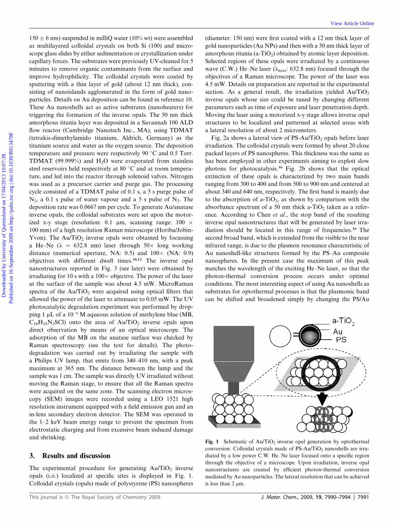
The colloidal crystals formed by PS-Au/TiO2 nanospheres were
prepared as follows. Monodisperse PS nanospheres (diameter:
This journal is ª The Royal Society of Chemistry 2009
during the first stages of this process, MB species converted from
dimers to monomers (MB2 ¼ 2MB), as indicated by the
appearance of the peak at 490 cm�1, which becomes stronger, and
the corresponding weakening of dimer signals.24 Then, the
photoreaction proceeded until the MB was completely removed,
and no signals could be detected. Although the power of the laser
was set at very low values, the removal of MB through this
process is faster than that occurring under UV irradiation.
Moreover, MB removal was also observed even when the power
of the laser was further reduced to 0.05 mW, as shown in ESI.†
Again, Au plays a key role in enhancing the efficiency of this
process. During the first stages the heating generated by light
absorption under resonance conditions promotes the dimer-to-
monomer conversion, which is an endothermic reaction. There-
after, the monomeric species are completely removed in a few
minutes. As MB photobleaching usually occurs at higher laser
power, in the present case the efficient photon-thermal conver-
sion of Au is a decisive factor for driving the degradation of the
dye. This is an important outcome, as it may allow the produc-
tion of microreactors using low power-solid state lasers as exci-
tation sources.
4. Conclusion
In conclusion, we demonstrated that optothermal conversion
can be exploited to generate nanostructured Au/TiO2 inverse
opals in opal substrates with high spatial resolution. This process
was accomplished by means of a C.W. laser at low power, with
a very simple experimental setup. The localized nature of plas-
monic heating provides a mean for generating inverse opals
without heating the remainder of the substrate, thus enabling the
integration of inverse opals into microreactors and nanodevices.
Gold acted as a very efficient photon-thermal converter, allow-
ing the a-TiO2 to be transformed into anatase. The overall
thermal effect was enhanced by the exothermic decomposition of
the underlying polymer. The enhancement of the electromag-
netic field due to the presence of Au NPs allowed the in-situ
microRaman characterization of the inverse opal nano-
structures. The same effect was exploited both for monitoring
and at the same time assisting reactions occurring at these sites
under optimal conditions of efficiency and light exposure. This
approach may be extended to other composite systems, including
Au (Ag)/ZnO or Au (Ag)/CeO2, thus opening attracting
scenarios for fabricating nanostructures and in situ character-
ization of physical and chemical processes occurring at these
interfaces.25
7994 | J. Mater. Chem., 2009, 19, 7990–7994
Acknowledgements
This work was supported by Fondazione Cariplo. Marcello Zucca
is gratefully acknowledged for assistance in deposition of TiO2.
Prof. Laura E. Depero is acknowledged for valuable discussions.
References
1 C. Burda, X. B. Chen, R. Narayan and M. A. El-Sayed, Chem. Rev.,2005, 105, 1025.
2 N. L. Rosi and C. A. Mirkin, Chem. Rev., 2005, 105, 1547.3 H. H. Richardson, Z. N. Hickman, A. O. Govorov, A. C. Thomas,
W. Zhang and M. E. Kordesch, Nano Lett., 2006, 6, 783.4 M. B. Cortie, N. Harris and M. Ford, Physica B, 2007, 394, 188.5 A. G. Skirtach, A. Mu~noz Javier, O. Kreft, K. K€ohler, A. P. Alberola,
H. M€ohwald, W. J. Parak and G. B. Sukhorukov, Angew. Chem., Int.Ed., 2006, 45, 4612.
6 C. Loo, A. Lowery, N. Halas, J. West and R. Drezek, Nano Lett.,2005, 5, 709.
7 L. R. Hirsch, R. J. Stafford, J. A. Bankson, S. R. Sershen, B. Rivera,R. E. Price, J. D. Hazle, N. J. Halas and J. L. West, Proc. Natl. Acad.Sci. U. S. A., 2003, 100, 13549.
8 D. A. Boyd, L. Greengard, M. Brongersma, M. Y. El-Naggar andD. G. Goodwin, Nano Lett., 2006, 6, 2592.
9 W. H. Hung, I.-K. Hsu, A. Bushmaker, R. Kumar, J. Theiss andS. B. Cronin, Nano Lett., 2008, 8, 3278.
10 I. Alessandri and L. E. Depero, Nanotechnology, 2008, 19, 305301.11 I. Alessandri, M. Ferroni and L. E. Depero, ChemPhysChem, 2009,
10, 1017.12 I. Alessandri and L. E. Depero, Chem. Commun., 2009, 2359.13 (a) G. Ozin, A. Arsenault and L. Cademartiri, Nanochemistry, RSC
Publishing: Cambridge, 2008; (b) A. Stein, F. Li and N. R. Denny,Chem. Mater., 2008, 20, 649.
14 S. Nishimura, N. Abrams, B. A. Lewis, L. I. Halaoui, T. E. Mallouk,K. D. Benkstein, K. van de Lagemaat and A. J. Frank, J. Am. Chem.Soc., 2003, 125, 6306.
15 I. Rodriguez, P. Atienzar, F. Ramiro-Manzano, F. Meseguer,A. Corma and H. Garcia, Photonics Nanostruct.: Fundam. Appl.,2005, 3, 148.
16 J. I. L. Chen, G. von Freymann, S. Y. Choi, V. Kitaev and G. A. Ozin,Adv. Mater., 2006, 18, 1915.
17 J. I. L. Chen, G. von Freymann, V. Kitaev and G. A. Ozin, J. Am.Chem. Soc., 2007, 129, 1196.
18 K. Awazu, M. Fujimaki, C. Rockstuhl, J. Tominaga, H. Murukami,Y. Ohki, N. Yoshida and T. Watanabe, J. Am. Chem. Soc., 2008, 130,1676.
19 S. J. Oldenburg, R. D. Averitt, S. L. Westcott and N. J. Halas, Chem.Phys. Lett., 1998, 288, 243.
20 C. Wang, J. Fu, G. M. Chow, P. C. Hsu and Y. K. Hwu, J. Am.Ceram. Soc., 2005, 88, 758.
21 I. Alessandri, M. Zucca, M. Ferroni, E. Bontempi and L. E. Depero,Small, 2009, 5, 336.
22 M. Jakob, H. Levanon and P. V. Kamat, Nano Lett., 2003, 3, 353.23 V. Subramanian, E. E. Wolff and P. V. Kamat, J. Am. Chem. Soc.,
2004, 126, 4943.24 S. H. A. Nicolai and J. C. Rubim, Langmuir, 19, p. 4291.25 A. V. Whitney, J. W. Elam, P. C. Stair and R. P. Van Duyne, J. Phys.
Chem. C, 2007, 111, 16827.
This journal is ª The Royal Society of Chemistry 2009