Exploring chemical bonding in molecules using Raman spectroscopy In the previous experiments, you were introduced to visible and fluorescence spectroscopy. You learned that different molecules absorb and emit different wavelengths of light and that you can characterize molecules based on these patterns. Now you will learn about another spectroscopic technique: Raman spectroscopy. You will use a combination of computer simulations and Raman spectroscopy to study chemical bonding. Exploring Chemical Bonding Using Raman Spectroscopy 4
Transcript
Exploring chemical bonding in molecules using Raman spectroscopy
In the previous experiments, you were introduced to visible and fluorescence spectroscopy. You
learned that different molecules absorb and emit different wavelengths of light and that you can
characterize molecules based on these patterns. Now you will learn about another spectroscopic
technique: Raman spectroscopy. You will use a combination of computer simulations and Raman
spectroscopy to study chemical bonding.
Exploring Chemical Bonding Using Raman Spectroscopy
4
Raman Spectroscopy
STELLARNET 1
Experiment 4: Exploring Chemical Bonding
INTRODUCTION
Learning Goals1 This experiment will introduce you to the technique of Raman spectroscopy and allows you to explore chemical bonding using both computer simulations and Raman spectroscopy. First, you will use a computer simulation of springs to model the properties of single, double, and triple covalent bonds and bonds with different types of atoms (different masses). You will explore what factors impact how bonds oscillate. Then, you will then explore the relationship between bond vibrations and Raman spectroscopy in hexane, 1-hexene, and 1-heptyne. Chemical Bonding
There are many different models for describing covalent chemical bonds and the motion of molecules. Molecules have three types of motion: translation, rotational, and vibrational. Translation motion is when molecules move from one position to another. Rotational motion entire molecule rotating or the internal parts of the molecule rotating with respect to one another. Vibrational motion is when bonds between atoms within a molecule move. There are many different types of molecular vibrations possible with such colorful names as symmetric and antisymmetric stretching, scissoring, rocking, wagging and twisting. Modeling a covalent bond as a spring helps illustrate the vibration of bonds in a molecule. When an atom is displaced from its equilibrium position in a molecule (e.g. when it stretches or wags or twists), it is subject to a restoring force. A spring follows this same pattern, which is called Hooke’s law. Thus, a chemical bond can be modeled as a spring that has weights (atoms) attached to its two ends. This system has a natural vibrational frequency which depends on the masses of the weights and the stiffness of the spring.
Think of your everyday experience with springs. Does the spring oscillate (move up and down) faster or slower with a heavy weight attached versus a light weight? What about the stiffness of the spring? Does a soft spring oscillate faster or slower than a stiff spring (with the same amount of weight attached)?
Bonds are always vibrating from thermal energy present in the surroundings. Light (electromagnetic radiation) can interact with these molecular vibrations in certain predictable ways. Raman 1 This experiment was adapted from: Borgsmiller, K. L., O’Connell, D. J., Klauenberg, K. M., Wilson, P. M., & Stromberg, C. J. (2012). Infrared and Raman Spectroscopy: A Discovery-Based Activity for the General Chemistry Curriculum. Journal of Chemical Education, 89(3), 365–369. http://doi.org/10.1021/ed2002835
Raman Spectroscopy
STELLARNET 2
Experiment 4: Exploring Chemical Bonding
spectroscopy uses the scattering of light from molecular vibrations can be used to characterize molecules. Raman Spectroscopy In the previous experiments, you were introduced to visible and fluorescence spectroscopy. You learned that different molecules absorb and emit different wavelengths of light and that you can characterize molecules based on these patterns. Now you are going to use another spectroscopic technique: Raman spectroscopy. Raman spectroscopy exposes a sample to a single wavelength of light, usually from a laser in the visible, infrared, or ultraviolet range. The laser light is then scattered by the sample. Scattering simply means that the light particles (photons) are forced to deviate from their straight path by some interaction with the sample. In Raman spectroscopy, the laser light interacts with molecular vibrations resulting in the energy of the laser photons being shifted up or down (e.g. Raman shift). The shift in energy provides useful information about the vibrational modes in the system and can be used to identify the type of bonds present in the compound. A Raman spectrum is unique to a material and thus is an excellent technique for identifying compounds. A Raman spectrum consists of a range of features, each associated with a vibrational mode (see Figure 1). Each type of bond (e.g. C-H, C-C, C=C) has unique vibrational modes, which leads to specific interactions with light and thus produce different Raman shifts (see Table 1). This information can be used to identify molecules and study chemical bonding. Table 1: Approximate Raman shifts (cm-1) for several common functional groups
Functional group Region
C−C ~600-1300 cm-1
C=C ~1600 cm-1
C≡C ~2100-2300 cm-1
C−H ~2700-3100 cm-1
Figure 1: Raman spectrum of acetaminophen
Raman Spectroscopy
STELLARNET 3
Experiment 4: Exploring Chemical Bonding
EXPERIMENT PREPARATION
Before you come to lab In preparation for this experiment, you should read the StellarNet Raman spectroscopy operation manual as directed by your instructor. Safety when working with a laser is of the utmost importance. • Caution – use of controls or adjustments
or performance of procedures other than those specified in the StellarNet manual may result in hazardous radiation exposure.
• Do not point laser or allow laser light to be directed or reflected toward other people or reflective objects.
• Always wear appropriate protective eyewear.
• Do not modify unit or remove protective covers or housings.
• Laser light emitted from this equipment may be sufficient to ignite some materials and initiate fire.
• Never operate laser if unit is defective or if safety covers, locks, and labels are damaged or missing.
Collaboration You will work with a partner to complete this laboratory experiment. You and your partner should discuss your answers to the prelab questions before you start the experiment. Feel free to talk about the discussion questions together but make sure the work you turn in is your own and you understand the material.
Safety In preparation for this experiment, you should look up hazard information on the chemicals and equipment used in this laboratory. This might include looking at the Material Safety Data Sheets (MSDS) or Safety Data Sheets (SDS) for the chemicals used in this experiment. Additionally, the Pharos Project’s Chemical and Materials database (https://www.pharosproject.net) provides a wealth of hazard and safety information on thousands of chemicals. You should identify the hazards associated with each chemical used in this experiment, the necessary personal protective equipment (e.g. lab coats, goggles) you should use, and any toxicology and ecological information. After completing this search, you should understand both how to handle chemicals safely in lab and how these chemicals effect the environment outside of your laboratory. Remember, the chemicals you use and the waste you generate don’t stay in lab – they have to be disposed of after you complete your experiment.
Raman Spectroscopy
STELLARNET 4
Experiment 4: Exploring Chemical Bonding
PRELAB QUESTIONS
1. Springs are often used as a model for
covalent chemical bonds. What makes springs a good model for bonds? What limitations does this model have?
2. Predict how you think the weight attached to a spring and the strength of the spring will impact the displacement and oscillation of the spring.
3. Predict the strength of carbon-carbon single, double, and triple bonds (rank C−C, C=C and C≡C from strongest to weakest). Explain your prediction.
4. Briefly describe what molecular properties are measured using Raman spectroscopy.
5. How does Raman spectroscopy relate to other types of spectroscopy (e.g. IR, fluorescence, UV-vis spectroscopies)?
6. What is the unit of frequency used in Raman spectroscopy?
7. Familiarize yourself with the spectrometer system you will use for this experiment. Using Figure 2 as a guide, explain how the spectrometer, laser, and computer (with spectroscopic software) will allow you to obtain Raman spectra.
8. Describe all the hazards associated with the spectroscopy system for this experiment. What precautions should you take when working with this system?
9. Describe the hazards associated with the chemicals for this experiment. What precautions should you take when working with these chemicals? What would you tell someone who wanted to pour these chemicals down the drain (remember, many sinks eventually connect into your local rivers, lakes, and coastal waters)?
Figure 2: StellarNet Raman spectroscopy system
Raman Spectroscopy
STELLARNET 5
Experiment 4: Exploring Chemical Bonding
PROCEDURE
Part 1: Chemical Bond Simulations
In this activity, you will use a spring simulation as model for covalent chemical bonds. A spring is a common model for covalent bonds and will allow you to study the oscillations of bonds. You will use this simulation to explore what factors which influence the frequency of bond vibrations. 1. Go to https://phet.colorado.edu/sims/mass-spring-lab/mass-spring-lab_en.html. PhET
provides free interactive physics, chemistry, biology, and math simulations (to learn more visit https://phet.colorado.edu/en/about). Today you will use a masses and springs simulation to see what factors influence the spring oscillations.
2. You should see a screen that looks like the one below. Spend a few minutes exploring the simulation. The great thing about simulations you can try out many different ideas quickly and safely! Once you’re done exploring refresh your screen to return to the original settings.
3. Hang one of the 100 gram weights on the middle spring (spring 2). You should see the weight move up and down many times (the weight is oscillating). A complete oscillation is the weight moving from its starting position (at the top) down the lowest position and then returning to its starting position.
4. How much time does it take the weight to oscillate three times? Use the stopwatch feature of the simulation to determine how long it takes for the 100-gram weight to oscillate three times. You can also change the time to 1/4 or 1/6 real time in order to more easily count the oscillations. Record this value in your lab notebook.
Raman Spectroscopy
STELLARNET 6
Experiment 4: Exploring Chemical Bonding
5. Once the weight has stopped oscillating measure the displacement of the spring caused by hanging the weight on it. You can drag the ruler around the screen to easily measure the displacement. Record this value in your lab notebook.
6. Repeat these steps for the 50-gram and 250- gram weights. You can use spring 1 and 3 so you can easily compare and contrast the response of the springs to these three different weights. Record the displacement of each spring and the oscillation time of each weight. How is displacement related to the mass of each weight? How do the oscillations change for each weight?
7. You will now adjust the softness of spring 3. This will essentially replicate having single, double, and triple spring sets.
8. First, remove the 50 and 250-gram weights from springs 1 and 3. Keep the 100-gram weight on spring 2.
9. Place the second 100-gram weight on spring 3. Once the weight stops oscillating you should notice that the two weights end at the same displacement.
10. Now, change the softness of spring 3 (using the sliding scale to the right of the springs). What happens when you make spring 3 softer than spring 2? What about when you make spring 3 less soft (harder) than spring 2?
11. Choose two spring softness settings (one that is softer than spring 2 and one that is harder than spring 2). What type of bond (single, double, and triple) do each of these spring softness settings represent?
12. Record the displacement of spring three for each softness setting. How is displacement related to the softness of the springs?
13. Calculate the oscillation time of the weight for each of these settings as well. How do the oscillations change for each spring softness?
Raman Spectroscopy
STELLARNET 7
Experiment 4: Exploring Chemical Bonding
Part 2: Raman Spectroscopy
Material and Equipment
Chemicals
ü Hexane
ü 1-Hexene
ü 1-Heptyne
Equipment
ü Spectrometer (Raman-HR-TEC)
ü Raman sample chamber
ü Ramulaser and Raman probe
ü Computer or tablet
ü Fiber optic cable
ü USB cable
In the "Springs and Bonds" activity, you observed macroscopic systems and saw that vibrational frequency depends on both bond (spring) strength and attached mass. This system was a model for covalent bond behavior. Unfortunately, we cannot view real molecular vibrations directly. In this experiment, you will investigate the molecular motions in hexane, 1-hexene, and 1-heptyne using Raman spectroscopy.
CAUTION The Raman spectroscopy system uses a laser source. Visible and/or invisible laser radiation can be emitted from the Raman probe. Appropriate laser safety procedures should be observed when operating the probe with a laser source, including the use of protective eyewear. Read StellarNet laser manual for precautions before operating this system.
1. Your instructor will demonstrate how to properly use the Raman spectroscopy system and discuss proper safety precautions you should take while working with this system. During this experiment, make sure to ask your instructor any questions you have about using the Raman spectrometer – don’t guess!
2. Obtain your samples of hexane, 1-hexene, and 1-heptyne in appropriate sample vials. Make sure to keep track of which vials contains each sample. However, don’t write or place tape on the clear glass sides of the sample vials – this will obstruct the path of the laser.
Raman Spectroscopy
STELLARNET 8
Experiment 4: Exploring Chemical Bonding
Spectrometer Setup 1. Transfer your samples to the spectrometer. Make sure that your samples are capped so you don’t
accidently spill solution on or near the spectrometer.
2. Review the spectrometer system setup and make sure that the spectrometer system resembles the setup below. Make sure that you can identify all the components of the spectrometer system.
Top view Front view
Make sure the Raman probe fiber optic is securely connected to the laser. While working with this spectrometer never remove Raman probe from the sample holder. The laser is powerful enough to cause serious eye injury.
3. Look at the back of the laser. First, make sure the key is not in the laser yet. If you are running
the laser on battery power push the red button on the back of the laser in. If you are NOT running the laser on batter power first make sure the red button on the back of the laser is not pressed in. Then make sure the power adaptor is connected to the outlet on the back of the laser.
Spectrometer Laser
Sample holder
Raman probe
Fiber optic
Battery
Power outlet
Key: Vertical = OFF
Horizontal = ON Remote interlock
Key Interlock jack
Raman Spectroscopy
STELLARNET 9
Experiment 4: Exploring Chemical Bonding
4. Make sure the interlock jack is placed in the remote interlock on the rear panel of the laser.
5. You may now you insert key into OFF (vertical) position.
6. You may now enable the laser power by turning the key to the ON (horizontal) position.
Taking Raman Measurements
1. Load your sample vial may be loaded into the sample holder.
2. Turn on the computer. Open the StellarNet software by double clicking on the desktop icon SpectraWiz to open the application
3. When you first open the software, you should see a screen that looks like:
Sample holder without sample
Sample holder with sample
Scope Icon
Spectral ID Application
Raman Spectroscopy
STELLARNET 10
Experiment 4: Exploring Chemical Bonding
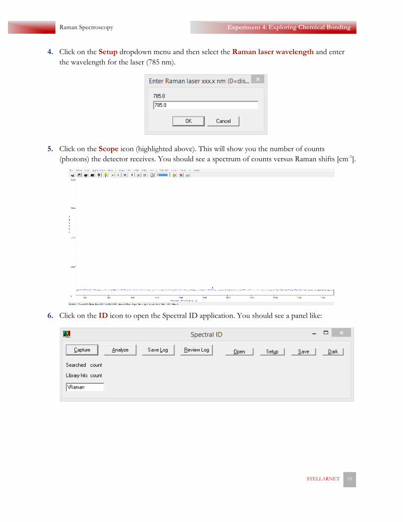
4. Click on the Setup dropdown menu and then select the Raman laser wavelength and enter the wavelength for the laser (785 nm).
5. Click on the Scope icon (highlighted above). This will show you the number of counts
(photons) the detector receives. You should see a spectrum of counts versus Raman shifts [cm-1].
6. Click on the ID icon to open the Spectral ID application. You should see a panel like:
Raman Spectroscopy
STELLARNET 11
Experiment 4: Exploring Chemical Bonding
7. Press and hold left front panel button (marked Fire Laser) to fire the laser.The LED bulb next to this button will light up red indicating that the laser is in operation. You should see a spectrum appear on the computer screen in the SpectraWiz software.
8. While still holding the Fire Laser button, click on the Capture button on the Spectral ID application to collect the displayed Raman spectrum. You can then release the Fire Laser button.
9. Record this spectrum in your lab notebook. Collect Raman spectra for the remaining samples.
Laser Shutdown
1. Once you have collected all of your spectra you can shut the laser down.
2. Make sure to release the Fire Laser button and observe that the red LED turns off, which indicates the laser is no longer active.
3. For complete shutdown, turn the key switch on the rear panel to the OFF (vertical) position. This allows key removal and disables ALL power.
Fire laser button
LED
Raman Spectroscopy
STELLARNET 12
Experiment 4: Exploring Chemical Bonding
DATA ANALYSIS
Part 1: Chemical Bond Simulations
1. From your oscillation data calculate the average time (in seconds) for one oscillation for each weight and spring softness. This is called the period (s). Calculate the inverse of the period; this is the frequency in Hertz (Hz, 1/s).
2. Create a table summarizing your data. It may look like the one below.
Spring weight (grams)
Spring softness
Displacement (cm)
Time for three oscillations (s)
Average period (s)
Average frequency (1/s, Hz)
50 Medium
100 Medium
250 Medium
100 Soft
100 Hard
3. What trends do you notice? How is the spring displacement effected by the weight? By the softness of the spring? How does the period and period and frequency change with changing weight and spring softness?
4. Which spring sets correspond to single, double, and triple bonds? Why?
Part 2: Raman Spectroscopy
1. Examine your collected Raman spectra.
a. What noticeably changes between each spectrum? Note these differences on your spectra.
b. Which bands correspond to specific function groups (e.g. C−C, C=C and C≡C)? Label the main bands on your spectra according to functional group.
Raman Spectroscopy
STELLARNET 13
Experiment 4: Exploring Chemical Bonding
DISCUSSION QUESTIONS
Part 1: Chemical Bond Simulations
1. Based on your data, how would the frequency of oscillation of a bond correlate with the bond strength?
2. In your prelab, you predicted the relative strengths of carbon-carbon single, double, and triple bonds.
a. Based on your observation from this experiment, predict the relative oscillation frequencies of these three bonds. Explain your thinking.
b. Do these earlier predictions on bond strength match your observations from this experiment? Why or why not?
3. Now consider three single bonds (assume that these bonds have equal strength): C−C, C−H, and C−O.
a. How did mass effect the frequency of oscillation of your springs?
b. Based on your observations from this experiment, predict the relative oscillation frequencies of these three bonds. Explain you reasoning.
Part 2: Raman Spectroscopy
1. Discuss the differences between the spectra for hexane, 1-hexene, and 1-heptyne. What causes these differences?
2. How does the frequency of the Raman peak associated with the C−C stretch compare to the frequency of the Raman peak for the C=C and C≡C stretches? Use your observations from Part 1 to support your explanation.
3. What information can Raman spectroscopy give you about bond strength?
4. Discuss the use and limitations of Raman spectroscopy. What advantages and disadvantages does it have over types of spectroscopy?