+ All Categories
Home > Documents > Exploring the Cardioprotective effect of synthetic Wine in ...

Exploring the Cardioprotective effect of synthetic Wine in ...

Date post: 18-Feb-2022
Category:
Upload: others
View: 6 times
Download: 0 times
Share this document with a friend
111
University of Cape Town II EXPLORING THE CARDIOPROTECTIVE EFFECT OF SYNTHETIC WINE IN LONG EVANS RATS LINDIZWE DLAMINI DLMSIB023 Submitted for the Degree of Master of Science in Medicine Faculty of Medicine, University of Cape Town February, 2015 Supervisor: A/Prof. Sandrine Lecour (Hatter Institute for Cardiology Research, University of Cape Town) Co-supervisor: Dr. Roisin Kelly-Laubscher (Department of biological sciences, University of Cape Town) Co-supervisor: Dr. Dee Blackhurst (Department of clinical laboratory sciences, University of Cape Town)
Transcript

Univers

ity of

Cap

e Tow

n

II

EXPLORING THE CARDIOPROTECTIVE

EFFECT OF SYNTHETIC WINE IN

LONG EVANS RATS

LINDIZWE DLAMINI

DLMSIB023

Submitted for the Degree of Master of Science in Medicine

Faculty of Medicine, University of Cape Town

February, 2015

Supervisor: A/Prof. Sandrine Lecour

(Hatter Institute for Cardiology Research, University of Cape Town)

Co-supervisor: Dr. Roisin Kelly-Laubscher

(Department of biological sciences, University of Cape Town)

Co-supervisor: Dr. Dee Blackhurst

(Department of clinical laboratory sciences, University of Cape Town)

The copyright of this thesis vests in the author. No quotation from it or information derived from it is to be published without full acknowledgement of the source. The thesis is to be used for private study or non-commercial research purposes only.

Published by the University of Cape Town (UCT) in terms of the non-exclusive license granted to UCT by the author.

Univers

ity of

Cap

e Tow

n

I

I

III

Acknowledgements

I would like to express my sincere gratitude to the following organisations and people:

The National Research Foundation (NRF) and The Wine Industry Network for Expertise and

Technology (Winetech) for financial assistance.

I would like to thank Dr. Sandrine Lecour who was not only my research supervisor but also

my mentor. Her guidance, patience and assistance during the duration of this study was

invaluable. Her broad knowledge of the topic has made this thesis possible.

My co-supervisors, Dr. Roisin Kelly-Laubscher and Dr. Dee Blackhurst for their

invaluable advice and assistance in completing this dissertation.

Professor Flourian Bauer from the Institute for Wine Biotechnology in Stellenbosch

University, for his assistance and allowing me to use his facilities to complete my project.

Animal Unit staff.

Tasneem Adams and Gerald Maarman for their assistance in laboratory protocols and

orders.

My fellow labmates in the Hatter Institute and ‘The purple lab’ for their friendship, motivation and advice. To my friends for the laughs and support during this journey.

My family for their support, both emotional and financial for the duration of this degree.

IV

Declaration

1. I Lindizwe Dlamini know that plagiarism is wrong. Plagiarism is using another’s

work and to pretend that it is one’s own.

2. I have used the Harvard reference method as the convention for citation

and referencing. Each significant contribution from the works of other people in this

dissertation has been attributed, cited and referenced.

3. This dissertation is my own work.

4. I have not allowed anyone to copy my work with the intention of passing it off as

his or her own work.

5. I acknowledge that copying someone else's work or part of it is wrong, and declare

that this is my own work

__Lindizwe.S.Dlamini________________

Signature

_______25/05/2015________________

Date

V

TABLE OF CONTENTS

VI

TABLE OF CONTENTS

TITLE PAGE ................................................................................................................................................................. #

ACKNOWLEDGEMENTS ..................................................................................................................... III

DECLARATION ..................................................................................................................................................... IV

TABLE OF CONTENTS ................................................................................................................................ V

ABBREVIATIONS................................................................................................................................................. X

LIST OF FIGURES ......................................................................................................................................... XIII

LIST OF TABLES.............................................................................................................................................. XV

ABSTRACT ............................................................................................................................................................. XVI

A. INTRODUCTION ...................................................................................... 1

1.1 PREVALENCE OF CARDIOVASCULAR DISEASE .................................................... 2

1.2 ISCHEMIA/REPERFUSION ................................................................................. 4

1.2.1 Definition .............................................................................................. 4

1.2.2 Pathophysiology ................................................................................... 5

1.2.3 Possible future cardioprotective therapies ............................................. 8

1.3 LIFESTYLE FACTORS FOR CARDIOVASCULAR DISEASE ........................................ 9

1.4 RED WINE AS A CARDIOPROTECTIVE AGENT .................................................... 11

1.4.1 Definition of red wine .......................................................................... 11

1.4.2 Cardiovascular benefit of red wine ...................................................... 12

1.4.3 Epidemiological evidence of red wine induced cardioprotection ........ 12

1.4.3.1 The French paradox ......................................................................... 12

1.4.4 Experimental evidence of red wine induced cardioprotection ........... 14

1.5 POSSIBLE CARDIOPROTECTIVE COMPONENTS IN RED WINE ............................... 14

1.5.1 Alcohol ............................................................................................... 15

1.5.1.1 Epidemiological evidence ................................................................ 15

1.5.1.2 Experimental evidence of alcohol induced cardioprotection ............ 16

1.5.1.3 Does alcohol contribute to red wine induced cardioprotection ........ 17

1.5.2 Resveratrol ........................................................................................ 18

1.5.2.1 Definition and structure ................................................................... 18

1.5.2.2 Cardiovascular benefit of resveratrol ............................................... 19

1.5.2.3 Does resveratrol contribute to red wine induced cardioprotection ... 21

VII

1.5.3 Melatonin ............................................................................................ 22

1.5.3.1 Definition and structure ................................................................... 22

1.5.3.2 Cardiovascular benefit of melatonin ................................................ 24

1.5.3.3 Does melatonin contribute to red wine induced cardioprotection ... 25

B. AIM AND OBJECTIVE .......................................................................... 27

C. MATERIALS AND METHODS ............................................................... 30

1. Production and validation of synthetic wine ................................................ 31

1.1 Production of synthetic wine ..................................................................... 31

1.2 Determination of concentration of total phenolics compounds .................. 33

1.3 Measurement of total antioxidant capacity in synthetic wine ................... 34

2. Testing the cardioprotective effect of synthetic wine .................................. 37

2.1 Animals .................................................................................................... 37

2.2 Experimental design ................................................................................. 37

2.3 The Langendorff preparation as a model of cardiovascular disease ........ 38

2.4 Experimental Langendorff perfused isolated rat heart preparation ........... 39

2.5 Experimental protocol ............................................................................... 40

2.6 Hemodynamic parameters ........................................................................ 41

2.7 Exclusion criteria ..................................................................................... 42

2.8 Infarct size ............................................................................................... 42

3. Measurement of total antioxidant capacity analysis of blood plasma .......... 43

3.1 Protein quantification ............................................................................... 43

3.2 Sample preparation: Protein extraction .................................................. 44

3.3 Thiobarbituric acid reactive substances (TBARS) assay .......................... 44

3.4 Catalase activity assay ............................................................................. 44

3.5 Superoxide dismutase (SOD) activity assay ............................................ 45

4. Statistical analysis ...................................................................................... 46

5. Chemical agents .......................................................................................... 46

D. RESULTS ............................................................................................... 47

1. Determination of stability of synthetic wine over 20 weeks ........................ 48

1.1 Determination of the total phenol content in synthetic wine ……………… ..48

1.2 Determination of the total antioxidant capacity of synthetic wine …………..49

VIII

1.3 Determination of the total antioxidant capacity of synthetic wine enriched with

resveratrol and/or melatonin ............................................................................ 50

2. Effect of synthetic wine enriched with melatonin and/or Resveratrol in isolated

hearts subjected to an ischemia/reperfusion insult ......................................... 51

2.1 Effect of chronic consumption of synthetic wine with melatonin and/or

resveratrol on heart rate ................................................................................. 51

2.2 Effect of chronic consumption of synthetic wine enriched with melatonin and/or

resveratrol on left ventricular developed pressure(LVDP) in hearts subjected to

ischemia/reperfusion injury ............................................................................. 52

2.3 Effect of chronic consumption of synthetic wine enriched with melatonin and/or

Resveratrol on functional recovery in hearts subjected to ischemia/reperfusion

injury ............................................................................................................... 53

2.4 Effect of chronic consumption of synthetic wine or water enriched with

melatonin and/or resveratrol on coronary flow in hearts subjected to

ischemia/reperfusion injury ............................................................................. 54

2.5 Effect of chronic consumption of synthetic wine enriched with melatonin and/or

resveratrol on infarct size ................................................................................. 55

2.5.1 Infarct size measured with IMAGE J analysis .......................................... 55

2.5.2 Infarct size measured with digitized tablet (SummaSketch graphics) ...... 56

3. Analysis of synthetic wine with and without melatonin and resveratrol on rat

plasma antioxidant activity ............................................................................... 56

3.1 Effect of chronic consumption of synthetic wine enriched with melatonin and/or

resveratrol on plasma total antioxidant activity ............................................... 57

3.2 Effect of chronic consumption of synthetic wine enriched with melatonin and/or

resveratrol on plasma levels of oxidative stress ............................................... 58

3.3 Effect of chronic consumption of synthetic wine or water enriched with/without

melatonin and/or resveratrol on plasma antioxidant enzyme catalase activity . 59

3.4 Effect of chronic consumption of synthetic wine or water enriched with/without

melatonin and/or resveratrol on plasma antioxidant enzyme superoxide

dismutase(SOD) activity ................................................................................. 60

E. DISCUSSION ................................................................................................ 61

4.1 Summary of results ..................................................................................... 62

4.2 Stability of synthetic wine ............................................................................ 63

IX

4.3 Cardioprotection with synthetic wine ........................................................... 63

4.3.1 Alcohol fails to induce cardioprotection ..................................................... 63

4.3.2 Resveratrol protects isolated hearts against ischemia/reperfusion injury 64

4.3.3 Melatonin protects isolated heart against ischemia/reperfusion injury ...... 65

4.3.4 Combination of resveratrol and melatonin induces cardioprotection ........ 66

4.4 Role of antioxidants in synthetic wine induced cardioprotection ................. 67

4.5 Limitations and future prospects .................................................................. 68

4.6 Conclusion .................................................................................................. 69

F. APPENDICES ............................................................................................... 70

G. REFERENCES ............................................................................................. 78

X

ABBREVIATIONS

XI

AAPH 2,2-azobis(2-amidinopropane) dihydrochloride

ANOVA Analysis of variance

ATP Adenosine triphosphate

ATPase Adenosine triphosphatase

BCL-2 B-cell lymphoma-2

BHT Butylated hydroxytoluene

Ca Calcium

cAMP cyclic adenosine monophosphate

CF Coronary flow

cGMP cyclic guanosine monophosphate

CHD Coronary heart disease

CVD Cardiovascular disease

FC Folin Ciocalteu

GAE Gallic acid equivalent

GI Global ischemia

GPCR G protein coupled receptor

GSH Glutathione

HDL High density lipoprotein

HR Heart rate

HSP Heat shock protein

IHD Ischemic heart disease

IR ischemia-reperfusion

K Potassium

LDH lactate dehydrogenase

LDL low density lipoprotein

LMIC Low-and middle income countries

LVDP Left ventricular developed pressure

LVEDP Left ventricular end diastolic pressure

LVESP Left ventricular end systolic pressure

MDA Malondialdehyde

Mel Melatonin

MI Myocardial Infarction

ml

Milliliters

mM Millimolar

MT1/2/3 Melatonin receptor(s)

Na Sodium

XII

NAD Nicotinamide adenine dinucleotide

NCD Non-communicable disease

NIH National Institutes of Health

NO Nitric Oxide

NOS Nitric oxide synthase

ORAC Oxygen radical absorbance capacity

pH Hydrogen potential

PKC Protein kinase C

R Reperfusion

Resv Resveratrol

RGJ Red grape juice

RISK Reperfusion Injury Salvage Kinase

RNS Reactive nitrogen species

ROS Reactive oxygen species

RPP Rate pressure product

S Stabilization

SAFE Survivor Activating Factor Enhancement

SEM Standard error of the mean

SOD Superoxide dismutase

SPT 8-(p-sulfophenyl)theophylline

SSA Sub-Saharan Africa

STAT-3 Signal transducer and activator of transcription-3

TBA Thiobarbituric acid

TBARS Thiobarbituric acid reactive substances

TE Trolox equivalent

TNFα Tumour necrosis factor alpha

TTC Triphenyltetrazolium chloride

USA United States of America

WHO World Health Organisation

YPD Yeast peptone dextrose

XIII

List of figures

A. Introduction

Figure 1 Distribution of global non-communicable disease by cause of death in both sexes

Figure 2 Proportion of deaths due to CVD by country income level

Figure 3 Graphical representation of acute myocardial infarction

Figure 4 Graphical representation of the pathogenesis of reperfusion injury

Figure 5

Schematic diagram showing the RISK and SAFE pathway, both pathways may confer

cardioprotection

Figure 6 Five lifestyle changes that can protect against cardiovascular death

Figure 7 Pie chart showing red wine component composition

Figure 8

Graph showing the low mortality rate of CHD in France in comparison to other

European countries despite similar intake of high saturated fats

Figure 9 A graphical representation of J-mortality curve for alcohol consumption

Figure 10 A graphical representation of the bioactive conformations of resveratrol.

Figure 11 Different types of red wine and their resveratrol concentrations

Figure 12 The multiple effects of resveratrol on cardiovascular health and disease

Figure 13 The molecular structure of melatonin

Figure 14 The classic biosynthetic pathways of melatonin in vertebrates

B. Aim and Objectives

Figure 15

A simplified diagram which illustrates a hypothetical setting whereby enriching

synthetic wine with resveratrol and/or melatonin may contribute to the cardioprotective

effect of chronic moderate consumption of wine

C. Materials and Methods

Figure 16 Simplified schematic diagram of the production of synthetic wine

Figure 17

Standard calibration curve of Gallic acid to determine total phenolic content in synthetic

wine

Figure 18

Decrease in fluorescence over time in different concentrations of the vitamin E

analogue trolox.

Figure 19

The area under each curve was for each concentration trolox in nmol was used to

generate a standard curve

Figure 20

A schematic diagram showing the different treatment groups used to assess the effect

of chronic moderate consumption of synthetic wine and red wine on ischemia/

reperfusion injury

Figure 21 A schematic representation of perfusion protocol

Figure 22 Langendorff perfusion retrograde apparatus

Figure 23 A Langendorff-perfused rat heart

XIV

Figure 24

Labchart trace showing various hemodynamic parameters recorded during

experimental protocol

C. Results

Figure 25 The total phenol content in synthetic wine over a period of 20 weeks

Figure 26 The total antioxidant capacity of synthetic wine over of 20 weeks

Figure 27

The total antioxidant capacity of synthetic wine and water enriched with melatonin

(Mel) and Resveratrol (Resv)

Figure 28

Effect of chronic consumption of synthetic wine enriched with/without melatonin(Mel)

and/or Resveratrol(Resv) on functional recovery after 60 minutes of reperfusion

Figure 29

Effect of chronic consumption of synthetic wine or water enriched with/without

melatonin(Mel) and/or Resveratrol(Resv) on infarct size after 60 minutes of reperfusion

using Image J.

Figure 30

Effect of chronic consumption of synthetic wine or water enriched with/without

melatonin(Mel) and/or Resveratrol(Resv) on infarct size after 60 minutes of reperfusion

using a digitized tablet

Figure 31

Oxygen radical absorbance capacity (ORAC) assay to determine the plasma

antioxidant capacity in Trolox equivalents(µmol/mL)

Figure 32

Results of Thiobarbituric acid reactive substances assay (TBARS) assay for the

measurement of malondialdehyde (MDA) in rat plasma

Figure 33

Effect of chronic consumption of synthetic wine or water enriched with/without

melatonin(Mel) and/or Resveratrol(Resv) on Catalase activity in rat plasma

XV

List of Tables

Table 1 Post-fermentation analysis of synthetic wine obtained from the Central Analytical Facility

Table 2

Effect of chronic consumption of synthetic wine or water enriched with/without melatonin and/or resveratrol on heart rate (beats/min) in isolated rat hearts subjected to ischemia/reperfusion injury

Table 3

Effect of chronic consumption of synthetic wine or water enriched with/without melatonin and/or resveratrol on LVDP(mmHg) in isolated rat hearts subjected to ischemia/reperfusion injury

Table 4

Effect of chronic consumption of synthetic wine or water enriched with/without melatonin and/or resveratrol on coronary flow in isolated rat hearts subjected to ischemia/reperfusion injury

XVI

ABSTRACT

XVII

Background: Moderate and chronic consumption of red wine protects against

cardiovascular disease. Wine is a complex matrix containing multiple molecules whose

concentrations can vary from one bottle to another. Therefore, the delineation of the putative

cardioprotective components in wine such as alcohol, resveratrol and melatonin is very

challenging when using commercially available red wine.

Aim: We aimed to use synthetic wine, whose composition is well characterized, to explore

whether the presence of alcohol, resveratrol and melatonin (as found in commercial wines)

contributes to the cardioprotective effect of chronic and moderate consumption of red wine

(equivalent to 2 glasses of wine/day) in an animal model. Additionally, we hypothesized that

synthetic wine enriched with resveratrol and melatonin confers cardioprotection via

improvement of overall antioxidant profile.

Methods: The drinking water of male Long Evans rats was supplemented with synthetic

wine (12% alcohol v/v) with/without resveratrol (100µg/L) and/or melatonin (0.075μg/L) to a

final concentration corresponding to the concentration found in 2 glasses of wine per day.

After 14 days of treatment, hearts were perfused on the Langendorff system and subjected

to 30 minutes global ischemia followed by 60 minutes of reperfusion. Functional parameters

were recorded throughout the experiments and infarct size was measured at the end of the

protocol. Functional recovery (heart rate x left ventricular developed pressure), measured at

60 minutes of reperfusion, was expressed as a percentage of baseline value. Blood plasma

was collected when harvesting the heart to measure total antioxidant capacity, lipid

peroxidation (thiobarbituric acid reactive substances (TBARS) assay), superoxide dismutase

(SOD) and catalase activity.

Results: Control hearts subjected to ischemia/reperfusion presented a functional recovery of

11±2%. Pre-treatment with synthetic wine with/without melatonin or resveratrol did not

improve functional recovery (15±6%,12±1%, 19±4% respectively, n.s. vs control). However,

addition of both melatonin and resveratrol in synthetic wine improved functional recovery to

32±5% (p<0.01 vs control). No significant changes were identified amongst the treatments

when quantifying antioxidant enzyme activity in the form of SOD or catalase. However,

synthetic wine enriched with both melatonin and resveratrol significantly increase lipid

peroxidation (TBARS: 16.3±0.9 µmol/mL vs. 8.5±0.4 µmol/mL for control; p<0.01).

Conclusion: In conclusion, our data strongly suggest that the presence of melatonin and

resveratrol in wine is required for cardioprotection with chronic moderate consumption of

wine. Our data do not provide a role for the antioxidant activities of the wine as a possible

mechanism for cardioprotection. Our findings support that the combination of melatonin with

resveratrol, at the concentration found in wine, may be considered as a therapeutic agent for

cardioprotection.

1

1

A. INTRODUCTION

2

2

1.1 Prevalence of cardiovascular disease

Non-communicable diseases (NCDs) can be defined as all the diseases, which are non-

infectious and non-transferable among people. NCDs include heart disease, most cancers,

diabetes, chronic kidney disease and Alzheimer’s disease amongst others. The World

Health Organisation (WHO) reports that NCDs are the leading cause of death in the world,

representing over 60% of all deaths (WHO, 2011). Of the 57 million global deaths in 2008,

36 million were due to NCDs. NCDs have surpassed communicable diseases as the world’s

major disease burden, with cardiovascular disease (CVD) remaining the principal global

cause of death, accounting for 17.3 million deaths per year (Smith, Collins et al. 2012)

(Figure.1).

CVD is expected to grow to more than 23.6 million by 2030 if no adequate

prevention/therapies are put in place (Mendis, Puska et al. 2012). CVD associated morbidity

Figure 1: Distribution of global non-communicable disease by cause of death in both sexes

From WHO/WHF/WSO: Global Atlas on Cardiovascular Disease Prevention and Control. Geneva. World Health Organisation. 2011

Diabetes mellitus

3

predominantly affects both men and women in low-and middle income countries (LMIC)

(Figure.2) where 80% of these deaths occur predominantly at younger ages compared with

higher income countries (Abegunde, Mathers et al. 2007). CVDs have reached epidemic

proportions in Sub-Saharan Africa (SSA) (Alberts, Urdal et al. 2005, Gersh, Sliwa et al.

2010, Ikem, Sumpio 2011). Previously, conditions such as ischemic heart disease (IHD) and

angina were considered a rarity in SSA (Walker, Sareli 1997, Seedat, Mayet et al. 1992).

However, there has been a recent increase in both prevalence and incidence in the number

of patients presenting with IHD (Mensah 2008), in part as a result of decreased access to

effective and equitable healthcare services in those countries (Abegunde, Mathers et al.

2007, Smith, Collins et al. 2012). Other key factors underlying the increasing prevalence of

CVD in developing countries such as South Africa include: the ongoing change in nutrition

patterns, the increase in weight and obesity, the decrease in physical activity and high levels

of stress as well as the increase of urbanization (Alberts, Urdal et al. 2005). These lifestyle

factors, associated with obesity and poor nutrition, lead to the emergence of well determined

risk factors for IHD (Yusuf, Hawken et al. 2004).

In South Africa, although the continued epidemic of human immunodeficiency virus/acquired

immunodeficiency syndrome (HIV/AIDS) is responsible for 29%and 36% of deaths in men

and women, respectively (Shisana, Rehle et al.2013), CVD represents a major burden. IHD,

hypertension and stroke account for more than a third of deaths in the population older than

65 years (Gaziano, Thomas A 2010). The burden of CVD is predicted to increase

Figure 2: Proportion of deaths due to CVD by country income level

From (Laslett, Alagona et al. 2012)

4

substantially in South Africa over the next decade if measures are not taken to slow down

this burden (Mayosi, Flisher et al. 2009).

1.2 Ischemia/ reperfusion

1.2.1 Definition

IHD is a condition that affects the supply of blood to the myocardium. Myocardial ischemia

occurs when blood flow to the myocardium is decreased by a partial blockage of the

coronary arteries and thus, reduces the myocardium oxygen supply. Complete blockage of

the coronary arteries results in deficient oxygenation and nutrient supply to the

cardiomyocytes, leading to damage and necrosis of the tissue, which is known as a

myocardial Infarction (MI) (Opie & Seedat, 2005)(Figure.3).

In a large majority of cases, myocardial ischemia is confined to specific regions of the

myocardium and is termed regional ischemia. However, there are some conditions which

can lead to the entire myocardium becoming ischemic (global ischemia), such as open-heart

surgery in patients undergoing coronary artery bypass grafting, valve replacement therapy or

heart transplant. While restoring blood flow to the ischemic area is essential to save

threatened cardiomyocytes either by the use of pharmacological therapy with thrombolytics

or through physical means with angioplasty, reperfusion is paradoxically associated with a

Figure 3: Graphical representation of acute myocardial infarction

From: www.medicinenet.com/heart_attack/page2.htm

5

cascade of deleterious effects in cardiomyocytes leading to ischemia/reperfusion (IR) injury

(Yellon, Hausenloy 2007).

1.2.2 Pathophysiology

Ischemic injury

The absence of oxygen halts cellular oxidative phosphorylation, leading to mitochondrial

membrane depolarization, adenosine triphosphate (ATP) depletion, and inhibition of

mitochondrial contractile function (Lesnefsky, Tandler et al. 1997, Reimer, Hill et al. 1981).

This process is aggravated by the breakdown of available ATP as a result of ATP hydrolysis

and increase in mitochondrial inorganic phosphate (Halestrap, Clarke et al. 2004). In the

absence of oxygen, cardiomyocyte metabolism changes to anaerobic glycolysis, resulting in

accumulation of lactate which reduces intracellular pH (Avkiran, Marber 2002, Kloner, Bolli et

al. 1998). These changes lead to modifications in cardiomyocyte physiology and structure,

including mitochondrial and sarcolemmal injury and alterations in intracellular calcium

handling (Meissner, Morgan 1995). If the ischemic insult is limited in time, the damage is

reversible, and restoration of blood flow during this period will lead to recovery of normal

function. However, if ischemia persists for an extended period of time, this damage becomes

irreversible and cell death occurs. Therefore, early restoration of oxygenated blood to the

ischemic myocardium is required to limit infarct size (Simoons, Brand et al. 1985). Ironically,

the return of blood can cause further cardiac damage and is referred to as reperfusion injury.

Reperfusion injury

The reintroduction of oxygen and energy into an ischemic cellular environment triggers

additional events that produce further cardiomyocyte damage collectively called reperfusion

injury. Several factors contribute to reperfusion injury. They include aggregation of

inflammatory mediators (Frangogiannis, Smith et al. 2002), calcium overload (Zimmerman,

Hülsmann 1966, Garcia-Dorado, Ruiz-Meana et al. 2012) and oxidative stress (Garlick,

Davies et al. 1987, Zweier, Flaherty et al. 1987) (Figure. 4). Oxidative stress is a disturbance

in the balance between the production of free radicals and antioxidant defences (Sies 2000).

Reperfusion of ischemic cells results in the formation of potentially toxic free radicals,

including hydroxyl radicals and superoxide anions. Free radicals harm cardiomyocytes

directly by altering membrane proteins and phospholipids leading to metabolic and structural

changes (Tsai, Sun et al. 2008, Kaplan, Babusikova et al. 2003). For instance, oxygen

radicals injure the sarcolemma and may impair contractile function of the cardiomyoctye

6

(Verma, Fedak et al. 2002).The role of free radicals as a source of significant myocardial

damage is further illustrated by studies demonstrating that free radical scavengers, such as

superoxide dismutase, administered during reperfusion help preserve myocardial function

(Salvemini, Cuzzocrea 2002).

Furthermore, during an ischemic period, intracellular calcium increases due to impaired

calcium handling and sarcolemmal damage (Meissner, Morgan 1995). This process can be

worsened with reperfusion. The restoration of a normal extracellular pH after reperfusion

produces a hydrogen gradient across the cell membrane. The sodium/hydrogen exchanger

is activated and causes an influx of sodium into the cytosol. Under physiological conditions,

the resulting increase in intracellular sodium would be corrected by the sodium/potassium

ATPase. However, this channel may not function normally after a period of ischemia due to a

lack of energy and structural damage. In this setting, the sodium excess causes the

sodium/calcium channel to run in reverse, producing an influx of calcium into the calcium-

overloaded cell. Although the pathophysiology of reperfusion injury offers itself to potential

therapeutic strategies, few therapies have made their way into clinical practice. A possible

Figure 4: Graphical representation of the pathogenesis of reperfusion injury

From (Lecour, Opie et al. 2012)

7

reason for this effect is that various mechanisms contribute to the consequences of IR injury.

Thus, the impact of a therapy targeted to a single component of the pathophysiology may be

weakened in clinical practice.

Oxidative stress

Oxidative stress is essentially an imbalance between the production of free radicals and the

ability of the body to counteract or detoxify their harmful effects through neutralization by

antioxidants (Sies 2000). Free radicals are oxygen containing molecules that have one or

more unpaired electrons, making them highly reactive with other molecules. Free radicals

can chemically interact with cellular components such as DNA, proteins or lipids and steal

their electrons in order to become stabilized. This, consequently, destabilizes the cellular

components or molecules which then seek an electron from another molecule, triggering a

large chain of free radical reactions. Oxygen by-products are relatively unreactive but some

of these can undergo metabolism within the biological system to give rise to highly reactive

oxidants. For instance, the diatomic oxygen molecule has two unpaired electrons, if this

molecule accepts an electron the product is a superoxide radical. Many free radicals have

important intermediates such as hydrogen peroxide and peroxynitrite which are not free

radicals but which are highly reactive and may be responsible for some of the biological

effects attributed to free radicals.

Free radicals and their non-radical reactants are recognized as critical mediators of cardiac

injury during ischemia and reperfusion. They have been implicated in cardiac cell death, post

ischemic contractile dysfunction and in chronic cardiovascular diseases. The main source of

free radicals in IR injury is the mitochondrial electron transport chain. During the reduction of

molecular oxygen into biological energy through the four mitochondrial complexes the

reduction process is not completely efficient and 1%-4% of available oxygen is normally

incompletely reduced and leaks from the electron transport chains in the form of a

superoxide radical. This process however becomes significantly accelerated at suboptimal

oxygen tensions or after mitochondrial injury and is believed to be the primary source of

ROS during IR injury. Cellular hypoxia decreases the activity of cytochrome oxidase. When

oxygen is reintroduced, leakage of free radicals from proximal complexes is greatly

accelerated. Although it was previously believed that ROS formation occurred primarily or

solely at reoxygenation after ischemia, it is now known that significant formation of ROS

occurs during ischemia from residual superoxide. This has been demonstrated in

cardiomyocytes (Vanden Hoek,Li et al.1997) and in the whole heart (Kevin, Camara et

al.2002,Kevin, Novalija et al.2003). Most peroxide is dismutated by manganese superoxide

dismutase in the mitochondrial matrix to hydrogen peroxide, which easily diffuses through

8

mitochondrial membranes. The remainder exits the mitochondria through anion channels in

the mitochondrial membrane and is then rapidly converted to hydrogen peroxide in the

cytoplasm, either spontaneously, or when catalyzed by copper superoxide dismutase.

Hydrogen peroxide is reduced to water and oxygen by catalase and glutathione peroxidase.

Alternatively, hydrogen peroxide reacts with transition metals, to generate hydroxyl radical.

Possible future cardioprotective therapies

Currently, there are few promising therapies that can effectively protect the heart against IR

injury. Novel approaches as potential adjunctive therapies to current reperfusion strategies

such as coronary angioplasty and thrombolytics are required to provide further

cardioprotection in the setting of MI to reduce morbidity and mortality. The discovery of

ischemic pre-conditioning and post-conditioning have emerged as promising experimental

therapies against IR injury. In each case, protection is conferred by repeated short bouts of

ischemia interspersed with bouts of reperfusion either before or after a potential lethal

ischemia (Murry, Jennings et al. 1986, Zhao, Corvera et al. 2003). Experimental and clinical

evidence suggests that both of these strategies are promising therapies to protect the heart

against IR injury (Murry, Jennings et al. 1986, Zhao, Corvera et al. 2003, Staat, Rioufol et al.

2005, Thibault, Piot et al. 2008). The signalling pathways that mediate ischemic pre- and

post-conditioning can be initiated by a large variety of agents including adenosine (Liu,

Thornton et al. 1991), bradykinin (Goto, Liu et al. 1995), sphingolipids (Lecour, Smith et al.

2002) and insulin (Jonassen, Sack et al. 2001). These protective effects are mostly mediated

via the activation of Reperfusion Injury Salvage Kinase (RISK) (Yellon, Hausenloy 2007,

Yellon, Baxter 1999) and Survivor Activating Factor Enhancement (SAFE) (Lecour 2009)

pathways (Figure.5). The RISK pathway includes activation of the prosurvival kinases Akt

and extracellular regulated kinase 1/2 (Erk1/2) at the time of reperfusion while the SAFE

pathway includes the activation of the cytokine tumour necrosis factor alpha (TNFα) and the

transcription factor signal transducer and activator of transcription-3 (STAT-3) (Hausenloy,

Lecour et al. 2011).

9

Additionally, ischemic conditioning can also be applied remotely when it is performed by

small episodes of ischemia-reperfusion in an organ separate to the heart (Przyklenk, Bauer

et al. 1993). However, despite an improved understanding of the pathophysiology of IR injury

and encouraging preclinical trials of multiple agents, most of the clinical trials to prevent

reperfusion injury have been disappointing. This could be attributed to several reasons; one

being that the presence of comorbidities may impact the efficacy of the treatment see review

(Heusch 2013). For instance, a study by Engbersen and colleagues demonstrated that

although type 1 diabetes patients were more tolerant to forearm IR injury compared to

healthy controls, the efficacy of ischemic preconditioning was reduced in patients with type 1

diabetes mellitus (Engbersen, Riksen et al. 2012). Despite these problems, adjunctive

therapies to limit IR injury remain an active area of investigation as there is a need for

alternative therapies which could limit the damage of IR injury. Thus, targeting lifestyles

would present a major benefit as it is inexpensive relative to medication and therefore would

be a better approach for low and middle income countries (LMIC).

1.3 Lifestyle factors for cardiovascular disease

The majority of CVD is caused by risk factors that can be controlled, treated or modified

such as high blood pressure, cholesterol, overweight/obesity, tobacco use, lack of physical

activity and diabetes (Yusuf, Hawken et al. 2004, Steyn, Sliwa et al. 2005). However, there

are also some major CVD risk factors that cannot be controlled which include age, gender

and family history (Jousilahti, Vartiainen et al. 1999).

Figure 5: Schematic diagram showing the RISK and SAFE pathway. Both pathways may confer cardioprotection

From Lacerda, Somers et al. 2009

10

With all the factors considered, an unhealthy lifestyle can contribute up to 80% of

cardiovascular deaths, whilst modest reductions in risk-associated behaviours can have

exponential benefits (Cheng, Zhao et al. 2009). For example, a 0.5% reduction in risk factors

can result in as much as a 23% decrease in mortality.

The role of diet is crucial in the development and prevention of CVD. Diet is one of the major

factors that can change an individual’s risk of acquiring CVD. IHD has a low incidence in

some developed countries such as Italy and France, leading to a higher life expectancy in

Mediterranean areas compared to Northern European countries and the United States of

America (USA) (Martínez-González, García-López et al. 2011, Pierucci, Misciagna et al.

2012). Diet and lifestyle related factors are suggested to be responsible for this advantage

(Estruch, Ros et al. 2013). The role of diet in IHD has been well documented for the past

century and substantial evidence about the protection by some food items and nutrients is

currently available (Valls-Pedret, Lamuela-Raventós et al. 2012, Urpi-Sarda, Casas et al.

2012). The Mediterranean diet, first studied by Keys and Grande in 1959 as the traditional

dietary pattern found in areas of Southern Italy and Crete, has attracted significant interest

(Keys, Anderson et al. 1965). The traditional Mediterranean diet is characterized by high

intake of olive oil, nuts, vegetables, and cereals, a moderate intake of fish and poultry, a low

intake of dairy products and wine in moderation (Huxley, Clifton 2013, Willett, Sacks et al.

1995). Several observational studies and secondary prevention trials such as the Lyon diet

heart study have consistently shown that adherence to the Mediterranean diet has

considerable benefit with respect to cardiovascular risk (Sofi, Abbate et al. 2010, Michel de

Lorgeril, Salen et al. 1999). Likewise, the INTERHEART study found five protective factors

which may guard against CVD and include: maintaining an ideal weight, regular exercise,

not smoking, eating a diet rich in fruit and vegetables as well as a moderate intake of alcohol

(2-3 glasses/day) (Yusuf, Hawken et al. 2004) (Figure.6). Until recently, alcohol consumption

was frequently overlooked as an important part of the diet. Alcohol, more specifically wine, is

an essential component of the Mediterranean diet.

.

11

1.4 Red wine as a cardioprotective agent

1.4.1 Definition of red wine

Wine is an alcoholic beverage made from fermented grapes. Although the primary

ingredients of wine include grape berry components including water, the end product yields a

complex composition of compounds mostly as a result of the fermentation process. The final

product can contain multiple chemical compounds varying in amounts from one part per

hundred to parts per billion. To date, more than 1000 compounds have been identified in

wine (Soleas, Diamandis et al. 1997). The water content represents from 80 to 85% of the

wine total mass and is principally derived from grape berries. The alcohol content differs

amongst wines from 9% to 16% and is achieved with fermentation by yeast which converts

sugars into alcohol and carbon dioxide. The most abundant alcohol in wine is ethanol. Under

standard fermentation conditions, it can sometimes accumulate to nearly 16%, but generally,

ethanol concentration ranges between 10 and 13%, depending mainly on the sugar content

of the grape, the temperature and the yeast strain. Phenols are derived from the seeds, skin

and vine stems and can be produced by yeast metabolism. Phenols affect the taste,

appearance, fragrance and antimicrobial properties of the wine. Other compounds found in

wine include: organic acids, glycerol, biogenic amines minerals and amino acids, most of

these compounds are found in low concentrations of not more than 100mg/L (Waterhouse,

2002) (Figure.7).

Figure 6: Five lifestyle changes that can protect against cardiovascular death

From Opie.L ,2011 , Living longer, living better: exploring the heart-mind connection , Oxford University Press, Oxford.

12

1.4.2 Cardiovascular benefit of red wine

In recent years, the benefits of daily moderate consumption of alcoholic beverages,

particularly of red wine, in the prevention of heart disease has received increasing attention

and debate (Klatsky, Armstrong et al. 1990). Alcohol intake from any type of alcoholic

beverage appears to be beneficial, but some studies suggest that red wine confers

additional health benefits (Thornton, Symes et al. 1983). The cardioprotective effects of red

wine have been accredited mostly to several polyphenolic antioxidants. The proposed

mechanisms for the observed cardioprotective effects have included, amongst others,

inhibition of low density lipoprotein (LDL) cholesterol (Frankel, German et al. 1993),

increased high density lipoprotein (HDL) cholesterol (Gaziano, Buring et al. 1993), reduction

or inhibition of platelet aggregation (Renaud, Beswick et al. 1992), increased free radical

scavengers (Sánchez‐Moreno, Larrauri et al. 1999, Saint-Cricq de Gaulejac, Glories et al.

1999) and the increased activation in expression of several cardioprotective oxidative stress

inducible proteins including heat shock proteins (HSPs) (Sato, Maulik et al. 2002).

1.4.3 Epidemiological evidence of red wine-induced

cardioprotection

1.4.3.1 The French paradox

In 1819, cardiologist Dr Samuel Black noticed a high incidence of coronary obstructions at

autopsies in Ireland, however, there was an unexplainable rarity of reports of such

obstructions coming from France. He attributed this difference to ‘the French’ habits and

Figure 7: Pie chart showing red wine component composition

From waterhouse.ucdavis.edu/whats-in-wine/red-wine-composition

13

modes of living’ see review (Evans 1995). In 1979, St Leger and colleagues drew attention

to the cardioprotective properties of wine when they described an inverse relationship

between wine consumption and the risk of mortality from CVD in several countries from

North America and Europe (St Leger, Cochrane et al. 1979). Almost two decades later in

1991, a popular investigative documentary television program in the United States, called 60

minutes, introduced to the public that chronic moderate consumption of red wine in France

could be responsible for the low incidence of coronary heart disease in this country, despite

an increased intake of saturated fat comparable to other developed countries (Renaud, de

Lorgeril 1992) (Figure.8). This observation became known as the “French paradox” and was

first published in the Lancet in 1992 (Renaud, de Lorgeril 1992).

It is possible that the supposed protection conferred by red wine may result from a complex

and partially understood association of wine intake with medical, psychosocial, religious

and/or demographic confounding factors. Since the possibility of a randomized controlled

study is low, the relationship between wine intake and the supposed lower risk of CVD

requires careful analysis. Growing evidence supports that red wine might afford a degree of

Figure 8: Graph showing the low mortality rate of CHD in France in comparison to other European countries despite similar intake of high saturated fats

From Renaud and de Lorgeril, Lancet, 1992

14

coronary protection in part due to multiple confounding factors. For example, moderate wine

drinkers may represent as a proxy of higher socioeconomic status, superior heath status and

lower CV risk (Hansel, Thomas et al. 2010). In addition, it has been demonstrated that

moderate wine drinkers consume a healthier diet when compared with heavy drinkers or

abstainers (Ruidavets, Bataille et al. 2004, Johansen, Friis et al. 2006).

1.4.4 Experimental evidence of red wine induced

cardioprotection

A few studies have demonstrated the cardioprotective effect of red wine against IR in an

isolated rat heart model. An acute treatment of red wine extract (1µg/ml) protects rat hearts

against IR injury by reducing infarct size as well as an improving developed pressure

compared to the control rats (Sato, Ray et al. 2000). The cardioprotective effect of red wine

was further illustrated in a study which examines whether the flesh and seeds of red grapes

possess any cardioprotective abilities. Hence, rats chronically fed with flesh of grapes

(2.5mg/kg) or seeds of grapes (2.5mg/kg) for 30 days are protected against IR injury, as

demonstrated by improved post-ischemic ventricular recovery and reduced myocardial

infarct size compared to the control groups treated with water only (Falchi, Bertelli et al.

2006). These studies demonstrate that red grapes contain components that are

cardioprotective independent of fermentation derived molecules which could be responsible

for this protective effect.

Unfortunately, most of these studies have investigated the cardioprotective effect of red wine

with particular interest in specific components of red wine and not the wine in its entirety.

One of the few studies that investigated the cardioprotective effect of whole red wine was

conducted by Lamont and colleagues who demonstrated that chronic pre-treatment with red

wine (for 10 days) at a concentration equivalent to 2-3 glasses/day was beneficial in male

Long Evans rats exposed to IR injury (Lamont, Blackhurst et al. 2012).

1.5 Possible cardioprotective components in red wine

Red wine contains a complex mixture of bioactive compounds, including flavonols,

monomeric and polymeric flavanoids, highly coloured anthocyanins, biogenic amines and

phenolic acids. Studies have shown that some of these compounds have health advantages

see review (Tsuda 2012, Xiao, Peng et al. 2011). To date, there have been three main

15

components in red wine that have been suggested to elicit cardioprotection: alcohol,

resveratrol and melatonin. Each will be reviewed in detail, specifically with regards to

biological mechanisms supporting the cardiovascular benefits of these components in

moderate consumption of wine.

1.5.1 Alcohol

1.5.1.1 Epidemiological evidence

Substantial evidence suggests the consistent negative correlation between alcohol

consumption and the incidence of CVD. Numerous studies from the late 1970s onwards

have reached a consensus that people who consume one to two drinks per day have a lower

CVD risk compared with abstainers and binge drinkers (Figure.9), a relationship described

as a J-shaped or U-shaped curve (St Leger, Cochrane et al. 1979, Connor 2006). Moderate

alcohol consumption mostly equivalent to 1 drink per day for women and 2 drinks per day for

men has been found to decrease the incidence and adverse consequences of heart disease

in several epidemiological studies (Mukamal, Chung et al. 2006). The definition of one

alcohol drink varies by country and publication. Terms such as light, moderate and heavy

drinking are unclear. For instance, one drink is 8 g of ethanol in England, 12 g in USA, and

20-24g in Japan. According to Dietary Guidelines for Americans, moderate drinking is no

more than 1 drink (12 g of ethanol) per day for women and no more than 2 drinks (24 g of

ethanol) per day for men (McGuire 2011).

Figure 9: A graphical representation of J-shaped mortality curve for alcohol consumption.

From Corrao, Rubbiati et al. 2000

Re

lati

ve r

isk

for

CV

D m

ort

alit

y

16

This phenomenon has been further illustrated by a study from Gronbaek et al, who

investigated the relationship between various types of alcoholic beverages and mortality in a

population comprised of men and women between ages 30 to 79 (Gronbaek, Deis et al.

1995). The findings included the reduction of the relative risk of death from 1.00 in

abstainers to 0.4 for those who drank 3 to 5 glasses of wine per day. With regards to the

intake of beer, 3 to 5 bottles per day conferred a reduction in risk of 0.72 compared to

abstainers. In contrast, consumption of 3-5 drinks of spirits per day was linked with

increased mortality. Furthermore, the study concluded that light and moderate wine drinking

is associated with dose-dependent decrease in all-cause mortality that is attributed to a

decrease in cardiovascular-related disease. The health benefits and mechanisms observed

might be heavily influenced by social, genetic and environmental factors. Hence, a recent

study by Leong et al demonstrated that alcohol consumers living in South Asia and the

Middle East, in contrast to the rest of the world, did not display protection against MI (Leong,

Smyth et al. 2014). In some instances, populations from particular South Asian countries

showed significantly elevated risk after adjusting for quality of diet, body composition and

classic vascular risk factors. This study suggests that the negative effects of alcohol are not

exclusive to frequent binge drinkers but, in addition, can extend to light-to-moderate drinkers.

Thus, the beneficial effects of alcohol intake in human health should be better defined, and

additional research is required before any suggestions can be made to initiate light-to-

moderate consumption of alcohol.

1.5.1.2 Experimental evidence of alcohol induced cardioprotection

Animal experiments have been performed to mimic human drinking patterns in order to

investigate whether moderate alcohol consumption could protect the heart against IR injury.

There is evidence that long-term alcohol consumption may improve survival after myocardial

infarction. Miyamae et al, found that prolonged consumption of 10% ethanol protects against

IR injury in guinea pig hearts (Miyamae, Diamond et al. 1997). Particularly, hearts isolated

from animals fed with ethanol for 3-12 weeks demonstrated better recovery and less

myocyte damage after IR injury compared to controls receiving water only. The authors

attributed the cardioprotective effect to an ethanol-induced adenosine receptor activation, an

important mediator of ischemic pre-conditioning. Furthermore, Kobuyashi et al,

demonstrated that ethanol added to the buffer of perfused rat hearts prior to anoxia, followed

by reoxygenation decreased myocardial injury (Kobayashi, Ashraf et al. 1987). However, this

study did not determine whether chronic ethanol consumption produced protection against

reperfusion injury in the absence of ethanol. Upon review of varied literature, the trend

appears to be that the concentration of ethanol required to produce an adaptive biological

17

response is inversely correlated to the duration of exposure (Diamond, Gordon 1994).

Studies by Miyamae et al have shown that doses as low as 2.5% and 5% ethanol produced

partial cardioprotection after 3 weeks of exposure, however full protection is maximal after 6

weeks of treatment independent of the dose of alcohol given (Miyamae, Diamond et al.

1997). Logically, higher concentrations of ethanol produced maximum protection at 3 weeks

and this was sustained as long as ethanol was consumed for a period of 12 weeks.

1.5.1.3 Does alcohol contribute to red wine-induced

cardioprotection?

Although multiple experimental and clinical studies support a cardiovascular benefit of

chronic consumption of alcohol, other studies strongly suggest that the cardioprotective

effect of red wine goes beyond alcohol content. A study conducted by Keevil et al, showed

substantial inhibition of platelet activity in healthy humans after drinking two cups of purple

grape juice for one week (Keevil, Osman et al. 2000). A study on coronary heart disease

patients showed that 250mL of de-alcoholized Greek red wine was able to decrease arterial

stiffness (Zilkens, Burke et al. 2005). Interestingly, red grape juice had similar

cardioprotective properties to that of red wine. Patients undergoing hemodialysis and who

consumed red grape juice for 14 days, had a significant reduction in plasma monocyte

chemoattractant protein 1 concentration and LDL concentration (Castilla, Echarri et al.

2006). In addition, patients displayed higher levels of HDL compared to patients not

consuming red grape juice. Experiments conducted at the Hatter Institute did not

demonstrate any cardioprotective effect of alcohol in isolated hearts subjected to IR injury

after 2 weeks of feeding with alcohol equivalent to 2-3 glasses of wine/day (Lamont,

Blackhurst et al. 2012). Rats pre-treated with alcohol only (6% and 12%) extracted from red

wine did not attain protection against IR injury compared to the untreated controls. However,

rats pre-treated with red wine containing either 6% or 12% alcohol demonstrated similar

cardioprotection against IR injury. Therefore suggesting that alcohol is not the sole

contributor in red wine induced cardioprotection.

18

1.5.2 Resveratrol

1.5.2.1 Definition and structure

Resveratrol (3,5,4’-trihydroxystilbene) was first isolated from the roots of white hellebore in

1940, and later, in 1963, from the roots of Polygonum Cuspidatum, a plant used in traditional

Chinese and Japanese medicine (Nonomura, Kagawnaa et al.1963). Resveratrol is a

stilbenoid, a type of natural phenol and a naturally occurring phytoalexin produced by a wide

variety of plants in response to stress, injury, ultraviolet (UV) irradiation and fungal infection

as part of their defence mechanism (Langcake, Pryce 1976). Resveratrol can be obtained

exogenously from various dietary sources which include red grapes, peanut butter, dark

chocolate and legumes (Cassidy, Hanley et al. 2000).

Resveratrol initially generated modest interest until 1992, when it was suggested to explain

the cardioprotective effects of red wine (Siemann, Creasy 1992). Subsequently, multiple

studies have shown that resveratrol can prevent or slow down the progression of a wide

variety of diseases including cancer (Jang, Cai et al. 1997), atherosclerosis (Wang, Zou et

al. 2005), heart failure (Rimbaud, Ruiz et al. 2011) and IHD (Ray, Maulik et al. 1999).

Resveratrol exists as two geometric isomers, a cis and a trans form (Figure.10). Trans-

resveratrol is suggested to be the isomer that contributes to the cardioprotective properties

of red wine (Orallo, Alvarez et al. 2002) through endothelium-dependent vasorelaxant

activity, possibly mediated by nitric oxide (NO) release from endothelial cells (Fitzpatrick,

Hirschfield et al. 1993).

Figure 10: A graphical representation of the bioactive conformations of resveratrol.

From Smith, Andreansky 2013

19

Cis-resveratrol is not a natural constituent of grape berries. However, cis-resveratrol has

been detected in all wine analyses (Siemann, Creasy 1992). It is likely that cis-resveratrol

derives from its trans isomer during vinification. Fresh grape skins contain 50 to 100 mg

resveratrol per gram (Jeandet, Sbaghi et al. 1995), however, resveratrol concentrations

show large variation in numerous types of wine (Figure.11). Concentrations of resveratrol in

wine depend on multiple factors which include geographical origin (Goldberg, Ng et al.

1996), wine type (Threlfall, Morris et al. 1999) and oenological practices (Jeandet, Bessis et

al. 1995, Soleas, Goldberg et al. 1995). Another major factor is the fermentation process,

contact with grape skins is important because resveratrol is largely produced by the skin and

not the pulp of grapes which would explain its negligible concentration in white wine

(Jeandet, Bessis et al. 1995).

1.5.2.2 Cardiovascular benefit of resveratrol

Many studies have demonstrated that resveratrol has a wide range of pharmacological

properties. In the cardiovascular system, resveratrol is suggested to mediate its

cardioprotective effects through several mechanisms such as its antioxidant activity,

inhibition of platelet aggregation and anti-inflammatory activity (Figure.12).

Figure 11: Different types of red wine and their resveratrol concentrations

From www.Nutritionexpress.com

20

Resveratrol has been shown to be effective in protecting against IR injury. In a study

conducted by Mokni and colleagues, rats which were pre-treated with resveratrol

(25mg/kg/day) for seven days and subjected to IR injury demonstrated cardioprotection as

shown by improved post-ischemic ventricular recovery, improved antioxidant enzyme activity

and reduced myocardial lipid peroxidation (Mokni, Hamlaoui et al. 2013).This effect was

thought to be mediated by a reduction in reactive oxygen species (ROS) production. In

another study, when resveratrol (100µmol/L) was administered prior to cardiomyocytes

being subjected to two hours of simulated ischemia, there was increased cell viability by

preventing apoptosis via increasing the expression of B-cell lymphoma 2, an anti-apoptotic

factor (Shen, Wu et al. 2012). Additionally, there was a decrease in lactate dehydrogenase

(LDH) release and increase in adenosine triphosphatase activity. These effects were

mediated by activation of the cyclic guanosine monophosphate pathway and protein kinase c

(PKC), a well-known mediator in ischemic preconditioning.

In 2007, a study conducted by Penumathsa and colleagues highlighted the effect of

resveratrol against IR injury. Male hypercholesterolemic Sprague-Dawley rats were fed a 2%

cholesterol diet for 8 weeks, followed by a chronic treatment of resveratrol (20mg/kg/day) for

2 weeks before being exposed to 30 minutes of global ischemia (Penumathsa,

Thirunavukkarasu et al. 2007). Resveratrol-treated rat hearts displayed a significant

reduction in infarct size, as well as improved functional recovery, compared to untreated

hypercholesterolemic rat hearts after an IR insult. In vitro human cardiac specimens treated

with resveratrol (10 μM) and placed in a microperfusion chamber, displayed a significant

Figure 12: The multiple effects of resveratrol on cardiovascular health and disease

21

reduction in apoptosis, compared to control cardiac specimens (Usta, Mustafi et al. 2011).

These findings suggest that resveratrol protects the heart against the detrimental effects of

IR injury. Although many studies have confirmed the cardioprotective effects of resveratrol,

most of them have used a concentration far larger than the resveratrol concentration found

in red wine (0.5 to 13.5mg/L).

1.5.2.3 Does resveratrol contribute to red wine

cardioprotection?

Lamont and colleagues observed that an acute treatment of resveratrol (2.3mg/L)

corresponding to the concentration found in red wine significantly reduced infarct size in

mouse hearts, but not in tumor necrosis factor (TNF) receptor 2 knockout or STAT3-deficient

mice (Lamont, Somers et al. 2011). This data suggests that resveratrol protects via the

SAFE prosurvival signalling pathway. In addition, when rats were pretreated with resveratrol

(7mg/L) chronically for 10 days, resveratrol failed to improve post-ischemic functional

recovery or reduce infarct size (Lamont 2009). Despite abundant experimental studies that

have been carried out in animal models, investigations regarding the safety and beneficial

effects of resveratrol in humans through randomized clinical trials are rare. Recently, a study

conducted by Semba and colleagues involving 800 people from the Chianti region of Italy

investigated whether dietary resveratrol had any links with cancer and CVD death rates

(Semba, Ferrucci et al. 2014) . The study found that the risk of death during the nine-year

follow-up period was no different for people with the highest levels of metabolites

(breakdown products) of resveratrol in their urine, compared to people with the lowest levels.

There were no differences in the risk of CVD. However, one of the limitations of the study

was that resveratrol levels were measured using 24 hour urine samples that looked for

breakdown products of resveratrol and this may not be representative of the participants’

usual pattern of consumption of red wine, berries and chocolate. Moreover, a recent study

suggests that resveratrol could counteract the benefits of cardiovascular exercise in older

men. The objective of the study was to investigate the effects of resveratrol supplements

during high-intensity exercise. For the study, the men were required to increase their

exercise levels and carry out high-intensity interval training three times a week for 4 weeks

(Gliemann, Schmidt et al. 2013). In addition, the men were randomized to receive either a

placebo or a 150-mg dose of resveratrol each day. The results showed that after 4 weeks,

the physical fitness of the men who received resveratrol supplementation did not improve.

However, those who received the placebo saw some benefits associated with physical

activity, such as an increase in superoxide dismutase 2 (SOD2) gene expression associated

with heart protection during exercise. However, the limitations of the study included a small

22

sample size and did not control for confounding drug use. Furthermore, when resveratrol is

given at a high dose it can become a pro-oxidant and could possibly cause damage in the

heart. A study demonstrating this was conducted by Gurusamy and colleagues who found

that resveratrol-induced autophagy occurred when resveratrol was given at higher doses

(100mg/kg/day) (Gurusamy, Lekli et al. 2010).This effect was mediated by inhibiting the

expression of rictor, a component that activates prosurvival kinase AKT. Of note, the field of

resveratrol research has been tainted by scientific fraud by Dr Dipak Das who was charged

with 145 cases of fabricated or false data (Naik 2011, Sen 2012). This has led to many of his

scientific articles being retracted.

1.5.3 Melatonin

1.5.3.1 Definition and structure

Melatonin (N-acetyl-5-methoxytryptamine) (Figure.13) was first isolated and identified in

bovine pineal tissue in the late 1950’s by Lerner and colleagues (Lerner, Case et al. 1958).

One of the earliest findings regarding the production of melatonin in the pineal gland was

that it is primarily synthesized and secreted at night and that the circadian rhythm of

melatonin is determined by the light-dark cycle (Reiter 1995). Apart from the pineal gland,

multiple organs have the capability of producing melatonin including the gastrointestinal tract

(Bubenik 2002) and the melanocytes in the skin (Slominski, Tobin et al. 2008). In more

recent years, melatonin has demonstrated multiple functions, which include; strengthening

the immune system (Maestroni 2001), slowing down cellular aging (Bonilla, Medina-

Leendertz et al. 2002) as well as regulating hormones involved in sexual maturation and

reproduction in females (Cavallo, Ritschel 1996) and regulation of leptin in the

gastrointestinal tract (Rasmussen, Boldt et al. 1999). Melatonin can be sourced exogenously

from certain foods such as cherries (Burkhardt, Tan et al. 2001), rice (Hattori, Migitaka et al.

1995) and meat (Tan, Zanghi et al. 2014). It can also be bought over-the-counter as a

supplement to alleviate jet lag (Herxheimer, Petrie 2002) and most importantly can be

sourced from red wine (Rodriguez-Naranjo, Gil-Izquierdo et al. 2011a). Melatonin is found in

Vitis vinifera seeds during the onset of ripening of the grapeberries (Vitalini, Gardana et al.

2011). Melatonin content in wine varies, depending on the grape type (Iriti, Rossoni et al.

2006), its environment, genetics, harvesting process and storage (Lachman, Šulc et al.

2009). Iriti and colleagues measured the melatonin content in eight different wines. The

berry skin of the Nebbiolo contained the highest melatonin concentration (428.3±32.1pg/ml),

whereas the Cabernet Franc contained the lowest concentration (2.4±0.6pg/ml) (Iriti,

Rossoni et al. 2006). Treatment of grape vines with benzothiadiazole, a plant defence

23

activator, results in an increase in the amount of melatonin in the skins of these grape

berries. The presence of melatonin has been detected in both white and red South African

wines, with large differences in quantites of melatonin from various wine estates (Albertyn

2012). This finding could be due, in part, to the characteristics of each wine, which can be

largely influenced by the agrometeorological conditions (i.e. influential factors in agricultural

crop development are the weather, climate, horticulture, animal husbandary and forestry).

There are several vineyards in the Western Cape region of South Africa. The location of

these vineyards varies, geographically, from areas close to the coast (eg. Hermanus) to in

dry land areas (eg. Robertson Valley).

Biosynthesis and physiological mechanism of action

In the biosynthesis of melatonin, tryptophan is first converted by tryptophan hydroxylase to

5-hydroxytryptophan, which is decarboxylated to serotonin. The synthesis of melatonin from

serotonin is catalyzed by two enzymes, arylalkylamine N-acetyltransferase which catalyzes

the N-acetylation of serotonin to N-acetylserotonin. This enzyme controls the circadian

rhythm of melatonin production by the pineal gland in all vertebrates. Its enzyme activity is

highest at night time and its activity decreases upon exposure to light. Hydroxyindole-o-

methyltransferase is the last enzyme of the melatonin biosynthesis pathway which catalyzes

the transfer of a methyl group from S-adenosyl-L-methionine onto N-acetyl-serotonin to

produce melatonin (Figure.14) (Sugden 1989).

Figure 13: The molecular structure of melatonin

24

Melatonin exerts its influence through membrane receptors. The three major membrane

receptors with varying affinities for melatonin are currently identified as melatonin 1 (MT1)

and melatonin 2 (MT2) (Morgan, Barrett et al. 1994), as well as relatively unknown melatonin

3 (MT3), which shows homology to human quinine reductase 2, a detoxification enzyme

(Mailliet, Ferry et al. 2004). Receptors MT1 and MT2 are members of the G protein coupled

receptor (GPCR) family with seven transmembrane domains. Depending on the specific cell,

melatonin activates a variety of different second messenger cascades after it binds to the

membrane receptor. Melatonin receptors are present in various localizations of the

cardiovascular system and both MT1 and MT2 have been found to be highly expressed in

sections of isolated coronary arteries, aorta and left ventricular specimens from healthy

hearts as well as patients with dilated and ischemic cardiomyopathy (Ekmekcioglu,

Haslmayer et al. 2001). When melatonin binds onto MT1 receptors, it has a vasoconstrictive

effect (Ting, Dunn et al. 1997) in contrast to MT2 receptors which has a vasodilatory effect.

This demonstrates that melatonin has the potential to regulate blood pressure (Masana,

Doolen et al. 2002). The activation of melatonin receptors results in a decrease in cyclic

adenosine monophosphate (cAMP) and in phospatidylino-inositol-4,5-bisphosphate

hydrolysis, which leads to vasoconstriction (Paulis, Simko 2007) .

1.6.3.2 Cardiovascular benefit of melatonin

In 1985, Muller and colleagues found that the likelihood for a MI to occur peaks between 9

am and 11 am (Muller, Stone et al. 1985). During the rest of the day, MI occurs at a similar

rate. This suggests that the occurrence of a MI is dependent on the circadian rhythm.

The production of melatonin is influenced by the detection of light and dark by the retina of

the eye. For instance, the production of melatonin is inhibited when the retina detects light

Figure 14: The classic biosynthetic pathways of melatonin in vertebrates

From Tan, Manchester et al.2007

25

and is stimulated in the absence of light. Thus, melatonin production is lower during the

daytime than at night-time see review (Reiter 1991).There is a correlation between MI

incidence and presence of melatonin. A study by Dominguez-Rodriguez and collegues

demonstrated that acute MI is associated with a nocturnal serum melatonin insufficiency as

well as increased oxidative stress (Domínguez‐Rodríguez, Abreu‐González et al. 2002).

Patients diagnosed with acute MI had lower glutathione peroxidise levels and did not show

diurnal variation. In addition, lipid peroxidation levels in acute MI patients were increased

and diurnal variation was also lost.

Melatonin, given at varying concentrations (1, 10 and 50 μM) attenuated cardiac arrhythmias

in an isolated male Sprague-Dawley rat heart model (Tan, Manchester et al. 1998).

Furthermore, 10 μM of melatonin and 50 μM of melatonin reduced reperfusion ventricular

induced fibrillation and arrhythmias in male Wistar rats subjected to IR injury (Szárszoi,

Asemu et al. 2001, Dobsak, Siegelová et al. 2003). In an ex vivo setting, Lee and colleagues

established the cardioprotective effect of melatonin against IR injury (Lee, Chen et al. 2002).

Male Sprague Dawley rat hearts treated with melatonin (1.0 and 5.0 mg/kg) 10 min before

occluding the left anterior descending artery and 45 min reperfusion, had a significant

reduction in infarct size, reduced tachycardia and fibrillation, compared to the control group.

Melatonin (10mg/kg) treatment of male Sprague Dawley rats for 4 weeks subjected to IR

injury reduced the infarct size and LDH release, compared to vehicle treated hypoxic rats

(Yeung, Hung et al. 2008). Male Wistar rats subjected to 30 min global ischemia and treated

acutely with melatonin (50μM) had a significant reduction in infarct size with a reduction in

LDH release, an indicator of necrosis, compared to untreated controls (Petrosillo,

Colantuono et al. 2009).

1.6.3.3 Does melatonin contribute to red wine-induced

cardioprotection?

Recent research conducted within the Hatter Institute for Cardiovascular Research in Africa

has suggested the role of melatonin in red wine-induced cardioprotection. Chronic pre-

treatment with melatonin at the concentration found in red wine, protected the heart against

IR injury (Lamont 2009). Likewise, isolated rat hearts treated acutely with melatonin at a

concentration similar to that found in red wine (75 ng/L), demonstrated a significant reduction

in infarct size in compared to untreated control hearts after an IR insult (Lamont, Somers et

al. 2011). In STAT-3 knockout or TNF-α knock-out mice, pretreatment with melatonin

(75ng/L) failed to reduce the infarct size after an IR insult. In addition, male Wistar rat hearts

subjected to an acute administration of melatonin (75ng/L) had a significant expression of

STAT-3 in the nucleus of cardiomyocytes. These data suggest that melatonin confers

cardioprotection against IR injury via the SAFE pathway. In another study conducted within

26

the Hatter Institute, the aim was to explore whether South African red and white wines confer

a cardioprotective effect in relation to their melatonin content (Albertyn 2012). The results

demonstrated that chronic moderate pre-treatment with both South African red and white

wines improved the cardiac function of rats subjected to IR injury. However, after measuring

the melatonin content in each South African wine, the data did not show a relationship

between the melatonin content present in the wine and its’ cardioprotective effect. As wine

may contain varying concentrations of melatonin and also other potential cardioprotective

molecules from one bottle to another, it is difficult to establish if melatonin plays a vital role in

red wine induced cardioprotection. Thus, in order to validate the role of melatonin in wine-

induced cardioprotection, we propose the use of a synthetic wine, whose composition is fully

known and controlled.

27

B. AIMS AND

OBJECTIVES

28

Aim and objectives

A large number of epidemiological studies have demonstrated that chronic and moderate

consumption of wine is associated with reduced cardiovascular disease. Elucidating the

components found in wine which contribute to this cardioprotective effect may lead to the

development of novel therapies against ischemic heart disease. Wine contains alcohol and

natural antioxidant compounds, including resveratrol and melatonin, that have all been

suggested to possess cardioprotective properties.

Red wine is a highly complex matrix which contains over a thousand different molecules

whose concentrations vary from one bottle of wine to another. Therefore, the delineation of

the exact components in wine which confers cardioprotection is very challenging when using

commercially available red wine. In the wine biotechnology field, the preparation of synthetic

wine, with a chemical composition which is perfectly known and controlled, is often used to

better understand the wine making process. This medium is designed to reflect the main

nutrients used by yeast that are present in freshly prepared, highly clarified, grape juice.

In the present study, we therefore hypothesised that synthetic wine, enriched with

cardioprotective components such as resveratrol and/or melatonin, contributes to the

cardioprotective effect of chronic and moderate consumption of wine.

To explore this hypothesis, the following objectives will be pursued:

1. In collaboration with the Wine Biotechnology Institute at the University of

Stellenbosch, we will prepare a synthetic wine whose chemical and functional

properties will be characterised.

2. To investigate the effect of chronic and moderate consumption of synthetic wine

enriched with melatonin and/or resveratrol in an ex vivo rat model of IR injury. Rats

will be pretreated with water or synthetic wine containing or not melatonin and/or

resveratrol. After 14 days of treatment, the hearts will be isolated and subjected to a

global ischemia-reperfusion insult. Functional parameters and infarct size will be

assessed as an endpoint

3. To investigate the effect of a chronic treatment of synthetic wine with/without

resveratrol and/or melatonin on antioxidant status in plasma of rats pretreated for 14

days. Antioxidant capacity (ORAC, catalase and SOD assay) and markers of

oxidative stress (TBARS assay) will be assessed in the plasma with the aim to

elucidate if cardioprotective effect is mediated by an improvement of the antioxidant

profile.

29

Figure 15: A simplified diagram which illustrates a hypothetical setting whereby enriching synthetic wine with resveratrol and/or melatonin may contribute to the cardioprotective effect of

chronic moderate consumption of wine

ORAC- Oxygen radical absorbance capacity

TBARS- Thiobarbituric acid reactive substances

SOD-Superoxide dismutase

30

C. MATERIALS AND

METHODS

31

1 Production and validation of synthetic wine

1.1 Production of synthetic wine

Wine is an extremely complex matrix combining over a thousand different chemicals in it. In

order to assess the effect of alcohol, resveratrol and/or melatonin in wine-induced

cardioprotection, a simplified synthetic wine, whose composition is perfectly controlled was

designed. Sugars (glucose and fructose) are used as the main energy source and organic

acids as well as amino acids are used as nitrogen sources.

The preparation of the synthetic wine is based on a method previously described by

Henschke and colleagues (Henschke, Jiranek 1993). The composition of the synthetic wine

is described in Appendix A. Briefly, sugars, organic acids, organic salts, amino acids, trace

elements, vitamins and lipids were added into 600mL of deionised water. The pH of the

medium was adjusted to 3.3 by adding potassium hydroxide and the volume was increased

to 1L with deionised water before inoculation.

Yeast strain and fermentation conditions

The yeast strain used in this study was VIN13 (Anchor yeast, South Africa), a diploid

saccharomyces cerevisiae strain used in industrial wine fermentations. Yeast cells were

cultivated at 30°C in Yeast Peptone Dextrose (YPD) synthetic media 1% yeast extract

(Biolab, Midrand, South Africa), 2% peptone, and 2% glucose (Sigma-Aldrich, Germany).

Solid medium was supplemented with 2% agar (Biolab, South Africa). Fermentation bottles

were inoculated with YPD cultures in the exponential growth phase to an OD600 of 0.1. All

fermentations were carried out under anaerobic conditions in 1L round-bottom flasks and

sealed with rubber stoppers to provide better exclusion of oxygen with a carbon dioxide

outlet. The fermentation temperature was maintained at approximately 25°C, and no

continuous stirring was performed during the course of the fermentation. The fermentations

followed a time course of 23 days, and the bottles were weighed daily to assess the

progress of fermentation. As the sugars were converted into alcohol and carbon dioxide by

the yeast, the fermentation flask would weigh less on each day. The fermentation was

considered complete when residual sugars in wine were lower than 2g/L and the

fermentation flask weight stabilized. Alcohol content was measured and equalled to 12%

(v/v). Once primary fermentation was completed there is a need for confirmation of the

success of the fermentation process. The central Analytical Facility in the Wine

biotechnology institute at the University of Stellenbosch completed these analyses including

a test for residual sugars to confirm the dryness of wine and cessation of fermentation. The

results of an analysis of the synthetic wine used in this study are shown in Table 1. Once full

32

Produce grape juice medium

Ferment grape juice medium for 23 days at 25°C

Aliquot synthetic wine and store at 4°C

analysis of the synthetic wine was complete, the wine was degassed with nitrogen before it

was aliquoted into standard Bordeaux 750mL wine bottles and sealed with screw-top caps.

The wine bottles were subsequently stored at 4°C until they were used for further

experiments.

Table 1: Post-fermentation analysis of the synthetic wine obtained from the Central

Analytical Facility

Acetic acid(g/L) D-Fructose(g/L) D-Glucose(g/L) Glycerol(g/L)

0.8 0.0187 -0.0042 6.006

A wine is considered dry when residual sugars (D-fructose and D-glucose) are less than

1g/L, in order to avoid re-fermentation of yeast. Analysis of volatile acids establishes a

baseline that enables monitoring of potential spoilage problems throughout the storage life of

a specific wine. At the post-fermentation stage, acetic acid, the main component of volatile

acidity, may be formed by oxidation of ethanol in an aerobic environment. The acceptable

upper limit for volatile acidity is 1.2g/L (Zoecklein, Fugelsang et al. 1990).

Nitrogen sources Vitamins Trace elements Lipids

Deionised water Carbon sources Acid-salts

Figure 16: Simplified schematic diagram summarising the different steps in the production of the synthetic wine

33

Functional assays of synthetic wine

A large number of epidemiological studies have demonstrated that moderate consumption of

red wine is associated with reduced mortality and reduced IHD, an association popularized

as the ‘French paradox”. Although red wines do not contain significant amounts of

antioxidant vitamins such as vitamin C and E, their protective effects have been largely

ascribed to phenolic components. It has been proposed that phenols act as antioxidants.

Phenols have shown to be effective radical scavengers, with respect to lipid, protein and

deoxyribonucleic acid (DNA) oxidation, which is determined mostly by their reactivity as

hydrogen or electron donating agents.

Red wine contains wood and yeast derived phenols, in addition to the large numbers of

phenol compounds which originate from grapes, particularly the skins which are removed

during the vinification of white wine. Thus, because of the different types of grapes used and

the different production processes implemented, there are some fundamental differences in

white and red wine composition in terms of the nature of polyphenols.

In order to investigate the effect of synthetic wine-induced protection, it is essential to

characterize the chemical and functional properties of the synthetic wine.

1.2 Determination of concentration of total phenolic compounds in synthetic

wine

The Folin-Ciocalteu assay is the most commonly used procedure to determine the total

phenolic content of food extracts. The concentration of synthetic wine phenolics was

measured by using the Folin-Ciocalteu (F-C) method adapted from Waterhouse

(Waterhouse 2002).The F-C assay is an in vitro colormetric assay based on the reduction of

a phosphotungstate –phosphomolybdate complex by phenolic compounds.

Wine samples (20µL) and gallic acid standards (20µL) were mixed with 1.58mL water in 5mL

plastic tubes. Undiluted commercially available F-C reagent (100µL) was added to the tube

and the mixture was vortexed and left at room temperature for 5 minutes. An aqueous

sodium carbonate (20% m/v) solution (300µL) (refer to Appendix B) was added to the tube,

vortexed and incubated at 40ºC for 30 minutes. The absorbance of the mixture was

determined by spectrophotometry (Spectramax Plus 384, Molecular Devices, Labotec, South

Africa) using Softmax Pro (Version 4.4) software at 765nm. A calibration curve was prepared

using gallic acid ranging from 0 to 500 mg/L (Figure.17). The results were expressed in mg/L

of gallic acid equivalents (GAE). Each sample and standard was measured in triplicate.

34

1.3 Measurement of total antioxidant capacity in the synthetic wine by the

oxygen radical absorbance capacity (ORAC) assay

Total antioxidant capacity was performed in synthetic wine using methods previously

described by Cao and collegues (Cao, Alessio et al. 1993) using the oxygen radical

absorbance capacity (ORAC) assay. This assay evaluates the intrinsic antioxidant protective

effect of samples against oxidative degradation of a fluorescent molecule, fluorescein (3,6,

dihydroxyspiro(isobenzofuran-1(3H), 9(9H)(xanthen)(disodium) (Sigma Aldrich, Germany),

following mixing with AAPH (2,2′-azobis(2-amidinopropane)dihydrochloride), a peroxyl

radical generator. The fluorescence intensity decreases as the oxidative damage of the free

radicals released by AAPH progresses (Figure.18).

y = 0.001x + 0.0735 R² = 0.9979

0.000

0.100

0.200

0.300

0.400

0.500

0.600

0.700

0 100 200 300 400 500 600

Ab

sorb

ance

at

76

5 n

m

mg/L gallic acid

Folin-Ciocalteau Gallic acid standard curve

Figure 17: Standard calibration curve of gallic acid to determine total phenolic content in synthetic wine

35

0

20

40

60

80

100

120

140

160

180

0 2 5 10 15 20 30 45 60 75 90

Re

lati

ve f

luo

resc

en

ce in

ten

sity

(%

)

Times (Minutes)

0.078

0.156

0.313

0.625

1.25

2.5

5

Synthetic wine

Blank

The protection against the free radicals by the synthetic wine is compared to known

concentrations of the vitamin E water-soluble analogue Trolox (6-hydroxy-2,5,7,8-

tetramethylchroman-2-carboxylicacid (Sigma-Aldrich, Germany). The reactions were carried

out in phosphate buffer (75mM, pH 7.4) and the final reaction volume was 300μL. To each

well of a 96-wellplate was added with diluted wine samples or trolox standards (50μL),

phosphate buffer (50μL) and fluorescein (100µL) (refer to Appendix C). The mixture was pre-

incubated for 15 minutes at 37°C. AAPH solution (100μL; 32.1µM final concentration) was

added rapidly using a multichannel pipette. The plate was automatically agitated prior to

each reading. The plate was immediately placed in the Varian Cary eclipse fluorescence

spectrophotometer reader (Set point tech., South Africa) and the fluorescence was recorded

every 5 minutes for the first 30 minutes and every 15 minutes thereafter for the duration of

the experiment. Fluorescence readings were carried out using excitation and emission filters

at 485nm and 520nm, respectively. A blank using phosphate buffer in place of the wine

sample, and eight calibration solutions using Trolox (between 1 and 8 nmol/mL final

concentration) were analysed in the same run. AAPH and trolox solutions in phosphate

buffer (75 mM,pH 7.4) were prepared daily and fluorescein was diluted from a stock solution

(1.196mM) in the same phosphate buffer that was stored at 4°C. All sample reactions were

measured in duplicate.

A standard curve was generated using the known concentrations of trolox, compared to the

area under the curve (Figure.19).

Figure 18: Decrease in fluorescence over time with different concentrations of the vitamin E analogue trolox.

36

The area under the curve is proportional to the antioxidant capacity as a small area indicates

faster decay and less protection against the free radicals generated by AAPH. The area

under the decay curve over time of the wine samples was calculated using Microsoft Excel

and the concentration of trolox equivalents of antioxidant capacity in nmol/mL was

extrapolated from the trolox standard curve.

y = 2074.8x + 5764 R² = 0.9973

0

2000

4000

6000

8000

10000

12000

14000

16000

18000

0 1 2 3 4 5 6

Are

a u

nd

er

the

cu

rve

Concentration (nmol)

Trolox standard calibration curve

Figure 19: The area under each curve was for each concentration trolox in nmol was used to generate a standard curve

37

2 Testing the cardioprotective effect of synthetic wine

2.1 Animals

All experimental procedures were carried out with the approval of the faculty of Health

Sciences Animal Ethics committee, University of Cape Town (Application 013/015 “Exploring

the cardioprotective effect of synthetic wine using Long Evans rats”). All protocols were

carried out in compliance with the guide for care and use of Laboratory Animals published by

the U.S National Institute for Health (NIH publication No. 85(23), revised 1996). Male Long

Evans rats (150-170g) were bred and obtained from the University of Cape Town animal

unit. Animals were allowed access to food ad libitum but water or treatment was given

according to body weight. The temperature was maintained at 22-24°C and lighting at 100-

300 lux. Rats were housed in a 12-hour light and dark cycle, with lights on at 06h00.

2.2 Experimental design

In order to assess the cardioprotective effect of synthetic wine with varying wine

components, Long Evans rats were pre-treated with synthetic or water for a period of 14

days. For this study, a total of 57 rats were divided into eight groups (Figure.20): Control

(n=12), synthetic wine only (n=6), synthetic wine enriched with resveratrol only (n=7),

synthetic wine enriched with melatonin only (n=7), synthetic wine enriched with both

melatonin and resveratrol (n=6), water enriched with resveratrol only (n=5), water enriched

with melatonin (n=8) and water enriched with both melatonin and resveratrol (n=6) .The

treatment solutions were prepared by adding one part of synthetic wine or distilled water

enriched with either resveratrol (100µg/L) or melatonin (75ng/L) (refer to Appendix F) to

seven parts of drinking water as previously described (Lamont, Blackhurst et al. 2012).

When the treatment was given to the rats, both body weight and the amount of drinking

water consumed by each rat was taken into account to ensure that the amount given per day

corresponded to an equivalent of 2 glasses of wine/day which would mimic the human

setting. The rats were treated for a period of 14 days. On the 15th day, rats were

anaesthetised, and ex vivo hemodynamic measurements were taken using the isolated

Langendorff heart system.

38

Long Evans rats

n=57

Synthetic wine & Resveratrol

n=7

Synthetic wine & Melatonin

n=7

Water & Resveratrol

n=5

Water & Melatonin

n=8

Control (Drinking water) n=12

n=12

Synthetic wine only

n=6

Synthetic wine, Melatonin & Resveratrol

n=6

Water, Melatonin & Resveratrol

n=6

2.3 The Langendorff heart preparation as a model for the study of

cardiovascular disease

The isolated retrograde perfused mammalian heart system was first pioneered by Oscar

Langendorff in 1895. The Langendorff system is a retrograde perfusion system which is

frequently used in cardiac physiology to study cardiac metabolism, contractile function and

coronary flow regulation.

Before calibrating the Langendorff system, the apparatus was washed out with boiling

distilled water several times to remove bacteria and debris. Once the apparatus was fully

cleaned the Krebs Henseleit buffer (refer to Appendix D) was prepared. The molecular

composition of the buffer resembles that of blood plasma in order to successfully perfuse the

heart ex vivo.The Langendorff heart preparation entails the cannulation of the aorta which is

then attached to a tank containing Krebs fluid oxygenated with 95% oxygen and 5% carbon

dioxide. The oxygenated Krebs fluid is delivered in a retrograde direction down the aorta at a

constant pressure maintained at 100cm with water. The aortic valves are forced shut and the

perfusion fluid is directed into the coronary ostia, in that way perfusing the entire ventricular

mass of the heart, draining into the right atrium via the coronary sinus.

Figure 20: A schematic diagram showing the different treatment groups used to

assess the effect of chronic moderate consumption of synthetic wine on ischemia/

reperfusion injury

39

The general procedure for Langendorff perfusion entails excision of the heart from the donor

anaesthesized animal. Immediately after excision, the heart is immersed in ice cold Krebs

fluid before cannulation and vascular perfusion. A water-jacketed reservoir positioned above

the aortic cannula, contains the Krebs fluid which is oxygenated through a sintered glass gas

distributor. The aorta is then eased over the end of the cannula, taking precaution not to

insert the cannula too far into the aorta. Once the heart is securely attached to the cannula,

any excess tissue is trimmed away. Once cannulation is completed and coronary perfusion

is initiated, contractile function and regular heart rhythm will return within a few seconds.

Throughout this time, various instrumentation is taken. Contractile activity of the heart, is

assessed through insertion of a compliant intraventricular balloon. The balloon is inflated

with water from a syringe until a left ventricle end diastolic pressure of between 4-12mm Hg

is obtained. Once the balloon is in position, left ventricular systolic, diastolic and developed

pressure can be recorded. Once instrumentation of the heart has been completed, a

temperature regulated heart chamber should be placed around the heart to maintain the

temperature at 37°C.

2.4 Experimental Langendorff perfused isolated rat heart preparation.

The Long Evans rats were anaesthetised with sodium pentobarbitone (60mg/kg) and heparin

(200I.U.) intraperitoneally. Prior to anaesthesia, animals were placed in a quiet room for 15

minutes to acclimatise them to the new environment. The depth of anaesthesia was judged

by the absence of the pedal withdrawal reflex, whereby the paw of the rat was pinched and

the perception of pain was observed. A thoracotomy was performed when the rat was

deemed to have reached surgical anaesthesia. An incision of the skin below the sternum

was delicately performed. The incision was continued along the proximal ends of the left and

right costal margins. The thoracic cavity was opened and the pericardium surrounding the

heart was removed, exposing the beating heart in the chest cavity. The heart was gently

cradled and hoisted between the thumb and the index finger and removed with one quick

precise cut without inflicting damage to the aorta. The isolated heart was immediately placed

into ice-cold Krebs buffer [NaCl 118.5mM, NaHCO3 25mM, KCl 4.7mM, MgSO4.7H2O1.2mM,

KH2PO4 1.2mM, Glucose 11.1mM, and CaCl2.2H2O1.3mM] as previously described (Krebs,

Henseleit 1932) (for preparation and abbreviations refer to Appendix D). Blood was removed

from the rat chest cavity and stored in a tube containing heparin (200 I.U). These tubes were

centrifuged immediately at a speed of 2000g for 10 minutes to separate the plasma from the

serum. The plasma was immediately stored at -80ºC and later used for oxidative stress

analyses.

40

Subsequently, the heart was mounted retrogradely onto the cannula of the Langendorff

apparatus within 3 minutes with two pairs of forceps stretching the aorta on either side to

ensure a fit onto the cannula (Figure.21).

This procedure was carried out under low flow to enable a fluid-fluid connection. The heart

was temporarily secured with a bulldog clamp in order to keep it in place. Thereafter, a thin

suture was tied around the aorta to secure the heart onto the cannula. The left ventricle was

exposed with a small excision of the atrial appendage surrounding the left atria, to enable

the insertion of a balloon. The intraventricular balloon was attached to a catheter filled with

water which was connected to a pressure transducer (MLT0670, Lasec, South Africa) on a

PowerLab system (ADInstruments, Australia) for hemodynamic measurements. Inside the

left ventricle, the balloon was inflated by filling it with water, thus creating a closed system to

measure the pressure experienced by the left ventricle.

2.5 Experimental protocol

All hearts were stabilized (S) for a period of 30 minutes and consequently subjected to 30

minutes of global ischemia (GI) followed by 60 minutes of reperfusion (R) (Figure.22).

Figure 21: A Langendorff-perfused rat heart. Figure key: (a) intraventricular balloon

inserted into the left ventricle and mounted on a 21-gauge bevelled needle; (b)

thermal couple for continual temperature monitoring inserted in pulmonary artery; (c)

cannula whereby heart is mounted onto system; (e) a 2-way tap placed between the

bubble trap and cannula and (d) water-jacketed reservoir

41

Global ischemia was achieved by complete cessation of perfusate while reperfusion was

restored by re-introducing perfusate. Temperature was measured with a thermocoupled wire

(Physitemp, NJ, United States of America) inserted into the pulmonary artery, and connected

to a Digitron 2600T temperature sensor (Torquay, United Kingdom)(Figure.23). Temperature

was controlled at 37°C with a water jacket and a desk lamp. At the end of reperfusion, hearts

were removed and stored at -80ºC before staining and infarct size analysis.

2.6 Hemodynamic parameters

Hemodynamic parameters were monitored throughout the experiments with LabChart Pro 7

software (ADInstruments, Australia) and included heart rate (HR) expressed in beats/min ,

left ventricular developed pressure at mmHg (LVDP: difference between left ventricular end

Figure 22: A schematic representation of perfusion protocol

14 days pre-treatment

100cm Krebs-Henseleit buffer, oxgenated with 95% 𝑂2 and 5% 𝐶𝑂2

LabChart recorder

Temperature control system

Pressure transducer linked to water inflatable intraventricular balloon

Plasma collection

Infarct size assessment

Figure 23: Langendorff perfusion retrograde apparatus

42

systolic pressure (LVESP) and end diastolic pressure (LVEDP)) and the coronary flow (CF)

expressed in ml/min. Functional recovery was assessed through the rate pressure product

(RPP) which was calculated as LVDP X HR at the stabilisation period compared to the end

of reperfusion. CF was measured with Falcon™ 15mL Conical Centrifuge Tubes.

Functional parameters were measured at 5, 10, 20 and 30 minute intervals during the

stabilisation period to ensure the inclusion criteria were met.

Functional parameters were also measured after 5,15,30,45 and 60 minutes of

reperfusion.

2.7 Exclusion criteria

Rats that did not meet the terms of the following criteria were not included in the study:

A. The heart rate should fall between 240-360 beats/min

B. The rate × pressure product must fall within the range 21 000-36 000

C. Left ventricular end diastolic pressure of 2-12mm Hg

D. The developed pressure should fall between 70-120mm Hg

E. The coronary flow rate should be between 8-18 ml/min

2.8 Infarct size

Hearts were sectioned transversely at 2mm thickness and stained by immersion into 2,3,5-

Triphenyltetrazolium chloride solution (TTC) (1% in phosphate buffer) at pH 7.4 for 10

minutes at 37ºC. TTC is a redox indicator used to differentiate between metabolically active

Heart rate

Systolic pressure

Diastolic pressure

Left ventricular developed

pressure

Figure 24: Labchart trace showing various hemodynamic parameters recorded during experimental

protocol

43

and inactive tissue. TTC is a colourless solution which is enzymatically reduced to a brick

red precipitation of formazan dye in living tissues with intact mitochondrial respiration due to

the activity of various dehydrogenases in the presence of the electron donor NADH. After

TTC staining, necrotic zones remain as pale areas of the heart as a result of enzymatic

wash-out in damaged cells. The slices were then fixed in 10% formalin for 24 hours and

scanned using a flatbed scanner. Infarct size was quantified by with both computerised

planimetry (Summa sketch II; Summa Graphics) and Image J (National Institute of Health,

United States of America).

3 Analysis of total antioxidant capacity of blood plasma

There is strong evidence to suggest that increased intracellular ROS are a central cause of

myocardial injury. The changes associated with myocardial injury result, in part in an

imbalance between the formation of oxidants and the availability of endogenous

antioxidants. Experiments were performed using the ORAC, thiobarbitituric acid reactive

substances (TBARS), catalase and superoxide dismutase assay to measure the total

antioxidant capabilities and levels of oxidative stress in plasma from animals that received

different treatments for 14 days.

3.1 Protein quantification

The Lowry Assay was used to quantify the concentration of plasma proteins (Lowry,

Rosebrough et al. 1951). A series of dilutions of 0.3mg/ml bovine serum albumin was

prepared to create a standard curve with concentrations which ranged from 0.03 to

1.15mg/ml. A volume of 200µL of sample or standard was added to 1ml of freshly mixed

complex-forming reagent, subsequently the solution was left to stand at room temperature

for 10 minutes. Afterwards, 100µL of diluted folin reagent was added. The mixture was left to

stand at room temperature for a further 30 minutes. The absorbance was read at 750nm for

protein concentration below 500ug/mL or at 550nm if the protein concentration was between

100µg/ml and 2000 µg/ml.

The standard curve of absorbance as a function of initial protein concentration was used to

determine the unknown protein concentrations.

44

3.2 Sample preparation: protein extraction

Plasma samples were thawed slowly and mixed well on a vortex. To extract the protein from

the plasma, 200μl ice cold ethanol (100%) was added to 100μl of plasma. The mixture was

then centrifuged at 12000 rpm at 4°C for 5 minutes. The protein free supernatant was diluted

363x in the phosphate buffer (Refer to Appendix E). Plasma antioxidant capacity was

determined by ORAC assay using deproteinized plasma using the same method as

previously described in section 1.3 of this chapter.

3.3 Thiobarbituric acid reactive substances (TBARS) assay

The thiobarbituric acid reactive substances (TBARS) are formed as part of lipid peroxidation

and may be detected using thiobarbituric acid (TBA) as a reagent. The assay mostly

measures malondialdehyde (MDA) in the plasma, a low molecular weight end product of lipid

peroxidation of polyunsaturated fatty acids. MDA and other TBARS in the plasma condense

with two equivalents of thiobarbituric acid to give a red derivative that can be assayed

spectrophotometrically. In order to measure the amount of MDA in the rat plasma, the

TBARS assay was performed according to the method of (Jentzsch, Bachmann et al.

1996)Jentzsch (Jentzsch, Bachmann et al. 1996).

Plasma was mixed with butylated hydroxytoluene (BHT) and 50µL orthophosphoric acid in

2mL Eppendorf micro test tubes and vortexed for 10 seconds. 25µL TBA reagent was added

and the mixture was vortexed again. The reaction mixture was then heated at 90ºC for 45

minutes. The tubes were put on ice to stop the reaction. After cooling to room temperature

for 5 minutes, TBARS were extracted once with 500µL n-butanol. To facilitate phase

separation, 50µl saturated sodium chloride solution was added, and the test tubes were

centrifuged at 2000 rpm for 2 minutes in a centrifuge. 300µL of the upper butanol phase was

placed in a flat-bottom 96-well plate. Absorption was read at 535nm on a spectrophotometer

(Spectramax Plus 384, Molecular Devices, Labotec, South Africa) using Softmax Pro

(Version 4.4) software

3.4 Catalase activity assay

The catalase activity assay is based on the principle that catalase, an antioxidant enzyme,

catalyses the reduction of hydrogen peroxide to water (Aebi 1984) and (Claiborne 1985).

For this assay, 30% hydrogen peroxide was diluted to a concentration of 12mM by adding

45

150µL of hydrogen peroxide to 100mL of phosphate buffer. The phosphate buffer was

prepared by adding KH2PO4(50mM) to Na2HPO4(50mM) to a final volume of one litre and

concentration of 50mM (ph 7.0). All the samples were diluted to a ratio of one in ten. 241µL

of phosphate buffer was added into the first two wells and 241µL of the hydrogen peroxide

was added to the next two wells. In the remainder of the wells, 10µL of the diluted samples

was added as well as 10µL of phosphate buffer.

The 96-well plate (Co-star,Lasec,SA) was inserted into the spectrophotometer (Spectramax

Plus 384, Molecular Devices, Labotec, South Africa) and a reading was recorded at 240nm

to confirm that the optical density of the diluted hydrogen peroxide was approximately

0.52±0.025. Thereafter, 221µL of the diluted hydrogen peroxide was added to each well and

the timer was started. A reading was recorded (at time point zero) and exactly one minute

later another reading was taken. The zero minute reading was subtracted from the one

minute reading. The difference between these two values was used to calculate the final

catalase activity which was expressed as international units per milligram protein.

3.5 Superoxide dismutase (SOD) activity assay

Plasma superoxide dismutase (SOD) activity was measured by using a method adapted

from ((Dalloz, Maingon et al. 1999). The first step was to prepare all components of the

assay including the phosphate buffer (50mM), xanthine (2mM), ammonium sulphate (3mM),

superoxide dismutase (stock solutions for the purpose of the standard curve )(5000IU/mL),

xanthine oxidase solution and the reaction solution. The second step was to determine the

reaction slope of the xanthine oxidase solution, during which the volume of xanthine oxidase

was either decreased or increased to bring the reaction slope to approximately 0.025±0.027.

To do this, 290µL of the reaction solution was added to wells A1 and A2 of a Greiner 96-well

plate, together with 6µL of phosphate buffer. Lastly, 3.5µL of xanthine oxidase was added

and the timer was started. Absorbance was read using a spectrometer (preheated to 37ºC)

at 550nm, with a micro plate data acquisition program Softmax Pro (Version 4.4) software.

This was considered the reading at time point zero and after exactly three minutes, another

reading was recorded. The reaction slope of the xanthine oxidase was then calculated by

subtracting the first reading from the last. Once this slope was adjusted to approximately

0.025±0.027, the rest of the assay was performed.

The third step was to prepare the range of SOD dilutions in order to generate the SOD

standard curve. The SOD stock solution was used to prepare five standards: zero IU/mL

46

(0µL SOD in 5mL phosphate buffer), 10IU/mL (10µL SOD in 4990µL phosphate buffer), 20

IU/mL (20µL SOD in 4980µL), 40IU/mL (40µL SOD in 4960µL phosphate buffer), 80IU/mL

(80 µL SOD in 4920µL phosphate buffer). In a clean 96 well plate, 3.5 µL of each standard

was pipette into wells A1 to A10 in a duplicate manner. In these wells 290 µL of reaction

solution was also added as well as 6 µL of phosphate buffer. During the fourth step of the

assay, 3.8 µL of each blood plasma sample was added to the separate well. After all the

additions were made, 3.5µL of xanthine oxidase was added to all the wells containing the

SOD and a reading was taken using a spectrophotometer. A reading was recorded at time

point zero with a micro plate data acquisition program and exactly three minutes later

another reading was recorded. The final reaction slope was determined and the final plasma

SOD activity was expressed in international units per milligram protein

4. Statistical analysis

All values were presented as mean ± standard error mean (SEM). Column statistics were

done to confirm if the data passed normal distribution with the Shapiro-Wilk normality tests.

For comparison of more than two groups, data were analysed by one-way analysis of

variance (ANOVA), followed by a Tukey-Kramer post hoc comparison using GraphPad

Prism 3.0. Differences within the data were considered statistically significant when P < 0.05.

5. Chemical agents

Unless stated in the text, all reagents were supplied by Sigma-Aldrich, Munich Germany.

47

D. RESULTS

48

1. Determination of stability of synthetic wine over 20 weeks The concentration of phenolic compounds and total antioxidant capacity was

determined over the course of 20 weeks in order to assess the stability of the

synthetic wine. Phenols are effective radical scavengers, during lipid, protein and

DNA oxidation. In addition, phenolic compounds may act as secondary antioxidants.

Rather than scavenging free radicals themselves, they are able to regenerate

antioxidants vitamins E and/or C when they accept or donate electrons from a free

radical. It was therefore important to analyse the stability of the antioxidant

functionality of the synthetic wine during the course of the project.

1.1 Determination of the total phenol content in synthetic

wine

The concentration of total phenolic compounds of the synthetic wine was determined

using the Folin-Ciocalteu method at 4 different time points of storage over 20 weeks

(Figure. 25). Results showed no statistically significant difference between the 4 time

points. The values were determined from a regression equation calibration curve for

each week and were expressed in gallic acid equivalents (GAE) per volume of wine.

The total phenol concentration ranged from 40.0±3.1 to 53.0±7.0 mg/mL GAE.

The intra- assay coefficient of variation was 8.0 %

Figure 25: The total phenol content in synthetic wine over a period of 20 weeks. n=3

GAE-Gallic acid equivalents

49

1.2 Determination of the total antioxidant capacity of synthetic

wine

The total antioxidant capacity of the synthetic wine was assessed using the ORAC

assay. The ORAC values of the synthetic wine at 4 different time points over 20

weeks ranged from 8.9±0.1 to 10.7±0.4 µmol/mL Trolox equivalents (TE)

(Figure.26). There were no statistically significant differences in the total antioxidant

capacity between the 4 time points of storage. The intra- assay coefficient of

variation was 5.9 %.

Figure 26: The total antioxidant capacity of synthetic wine over 20 weeks. n=4

TE-Trolox equivalents

yuftguy

50

1.3 Determination of the total antioxidant capacity of synthetic

wine enriched with resveratrol and/or melatonin

The total antioxidant capacity of both water and synthetic wine enriched with

melatonin and/or resveratrol was determined in order to assess functionality before

feeding to animals. The ORAC values ranged from 9.2±0.7 to 15.5±1.9 µmol/mL

Trolox antioxidant equivalents (TE) (Figure.27). There was significant difference in

the total antioxidant capacity between water and all the other different treatments.

Figure 27: The total antioxidant capacity of synthetic wine and water enriched with melatonin (Mel) and

Resveratrol (Resv) n=2 Mel- Melatonin Resv-Resveratrol SW-Synthetic wine *p<0.05 vs. Water, **p<0.01 vs.

Water ***p<0.001 vs. Water n=4

TE-Trolox equivalents

yuftguy

*** **

**

* * **

**

51

2. Effect of synthetic wine enriched with melatonin and/or resveratrol in

isolated hearts subjected to an ischemia/reperfusion insult

2.1 Effect of chronic consumption of synthetic wine with melatonin and/or

resveratrol on heart rate Prior to an ischemic insult, control hearts had a heart rate of 278±8 beats/min (Table 2). This

is consistent with findings previously described in the literature (Lecour, Smith et al. 2002).

Pre-treatment with melatonin or resveratrol with and without synthetic wine had no significant

effect on the pre-ischemic values of heart rate compared to the control group. After 30

minutes of ischemia and 60 minutes of reperfusion, the control hearts had a heart rate of

256±15 beats/min (p>0.05 vs. pre-ischemia). None of the treatments affected the heart rate

at the end of reperfusion compared to the control group.

Table 2: Effect of chronic consumption of synthetic wine with melatonin and/or resveratrol on

heart rate (beats/min) in isolated rat hearts subjected to ischemia/reperfusion injury

Heart rate (beats/min)

Pre-ischemia Reperfusion 5 min

Reperfusion 15 min

Reperfusion 30 min

Reperfusion 45 min

Reperfusion 60 min

Water (Control) 278± 8 242 ± 44 250± 36 303± 42 308± 57 256± 15

Water & melatonin 276± 12 131 ± 26 146± 35 288± 20 300± 15 301± 12

Water & resveratrol

292± 17 214 ± 95 309± 81 326± 34 337± 41 280± 12

Water, resveratrol & melatonin

276± 17 151 ± 49 241± 39 242± 41 262± 45 269± 20

Synthetic wine 270± 7 162 ± 52 179± 40 174± 20 221± 22 228± 19

Synthetic wine & melatonin

266± 7 220 ± 57 127± 29 234± 34 225± 17 208± 43

Synthetic wine & resveratrol

278± 11 123 ± 35 232± 67 243± 31 250± 18 244± 13

Synthetic wine, resveratrol & melatonin

281± 14 144 ± 38 234± 25 255± 35 263± 27 276± 17

Control n=12, Synthetic wine n=6, SW & M-synthetic wine & Melatonin n=7 wine, SW & R-synthetic wine & Resveratrol n=7, W

& M-Water & Melatonin n=8, W & R-Water & Resveratrol n=5, W,M & R-Water,Melatonin & Resveratrol n=6, SW, M & R-

Synthetic wine, melatonin and Resveratrol n=6.

52

2.2 Effect of chronic consumption of synthetic wine enriched with melatonin

and/or resveratrol on left ventricular developed pressure (LVDP) in hearts subjected to ischemia/reperfusion injury At baseline, control hearts had a LVDP of 88±2 mmHg (Table 3). Pre-treatment with

melatonin or resveratrol with and without synthetic wine had no significant effect on the pre-

ischemic LVDP values compared to the control group. After 30 minutes of ischemia and 60

minutes of reperfusion, the control hearts had a LVDP of 12±2 mmHg (#p<0.05 vs. pre-

ischemia). After 60 minutes of reperfusion, pre-treatment with synthetic wine did not improve

the LVDP compared to controls. Addition of melatonin or resveratrol in the synthetic wine did

not alter the LVDP. Pre-treatment with melatonin or resveratrol without synthetic wine did not

display a significant improvement in the LVDP compared to the untreated controls. After 30,

45 and 60 minutes of reperfusion, pre-treatment with synthetic wine enriched with both

resveratrol and melatonin improved LVDP to 24±5mmHg, 30±5mmHg and 29±5mmHg

respectively, (*p<0.05 vs. control.). After 45minutes of reperfusion, water enriched with both

resveratrol and melatonin significantly improved LVDP to 31± 8mmHg (*p<0.05 vs. control)

however, this effect was lost at 60 minutes.

Table 3: Effect of chronic consumption of synthetic wine or water enriched with melatonin

and/or resveratrol on LVDP (mmHg) in isolated rat hearts subjected to ischemia/reperfusion

injury

LVDP (mmHg) Pre-ischemia Reperfusion 5 min

Reperfusion 15 min

Reperfusion 30 min

Reperfusion 45 min

Reperfusion 60 min

Water(Control) 88± 2 2± 1 6± 2 6± 2 10± 3 12± 2#

Synthetic wine 94± 6 9± 4 9± 2 13± 5 14± 5 15± 5#

Synthetic wine & melatonin

82± 2 2± 1 2± 1 6± 1 9± 2 10± 2#

Synthetic wine & resveratrol

83± 5 3± 2 4± 2 10± 2 16± 3 18± 4#

Synthetic wine, resveratrol & melatonin

88± 4 4± 2 8± 4 24± 5* 30± 5* 29± 5*#

Water & melatonin

79± 5 4± 1 4± 2 11± 4 18± 4 20± 4#

Water & resveratrol

85± 4 3± 2 5± 2 17± 3 22± 6 22± 4#

Water, resveratrol & melatonin

88± 4 3± 2 6± 3 20± 6 31± 8* 28± 7#

Control n=12, Synthetic wine n=6, SW & M-synthetic wine & Melatonin n=7 wine, SW & R-synthetic wine & Resveratrol n=7, W

& M-Water & Melatonin n=8, W & R-Water & Resveratrol n=5, W,M & R-Water,Melatonin & Resveratrol n=6, SW, M & R-

Synthetic wine, melatonin and Resveratrol n=6.

*p<0.05 vs. the control group at same time of reperfusion

#p<0.05 60 minutes of reperfusion vs. pre ischemia

53

2.3 Effect of chronic consumption of synthetic wine enriched with melatonin

and/or resveratrol on functional recovery in hearts subjected to ischemia/reperfusion injury

After 60min of reperfusion the control hearts had a functional recovery of 11±2% (which is

expressed as a percentage of baseline).The pre-treatment with resveratrol significantly

enhanced the functional recovery compared with control groups (*p <0.05) and protected to

a similar degree to animals pre-treated with melatonin only (25±3% vs. 26±5%). Synthetic

wine alone did not significantly improve the functional recovery after 60 minutes of

reperfusion compared with controls (15±6% vs. control). Addition of either melatonin or

resveratrol in the synthetic wine did not significantly improve the functional recovery

compared to the untreated control hearts. Pre-treatment with synthetic wine enriched with

both resveratrol and melatonin enhanced the functional recovery compared with control

groups (**p<0.01) and protected to a similar degree to water enriched with both resveratrol

and water (functional recovery: 32±5% vs. 32±8%).

Figure 28: Effect of chronic consumption of synthetic wine enriched with melatonin(Mel) and/or Resveratrol(Resv) on functional recovery after 60 minutes of reperfusion. Control n=12, Synthetic wine n=6, SW & M-synthetic wine & Melatonin n=7 wine, SW & R-synthetic wine & Resveratrol n=7, W & M-Water & Melatonin n=8, W & R-Water & Resveratrol n=5, W,M & R-Water,Melatonin & Resveratrol n=6, SW, M & R-Synthetic wine, melatonin and Resveratrol n=6. *p<0.05 vs. control **p<0.01 vs. Control $p<0.05 vs. Water or synthetic wine enriched with melatonin and resveratrol

$

54

2.4 Effect of chronic consumption of synthetic wine or water enriched with melatonin and/or resveratrol on coronary flow in hearts subjected to ischemia/reperfusion injury At baseline, control hearts had a coronary flow of 12.0 ±1.2 mL/min (Table 4). This is

consistent with findings previously described in the literature (Lecour, Smith et al. 2002).

Pre-treatment with synthetic wine or water with/ without melatonin and/or resveratrol had no

significant effect on the pre-ischemic values of coronary flow compared to the control group.

After 30 minutes of ischemia and 60 minutes of reperfusion the control hearts had a

coronary flow of 6.4±0.5 mL/min (p<0.05 vs. pre-ischemia) none of the treatments affected

coronary flow at the end of reperfusion compared to the control group.

Table 4: Effect of chronic consumption of synthetic wine enriched with melatonin and/or

resveratrol on coronary flow (mL/min) in isolated rat hearts subjected to ischemia/reperfusion

injury

Coronary flow (mL/min)

Pre-ischemia Reperfusion 5 min

Reperfusion 15 min

Reperfusion 30 min

Reperfusion 45 min

Reperfusion 60 min

Water(Control) 12.3± 1.2 5.0± 0.7 6.3± 0.7 5.1± 0.6 6.1± 0.5 6.1± 0.5#

Synthetic wine 12.1± 0.1 5.2± 0.5 5.2± 0.7 5.4± 0.7 6.3± 0.8 6.1± 0.6#

Synthetic wine & melatonin

13.6± 0.6 6.4± 0.5 6.0± 0.5 7.0± 0.4 7.1± 0.5 7.3± 0.3#

Synthetic wine & resveratrol

12.5± 1.0 5.1± 0.7 6.4± 0.8 6.1± 0.8 6.1± 0.9 6.0± 0.6#

Synthetic wine, resveratrol & melatonin

14.4± 1.1 3.3± 0.8 5.5± 0.9 7.4±0.2 7.2±0.3 7.4± 0.2#

Water & melatonin

13.3± 1.2 6.3± 1.0 7.1± 1.0 7.3± 0.8 7.0± 0.9 7.1± 0.8#

Water & resveratrol

14.7± 1.7 4.4± 0.5 6.3± 0.7 7.1± 0.5 7.4± 0.5 7.2± 0.5#

Water, resveratrol & melatonin

13.1± 0.9 4.2± 0.1 6.4± 0.2 7.1±0.5 7.3±0.7 7.1±0.5#

#p<0.05 60 minutes of reperfusion vs. pre ischemia

Control n=12, Synthetic wine n=6, SW & M-synthetic wine & Melatonin n=7 wine, SW & R-synthetic

wine & Resveratrol n=7, W & M-Water & Melatonin n=8, W & R-Water & Resveratrol n=5, W,M & R-

Water,Melatonin & Resveratrol n=6, SW, M & R-Synthetic wine, melatonin and Resveratrol n=6.

55

2.5 Effect of chronic consumption of synthetic wine enriched with melatonin and/or resveratrol on infarct size In order to quantify the total infarct area, two different forms of computerised planimetry were

used namely Image J (NIH) and a digitizing tablet (SummaSketch graphics). The analysis

was performed by a researcher blinded to the treatments.

2.5.1 Infarct size measured with IMAGE J analysis

The control hearts subjected to 30 min global ischemia followed by 60 min of reperfusion

had an infarct size of 49.2±4.7%. Pre-treatment with synthetic wine did not reduce infarct

size compared to controls (44.0±8.0% vs. control). None of the treatment combinations

significantly reduced the infarct size (Figure.29).

Figure 29: Effect of chronic consumption of synthetic wine enriched with melatonin(Mel) and/or Resveratrol(Resv)

on infarct size after 60 minutes of reperfusion using Image J. Control n=12, Synthetic wine n=6, SW & M-synthetic wine

& Melatonin n=7 wine, SW & R-synthetic wine & Resveratrol n=7, W & M-Water & Melatonin n=8, W & R-Water &

Resveratrol n=5, W,M & R-Water,Melatonin & Resveratrol n=6, SW, M & R-Synthetic wine, melatonin and Resveratrol n=6.

yuftguy

56

2.5.2 Infarct size measured with digitized tablet (SummaSketch graphics)

The control hearts subjected to 30 min global ischemia followed by 60 min of

reperfusion had an infarct size of 61.2±4.0% using a digitized tablet. Pre-treatment

with synthetic wine did not reduce infarct size compared to controls (53.4±8.0% vs

control). None of the treatment combinations significantly reduced the infarct size

(Figure.30).

Figure 30: Effect of chronic consumption of synthetic wine enriched with melatonin(Mel) and/or

Resveratrol(Resv) on infarct size after 60 minutes of reperfusion using a digitized tablet. Control n=12,

Synthetic wine n=6, SW & M-synthetic wine & Melatonin n=7 wine, SW & R-synthetic wine & Resveratrol n=7, W & M-

Water & Melatonin n=8, W & R-Water & Resveratrol n=5, W,M & R-Water,Melatonin & Resveratrol n=6, SW, M & R-

Synthetic wine, melatonin and Resveratrol n=6.

yuftguy

57

3. Analysis of the antioxidant effect of synthetic wine with and without

melatonin and Resveratrol on rat plasma antioxidant activity

3.1 Effect of chronic consumption of synthetic wine enriched with

melatonin and/or resveratrol on plasma total antioxidant activity

The total antioxidant capacity of the rat plasma after various treatments was assessed by

using the ORAC assay (Figure.31). Control animals had a total antioxidant capacity of

9.4±2.2 μmol/mL trolox equivalents (TE). There was no difference between animals pre-

treated with synthetic wine compared to the control group (15.2±3.6 μmol/mL vs control).

Synthetic wine enriched with either melatonin or resveratrol did not alter the total antioxidant

activity relative to the control group. The ORAC values did not differ between animals pre-

treated with water enriched with melatonin (7.7±2.5 μmol/mL) and water enriched with

resveratrol (6.5±2.4 μmol/mL). However, animals pre-treated with synthetic wine and water

enriched with both resveratrol and melatonin demonstrated a significant reduction in total

antioxidant capacity relative to animals treated with synthetic wine only (**p<0.01;1.1±2.9

mol/mL, 1.0±0.4 μmol/mL vs synthetic wine). Similarly, animals pre-treated with synthetic

wine and water enriched with both resveratrol and melatonin showed a significant reduction

in total antioxidant capacity relative to animals treated with synthetic wine enriched with

melatonin only (**p<0.001; 1.1±2.9 μmol/mL, 1.0±0.4 μmol/mL vs 15.4±3.3).

Figure 31: Oxygen radical absorbance capacity (ORAC) assay to determine the plasma antioxidant capacity in Trolox

equivalents (µmol/mL). Mel-Melatonin and Resv-Resveratrol.**p<0.01 vs. Synthetic wine, ***p<0.001 vs. Synthetic

wine enriched with both resveratrol and melatonin/Water enriched with both resveratrol and melatonin. Control n=12,

Synthetic wine n=6, SW & M-synthetic wine & Melatonin n=7 wine, SW & R-synthetic wine & Resveratrol n=7, W & M-

Water & Melatonin n=8, W & R-Water & Resveratrol n=5, W,M & R-Water,Melatonin & Resveratrol n=6, SW, M & R-

Synthetic wine, melatonin and Resveratrol n=6.

58

3.2 Effect of chronic consumption of synthetic wine enriched with

melatonin and/or resveratrol on plasma levels of oxidative stress

markers

To measure markers of oxidative stress in the plasma after various treatments, a marker of

lipid peroxidation, the TBARS assay measuring plasma MDA concentrations was carried out.

Water treated animals had a MDA concentration of 11.5±1.1 μmol/L (Figure.32). There was

no significant difference between animals pre-treated with synthetic wine compared to the

control group (12.6±2.2 μmol/L vs control). Addition of either melatonin or resveratrol in the

synthetic wine did not significantly alter MDA levels relative to the control group (8.5±0.4

μmol/L & 11.7±1.6μmol/L, respectively). Furthermore, the MDA concentrations did not

significantly differ between animals treated with water enriched with melatonin (9.8±0.9

μmol/L) and water enriched with resveratrol alone (9.76±0.69 μmol/L). Animals pre-treated

with synthetic wine combined with both resveratrol and melatonin displayed significantly

higher MDA concentrations relative to animals treated with synthetic wine and melatonin

only (16.3±0.9 μmol/L vs synthetic wine and melatonin). In addition, animals treated with

water combined with resveratrol and melatonin showed significantly increased MDA

concentrations compared to animals treated with synthetic wine enriched with melatonin

(16.6±0.1 μmol/L vs 8.5±0.4)

Figure 32: Results of Thiobarbituric acid reactive substances assay (TBARS) assay for the measurement of

malondialdehyde (MDA) in rat plasma. Mel-Melatonin and Resv-Resveratrol. #p<0.01 vs. Synthetic wine and melatonin.

Control n=12, Synthetic wine n=6, SW & M-synthetic wine & Melatonin n=7 wine, SW & R-synthetic wine & Resveratrol

n=7, W & M-Water & Melatonin n=8, W & R-Water & Resveratrol n=5, W,M & R-Water,Melatonin & Resveratrol n=6, SW,

M & R-Synthetic wine, melatonin and Resveratrol n=6.

59

3.3 Effect of chronic consumption of synthetic wine or water enriched

with/without melatonin and/or resveratrol on plasma antioxidant enzyme

catalase activity

Catalase activity of the plasma obtained from various treatment groups was assessed.

Results shown in Figure 32 illustrate control animals had a total catalase activity of

39.89±9.47 IU/mg protein. There was no statistically significant difference between animals

pre-treated with synthetic wine compared to the group treated with water only (25.5±11.5

IU/mg protein vs control). Addition of melatonin or resveratrol in the synthetic wine did not

significantly alter catalase activity relative to the control group (28.7±5.3 IU/mg protein &

36.9±8.3IU/mg protein, respectively). Furthermore, the catalase activity did not differ

between animals treated with melatonin alone (29.2±7.2 IU/mg protein) and resveratrol

alone (24.7±6.6 IU/mg protein). Synthetic wine or water enriched with melatonin and

resveratrol did not significantly change the catalase activity of the plasma obtained from the

respective groups.

Figure 32: Effect of chronic consumption of synthetic wine or water enriched with/without melatonin(Mel) and/or

Resveratrol(Resv) on Catalase activity in rat plasma. Control n=12, Synthetic wine n=6, SW & M-synthetic wine &

Melatonin n=7 wine, SW & R-synthetic wine & Resveratrol n=7, W & M-Water & Melatonin n=8, W & R-Water &

Resveratrol n=5, W,M & R-Water,Melatonin & Resveratrol n=6, SW, M & R-Synthetic wine, melatonin and Resveratrol

n=6.

yuftguy

60

3.4 Effect of chronic consumption of synthetic wine or water enriched

with/without melatonin and/or resveratrol on plasma antioxidant enzyme

superoxide dismutase (SOD) activity

SOD activity of the plasma obtained from various treatment groups was assessed. Results

shown in Figure.33 illustrate control animals had a total SOD activity of 6.6±0.8 IU/mg

protein. There was no statistically significant difference between animals pre-treated with

synthetic wine compared to the control group (7.4±0.5 IU/mg protein vs control). Addition of

melatonin or resveratrol in the synthetic wine did not significantly alter SOD activity relative

to the control group (6.0±0.3 IU/mg protein & 6.2±0.7 IU/mg protein, respectively).

Furthermore, SOD activity did not differ between animals treated with melatonin alone

(7.7±0.4 IU/mg protein) and resveratrol alone (7.0±0.3 IU/mg protein). Synthetic wine or

water enriched with both melatonin and resveratrol did not significantly change the SOD

activity of the plasma obtained from the respective groups.

Figure 33: Effect of chronic consumption of synthetic wine or water enriched with/without melatonin (Mel) and/or

Resveratrol (Resv) on superoxide dismutase activity in rat plasma. Control n=12, Synthetic wine n=6, SW & M-

synthetic wine & Melatonin n=7 wine, SW & R-synthetic wine & Resveratrol n=7, W & M-Water & Melatonin n=8, W &

R-Water & Resveratrol n=5, W,M & R-Water,Melatonin & Resveratrol n=6, SW, M & R-Synthetic wine, melatonin and

Resveratrol n=6.

yuftguy

61

E. DISCUSSION

62

4.1 Summary of results

The aim of our study was to use synthetic wine whose composition is well characterized to

explore whether the presence of alcohol, resveratrol and melatonin (as found in commercial

wine) contributes to the cardioprotective effect of chronic and moderate (2 glasses wine/day)

consumption of red wine in an animal model. Additionally, we hypothesized that synthetic

wine enriched with these components confers cardioprotection via improvement of overall

antioxidant profile.

In this study, we have confirmed that chronic and moderate consumption of resveratrol or

melatonin, given in water for 14 days, has the ability to protect the heart against IR injury in

an animal model. Synthetic wine, given on its own for 14 days, did not confer any

cardioprotective benefit. However, a chronic treatment of synthetic wine enriched with both

resveratrol and melatonin conferred cardioprotection against IR injury. Surprisingly, this

cardioprotective effect was lost when the synthetic wine was given with resveratrol or

melatonin only.

A chronic treatment with synthetic wine enriched with both melatonin and resveratrol did not

affect plasma antioxidant enzymes activities and plasma lipid peroxidation in rats. These

data therefore do not support that red wine protects via a modulation of the antioxidant

profile.

Our data therefore suggest that the presence of both melatonin and resveratrol is critical for

chronic and moderate consumption of red wine-induced cardioprotection.

63

4.2 Stability of synthetic wine

A synthetic wine is traditionally used in wine biotechnology to optimize yeast fermentations

and to assess the efficiency of various yeast strains. To the best of our knowledge, this is the

first study to use synthetic wine in order to investigate its effect against IR injury. Since there

was virtually no information available on the chemical and functional properties of the

synthetic wine, the first objective of our study was to investigate synthetic wines stability and

functionality before feeding it to our animals. The total antioxidant capacity and total phenol

of the synthetic wine remained relatively the same throughout the 20 week storage period.

These measures therefore suggest that the synthetic wines chemical and functional

properties remain constant throughout the duration of the project and this information was

useful to avoid any confounders due to artefacts of ageing in the wine.

4.3 Cardioprotection with synthetic wine

4.3.1 Alcohol fails to induce cardioprotection

Alcohol is thought to contribute to the cardioprotective effect of red wine against IR injury. In

this study, we were able to demonstrate that synthetic wine, an alcohol based matrix, did not

confer protection against IR injury. This result suggests that alcohol solely does not play a

vital role in wine induced cardioprotection.

Previous studies have demonstrated that protection conferred by alcohol against IR injury

proceeds through an ischemic preconditioning-like mechanism which involves activation of

protein kinase C (PKC) and that this is dependent on the time and duration of ethanol

exposure. Hence, a study has shown that rats treated with 36% alcohol for 16 weeks and

ginea pigs treated with 20 % alcohol for 16 weeks protected the hearts against IR injury

(Miyamae, Camacho et al. 1998). Likewise, guinea pigs fed with 2.5% alcohol for four

months showed improved recovery of post-ischemic left ventricular developed pressure and

decreased necrosis compared to control ginea pigs fed with water (Miyamae, Domae et al.

2003). In contrast, in our study the chronic consumption of synthetic wine (12%) for 14 days

did not improve functional recovery nor reduce infarct size. Possibly, if the rats were pre-

treated for a longer duration or given the treatments through oral gavage, cardioproection

would have been observed. Of note, in our study, infarct size was quantified using two well

64

established planimetry techniques, Image J and Summa sketch. Both techniques showed

similar patterns in measuring the level of necrosis in the heart tissue. However, Image J

computerised planimetry in our opinion, is more objective relative to Summa sketch since it

detects infarct size based on threshold settings predetermined by the program. In contrast,

Summa sketch detection of infarct size is dependent on the skill of the person analysing the

hearts.

4.3.2 Resveratrol protects isolated hearts against

ischemia/reperfusion injury

Substantial experimental evidence has demonstrated that acute administration of resveratrol

can provide cardioprotection against IR injury. Resveratrol given at a concentration of

228µg/L elicits its cardioprotective effect by improving post-ischemic ventricular function and

aortic flow, reduces infarct size and translocates glycogen synthase kinase 3 β (GSK-3 β)

from the cytosol to the mitochondria presumably by targeting the mitochondria permeability

transition pore (mPTP)(Xi, Wang et al. 2009) (Ray, Maulik et al. 1999a). However, most

studies administered resveratrol at a much higher concentration than that found in wine.

Lamont and colleagues were able to observe a cardioprotective effect when resveratrol

(2.3mg/L) was given acutely to hearts exposed to IR injury. Furthermore, resveratrol was

shown to protect through the activation of the SAFE pathway (Lamont, Somers et al. 2011).

However, when rats were pre-treated with resveratrol (7mg/L) chronically for 10 days,

Resveratrol failed to improve post-ischemic functional recovery or reduced infarct size

(Lamont 2009b).

To date, there have not been any studies that explored the cardioprotective effect of chronic

administration of resveratrol against IR injury. To the best of our knowledge, our study is the

first to demonstrate that the chronic consumption of resveratrol at a concentration of 100µg/L

can protect against IR injury. We observed that hearts pre-treated with resveratrol

independent of synthetic wine showed improved functional recovery after IR insults.

However, our antioxidant and oxidative stress data do not show significant differences

between resveratrol and other treatment groups, suggesting that resveratrol might elicit its

cardioprotective effects independent of its’ direct radical scavenging effect. Treatment of

normal rats with resveratrol does not affect lipid peroxidation, as reflected by the presence of

TBARS (Turrens, Lariccia et al. 1997). However, in spontaneously hypertensive rats, pre-

treatment with resveratrol (2.3mg/body weight) for 8 weeks significantly reduced oxidative

stress and inflammation (Thandapilly, Louis et al. 2013). In addition, obese rats

65

supplemented with resveratrol for 2 months had a higher survival rate than their control

counterparts after isoproterenol injection (60 mg/kg) and this effect was largely associated

with increased antioxidant enzyme activities and reduced oxidative damage (Avila, Marques

et al. 2013). These studies show that resveratrol can suppress pathological increases in the

peroxidation of lipids and other macromolecules in vivo. However, it is possible that

resveratrol exerts its cardioprotective effect through NO and adenosine. Indeed, in a similar

model to ours, rats provided with resveratrol (1mg/body weight) for 15 days showed

improved recovery in function and coronary flow of isolated hearts even 24 hours after

resveratrol treatment was stopped (Bradamante, Barenghi et al. 2003). This protective effect

was hindered by L-NAME, a nitric oxide synthase (NOS) inhibitor or 8-(p-

sulfophenyl)theophylline (SPT), an adenosine receptor antagonist. This could possibly

suggest that the activation of adenosine receptors play a role in the delayed effects of

resveratrol. Surprisingly, the addition of resveratrol in synthetic wine did not improve post-

ischemic left ventricular function. This could be due, in part, to a quenching effect of the

synthetic wine, however, more studies would have to be done in examining interactions

between properties of synthetic wine.

4.3.3 Melatonin protects isolated heart against

ischemia/reperfusion injury

Previous studies have shown that melatonin administered both chronically and acutely

reduced myocardial IR injury in an isolated heart model. Indeed, isolated hearts exposed to

an acute concentration (100µM) of melatonin demonstrated a significant reduction in MDA,

improve left ventricular function and decreased duration of ventricular tachycardia and

ventricular fibrillation (Kaneko, Okumura et al. 2000). In an in vivo model, rats exposed to IR

injury and treated with melatonin (10mg/kg) before ischemia showed a significant reduction

in MDA levels, infarct size and increased glutathione (GSH) levels (Sahna, Parlakpinar et al.

2005). However, the concentration of melatonin used in these studies is much higher than

the concentration found in red wine, which usually ranges between 74ng/mL to 423 ng/mL

subject to the grape variety (Rodriguez-Naranjo, Gil-Izquierdo et al. 2011b).

In our experiments, chronic consumption of melatonin given alone at a concentration of

75ng/L was sufficient to improve functional recovery as previously described size (Lamont

2009b). However, the treatment failed to reduce infarct size. Previous studies in our lab have

demonstrated a reduction in infarct size after pre-treated with melatonin (75ng/L) for 10 days

(Lamont 2009). However, in our experiments we failed to observe a reduction in infarct size

66

after 14 days of pre-treatment. This could be attributed to the levels of stress the rats were

exposed to at the time of these experiments as the building in which the rats were housed

was undergoing extensive renovations and the noise may have significantly altered the

behaviours of the experimental subjects. In addition, our oxidative stress and antioxidant

data does not show significant differences between melatonin and the control group, thus

suggesting that the free radical scavenging properties of melatonin may not play a major role

in its cardioprotective effect in our model. It is possible melatonin conferred its effect through

melatonin receptors. Previously, it has been shown that melatonin mediates its

cardioprotective effect by activating pro-survival RISK and SAFE pathways during

reperfusion and inhibiting the opening of the MPTP (Smith, Dixon et al. 2010). Lamont and

colleagues demonstrated convincingly the significance of melatonin receptors in melatonin

induced cardioprotective by demonstrating that luzindole, selective melatonin receptor

antagonist, abolished it cardioprotective effect against IR injury (Lamont 2009).

4.3.4 Combination of resveratrol and melatonin induces additional

cardioprotection

Red wine has been shown to be cardioprotective against IR injury. Multiple studies have

demonstrated the protective effect of red wine using red wine extracts, singular components

of red wine such as resveratrol or ethanol (Sato, Ray et al. 2000) or whole red wine (Lamont,

Blackhurst et al. 2012, Lamont 2009). A previous study in our lab surprisingly demonstrated

an inverse relationship between resveratrol and melatonin content i.e South African wines

with high melatonin content had a low resveratrol content and conversely (Albertyn 2012).

Nonetheless, wines which displayed this relationship showed improved functional

parameters after IR injury. This demonstrates melatonin and resveratrol in wine may act

synergistically to provide a cardioprotective effect against IR injury. One of the chief

limitations of this study was that it was difficult to attribute the cardioprotection to melatonin

specifically as wine is a complex matrix with multiple components which may be responsible

for wine-induced cardioprotection.

In our experiments, a chronic pre-treatment of synthetic wine enriched with both melatonin

and resveratrol at concentrations 75ng/L and 100µg/L respectively, was enough to further

improve post-ischemic functional recovery compared to groups which either received

melatonin or resveratrol only. This effect could be attributed to melatonin binding onto

67

quinone reductase 2, a third melatonin binding site (MT3). Interestingly, a recent study has

shown that quinone reductase 2 has a similar affinity for resveratrol and melatonin (Ferry,

Hecht et al. 2010). We speculate that the synergy of both compounds leads to the activation

of quinone reductase 2 as well. This could partially explain why wine-induced

cardioprotection was not eradicated in the presence of luzindole, a non-selective inhibitor

which binds onto MT1 and MT2, but protection was lost in melatonin-induced

cardioprotection when luzindole was co-administered (Lamont 2009). It would be interesting

to investigate whether the administration of both luzindole and S29434, a novel competitive

inhibitor of quinine reductase 2 (Reybier, Perio et al. 2011) may alter synthetic wine enriched

with both melatonin and resveratrol induced cardioprotection. Thus, co-administration of

melatonin and resveratrol, given at concentrations similar to those found in red wine, had an

synergistic effect and protected the myocardium against IR injury by improving functional

recovery to a better degree than their own individual parts. Interestingly, rats pre-treated with

synthetic wine (12% in alcohol) enriched with either resveratrol or melatonin did not

demonstrate improved post-ischemic functional recovery or reduced infarct size. Perhaps

components within the synthetic wine have a quenching effect on melatonin and resveratrol

and thus these two components are unable to fully execute their cardioprotective

capabilities.

4.4 Role of antioxidants in synthetic wine induced cardioprotection

Reperfusion injury worsens myocardial injury and considerable evidence attributes free

radicals either produced by the cardiomyocyte or by inflammatory cells (Bolli, Jeroudi et al.

1989) as an early event in IR injury. Once produced, free radicals can lead to cellular

damage through multiple pathways including direct damage to membranes (Toyokuni 1999)

or indirect damage through activation of pro-apoptotic pathways (Bialik, Cryns et al. 1999).

While using antioxidants to scavenge free radicals may be a potential approach to reduce

myocardial IR injury, multiple clinical trials using antioxidant therapies have been largely

disappointing see review (Steinhubl 2008).

Well known components of red wine such as resveratrol and melatonin are suggested to

protect against IR injury through their antioxidant properties. In this study, chronic

consumption of the various treatments did not increase antioxidant enzymes catalase and

SOD activity or reduce the amount of lipid peroxidation. In addition, most of the treatments

did not significantly increase the total antioxidant capacity of the animals receiving the

treatment. Surprisingly, animals pre-treated with synthetic wine or water enriched with both

melatonin and resveratrol demonstrated significant increases in lipid peroxidation and a

68

significant decrease in their total antioxidant capacity. Paradoxically, these are the two

groups which demonstrated improved post-ishemic recovery relative to the animals which

received water only. Our experiments were conducted in baseline conditions and it would be

of interest to explore the antioxidant status of the rats in both plasma and heart tissue

following an IR insult. Of note, free radicals have been implicated as mediators of diverse

physiologic and pathophysiologic events however, there is growing evidence which suggests

that free radicals may have a dual effect, becoming protective in some pathological

conditions. ROS generation has been suggested to mediate in ischemic preconditioning see

review (Yellon, Downey 2003) and the role of free radicals in TNFα induced cardioprotection

(Lecour, Rochette et al. 2005).

4.5 Limitations and future prospects

a) It would be of interest to simulate our experiments using an in vivo model of IR injury,

as the isolated heart does not take into consideration the confounding effects of other

organs systemic and circulating neuro-hormonal factors.

b) The oxidant and antioxidant results predominantly did not show any level of

significance. It would have been interesting if animal tissue such as the heart and

liver were examined to see if there were any changes in the pro- and anti-oxidant

status. In addition, if hearts subjected to IR injury were examined for changes in the

oxidative profile, there could have been more robust differences in our measures.

c) During the duration of this project, the building in which our animals were being

housed was being refurbished. The noise from the on-going construction could have

stressed the animals and thus, might have been responsible for the lack of

differences between the groups when measuring infarct size.

d) Most of the results demonstrated large variation within groups. If the sample size

within each group were to be increased statistical significance could possibly be

reached

e) There was no way to ascertain if all the rats in one cage received the same amount

of treatment in a day. Thus, giving the rats their treatment through oral gavage would

be a more accurate method.

f) In this study, we only used resveratrol and melatonin to enrich the synthetic wine.

There are other components which could be added into the matrix such as

ethanolamine which could be a step further in mimicking the composition of wine.

69

g) This study demonstrated synthetic wine enriched with both resveratrol and melatonin

improved post-ischemic functional recovery. However, we did not elucidate through

which mechanism it achieves this effect. It would be of interest if we could use both

melatonin inhibitors luzindole and S29434 and examine if the treatment elicits it effect

through pro-survival SAFE and RISK pathways.

4.6 Conclusion

In conclusion, our data strongly suggest that chronic and moderate consumption of synthetic

wine enriched with both melatonin and resveratrol protects against IR injury. Our data did not

support a role for alcohol in red wine-induced cardioprotection. Our findings support the use

of dietary concentrations of combined resveratrol and melatonin as a safe, inexpensive and

simple therapy against ischemic heart disease.

70

F. APPENDICES

71

APPENDIX A

Production of synthetic wine

Component Amount per litre Carbon sources

Glucose

Fructose

100 g

100 g

Acids

PotassiumTartrate

L-Malic acid

Citric acid

2.5 g

3.0 g

0.2 g

Salts

Dipotassium phosphate

magnesium sulfate heptahydrate

Calcium Chloride Dihydrate

1.14 g

1.23 g

0.44 g

Nitrogen sources

Ammonium phosphate dibasic

Alanine

Arginine

Asparagine

Aspartic acid

Glutamine

Glutamic acid

Glycine

Histidine

Isoleucine

Leucine

Lysine

Methionine

Phenylalanine

Proline

Serine

Threonine

Tryptophan

Tyrosine

Valine

500 mg

100 mg

750 mg

150 mg

350 mg

200 mg

500 mg

50 mg

150 mg

200 mg

300 mg

250 mg

150 mg

150 mg

500 mg

400 mg

350 mg

100 mg

20 mg

200 mg

72

Trace elements

Manganese(II)Chloride

Tetrahydrate

Zinc chloride

Iron(II) chloride

Copper(II) chloride

Cobalt(II) nitrate hexahydrate

Sodium molybdate dihydrate

Potassium iodate

200 µg

135 µg

30 µg

15 µg

5 µg

30 µg

25 µg

10 µg

Vitamins

Myo-Inositol

Pyridoxine.HCL

Nicotinic acid

Ca Pantothenate

Thiamin.HCL

Para-aminobenzoic acid.K

Riboflavin

Biotin

Folic acid

100 mg

2 mg

2 mg

1 mg

0.5 mg

0.2 mg

0.2 mg

0.125 mg

0.2 mg

Lipids/Oxygen

Ergosterol

Tween 80

Air saturated or

O2 free

10 mg

0.5 mL

0-9 ppm

pH 3.2 - 3.5

73

APPENDIX B

Folin-ciocalteau assay

a) 20% (m/v) Gallic acid in ethanol

0.5 g of Gallic acid in 10mL of ethanol

b) Gallic acid standard curve:

100µL of Gallic acid + 9900µL distilled water=500mg/L

400µL 0f Gallic acid + 100µL distilled water=400mg/L

300µL of Gallic acid + 200µL distilled water=300mg/L

200µL of Gallic acid + 300µL distilled water=200mg/L

100µL of Gallic acid + 400µL distilled water=100mg/L

50µL of Gallic acid + 450µL distilled water=50mg/L

0µL of Gallic acid + 500µL distilled water=0mg/L

c) 20% (m/v) Sodium carbonate (𝐍𝐚𝟐𝐂𝐎𝟑)

Measure 20g anhydrous Na2CO3 in 80mL distilled water and bring to boil.

After cooling, add few crystals of Na2CO3 and leave for 24 hours. Filter and add

distilled water to 100mL.

74

APPENDIX C

Oxygen radical absorbance capacity (ORAC) assay

a) Phosphate buffer(0.075M pH=7.4):

0.75M K2HPO4 .3H2O (MW= 228.23: 85.6g →500mL Distilled water)

0.75M Na2H2P04. H2O (MW= 137.99: 51.8g →500mL Distilled water)

90mL K2HPO4 +24mL Na2H2P04

Make up to 900mL then pH , make up to 1L

b) Trolox(6-OH-2,5,7,8-tetromethylchroman-2-carboxylic acid) standard curve

0.005g Trolox in 200μl ethanol = 100mM

1. 100μl + 9.9 μl buffer =1000μM

2. 1mL + 9mL buffer =100μM

3. 300μl +300 μl buffer =50 μM

4. 300μl +300 μl buffer =25 μM

5. 300μl +300 μl buffer =12.5 μM

6. 300μl +300 μl buffer =6.25 μM

7. 300μl +300 μl buffer =3.13 μM

8. 300μl +300 μl buffer =1.37 μM

c) Fluorescein (3,6-dihydroxyspiro(isoberyofuran-1(3H),9(9H)-xanthen) (disodium)

Stock 1: 0.0225g in 50mL buffer (0.0011959 mol/L)

Stock 2: 50μl stock 1 in 10mL buffer 1 in 10m/L buffer (5.98μmol/L)

Working solution: 320μl stock 2 in 20mL buffer (95.7nmol/L)

d) AAPH: (2,2'-Azobis(2-amidinopropane) dihydrochloride)

NB: Prepare immediately before use in phosphate buffer pre washed to 37°C

32.1 μmol per well

0.696 + 7840 warm buffer

75

APPENDIX D

Krebs Henseleit buffer for Langendorff perfusion (5L)

Weight(g)

Sodium chloride-NaCl 34.63

Sodium bicarbonate-NaHCO3 10.5

Sodium chloride-KCl 1.77

Magnesium Sulfate Heptahydrate-MgSO

4.7H

2O

1.47

Monopotassium phosphate-KH2 PO

4 0.80

Glucose

10.99

Calcium chloride-CaCl 2.2H

2 O 1.00

76

APPENDIX E

Triphenyltetrazolium chloride staining for defrosted heart sections

Solution 1: 100mM Monobasic sodium phosphate

15.6g NaH2 PO

4 in 1000mL distilled water

Solution 2: 100mM Dibasic sodium phosphate

14.2g Na2 HPO4 in 1000mL distilled water

Method used for 1% Triphenyltetrazolium chloride solution

Mix 4 parts solution 2 : 1 part solution 1 and titrate to pH 7.4

Add 250 mg TTC in 25ml triphenyltetrazolium buffer solution

77

APPENDIX F

Solubility of compounds at 20°C

Resveratrol : 0.03g/L in water at 20°C

50.0g/L in ethanol at 20°C

Melatonin: 2g/L in water at 20°C

182g/L in ethanol at 20°C

Preparation of Melatonin (75 ng /L)

Measure 15mg into 200ml of solvent (ethanol) to create stock solution (A)

Pipette 10µL of solution (A) into 10000µL of ethanol to create solution (B)

Pipette 10 µL of solution (B) into 10000µL of synthetic wine to create solution (C)

Dilute 1 part of solution (C) into 7 parts of distilled water

Preparation of Resveratrol (100µg/L)

Measure 10mg in 100ml of ethanol to create stock solution (A)

Pipette 10 µL of solution (A) into 10000µL of wine to create solution (B)

Dilute 1 part solution (B) into 7 parts of distilled water

78

G. REFERENCES

79

ABEGUNDE, D.O., MATHERS, C.D., ADAM, T., ORTEGON, M. and STRONG, K., 2007a. The burden and costs of chronic diseases in low-income and middle-income countries. The Lancet, 370(9603), pp. 1929-1938.

AEBI, H., 1984. Catalase in vitro. Methods Enzymol., 105, pp. 121-126.

ALBERTS, M., URDAL, P., STEYN, K., STENSVOLD, I., TVERDAL, A., NEL, J.H. and STEYN, N.P., 2005. Prevalence of cardiovascular diseases and associated risk factors in a rural black population of South Africa. European journal of cardiovascular prevention and rehabilitation : official journal of the European Society of Cardiology, Working Groups on Epidemiology & Prevention and Cardiac Rehabilitation and Exercise Physiology, 12(4), pp. 347-354.

ALBERTYN, Z., 2012. The Role of Toll-like Receptor 4 (TLR-4) in Wine-induced Cardioprotection,(Masters' dissertation) University of Cape Town .

AVILA, P.R., MARQUES, S.O., LUCIANO, T.F., VITTO, M.F., ENGELMANN, J., SOUZA, D.R., PEREIRA, S.V., PINHO, R.A., LIRA, F.S. and DE SOUZA, C.T., 2013. Resveratrol and fish oil reduce catecholamine-induced mortality in obese rats: role of oxidative stress in the myocardium and aorta. British Journal of Nutrition, 110(09), pp. 1580-1590.

AVKIRAN, M. and MARBER, M.S., 2002. Na /H exchange inhibitors for cardioprotective therapy: progress, problems and prospects. Journal of the American College of Cardiology, 39(5), pp. 747-753.

BIALIK, S., CRYNS, V.L., DRINCIC, A., MIYATA, S., WOLLOWICK, A.L., SRINIVASAN, A. and KITSIS, R.N., 1999. The mitochondrial apoptotic pathway is activated by serum and glucose deprivation in cardiac myocytes. Circulation research, 85(5), pp. 403-414.

BOLLI, R., JEROUDI, M.O., PATEL, B.S., DUBOSE, C.M., LAI, E.K., ROBERTS, R. and MCCAY, P.B., 1989. Direct evidence that oxygen-derived free radicals contribute to postischemic myocardial dysfunction in the intact dog. Proceedings of the National Academy of Sciences of the United States of America, 86(12), pp. 4695-4699.

BONILLA, E., MEDINA-LEENDERTZ, S. and DIAZ, S., 2002. Extension of life span and stress resistance of Drosophila melanogaster by long-term supplementation with melatonin. Experimental gerontology, 37(5), pp. 629-638.

BRADAMANTE, S., BARENGHI, L., PICCININI, F., BERTELLI, A.A., DE JONGE, R., BEEMSTER, P. and DE JONG, J.W., 2003. Resveratrol provides late-phase cardioprotection by means of a nitric oxide-and adenosine-mediated mechanism. European journal of pharmacology, 465(1), pp. 115-123.

BUBENIK, G.A., 2002. Review: gastrointestinal melatonin: localization, function, and clinical relevance. Digestive diseases and sciences, 47(10), pp. 2336-2348.

BURKHARDT, S., TAN, D.X., MANCHESTER, L.C., HARDELAND, R. and REITER, R.J., 2001. Detection and quantification of the antioxidant melatonin in Montmorency and Balaton tart cherries (Prunus cerasus). Journal of Agricultural and Food Chemistry, 49(10), pp. 4898-4902.

CAO, G., ALESSIO, H.M. and CUTLER, R.G., 1993. Oxygen-radical absorbance capacity assay for antioxidants. Free Radical Biology and Medicine, 14(3), pp. 303-311.

80

CASSIDY, A., HANLEY, B. and LAMUELA‐RAVENTOS, R.M., 2000. Isoflavones, lignans and stilbenes–origins, metabolism and potential importance to human health. Journal of the science of food and agriculture, 80(7), pp. 1044-1062.

CASTILLA, P., ECHARRI, R., DAVALOS, A., CERRATO, F., ORTEGA, H., TERUEL, J.L., LUCAS, M.F., GOMEZ-CORONADO, D., ORTUNO, J. and LASUNCION, M.A., 2006. Concentrated red grape juice exerts antioxidant, hypolipidemic, and antiinflammatory effects in both hemodialysis patients and healthy subjects. The American Journal of Clinical Nutrition, 84(1), pp. 252-262.

CAVALLO, A. and RITSCHEL, W.A., 1996. Pharmacokinetics of melatonin in human sexual maturation. The Journal of clinical endocrinology and metabolism, 81(5), pp. 1882-1886.

CHENG, J., ZHAO, D., ZENG, Z., CRITCHLEY, J.A., LIU, J., WANG, W., SUN, J. and CAPEWELL, S., 2009. The impact of demographic and risk factor changes on coronary heart disease deaths in Beijing, 1999-2010. BioMed Central public health, 9, pp. 30-2458-9-30.

CLAIBORNE, A., 1985. Catalase activity. CRC handbook of methods for oxygen radical research, 1, pp. 283-284.

CONNOR, J., 2006. The life and times of the J-shaped curve. Alcohol and Alcoholism (Oxford, Oxfordshire), 41(6), pp. 583-584.

CORRAO, G., RUBBIATI, L., BAGNARDI, V., ZAMBON, A. and POIKOLAINEN, K., 2000. Alcohol and coronary heart disease: a meta‐analysis. Addiction, 95(10), pp. 1505-1523.

DALLOZ, F., MAINGON, P., COTTIN, Y., BRIOT, F., HORIOT, J. and ROCHETTE, L., 1999. Effects of combined irradiation and doxorubicin treatment on cardiac function and antioxidant defenses in the rat. Free Radical Biology and Medicine, 26(7), pp. 785-800.

DIAMOND, I. and GORDON, A., 1994. The role of adenosine in mediating cellular and molecular responses to ethanol. Toward a molecular basis of alcohol use and abuse. Springer, pp. 175-183.

DOBSAK, P., SIEGELOVÁ, J., EICHER, J., JANCIK, J., SVACINOVA, H., VASKU, J., KUCHTICKOVA, S., HORKY, M. and WOLF, J., 2003. Melatonin protects against ischemia-reperfusion injury and inhibits apoptosis in isolated working rat heart. Pathophysiology, 9(3), pp. 179-187.

DOMÍNGUEZ‐RODRÍGUEZ, A., ABREU‐GONZÁLEZ, P., GARCÍA, M.J., SANCHEZ, J., MARRERO, F. and ARMAS‐TRUJILLO, D.D., 2002. Decreased nocturnal melatonin levels during acute myocardial infarction. Journal of pineal research, 33(4), pp. 248-252.

EKMEKCIOGLU, C., HASLMAYER, P., PHILIPP, C., MEHRABI, M.R., GLOGAR, H.D., GRIMM, M., LEIBETSEDER, V.J., THALHAMMER, T. and MARKTL, W., 2001. Expression of the MT1 melatonin receptor subtype in human coronary arteries. Journal of Receptors and Signal Transduction, 21(1), pp. 85-91.

ENGBERSEN, R., RIKSEN, N.P., MOL, M.J., BRAVENBOER, B., BOERMAN, O.C., MEIJER, P., OYEN, W., TACK, C., RONGEN, G.A. and SMITS, P., 2012. Improved resistance to ischemia and reperfusion, but impaired protection by ischemic preconditioning

81

in patients with type 1 diabetes mellitus: a pilot study. Cardiovascular Diabetology, 11, pp. 124.

ESTRUCH, R., ROS, E., SALAS-SALVADÓ, J., COVAS, M., CORELLA, D., ARÓS, F., GÓMEZ-GRACIA, E., RUIZ-GUTIÉRREZ, V., FIOL, M. and LAPETRA, J., 2013. Primary prevention of cardiovascular disease with a Mediterranean diet. New England Journal of Medicine, 368(14), pp. 1279-1290.

EVANS, A., 1995. Dr Black's favourite disease. British heart journal, 74(6), pp. 696-697.

FALCHI, M., BERTELLI, A., LO SCALZO, R., MORASSUT, M., MORELLI, R., DAS, S., CUI, J. and DAS, D.K., 2006. Comparison of cardioprotective abilities between the flesh and skin of grapes. Journal of Agricultural and Food Chemistry, 54(18), pp. 6613-6622.

FERRY, G., HECHT, S., BERGER, S., MOULHARAT, N., COGE, F., GUILLAUMET, G., LECLERC, V., YOUS, S., DELAGRANGE, P. and BOUTIN, J.A., 2010. Old and new inhibitors of quinone reductase 2. Chemico-biological interactions, 186(2), pp. 103-109.

FITZPATRICK, D.F., HIRSCHFIELD, S.L. and COFFEY, R.G., 1993. Endothelium-dependent vasorelaxing activity of wine and other grape products. American Journal of Physiology, 265, pp. H774-H774.

FRANGOGIANNIS, N.G., SMITH, C.W. and ENTMAN, M.L., 2002. The inflammatory response in myocardial infarction. Cardiovascular research, 53(1), pp. 31-47.

FRANKEL, E., GERMAN, J., KINSELLA, J., PARKS, E. and KANNER, J., 1993. Inhibition of oxidation of human low-density lipoprotein by phenolic substances in red wine. The Lancet, 341(8843), pp. 454-457.

GARCIA-DORADO, D., RUIZ-MEANA, M., INSERTE, J., RODRIGUEZ-SINOVAS, A. and PIPER, H.M., 2012. Calcium-mediated cell death during myocardial reperfusion. Cardiovascular research, 94(2), pp. 168-180.

GARLICK, P.B., DAVIES, M.J., HEARSE, D.J. and SLATER, T.F., 1987. Direct detection of free radicals in the reperfused rat heart using electron spin resonance spectroscopy. Circulation research, 61(5), pp. 757-760.

GAZIANO, J.M., BURING, J.E., BRESLOW, J.L., GOLDHABER, S.Z., ROSNER, B., VANDENBURGH, M., WILLETT, W. and HENNEKENS, C.H., 1993. Moderate alcohol intake, increased levels of high-density lipoprotein and its subfractions, and decreased risk of myocardial infarction. New England Journal of Medicine, 329(25), pp. 1829-1834.

GERSH, B.J., SLIWA, K., MAYOSI, B.M. and YUSUF, S., 2010. Novel therapeutic concepts: the epidemic of cardiovascular disease in the developing world: global implications. European heart journal, 31(6), pp. 642-648.

GLIEMANN, L., SCHMIDT, J.F., OLESEN, J., BIENSO, R.S., PERONARD, S.L., GRANDJEAN, S.U., MORTENSEN, S.P., NYBERG, M., BANGSBO, J., PILEGAARD, H. and HELLSTEN, Y., 2013. Resveratrol blunts the positive effects of exercise training on cardiovascular health in aged men. The Journal of physiology, 591(Pt 20), pp. 5047-5059.

82

GOLDBERG, D., NG, E., YAN, J., KARUMANCHIRI, A., SOLEAS, G. and DIAMANDIS, E., 1996. Regional differences in resveratrol isomer concentrations of wines from various cultivars. Journal of Wine Research, 7(1), pp. 13-24.

GOTO, M., LIU, Y., YANG, X., ARDELL, J.L., COHEN, M.V. and DOWNEY, J.M., 1995. Role of bradykinin in protection of ischemic preconditioning in rabbit hearts. Circulation research, 77(3), pp. 611-621.

GRONBAEK, M., DEIS, A., SORENSEN, T.I., BECKER, U., SCHNOHR, P. and JENSEN, G., 1995. Mortality associated with moderate intakes of wine, beer, or spirits. British Medical Journal (Clinical research ed.), 310(6988), pp. 1165-1169.

GURUSAMY, N., LEKLI, I., MUKHERJEE, S., RAY, D., AHSAN, M.K., GHERGHICEANU, M., POPESCU, L.M. and DAS, D.K., 2010. Cardioprotection by resveratrol: a novel mechanism via autophagy involving the mTORC2 pathway. Cardiovascular research, 86(1), pp. 103-112.

HALESTRAP, A.P., CLARKE, S.J. and JAVADOV, S.A., 2004. Mitochondrial permeability transition pore opening during myocardial reperfusion--a target for cardioprotection. Cardiovascular research, 61(3), pp. 372-385.

HANSEL, B., THOMAS, F., PANNIER, B., BEAN, K., KONTUSH, A., CHAPMAN, M., GUIZE, L. and BRUCKERT, E., 2010. Relationship between alcohol intake, health and social status and cardiovascular risk factors in the urban Paris-Ile-De-France Cohort: is the cardioprotective action of alcohol a myth&quest. European journal of clinical nutrition, 64(6), pp. 561-568.

HATTORI, A., MIGITAKA, H., IIGO, M., ITOH, M., YAMAMOTO, K., OHTANI-KANEKO, R., HARA, M., SUZUKI, T. and REITER, R.J., 1995. Identification of melatonin in plants and its effects on plasma melatonin levels and binding to melatonin receptors in vertebrates. Biochemistry and molecular biology international, 35(3), pp. 627-634.

HAUSENLOY, D.J., LECOUR, S. and YELLON, D.M., 2011. Reperfusion injury salvage kinase and survivor activating factor enhancement prosurvival signaling pathways in ischemic postconditioning: two sides of the same coin. Antioxidants & redox signaling, 14(5), pp. 893-907.

HENSCHKE, P. and JIRANEK, V., 1993. Yeasts-metabolism of nitrogen compounds. Wine microbiology and biotechnology, , pp. 77-164.

HERXHEIMER, A. and PETRIE, K., 2002. Melatonin for the prevention and treatment of jet lag. Cochrane Database Syst Rev, 2.

HEUSCH, G., 2013. Cardioprotection: chances and challenges of its translation to the clinic. The Lancet, 381(9861), pp. 166-175.

HOEK, T.L.V., LI, C., SHAO, Z., SCHUMACKER, P.T. and BECKER, L.B., 1997. Significant levels of oxidants are generated by isolated cardiomyocytes during ischemia prior to reperfusion. Journal of Molecular and Cellular Cardiology, 29(9), pp. 2571-2583.

HUXLEY, R.R. and CLIFTON, P., 2013. Mediterranean Diet and Cardiovascular Risk–Are We There Yet? Current Cardiovascular Risk Reports, 7(6), pp. 520-526.

83

IKEM, I. and SUMPIO, B.E., 2011. Cardiovascular disease: the new epidemic in sub-Saharan Africa. Vascular, 19(6), pp. 301-307.

IRITI, M., ROSSONI, M. and FAORO, F., 2006. Melatonin content in grape: myth or panacea? Journal of the science of food and agriculture, 86(10), pp. 1432-1438.

JANG, M., CAI, L., UDEANI, G.O., SLOWING, K.V., THOMAS, C.F., BEECHER, C.W., FONG, H.H., FARNSWORTH, N.R., KINGHORN, A.D., MEHTA, R.G., MOON, R.C. and PEZZUTO, J.M., 1997. Cancer chemopreventive activity of resveratrol, a natural product derived from grapes. Science (New York, N.Y.), 275(5297), pp. 218-220.

JEANDET, P., BESSIS, R., MAUME, B.F., MEUNIER, P., PEYRON, D. and TROLLAT, P., 1995. Effect of enological practices on the resveratrol isomer content of wine. Journal of Agricultural and Food Chemistry, 43(2), pp. 316-319.

JEANDET, P., SBAGHI, M., BESSIS, R. and MEUNIER, P., 1995. The potential relationship of stilbene(resveratrol) synthesis to anthocyanin content in grape berry skins. Vitis, 34(2), pp. 91-94.

JENTZSCH, A.M., BACHMANN, H., FÜRST, P. and BIESALSKI, H.K., 1996. Improved analysis of malondialdehyde in human body fluids. Free Radical Biology and Medicine, 20(2), pp. 251-256.

JOHANSEN, D., FRIIS, K., SKOVENBORG, E. and GRONBAEK, M., 2006. Food buying habits of people who buy wine or beer: cross sectional study. British Medical Journal (Clinical research ed.), 332(7540), pp. 519-522.

JONASSEN, A.K., SACK, M.N., MJØS, O.D. and YELLON, D.M., 2001. Myocardial protection by insulin at reperfusion requires early administration and is mediated via Akt and p70s6 kinase cell-survival signaling. Circulation research, 89(12), pp. 1191-1198.

JOUSILAHTI, P., VARTIAINEN, E., TUOMILEHTO, J. and PUSKA, P., 1999. Sex, age, cardiovascular risk factors, and coronary heart disease: a prospective follow-up study of 14 786 middle-aged men and women in Finland. Circulation, 99(9), pp. 1165-1172.

KANEKO, S., OKUMURA, K., NUMAGUCHI, Y., MATSUI, H., MURASE, K., MOKUNO, S., MORISHIMA, I., HIRA, K., TOKI, Y. and ITO, T., 2000. Melatonin scavenges hydroxyl radical and protects isolated rat hearts from ischemic reperfusion injury. Life Sciences, 67(2), pp. 101-112.

KAPLAN, P., BABUSIKOVA, E., LEHOTSKY, J. and DOBROTA, D., 2003. Free radical-induced protein modification and inhibition of Ca2 -ATPase of cardiac sarcoplasmic reticulum. Molecular and cellular biochemistry, 248(1-2), pp. 41-47.

KEEVIL, J.G., OSMAN, H.E., REED, J.D. and FOLTS, J.D., 2000. Grape juice, but not orange juice or grapefruit juice, inhibits human platelet aggregation. The Journal of nutrition, 130(1), pp. 53-56.

KEVIN, L.G., NOVALIJA, E., RIESS, M.L., CAMARA, A.K., RHODES, S.S. and STOWE, D.F., 2003. Sevoflurane exposure generates superoxide but leads to decreased superoxide during ischemia and reperfusion in isolated hearts. Anesthesia & Analgesia, 96(4), pp. 949-955.

84

KEVIN, L.G., CAMARA, A.K., RIESS, M.L., NOVALIJA, E. and STOWE, D.F., 2003. Ischemic preconditioning alters real-time measure of O2 radicals in intact hearts with ischemia and reperfusion. American journal of physiology.Heart and circulatory physiology, 284(2), pp. H566-74.

KEYS, A., ANDERSON, J.T. and GRANDE, F., 1965. Serum cholesterol response to changes in the diet: IV. Particular saturated fatty acids in the diet. Metabolism, 14(7), pp. 776-787.

KLATSKY, A.L., ARMSTRONG, M.A. and FRIEDMAN, G.D., 1990. Risk of cardiovascular mortality in alcohol drinkers, ex-drinkers and nondrinkers. The American Journal of Cardiology, 66(17), pp. 1237-1242.

KLONER, R.A., BOLLI, R., MARBAN, E., REINLIB, L. and BRAUNWALD, E., 1998. Medical and cellular implications of stunning, hibernation, and preconditioning: an NHLBI workshop. Circulation, 97(18), pp. 1848-1867.

KOBAYASHI, H., ASHRAF, M., RAHAMATHULLA, P. and MINAMI, M., 1987. Moderating effect of low doses of ethanol on reoxygenation injury in the anoxic myocardium. Pathology-Research and Practice, 182(5), pp. 676-684.

KREBS, H.A. and HENSELEIT, K., 1932. Untersuchungen uber die Harnstoffbildung im Tierkörper. Hoppe-Seyler´ s Zeitschrift für physiologische Chemie, 210(1-2), pp. 33-66.

LACHMAN, J., ŠULC, M., FAITOVÁ, K. and PIVEC, V., 2009. Major factors influencing antioxidant contents and antioxidant activity in grapes and wines. International Journal of Wine Research, 1(1), pp. 101-121.

LAMONT, K.T., SOMERS, S., LACERDA, L., OPIE, L.H. and LECOUR, S., 2011. Is red wine a SAFE sip away from cardioprotection? Mechanisms involved in resveratrol‐and melatonin‐induced cardioprotection. Journal of pineal research, 50(4), pp. 374-380.

LAMONT, K., 2009. Delineation of the Cardioprotective Agents Found in Red Wine(Masters dissertation)University of Cape Town .

LAMONT, K.T., SOMERS, S., LACERDA, L., OPIE, L.H. and LECOUR, S., 2011. Is red wine a SAFE sip away from cardioprotection? Mechanisms involved in resveratrol‐and melatonin‐induced cardioprotection. Journal of pineal research, 50(4), pp. 374-380.

LAMONT, K., BLACKHURST, D., ALBERTYN, Z., MARAIS, D. and LECOUR, S., 2012. Lowering the alcohol content of red wine does not alter its cardioprotective properties. SAMJ: South African Medical Journal, 102(6), pp. 565-567.

LANGCAKE, P. and PRYCE, R., 1976. The production of resveratrol by< i> Vitis vinifera</i> and other members of the Vitaceae as a response to infection or injury. Physiological Plant Pathology, 9(1), pp. 77-86.

LASLETT, L.J., ALAGONA, P., CLARK, B.A., DROZDA, J.P., SALDIVAR, F., WILSON, S.R., POE, C. and HART, M., 2012. The worldwide environment of cardiovascular disease: prevalence, diagnosis, therapy, and policy issues: a report from the American College of Cardiology. Journal of the American College of Cardiology, 60(25_S), pp. S1-S49.

85

LECOUR, S., OPIE, L. and SOMERS, S.J., 2012. Cardiac Postconditioning: An Additional Therapy to Limit Cell Death Following Myocardial Infarction. INTECH Open Access Publisher.

LECOUR, S., 2009. Activation of the protective Survivor Activating Factor Enhancement (SAFE) pathway against reperfusion injury: Does it go beyond the RISK pathway? Journal of Molecular and Cellular Cardiology, 47(1), pp. 32-40.

LECOUR, S., SMITH, R.M., WOODWARD, B., OPIE, L.H., ROCHETTE, L. and SACK, M.N., 2002. Identification of a Novel Role for Sphingolipid Signaling in TNF< i> α</i> and Ischemic Preconditioning Mediated Cardioprotection. Journal of Molecular and Cellular Cardiology, 34(5), pp. 509-518.

LECOUR, S., ROCHETTE, L. and OPIE, L., 2005. Free radicals trigger TNF alpha-induced cardioprotection. Cardiovascular research, 65(1), pp. 239-243.

LEE, Y., CHEN, H., HSIAO, G., SHEU, J., WANG, J. and YEN, M., 2002. Protective effects of melatonin on myocardial ischemia/reperfusion injury in vivo. Journal of pineal research, 33(2), pp. 72-80.

LEONG, D.P., SMYTH, A., TEO, K.K., MCKEE, M., RANGARAJAN, S., PAIS, P., LIU, L., ANAND, S.S., YUSUF, S. and INTERHEART INVESTIGATORS, 2014. Patterns of alcohol consumption and myocardial infarction risk: observations from 52 countries in the INTERHEART case-control study. Circulation, 130(5), pp. 390-398.

LERNER, A.B., CASE, J.D., TAKAHASHI, Y., LEE, T.H. and MORI, W., 1958. Isolation of melatonin, the pineal gland factor that lightens melanocyteS1. Journal of the American Chemical Society, 80(10), pp. 2587-2587.

LESNEFSKY, E.J., TANDLER, B., YE, J., SLABE, T.J., TURKALY, J. and HOPPEL, C.L., 1997. Myocardial ischemia decreases oxidative phosphorylation through cytochrome oxidase in subsarcolemmal mitochondria. American Journal of Physiology-Heart and Circulatory Physiology, 42(3), pp. H1544.

LIU, G.S., THORNTON, J., VAN WINKLE, D.M., STANLEY, A.W., OLSSON, R.A. and DOWNEY, J.M., 1991. Protection against infarction afforded by preconditioning is mediated by A1 adenosine receptors in rabbit heart. Circulation, 84(1), pp. 350-356.

LOWRY, O.H., ROSEBROUGH, N.J., FARR, A.L. and RANDALL, R.J., 1951. Protein measurement with the Folin phenol reagent. Journal biological Chememistry,, 193(1), pp. 265-275.

MAESTRONI, G.J., 2001. The immunotherapeutic potential of melatonin. Expert opinion on investigational drugs, 10(3), pp. 467-476.

MAILLIET, F., FERRY, G., VELLA, F., THIAM, K., DELAGRANGE, P. and BOUTIN, J.A., 2004. Organs from mice deleted for NRH: quinone oxidoreductase 2 are deprived of the melatonin binding site< i> MT</i>< sub> 3</sub>. FEBS letters, 578(1), pp. 116-120.

MARTÍNEZ-GONZÁLEZ, M.A., GARCÍA-LÓPEZ, M., BES-RASTROLLO, M., TOLEDO, E., MARTÍNEZ-LAPISCINA, E.H., DELGADO-RODRIGUEZ, M., VAZQUEZ, Z., BENITO, S.

86

and BEUNZA, J.J., 2011. Mediterranean diet and the incidence of cardiovascular disease: a Spanish cohort. Nutrition, Metabolism and Cardiovascular Diseases, 21(4), pp. 237-244.

MASANA, M.I., DOOLEN, S., ERSAHIN, C., AL-GHOUL, W.M., DUCKLES, S.P., DUBOCOVICH, M.L. and KRAUSE, D.N., 2002. MT(2) melatonin receptors are present and functional in rat caudal artery. The Journal of pharmacology and experimental therapeutics, 302(3), pp. 1295-1302.

MCGUIRE, S., 2011. U.S. Department of Agriculture and U.S. Department of Health and Human Services, Dietary Guidelines for Americans, 2010. 7th Edition, Washington, DC: U.S. Government Printing Office, January 2011. Advances in nutrition (Bethesda, Md.), 2(3), pp. 293-294.

MEISSNER, A. and MORGAN, J.P., 1995. Contractile dysfunction and abnormal Ca2 modulation during postischemic reperfusion in rat heart. American Journal of Physiology-Heart and Circulatory Physiology, 37(1), pp. H100.

MENSAH, G.A., 2008. Ischaemic heart disease in Africa. Heart (British Cardiac Society), 94(7), pp. 836-843.

MICHEL DE LORGERIL, M., SALEN, P., MARTIN, J., MONJAUD, I., DELAYE, J. and MAMELLE, N., 1999. Mediterranean diet, traditional risk factors, and the rate of cardiovascular complications after myocardial infarction. Heart failure, 11, pp. 6.

MIYAMAE, M., CAMACHO, S.A., ZHOU, H.Z., DIAMOND, I. and FIGUEREDO, V.M., 1998. Alcohol consumption reduces ischemia-reperfusion injury by species-specific signaling in guinea pigs and rats. The American Journal of Physiology, 275(1 Pt 2), pp. H50-6.

MIYAMAE, M., DIAMOND, I., WEINER, M.W., CAMACHO, S.A. and FIGUEREDO, V.M., 1997. Regular alcohol consumption mimics cardiac preconditioning by protecting against ischemia-reperfusion injury. Proceedings of the National Academy of Sciences of the United States of America, 94(7), pp. 3235-3239.

MIYAMAE, M., DOMAE, N., ZHOU, H.Z., SUGIOKA, S., DIAMOND, I. and FIGUEREDO, V.M., 2003. Phospholipase C activation is required for cardioprotection by ethanol consumption. Experimental and clinical cardiology, 8(4), pp. 184-188.

MOKNI, M., HAMLAOUI, S., KARKOUCH, I., AMRI, M., MARZOUKI, L., LIMAM, F. and AOUANI, E., 2013. Resveratrol Provides Cardioprotection after Ischemia/reperfusion Injury via Modulation of Antioxidant Enzyme Activities. Iranian journal of pharmaceutical research: IJPR, 12(4), pp. 867.

MORGAN, P.J., BARRETT, P., HOWELL, H.E. and HELLIWELL, R., 1994. Melatonin receptors: localization, molecular pharmacology and physiological significance. Neurochemistry international, 24(2), pp. 101-146.

MUKAMAL, K.J., CHUNG, H., JENNY, N.S., KULLER, L.H., LONGSTRETH, W., MITTLEMAN, M.A., BURKE, G.L., CUSHMAN, M., PSATY, B.M. and SISCOVICK, D.S., 2006. Alcohol consumption and risk of coronary heart disease in older adults: the Cardiovascular Health Study. Journal of the American Geriatrics Society, 54(1), pp. 30-37.

MULLER, J.E., STONE, P.H., TURI, Z.G., RUTHERFORD, J.D., CZEISLER, C.A., PARKER, C., POOLE, W.K., PASSAMANI, E., ROBERTS, R. and ROBERTSON, T., 1985. Circadian

87

variation in the frequency of onset of acute myocardial infarction. New England Journal of Medicine, 313(21), pp. 1315-1322.

MURRY, C.E., JENNINGS, R.B. and REIMER, K.A., 1986. Preconditioning with ischemia: a delay of lethal cell injury in ischemic myocardium. Circulation, 74(5), pp. 1124-1136.

NAIK, G., 2011. Mistakes in scientific studies surge. The Wall Street Journal, .

NONOMURA, S., KANAGAWA, H. and MAKIMOTO, A., 1963. Chemical Constituents of Polygonaceous Plants. I. Studies on the Components of Ko-J O-Kon. (Polygonum Cuspidatum Sieb. Et Zucc.). Yakugaku zasshi : Journal of the Pharmaceutical Society of Japan, 83, pp. 988-990.

ORALLO, F., ALVAREZ, E., CAMINA, M., LEIRO, J.M., GOMEZ, E. and FERNANDEZ, P., 2002. The possible implication of trans-Resveratrol in the cardioprotective effects of long-term moderate wine consumption. Molecular pharmacology, 61(2), pp. 294-302.

PAULIS, L. and SIMKO, F., 2007. Blood pressure modulation and cardiovascular protection by melatonin: potential mechanisms behind. Physiological research, 56(6), pp. 671.

PENUMATHSA, S.V., THIRUNAVUKKARASU, M., KONERU, S., JUHASZ, B., ZHAN, L., PANT, R., MENON, V.P., OTANI, H. and MAULIK, N., 2007. Statin and resveratrol in combination induces cardioprotection against myocardial infarction in hypercholesterolemic rat. Journal of Molecular and Cellular Cardiology, 42(3), pp. 508-516.

PETROSILLO, G., COLANTUONO, G., MORO, N., RUGGIERO, F.M., TIRAVANTI, E., DI VENOSA, N., FIORE, T. and PARADIES, G., 2009. Melatonin protects against heart ischemia-reperfusion injury by inhibiting mitochondrial permeability transition pore opening. American journal of physiology.Heart and circulatory physiology, 297(4), pp. H1487-93.

PIERUCCI, P., MISCIAGNA, G., VENTURA, M., INGUAGGIATO, R., CISTERNINO, A., GUERRA, V., SUPPRESSA, P., RESTA, F. and SABBÀ, C., 2012. Diet and myocardial infarction: A nested case-control study in a cohort of elderly subjects in a Mediterranean area of southern Italy. Nutrition, Metabolism and Cardiovascular Diseases, 22(9), pp. 727-733.

PRZYKLENK, K., BAUER, B., OVIZE, M., KLONER, R.A. and WHITTAKER, P., 1993. Regional ischemic 'preconditioning' protects remote virgin myocardium from subsequent sustained coronary occlusion. Circulation, 87(3), pp. 893-899.

RASMUSSEN, D.D., BOLDT, B.M., WILKINSON, C., YELLON, S.M. and MATSUMOTO, A.M., 1999. Daily melatonin administration at middle age suppresses male rate visceral fat, plasma leptin, and plasma insulin to youthful levels. Endocrinology, 140(2), pp. 1009-1012.

RAY, P.S., MAULIK, G., CORDIS, G.A., BERTELLI, A., BERTELLI, A. and DAS, D.K., 1999a. The red wine antioxidant resveratrol protects isolated rat hearts from ischemia reperfusion injury. Free radical biology & medicine, 27(1-2), pp. 160.

REIMER, K.A., HILL, M.L. and JENNINGS, R.B., 1981. Prolonged depletion of ATP and of the adenine nucleotide pool due to delayed resynthesis of adenine nucleotides following

88

reversible myocardial ischemic injury in dogs. Journal of Molecular and Cellular Cardiology, 13(2), pp. 229-239.

REITER, R.J., 1995. Functional pleiotropy of the neurohormone melatonin: antioxidant protection and neuroendocrine regulation. Frontiers in neuroendocrinology, 16(4), pp. 383-415.

REITER, R.J., 1991. Pineal Melatonin: Cell Biology of Its Synthesis and of Its Physiological Interactions*. Endocrine reviews, 12(2), pp. 151-180.

RENAUD, S.D. and DE LORGERIL, M., 1992. Wine, alcohol, platelets, and the French paradox for coronary heart disease. The Lancet, 339(8808), pp. 1523-1526.

RENAUD, S.C., BESWICK, A.D., FEHILY, A.M., SHARP, D.S. and ELWOOD, P.C., 1992. Alcohol and platelet aggregation: the Caerphilly Prospective Heart Disease Study. The American Journal of Clinical Nutrition, 55(5), pp. 1012-1017.

REYBIER, K., PERIO, P., FERRY, G., BOUAJILA, J., DELAGRANGE, P., BOUTIN, J.A. and NEPVEU, F., 2011. Insights into the redox cycle of human quinone reductase 2. Free radical research, 45(10), pp. 1184-1195.

RIMBAUD, S., RUIZ, M., PIQUEREAU, J., MATEO, P., FORTIN, D., VEKSLER, V., GARNIER, A. and VENTURA-CLAPIER, R., 2011. Resveratrol improves survival, hemodynamics and energetics in a rat model of hypertension leading to heart failure. PLoS One, 6(10), pp. e26391.

RODRIGUEZ-NARANJO, M.I., GIL-IZQUIERDO, A., TRONCOSO, A.M., CANTOS, E. and GARCIA-PARRILLA, M.C., 2011a. Melatonin: a new bioactive compound in wine. Journal of Food Composition and Analysis, 24(4), pp. 603-608.

RODRIGUEZ-NARANJO, M.I., GIL-IZQUIERDO, A., TRONCOSO, A.M., CANTOS-VILLAR, E. and GARCIA-PARRILLA, M.C., 2011b. Melatonin is synthesised by yeast during alcoholic fermentation in wines. Food Chemistry, 126(4), pp. 1608-1613.

RUIDAVETS, J.B., BATAILLE, V., DALLONGEVILLE, J., SIMON, C., BINGHAM, A., AMOUYEL, P., ARVEILER, D., DUCIMETIERE, P. and FERRIERES, J., 2004. Alcohol intake and diet in France, the prominent role of lifestyle. European heart journal, 25(13), pp. 1153-1162.

SAHNA, E., PARLAKPINAR, H., TURKOZ, Y. and ACET, A., 2005. Protective effects of melatonin on myocardial ischemia-reperfusion induced infarct size and oxidative changes. Physiological research, 54(5), pp. 491.

SAINT-CRICQ DE GAULEJAC, N., GLORIES, Y. and VIVAS, N., 1999. Free radical scavenging effect of anthocyanins in red wines. Food Research International, 32(5), pp. 327-333.

SALVEMINI, D. and CUZZOCREA, S., 2002. Superoxide, superoxide dismutase and ischemic injury. Current opinion in investigational drugs (London, England : 2000), 3(6), pp. 886-895.

89

SÁNCHEZ‐MORENO, C., LARRAURI, J.A. and SAURA‐CALIXTO, F., 1999. Free radical scavenging capacity of selected red, rose and white wines. Journal of the science of food and agriculture, 79(10), pp. 1301-1304.

SATO, M., MAULIK, N. and DAS, D.K., 2002. Cardioprotection with alcohol. Annals of the New York Academy of Sciences, 957(1), pp. 122-135.

SATO, M., RAY, P.S., MAULIK, G., MAULIK, N., ENGELMAN, R.M., BERTELLI, A., BERTELLI, A. and DAS, D.K., 2000. Myocardial protection with red wine extract. Journal of cardiovascular pharmacology, 35(2), pp. 263-268.

SEEDAT, Y.K., MAYET, F.G., LATIFF, G.H. and JOUBERT, G., 1992. Risk factors and coronary heart disease in Durban blacks--the missing links. South African medical journal = Suid-Afrikaanse tydskrif vir geneeskunde, 82(4), pp. 251-256.

SEMBA, R.D., FERRUCCI, L., BARTALI, B., URPI-SARDA, M., ZAMORA-ROS, R., SUN, K., CHERUBINI, A., BANDINELLI, S. and ANDRES-LACUEVA, C., 2014. Resveratrol levels and all-cause mortality in older community-dwelling adults. JAMA internal medicine, 174(7), pp. 1077-1084.

SEN, C.K., 2012. Commitment to Intellectual Honesty and Personal Responsibility. Antioxidants & Redox Signaling, 16(7), pp. 635-635.

SHEN, M., WU, R., ZHAO, L., LI, J., GUO, H., FAN, R., CUI, Y., WANG, Y., YUE, S. and PEI, J., 2012. Resveratrol attenuates ischemia/reperfusion injury in neonatal cardiomyocytes and its underlying mechanism. PloS one, 7(12), pp. e51223.

SIEMANN, E. and CREASY, L., 1992. Concentration of the phytoalexin resveratrol in wine. American Journal of Enology and Viticulture, 43(1), pp. 49-52.

SIES, H., 2000. What is oxidative stress? Oxidative stress and vascular disease. Springer, pp. 1-8.

SIMOONS, M., BRAND, M., ZWAAN, C.D., VERHEUGT, F., REMME, W., SERRUYS, P., BÄR, F., KRAUSS, X., VERMEER, F. and LUBSEN, J., 1985. Improved survival after early thrombolysis in acute myocardial infarction: a randomised trial by the Interuniversity Cardiology Institute in The Netherlands. The Lancet, 326(8455), pp. 578-581.

SLOMINSKI, A., TOBIN, D.J., ZMIJEWSKI, M.A., WORTSMAN, J. and PAUS, R., 2008. Melatonin in the skin: synthesis, metabolism and functions. Trends in Endocrinology & Metabolism, 19(1), pp. 17-24.

SMITH, A.R. and ANDREANSKY, S., 2013. Antitumor Immunity and Dietary Compounds. Medical Sciences, 2(1), pp. 1-22.

SMITH, S.C., COLLINS, A., FERRARI, R., HOLMES, D.R., LOGSTRUP, S., MCGHIE, D.V., RALSTON, J., SACCO, R.L., STAM, H. and TAUBERT, K., 2012. Our time: a call to save preventable death from cardiovascular disease (heart disease and stroke). Journal of the American College of Cardiology, 60(22), pp. 2343-2348.

SMITH, C.C., DIXON, R.A., WYNNE, A.M., THEODOROU, L., ONG, S.G., SUBRAYAN, S., DAVIDSON, S.M., HAUSENLOY, D.J. and YELLON, D.M., 2010. Leptin-induced cardioprotection involves JAK/STAT signaling that may be linked to the mitochondrial

90

permeability transition pore. American journal of physiology.Heart and circulatory physiology, 299(4), pp. H1265-70.

SOFI, F., ABBATE, R., GENSINI, G.F. and CASINI, A., 2010. Accruing evidence on benefits of adherence to the Mediterranean diet on health: an updated systematic review and meta-analysis. The American Journal of Clinical Nutrition, 92(5), pp. 1189-1196.

SOLEAS, G.J., DIAMANDIS, E.P. and GOLDBERG, D.M., 1997. Wine as a biological fluid: history, production, and role in disease prevention. Journal of clinical laboratory analysis, 11(5), pp. 287-313.

SOLEAS, G.J., GOLDBERG, D.M., KARUMANGHIRI, A., DIAMANDIS, E.P. and NG, E., 1995. Influences of viticultural and oenological factors on changes in cis‐and trans‐resveratrol in commercial wines. Journal of Wine Research, 6(2), pp. 107-121.

SOMERS, S.J., FRIAS, M., LACERDA, L., OPIE, L.H. and LECOUR, S., 2012. Interplay between SAFE and RISK pathways in sphingosine-1-phosphate–induced cardioprotection. Cardiovascular drugs and therapy, 26(3), pp. 227-237.

ST LEGER, A., COCHRANE, A. and MOORE, F., 1979. Factors associated with cardiac mortality in developed countries with particular reference to the consumption of wine. The Lancet, 313(8124), pp. 1017-1020.

STAAT, P., RIOUFOL, G., PIOT, C., COTTIN, Y., CUNG, T.T., L'HUILLIER, I., AUPETIT, J.F., BONNEFOY, E., FINET, G., ANDRE-FOUET, X. and OVIZE, M., 2005. Postconditioning the human heart. Circulation, 112(14), pp. 2143-2148.

STEINHUBL, S.R., 2008. Why have antioxidants failed in clinical trials? The American Journal of Cardiology, 101(10), pp. S14-S19.

STEYN, K., SLIWA, K., HAWKEN, S., COMMERFORD, P., ONEN, C., DAMASCENO, A., OUNPUU, S., YUSUF, S. and INTERHEART INVESTIGATORS IN AFRICA, 2005. Risk factors associated with myocardial infarction in Africa: the INTERHEART Africa study. Circulation, 112(23), pp. 3554-3561.

SUGDEN, D., 1989. Melatonin biosynthesis in the mammalian pineal gland. Experientia, 45(10), pp. 922-932.

SZÁRSZOI, O., ASEMU, G., VANĚČEK, J., OŠT'ÁDAL, B. and KOLÁŘ, F., 2001. Effects of melatonin on ischemia and reperfusion injury of the rat heart. Cardiovascular drugs and therapy, 15(3), pp. 251-257.

TAN, D., MANCHESTER, L.C., REITER, R.J., QI, W., KIM, S.J. and EL‐SOKKARY, G.H., 1998. Ischemia/reperfusion‐induced arrhythmias in the isolated rat heart: Prevention by melatonin. Journal of pineal research, 25(3), pp. 184-191.

TAN, D., MANCHESTER, L.C., TERRON, M.P., FLORES, L.J. and REITER, R.J., 2007. One molecule, many derivatives: A never‐ending interaction of melatonin with reactive oxygen and nitrogen species? Journal of pineal research, 42(1), pp. 28-42.

TAN, D., ZANGHI, B.M., MANCHESTER, L.C. and REITER, R.J., 2014. Melatonin identified in meats and other food stuffs: potentially nutritional impact. Journal of pineal research, 57(2), pp. 213-218.

91

THANDAPILLY, S.J., LOUIS, X.L., BEHBAHANI, J., MOVAHED, A., YU, L., FANDRICH, R., ZHANG, S., KARDAMI, E., ANDERSON, H.D. and NETTICADAN, T., 2013. Reduced hemodynamic load aids low-dose resveratrol in reversing cardiovascular defects in hypertensive rats. Hypertension Research, 36(10), pp. 866-872.

THIBAULT, H., PIOT, C., STAAT, P., BONTEMPS, L., SPORTOUCH, C., RIOUFOL, G., CUNG, T.T., BONNEFOY, E., ANGOULVANT, D., AUPETIT, J.F., FINET, G., ANDRE-FOUET, X., MACIA, J.C., RACZKA, F., ROSSI, R., ITTI, R., KIRKORIAN, G., DERUMEAUX, G. and OVIZE, M., 2008. Long-term benefit of postconditioning. Circulation, 117(8), pp. 1037-1044.

THORNTON, J., SYMES, C. and HEATON, K., 1983. Moderate alcohol intake reduces bile cholesterol saturation and raises HDL cholesterol. The Lancet, 322(8354), pp. 819-822.

THRELFALL, R., MORRIS, J. and MAUROMOUSTAKOS, A., 1999. Effect of variety, ultraviolet light exposure, and enological methods on the trans-resveratrol level of wine. American Journal of Enology and Viticulture, 50(1), pp. 57-64.

TING, K., DUNN, W., DAVIES, D., SUGDEN, D., DELAGRANGE, P., GUARDIOLA‐LEMAÎTRE, B., SCALBERT, E. and WILSON, V., 1997. Studies on the vasoconstrictor action of melatonin and putative melatonin receptor ligands in the tail artery of juvenile Wistar rats. British journal of pharmacology, 122(7), pp. 1299-1306.

TOYOKUNI, S., 1999. Reactive oxygen species‐induced molecular damage and its application in pathology. Pathology international, 49(2), pp. 91-102.

TSAI, M.S., SUN, S., TANG, W., RISTAGNO, G., CHEN, W.J. and WEIL, M.H., 2008. Free radicals mediate postshock contractile impairment in cardiomyocytes. Critical Care Medicine, 36(12), pp. 3213-3219.

TSUDA, T., 2012. Dietary anthocyanin‐rich plants: biochemical basis and recent progress in health benefits studies. Molecular nutrition & food research, 56(1), pp. 159-170.

TURRENS, J.F., LARICCIA, J. and NAIR, M.G., 1997. Resveratrol has no effect on lipoprotein profile and does not prevent peroxidation of serum lipids in normal rats. Free radical research, 27(6), pp. 557-562.

URPI-SARDA, M., CASAS, R., CHIVA-BLANCH, G., ROMERO-MAMANI, E.S., VALDERAS-MARTÍNEZ, P., ARRANZ, S., ANDRES-LACUEVA, C., LLORACH, R., MEDINA-REMÓN, A. and LAMUELA-RAVENTOS, R.M., 2012. Virgin olive oil and nuts as key foods of the Mediterranean diet effects on inflammatory biomarkers related to atherosclerosis. Pharmacological Research, 65(6), pp. 577-583.

USTA, E., MUSTAFI, M., WALKER, T. and ZIEMER, G., 2011. Resveratrol suppresses apoptosis in intact human cardiac tissue - in vitro model simulating extracorporeal circulation. The Journal of cardiovascular surgery, 52(3), pp. 399-409.

VALLS-PEDRET, C., LAMUELA-RAVENTÓS, R.M., MEDINA-REMÓN, A., QUINTANA, M., CORELLA, D., PINTÓ, X., MARTÍNEZ-GONZÁLEZ, M.Á., ESTRUCH, R. and ROS, E., 2012. Polyphenol-rich foods in the Mediterranean diet are associated with better cognitive function in elderly subjects at high cardiovascular risk. Journal of Alzheimer's Disease, 29(4), pp. 773-782.

92

VERMA, S., FEDAK, P.W., WEISEL, R.D., BUTANY, J., RAO, V., MAITLAND, A., LI, R.K., DHILLON, B. and YAU, T.M., 2002. Fundamentals of reperfusion injury for the clinical cardiologist. Circulation, 105(20), pp. 2332-2336.

VITALINI, S., GARDANA, C., ZANZOTTO, A., SIMONETTI, P., FAORO, F., FICO, G. and IRITI, M., 2011. The presence of melatonin in grapevine (Vitis vinifera L.) berry tissues. Journal of pineal research, 51(3), pp. 331-337.

WALKER, A.R. and SARELI, P., 1997. Coronary heart disease: outlook for Africa. Journal of the Royal Society of Medicine, 90(1), pp. 23-27.

WANG, Z., ZOU, J., CAO, K., HSIEH, T., HUANG, Y. and WU, J.M., 2005. Dealcoholized red wine containing known amounts of resveratrol suppresses atherosclerosis in hypercholesterolemic rabbits without affecting plasma lipid levels. International journal of molecular medicine, 16(4), pp. 533-540.

WATERHOUSE, A.L., 2002. Determination of total phenolics. Current protocols in food analytical chemistry, .

WILLETT, W.C., SACKS, F., TRICHOPOULOU, A., DRESCHER, G., FERRO-LUZZI, A., HELSING, E. and TRICHOPOULOS, D., 1995. Mediterranean diet pyramid: a cultural model for healthy eating. The American Journal of Clinical Nutrition, 61(6 Suppl), pp. 1402S-1406S.

XI, J., WANG, H., MUELLER, R.A., NORFLEET, E.A. and XU, Z., 2009. Mechanism for resveratrol-induced cardioprotection against reperfusion injury involves glycogen synthase kinase 3β and mitochondrial permeability transition pore. European journal of pharmacology, 604(1), pp. 111-116.

XIAO, Z., PENG, Z., PENG, M., YAN, W., OUYANG, Y. and ZHU, H., 2011. Flavonoids health benefits and their molecular mechanism. Mini reviews in medicinal chemistry, 11(2), pp. 169-177.

YELLON, D.M. and BAXTER, G.F., 1999. Reperfusion injury revisited: is there a role for growth factor signaling in limiting lethal reperfusion injury? Trends in cardiovascular medicine, 9(8), pp. 245-249.

YELLON, D.M. and HAUSENLOY, D.J., 2007. Myocardial reperfusion injury. New England Journal of Medicine, 357(11), pp. 1121-1135.

YELLON, D.M. and DOWNEY, J.M., 2003. Preconditioning the myocardium: from cellular physiology to clinical cardiology. Physiological Reviews, 83(4), pp. 1113-1151.

YEUNG, H., HUNG, M. and FUNG, M., 2008. Melatonin ameliorates calcium homeostasis in myocardial and ischemia–reperfusion injury in chronically hypoxic rats. Journal of pineal research, 45(4), pp. 373-382.

YUSUF, S., HAWKEN, S., ÔUNPUU, S., DANS, T., AVEZUM, A., LANAS, F., MCQUEEN, M., BUDAJ, A., PAIS, P. and VARIGOS, J., 2004. Effect of potentially modifiable risk factors associated with myocardial infarction in 52 countries (the INTERHEART study): case-control study. The Lancet, 364(9438), pp. 937-952.

ZHAO, Z.Q., CORVERA, J.S., HALKOS, M.E., KERENDI, F., WANG, N.P., GUYTON, R.A. and VINTEN-JOHANSEN, J., 2003. Inhibition of myocardial injury by ischemic

93

postconditioning during reperfusion: comparison with ischemic preconditioning. American journal of physiology.Heart and circulatory physiology, 285(2), pp. H579.

ZILKENS, R.R., BURKE, V., HODGSON, J.M., BARDEN, A., BEILIN, L.J. and PUDDEY, I.B., 2005. Red wine and beer elevate blood pressure in normotensive men. Hypertension, 45(5), pp. 874-879.

ZIMMERMAN, A. and HÜLSMANN, W., 1966. Paradoxical influence of calcium ions on the permeability of the cell membranes of the isolated rat heart.

ZOECKLEIN, B.W., FUGELSANG, K.C., GUMP, B.H. and NURY, F.S., 1990. Volatile acidity. Production Wine Analysis. Springer, pp. 98-113.

ZWEIER, J.L., FLAHERTY, J.T. and WEISFELDT, M.L., 1987. Direct measurement of free radical generation following reperfusion of ischemic myocardium. Proceedings of the National Academy of Sciences of the United States of America, 84(5), pp. 1404-1407.
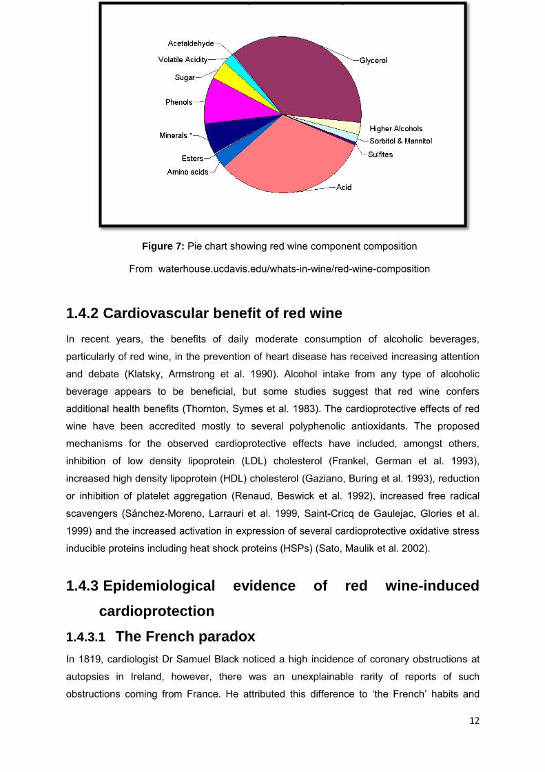
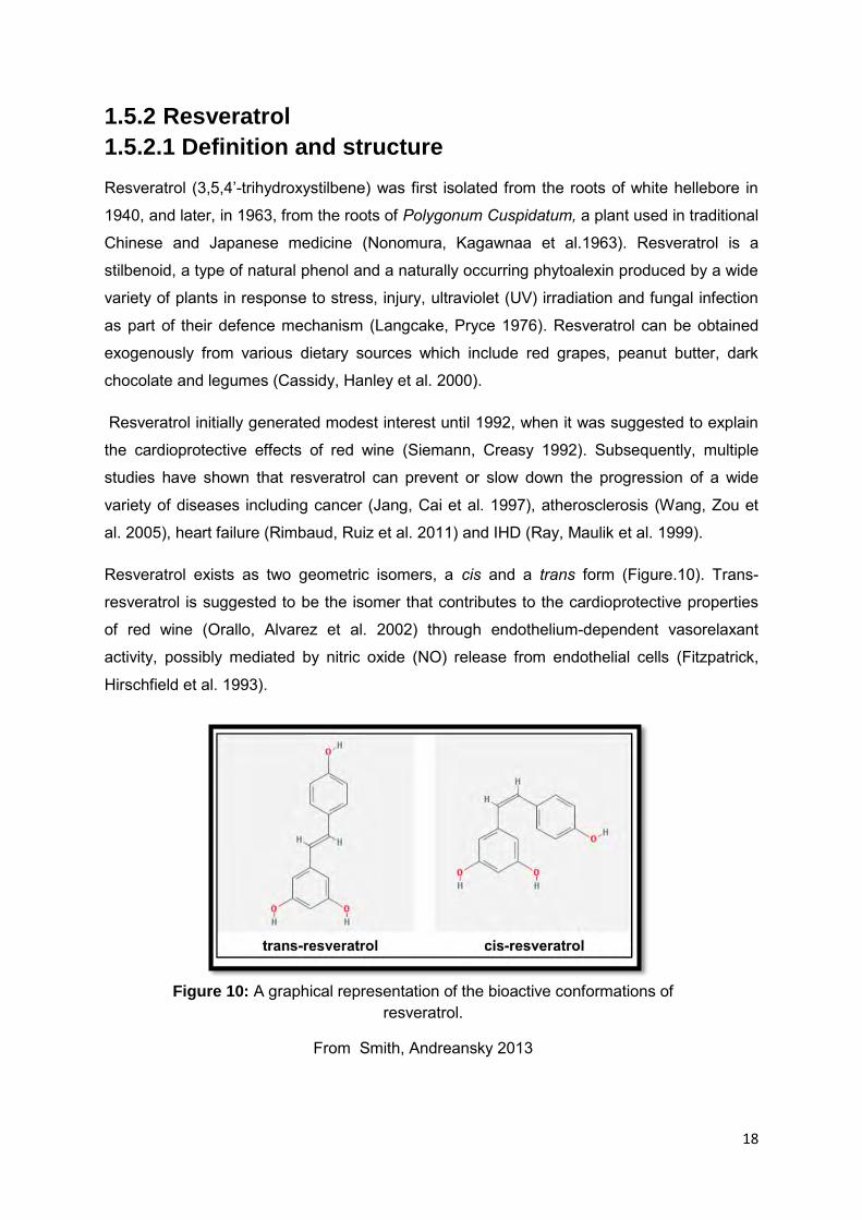
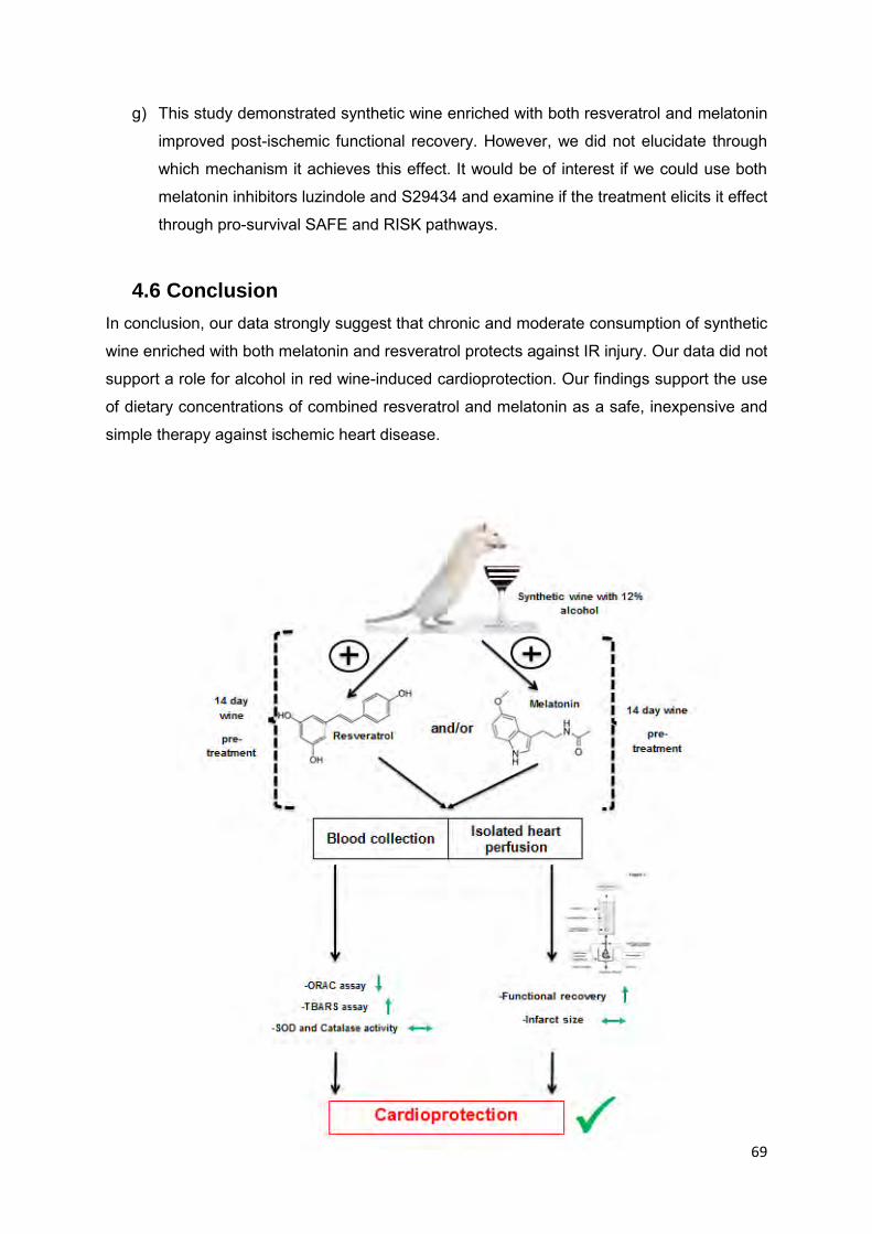
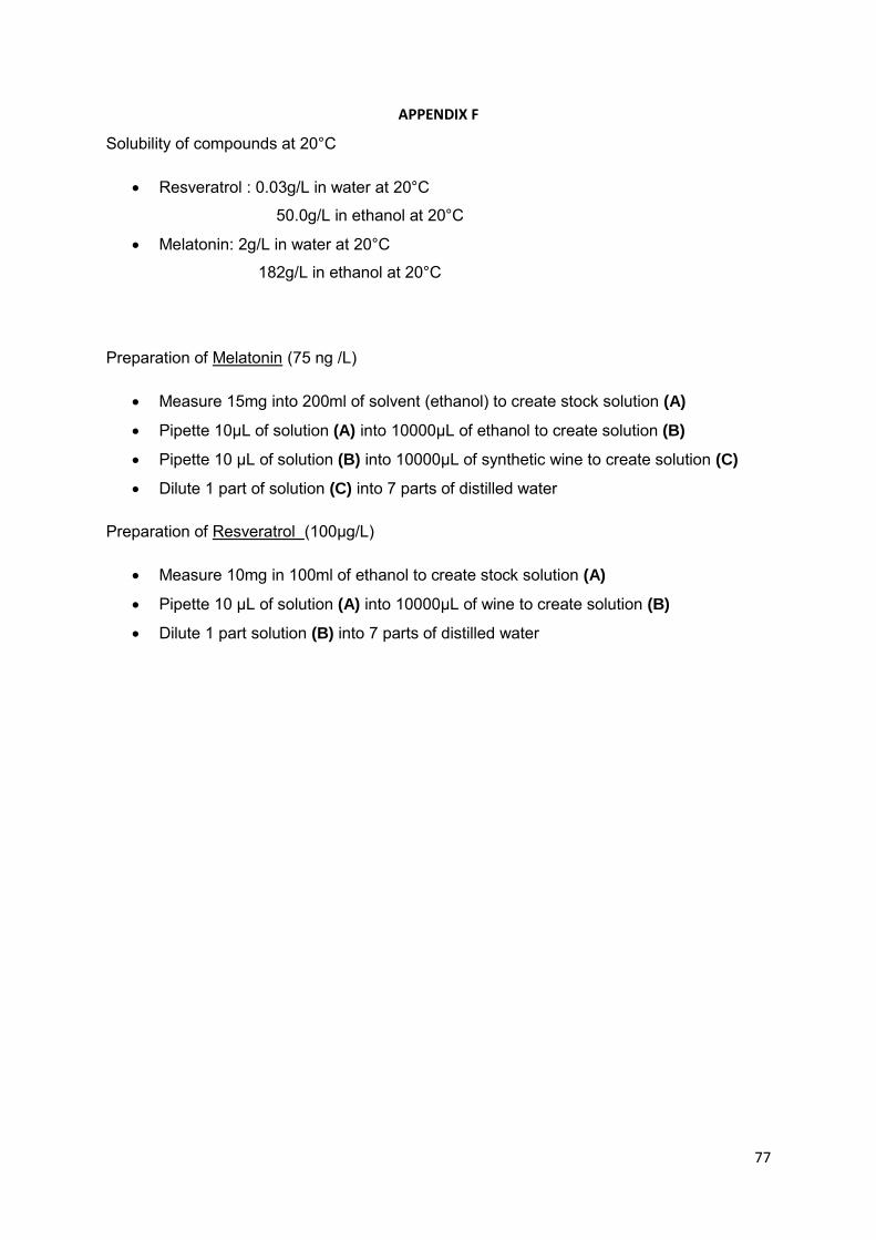

Recommended