Exported Proteins Required for Virulence and Rigidity of Plasmodium falciparum - Infected Human Erythrocytes Alexander G. Maier, 1 Melanie Rug, 1 Matthew T. O’Neill, 1 Monica Brown, 1 Srabasti Chakravorty, 2 Tadge Szestak, 2 Joanne Chesson, 1 Yang Wu, 2 Katie Hughes, 2 Ross L. Coppel, 3 Chris Newbold, 4 James G. Beeson, 1 Alister Craig, 2 Brendan S. Crabb, 1 and Alan F. Cowman 1, * 1 The Walter and Eliza Hall Institute of Medical Research, Melbourne 3050, Australia 2 Liverpool School of Tropical Medicine, Liverpool L3 5QA, UK 3 Monash University, Department of Microbiology, Clayton 3800, Australia 4 University of Oxford, Weatherall Institute of Molecular Medicine, John Radcliffe Hospital, Oxford OX3 9DS, UK *Correspondence: [email protected]DOI 10.1016/j.cell.2008.04.051 SUMMARY A major part of virulence for Plasmodium falciparum malaria infection, the most lethal parasitic disease of humans, results from increased rigidity and adhe- siveness of infected host red cells. These changes are caused by parasite proteins exported to the erythrocyte using novel trafficking machinery assem- bled in the host cell. To understand these unique modifications, we used a large-scale gene knockout strategy combined with functional screens to identify proteins exported into parasite-infected erythro- cytes and involved in remodeling these cells. Eight genes were identified encoding proteins required for export of the parasite adhesin PfEMP1 and as- sembly of knobs that function as physical platforms to anchor the adhesin. Additionally, we show that multiple proteins play a role in generating increased rigidity of infected erythrocytes. Collectively these proteins function as a pathogen secretion system, similar to bacteria and may provide targets for anti- virulence based therapies to a disease responsible for millions of deaths annually. INTRODUCTION Plasmodium falciparum causes the most severe form of malaria in humans with 1 to 3 million deaths annually. Once in the blood, multiplication of the parasite inside erythrocytes is responsible for associated morbidity and mortality. Profound structural and morphological changes occur in erythrocytes after parasite inva- sion, dramatically altering their physical properties and impairing circulation in vivo (Cooke et al., 2004). In contrast to normal erythrocytes, parasitised cells are rigid and adhere to host endo- thelium as well as other cell types (Barnwell, 1989). The in- creased rigidity and adhesiveness of P. falciparum-infected erythrocytes result in augmented haemodynamic resistance in the microvasculature (Raventos et al., 1985) and play an important role in the pathogenesis of malaria. Adherence of infected red cells to vascular endothelium is me- diated by P. falciparum erythrocyte membrane protein (PfEMP1) (Leech et al., 1984), an antigenically diverse protein family traf- ficked to the infected red cell surface (Baruch et al., 1995; Smith et al., 1995; Su et al., 1995). This in turn is anchored at the red cell membrane skeleton by knobs, macromolecular complexes con- sisting of knob associated histidine-rich protein (KAHRP) (Crabb et al., 1997). In the absence of knobs, PfEMP1 cannot form adhesive interactions of sufficient strength to withstand disrup- tion by forces of blood flow (Crabb et al., 1997). KAHRP binding with the membrane skeleton leads to an increased rigidity, blockage of blood vessels and resistance to flow (Pei et al., 2005). The parasite proteins involved are transported through host cells without trafficking machinery and inserted into a highly organized membrane skeleton structure. The formation of a de novo transport system and trafficking of parasite proteins to diverse locations in the host cell is unique in cell biology (Marti et al., 2005). Parasite proteins such as PfEMP1 and KAHRP have to traverse several membranes to reach their destination (Marti et al., 2005). A pentameric sequence has been identified required for translocation of proteins across the parasitophorous vacuole membrane termed the P. falciparum Export Element (PEXEL) (Marti et al., 2004) or Vacuolar Targeting Signal (VTS) (Hiller et al., 2004). Indeed, a similar sequence has been identified in the parasitic fungi Phytophtora infestans that is required for export of proteins into infected plant cells (Whisson et al., 2007). Searching of the P. falciparum genome sequence has revealed 8% of P. falciparum genes contain this sequence (Hiller et al., 2004; Marti et al., 2004; Sargeant et al., 2006). Many of these are likely to encode proteins that play an important role in remodelling infected erythrocytes (Marti et al., 2005). Translocation across the parasitophorous vacuole membrane via a PEXEL motif is functionally conserved across all Plasmo- dium species. However the ‘exportome’ for P. falciparum is 5-10 times larger than that of other malaria parasites partly because of radiation and expansion of gene families including 48 Cell 134, 48–61, July 11, 2008 ª2008 Elsevier Inc. Open access under CC BY license.
Transcript
Exported Proteins Required for Virulenceand Rigidity of Plasmodium falciparum-Infected Human ErythrocytesAlexander G. Maier,1 Melanie Rug,1 Matthew T. O’Neill,1 Monica Brown,1 Srabasti Chakravorty,2
Tadge Szestak,2 Joanne Chesson,1 Yang Wu,2 Katie Hughes,2 Ross L. Coppel,3 Chris Newbold,4
James G. Beeson,1 Alister Craig,2 Brendan S. Crabb,1 and Alan F. Cowman1,*1The Walter and Eliza Hall Institute of Medical Research, Melbourne 3050, Australia2Liverpool School of Tropical Medicine, Liverpool L3 5QA, UK3Monash University, Department of Microbiology, Clayton 3800, Australia4University of Oxford, Weatherall Institute of Molecular Medicine, John Radcliffe Hospital, Oxford OX3 9DS, UK
A major part of virulence for Plasmodium falciparummalaria infection, the most lethal parasitic disease ofhumans, results from increased rigidity and adhe-siveness of infected host red cells. These changesare caused by parasite proteins exported to theerythrocyte using novel trafficking machinery assem-bled in the host cell. To understand these uniquemodifications, we used a large-scale gene knockoutstrategy combined with functional screens to identifyproteins exported into parasite-infected erythro-cytes and involved in remodeling these cells. Eightgenes were identified encoding proteins requiredfor export of the parasite adhesin PfEMP1 and as-sembly of knobs that function as physical platformsto anchor the adhesin. Additionally, we show thatmultiple proteins play a role in generating increasedrigidity of infected erythrocytes. Collectively theseproteins function as a pathogen secretion system,similar to bacteria and may provide targets for anti-virulence based therapies to a disease responsiblefor millions of deaths annually.
INTRODUCTION
Plasmodium falciparum causes the most severe form of malaria
in humans with 1 to 3 million deaths annually. Once in the blood,
multiplication of the parasite inside erythrocytes is responsible
for associated morbidity and mortality. Profound structural and
morphological changes occur in erythrocytes after parasite inva-
sion, dramatically altering their physical properties and impairing
circulation in vivo (Cooke et al., 2004). In contrast to normal
erythrocytes, parasitised cells are rigid and adhere to host endo-
thelium as well as other cell types (Barnwell, 1989). The in-
creased rigidity and adhesiveness of P. falciparum-infected
erythrocytes result in augmented haemodynamic resistance in
48 Cell 134, 48–61, July 11, 2008 ª2008 Elsevier Inc.
the microvasculature (Raventos et al., 1985) and play an
important role in the pathogenesis of malaria.
Adherence of infected red cells to vascular endothelium is me-
diated by P. falciparum erythrocyte membrane protein (PfEMP1)
(Leech et al., 1984), an antigenically diverse protein family traf-
ficked to the infected red cell surface (Baruch et al., 1995; Smith
et al., 1995; Su et al., 1995). This in turn is anchored at the red cell
membrane skeleton by knobs, macromolecular complexes con-
sisting of knob associated histidine-rich protein (KAHRP) (Crabb
et al., 1997). In the absence of knobs, PfEMP1 cannot form
adhesive interactions of sufficient strength to withstand disrup-
tion by forces of blood flow (Crabb et al., 1997). KAHRP binding
with the membrane skeleton leads to an increased rigidity,
blockage of blood vessels and resistance to flow (Pei et al.,
2005). The parasite proteins involved are transported through
host cells without trafficking machinery and inserted into a highly
organized membrane skeleton structure. The formation of a de
novo transport system and trafficking of parasite proteins to
diverse locations in the host cell is unique in cell biology (Marti
et al., 2005).
Parasite proteins such as PfEMP1 and KAHRP have to
traverse several membranes to reach their destination (Marti
et al., 2005). A pentameric sequence has been identified required
for translocation of proteins across the parasitophorous vacuole
membrane termed the P. falciparum Export Element (PEXEL)
(Marti et al., 2004) or Vacuolar Targeting Signal (VTS) (Hiller
et al., 2004). Indeed, a similar sequence has been identified in
the parasitic fungi Phytophtora infestans that is required for
export of proteins into infected plant cells (Whisson et al.,
2007). Searching of the P. falciparum genome sequence has
revealed 8% of P. falciparum genes contain this sequence (Hiller
et al., 2004; Marti et al., 2004; Sargeant et al., 2006). Many of
these are likely to encode proteins that play an important role
in remodelling infected erythrocytes (Marti et al., 2005).
Translocation across the parasitophorous vacuole membrane
via a PEXEL motif is functionally conserved across all Plasmo-
dium species. However the ‘exportome’ for P. falciparum is
5-10 times larger than that of other malaria parasites partly
because of radiation and expansion of gene families including
(CDUP) gene (Figure 2A) (Maier et al., 2006). The plasmids
were transfected into CS2, a strain of P. falciparum conferring
adhesion of infected erythrocytes to chondroitin sulfate A
(CSA) via a PfEMP1 encoded by var2csa (Salanti et al., 2004).
This parasite line was chosen because expression of PfEMP1
encoded by var2csa is very stable over time. As most PfEMP1
genes undergo rapid transcriptional switches to other family
members as a means of immune evasion these switching events
could confound our subsequent analysis, the choice of var2csa
minimizes this problem.
In P. falciparum the transfected plasmids are maintained as
episomal circles and integration by double crossover homolo-
gous recombination occurs at low frequency (Maier et al.,
2006). Growth on WR99210 (positive selection) and 50-fluorocy-
tosine (negative selection) favors the survival of transfected par-
asites with homologous integration into the target gene and loss
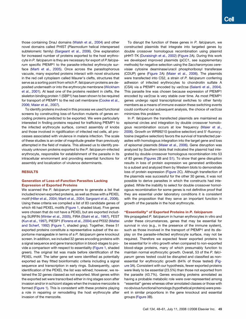
of episomal plasmids (Maier et al., 2006). Gene disruption was
analyzed by Southern blots that indicated the plasmid had inte-
grated by double-crossover homologous recombination into 53
of 83 genes (Figures 2B and S1). To show that gene disruption
results in loss of protein expression we generated antibodies
to a subset and analyzed them by Western blots to demonstrate
loss of protein expression (Figure 2C). Although transfection of
the plasmids was successful for the other 30 genes, it was not
possible to derive parasites in which the constructs had inte-
grated. While the inability to select for double crossover homol-
ogous recombination for some genes is not definitive proof that
they are essential under laboratory conditions it is consistent
with the proposition that they serve an important function in
growth of the parasite in the host erythrocyte.
‘‘Essentiality’’ of Exported Proteins in P. falciparum
We propagated P. falciparum in human erythrocytes in vitro and
under these circumstances, genes that may be essential for
survival in vivo (e.g., in the presence of the immune system),
such as those involved in the transport of PfEMP1 and its dis-
play on the parasite-infected erythrocyte surface, may not be
required. Therefore we expected fewer exported proteins to
be essential for in vitro growth when compared to non-exported
blood-stage proteins, many of which presumably function to
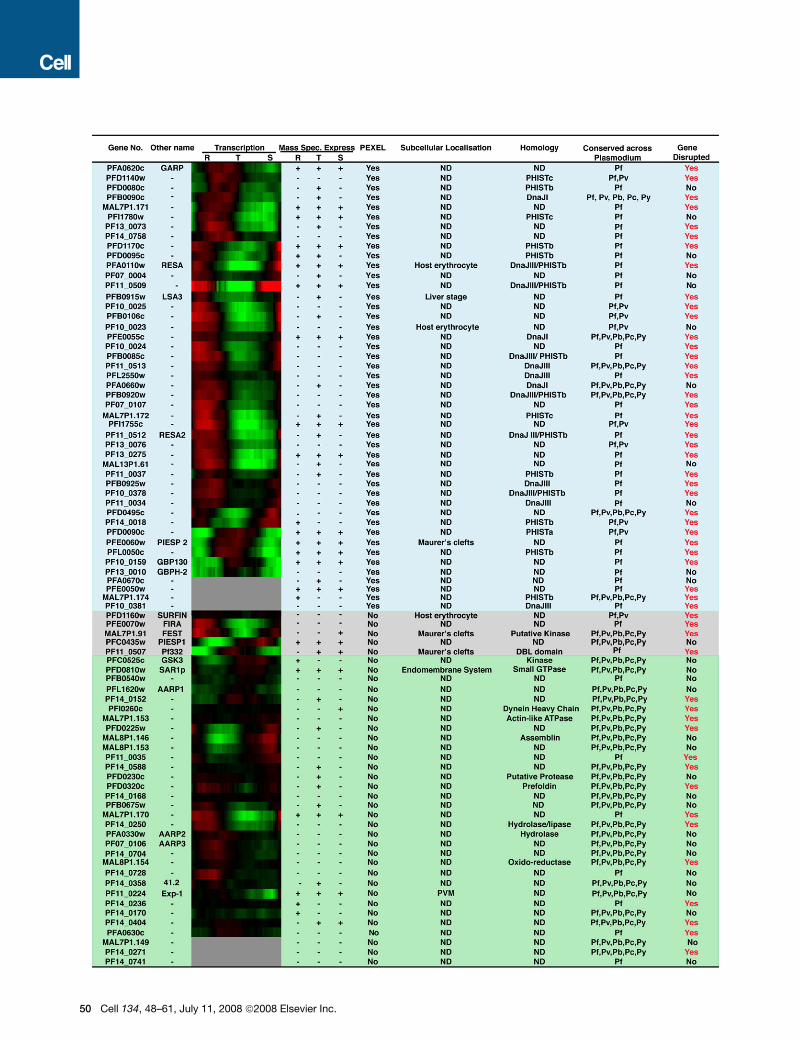
maintain normal erythrocytic growth. Overall, 53 of the P. falci-
parum genes tested could be disrupted and classified as non-
essential for erythrocytic growth (64% of those tested) (Fig-
ure 3A). Consistent with our hypothesis, fewer exported proteins
were likely to be essential (23.5%) than those not exported from
the parasite (43.7%). Genes encoding proteins annotated as
having a probable metabolic role were over-represented among
‘‘essential’’ genes whereas other annotated classes or those with
no obvious functional homologs (hypothetical proteins) were pres-
ent in similar proportions in the gene knockout and essential
groups (Figure 3B).
Cell 134, 48–61, July 11, 2008 ª2008 Elsevier Inc. 49
5
0 Cell 134, 48–61, July 11, 2008 ª2008 Elsevier Inc.
Interestingly, for genes PFD0095c, MAL7P1.149 and
MAL8P1.153 we were able to disrupt the endogenous loci but
this was accompanied by a duplication event maintaining ex-
pression of the gene (Figure S2). We concluded these genes
are essential for in vitro growth. No matter which sub-classifica-
tion was used to group different genes, a higher proportion of
non-exported proteins were considered essential (Figure 3C).
Among the genes encoding exported proteins, both disruptable
and non-disruptable examples were found within the PHIST fam-
ily and those containing DnaJ domains. The latter suggests that
some co-chaperone functions may be essential, while others
may not (Walsh et al., 2004) (Figure 2D).
Identification of Genes Required for PfEMP1 SurfaceExpressionTo identify genes required for trafficking, display and function of
PfEMP1 on the surface of P. falciparum-infected erythrocytes we
screened mutant lines for recognition of surface antigens by an-
tibodies from malaria-exposed individuals (Beeson et al., 2006).
Figure 2. Genetic Disruption of Genes En-
coding Exported Proteins
(A) Strategy to delete genes as exemplified for
PF14_0758. The vectors used were pHHT-TK
(TK) or pCC1 (CDUP). The restriction enzyme
used for PF14_0758 was EcoRI (E). The flanks
for recombination are shaded gray or black. These
flanks were probes for Southern blots and the size
of DNA fragments based on 3D7 sequence are
indicated in kilobases (kb).
(B) Example of a Southern blot to verify disruption
for PF14_0758 (all disrupted genes are shown in
Fig. S1). (C) Western blots to confirm absence of
protein expression in parasites lines in which
the genes PFA0110w, PFB0106c, PFE0060w,
MAL7P1.172, PF10_0159, PF11_0037, PF13_0275,
PF14_0018 and PF14_0758 had been disrupted.
CS2 is shown in each panel and equal loading dem-
onstrated with Hsp70 antibodies (bottom panel).
The CS2 parasite line used here ex-
presses the var2csa gene (PFL0030c)
(Duffy et al., 2006), which encodes
a PfEMP1 responsible for adhesion to
CSA (Salanti et al., 2004). In vivo, para-
sites expressing this var gene tend to be
found in primigravid women and very rarely in males or mutligra-
vid women who have developed antibodies specific for the var2-
csa PfEMP1. We can thus use sera from multigravid women to
detect surface expressed PfEMP1 on CS2-infected erythrocytes
(Beeson et al., 2006). An initial screen with such sera showed
that 10 of the 53 parasite-infected erythrocyte lines had a de-
crease in reactivity of R 70% compared to parental CS2-in-
fected red cells (Figure 4A) and we chose these as a cut off for
further analyses.
To confirm the reduced level of PfEMP1 on the surface of
these cells we used an assay in which surface exposed protein
is cleaved by trypsin and the conserved C terminus of the protein
detected by western blot with antibodies against the acidic ter-
minal segment (ATS) (Waterkeyn et al., 2000). This differentiates
surface exposed PfEMP1 from the intracellular pool. Surface ex-
posed PfEMP1 in parental line CS2 results in cleavage products
of 90 and 70 kDa (Figure 4B) whereas the internal pool of PfEMP1
in these cells migrates at approximately 300 kDa. This antibody
also shows crossreaction with host spectrin (Rug et al., 2006).
Figure 1. Expression and Homology of Genes Selected for Gene Knockout Screen in P. falciparum
The P. falciparum genome was scanned to generate a list that included known exported proteins, as well as those with a PEXEL motif (Hiller et al., 2004; Marti et
al., 2004; Sargeant et al., 2006). Using these criteria we compiled a list of 83 candidate genes of which 46 had PEXEL motifs (shaded blue). Those not containing
a PEXEL but known to be exported are shaded gray. Genes shaded green are not known to be exported or are present on the parasitophorous vacuole membrane
(PVM) e.g. Exp-1 (PF11_0224). Shown in the first column is the gene number which can be found at http://www.plasmodb.org. The second column refers to
a known name of the corresponding protein. The third column shows a transcription profile where red is an increased period of transcription and green a de-
creased period or no transcription. Gene transcription was obtained from the microarray data available on http://malaria.ucsf.edu/ (Bozdech et al., 2003). In
the fourth column proteomic data is shown and + refers to peptides detected in ring (R), trophozoite (T) or schizont (S) asexual life cycle stages. Proteomic
data is available on www.plasmodb.org. The fifth column refers to the presence (Yes) or absence (No) of a Plasmodium export element or PEXEL (Hiller
et al., 2004; Marti et al., 2004; Sargeant et al., 2006). The sixth column refers to the subcellular localization of the protein in the P. falciparum-infected erythrocyte
if known and if not known shown as ND. The seventh column shows the homology detected and in some cases putative function of the protein and where none
was detected is shown as ND. The eighth column shows the conservation of genes within different Plasmodia spp.: Pf, P. falciparum; Pv, P. vivax; Pb, P. berghei;
Pc, P. chabaudi; Py, P. yoelii. The ninth column refers to whether the gene can be genetically disrupted (Yes) or not (No).
Cell 134, 48–61, July 11, 2008 ª2008 Elsevier Inc. 51
et al., 1998) (Figures 6A and 6B). The deformability ratio of eryth-
rocytes infected with wild-type parasite to erythrocytes infected
with mutant parasites for the four highest shear stresses was
calculated and plotted to compare the influence of the deleted
Cell 134, 48–61, July 11, 2008 ª2008 Elsevier Inc. 53
54 Cell 134, 48–61, July 11, 2008 ª2008 Elsevier Inc.
protein on the rigidity of the infected erythrocyte (Figure 6A). The
average ratio for uninfected erythrocytes was 0.67. Many of the
mutant lines demonstrated small alterations in rigidity of the in-
fected erythrocyte suggesting a large number of proteins poten-
tially have a minor effect on this host cell property.
However a number of mutant cell lines had a significantly re-
duced level of rigidity and we used CS2DPFA0110w as the cut
off for significance (�0.13 ± 0.02) compared to CS2 as disruption
of the RESA gene has been shown previously to affect rigidity
(Silva et al., 2005) (Figure 7). Four cell lines CS2DPFB0920w,
CS2DPF10_0159, CS2DPF13_0073 and CS2DPF14_0758
showed a significant increase in membrane rigidity (Figure 6A,
Figure 7). Interestingly, CS2DPFB0920w, CS2DPF10_0159 and
CS2DPF13_0073 were also high binders in the CS2 adhesion as-
say (Figure 4C) and in contrast, CS2DPF14_0758 lacked erythro-
cyte surface PfEMP1 (Figure 4B). These results suggest that
a number of P. falciparum proteins combine to determine the
overall rigidity of the parasite-infected erythrocyte (Figure 7).
DISCUSSION
The P. falciparum-infected erythrocyte undergoes a series of
modifications after invasion converting a terminally differentiated
cell into one in which the parasite can access nutrients and grow
within a niche relatively protected from host responses. The
mediators responsible for remodeling the erythrocyte are most
likely exported proteins (Sargeant et al., 2006). However, there
is information on specific roles for only a handful of these pro-
teins. In order to address the function of exported proteins we
used a gene knockout strategy combined with functional assays.
Using this approach we identified exported proteins required for
trafficking, display and function of the cytoadherence protein
PfEMP1, assembly of knobs and rigidification of the infected
red cell, properties that are all thought to be important in malaria
pathogenesis (Figure 7).
The virulence protein PfEMP1 is expressed early post inva-
sion; however, it does not appear on the P. falciparum-infected
erythrocyte surface until 16 hr after invasion when the host
cells become adherent (Kriek et al., 2003). The mechanism and
proteins required for trafficking of PfEMP1 through the parasito-
phorous vacuole membrane into Maurer’s clefts and to the eryth-
rocyte membrane are unknown. In this study we have identified
six proteins that have an effect on normal trafficking of PfEMP1
(Figures 6C, 6D, and 7). Disruption of function for PFB0106c,
MAL7P1.172, PF14_0758, and PF13_0076 resulted in a com-
plete lack or greatly reduced levels of PfEMP1 on the parasite-in-
fected erythrocyte suggesting they are required for subcellular
localization of this virulence protein. Trafficking of other exported
proteins such as the classical PEXEL-containing proteins
KAHRP and PfEMP3 (Figures 5A and S5) and the non-PEXEL
containing exported protein SBP1 (Figure S5) is not affected
suggesting that the proteins identified are specifically required
for localization of PfEMP1.
The gene products of PFB0106c, MAL7P1.171 and
PF10_0025 seem to interfere with early steps of PfEMP1 trans-
port, since less PfEMP1 is detected in erythrocytes infected
with parasites deficient of these molecules. In parasite lines de-
ficient in either MAL7P1.172, PF14_0758 or PF13_0076, PfEMP1
was trafficked to Maurer’s clefts suggesting the function of the
relevant proteins is in transfer from this parasite structure to
the erythrocyte membrane (Figure 6D). Previous studies have
identified SBP1 as functioning at or just prior to this point (Cooke
et al., 2006; Maier et al., 2007) and additional molecular players in
this step are now revealed. The precise interplay between these
proteins will require further studies. In contrast, PfEMP1 in
CS2DPFB0106c does not appear to be transferred to Maurer’s
clefts suggesting this protein functions early when PfEMP1 is
loaded into these structures (Figure 6D). Interestingly, in
CS2DMAL7P1.172 parasites PfEMP1 was not readily detected
on Western blots using the standard solubilization procedure
for this protein. One explanation could be that PfEMP1 in this
line has different solubility characteristics perhaps due to its
blockage at Maurer’s clefts in its trafficking route. This is consis-
tent with previous data showing that the solubility of PfEMP1
changes during its transport pathway (Papakrivos et al., 2005).
Consistent with the hypothesis that expansion of the exportome
in P. falciparum is primarily for trafficking and function of PfEMP1
and human specific pathogenicity mechanisms is the observa-
tion that the identified molecules are either P. falciparum specific
or found exclusively in the other Plasmodia of primates.
Our screen revealed that disruption of PFD1170c and
PF10_0381 protein function leads to absent or greatly decreased
knob structures with an abnormal distribution. These same
disruptants also showed reduced cytoadherence providing
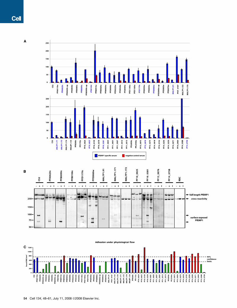
Figure 4. Identification of Proteins Required for Display and Function of PfEMP1 on the Surface of P. falciparum-Infected Erythrocytes
(A) Screening of mutant parasite strains to identify those with altered reactivity to anti-var2csa antibodies by FACS. Mutant parasite lines with specific gene dis-
ruptions were tested for reactivity with serum antibodies from malaria-exposed multigravid women (blue bars) and non-exposed controls (red bars). Reactivity
was expressed as relative to the parental line CS2, which was set at 100%. Gene names in blue signify candidates for a trypsin cleavage assay (Figure 3B) and
gene names in italics indicate a subsequently identified influence on PfEMP1 transport (Figure 7). Error bars indicate % range.
(B) Trypsin treatment of P. falciparum-infected erythrocytes to determine presence of PfEMP1 on the host erythrocyte. The full-length PfEMP1 and cytoplasmic
tail were detected using antibodies to the cytoplasmic acidic terminal segment (ATS). Full-length PfEMP1 was a > 300 kDa band. Surface pool of PfEMP1 was
detected by a trypsin-resistant band between 70 and 90 kDa. The lanes in each panel show Triton-X insoluble/SDS soluble extractions of parasite-infected eryth-
rocytes: first lane, untreated (�); second lane, trypsin-treated (+); third lane, trypsin plus soybean trypsin inhibitor (i). The parasite lines shown are those that when
screened by antibodies against var2csa PfEMP1 were less than 30% reactive compared to CS2 or knob-deficient (panel A). The red cell control is shown in the
last panel. The anti-ATS antibody cross-reacts with spectrin (Maier et al., 2007; Rug et al., 2006). Lack of a band between 70 and 90 kDa in the trypsin-treated
lanes shows absence of PfEMP1 on the erythrocyte surface. Full-length PfEMP1 is observed because there is a large pool of internal protein resistant to trypsin
(Waterkeyn et al., 2000).
(C) Adherence of mutant P. falciparum-infected erythrocytes to CSA under flow conditions. Each of the P. falciparum mutant strains was tested for binding to CSA
under flow conditions and parasitised cells counted as bound infected red blood cells/mm2. 95% confidence intervals for CS2 wild-type binding is presented
(dashed lines).
Cell 134, 48–61, July 11, 2008 ª2008 Elsevier Inc. 55
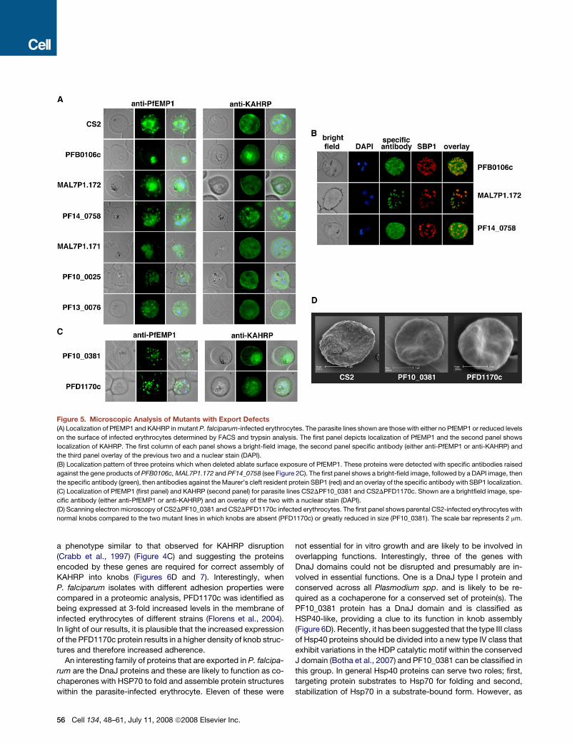
Figure 5. Microscopic Analysis of Mutants with Export Defects
(A) Localization of PfEMP1 and KAHRP in mutant P. falciparum-infected erythrocytes. The parasite lines shown are those with either no PfEMP1 or reduced levels
on the surface of infected erythrocytes determined by FACS and trypsin analysis. The first panel depicts localization of PfEMP1 and the second panel shows
localization of KAHRP. The first column of each panel shows a bright-field image, the second panel specific antibody (either anti-PfEMP1 or anti-KAHRP) and
the third panel overlay of the previous two and a nuclear stain (DAPI).
(B) Localization pattern of three proteins which when deleted ablate surface exposure of PfEMP1. These proteins were detected with specific antibodies raised
against the gene products of PFB0106c, MAL7P1.172 and PF14_0758 (see Figure 2C). The first panel shows a bright-field image, followed by a DAPI image, then
the specific antibody (green), then antibodies against the Maurer’s cleft resident protein SBP1 (red) and an overlay of the specific antibody with SBP1 localization.
(C) Localization of PfEMP1 (first panel) and KAHRP (second panel) for parasite lines CS2DPF10_0381 and CS2DPFD1170c. Shown are a brightfield image, spe-
cific antibody (either anti-PfEMP1 or anti-KAHRP) and an overlay of the two with a nuclear stain (DAPI).
(D) Scanning electron microscopy of CS2DPF10_0381 and CS2DPFD1170c infected erythrocytes. The first panel shows parental CS2-infected erythrocytes with
normal knobs compared to the two mutant lines in which knobs are absent (PFD1170c) or greatly reduced in size (PF10_0381). The scale bar represents 2 mm.
a phenotype similar to that observed for KAHRP disruption
(Crabb et al., 1997) (Figure 4C) and suggesting the proteins
encoded by these genes are required for correct assembly of
KAHRP into knobs (Figures 6D and 7). Interestingly, when
P. falciparum isolates with different adhesion properties were
compared in a proteomic analysis, PFD1170c was identified as
being expressed at 3-fold increased levels in the membrane of
infected erythrocytes of different strains (Florens et al., 2004).
In light of our results, it is plausible that the increased expression
of the PFD1170c protein results in a higher density of knob struc-
tures and therefore increased adherence.
An interesting family of proteins that are exported in P. falcipa-
rum are the DnaJ proteins and these are likely to function as co-
chaperones with HSP70 to fold and assemble protein structures
within the parasite-infected erythrocyte. Eleven of these were
56 Cell 134, 48–61, July 11, 2008 ª2008 Elsevier Inc.
not essential for in vitro growth and are likely to be involved in
overlapping functions. Interestingly, three of the genes with
DnaJ domains could not be disrupted and presumably are in-
volved in essential functions. One is a DnaJ type I protein and
conserved across all Plasmodium spp. and is likely to be re-
quired as a cochaperone for a conserved set of protein(s). The
PF10_0381 protein has a DnaJ domain and is classified as
HSP40-like, providing a clue to its function in knob assembly
(Figure 6D). Recently, it has been suggested that the type III class
of Hsp40 proteins should be divided into a new type IV class that
exhibit variations in the HDP catalytic motif within the conserved
J domain (Botha et al., 2007) and PF10_0381 can be classified in
this group. In general Hsp40 proteins can serve two roles; first,
targeting protein substrates to Hsp70 for folding and second,
stabilization of Hsp70 in a substrate-bound form. However, as
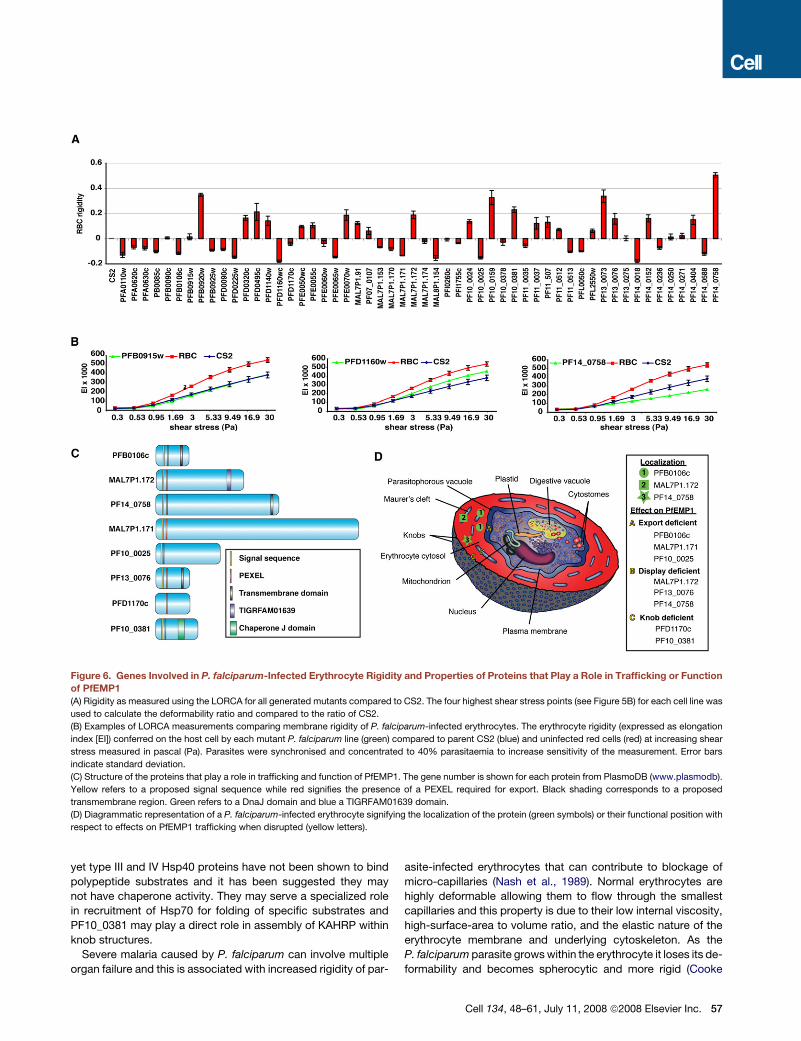
Figure 6. Genes Involved in P. falciparum-Infected Erythrocyte Rigidity and Properties of Proteins that Play a Role in Trafficking or Function
of PfEMP1
(A) Rigidity as measured using the LORCA for all generated mutants compared to CS2. The four highest shear stress points (see Figure 5B) for each cell line was
used to calculate the deformability ratio and compared to the ratio of CS2.
(B) Examples of LORCA measurements comparing membrane rigidity of P. falciparum-infected erythrocytes. The erythrocyte rigidity (expressed as elongation
index [EI]) conferred on the host cell by each mutant P. falciparum line (green) compared to parent CS2 (blue) and uninfected red cells (red) at increasing shear
stress measured in pascal (Pa). Parasites were synchronised and concentrated to 40% parasitaemia to increase sensitivity of the measurement. Error bars
indicate standard deviation.
(C) Structure of the proteins that play a role in trafficking and function of PfEMP1. The gene number is shown for each protein from PlasmoDB (www.plasmodb).
Yellow refers to a proposed signal sequence while red signifies the presence of a PEXEL required for export. Black shading corresponds to a proposed
transmembrane region. Green refers to a DnaJ domain and blue a TIGRFAM01639 domain.
(D) Diagrammatic representation of a P. falciparum-infected erythrocyte signifying the localization of the protein (green symbols) or their functional position with
respect to effects on PfEMP1 trafficking when disrupted (yellow letters).
yet type III and IV Hsp40 proteins have not been shown to bind
polypeptide substrates and it has been suggested they may
not have chaperone activity. They may serve a specialized role
in recruitment of Hsp70 for folding of specific substrates and
PF10_0381 may play a direct role in assembly of KAHRP within
knob structures.
Severe malaria caused by P. falciparum can involve multiple
organ failure and this is associated with increased rigidity of par-
asite-infected erythrocytes that can contribute to blockage of
micro-capillaries (Nash et al., 1989). Normal erythrocytes are
highly deformable allowing them to flow through the smallest
capillaries and this property is due to their low internal viscosity,
high-surface-area to volume ratio, and the elastic nature of the
erythrocyte membrane and underlying cytoskeleton. As the
P. falciparum parasite grows within the erythrocyte it loses its de-
formability and becomes spherocytic and more rigid (Cooke
Cell 134, 48–61, July 11, 2008 ª2008 Elsevier Inc. 57
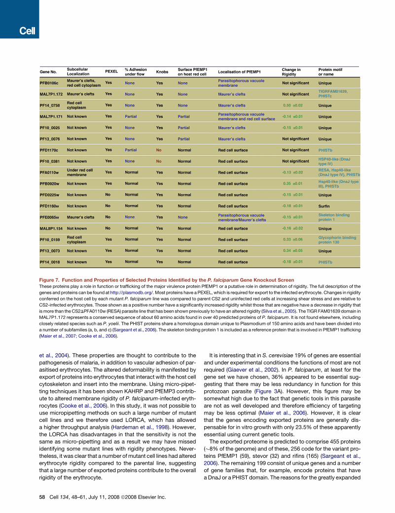
Figure 7. Function and Properties of Selected Proteins Identified by the P. falciparum Gene Knockout Screen
These proteins play a role in function or trafficking of the major virulence protein PfEMP1 or a putative role in determination of rigidity. The full description of the
genes and proteins can be found at http://plasmodb.org/. Most proteins have a PEXEL, which is required for export to the infected erythrocyte. Changes in rigidity
conferred on the host cell by each mutant P. falciparum line was compared to parent CS2 and uninfected red cells at increasing shear stress and are relative to
CS2-infected erythrocytes. Those shown as a positive number have a significantly increased rigidity whilst those that are negative have a decrease in rigidity that
is more than the CS2DPFA0110w (RESA) parasite line that has been shown previously to have an altered rigidity (Silva et al., 2005). The TIGR FAM01639 domain in
MAL7P1.172 represents a conserved sequence of about 60 amino acids found in over 40 predicted proteins of P. falciparum. It is not found elsewhere, including
closely related species such as P. yoelii. The PHIST proteins share a homologous domain unique to Plasmodium of 150 amino acids and have been divided into
a number of subfamilies (a, b, and c) (Sargeant et al., 2006). The skeleton binding protein 1 is included as a reference protein that is involved in PfEMP1 trafficking
(Maier et al., 2007; Cooke et al., 2006).
et al., 2004). These properties are thought to contribute to the
pathogenesis of malaria, in addition to vascular adhesion of par-
asitised erythrocytes. The altered deformability is manifested by
export of proteins into erythrocytes that interact with the host cell
cytoskeleton and insert into the membrane. Using micro-pipet-
ting techniques it has been shown KAHRP and PfEMP3 contrib-
ute to altered membrane rigidity of P. falciparum-infected eryth-
rocytes (Cooke et al., 2006). In this study, it was not possible to
use micropipetting methods on such a large number of mutant
cell lines and we therefore used LORCA, which has allowed
a higher throughput analysis (Hardeman et al., 1998). However,
the LORCA has disadvantages in that the sensitivity is not the
same as micro-pipetting and as a result we may have missed
identifying some mutant lines with rigidity phenotypes. Never-
theless, it was clear that a number of mutant cell lines had altered
erythrocyte rigidity compared to the parental line, suggesting
that a large number of exported proteins contribute to the overall
rigidity of the erythrocyte.
58 Cell 134, 48–61, July 11, 2008 ª2008 Elsevier Inc.
It is interesting that in S. cerevisiae 19% of genes are essential
and under experimental conditions the functions of most are not
required (Giaever et al., 2002). In P. falciparum, at least for the
gene set we have chosen, 36% appeared to be essential sug-
gesting that there may be less redundancy in function for this
protozoan parasite (Figure 3A). However, this figure may be
somewhat high due to the fact that genetic tools in this parasite
are not as well developed and therefore efficiency of targeting
may be less optimal (Maier et al., 2006). However, it is clear
that the genes encoding exported proteins are generally dis-
pensable for in vitro growth with only 23.5% of these apparently
essential using current genetic tools.
The exported proteome is predicted to comprise 455 proteins
(�8% of the genome) and of these, 256 code for the variant pro-
teins PfEMP1 (59), stevor (32) and rifins (165) (Sargeant et al.,
2006). The remaining 199 consist of unique genes and a number
of gene families that, for example, encode proteins that have
a DnaJ or a PHIST domain. The reasons for the greatly expanded