Exposure to gestational diabetes mellitusinduces neuroinflammation, derangementof hippocampal neurons, and cognitivechanges in rat offspringBilly Vuong1,2, Gary Odero1,2, Stephanie Rozbacher2, Mackenzie Stevenson2, Stephanie M. Kereliuk1,3,Troy J. Pereira1,3, Vernon W. Dolinsky1,3 and Tiina M. Kauppinen1,2,3*
Abstract
Background: Birth cohort studies link gestational diabetes mellitus (GDM) with impaired cognitive performance inthe offspring. However, the mechanisms involved are unknown. We tested the hypothesis that obesity-associatedGDM induces chronic neuroinflammation and disturbs the development of neuronal circuitry resulting in impairedcognitive abilities in the offspring.
Methods: In rats, GDM was induced by feeding dams a diet high in sucrose and fatty acids. Brains of neonatal(E20) and young adult (15-week-old) offspring of GDM and lean dams were analyzed by immunohistochemistry,cytokine assay, and western blotting. Young adult offspring of GDM and lean dams went also through cognitiveassessment. Cultured microglial responses to elevated glucose and/or fatty acids levels were analyzed.
Results: In rats, impaired recognition memory was observed in the offspring of GDM dams. GDM exposure combinedwith a postnatal high-fat and sucrose diet resulted in atypical inattentive behavior in the offspring. These cognitivechanges correlated with reduced density and derangement of Cornu Ammonis 1 pyramidal neuronal layer, decreasedhippocampal synaptic integrity, increased neuroinflammatory status, and reduced expression of CX3CR1, the microglialfractalkine receptor regulating microglial pro-inflammatory responses and synaptic pruning. Primary microglial culturesthat were exposed to high concentrations of glucose and/or palmitate were transformed into an activated, amoeboidmorphology with increased nitric oxide and superoxide production, and altered their cytokine release profile.
Conclusions: These findings demonstrate that GDM stimulates microglial activation and chronic inflammatory responsesin the brain of the offspring that persist into young adulthood. Reactive gliosis correlates positively with hippocampalsynaptic decline and cognitive impairments. The elevated pro-inflammatory cytokine expression at the critical period ofhippocampal synaptic maturation suggests that neuroinflammation might drive the synaptic and cognitive decline in theoffspring of GDM dams. The importance of microglia in this process is supported by the reduced Cx3CR1 expression asan indication of the loss of microglial control of inflammatory responses and phagocytosis and synaptic pruning in GDMoffspring.
* Correspondence: [email protected] of Pharmacology and Therapeutics, University of Manitoba, 753McDermot Avenue, Winnipeg, Manitoba R3E 0T6, Canada2Neuroscience Research Program, Kleysen Institute for Advanced Medicine,Health Sciences Center, SR434 – 710 William Avenue, Winnipeg, ManitobaR3E 0Z3, CanadaFull list of author information is available at the end of the article
BackgroundGestational diabetes mellitus (GDM) is a state of glucoseintolerance and hyperglycemia with first onset duringpregnancy and is the most common complication ofpregnancy, affecting up to 10% of expectant mothers [1].The prevalence of GDM is on the rise since morewomen are overweight or obese when they becomepregnant. The combination of human cohort studies andresearch using rodent models with controlled pre- andpostnatal conditions has demonstrated that diabetes dur-ing pregnancy during critical stages of development af-fects health outcomes in the offspring [2]. Severalhuman cohort studies have linked diabetes during preg-nancy with impaired cognitive abilities [3–6] and psychi-atric disorders [7, 8] in the offspring. Similar correlationsexist between poor quality early childhood diets and cog-nitive performance [9, 10]. Brain development is particu-larly sensitive to environmental influences during infancy[11]. Animal studies utilizing controlled conditions con-firmed that maternal high-sugar intake caused deficits incognitive functions in the offspring [12, 13]. However, themechanisms that link GDM to the development of cogni-tive impairments in the offspring remain to be elucidated.GDM is associated with mild but chronic systemic in-
flammation [14] and elevated circulating free fatty acids[15, 16]. Although the transfer of cytokines across theplacenta from mother to fetus is restricted [17], maternalhyperglycemia and hyperlipidemia directly impact thefetus through the placental overproduction of pro-inflammatory cytokines [14, 18]. We have also recentlyshown that obesity-associated GDM promotes the pro-duction of the pro-inflammatory cytokine, interleukin(IL)-1β, and toll-like receptor activity, in the spleen cellsof neonatal and 15-week-old offspring that may contrib-ute to chronic low-grade systemic inflammation [19].Cytokines can induce pro-inflammatory responses in thecentral nervous system (CNS) resulting in neuroinflam-mation, which includes the release of cytokines, chemo-kines, adhesion molecules, free radicals, and destructiveenzymes. Activation of microglia, resident brain immunecells, is a central event and driving force in neuroinflam-mation [20]. Pro-inflammatory responses of activatedmicroglia can jeopardize neurogenesis and promote neu-rodegeneration [21]. Microglia have an important role inneuroplasticity as they assist in the development andmaintenance of neuronal networks via trophic factorrelease and controlled phagocytosis, which allows micro-glia to prune synapses and remodel neuronal transmis-sion [22, 23]. Microglial pro-inflammatory responses[20] can also indirectly affect the neuronal vitality bychanging the function of astroglial cells, which have sup-portive role in neuronal metabolism and modulation ofsynaptic transmission [24]. GDM-induced inflammationduring embryogenesis could perturb microglial functions
resulting in a limited ability to support the developmentof a healthy neuronal network that detrimentally influ-ences brain development in the offspring.We utilized a high-fat and sucrose (HFS) diet-induced
GDM model [19, 25] to evaluate the effects of GDM ex-posure and postnatal diets on cognitive abilities andneuroinflammation in the offspring. In this GDM model,hyperglycemia is observed in mid-gestation and resolvesafter the dam litters out, thus mimicking some featuresof the clinical presentation of GDM in overweight andobese women [19, 25]. We determined that GDM expos-ure impaired recognition memory in the offspring andwhen combined with postnatal HFS diet, resulted inatypical inattentive behavior. These cognitive changeswere associated with reduced density and derangementof the Cornu Ammonis 1 (CA1) pyramidal neuronallayer, decreased hippocampal synaptic integrity, andincreased neuroinflammation. Exposure of primary cul-tures of microglia to elevated levels of glucose and/orfatty acids triggered pro-inflammatory responses capableof disturbing development of neuronal circuitry andfunctions, suggesting that gluco- and lipotoxicity con-tribute to microglial-mediated neuroinflammation.
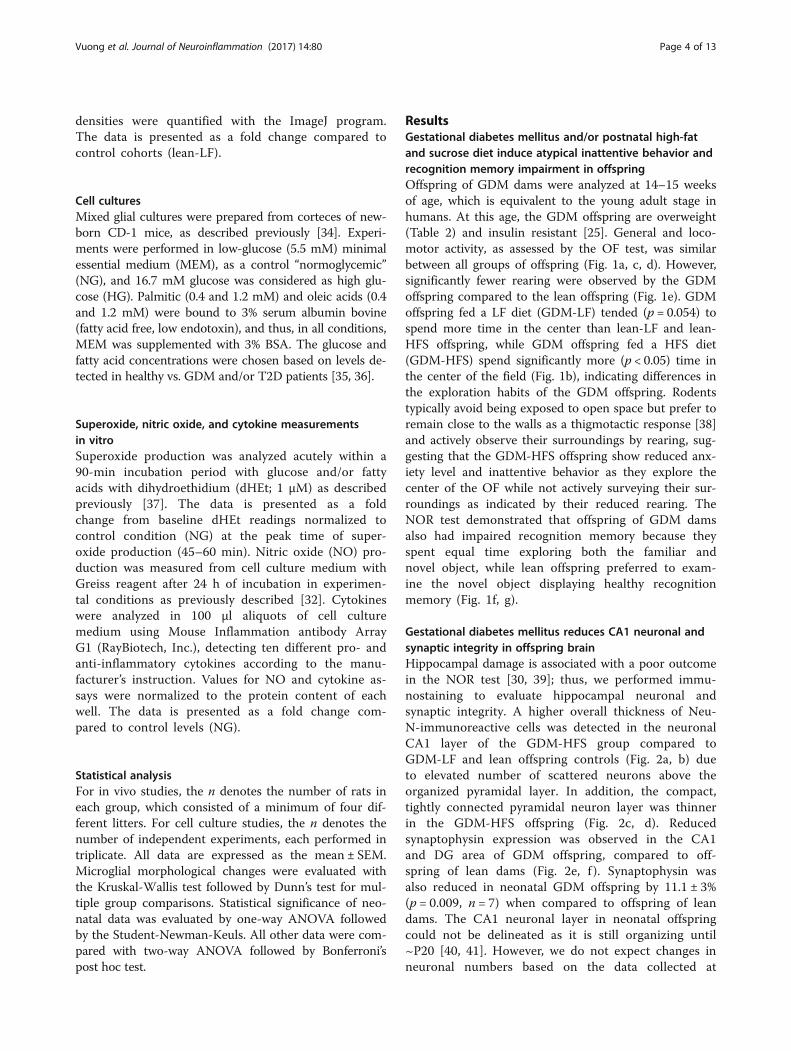
MethodsMaterialsCell culture reagents were obtained from ThermoFisher,and all other reagents were from Sigma/Aldrich (St.Louis, MO) except where otherwise stated.
Animal modelAll animal procedures were approved by the AnimalWelfare Committee of the University of Manitoba whichfollows the guidelines of the Canadian Council onAnimal Care and the Council for International Organi-zations of Medical Sciences. Rats were given ad libitumaccess to food and water and were housed two per cage.As described previously [25], GDM was induced in fe-male Sprague-Dawley rats using a high-fat and sucrosediet (HFS; 45% fat, 4.73 kcal/gm, Research DietsD12451; New Brunswick, NJ, USA) for 6 weeks priormating, throughout gestation and lactation, while leancontrol dams were fed a low-fat diet (LF, 10% fat,3.85 kcal/gm, Research Diets D12450B). All femaleswere mated with lean males. The dams experienced in-creased weight gain during pregnancy and starting mid-gestation developed hyperinsulinemia and moderatehyperglycemia (Table 1). This model mimics clinicalsymptoms of GDM seen in mothers as well as theiroffspring that developed obesity and insulin resistance[2, 25]. Litters were randomly reduced to eight pups inorder to avoid competition for food. The remaining off-spring were weaned at 3 weeks of age, and males fromeach litter were randomly assigned to a LF or HFS
Vuong et al. Journal of Neuroinflammation (2017) 14:80 Page 2 of 13
postnatal diet. This created four experimental groups: off-spring from LF-fed dams (“lean”) that were fed a LF orHFS postweaning diet and offspring from HFS-fed dams(“GDM”) that were fed a LF or HFS postweaning diet. Alloffspring underwent behavioral testing at 14 weeks of ageand were sacrificed at 15 weeks of age, as young adults,when they were insulin resistant [25] but hyperglycemiawas not observed (Table 2). In addition, neonatal (E20)offspring were collected for analysis.
Behavioral testingYoung adult rats had behavioral tests starting 1 weekprior their sacrifice to analyze whether GDM and/orpostweaning diet affected their cognition. The behavioraltests focus on analyzing the anxiety, attention, explor-ation, memory, and learning abilities. For the open field(OF) test, an arena with walls to prevent escape wasused and the test was videotaped for later analysis to as-sess general activity level, anxiety, attention, and explor-ation tendency [26]. The rats were placed in OF (whitesquare plexiglass chamber; L 75 cm ×W 75 cm ×H75 cm, illuminated at 8–13 lx red light) for 10 min.Locomotor activity, rearing, and the amount of timespent in the center area of the OF were calculated. Thenovel object recognition (NOR) test of recognitionmemory which is affected by hippocampal and perirhinalcortex damage [27–31] was performed after the OF test.On the first day, rats were placed back to the testingarena which had now two identical objects. Rats wereallowed to explore the objects freely for a 10-min train-ing session. On the second day, rats were placed backinto the testing arena for the 10-min test session, duringwhich they were presented with one of the objects usedduring training (familiar object) and with a novel,unfamiliar object of different shape and texture. Objectlocations were kept constant during training and testsessions for any given rat. Arenas and objects were
cleaned with 70% ethanol between each rat. Frequencyof object interactions and time spent exploring each ob-ject was recorded with a HVS Image 2100 Plus videotracking system software (HVS Image Ltd., Twickenham,Middlesex, UK). Frequency of object interactions duringthe first 5 min was used for analyses.
Brain immunostaining and cytokine measurementsImmunostaining was performed on 4% formaldehydefixed 30-μm coronal sections with anti-NeuN (Chemiconinternational, 1:500), anti-synaptophysin (1:500), anti-Iba1(Wako, 1:500), and anti-GFAP (EMD Millipore, 1:500)followed by Dylight 488 or 594 conjugated anti-IgG(Thermofisher, 1:500) as described previously [32]. ImageJprogram (NIH) was used to measure hippocampal neur-onal CA1 layer thickness, and synaptophysin and GFAPexpressions were determined by the mean optical densityin the designated, uniform-sized regions of interest[32, 33]. Microglial morphology identification wasbased on assessment of cell soma and processes (Table 3).Values were measured on a minimum of three matchinghippocampal regions from each rat, background valueswere subtracted, and the resulting values were averaged togive one value per rat.For cytokine assays, the brain tissue (the whole neo-
natal hemisphere, forebrain hemisphere of young adult)were homogenized 1:3 weight to volume in RIPA buffer(Thermo Scientific) with complete protease inhibitor,followed by centrifugation. Cytokines were analyzed in100 μl aliquots of tissue homogenates using Rat Inflam-mation antibody Array G1 (RayBiotech, Inc.) accordingto the manufacturer’s instructions [32]. Values of thecytokine assay were normalized to protein content oftissue homogenate. The data is presented as a foldchange compared to control cohorts (lean or lean-LF).
Western blottingBrain homogenates were analyzed by immunoblottingon 8% SDS-PAGE gel with anti-CX3CR1 (Novus, 1:500dilutions) and anti-β-actin (1;5000), followed by peroxidase-conjugated anti-IgG (Vector Laboratories, Burlingame, CA,1:5000 dilution). The protein bands were visualized usingECL™ WB Detection kit (Amersham-Pharmacia Biotech)and ChemiDoc MP imaging system (BioRad). Band
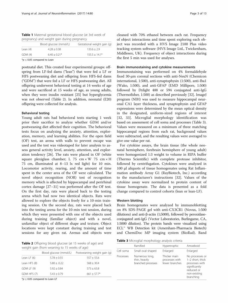
Table 2 Offspring blood glucose (at 15 weeks of age) andweight gain (from weaning to 15 weeks of age)
Blood glucose (mmol/L) Postweaning weight gain (g)
Lean LF (6) 5.78 ± 0.55 557 ± 55.6
Lean HFS (8) 5.80 ± .0.22 568 ± 30.4
GDM LF (9) 5.92 ± 0.84 573 ± 63.8
GDM HFS (7) 5.43 ± 0.79 661 ± 57.7*
*p ≤ 0.05 compared to Lean LF
Table 1 Maternal gestational blood glucose (at 3rd week ofpregnancy) and weight gain during pregnancy
Blood glucose (mmol/L) Gestational weight gain (g)
No processes or1–2 short, thickprocesses withsignificantlyreduced ornon-existingbranching
Vuong et al. Journal of Neuroinflammation (2017) 14:80 Page 3 of 13
densities were quantified with the ImageJ program.The data is presented as a fold change compared tocontrol cohorts (lean-LF).
Cell culturesMixed glial cultures were prepared from corteces of new-born CD-1 mice, as described previously [34]. Experi-ments were performed in low-glucose (5.5 mM) minimalessential medium (MEM), as a control “normoglycemic”(NG), and 16.7 mM glucose was considered as high glu-cose (HG). Palmitic (0.4 and 1.2 mM) and oleic acids (0.4and 1.2 mM) were bound to 3% serum albumin bovine(fatty acid free, low endotoxin), and thus, in all conditions,MEM was supplemented with 3% BSA. The glucose andfatty acid concentrations were chosen based on levels de-tected in healthy vs. GDM and/or T2D patients [35, 36].
Superoxide, nitric oxide, and cytokine measurementsin vitroSuperoxide production was analyzed acutely within a90-min incubation period with glucose and/or fattyacids with dihydroethidium (dHEt; 1 μM) as describedpreviously [37]. The data is presented as a foldchange from baseline dHEt readings normalized tocontrol condition (NG) at the peak time of super-oxide production (45–60 min). Nitric oxide (NO) pro-duction was measured from cell culture medium withGreiss reagent after 24 h of incubation in experimen-tal conditions as previously described [32]. Cytokineswere analyzed in 100 μl aliquots of cell culturemedium using Mouse Inflammation antibody ArrayG1 (RayBiotech, Inc.), detecting ten different pro- andanti-inflammatory cytokines according to the manu-facturer’s instruction. Values for NO and cytokine as-says were normalized to the protein content of eachwell. The data is presented as a fold change com-pared to control levels (NG).
Statistical analysisFor in vivo studies, the n denotes the number of rats ineach group, which consisted of a minimum of four dif-ferent litters. For cell culture studies, the n denotes thenumber of independent experiments, each performed intriplicate. All data are expressed as the mean ± SEM.Microglial morphological changes were evaluated withthe Kruskal-Wallis test followed by Dunn’s test for mul-tiple group comparisons. Statistical significance of neo-natal data was evaluated by one-way ANOVA followedby the Student-Newman-Keuls. All other data were com-pared with two-way ANOVA followed by Bonferroni’spost hoc test.
ResultsGestational diabetes mellitus and/or postnatal high-fatand sucrose diet induce atypical inattentive behavior andrecognition memory impairment in offspringOffspring of GDM dams were analyzed at 14–15 weeksof age, which is equivalent to the young adult stage inhumans. At this age, the GDM offspring are overweight(Table 2) and insulin resistant [25]. General and loco-motor activity, as assessed by the OF test, was similarbetween all groups of offspring (Fig. 1a, c, d). However,significantly fewer rearing were observed by the GDMoffspring compared to the lean offspring (Fig. 1e). GDMoffspring fed a LF diet (GDM-LF) tended (p = 0.054) tospend more time in the center than lean-LF and lean-HFS offspring, while GDM offspring fed a HFS diet(GDM-HFS) spend significantly more (p < 0.05) time inthe center of the field (Fig. 1b), indicating differences inthe exploration habits of the GDM offspring. Rodentstypically avoid being exposed to open space but prefer toremain close to the walls as a thigmotactic response [38]and actively observe their surroundings by rearing, sug-gesting that the GDM-HFS offspring show reduced anx-iety level and inattentive behavior as they explore thecenter of the OF while not actively surveying their sur-roundings as indicated by their reduced rearing. TheNOR test demonstrated that offspring of GDM damsalso had impaired recognition memory because theyspent equal time exploring both the familiar andnovel object, while lean offspring preferred to exam-ine the novel object displaying healthy recognitionmemory (Fig. 1f, g).
Gestational diabetes mellitus reduces CA1 neuronal andsynaptic integrity in offspring brainHippocampal damage is associated with a poor outcomein the NOR test [30, 39]; thus, we performed immu-nostaining to evaluate hippocampal neuronal andsynaptic integrity. A higher overall thickness of Neu-N-immunoreactive cells was detected in the neuronalCA1 layer of the GDM-HFS group compared toGDM-LF and lean offspring controls (Fig. 2a, b) dueto elevated number of scattered neurons above theorganized pyramidal layer. In addition, the compact,tightly connected pyramidal neuron layer was thinnerin the GDM-HFS offspring (Fig. 2c, d). Reducedsynaptophysin expression was observed in the CA1and DG area of GDM offspring, compared to off-spring of lean dams (Fig. 2e, f ). Synaptophysin wasalso reduced in neonatal GDM offspring by 11.1 ± 3%(p = 0.009, n = 7) when compared to offspring of leandams. The CA1 neuronal layer in neonatal offspringcould not be delineated as it is still organizing until~P20 [40, 41]. However, we do not expect changes inneuronal numbers based on the data collected at
Vuong et al. Journal of Neuroinflammation (2017) 14:80 Page 4 of 13
15 weeks of age, when only GDM-HFS diet resultedin derangement in the CA1 neuronal layer. This sug-gests that fetal GDM exposure conditions the cellswhile the second stress induced by the postnatal HFSdiet causes derangement in the CA1 layer.
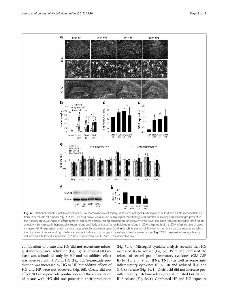
Gestational diabetes mellitus induces chronic inflammationin the offspring brainSince GDM can promote a pro-inflammatory environ-ment [14], we evaluated activation of microglia, theresident immune cells of the brain. Iba1 immunohisto-chemical analysis of the hippocampi from neonates re-vealed microglial morphological activation, as judged bythe reduced proportion of ramified microglia (lean 65 ±11% vs GDM 19 ± 6%), and an increased proportion of
microglia exhibiting amoeboid morphology (lean 8 ± 2%vs GDM 46 ± 8%) (Fig. 3a–c). Increased GFAP expres-sion (Fig. 3d, e) in the GDM neonates indicated elevatedhippocampal astogliosis. Increased levels of pro-inflammatory cytokines IFNγ, IL-1α, IL-4, MCP-1, andTNFα were also observed in the brain of the neonataloffspring of GDM dams (Fig. 3f ). Overall, our findingsshow that GDM increased neuroinflammation status inthe neonatal offspring.The microglial morphological transformation and astro-
gliosis observed in the GDM offspring persisted intoyoung adulthood (Fig. 4). A reduced proportion of rami-fied microglia and increased hypertrophic microglia withinthe hippocampus were observed in the lean-HFS group(Fig. 4a, b). However, both GDM-LF and GDM-HFS
Fig. 1 Gestational diabetes mellitus induces atypically exploratory and inattentive behavior and impairs recognition memory in offspring. Behavioraltest analysis of 14- to 15-week-old rats. a, b Open field heat map demonstrates that GDM offspring on postnatal HFS diet stayed more in the center ofthe field. #p < 0.05, compared to lean LF cohorts, n = 8–10. c–e GDM offspring also showed reduced rearing, but their moving activity was not differentfrom the lean offspring cohorts. f, g In NOR, GDM offspring failed to differentiate the familiar object from the novel object presented to them a day afterthe training, while lean offspring spent significantly more time exploring the novel object on test day.*p < 0.05, n = 6–10
Vuong et al. Journal of Neuroinflammation (2017) 14:80 Page 5 of 13
groups exhibited reduced ramified microglia and in-creased hypertrophic and amoeboid microglia indicatingneuroinflammation (Fig. 4a, b). Astrogliosis assessed byGFAP staining was elevated in the lean-HFS, GDM-LF,and GDM-HFS offspring groups (Fig. 4a, d). Interestingly,increased hippocampal microglial numbers were observedin the lean-HFS and the GDM-LF groups (32 ± 17.2 and40 ± 17.7%, respectively), whereas the GDM-HFS groupexhibited a 20.4 ± 0.1% reduction in microglial numberscompared to GDM-LF offspring (Fig. 4c). Microglial num-bers in the GDM-HFS group were not below the levelsobserved in lean-LF offspring. Cytokine analysis of theyoung adults from the various groups did not reveal dras-tic changes in levels of cytokines in the brain tissue(Fig. 4e). Among pro-inflammatory cytokines measured,only MCP-1 was elevated in the brain of lean-HFS off-spring (Fig. 4e), whereas the anti-inflammatory cytokine,
IL-10 was increased in the brain of the both lean-HFS andGDM-LF offspring groups (Fig. 4e).
GDM reduces microglial CX3CR1 levelsSince microglial morphological activation correlates withCA1 neuronal derangement and reduction in synapto-physin expression in the GDM offspring, we evaluatedwhether GDM affects microglial CX3CR1 expression,which has been suggested to have an important role inregulation of microglial functions associated with restingsignals and phagocytosis [42, 43]. While a postnatal HFSdiet in lean offspring tended to reduce CX3CR1 expres-sion in the brain (31.6 ± 9.3%, p = 0.055), GDM exposurealso tended to reduce CX3CR1 expression in LF-fed off-spring (30.6 ± 10.5%, p = 0.057) and an additive effect ofGDM exposure and postnatal HFS diet consumption on
Fig. 2 Gestational diabetes mellitus reduces hippocampal synaptic protein expression and deranges CA1 neuronal layer, when combined withpostnatal HFS diet. a, b, e Micrographs of 15-week-old rat hippocampus with NeuN and synaptophysin. a–d NeuN staining did not show clearreduction in neuronal numbers, but changes in CA1 pyramidal neuronal layer thickness and density were evident. e, f Hippocampal CA1 area inGDM offspring had decreased synaptophysin expression suggesting decreased synaptic terminal integrity. *p≤ 0.05 compared to lean, n = 6
Vuong et al. Journal of Neuroinflammation (2017) 14:80 Page 6 of 13
CX3CR1 expression was observed in the GDM-HFS off-spring (54.6 ± 7.3%, p = 0.015) (Fig. 4f, g).
High glucose and palmitic acid exposure stimulate pro-inflammatory responses in primary microglial culturesTo test the hypothesis that GDM conditions and/or in-dividual components of a HFS diet can trigger microglialactivation, primary microglia were cultured in elevatedglucose and palmitic acid concentrations that are similarto levels observed in the circulation of patients with type 2
diabetes [44, 45]. High glucose (HG) stimulated microglialtransformation towards activated morphology (Fig. 5a).Oleic acid, a monounsaturated fatty acid, promoted mor-phological activation increasing the percentage of hyper-trophic microglia, whereas palmitic acid, a saturated fattyacid, increased the percentage of amoeboid microglia(Fig. 5a) and overall microglial numbers (Fig. 5b). Interest-ingly, the combination of HG and high palmitate (HP)had the most profound effect on increasing the proportionof activated microglia with amoeboid morphology. The
Fig. 3 Gestational diabetes mellitus induces neuroinflammation in the offspring brain (at 20E). Micrographs of immunostaining with Iba1 andGFAP demonstrate elevated neuroinflammation status throughout the rat embryo brain. a, b Iba1 allows visualization of microglial morphology.The microglia of embryos from GDM dams have more activated, amoeboid morphology compared to embryos from dams with healthy pregnancy ofwhich majority possess resting, ramified microglial morphology. d Increase in GFAP expression seen in embryos of GDM dams reflects on astroglial activationand chronic inflammation. Quantitative analysis of hippocampal Iba1-positive cells (c) and GFAP staining (e) confirms the elevated neuroinflammation inGDM offspring. f Cytokine analysis demonstrates increase in pro-inflammatory cytokines (IFNγ, TNFα, IL-1, MCP-1), while only IL-4 was elevated from theanti-inflammatory cytokines (IL-4, IL-10, IL-13). *p≤ 0.05 compared to lean, n= 6
Vuong et al. Journal of Neuroinflammation (2017) 14:80 Page 7 of 13
combination of oleate and HG did not accentuate micro-glial morphological activation (Fig. 5a). Microglial NO re-lease was stimulated only by HP and no additive effectwas observed with HP and HG (Fig. 5c). Superoxide pro-duction was increased by HG or HP, but additive effects ofHG and HP were not observed (Fig. 5d). Oleate did notaffect NO or superoxide production and the combinationof oleate with HG did not potentiate their production
(Fig. 5c, d). Microglial cytokine analysis revealed that HGincreased IL-1α release (Fig. 5e). Palmitate increased therelease of several pro-inflammatory cytokines (GM-CSF,IL-1α, 1β, 2, 3, 9, 23, IFNγ, TNFα) as well as some anti-inflammatory cytokines (IL-4, 10) and reduced IL-6 andG-CSF release (Fig. 5e, f ). Oleic acid did not increase pro-inflammatory cytokine release, but stimulated G-CSF andIL-6 release (Fig. 5e, f ). Combined HP and HG exposure
Fig. 4 Gestational diabetes mellitus promotes neuroinflammation in offspring (at 15 weeks of age). a Micrographs of Iba1 and GFAP immunostainingsfrom 15-week-old rat hippocampi. b, c Iba1 staining allows visualization of microglial morphology and number of microglia/macrophages present inthe hippocampus. Microglia in offspring from lean dams possess resting, ramified morphology, whereas GDM exposure reduced microglial ramification,as evident by increase in hypertrophic morphology and “fully activated” amoeboid morphology in GDM offspring brain. d GDM offspring also showedincreased GFAP expression which demonstrates astroglial activation upon GDM. e Cytokine analysis of 15-week-old rat brain cortical section includingthe hippocampi, cortex, and hypothalamus does not indicate big changes in cytokine profiles between groups. f, g CX3CR1 expression was significantlyreduced in GDM-HFS offspring brain. #p≤ 0.05 compared to lean LF, *p≤ 0.05 as indicated, n= 6
Vuong et al. Journal of Neuroinflammation (2017) 14:80 Page 8 of 13
only increased IL-1α release and reduced IL-6, 13, andG-CSF (Fig. 5e, f ). The combination of oleate and HG at-tenuated the oleate-induced production of IL-6 (Fig. 5f).
DiscussionNeurons in the CNS require a healthy microenviron-ment to survive, which is a central function of microglia,the resident immune cells of the brain. In the adultbrain, microglia phagocytose invading pathogens andclear away debris to maintain a healthy CNS. Duringbrain development in utero, microglia control the wiring
of the brain by performing synaptic pruning and assist-ing in synapse maturation [23, 46]. Microglial phagocyt-osis and cytokine release profile also affect neurogenesisand neuronal viability [20, 46]. In obese adult humansand rodents, a prolonged hypercaloric challenge coin-cides with activation of microglia, which is initially tran-sient but, when sustained, leads to detrimental effects onthe surrounding neurons [47].Our data provides the first evidence that in the rat,
maternal obesity-associated GDM influences microglialactivation and neuroinflammation in newborn offspring.
Fig. 5 High glucose and palmitic acid exposure stimulates pro-inflammatory response in primary microglial cultures. a–f Responses of culturedmicroglia exposed to conditions mimicking T2D; utilizing glucose and fatty acid levels find in T2D vs. healthy patients. High fat (HF; 1.2 mM palmiticacid) alone and with elevated glucose (HG; 16.7 mM) induces microglial transformation towards activated amoeboid morphology (a), stimulatesmicroglial proliferation (b), triggers release of nitric oxide (NO) (c), production of superoxide (SO) (d), and alters pro-inflammatory (e) andanti-inflammatory (b) cytokine release profiles in primary microglial cell cultures. #p≤ 0.05 compared to control (NG), *p ≤ 0.05 as indicated, n = 3-6
Vuong et al. Journal of Neuroinflammation (2017) 14:80 Page 9 of 13
Moreover, we show for the first time that microglial acti-vation in GDM offspring is sustained into young adult-hood and is associated with astrogliosis and derangementof the hippocampal CA1 layer. Interestingly, these alter-ations in neuronal arrangement in the young adult GDMoffspring were associated with impaired recognition mem-ory, reduced anxiety level, and inattentive behavior.Therefore, offspring of GDM dams exhibit many of thecognitive changes reported in some clinical cohort studiesof populations of children from mothers diagnosed withdiabetes during pregnancy [3–6].Our study used obesity-associated GDM model, in
which dams are fed a diet enrich in high levels of fatsand simple sugars. The diet increased the weight of thedams and caused a pregnancy-driven hyperglycemia andhyperinsulinemia, as the glucose intolerance starts aftermid-pregnancy [19, 25]. The offspring of our GDM micehave elevated weight gain and, at 15 weeks, show mildinsulin resistance, but no glucose intolerance or hyper-glycemia [2, 25]. While these symptoms in both damsand offspring are consistent with the clinical presenta-tion of obesity-associated GDM, our GDM rat modeldoes not recapitulate all of the aspects of non-obeseGDM that has different etiology [48].At present, little is known about how excess saturated
fats and simple sugars affect microglial activation.Recent research has shown that high-glucose levels in-crease microglial activation [49] and cytokine production[50, 51]. Similarly, a diet rich in saturated fats [52] aswell as intracerebral ventricular injections of saturatedfatty acids [53] stimulated microglial activation and in-flammatory responses. Previous clinical research [54, 55]and results from experimental mouse models [56]showed that diets high in saturated fat and simple sugarslead to cognitive impairments. Obesity has also reportedto induce microglial activation and IL-1R-mediated defi-cits in hippocampal synaptic plasticity in mice [57]. Inagreement with these findings, we observed elevatedneuroinflammatory status (microglial morphological ac-tivation and astrogliosis) in lean-HFS offspring. We ex-tend these findings to show for the first time that GDMinduced by a diet high in saturated fat and simple sugarsduring pregnancy caused reactive gliosis and elevatedpro-inflammatory cytokine (IFN-γ, IL-1α, MCP-1, TNFα)levels in the neonate offspring. Reactive gliosis was main-tained until early adulthood; however, inflammatory cyto-kine levels in the brain were not. Together, this datasuggested that fetal exposure to maternal obesity-associated GDM induced robust neuroinflammation inthe offspring brain during the critical period of hippocam-pal synaptic development (up to 3–4 weeks of age). Inter-estingly, derangement of the CA1 pyramidal neurons andbehavioral changes in the GDM offspring were observedonly when a postnatal HFS diet was consumed, suggesting
that GDM conditions microglia to be in an activated stateand the addition of the HFS diet further damages the hip-pocampal CA1 synaptic organization and maintenance.Previous research using primary microglial culturesshowed that raising the glucose concentration from 25 to50 mM or from 10 to 25 mM increased TNFα secretiontwo- or [51] fourfold [50], respectively. However, 25 mMglucose is rarely observed in the brain [44]. The glucoselevels in adult are about 70–80% lower in CSF than inplasma and do not generally exceed 16.7 mM (300 mg/dl)[58]. However, in neonates and infants, CSF/plasma ratiois higher than in adults 0.88–1.1 [59]. As glucose levelsfluctuate due to feeding patterns, we decided to use a5.5 mM (100 mg/dl) glucose as a normoglycemic condi-tion and significantly higher, 16.7 mM glucose (300 mg/dl)as a high-glucose condition in order to capture the fluctu-ation maximum in these 24-hour microglial culture exper-iments in order to assess the effects of gestational glucoseexposure. This glucose increase (from 5.5 to 16.7 mM)stimulated morphological activation of microglia withoutincreasing TNFα production, nor other cytokines in gen-eral. On the other hand, we observed that elevated palmi-tate markedly stimulated microglial activation includingpro-inflammatory cytokine production, in agreement withprevious findings [52]. While the combination of HP andHG triggered superoxide production and morphologicalactivation, it only increased IL-1α cytokine release,whereas HP alone elevated most of the measured pro-inflammatory cytokines. The combination of HP and HGmaintained anti-inflammatory cytokines at basal levels(IL-4 and IL-10) or below (IL-13), while HP alone in-creased IL-4 and 10 levels. While the fold changes in cyto-kine levels in our study were not profound, the singularcytokine levels may not be as crucial as the sum of thechanges in the whole cytokine profile. Based on thesefindings, elevated saturated fatty acid concentrations ap-peared to contribute a larger effect on microglial activa-tion and neuroinflammation than elevated glucose levels.This is not surprising given that microglia express thetoll-like receptor [53] as well as lipoprotein lipase andacyl-CoA synthases that facilitate fatty acid uptake by cells[51]. Nonetheless, additional studies are required to eluci-date the effects of glucose and fatty acid supplementationon microglial cytokine production.While our study provides strong evidence that
microglial actions promote the pathological changesin the hippocampus of GDM offspring, it is importantto keep in mind that these changes are likely multi-factorial and represent a combination of elevatedmaternal glucose, fatty acids, and inflammation/cyto-kines. In fact, a HFS diet has been reported to reducehippocampal neuronal viability via affecting micro-vascular insulin sensitivity [60] and fatty acids can bedirectly neurotoxic [61, 62].
Vuong et al. Journal of Neuroinflammation (2017) 14:80 Page 10 of 13
Hippocampal microglial numbers were increased inlean-HFS as well as GDM-LF offspring, while the com-bination of GDM exposure and a HFS postnatal diet re-duced microglial cell numbers. One explanation for thisobservation is that the HFS diet promotes GDM inducedchronic microglial activation to a level where chronicpro-inflammatory activation jeopardized microglial via-bility. It has been postulated that microglial senescenceand lack of microglial supportive functions could lead toneurodegeneration associated with various neurodegen-erative diseases [63]. Cytokine analysis of the youngadults partially supports this notion, since IL-10, 4, and13 levels were reduced in the GDM-HFS offspring com-pared to the GDM-LF offspring.An additional explanation for the cognitive deficits
seen in young adult GDM offspring was reduced levelsof synaptic proteins and a drastic decrease in microglialexpression of the fractalkine receptor, CX3CR1. Thefractalkine (CX3CL1)-CX3CR1 signaling axis promotesmicroglial remodeling of synaptic circuits by regulatingmicroglial motility, trophic factor release, and phagocyt-osis activity [43]. Microglial CX3CR1 expression in thedeveloping CNS is essential for hippocampal synapsedevelopment [23], synaptic plasticity, and cognitivefunctions [64]. In neurodegenerative disease models,CX3CR1 depletion was reported to sustain microglial in-flammatory responses and degrade synaptic circuits, thuspromoting neurodegeneration [65]. Our in vivo findingsare in line with these findings and reduced CX3CR1 ex-pression provides a logical mechanistic explanationlinking microglial changes to hippocampal synaptic deg-radation, neuronal derangement, and cognitive changesin GDM offspring. A recent report further supports theview that prenatal stress can program microglial CX3CR1expression and promote microglial pro-inflammatory re-sponses [66]. Given that neuroinflammation is largelydriven by microglia, and our data shows that GDM causesmicroglial activation that is associated with hippocampalneuronal weakening and cognitive deficits, a reduction inmicroglial CX3CR1 expression could be central in the sus-tained microglial activation observed in the 15-week-oldoffspring of GDM dams.
ConclusionsOur findings suggested that obesity-associated GDM in-duces neuroinflammation in offspring during the criticalperiod of hippocampal synaptic development dramatic-ally affecting hippocampal neuronal synaptic integrityand cognitive abilities. GDM exposure combined withpostnatal HFS feeding to the offspring induced furtherhippocampal damage as indicated by the derangement ofhippocampal pyramidal neurons in the CA1 area.Mechanistically, postnatal HFS diets and prenatal GDMexposure induced reduced CX3CR1 expression that
could explain impaired microglial functions, prolongedpro-inflammatory microglial responses, and uncon-trolled synaptic phagocytosis. Therefore, microglial acti-vation in the fetal brain could serve as an adaptiveresponse to elevated fatty acids and glucose during aGDM pregnancy, which appears to condition microgliato a neuroinflammatory environment such that sus-tained exposure to postnatal HFS diet resulted in de-rangement of the hippocampal CA1 pyramidal neuronsand cognitive impairments in young adulthood. Under-standing the mechanisms responsible for the effects ofGDM on cognitive development in the offspring couldprovide new ways to intervene and thus modify the neu-rodevelopmental effects of GDM.
AcknowledgementsWe thank Drs. Marc Del Bigio and Gilbert Kirouac for the assistance in thebehavioral tests and Domenico Di Curzio and Mario Fonseca for the experttechnical assistance.
FundingThis work was supported by the grants from the Manitoba Health ResearchCouncil, Children’s Hospital Foundation, Research Manitoba and the AlzheimerSociety Canada to TMK, CIHR (MOP136885), and Research Manitoba to VWD.VWD is the Ken Hughes Young Investigator and the Dr. J.A. Moorhouse fellowof the Diabetes Foundation of Manitoba. BV and TJP are the recipients ofManitoba Health Research Council studentships.
Availability of data and materialsThe authors declare that the data supporting the findings of this study areavailable within the article.
Authors’ contributionsBV performed and analyzed the immunohistochemical staining, immunoblots,and cell culture experiments and assisted in the behavioral test. GO performedthe immunohistochemistry stainings and behavioral tests. SR performed theimmunohistochemical staining and cell culture experiments. MS performed theimmunohistochemical staining and cell culture experiments and assisted in thebehavioral tests. SMK and TJP produced the GDM dams and offspring. VWDprovided the GDM animal model and offspring and assisted in the experimentaldesign and writing. TMK designed the study, performed and analyzed thecytokine assays, analyzed and combined all the data, and wrote the paper. Allauthors reviewed the results and approved the final version of the manuscript.
Competing interestsThe authors declare that they have no competing interests.
Consent for publicationNot applicable.
Ethics approval and consent to participateNot applicable.
Publisher’s NoteSpringer Nature remains neutral with regard to jurisdictional claims inpublished maps and institutional affiliations.
Vuong et al. Journal of Neuroinflammation (2017) 14:80 Page 11 of 13
Author details1Department of Pharmacology and Therapeutics, University of Manitoba, 753McDermot Avenue, Winnipeg, Manitoba R3E 0T6, Canada. 2NeuroscienceResearch Program, Kleysen Institute for Advanced Medicine, Health SciencesCenter, SR434 – 710 William Avenue, Winnipeg, Manitoba R3E 0Z3, Canada.3The Children’s Hospital Research Institute of Manitoba, 601 John BuhlerResearch Centre, 715 McDermott Avenue, Winnipeg, Manitoba R3E 3P4,Canada.
Received: 8 December 2016 Accepted: 30 March 2017
References1. Simeoni U, Barker DJ. Offspring of diabetic pregnancy: long-term outcomes.
overnutrition and gestational diabetes on the programming of metabolichealth outcomes in the offspring: experimental evidence. Biochem Cell Biol.2014;93:1–14.
3. Cai S, Qiu A, Broekman BF, Wong EQ, Gluckman PD, Godfrey KM, Saw SM,Soh SE, Kwek K, Chong YS, Meaney MJ, Kramer MS, Rifkin-Graboi A, group,G. s. The influence of gestational diabetes on neurodevelopment of children inthe first two years of life: a prospective study. PLoS ONE. 2016;11:e0162113.
4. Fraser A, Nelson SM, Macdonald-Wallis C, Lawlor DA. Associations ofexisting diabetes, gestational diabetes, and glycosuria with offspring IQ andeducational attainment: the Avon Longitudinal Study of Parents and Children.Exp Diabetes Res. 2012;2012:963735.
5. Nielsen GL, Andersen E, Lundbye-Christensen S. Maternal blood glucose indiabetic pregnancies and cognitive performance in offspring in young adulthood:a Danish cohort study. Diabet Med. 2010;27:786–90.
6. Nomura Y, Marks DJ, Grossman B, Yoon M, Loudon H, Stone J, Halperin JM.Exposure to gestational diabetes mellitus and low socioeconomic status:effects on neurocognitive development and risk of attention-deficit/hyperactivitydisorder in offspring. Arch Pediatr Adolesc Med. 2012;166:337–43.
7. Xiang AH, Wang X, Martinez MP, Walthall JC, Curry ES, Page K, Buchanan TA,Coleman KJ, Getahun D. Association of maternal diabetes with autism inoffspring. JAMA. 2015;313:1425–34.
8. Cannon M, Jones PB, Murray RM. Obstetric complications and schizophrenia:historical and meta-analytic review. Am J Psychiatry. 2002;159:1080–92.
9. Wiles NJ, Northstone K, Emmett P, Lewis G. ‘Junk food’ diet and childhoodbehavioural problems: results from the ALSPAC cohort. Eur J Clin Nutr. 2009;63:491–8.
10. Peacock PJ, Lewis G, Northstone K, Wiles NJ. Childhood diet andbehavioural problems: results from the ALSPAC cohort. Eur J Clin Nutr. 2011;65:720–6.
11. Becerra JE, Khoury MJ, Cordero JF, Erickson JD. Diabetes mellitus duringpregnancy and the risks for specific birth defects: a population-based case-control study. Pediatrics. 1990;85:1–9.
12. Fuente-Martin E, Garcia-Caceres C, Diaz F, Argente-Arizon P, Granado M,Barrios V, Argente J, Chowen JA. Hypothalamic inflammation withoutastrogliosis in response to high sucrose intake is modulated by neonatalnutrition in male rats. Endocrinology. 2013;154:2318–30.
13. Bilbo SD, Tsang V. Enduring consequences of maternal obesity for braininflammation and behavior of offspring. FASEB J. 2010;24:2104–15.
14. Radaelli T, Varastehpour A, Catalano P, Hauguel-de Mouzon S. Gestationaldiabetes induces placental genes for chronic stress and inflammatory pathways.Diabetes. 2003;52:2951–8.
15. Ortega-Senovilla H, Schaefer-Graf U, Meitzner K, Abou-Dakn M, Graf K,Kintscher U, Herrera E. Gestational diabetes mellitus causes changes in theconcentrations of adipocyte fatty acid-binding protein and other adipocytokinesin cord blood. Diabetes Care. 2011;34:2061–6.
16. Montelongo A, Lasuncion MA, Pallardo LF, Herrera E. Longitudinal study ofplasma lipoproteins and hormones during pregnancy in normal and diabeticwomen. Diabetes. 1992;41:1651–9.
17. Zaretsky MV, Alexander JM, Byrd W, Bawdon RE. Transfer of inflammatorycytokines across the placenta. Obstet Gynecol. 2004;103:546–50.
18. Desoye G, Hauguel-de Mouzon S. The human placenta in gestationaldiabetes mellitus. The insulin and cytokine network. Diabetes Care. 2007;30Suppl 2:S120–6.
19. Li Q, Pereira TJ, Moyce BL, Mahood TH, Doucette CA, Rempel J, DolinskyVW. In utero exposure to gestational diabetes mellitus conditions TLR4 and
TLR2 activated IL-1beta responses in spleen cells from rat offspring. BiochimBiophys Acta. 2016;1862:2137–46.
21. Minghetti L. Role of inflammation in neurodegenerative diseases. Curr OpinNeurol. 2005;18:315–21.
22. Hughes V. Microglia: the constant gardeners. Nature. 2012;485:570–2.23. Paolicelli RC, Bolasco G, Pagani F, Maggi L, Scianni M, Panzanelli P, Giustetto M,
Ferreira TA, Guiducci E, Dumas L, Ragozzino D, Gross CT. Synaptic pruning bymicroglia is necessary for normal brain development. Science. 2011;333:1456–8.
24. Zhang D, Hu X, Qian L, O’Callaghan JP, Hong JS. Astrogliosis in CNSpathologies: is there a role for microglia? Mol Neurobiol. 2010;41:232–41.
25. Pereira TJ, Fonseca MA, Campbell KE, Moyce BL, Cole LK, Hatch GM, DoucetteCA, Klein J, Aliani M, Dolinsky VW. Maternal obesity characterized bygestational diabetes increases the susceptibility of rat offspring to hepaticsteatosis via a disrupted liver metabolome. J Physiol. 2015;593:3181–97.
26. Gould TD, Dao DT, Kovacsics CE. The open field test. In: mood and anxietyrelated phenotypes in mice. D, GT. editor. Humana Press; 2009. http://www.springerprotocols.com/Abstract/doi/10.1007/978-1-60761-303-9_1. Accessed4 Apr 2017.
27. Moses SN, Cole C, Driscoll I, Ryan JD. Differential contributions of hippocampus,amygdala and perirhinal cortex to recognition of novel objects, contextual stimuliand stimulus relationships. Brain Res Bull. 2005;67:62–76.
29. Wakade C, Sukumari-Ramesh S, Laird MD, Dhandapani KM, Vender JR.Delayed reduction in hippocampal postsynaptic density protein-95 expressiontemporally correlates with cognitive dysfunction following controlled corticalimpact in mice. J Neurosurg. 2010;113:1195–201.
30. Stackman Jr RW, Cohen SJ, Lora JC, Rios LM. Temporary inactivation revealsthat the CA1 region of the mouse dorsal hippocampus plays an equivalentrole in the retrieval of long-term object memory and spatial memory.Neurobiol Learn Mem. 2016;133:118–28.
31. Kinnavane L, Amin E, Olarte-Sanchez CM, Aggleton JP. Detecting anddiscriminating novel objects: the impact of perirhinal cortex disconnectionon hippocampal activity patterns. Hippocampus. 2016;26:1393–413.
32. Kauppinen TM, Suh SW, Higashi Y, Berman AE, Escartin C, Won SJ, Wang C,Cho SH, Gan L, Swanson RA. Poly(ADP-ribose)polymerase-1 modulatesmicroglial responses to amyloid beta. J Neuroinflammation. 2011;8:152.
35. Forouhi NG, Koulman A, Sharp SJ, Imamura F, Kroger J, Schulze MB, CroweFL, Huerta JM, Guevara M, Beulens JW, van Woudenbergh GJ, Wang L,Summerhill K, Griffin JL, Feskens EJ, Amiano P, Boeing H, Clavel-Chapelon F,Dartois L, Fagherazzi G, Franks PW, Gonzalez C, Jakobsen MU, Kaaks R, KeyTJ, Khaw KT, Kuhn T, Mattiello A, Nilsson PM, Overvad K, Pala V, Palli D,Quiros JR, Rolandsson O, Roswall N, Sacerdote C, Sanchez MJ, Slimani N,Spijkerman AM, Tjonneland A, Tormo MJ, Tumino R, van der AD, van derSchouw YT, Langenberg C, Riboli E, Wareham NJ. Differences in theprospective association between individual plasma phospholipid saturatedfatty acids and incident type 2 diabetes: the EPIC-InterAct case-cohortstudy. Lancet Diabetes Endocrinol. 2014;2:810-18
36. Barrett HL, Dekker Nitert M, McIntyre HD, Callaway LK. Normalizing metabolism indiabetic pregnancy: is it time to target lipids? Diabetes Care. 2014;37:1484–93.
37. Choi BY, Kim JH, Kho AR, Kim IY, Lee SH, Lee BE, Choi E, Sohn M, Stevenson M,Chung TN, Kauppinen TM, Suh SW. Inhibition of NADPH oxidase activationreduces EAE-induced white matter damage in mice. J Neuroinflammation.2015;12:104.
38. Lamprea MR, Cardenas FP, Setem J, Morato S. Thigmotactic responses in anopen-field. Braz J Med Biol Res. 2008;41:135–40.
39. Broadbent NJ, Squire LR, Clark RE. Spatial memory, recognition memory,and the hippocampus. Proc Natl Acad Sci U S A. 2004;101:14515–20.
40. Baudry M, Arst D, Oliver M, Lynch G. Development of glutamate binding sitesand their regulation by calcium in rat hippocampus. Brain Res. 1981;227:37–48.
41. Dudek SM, Bear MF. Bidirectional long-term modification of synapticeffectiveness in the adult and immature hippocampus. J Neurosci. 1993;13:2910–8.
Vuong et al. Journal of Neuroinflammation (2017) 14:80 Page 12 of 13
42. Zabel MK, Zhao L, Zhang Y, Gonzalez SR, Ma W, Wang X, Fariss RN, WongWT. Microglial phagocytosis and activation underlying photoreceptordegeneration is regulated by CX3CL1-CX3CR1 signaling in a mouse modelof retinitis pigmentosa. Glia. 2016;64:1479–91.
43. Paolicelli RC, Bisht K, Tremblay ME. Fractalkine regulation of microglial physiologyand consequences on the brain and behavior. Front Cell Neurosci. 2014;8:129.
44. Silver IA, Erecinska M. Extracellular glucose concentration in mammalianbrain: continuous monitoring of changes during increased neuronal activityand upon limitation in oxygen supply in normo-, hypo-, and hyperglycemicanimals. J Neurosci. 1994;14:5068–76.
45. Mozaffarian D. Saturated fatty acids and type 2 diabetes: more evidence tore-invent dietary guidelines. Lancet Diabetes Endocrinol. 2014;2:770–2.
46. Wu Y, Dissing-Olesen L, MacVicar BA, Stevens B. Microglia: dynamic mediatorsof synapse development and plasticity. Trends Immunol. 2015;36:605–13.
47. Thaler JP, Yi CX, Schur EA, Guyenet SJ, Hwang BH, Dietrich MO, Zhao X,Sarruf DA, Izgur V, Maravilla KR, Nguyen HT, Fischer JD, Matsen ME, WisseBE, Morton GJ, Horvath TL, Baskin DG, Tschop MH, Schwartz MW. Obesity isassociated with hypothalamic injury in rodents and humans. J Clin Invest.2012;122:153–62.
48. Jawerbaum A, White V. Animal models in diabetes and pregnancy. EndocrRev. 2010;31:680–701.
49. Sonneville R, den Hertog HM, Guiza F, Gunst J, Derese I, Wouters PJ,Brouland JP, Polito A, Gray F, Chretien F, Charlier P, Annane D, Sharshar T,Van den Berghe G, Vanhorebeek I. Impact of hyperglycemia on neuropathologicalalterations during critical illness. J Clin Endocrinol Metab. 2012;97:2113–23.
50. Quan Y, Jiang CT, Xue B, Zhu SG, Wang X. High glucose stimulates TNFalphaand MCP-1 expression in rat microglia via ROS and NF-kappaB pathways. ActaPharmacol Sin. 2011;32:188–93.
51. Zhang X, Dong H, Zhang S, Lu S, Sun J, Qian Y. Enhancement of LPS-induced microglial inflammation response via TLR4 under high glucoseconditions. Cell Physiol Biochem. 2015;35:1571–81.
52. Valdearcos M, Robblee MM, Benjamin DI, Nomura DK, Xu AW, Koliwad SK.Microglia dictate the impact of saturated fat consumption on hypothalamicinflammation and neuronal function. Cell Rep. 2014;9:2124–38.
53. Milanski M, Degasperi G, Coope A, Morari J, Denis R, Cintra DE, TsukumoDM, Anhe G, Amaral ME, Takahashi HK, Curi R, Oliveira HC, Carvalheira JB,Bordin S, Saad MJ, Velloso LA. Saturated fatty acids produce aninflammatory response predominantly through the activation of TLR4signaling in hypothalamus: implications for the pathogenesis of obesity.J Neurosci. 2009;29:359–70.
54. Noble EE, Kanoski SE. Early life exposure to obesogenic diets and learningand memory dysfunction. Curr Opin Behav Sci. 2016;9:7–14.
55. Attuquayefio T, Stevenson RJ, Boakes RA, Oaten MJ, Yeomans MR, MahmutM, Francis HM. A high-fat high-sugar diet predicts poorer hippocampal-related memory and a reduced ability to suppress wanting under satiety.J Exp Psychol Anim Learn Cogn. 2016;42:215–28.
56. Hao S, Dey A, Yu X, Stranahan AM. Dietary obesity reversibly inducessynaptic stripping by microglia and impairs hippocampal plasticity. BrainBehav Immun. 2016;51:230–9.
57. Erion JR, Wosiski-Kuhn M, Dey A, Hao S, Davis CL, Pollock NK, Stranahan AM.Obesity elicits interleukin 1-mediated deficits in hippocampal synapticplasticity. J Neurosci. 2014;34:2618–31.
58. Seehusen DA, Reeves MM, Fomin DA. Cerebrospinal fluid analysis. Am FamPhysician. 2003;68:1103–8.
59. Leen WG, Willemsen MA, Wevers RA, Verbeek MM. Cerebrospinal fluidglucose and lactate: age-specific reference values and implications forclinical practice. PLoS ONE. 2012;7:e42745.
60. Fu Z, Wu J, Nesil T, Li MD, Aylor KW, Liu Z. Long-term high-fat diet induceshippocampal microvascular insulin resistance and cognitive dysfunction. AmJ Physiol Endocrinol Metab. 2017;312:E89–97.
61. Reddy SS, Shruthi K, Reddy VS, Raghu G, Suryanarayana P, Giridharan NV,Reddy GB. Altered ubiquitin-proteasome system leads to neuronal celldeath in a spontaneous obese rat model. Biochim Biophys Acta. 2014;1840:2924–34.
62. Ulloth JE, Casiano CA, De Leon M. Palmitic and stearic fatty acids inducecaspase-dependent and -independent cell death in nerve growth factordifferentiated PC12 cells. J Neurochem. 2003;84:655–68.
63. Streit WJ, Xue QS. Life and death of microglia. J Neuroimmune Pharmacol.2009;4:371–9.
64. Rogers JT, Morganti JM, Bachstetter AD, Hudson CE, Peters MM, GrimmigBA, Weeber EJ, Bickford PC, Gemma C. CX3CR1 deficiency leads to impairment
of hippocampal cognitive function and synaptic plasticity. J Neurosci. 2011;31:16241–50.
65. Cardona AE, Pioro EP, Sasse ME, Kostenko V, Cardona SM, Dijkstra IM, HuangD, Kidd G, Dombrowski S, Dutta R, Lee JC, Cook DN, Jung S, Lira SA, LittmanDR, Ransohoff RM. Control of microglial neurotoxicity by the fractalkinereceptor. Nat Neurosci. 2006;9:917–24.
66. Slusarczyk J, Trojan E, Glombik K, Chamera K, Roman A, Budziszewska B,Basta-Kaim A. Fractalkine attenuates microglial cell activation induced byprenatal stress. Neural Plast. 2016;2016:7258201.
• We accept pre-submission inquiries
• Our selector tool helps you to find the most relevant journal
• We provide round the clock customer support
• Convenient online submission
• Thorough peer review
• Inclusion in PubMed and all major indexing services
• Maximum visibility for your research
Submit your manuscript atwww.biomedcentral.com/submit
Submit your next manuscript to BioMed Central and we will help you at every step:
Vuong et al. Journal of Neuroinflammation (2017) 14:80 Page 13 of 13