HIV Exposure to SIV in utero results in reduced viral loads and altered responsiveness to postnatal challenge Chris A. R. Baker, 1,2 Louise Swainson, 2 Din L. Lin, 2 Samson Wong, 2 Dennis J. Hartigan-O’Connor, 2,3,4 Jeffrey D. Lifson, 5 Alice F. Tarantal, 6,7,8 Joseph M. McCune 2 * HIV disease progression appears to be driven by increased immune activation. Given observations that fetal ex- posure to infectious pathogens in utero can result in reduced immune responses, or tolerance, to those patho- gens postnatally, we hypothesized that fetal exposure to HIV may render the fetus tolerant to the virus, thus reducing damage caused by immune activation during infection later in life. To test this hypothesis, fetal rhesus macaques (Macaca mulatta) were injected with the attenuated virus SIVmac1A11 in utero and challenged with pathogenic SIVmac239 1 year after birth. SIVmac1A11-injected animals had significantly reduced plasma RNA viral loads (P < 0.02) up to 35 weeks after infection. Generalized estimating equations analysis was performed to identify immunologic and clinical measurements associated with plasma RNA viral load. A positive association with plasma RNA viral load was observed with the proportion of CD8 + T cells expressing the transcription factor, FoxP3, and the proportion of CD4 + T cells producing the lymphoproliferative cytokine, IL-2. In contrast, an inverse relationship was found with the frequencies of circulating CD4 + and CD8 + T cells displaying intermediate expres- sion of the proliferation marker, Ki-67. Animals exposed to simian immunodeficiency virus (SIV) in utero appeared to have enhanced SIV-specific immune responses, a lower proportion of CD8 + T cells expressing the exhaustion marker PD-1, and more circulating T H 17 cells than controls. Although the development of tolerance was not dem- onstrated, these data suggest that rhesus monkeys exposed to SIVmac1A11 in utero had distinct immune re- sponses associated with the control of viral replication after postnatal challenge. INTRODUCTION Vaccines are traditionally designed to induce neutralizing antibodies and/or cytotoxic T cells that can specifically recognize and destroy a given target, for example, an infectious agent. Although highly suc- cessful against many acute infectious agents, this approach has not only failed to protect against HIV but also, in some instances, been associ- ated with more infections, not fewer (1). Unlike other acute infectious agents, HIV persists and results in progressive disease in the setting of an activated immune system, one that is associated with altered ho- meostasis of CD4 + T cells (2, 3) and with the elicitation of proinflam- matory cytokines (4, 5). The fact that viral replication is necessary but not sufficient for disease progression has been revealed by examina- tion of nonhuman primate responses to infection by simian immuno- deficiency virus (SIV). Thus, sooty mangabeys and African green monkeys, species that are naturally infected with SIV, exhibit high viral loads but manifest only low levels of chronic inflammation and incur few, if any, clinical complications after infection, whereas rhesus and pigtailed macaques, species not naturally infected with SIV in the wild, have equally high viral loads and display persistent pathological in- flammation that is accompanied by disease progression (6–10). Be- cause traditional vaccines generally induce activation of the immune system (11), they may paradoxically favor viral replication and spread (12). If this is the case, then protection against HIV may best be achieved by an immune response that is wholly different from those normally induced by traditional vaccines. An alternative approach to prevent the replication and spread of HIV in vivo would be to create a vaccine that instead suppresses an im- munoreactive response against the virus, for example, one that gen- erates tolerance in an antigen-specific manner. If such a response were to dampen the rate of viral spread, then the virus might instead be cleared by the normal processes of cell turnover (13). Because lenti- viral infection appears to target a number of CD4 + T cell subpopula- tions, including long-lived memory CD4 + T cells (14, 15), prevention of inflammation during the initial stages of infection may reduce the spread of virus to such cells and create a pool of infected cells that can be cleared by natural homeostatic mechanisms. Such a dynamic might account for the comparatively rapid loss of viral reservoirs found in babies and in some adults treated shortly after infection (16, 17). Several routes of immunization have been historically associated with the induction of tolerance, including administration of low or high doses of antigen in the absence of costimulation, oral administration of antigen, and exposure to antigen in utero (18–21). Notably, the latter route is one that occurs as a matter of course during gestation of the human fetus in an untreated, HIV-infected mother. Because more than 50% of those around the world who are infected by HIV are women of childbearing age (who, unfortunately, are often not on suppressive anti- retroviral therapy during the course of pregnancy), such exposures are also quite common. Yet, remarkably, only about 5 to 10% of babies born to such mothers are found to have been infected in utero (22, 23). We wondered whether the apparent protection of the human fetus from HIV infection might be related to the fact that the human fetal immune system is more likely to generate a tolerogenic, as opposed 1 Graduate Group in Infectious Diseases and Immunity, School of Public Health, University of California, Berkeley, Berkeley, CA 94720, USA. 2 Division of Experimental Medicine, University of California, San Francisco, San Francisco, CA 94110, USA. 3 Department of Medical Microbiology and Immunology, University of California, Davis, Davis, CA 95616, USA. 4 California National Primate Research Center, Davis, CA 95616, USA. 5 AIDS and Cancer Virus Program, Leidos Biomedical Research Inc., Frederick National Laboratory, Frederick, MD 21702, USA. 6 Center for Fetal Monkey Gene Transfer for Heart, Lung, and Blood Diseases, California National Primate Research Center, Davis, CA 95616, USA. 7 Depart- ment of Pediatrics, University of California, Davis, Davis, CA 95616, USA. 8 Department of Cell Biology and Human Anatomy, University of California, Davis, Davis, CA 95616, USA. *Corresponding author. E-mail: [email protected]RESEARCH ARTICLE www.ScienceTranslationalMedicine.org 12 August 2015 Vol 7 Issue 300 300ra125 1 by guest on July 16, 2019 http://stm.sciencemag.org/ Downloaded from
Transcript
R E S EARCH ART I C L E
H IV
http://stmD
ownloaded from
Exposure to SIV in utero results in reduced viral loadsand altered responsiveness to postnatal challengeChris A. R. Baker,1,2 Louise Swainson,2 Din L. Lin,2 Samson Wong,2
Dennis J. Hartigan-O’Connor,2,3,4 Jeffrey D. Lifson,5 Alice F. Tarantal,6,7,8 Joseph M. McCune2*
HIV disease progression appears to be driven by increased immune activation. Given observations that fetal ex-posure to infectious pathogens in utero can result in reduced immune responses, or tolerance, to those patho-gens postnatally, we hypothesized that fetal exposure to HIV may render the fetus tolerant to the virus, thusreducing damage caused by immune activation during infection later in life. To test this hypothesis, fetal rhesusmacaques (Macaca mulatta) were injected with the attenuated virus SIVmac1A11 in utero and challenged withpathogenic SIVmac239 1 year after birth. SIVmac1A11-injected animals had significantly reduced plasma RNAviral loads (P < 0.02) up to 35 weeks after infection. Generalized estimating equations analysis was performedto identify immunologic and clinical measurements associated with plasma RNA viral load. A positive associationwith plasma RNA viral load was observed with the proportion of CD8+ T cells expressing the transcription factor,FoxP3, and the proportion of CD4+ T cells producing the lymphoproliferative cytokine, IL-2. In contrast, an inverserelationship was found with the frequencies of circulating CD4+ and CD8+ T cells displaying intermediate expres-sion of the proliferationmarker, Ki-67. Animals exposed to simian immunodeficiency virus (SIV) in utero appearedto have enhanced SIV-specific immune responses, a lower proportion of CD8+ T cells expressing the exhaustionmarker PD-1, andmore circulating TH17 cells than controls. Although the development of tolerance was not dem-onstrated, these data suggest that rhesus monkeys exposed to SIVmac1A11 in utero had distinct immune re-sponses associated with the control of viral replication after postnatal challenge.
.sc
by guest on July 16, 2019
iencemag.org/
INTRODUCTION
Vaccines are traditionally designed to induce neutralizing antibodiesand/or cytotoxic T cells that can specifically recognize and destroy agiven target, for example, an infectious agent. Although highly suc-cessful against many acute infectious agents, this approach has not onlyfailed to protect against HIV but also, in some instances, been associ-ated with more infections, not fewer (1). Unlike other acute infectiousagents, HIV persists and results in progressive disease in the setting ofan activated immune system, one that is associated with altered ho-meostasis of CD4+ T cells (2, 3) and with the elicitation of proinflam-matory cytokines (4, 5). The fact that viral replication is necessary butnot sufficient for disease progression has been revealed by examina-tion of nonhuman primate responses to infection by simian immuno-deficiency virus (SIV). Thus, sooty mangabeys and African greenmonkeys, species that are naturally infected with SIV, exhibit high viralloads but manifest only low levels of chronic inflammation and incurfew, if any, clinical complications after infection, whereas rhesus andpigtailedmacaques, species not naturally infectedwith SIV in the wild,have equally high viral loads and display persistent pathological in-flammation that is accompanied by disease progression (6–10). Be-cause traditional vaccines generally induce activation of the immune
1Graduate Group in Infectious Diseases and Immunity, School of Public Health, Universityof California, Berkeley, Berkeley, CA 94720, USA. 2Division of Experimental Medicine,University of California, San Francisco, San Francisco, CA 94110, USA. 3Department ofMedical Microbiology and Immunology, University of California, Davis, Davis, CA 95616,USA. 4California National Primate Research Center, Davis, CA 95616, USA. 5AIDS andCancer Virus Program, Leidos Biomedical Research Inc., Frederick National Laboratory,Frederick, MD 21702, USA. 6Center for Fetal Monkey Gene Transfer for Heart, Lung, andBlood Diseases, California National Primate Research Center, Davis, CA 95616, USA. 7Depart-ment of Pediatrics, University of California, Davis, Davis, CA 95616, USA. 8Department of CellBiology and Human Anatomy, University of California, Davis, Davis, CA 95616, USA.*Corresponding author. E-mail: [email protected]
www.Scienc
system (11), they may paradoxically favor viral replication and spread(12). If this is the case, then protection againstHIVmaybest be achievedby an immune response that is wholly different from those normallyinduced by traditional vaccines.
An alternative approach to prevent the replication and spread ofHIV in vivo would be to create a vaccine that instead suppresses an im-munoreactive response against the virus, for example, one that gen-erates tolerance in an antigen-specific manner. If such a responsewere to dampen the rate of viral spread, then the virus might insteadbe cleared by the normal processes of cell turnover (13). Because lenti-viral infection appears to target a number of CD4+ T cell subpopula-tions, including long-lived memory CD4+ T cells (14, 15), preventionof inflammation during the initial stages of infection may reduce thespread of virus to such cells and create a pool of infected cells thatcan be cleared by natural homeostatic mechanisms. Such a dynamicmight account for the comparatively rapid loss of viral reservoirs foundin babies and in some adults treated shortly after infection (16, 17).
Several routes of immunization have been historically associatedwith the induction of tolerance, including administration of low or highdoses of antigen in the absence of costimulation, oral administration ofantigen, and exposure to antigen in utero (18–21). Notably, the latterroute is one that occurs as a matter of course during gestation of thehuman fetus in an untreated, HIV-infected mother. Because more than50% of those around the world who are infected by HIV are women ofchildbearing age (who, unfortunately, are often not on suppressive anti-retroviral therapy during the course of pregnancy), such exposures arealso quite common. Yet, remarkably, only about 5 to 10%of babies bornto such mothers are found to have been infected in utero (22, 23).
We wondered whether the apparent protection of the human fetusfrom HIV infection might be related to the fact that the human fetalimmune system is more likely to generate a tolerogenic, as opposed
eTranslationalMedicine.org 12 August 2015 Vol 7 Issue 300 300ra125 1
to an immunoreactive, response to exoge-nous antigen. In previous studies, we haveshown that the T and myeloid lineages inutero are derived from a hematopoieticprogenitor cell that is distinct from thatfound in the adult (24). When stimulated,fetal naïve CD4+ T cells are polarizedtoward a FoxP3+ T cell lineage with immu-nosuppressive properties (25), whereasfetal CD14+CD16− monocytes are rela-tively deficient in their ability to up-regulatethe surface molecules necessary for anti-gen presentation and delivery of costimu-latory signals (26). We have also shownthat in utero development of the non-human primate (rhesus macaque) im-mune system has many parallels to thatof the human (27). A previous report inwhich fetal macaques were injected withnonpathogenic SIVmac1A11 in utero alsofound that exposed animals were betterable to control viral replication when chal-lenged with pathogenic virus at 1 year ofage, although themechanismof protectionwas unclear (28). We have now extendedthese studies to evaluate a larger array ofimmune parameters and to specificallyaddress the possibility that inoculationof nonpathogenic SIV into the developingrhesus macaque fetus might generate atolerogenic immune response, one thatprotects against aberrant immune activa-
guest on July 1
tion and disease progression after postnatal challenge. Our resultssuggest that in utero exposure to SIV has important effects on the de-velopment of the fetal immune system that are associated with, andpossibly causal of, partial control of viral replication upon subsequentchallenge.
6, 2019
RESULTS
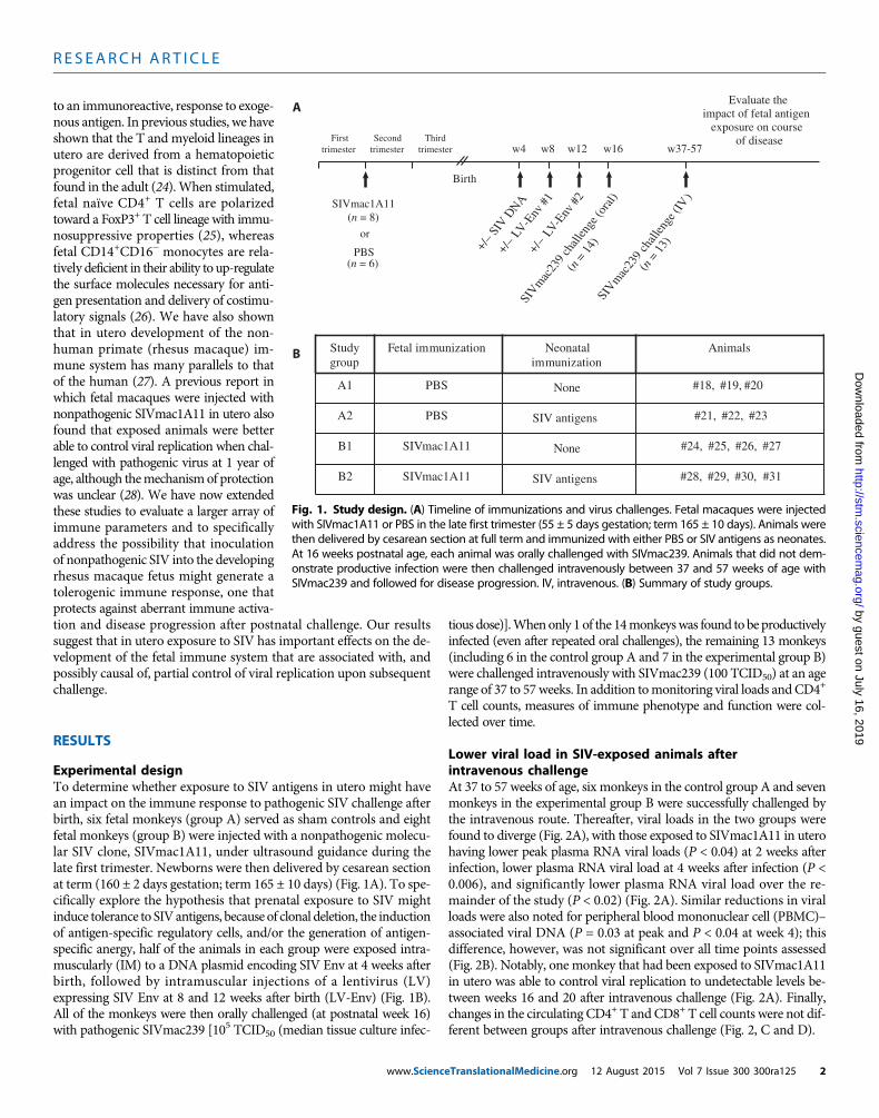
Experimental designTo determine whether exposure to SIV antigens in utero might havean impact on the immune response to pathogenic SIV challenge afterbirth, six fetal monkeys (group A) served as sham controls and eightfetal monkeys (group B) were injected with a nonpathogenic molecu-lar SIV clone, SIVmac1A11, under ultrasound guidance during thelate first trimester. Newborns were then delivered by cesarean sectionat term (160 ± 2 days gestation; term 165 ± 10 days) (Fig. 1A). To spe-cifically explore the hypothesis that prenatal exposure to SIV mightinduce tolerance to SIVantigens, because of clonal deletion, the inductionof antigen-specific regulatory cells, and/or the generation of antigen-specific anergy, half of the animals in each group were exposed intra-muscularly (IM) to a DNA plasmid encoding SIV Env at 4 weeks afterbirth, followed by intramuscular injections of a lentivirus (LV)expressing SIV Env at 8 and 12 weeks after birth (LV-Env) (Fig. 1B).All of the monkeys were then orally challenged (at postnatal week 16)with pathogenic SIVmac239 [105 TCID50 (median tissue culture infec-
www.Scienc
tious dose)].Whenonly 1 of the 14monkeyswas found to be productivelyinfected (even after repeated oral challenges), the remaining 13 monkeys(including 6 in the control group A and 7 in the experimental group B)were challenged intravenously with SIVmac239 (100 TCID50) at an agerange of 37 to 57weeks. In addition tomonitoring viral loads andCD4+
T cell counts, measures of immune phenotype and function were col-lected over time.
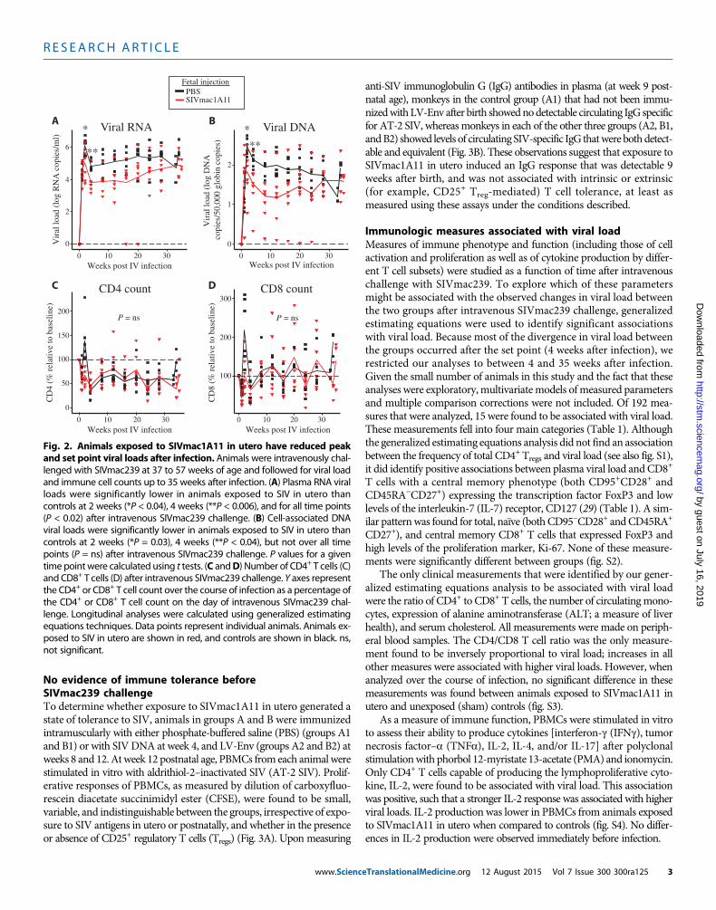
Lower viral load in SIV-exposed animals afterintravenous challengeAt 37 to 57 weeks of age, six monkeys in the control group A and sevenmonkeys in the experimental group B were successfully challenged bythe intravenous route. Thereafter, viral loads in the two groups werefound to diverge (Fig. 2A), with those exposed to SIVmac1A11 in uterohaving lower peak plasma RNA viral loads (P < 0.04) at 2 weeks afterinfection, lower plasma RNA viral load at 4 weeks after infection (P <0.006), and significantly lower plasma RNA viral load over the re-mainder of the study (P < 0.02) (Fig. 2A). Similar reductions in viralloads were also noted for peripheral blood mononuclear cell (PBMC)–associated viral DNA (P = 0.03 at peak and P < 0.04 at week 4); thisdifference, however, was not significant over all time points assessed(Fig. 2B). Notably, one monkey that had been exposed to SIVmac1A11in utero was able to control viral replication to undetectable levels be-tween weeks 16 and 20 after intravenous challenge (Fig. 2A). Finally,changes in the circulating CD4+ T and CD8+ T cell counts were not dif-ferent between groups after intravenous challenge (Fig. 2, C and D).
A
B Study group
Fetal immunization Neonatal immunization
Animals
A1 PBS None #18, #19, #20
A2 PBS SIV antigens #21, #22, #23
B1 SIVmac1A11 #24, #25, #26, #27
B2 SIVmac1A11 #28, #29, #30, #31SIV antigens
None
SIVmac1A11 (n = 8)
or
PBS (n = 6)
SIV
mac
239
chall
enge
(ora
l)
(n =
14)
Evaluate the impact of fetal antigen
exposure on course of disease
Birth
+/–
LV-E
nv #
1
w8 w12 w16 w4
+/–
SIV D
NA +/
– LV
-Env
#2
w37-57
SIVm
ac23
9 ch
allen
ge (I
V)
(n =
13)
First trimester
Second trimester
Third trimester
Fig. 1. Study design. (A) Timeline of immunizations and virus challenges. Fetal macaques were injectedwith SIVmac1A11 or PBS in the late first trimester (55 ± 5 days gestation; term 165 ± 10 days). Animals were
then delivered by cesarean section at full term and immunized with either PBS or SIV antigens as neonates.At 16 weeks postnatal age, each animal was orally challenged with SIVmac239. Animals that did not dem-onstrate productive infection were then challenged intravenously between 37 and 57 weeks of age withSIVmac239 and followed for disease progression. IV, intravenous. (B) Summary of study groups.
eTranslationalMedicine.org 12 August 2015 Vol 7 Issue 300 300ra125 2
No evidence of immune tolerance beforeSIVmac239 challengeTo determine whether exposure to SIVmac1A11 in utero generated astate of tolerance to SIV, animals in groups A and B were immunizedintramuscularly with either phosphate-buffered saline (PBS) (groups A1and B1) or with SIVDNA at week 4, and LV-Env (groups A2 and B2) atweeks 8 and 12. Atweek 12 postnatal age, PBMCs fromeach animalwerestimulated in vitro with aldrithiol-2–inactivated SIV (AT-2 SIV). Prolif-erative responses of PBMCs, as measured by dilution of carboxyfluo-rescein diacetate succinimidyl ester (CFSE), were found to be small,variable, and indistinguishable between the groups, irrespective of expo-sure to SIV antigens in utero or postnatally, and whether in the presenceor absence of CD25+ regulatory T cells (Tregs) (Fig. 3A). Upon measuring
www.Scienc
anti-SIV immunoglobulin G (IgG) antibodies in plasma (at week 9 post-natal age), monkeys in the control group (A1) that had not been immu-nizedwithLV-Env after birth showednodetectable circulating IgG specificfor AT-2 SIV, whereasmonkeys in each of the other three groups (A2, B1,andB2) showed levels of circulating SIV-specific IgG thatwere bothdetect-able and equivalent (Fig. 3B). These observations suggest that exposure toSIVmac1A11 in utero induced an IgG response that was detectable 9weeks after birth, and was not associated with intrinsic or extrinsic(for example, CD25+ Treg-mediated) T cell tolerance, at least asmeasured using these assays under the conditions described.
Immunologic measures associated with viral loadMeasures of immune phenotype and function (including those of cellactivation and proliferation as well as of cytokine production by differ-ent T cell subsets) were studied as a function of time after intravenouschallenge with SIVmac239. To explore which of these parametersmight be associated with the observed changes in viral load betweenthe two groups after intravenous SIVmac239 challenge, generalizedestimating equations were used to identify significant associationswith viral load. Because most of the divergence in viral load betweenthe groups occurred after the set point (4 weeks after infection), werestricted our analyses to between 4 and 35 weeks after infection.Given the small number of animals in this study and the fact that theseanalyses were exploratory,multivariatemodels ofmeasured parametersand multiple comparison corrections were not included. Of 192 mea-sures that were analyzed, 15 were found to be associated with viral load.These measurements fell into four main categories (Table 1). Althoughthe generalized estimating equations analysis did not find an associationbetween the frequency of total CD4+ Tregs and viral load (see also fig. S1),it did identify positive associations between plasma viral load and CD8+
T cells with a central memory phenotype (both CD95+CD28+ andCD45RA−CD27+) expressing the transcription factor FoxP3 and lowlevels of the interleukin-7 (IL-7) receptor, CD127 (29) (Table 1). A sim-ilar patternwas found for total, naïve (both CD95−CD28+ andCD45RA+
CD27+), and central memory CD8+ T cells that expressed FoxP3 andhigh levels of the proliferation marker, Ki-67. None of these measure-ments were significantly different between groups (fig. S2).
The only clinical measurements that were identified by our gener-alized estimating equations analysis to be associated with viral loadwere the ratio of CD4+ to CD8+ T cells, the number of circulatingmono-cytes, expression of alanine aminotransferase (ALT; a measure of liverhealth), and serum cholesterol. All measurements were made on periph-eral blood samples. The CD4/CD8 T cell ratio was the only measure-ment found to be inversely proportional to viral load; increases in allother measures were associated with higher viral loads. However, whenanalyzed over the course of infection, no significant difference in thesemeasurements was found between animals exposed to SIVmac1A11 inutero and unexposed (sham) controls (fig. S3).
As a measure of immune function, PBMCs were stimulated in vitroto assess their ability to produce cytokines [interferon-g (IFNg), tumornecrosis factor–a (TNFa), IL-2, IL-4, and/or IL-17] after polyclonalstimulationwith phorbol 12-myristate 13-acetate (PMA) and ionomycin.Only CD4+ T cells capable of producing the lymphoproliferative cyto-kine, IL-2, were found to be associated with viral load. This associationwas positive, such that a stronger IL-2 response was associated with higherviral loads. IL-2 production was lower in PBMCs from animals exposedto SIVmac1A11 in utero when compared to controls (fig. S4). No differ-ences in IL-2 production were observed immediately before infection.
Fetal injectionPBSSIVmac1A11
B
Vir
al lo
ad (
log
DN
A
copi
es/5
0,00
0 gl
obin
cop
ies)
Viral DNA
Weeks post IV infection
0
1
2
10 20 300
*
**
*
**
Viral RNA
0
2
4
6
0 10 20 30
Vir
al lo
ad (
log
RN
A c
opie
s/m
l)
Weeks post IV infection
C
0
50
100
150
200
CD
4 (%
rel
ativ
e to
bas
elin
e)
CD4 count
10 20 300
P = ns
Weeks post IV infection
CD8 count
CD
8 (%
rel
ativ
e to
bas
elin
e)
300
200
100
10 20 300Weeks post IV infection
P = ns
D
A
Fig. 2. Animals exposed to SIVmac1A11 in utero have reduced peakand set point viral loads after infection. Animals were intravenously chal-
lenged with SIVmac239 at 37 to 57 weeks of age and followed for viral loadand immune cell counts up to 35 weeks after infection. (A) Plasma RNA viralloads were significantly lower in animals exposed to SIV in utero thancontrols at 2 weeks (*P < 0.04), 4 weeks (**P < 0.006), and for all time points(P < 0.02) after intravenous SIVmac239 challenge. (B) Cell-associated DNAviral loads were significantly lower in animals exposed to SIV in utero thancontrols at 2 weeks (*P = 0.03), 4 weeks (**P < 0.04), but not over all timepoints (P = ns) after intravenous SIVmac239 challenge. P values for a giventime point were calculated using t tests. (C andD) Number of CD4+ T cells (C)and CD8+ T cells (D) after intravenous SIVmac239 challenge. Y axes representthe CD4+ or CD8+ T cell count over the course of infection as a percentage ofthe CD4+ or CD8+ T cell count on the day of intravenous SIVmac239 chal-lenge. Longitudinal analyses were calculated using generalized estimatingequations techniques. Data points represent individual animals. Animals ex-posed to SIV in utero are shown in red, and controls are shown in black. ns,not significant.
eTranslationalMedicine.org 12 August 2015 Vol 7 Issue 300 300ra125 3
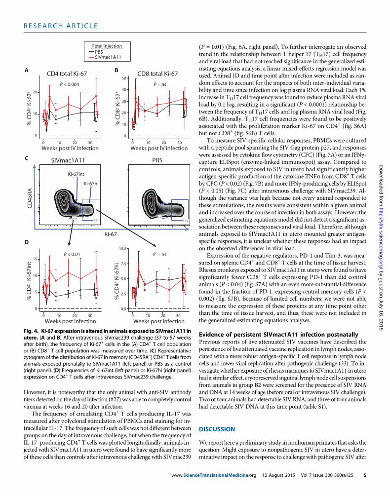
The frequency of both CD4+ and CD8+ T cells expressing Ki-67was inversely correlated with viral load over the course of infection,except in the case where Ki-67+ cells coexpressed FoxP3 (as discussedabove). When plotted longitudinally, it became apparent that Ki-67–expressingCD4+T cells weremore abundant in rhesusmonkeys exposedto SIVmac1A11 in utero compared to controlmonkeys not exposed to
www.ScienceTranslationalMedicine.org 12
virus in utero (P < 0.004) (Fig. 4A). Nodifference between experimental groupswas found in CD8+ T cells (Fig. 4B). Ki-67was found to be expressed in CD4+ T cellsfrom SIVmac1A11-exposed monkeys in aunique manner, with many more cells ex-pressing intermediate levels of Ki-67 pro-tein relative to those observed in CD4+ Tcells from sham control rhesus monkeys(Fig. 4C). Such Ki-67 intermediate (Ki-67int)cells were not differentially abundant atthe time of intravenous SIVmac239 chal-lenge, but did increase in abundance in theSIVmac1A11-exposed animals as a functionof time thereafter when compared to thesham-injected control animals (P < 0.01)(Fig. 4D, left panel). By contrast, there wasno difference between the groups in CD4+
T cells expressing high levels of Ki-67 (Fig.4D, right panel).
Including Ki-67 high-expressing (Ki-67hi) and Ki-67 intermediate-expressing(Ki-67int) CD4+ and CD8+ T cells into
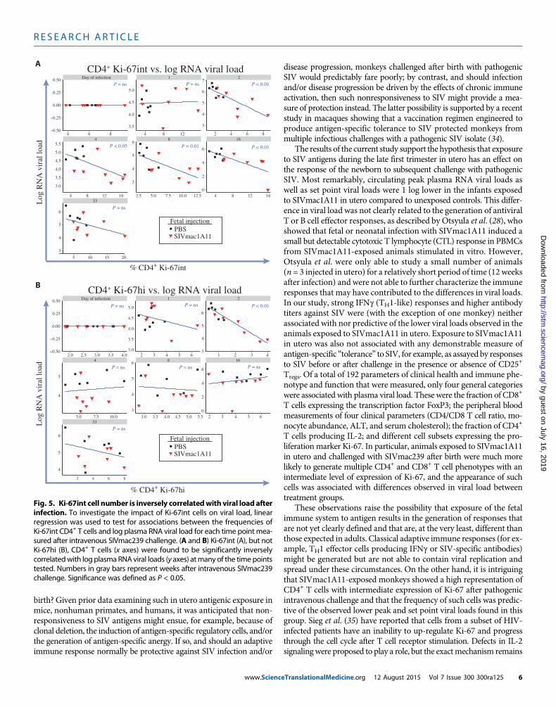
the generalized estimating equations analysis revealed multiple (andpotentially overlapping) subsets that were significantly inversely as-sociated with viral load over the course of infection. Linear regressionmodels were then used to analyze these associations for every time pointmeasured after infection. Plots for each measurement of viral load witheach measurement of CD4+ Ki-67int T cells are shown in Fig. 5A, withsignificant associations found at weeks 2, 4, 8, 16, and 24 after intra-venous SIVmac239 challenge. These associations remained significanteven after removing the animal that controlled viral replication to un-detectable levels (animal #27). By contrast, it was only at week 2 afterintravenous SIVmac239 challenge that the frequency of CD4+ Ki-67hiT cells had an association with viral load (Fig. 5B).
Immunologic measures not associated with viral load butdifferent between groupsMultiple previous studies with SIV-infected rhesusmacaques andHIV-infected humans have raised the possibility that viral replication andspread can be suppressed by adaptive immunity (for example, virus-specific neutralizing antibodies and effector T cells) and augmentedby the immunologic effects of chronic activation (for example, resultingin the release of proinflammatory cytokines and the generation of func-tionally “exhausted”T cells) (30–32). Although the generalized estimat-ing equations models used in Table 1 did not show that any suchmeasures predicted lower viral loads in the SIVmac1A11-exposed rhesusmonkeys at any time point after intravenous challenge with SIVmac239,several merit mention.
Although monkeys from control group A1 (with no exposure toSIVmac1A11 in utero and no postnatal vaccination) did not developdetectable antibody responses against AT-2 SIV until after the peak ofviremia (2 weeks after infection), monkeys in each of the other groupshad detectable anti-SIV antibody titers by 2 weeks after infection (anda single animal had detectable antibody titers on the day of intravenousinfection) (fig. S5). In a generalized estimating equations model, thesetiters showed no significant relationship to viral RNA levels or to theobserveddifferences in viral loadbetween groupAandgroupBmonkeys.
BAnti-SIV IgG titers
Mid
poin
t tite
r (–
logE
C50
)
0
2
4
6
A1 A2 B1 B2
SIV-specific
CD4 T cell proliferation
–1
% C
D4
prol
ifer
atio
n
PBS SIV
All cells
CD25–
All cells
CD25–
0
1
2
3
4 ns
Fetal injection
A
Study group
ns
ns
ns
ns
Fig. 3. No evidence of antigen-specific tolerance before oral challenge. Fetal exposure to SIVmac1A11followed by neonatal vaccination elicited detectable humoral but not cellular responses before oral chal-
lenge. (A) Proliferating CD4+ T cells in total PBMCs from 12-week-old rhesus monkeys were labeled withCFSE and incubated with AT-2 SIV for 6 days. (B) IgG titers against AT-2 SIV in plasma from 9-week-oldanimals were not detectable in group A1, but were detectable in groups A2, B1, and B2. No significant dif-ferenceswere foundbetween the threegroupswith detectable titers. Differenceswere assessed using t testswith significance defined as P < 0.05. ns, not significant.
Table 1. Plasma RNA viral loads are associated with multiple immuno-logic and clinical measurements. A panel of 192 immunologic and clinicalmeasurements, taken at multiple time points after intravenous SIVmac239challenge, was analyzed for associations with log plasma RNA viral load bygeneralized estimating equations. Fifteen measurements were identified asbeing significantly associated with viral load (P < 0.05).
However, it is noteworthy that the only animal with anti-SIV antibodytiters detected on the day of infection (#27)was able to completely controlviremia at weeks 16 and 20 after infection.
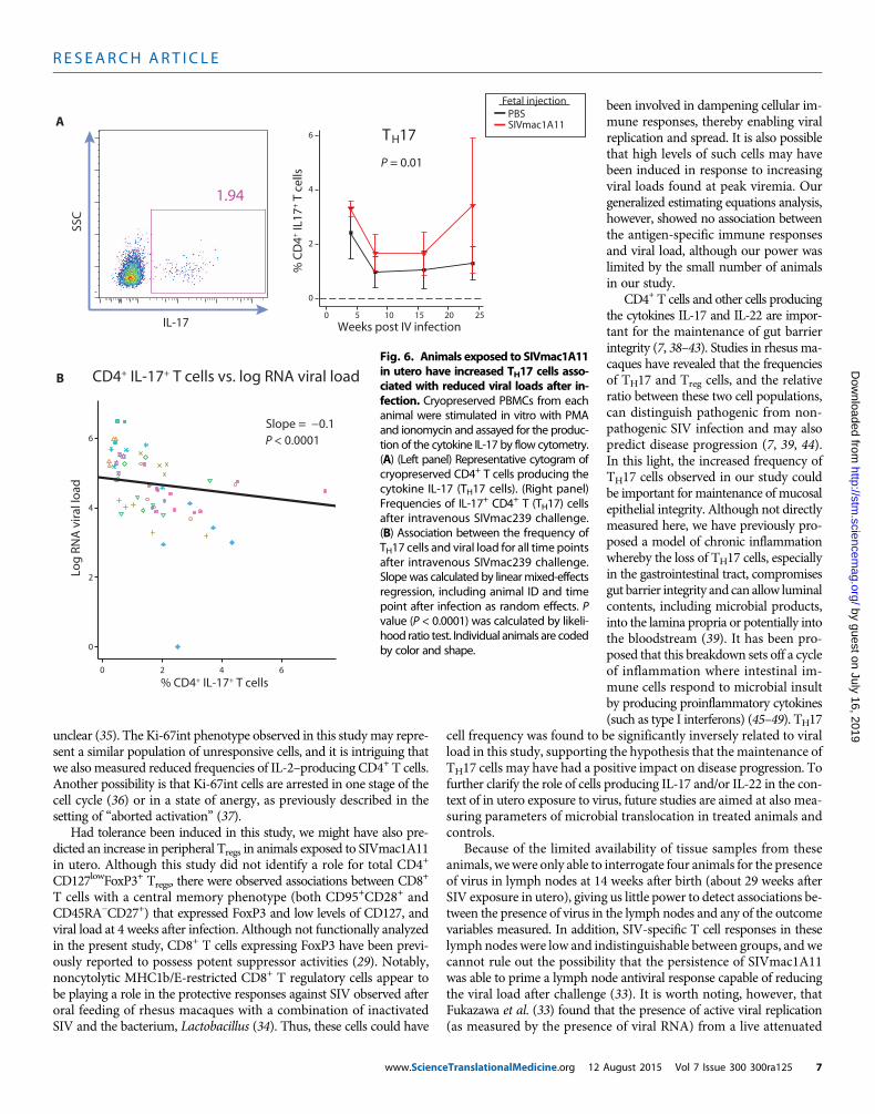
The frequency of circulating CD4+ T cells producing IL-17 wasmeasured after polyclonal stimulation of PBMCs and staining for in-tracellular IL-17. The frequency of such cells was not different betweengroups on the day of intravenous challenge, but when the frequency ofIL-17–producing CD4+ T cells was plotted longitudinally, animals in-jected with SIVmac1A11 in utero were found to have significantlymoreof these cells than controls after intravenous challenge with SIVmac239
www.Scienc
(P = 0.01) (Fig. 6A, right panel). To further interrogate an observedtrend in the relationship between T helper 17 (TH17) cell frequencyand viral load that had not reached significance in the generalized esti-mating equations analysis, a linear mixed-effects regression model wasused. Animal ID and time point after infection were included as ran-dom effects to account for the impacts of both inter-individual varia-bility and time since infection on log plasma RNA viral load. Each 1%increase in TH17 cell frequency was found to reduce plasma RNA viralload by 0.1 log, resulting in a significant (P < 0.0001) relationship be-tween the frequency of TH17 cells and log plasma RNA viral load (Fig.6B). Additionally, TH17 cell frequencies were found to be positivelyassociated with the proliferation marker Ki-67 on CD4+ (fig. S6A)but not CD8+ (fig. S6B) T cells.
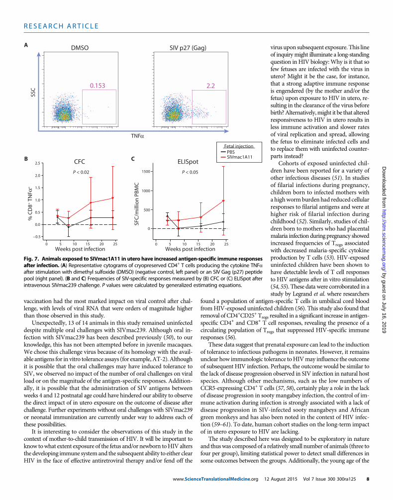
To measure SIV-specific cellular responses, PBMCs were culturedwith a peptide pool spanning the SIV Gag protein p27, and responseswere assessed by cytokine flow cytometry (CFC) (Fig. 7A) or an IFNg-capture ELISpot (enzyme-linked immunospot) assay. Compared tocontrols, animals exposed to SIV in utero had significantly higherantigen-specific production of the cytokine TNFa from CD8+ T cellsby CFC (P < 0.02) (Fig. 7B) andmore IFNg-producing cells by ELISpot(P < 0.05) (Fig. 7C) after intravenous challenge with SIVmac239. Al-though the variance was high because not every animal responded tothese stimulations, the results were consistent within a given animaland increased over the course of infection in both assays. However, thegeneralized estimating equationsmodel did not detect a significant as-sociation between these responses and viral load. Therefore, althoughanimals exposed to SIVmac1A11 in utero mounted greater antigen-specific responses, it is unclear whether these responses had an impacton the observed differences in viral load.
Expression of the negative regulators, PD-1 and Tim-3, was mea-sured on splenic CD4+ and CD8+ T cells at the time of tissue harvest.Rhesusmonkeys exposed to SIVmac1A11 in utero were found to havesignificantly fewer CD8+ T cells expressing PD-1 than did controlanimals (P < 0.04) (fig. S7A) with an even more substantial differencefound in the fraction of PD-1–expressing central memory cells (P <0.002) (fig. S7B). Because of limited cell numbers, we were not ableto measure the expression of these proteins at any time point otherthan the time of tissue harvest, and thus, these were not included inthe generalized estimating equations analyses.
Evidence of persistent SIVmac1A11 infection postnatallyPrevious reports of live attenuated SIV vaccines have described thepersistence of live attenuated vaccine replication in lymphnodes, asso-ciated with a more robust antigen-specific T cell response in lymph nodecells and lower viral replication after pathogenic challenge (33). To in-vestigatewhether exposure of rhesusmacaques to SIVmac1A11 in uterohad a similar effect, cryopreserved inguinal lymphnode cell suspensionsfrom animals in group B2 were screened for the presence of SIV RNAandDNA at 14 weeks of age (before oral or intravenous SIV challenge).Two of four animals had detectable SIV RNA, and three of four animalshad detectable SIV DNA at this time point (table S1).
DISCUSSION
Wereport here a preliminary study in nonhumanprimates that asks thequestion: Might exposure to nonpathogenic SIV in utero have a deter-minative impact on the response to challenge with pathogenic SIV after
A
0
10
20
% C
D4+
Ki-
67+
P = ns
0
10
20
30
40
50
% C
D8+
Ki-
67+
CD4 total Ki-67 CD8 total Ki-67 B
Weeks post IV infection3020100
Weeks post IV infection
4.279.19
SIVmac1A11
5.79
5.5
PBS
Ki-67
CD
45RA
Ki-67hi
Ki-67int
C
D
P < 0.004
3020100
0
5
10
15
Weeks post infection
% C
D4+
Ki-
67in
t
P = ns
0.0
2.5
5.0
7.5
10.0
% C
D4+
Ki-
67h
i
3020100
P < 0.01
Weeks post infection3020100
Fetal injectionPBSSIVmac1A11
Fig. 4. Ki-67expression is altered inanimals exposed toSIVmac1A11 inutero. (A and B) After intravenous SIVmac239 challenge (37 to 57 weeks
after birth), the frequency of Ki-67+ cells in the (A) CD4+ T cell populationor (B) CD8+ T cell population was measured over time. (C) Representativecytogramof the distribution of Ki-67 inmemory (CD45RA−) CD4+ T cells fromanimals exposed prenatally to SIVmac1A11 (left panel) or PBS as a control(right panel). (D) Frequencies of Ki-67int (left panel) or Ki-67hi (right panel)expression on CD4+ T cells after intravenous SIVmac239 challenge.
eTranslationalMedicine.org 12 August 2015 Vol 7 Issue 300 300ra125 5
birth? Given prior data examining such in utero antigenic exposure inmice, nonhuman primates, and humans, it was anticipated that non-responsiveness to SIV antigens might ensue, for example, because ofclonal deletion, the induction of antigen-specific regulatory cells, and/orthe generation of antigen-specific anergy. If so, and should an adaptiveimmune response normally be protective against SIV infection and/or
www.Scienc
disease progression, monkeys challenged after birth with pathogenicSIV would predictably fare poorly; by contrast, and should infectionand/or disease progression be driven by the effects of chronic immuneactivation, then such nonresponsiveness to SIV might provide a mea-sure of protection instead. The latter possibility is supported by a recentstudy in macaques showing that a vaccination regimen engineered toproduce antigen-specific tolerance to SIV protected monkeys frommultiple infectious challenges with a pathogenic SIV isolate (34).
The results of the current study support the hypothesis that exposureto SIV antigens during the late first trimester in utero has an effect onthe response of the newborn to subsequent challenge with pathogenicSIV. Most remarkably, circulating peak plasma RNA viral loads aswell as set point viral loads were 1 log lower in the infants exposedto SIVmac1A11 in utero compared to unexposed controls. This differ-ence in viral load was not clearly related to the generation of antiviralT or B cell effector responses, as described by Otsyula et al. (28), whoshowed that fetal or neonatal infection with SIVmac1A11 induced asmall but detectable cytotoxic T lymphocyte (CTL) response in PBMCsfrom SIVmac1A11-exposed animals stimulated in vitro. However,Otsyula et al. were only able to study a small number of animals(n = 3 injected in utero) for a relatively short period of time (12 weeksafter infection) and were not able to further characterize the immuneresponses that may have contributed to the differences in viral loads.In our study, strong IFNg (TH1-like) responses and higher antibodytiters against SIV were (with the exception of one monkey) neitherassociated with nor predictive of the lower viral loads observed in theanimals exposed to SIVmac1A11 in utero. Exposure to SIVmac1A11in utero was also not associated with any demonstrable measure ofantigen-specific “tolerance” to SIV, for example, as assayed by responsesto SIV before or after challenge in the presence or absence of CD25+
Tregs. Of a total of 192 parameters of clinical health and immune phe-notype and function that were measured, only four general categorieswere associated with plasma viral load. These were the fraction of CD8+
T cells expressing the transcription factor FoxP3; the peripheral bloodmeasurements of four clinical parameters (CD4/CD8 T cell ratio, mo-nocyte abundance, ALT, and serum cholesterol); the fraction of CD4+
T cells producing IL-2; and different cell subsets expressing the pro-liferation marker Ki-67. In particular, animals exposed to SIVmac1A11in utero and challenged with SIVmac239 after birth were much morelikely to generate multiple CD4+ and CD8+ T cell phenotypes with anintermediate level of expression of Ki-67, and the appearance of suchcells was associated with differences observed in viral load betweentreatment groups.
These observations raise the possibility that exposure of the fetalimmune system to antigen results in the generation of responses thatare not yet clearly defined and that are, at the very least, different thanthose expected in adults. Classical adaptive immune responses (for ex-ample, TH1 effector cells producing IFNg or SIV-specific antibodies)might be generated but are not able to contain viral replication andspread under these circumstances. On the other hand, it is intriguingthat SIVmac1A11-exposed monkeys showed a high representation ofCD4+ T cells with intermediate expression of Ki-67 after pathogenicintravenous challenge and that the frequency of such cells was predic-tive of the observed lower peak and set point viral loads found in thisgroup. Sieg et al. (35) have reported that cells from a subset of HIV-infected patients have an inability to up-regulate Ki-67 and progressthrough the cell cycle after T cell receptor stimulation. Defects in IL-2signalingwere proposed to play a role, but the exactmechanism remains
Day of infection 1 2
4 8 16
33
−0.50
−0.25
0.00
0.25
0.50
3.0
3.5
4.0
4.5
5.0
3
4
5
6
4
5
3
4
5
6
0
2
4
6
4
5
6
2 3 4 5 6 1 2 3 4
5.0 7.5 10.0 2 3 4 5 6
2 4 6 8
% CD4+ Ki-67hi
Log
RN
A v
iral
load
CD4+ Ki-67hi vs. log RNA viral load
P = ns P < 0.01
P < 0.05 P = 0.01
Day of infection 1 2
4 8 16
33
−0.50
−0.25
0.00
0.25
0.50
3.5
4.0
4.5
5.0
3
4
5
6
7
3.0
3.5
4.0
4.5
5.0
5.5
3
4
5
6
0
2
4
6
3
4
5
6
4 6 8 4 8 12 2 4 6 8
4 8 12 16 4 8 12 16
5 10 15 20
CD4+ Ki-67int vs. log RNA viral loadA
B
P = ns
P < 0.01
P = ns
P = ns P = ns
P = ns P = ns P = ns
P < 0.01
P = ns
Fetal injectionPBSSIVmac1A11
Fetal injectionPBSSIVmac1A11
3.0 3.5 4.0 4.5 5.0 5.5
% CD4+ Ki-67int
Log
RN
A v
iral
load
12.510.07.55.02.5
2.0 2.5 3.0 3.5 4.0
Fig. 5. Ki-67int cell number is inversely correlatedwith viral load afterinfection. To investigate the impact of Ki-67int cells on viral load, linear
regression was used to test for associations between the frequencies ofKi-67int CD4+ T cells and log plasma RNA viral load for each time point mea-sured after intravenous SIVmac239 challenge. (A and B) Ki-67int (A), but notKi-67hi (B), CD4+ T cells (x axes) were found to be significantly inverselycorrelatedwith log plasmaRNA viral loads (y axes) atmany of the timepointstested. Numbers in gray bars represent weeks after intravenous SIVmac239challenge. Significance was defined as P < 0.05.
eTranslationalMedicine.org 12 August 2015 Vol 7 Issue 300 300ra125 6
unclear (35). The Ki-67int phenotype observed in this studymay repre-sent a similar population of unresponsive cells, and it is intriguing thatwe also measured reduced frequencies of IL-2–producing CD4+ T cells.Another possibility is that Ki-67int cells are arrested in one stage of thecell cycle (36) or in a state of anergy, as previously described in thesetting of “aborted activation” (37).
Had tolerance been induced in this study, we might have also pre-dicted an increase in peripheral Tregs in animals exposed to SIVmac1A11in utero. Although this study did not identify a role for total CD4+
CD127lowFoxP3+ Tregs, there were observed associations between CD8+
T cells with a central memory phenotype (both CD95+CD28+ andCD45RA−CD27+) that expressed FoxP3 and low levels of CD127, andviral load at 4 weeks after infection. Although not functionally analyzedin the present study, CD8+ T cells expressing FoxP3 have been previ-ously reported to possess potent suppressor activities (29). Notably,noncytolytic MHC1b/E-restricted CD8+ T regulatory cells appear tobe playing a role in the protective responses against SIV observed afteroral feeding of rhesus macaques with a combination of inactivatedSIV and the bacterium, Lactobacillus (34). Thus, these cells could have
www.ScienceTranslationalMedicine.org 12
been involved in dampening cellular im-mune responses, thereby enabling viralreplication and spread. It is also possiblethat high levels of such cells may havebeen induced in response to increasingviral loads found at peak viremia. Ourgeneralized estimating equations analysis,however, showed no association betweenthe antigen-specific immune responsesand viral load, although our power waslimited by the small number of animalsin our study.
CD4+ T cells and other cells producingthe cytokines IL-17 and IL-22 are impor-tant for the maintenance of gut barrierintegrity (7, 38–43). Studies in rhesus ma-caques have revealed that the frequenciesof TH17 and Treg cells, and the relativeratio between these two cell populations,can distinguish pathogenic from non-pathogenic SIV infection and may alsopredict disease progression (7, 39, 44).In this light, the increased frequency ofTH17 cells observed in our study couldbe important for maintenance of mucosalepithelial integrity. Although not directlymeasured here, we have previously pro-posed a model of chronic inflammationwhereby the loss of TH17 cells, especiallyin the gastrointestinal tract, compromisesgut barrier integrity and can allow luminalcontents, including microbial products,into the lamina propria or potentially intothe bloodstream (39). It has been pro-posed that this breakdown sets off a cycleof inflammation where intestinal im-mune cells respond to microbial insultby producing proinflammatory cytokines(such as type I interferons) (45–49). TH17
cell frequency was found to be significantly inversely related to viralload in this study, supporting the hypothesis that the maintenance ofTH17 cells may have had a positive impact on disease progression. Tofurther clarify the role of cells producing IL-17 and/or IL-22 in the con-text of in utero exposure to virus, future studies are aimed at also mea-suring parameters of microbial translocation in treated animals andcontrols.
Because of the limited availability of tissue samples from theseanimals, wewere only able to interrogate four animals for the presenceof virus in lymph nodes at 14 weeks after birth (about 29 weeks afterSIV exposure in utero), giving us little power to detect associations be-tween the presence of virus in the lymph nodes and any of the outcomevariables measured. In addition, SIV-specific T cell responses in theselymph nodes were low and indistinguishable between groups, andwecannot rule out the possibility that the persistence of SIVmac1A11was able to prime a lymph node antiviral response capable of reducingthe viral load after challenge (33). It is worth noting, however, thatFukazawa et al. (33) found that the presence of active viral replication(as measured by the presence of viral RNA) from a live attenuated
P = 0.01
0
2
4
6
% C
D4+
IL17
+ T
cel
ls
TH17
25200
Weeks post IV infection15105
1.94
SSC
IL-17
A
% CD4+ IL-17+ T cells
Log
RN
A v
iral
load
CD4+ IL-17+ T cells vs. log RNA viral loadB
0
2
4
6
Slope = −0.1
0 2 4 6
P < 0.0001
Fetal injectionPBSSIVmac1A11
Fig. 6. Animals exposed to SIVmac1A11in utero have increased TH17 cells asso-ciated with reduced viral loads after in-fection. Cryopreserved PBMCs from eachanimal were stimulated in vitro with PMAand ionomycin and assayed for the produc-tion of the cytokine IL-17 by flow cytometry.(A) (Left panel) Representative cytogram ofcryopreserved CD4+ T cells producing thecytokine IL-17 (TH17 cells). (Right panel)Frequencies of IL-17+ CD4+ T (TH17) cellsafter intravenous SIVmac239 challenge.(B) Association between the frequency ofTH17 cells and viral load for all time pointsafter intravenous SIVmac239 challenge.Slopewas calculated by linearmixed-effectsregression, including animal ID and timepoint after infection as random effects. Pvalue (P < 0.0001) was calculated by likeli-hood ratio test. Individual animals are codedby color and shape.
vaccination had the most marked impact on viral control after chal-lenge, with levels of viral RNA that were orders of magnitude higherthan those observed in this study.
Unexpectedly, 13 of 14 animals in this study remained uninfecteddespite multiple oral challenges with SIVmac239. Although oral in-fection with SIVmac239 has been described previously (50), to ourknowledge, this has not been attempted before in juvenile macaques.We chose this challenge virus because of its homology with the avail-able antigens for in vitro tolerance assays (for example, AT-2). Althoughit is possible that the oral challenges may have induced tolerance toSIV, we observed no impact of the number of oral challenges on viralload or on the magnitude of the antigen-specific responses. Addition-ally, it is possible that the administration of SIV antigens betweenweeks 4 and 12 postnatal age could have hindered our ability to observethe direct impact of in utero exposure on the outcome of disease afterchallenge. Further experiments without oral challenges with SIVmac239or neonatal immunization are currently under way to address each ofthese possibilities.
It is interesting to consider the observations of this study in thecontext of mother-to-child transmission of HIV. It will be important toknow towhat extent exposure of the fetus and/or newborn toHIV altersthe developing immune systemand the subsequent ability to either clearHIV in the face of effective antiretroviral therapy and/or fend off the
www.ScienceTranslationalMedicine.org 12
virus upon subsequent exposure. This lineof inquirymight illuminate a long-standingquestion in HIV biology:Why is it that sofew fetuses are infected with the virus inutero? Might it be the case, for instance,that a strong adaptive immune responseis engendered (by the mother and/or thefetus) upon exposure to HIV in utero, re-sulting in the clearance of the virus beforebirth?Alternatively,might it be that alteredresponsiveness to HIV in utero results inless immune activation and slower ratesof viral replication and spread, allowingthe fetus to eliminate infected cells andto replace them with uninfected counter-parts instead?
Cohorts of exposed uninfected chil-dren have been reported for a variety ofother infectious diseases (51). In studiesof filarial infections during pregnancy,children born to infected mothers witha highwormburden had reduced cellularresponses to filarial antigens and were athigher risk of filarial infection duringchildhood (52). Similarly, studies of chil-dren born to mothers who had placentalmalaria infection during pregnancy showedincreased frequencies of Tregs associatedwith decreased malaria-specific cytokineproduction by T cells (53). HIV-exposeduninfected children have been shown tohave detectable levels of T cell responsesto HIV antigens after in vitro stimulation(54, 55). These data were corroborated in astudy by Legrand et al. where researchers
found a population of antigen-specific T cells in umbilical cord bloodfromHIV-exposed uninfected children (56). This study also found thatremoval of CD4+CD25+ Tregs resulted in a significant increase in antigen-specific CD4+ and CD8+ T cell responses, revealing the presence of acirculating population of Tregs that suppressed HIV-specific immuneresponses (56).
These data suggest that prenatal exposure can lead to the inductionof tolerance to infectious pathogens in neonates. However, it remainsunclear how immunologic tolerance toHIVmay influence the outcomeof subsequent HIV infection. Perhaps, the outcome would be similar tothe lack of disease progression observed in SIV infection in natural hostspecies. Although other mechanisms, such as the low numbers ofCCR5-expressing CD4+ T cells (57, 58), certainly play a role in the lackof disease progression in sooty mangabey infection, the control of im-mune activation during infection is strongly associated with a lack ofdisease progression in SIV-infected sooty mangabeys and Africangreen monkeys and has also been noted in the context of HIV infec-tion (59–61). To date, human cohort studies on the long-term impactof in utero exposure to HIV are lacking.
The study described here was designed to be exploratory in natureand thuswas composed of a relatively small number of animals (three tofour per group), limiting statistical power to detect small differences insome outcomes between the groups. Additionally, the young age of the
B
P < 0.02
–0.5
0.0
0.5
1.0
1.5
2.0
2.5
% C
D8+
TN
F+
CFC
0 10 15 20Weeks post infection
5 25
P < 0.05
0
500
1000
1500
10 15 20 25
SFC
/mill
ion
PB
MC
C ELISpot
Weeks post infection50
0.153
DMSO
2.2
SIV p27 (Gag)A
TNF
SSC
Fetal injectionPBSSIVmac1A11
α
α
Fig. 7. Animals exposed to SIVmac1A11 in utero have increased antigen-specific immune responsesafter infection. (A) Representative cytograms of cryopreserved CD4+ T cells producing the cytokine TNFa
after stimulation with dimethyl sulfoxide (DMSO) (negative control, left panel) or an SIV Gag (p27) peptidepool (right panel). (B and C) Frequencies of SIV-specific responses measured by (B) CFC or (C) ELISpot afterintravenous SIVmac239 challenge. P values were calculated by generalized estimating equations.
animals provided some challenges in detecting SIV-specific responses,both related to themagnitude of the responses, and because the quantityof PBMCs was limited since small volumes of blood were collected ateach time point based on bodyweight. Despite this, we believe that thesestudies, although preliminary, provide a relevant and interesting avenueof research to pursue.
Although these studies, like those of Andrieu and colleagues (34, 62),should be viewed as preliminary, it is striking that interventions de-signed to promote the generation of a tolerogenic immune responseare in each case associated with partial or complete protection againstpathogenic SIV challenge. If our findings can be confirmed and extended,then one might envisage developing vaccines that induce a tolerogenicresponse in people at risk for HIV infection. Similar vaccines might beuseful against other chronic infectious agents where, as in HIV, pathol-ogy and spread of the pathogen are often associated with an activatedimmune system. Further work on tolerogenic vaccines might findapplication in the prevention or treatment of inflammatory autoim-mune diseases.
by guest on July 16, 2019http://stm
.sciencemag.org/
nloaded from
MATERIALS AND METHODS
Study designFemale rhesus macaques (n = 14) negative for the MHC (major histo-compatibility complex) alleles Mamu-A*01 and Mamu-A*02 as wellas for Mamu alleles associated with “elite control” of SIV viremia (forexample, Mamu-B*08 and Mamu-B*17) (63) were time-mated withmales who were heterozygous for Mamu-A*01 and Mamu-A*02.Animals were identified as pregnant by ultrasound, using establishedmethods (64), and randomly assigned to two treatment groups. Six fetalmonkeys (group A) served as sham controls, and eight fetal monkeys(group B) were injected with a nonpathogenic molecular SIV clone,SIVmac1A11, under ultrasound guidance during the late first trimester.Newbornswere then delivered by cesarean section at term (160 ± 2 daysgestation; term 165 ± 10 days) (Fig. 1A). To specifically explore the hy-pothesis that prenatal exposure to SIV might induce tolerance to SIVantigens, because of clonal deletion, the induction of antigen-specificregulatory cells, and/or the generation of antigen-specific anergy, halfof the animals in each group were exposed intramuscularly to a DNAplasmid encoding SIV Env at 4 weeks after birth, followed by intra-muscular injections of an LV expressing SIV Env at 8 and 12 weeksafter birth (LV-Env) (groups A2 and B2, Fig. 1B). All of the monkeyswere then orally challenged (at postnatal week 16) with pathogenicSIVmac239 (105 TCID50).When only 1 of the 14monkeyswas found tobe productively infected (even after repeated oral challenges), the re-maining 13 monkeys (including 6 in the control group A and 7 in theexperimental group B) were challenged intravenously with SIVmac239(100 TCID50) at an age range of 37 to 57 weeks. In addition to monitor-ing viral loads and CD4+ T cell counts, measures of immune pheno-type and function were collected over time. An outline of the study isshown in Fig. 1A.
Rhesus monkeysAll animal procedures conformed to the requirements of the AnimalWelfare Act, and protocols were approved before implementation bythe Institutional Animal Care andUse Committee at the University ofCalifornia, Davis. Activities related to animal care were performedaccording to California National Primate Research Center (CNPRC)
www.Scienc
standard operating procedures. Newborns were delivered by cesareansection at term (160 ± 2 days gestation; term 165 ± 10 days) usingstandardized protocols, then nursery-reared through 3 months post-natal age. Infant health, food intake, and body weights were recordeddaily in the nursery and then on a regular basis when moved into ju-venile housing and according to established protocols. Blood samples(~3 to 6ml, dependent on age) were collected from a peripheral vesseltomonitor complete blood counts, clinical chemistry panels, and viralloads after inoculation. At defined time points, an aliquot (1 to 3 ml)was also used for immunologic assays.
SIV antigensLentiviral vectors expressing the ectodomain of the SIV envelope pro-tein (gp140) were produced using plasmids by the Penn Vector Core,University of Pennsylvania, Philadelphia, PA. A second plasmid con-taining a gene insert for SIV gp140was used for nakedDNA injections(SIVDNA). Invitro assayswere stimulatedwithwhole,AT-2–inactivatedSIVmac239 or microvesicle control (MV) (Biological Products Core,AIDS and Cancer Virus Program, Leidos Biomedical Research Inc.,Frederick National Laboratory), or SIVmac239 15-mer peptide poolscorresponding to p27 (Gag) or gp120 (Env) proteins [National Insti-tutes of Health (NIH) AIDS Reagent Resource Program].
ImmunizationsIn the late first trimester, eight fetuses were injected under ultra-sound guidance using an intraperitoneal approach with nonpathogenicSIVmac1A11 (100 TCID50) and established methods. Controls wereadministered PBS. Beginning at 4 weeks postnatal age, four infants thathad received SIVmac1A11 prenatally and three sham controls wereimmunizedwith SIVplasmidDNA(1.4mg, IM).At 8 and12weekspost-natal age, they were administered the LV-Env (LV#1, LV#2) (~107 in-fectious units) (Fig. 1A). A summary of the treatment groups is shownin Fig. 1B.
Virus preparationsAll virus stocks were obtained through the CNPRC Immunology andPathogen Detection Resources Core, prepared and titered by endpointdilution in CEMx174 cells according to standard protocols (65), andstored frozen at≤−135°C until use. Aliquots were thawed immediatelybefore inoculation.
Virus challengeAt 16 weeks postnatal age, all animals were challenged orally with1 ml of SIVmac239 (105 TCID50). Only one animal (group B2) haddetectable plasma viral loads after infection. Consequently, all remain-ing animals were injected intravenously with SIVmac239 (100 TCID50)at a range of 37 to 57 weeks.
Sample preparationFreshly isolated peripheral blood was spun at 300g in a benchtopcentrifuge to separate cells from plasma. The plasma fraction wasremoved, spun again at 500g to pellet any contaminating cells, placedin aliquots, and frozen at ≤−80°C. PBMCs were isolated from thecellular fraction by diluting samples 1:2 in PBS, layered onto Ficoll-Hypaque (Sigma), and centrifuged at 800g for 20 min. The leukocytelayer was removed by pipette and diluted in PBS containing 2% fetalbovine serum (FBS), and cells were pelleted by centrifugation at 350gfor 5 min. Cells were washed twice with PBS containing 2% FBS and
eTranslationalMedicine.org 12 August 2015 Vol 7 Issue 300 300ra125 9
resuspended in fresh RPMI 1640 supplemented with 10% FBS, 2 nML-glutamine, and penicillin (100 U/ml) and streptomycin (R10), andleft overnight at 4°C.
ELISpot assayELISpot plates (Millipore) were washed with PBS and coated with anti-IFNg capture antibody (clone GZ-4, Mabtech, 16 mg/ml) for 1 hour atroom temperature. Plates werewashed four timeswith PBS and blockedwith R10 medium for 1 hour at 37°C and 5% CO2. AT-2 SIV (10 mgcapsid/ml), MV (dose matched to total protein content of AT-2 anti-gen), p27 peptides (10 mg/ml), or gp120 peptides (10 mg/ml) were addedin triplicate wells, along with the costimulatory antibodies, CD28 (BDBiosciences, 4 mg/ml) and CD49d (BD Biosciences, 4 mg/ml). Cells (1 ×10⁵ per well) in R10 medium were added, and plates were incubated at37°C and 5%CO2 for 16 to 18 hours. Amixture of PMA (20 ng/ml) andionomycin (1 mg/ml) was used as a positive control. Plates were thenwashed twice in PBS, washed twomore times in PBSwith 0.05%Tween20 (PBST), and incubated with a biotinylated anti-IFNg secondaryantibody (clone 7B6-1, Mabtech, 1 mg/ml) and incubated for 1 hourat 37°C and 5% CO2. Plates were next washed twice in PBST and incu-bated with streptavidin–alkaline phosphatase for 1 hour at room tem-perature, washedwith PBST, and soaked in a bath of PBST for 1 hour atroom temperature. PBST was then removed, and spots were developedwith Vector Blue substrate in the dark for 5 to 15 min, after which thereaction was stopped by rinsing plates with water. When plates weredry, spots were counted using an S5 Analyzer (CTL Analyzers LLC).Results were reported as spot-forming cells (SFC) per million PBMCsafter subtraction from background (DMSO-only wells).
Phenotypic analysis of lymphocyte populationsFreshly isolated PBMCs (5 × 10⁵ cells) were surface stained with theviability dye, Aqua amine reactive dye (Invitrogen), as well as mono-clonal antibodies directed against CD3 (clone SP 34-2, BDBiosciences),CD4 (NIH Nonhuman Primate Reagent Resource Program), CD8(clone 3B5, Invitrogen), CD25 (cloneM-A251, BD Biosciences or clone4E3, Miltenyi), CD95 (clone DX2, BD Biosciences) or CD45RA (clone2H4, Beckman Coulter), CD27 (clone M-T271, BD Biosciences) orCD28 (clone CD28.2, BD Biosciences), and CD127 (clone hIL-7R-M21,BD Biosciences) for 20 min at room temperature, fixed, and then per-meabilized with Affymetrix FoxP3 Fix/Perm Buffers (Affymetrix) perthe manufacturer’s instructions. Permeabilized cells were then stainedintracellularly for Ki-67 (clone B56, BD Biosciences) and FoxP3 (clonePCH101, Affymetrix) for 30 min at 4°C, washed in PBS–2% FBS, andanalyzed by flow cytometry. Aminimumof 150,000 eventswere acquiredon a BD LSRII flow cytometer (BD Biosciences) and analyzed usingFlowJo software (Tree Star Inc.).
Treg depletion assaysTreg cells were depleted from PBMC cultures using anti-CD25–labeledparamagnetic microbeads and MS columns, according to the manu-facturer’s instructions (Miltenyi Biotec).
Antibody productionAntigen-specific antibody production was measured by enzyme-linkedimmunosorbent assay (ELISA); 96-well ELISA plates (Nunc Inc.) werecoated overnight with AT-2 SIV (1 mg capsid/ml) or PBS. Plates werethen washed three times with PBST and blocked with blocking buffer(PBS containing 2.5% bovine serum albumin) for 1 hour at room tem-
www.ScienceT
perature. Frozen plasma samples were thawed, and eight 4-fold serialdilutions were made in blocking buffer. Diluted plasma (100 ml) wasthen added to duplicate wells and incubated at room temperaturefor 2 hours. Wells were washed three times in PBST and incubatedwith an anti-nonhuman primate IgG secondary antibody conjugatedto horseradish peroxidase (12.5 mg/ml; Rockland Immunochemicals).Plates were then incubated for 1 hour at room temperature and devel-oped using a TMB substrate kit (BD Biosciences), and the reaction wasstopped with dilute sulfuric acid (2N). Plates were read at 450 nm on anELISA plate reader and at 690 nm for the background. Midpoint titerswere calculated from sigmoidal dilution curves using Prism Software(GraphPad Inc.).
Predictors of viral loadRegression analyses were used to examine potential associations oflog-transformed plasma viral load measurements with a panel of192 clinical and immunologic parameters. Measurements were takenfrom each animal at multiple time points after infection; becausemea-surements from the same animal are more likely to be correlated thanmeasurements taken from different animals, we used generalized es-timating equations techniques to conduct repeated-measures regressionanalyses. Generalized estimating equations have the effect of adjustingSEs to account for both within-animal and between-animal variability,while averaging over all animals to generate mean estimates of the out-comes. We limited the analyses to time points following the viral loadset point (4 weeks after infection), the time at which viral loadmeasure-ments were observed to increase and diverge between the two groups.Because this analysis was exploratory, adjustments were not made formultiple comparisons.
Statistical analysesTwo-sided t tests (results of which appear in Fig. 2) were performedwithGraphPad Prism version 5.0 (GraphPad Software, www.graphpad.com). Longitudinal analyses and associations with viral load were gen-erated using R software (http://CRAN.R-project.org) with a generalizedestimating equations package (http://cran.r-project.org/web/packages/gee/index.html). Linear regression models were generated in R usingln commands. Linearmixed-effects regression was generated in R usinglmer commands. P values for lmer analysis were generated using thelikelihood ratio test by generating two lmer models with log plasmaRNA viral load as an outcome and including the random effects of ani-mal ID and time point after infection with or without the fixed effect ofTH17 cell frequency. The two models were then compared using anal-ysis of variance (ANOVA) to assess how TH17 cell frequency helps toexplain the variability in plasma RNA viral load. Significance was de-fined as P < 0.05.
SUPPLEMENTARY MATERIALS
www.sciencetranslationalmedicine.org/cgi/content/full/7/300/300ra125/DC1Materials and MethodsFig. S1. Animals exposed to SIVmac1A11 in utero show no difference in the frequency of Tregsafter infection.Fig. S2. CD8+ T cell subsets associated with viral load are not different between groups.Fig. S3. Clinical measurements associated with viral load are not different between groups.Fig. S4. Animals exposed to SIVmac1A11 in utero have a lower frequency of IL-2–producingCD4+ T cells after infection.Fig. S5. Animals exposed to SIVmac1A11 in utero have reduced humoral responses afterinfection.
ranslationalMedicine.org 12 August 2015 Vol 7 Issue 300 300ra125 10
Fig. S6. TH17 cells are directly correlated with CD4+ but not CD8+ T cell proliferation at multipletime points after infection.Fig. S7. Splenocytes from animals exposed to SIVmac1A11 in utero have lower expression ofexhaustion markers on CD8+ T cells.Table S1. Residual SIVmac1A11 nucleic acid was found in lymph nodes postnatally.Reference (66)
by guest on July 16, 2019http://stm
.sciencemag.org/
Dow
nloaded from
REFERENCES AND NOTES
1. A. S. Fauci, M. A. Marovich, C. W. Dieffenbach, E. Hunter, S. P. Buchbinder, Immunology.Immune activation with HIV vaccines. Science 344, 49–51 (2014).
2. J. M. McCune, The dynamics of CD4+ T-cell depletion in HIV disease. Nature 410, 974–979(2001).
3. A. A. Okoye, L. J. Picker, CD4+ T-cell depletion in HIV infection: Mechanisms of immuno-logical failure. Immunol. Rev. 254, 54–64 (2013).
4. B. Autran, B. Descours, C. Bacchus, Immune control of HIV-1 reservoirs. Curr. Opin. HIV AIDS8, 204–210 (2013).
5. N. R. Klatt, N. Chomont, D. C. Douek, S. G. Deeks, Immune activation and HIV persistence:Implications for curative approaches to HIV infection. Immunol. Rev. 254, 326–342 (2013).
6. A. Chahroudi, S. E. Bosinger, T. H. Vanderford, M. Paiardini, G. Silvestri, Natural SIV hosts:Showing AIDS the door. Science 335, 1188–1193 (2012).
7. D. Favre, S. Lederer, B. Kanwar, Z. M. Ma, S. Proll, Z. Kasakow, J. Mold, L. Swainson, J. D. Barbour,C. R. Baskin, R. Palermo, I. Pandrea, C. J. Miller, M. G. Katze, J. M. McCune, Critical loss of thebalance between Th17 and T regulatory cell populations in pathogenic SIV infection. PLOSPathog. 5, e1000295 (2009).
8. S. Lederer, D. Favre, K. A. Walters, S. Proll, B. Kanwar, Z. Kasakow, C. R. Baskin, R. Palermo,J. M. McCune, M. G. Katze, Transcriptional profiling in pathogenic and non-pathogenicSIV infections reveals significant distinctions in kinetics and tissue compartmentalization.PLOS Pathog. 5, e1000296 (2009).
9. I. V. Pandrea, R. Gautam, R. M. Ribeiro, J. M. Brenchley, I. F. Butler, M. Pattison, T. Rasmussen,P. A. Marx, G. Silvestri, A. A. Lackner, A. S. Perelson, D. C. Douek, R. S. Veazey, C. Apetrei, Acuteloss of intestinal CD4+ T cells is not predictive of simian immunodeficiency virus virulence.J. Immunol. 179, 3035–3046 (2007).
10. G. Silvestri, AIDS pathogenesis: A tale of two monkeys. J. Med. Primatol. 37 (Suppl. 2), 6–12(2008).
11. P. H. Naylor, J. W. Hadden, T cell targeted immune enhancement yields effective T celladjuvants. Int. Immunopharmacol. 3, 1205–1215 (2003).
12. P. H. Lambert, M. Liu, C. A. Siegrist, Can successful vaccines teach us how to induce efficientprotective immune responses? Nat. Med. 11, S54–S62 (2005).
13. S. G. Hansen, C. Vieville, N. Whizin, L. Coyne-Johnson, D. C. Siess, D. D. Drummond,A. W. Legasse, M. K. Axthelm, K. Oswald, C. M. Trubey, M. Piatak, J. D. Lifson, J. A. Nelson,M. A. Jarvis, L. J. Picker, Effector memory T cell responses are associated with protectionof rhesus monkeys from mucosal simian immunodeficiency virus challenge. Nat. Med.15, 293–299 (2009).
14. T. W. Chun, D. Engel, M. M. Berrey, T. Shea, L. Corey, A. S. Fauci, Early establishment of apool of latently infected, resting CD4+ T cells during primary HIV-1 infection. Proc. Natl.Acad. Sci. U.S.A. 95, 8869–8873 (1998).
15. J. J. Mattapallil, D. C. Douek, B. Hill, Y. Nishimura, M. Martin, M. Roederer, Massive infectionand loss of memory CD4+ T cells in multiple tissues during acute SIV infection. Nature 434,1093–1097 (2005).
16. D. Persaud, H. Gay, C. Ziemniak, Y. H. Chen, M. Piatak, T. W. Chun, M. Strain, D. Richman,K. Luzuriaga, Absence of detectable HIV-1 viremia after treatment cessation in an infant.N. Engl. J. Med. 369, 1828–1835 (2013).
17. A. Sáez-Cirión, C. Bacchus, L. Hocqueloux, V. Avettand-Fenoel, I. Girault, C. Lecuroux,V. Potard, P. Versmisse, A. Melard, T. Prazuck, B. Descours, J. Guergnon, J. P. Viard, F. Boufassa,O. Lambotte, C. Goujard, L. Meyer, D. Costagliola, A. Venet, G. Pancino, B. Autran, C. Rouzioux;ANRS VISCONTI Study Group, Post-treatment HIV-1 controllers with a long-term virologicalremission after the interruption of early initiated antiretroviral therapy ANRS VISCONTI study.PLOS Pathog. 9, e1003211 (2013).
18. N. Gupta, S. Culina, Y. Meslier, J. Dimitrov, C. Arnoult, S. Delignat, B. Gangadharan,M. Lecerf, S. Justesen, V. Gouilleux-Gruart, B. L. Salomon, D. W. Scott, S. V. Kaveri, R. Mallone,S. Lacroix-Desmazes, Regulation of immune responses to protein therapeutics by trans-placental induction of T cell tolerance. Sci. Transl. Med. 7, 275ra21 (2015).
19. J. E. Mold, J. M. McCune, Immunological tolerance during fetal development: From mouseto man. Adv. Immunol. 115, 73–111 (2012).
20. R. Rachid, D. T. Umetsu, Immunological mechanisms for desensitization and tolerance infood allergy. Semin. Immunopathol. 34, 689–702 (2012).
21. M. H. Shamji, S. R. Durham, Mechanisms of immunotherapy to aeroallergens. Clin. Exp.Allergy 41, 1235–1246 (2011).
www.ScienceT
22. D. A. Lehman, C. Farquhar, Biological mechanisms of vertical human immunodeficiencyvirus (HIV-1) transmission. Rev. Med. Virol. 17, 381–403 (2007).
23. G. Scarlatti, Paediatric HIV infection. Lancet 348, 863–868 (1996).24. J. E. Mold, S. Venkatasubrahmanyam, T. D. Burt, J. Michaëlsson, J. M. Rivera, S. A. Galkina,
K. Weinberg, C. A. Stoddart, J. M. McCune, Fetal and adult hematopoietic stem cells giverise to distinct T cell lineages in humans. Science 330, 1695–1699 (2010).
25. J. E. Mold, J. Michaëlsson, T. D. Burt, M. O. Muench, K. P. Beckerman, M. P. Busch, T.-H. Lee,D. F. Nixon, J. M. McCune, Maternal alloantigens promote the development of tolerogenicfetal regulatory T cells in utero. Science 322, 1562–1565 (2008).
26. E. R. Krow-Lucal, C. C. Kim, T. D. Burt, J. M. McCune, Distinct functional programming ofhuman fetal and adult monocytes. Blood 123, 1897–1904 (2014).
27. C. A. Batchelder, N. Duru, C. I. Lee, C. A. Baker, L. Swainson, J. M. McCune, A. F. Tarantal,Myeloid-Lymphoid ontogeny in the rhesus monkey (Macaca mulatta). Anat. Rec. 297,1392–1406 (2014).
28. M. G. Otsyula, C. J. Miller, A. F. Tarantal, M. L. Marthas, T. P. Greene, J. R. Collins, K. K. van Rompay,M. B. McChesney, Fetal or neonatal infection with attenuated simian immunodeficiency virusresults in protective immunity against oral challenge with pathogenic SIVmac251. Virology 222,275–278 (1996).
29. R. J. Robb, K. E. Lineburg, R. D. Kuns, Y. A. Wilson, N. C. Raffelt, S. D. Olver, A. Varelias,K. A. Alexander, B. E. Teal, T. Sparwasser, G. J. Hammerling, K. A. Markey, M. Koyama,A. D. Clouston, C. R. Engwerda, G. R. Hill, K. P. MacDonald, Identification and expansion ofhighly suppressive CD8+Foxp3+ regulatory T cells after experimental allogeneic bone marrowtransplantation. Blood 119, 5898–5908 (2012).
30. C. L. Day, D. E. Kaufmann, P. Kiepiela, J. A. Brown, E. S. Moodley, S. Reddy, E. W. Mackey,J. D. Miller, A. J. Leslie, C. DePierres, Z. Mncube, J. Duraiswamy, B. Zhu, Q. Eichbaum, M. Altfeld,E. J. Wherry, H. M. Coovadia, P. J. Goulder, P. Klenerman, R. Ahmed, G. J. Freeman, B. D. Walker,PD-1 expression on HIV-specific T cells is associated with T-cell exhaustion and disease pro-gression. Nature 443, 350–354 (2006).
31. A. Sakhdari, S. Mujib, B. Vali, F. Y. Yue, S. MacParland, K. Clayton, R. B. Jones, J. Liu, E. Y. Lee,E. Benko, C. Kovacs, J. Gommerman, R. Kaul, M. A. Ostrowski, Tim-3 negatively regulatescytotoxicity in exhausted CD8+ T cells in HIV infection. PLOS One 7, e40146 (2012).
32. J. Y. Zhang, Z. Zhang, X. Wang, J. L. Fu, J. Yao, Y. Jiao, L. Chen, H. Zhang, J. Wei, L. Jin, M. Shi,G. F. Gao, H. Wu, F. S. Wang, PD-1 up-regulation is correlated with HIV-specific memoryCD8+ T-cell exhaustion in typical progressors but not in long-term nonprogressors. Blood109, 4671–4678 (2007).
33. Y. Fukazawa, H. Park, M. J. Cameron, F. Lefebvre, R. Lum, N. Coombes, E. Mahyari, S. I. Hagen,J. Y. Bae, M. D. Reyes III, T. Swanson, A. W. Legasse, A. Sylwester, S. G. Hansen, A. T. Smith,P. Stafova, R. Shoemaker, Y. Li, K. Oswald, M. K. Axthelm, A. McDermott, G. Ferrari, D. C. Montefiori,P. T. Edlefsen, M. Piatak Jr., J. D. Lifson, R. P. Sékaly, L. J. Picker, Lymph node T cell responsespredict the efficacy of live attenuated SIV vaccines. Nat. Med. 18, 1673–1681 (2012).
34. W. Lu, S. Chen, C. Lai, W. Guo, L. Fu, J. M. Andrieu, Induction of CD8+ regulatory T cellsprotects macaques against SIV challenge. Cell Rep. 2, 1736–1746 (2012).
35. S. F. Sieg, D. A. Bazdar, M. M. Lederman, Impaired TCR-mediated induction of Ki67 by naiveCD4+ T cells is only occasionally corrected by exogenous IL-2 in HIV-1 infection. J. Immunol.171, 5208–5214 (2003).
36. M. Tsurusawa, T. Fujimoto, Cell cycle progression and phenotypic modification of Ki67antigen-negative G1- and G2-phase cells in phorbol ester-treated Molt-4 human leukemiacells. Cytometry 20, 146–153 (1995).
37. B. Knoechel, J. Lohr, S. Zhu, L. Wong, D. Hu, L. Ausubel, A. K. Abbas, Functional and mo-lecular comparison of anergic and regulatory T lymphocytes. J. Immunol. 176, 6473–6483(2006).
38. V. Cecchinato, G. Franchini, Th17 cells in pathogenic simian immunodeficiency virus infec-tion of macaques. Curr. Opin. HIV AIDS 5, 141–145 (2010).
39. D. Favre, J. Mold, P. W. Hunt, B. Kanwar, P. Loke, L. Seu, J. D. Barbour, M. M. Lowe, A. Jayawardene,F. Aweeka, Y. Huang, D. C. Douek, J. M. Brenchley, J. N. Martin, F. M. Hecht, S. G. Deeks,J. M. McCune, Tryptophan catabolism by indoleamine 2,3-dioxygenase 1 alters the balanceof TH17 to regulatory T cells in HIV disease. Sci. Transl. Med. 2, 32ra36 (2010).
40. B. Kanwar, D. Favre, J. M. McCune, Th17 and regulatory T cells: Implications for AIDS patho-genesis. Curr. Opin. HIV AIDS 5, 151–157 (2010).
41. N. R. Klatt, J. D. Estes, X. Sun, A. M. Ortiz, J. S. Barber, L. D. Harris, B. Cervasi, L. K. Yokomizo,L. Pan, C. L. Vinton, B. Tabb, L. A. Canary, Q. Dang, V. M. Hirsch, G. Alter, Y. Belkaid, J. D. Lifson,G. Silvestri, J. D. Milner, M. Paiardini, E. K. Haddad, J. M. Brenchley, Loss of mucosal CD103+DCs and IL-17+ and IL-22+ lymphocytes is associated with mucosal damage in SIV infection.Mucosal Immunol. 5, 646–657 (2012).
42. R. K. Reeves, P. A. Rajakumar, T. I. Evans, M. Connole, J. Gillis, F. E. Wong, Y. V. Kuzmichev,A. Carville, R. P. Johnson, Gut inflammation and indoleamine deoxygenase inhibit IL-17production and promote cytotoxic potential in NKp44+ mucosal NK cells during SIV infection.Blood 118, 3321–3330 (2011).
43. H. Xu, X. Wang, D. X. Liu, T. Moroney-Rasmussen, A. A. Lackner, R. S. Veazey, IL-17-producing innate lymphoid cells are restricted to mucosal tissues and are depleted inSIV-infected macaques. Mucosal Immunol. 5, 658–669 (2012).
ranslationalMedicine.org 12 August 2015 Vol 7 Issue 300 300ra125 11
44. D. J. Hartigan-O’Connor, K. Abel, K. K. Van Rompay, B. Kanwar, J. M. McCune, SIV replicationin the infected rhesus macaque is limited by the size of the preexisting Th17 cell com-partment. Sci. Transl. Med. 4, 136ra69 (2012).
45. P. Ancuta, P. Monteiro, R. P. Sekaly, Th17 lineage commitment and HIV-1 pathogenesis.Curr. Opin. HIV AIDS 5, 158–165 (2010).
46. A. Elhed, D. Unutmaz, Th17 cells and HIV infection. Curr. Opin. HIV AIDS 5, 146–150 (2010).47. P. W. Hunt, Th17, gut, and HIV: Therapeutic implications. Curr. Opin. HIV AIDS 5, 189–193 (2010).48. C. H. Kim, Migration and function of Th17 cells. Inflamm. Allergy Drug Targets 8, 221–228
(2009).49. M. Paiardini, Th17 cells in natural SIV hosts. Curr. Opin. HIV AIDS 5, 166–172 (2010).50. Y. S. Suh, K. S. Park, U. Sauermann, K. S. Kim, S. S. Ahn, M. Franz, R. Schulte, D. Wilfingseder,
H. Stoiber, K. Uberla, G. Hunsmann, C. Stahl-Hennig, Y. C. Sung, Prolonged survival of vac-cinated macaques after oral SIVmac239 challenge regardless of viremia control in thechronic phase. Vaccine 26, 6690–6698 (2008).
51. N. Dauby, T. Goetghebuer, T. R. Kollmann, J. Levy, A. Marchant, Uninfected but not un-affected: Chronic maternal infections during pregnancy, fetal immunity, and susceptibilityto postnatal infections. Lancet Infect. Dis. 12, 330–340 (2012).
52. I. Malhotra, P. L. Mungai, A. N. Wamachi, D. Tisch, J. M. Kioko, J. H. Ouma, E. Muchiri,J. W. Kazura, C. L. King, Prenatal T cell immunity to Wuchereria bancrofti and its effect onfilarial immunity and infection susceptibility during childhood. J. Infect. Dis. 193, 1005–1013(2006).
53. M. S. Mackroth, I. Malhotra, P. Mungai, D. Koech, E. Muchiri, C. L. King, Human cord bloodCD4+CD25hi regulatory T cells suppress prenatally acquired T cell responses to Plasmodiumfalciparum antigens. J. Immunol. 186, 2780–2791 (2011).
54. L. Kuhn, S. Meddows-Taylor, G. Gray, C. Tiemessen, Human immunodeficiency virus (HIV)–specific cellular immune responses in newborns exposed to HIV in utero. Clin. Infect. Dis.34, 267–276 (2002).
55. K. C. Rich, J. N. Siegel, C. Jennings, R. J. Rydman, A. L. Landay, Function and phenotype ofimmature CD4+ lymphocytes in healthy infants and early lymphocyte activation in uninfectedinfants of human immunodeficiency virus-infected mothers. Clin. Diagn. Lab. Immunol. 4,358–361 (1997).
56. F. A. Legrand, D. F. Nixon, C. P. Loo, E. Ono, J. M. Chapman, M. Miyamoto, R. S. Diaz,A. M. N. Santos, R. C. M. Succi, J. Abadi, M. G. Rosenberg, M. I. de Moraes-Pinto, E. G. Kallas,Strong HIV-1-specific T cell responses in HIV-1-exposed uninfected infants and neonatesrevealed after regulatory T cell removal. PLOS One 1, e102 (2006).
57. H. M. McClure, D. C. Anderson, P. N. Fultz, A. A. Ansari, T. Jehuda-Cohen, F. Villinger, S. A. Klumpp,W. Switzer, E. Lockwood, A. Brodie, Maternal transmission of SIVsmm in rhesus macaques.J. Med. Primatol. 20, 182–187 (1991).
58. I. Pandrea, N. F. Parrish, K. Raehtz, T. Gaufin, H. J. Barbian, D. Ma, J. Kristoff, R. Gautam, F. Zhong,G. S. Haret-Richter, A. Trichel, G. M. Shaw, B. H. Hahn, C. Apetrei, Mucosal simian immuno-deficiency virus transmission in African green monkeys: Susceptibility to infection is propor-tional to target cell availability at mucosal sites. J. Virol. 86, 4158–4168 (2012).
59. P. W. Hunt, J. Brenchley, E. Sinclair, J. M. McCune, M. Roland, K. Page-Shafer, P. Hsue, B. Emu,M. Krone, H. Lampiris, D. Douek, J. N. Martin, S. G. Deeks, Relationship between T cell acti-vation and CD4+ T cell count in HIV-seropositive individuals with undetectable plasma HIVRNA levels in the absence of therapy. J. Infect. Dis. 197, 126–133 (2008).
60. H. Ipp, A. E. Zemlin, R. T. Erasmus, R. H. Glashoff, Role of inflammation in HIV-1 diseaseprogression and prognosis. Crit. Rev. Clin. Lab. Sci. 51, 98–111 (2014).
www.ScienceT
61. C. Katlama, S. G. Deeks, B. Autran, J. Martinez-Picado, J. van Lunzen, C. Rouzioux, M. Miller,S. Vella, J. E. Schmitz, J. Ahlers, D. D. Richman, R. P. Sekaly, Barriers to a cure for HIV: Newways to target and eradicate HIV-1 reservoirs. Lancet 381, 2109–2117 (2013).
62. J. M. Andrieu, S. Chen, C. Lai, W. Guo, W. Lu, Mucosal SIV vaccines comprising inactivatedvirus particles and bacterial adjuvants induce CD8+ T-regulatory cells that suppress SIV-positive CD4+ T-cell activation and prevent SIV infection in the macaque model. Front.Immunol. 5, 297 (2014).
63. J. T. Loffredo, J. Maxwell, Y. Qi, C. E. Glidden, G. J. Borchardt, T. Soma, A. T. Bean, D. R. Beal,N. A. Wilson, W. M. Rehrauer, J. D. Lifson, M. Carrington, D. I. Watkins, Mamu-B*08-positivemacaques control simian immunodeficiency virus replication. J. Virol. 81, 8827–8832(2007).
64. A. F. Tarantal, Ultrasound imaging in rhesus (Macaca mulatta) and long-tailed (Macacafascicularis) macaques: Reproductive and research applications, in The Laboratory Primate(Elsevier Academic Press, San Diego, CA, 2005), chap. 20.
65. M. L. Marthas, R. A. Ramos, B. L. Lohman, K. K. Van Rompay, R. E. Unger, C. J. Miller, B. Banapour,N. C. Pedersen, P. A. Luciw, Viral determinants of simian immunodeficiency virus (SIV) virulencein rhesus macaques assessed by using attenuated and pathogenic molecular clones ofSIVmac. J. Virol. 67, 6047–6055 (1993).
66. S. G. Hansen, M. Piatak Jr., A. B. Ventura, C. M. Hughes, R. M. Gilbride, J. C. Ford, K. Oswald,R. Shoemaker, Y. Li, M. S. Lewis, A. N. Gilliam, G. Xu, N. Whizin, B. J. Burwitz, S. L. Planer,J. M. Turner, A. W. Legasse, M. K. Axthelm, J. A. Nelson, K. Früh, J. B. Sacha, J. D. Estes, B. F. Keele,P. T. Edlefsen, J. D. Lifson, L. J. Picker, Immune clearance of highly pathogenic SIV infection.Nature 502, 100–104 (2013).
Acknowledgments: We would like to thank N. Hills of the University of California, San Francisco(UCSF) Clinical and Translational Sciences Institute for help with the statistical analyses andB. J. Bosche for technical assistance with cell-associated viral load analysis for residual SIVmac1A11.Funding: This work was supported by NIH grants OD000329, R01 AI090677, and R01 AI084109(to J.M.M.); contract no. HHSN261200800001E (to J.D.L.) from the National Cancer Institute; theNational Heart, Lung, and Blood Institute Center for Fetal Gene Transfer for Heart, Lung, andBlood Diseases (HL085794, to A.T.); the CNPRC base-operating grant (OD011107); the UCSF Clinicaland Translational Research Institute (UL1 RR024131); and the Harvey V. Berneking Living Trust.J.M.M. is a recipient of the NIH Director’s Pioneer Award Program, part of the NIH Roadmap forMedical Research, through grant DPI OD00329. Author contributions: D.J.H.-O.C., A.F.T., andJ.M.M. designed the experiment. C.A.R.B., L.S., D.L.L., and S.W. performed the sample prepara-tion and in vitro assays. J.D.L. performed the lymph node viral load assay. A.F.T. performed theinjections, animal-related procedures, and harvested tissues. C.A.R.B., L.S., D.J.H.-O.C., J.D.L., A.F.T.,and J.M.M. consulted on the manuscript preparation. C.A.R.B., L.S., and J.M.M. wrote the paper.Competing interests: The authors declare that they have no competing interests.
Submitted 11 May 2015Accepted 23 July 2015Published 12 August 201510.1126/scitranslmed.aac5547
Citation: C. A. R. Baker, L. Swainson, D. L. Lin, S. Wong, D. J. Hartigan-O’Connor, J. D. Lifson,A. F. Tarantal, J. M. McCune, Exposure to SIV in utero results in reduced viral loads and alteredresponsiveness to postnatal challenge. Sci. Transl. Med. 7, 300ra125 (2015).
9
ranslationalMedicine.org 12 August 2015 Vol 7 Issue 300 300ra125 12
postnatal challengeExposure to SIV in utero results in reduced viral loads and altered responsiveness to
Tarantal and Joseph M. McCuneChris A. R. Baker, Louise Swainson, Din L. Lin, Samson Wong, Dennis J. Hartigan-O'Connor, Jeffrey D. Lifson, Alice F.
DOI: 10.1126/scitranslmed.aac5547, 300ra125300ra125.7Sci Transl Med
avenue for HIV vaccine design.altered immune responses that were associated with the control of viral replication. This study supports a new evidence of tolerance to SIV; however, the animals did have significantly lower viral loads after infection anddisease than unexposed animals. They found that macaques exposed to SIV in utero did not display any direct less aggressive immune responses against the virus, have lower immune activation, and may have less severefrom SIV. When experimentally infected after birth, the authors hypothesized that exposed macaques might mount that this would cause the macaque's immune system to become less reactive to, or tolerant of, material derivedmacaques to simian immunodeficiency virus (SIV), a close relative of HIV, during a prenatal period, hypothesizing
. exposed fetal rhesuset alimmune dysfunction and delay the progression of the disease. In a new study, Baker dysfunction of the immune system. Control of immune activation in infected patients may help to protect against
HIV disease is characterized by a state of chronic immune activation that appears to contribute to theGetting an early start in HIV protection