Expression and Characterization of Drosophila Signal Peptide Peptidase-Like (sppL), a Gene That Encodes an Intramembrane Protease David J. Casso 1¤ , Songmei Liu 2 , Brian Biehs 1 , Thomas B. Kornberg 1,2 * 1 Department of Biochemistry and Biophysics, University of California San Francisco, San Francisco, California, United States of America, 2 Cardiovascular Research Institute, University of California San Francisco, San Francisco, California, United States of America Abstract Intramembrane proteases of the Signal Peptide Peptidase (SPP) family play important roles in developmental, metabolic and signaling pathways. Although vertebrates have one SPP and four SPP-like (SPPL) genes, we found that insect genomes encode one Spp and one SppL. Characterization of the Drosophila sppL gene revealed that the predicted SppL protein is a highly conserved structural homolog of the vertebrate SPPL3 proteases, with a predicted nine-transmembrane topology, an active site containing aspartyl residues within a transmembrane region, and a carboxy-terminal PAL domain. SppL protein localized to both the Golgi and ER. Whereas spp is an essential gene that is required during early larval stages and whereas spp loss-of-function reduced the unfolded protein response (UPR), sppL loss of function had no apparent phenotype. This was unexpected given that genetic knockdown phenotypes in other organisms suggested significant roles for Spp-related proteases. Citation: Casso DJ, Liu S, Biehs B, Kornberg TB (2012) Expression and Characterization of Drosophila Signal Peptide Peptidase-Like (sppL), a Gene That Encodes an Intramembrane Protease. PLoS ONE 7(3): e33827. doi:10.1371/journal.pone.0033827 Editor: Maria Gasset, Consejo Superior de Investigaciones Cientificas, Spain Received March 18, 2011; Accepted February 20, 2012; Published March 16, 2012 Copyright: ß 2012 Casso et al. This is an open-access article distributed under the terms of the Creative Commons Attribution License, which permits unrestricted use, distribution, and reproduction in any medium, provided the original author and source are credited. Funding: The work was funded by National Institutes of Health grant #GM077407. The funders had no role in study design, data collection and analysis, decision to publish, or preparation of the manuscript. Competing Interests: The authors have declared that no competing interests exist. * E-mail: [email protected]¤ Current address: Department of Cell and Tissue Biology, University of California San Francisco, San Francisco, California, United States of America Introduction Transmembrane segments of integral membrane proteins can be cleaved by Intramembrane Cleaving Proteases (I-CLiPs; reviewed in) [1]. These integral membrane proteins are remarkable enzymes, with catalytic sites situated within the lipid bilayer. Known I-CLiPs have been categorized into four families: c-secretase aspartyl proteases, rhomboid serine proteases, Site 2 Proteases (S2P), and signal peptide peptidase (SPP) aspartyl proteases. I-CLiPs carry out essential steps in metabolic and cell signaling pathways, including activation of Notch by Presenilin, the aspartyl protease in the c-secretase complex [2,3,4], cleavage and release of the Drosophila EGF-like proteins by Rhomboids [5], and cleavage and activation of SREBP by Site-2 Protease (S2P) [6]. Mammalian SPP was first identified as an enzymatic activity that proteolyzes signal peptides generated by proteolysis in the endoplasmic reticulum (ER) [7,8]. Its characteriza- tion has revealed that in addition to a role in housekeeping functions such as cleansing the membrane of signal peptides, it also cleaves substrates to release bioactive peptides from lipid bilayers. Substrates for SPP include HLA-E [9], hepatitis C virus polyprotein [10], preprolactin [11], and class I MHC heavy chains in cytomegalovirus infected cells [12]. The activities of Drosophila Spp are less well characterized, but a recent report identified Crumbs, a transmem- brane protein controlling cell polarity and morphogenesis that has an unusually long signal peptide, as a target substrate [13]. Putative SPP homologs (‘‘SPP-like’’ proteases (SPPLs)) have been identified in the genomes of mammals, amphibians, fish, insects, and nematodes, and related sequences have been found in rice, corn and Arabidopsis [8,14]. Like SPPs, these proteins are characterized by a nine-transmembrane topology, an aspartyl diad (YD and GXGD) in the presumptive catalytic site situated within two transmembrane domains, and a PAL motif of unknown function near the carboxy terminus. Vertebrate genomes encode five SPP family members: SPP itself, and related proteins that have been named, SPPL2a/b/c and SPPL3. Fungal genomes also encode a fifth member, SPPL4. The SPP, SPPL2a/b/c and SPPL3 proteins all appear to have the same relative orientation, placing their catalytic sites in a similar manner within the membrane. This conserved orientation is consistent with the idea that all of these family members cleave type 2 transmembrane proteins by a similar process [15]. To date, target substrates have been identified for only the SPPL2 enzymes. These substrates are TNF-a, Bri2, and FasL [16,17,18,19]. In addition to the biochemical approaches that have been taken to investigate the functions of SPP proteases, genetic studies have been carried out in C. elegans, D. rerio and D. melanogaster that have suggested several types of essential roles for SPP. RNAi knockdown of C. elegans IMP-2 (spp) caused embryonic lethality, abnormal larval molting, adult egg production defects and sterility [20]. In D. rerio, knockdown phenotypes for spp and sppl3 included neural lethality, and knockdown of sppl2b caused vasculature and blood abnormalities [21]. Reduction of spp function in A. thaliana compromised pollen formation [22]. We previously characterized the expression and genetics of the Drosophila spp gene [23]. Expression of spp was first detected in PLoS ONE | www.plosone.org 1 March 2012 | Volume 7 | Issue 3 | e33827
Transcript
Expression and Characterization of Drosophila SignalPeptide Peptidase-Like (sppL), a Gene That Encodes anIntramembrane ProteaseDavid J. Casso1¤, Songmei Liu2, Brian Biehs1, Thomas B. Kornberg1,2*
1 Department of Biochemistry and Biophysics, University of California San Francisco, San Francisco, California, United States of America, 2 Cardiovascular Research
Institute, University of California San Francisco, San Francisco, California, United States of America
Abstract
Intramembrane proteases of the Signal Peptide Peptidase (SPP) family play important roles in developmental, metabolicand signaling pathways. Although vertebrates have one SPP and four SPP-like (SPPL) genes, we found that insect genomesencode one Spp and one SppL. Characterization of the Drosophila sppL gene revealed that the predicted SppL protein is ahighly conserved structural homolog of the vertebrate SPPL3 proteases, with a predicted nine-transmembrane topology, anactive site containing aspartyl residues within a transmembrane region, and a carboxy-terminal PAL domain. SppL proteinlocalized to both the Golgi and ER. Whereas spp is an essential gene that is required during early larval stages and whereasspp loss-of-function reduced the unfolded protein response (UPR), sppL loss of function had no apparent phenotype. Thiswas unexpected given that genetic knockdown phenotypes in other organisms suggested significant roles for Spp-relatedproteases.
Citation: Casso DJ, Liu S, Biehs B, Kornberg TB (2012) Expression and Characterization of Drosophila Signal Peptide Peptidase-Like (sppL), a Gene That Encodes anIntramembrane Protease. PLoS ONE 7(3): e33827. doi:10.1371/journal.pone.0033827
Editor: Maria Gasset, Consejo Superior de Investigaciones Cientificas, Spain
Received March 18, 2011; Accepted February 20, 2012; Published March 16, 2012
Copyright: � 2012 Casso et al. This is an open-access article distributed under the terms of the Creative Commons Attribution License, which permitsunrestricted use, distribution, and reproduction in any medium, provided the original author and source are credited.
Funding: The work was funded by National Institutes of Health grant #GM077407. The funders had no role in study design, data collection and analysis, decisionto publish, or preparation of the manuscript.
Competing Interests: The authors have declared that no competing interests exist.
CAG) were carried out using a 60uC annealing step. Products of
100 and 77 base pairs were resolved on 2% Omnipur low melting
Drosophila Signal Peptide Peptidase-Like (sppL)
PLoS ONE | www.plosone.org 2 March 2012 | Volume 7 | Issue 3 | e33827
agarose (EM Scientific) gels, with the smaller band indicative of the
UPR. Relative band intensities were measured using ImageJ.
Life span determinationTen virgin flies were placed into vials containing standard
cornmeal/yeast/agar medium. A census of each vial was taken
every seven days, and the surviving flies transferred to a fresh vial
until all the flies had died.
Results
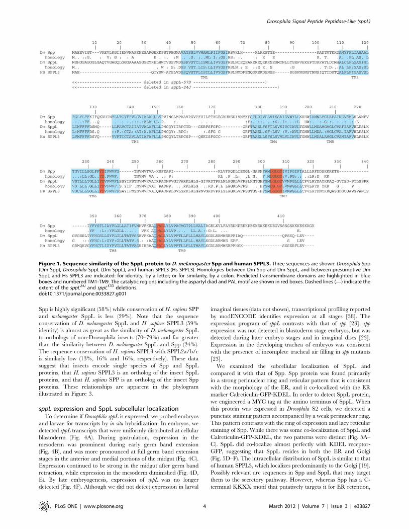
The SppL proteinIn 2002, a search of sequence databases identified the I-CLiP
family of presenilin homologs [14] that includes Drosophila
CG11840 (spp) [23] and CG17370. We have investigated the
CG17370 sequence and here show that it encodes a SPPL
homolog. CG17370 is predicted to encode two proteins (417 and
422 residues) that could be derived by alternative splicing. These
proteins have limited similarity to the 390 residue Drosophila Spp
(24% identity with the 417 residue form of CG17370). However,
several key features and regions in these proteins suggest their
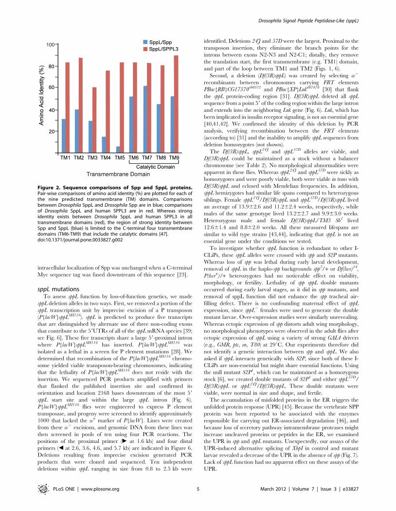
functional homology. Both Spp and CG17370 proteins have nine
predicted transmembrane helices (Fig. 1), of which the four C-
terminal helices that include the presumed catalytic domain have
significant sequence homology (55% identity in this region; Figs. 1,
2). Sequence similarity is particularly high in the immediate
vicinity of the catalytic YD and GXGD aspartyl diad, as well as
near the distal PAL motif. The putative ER retention motif
KKXX found at the carboxy-terminus of SPP proteins [8] is not
conserved in CG17370 or SPPL3. The overall relatedness of
human SPPL3 to the CG17370 protein (59% overall identity) is
significantly higher than is the kinship of Drosophila Spp and
CG17370 (24% identity). All nine predicted transmembrane
helices are highly conserved between human SPPL3 and Drosophila
CG17370 (80% identity; Fig. 2), and significant sequence
similarity is also distributed in the non-transmembrane regions
(46% identity). We henceforth refer to the CG17370 protein as
SppL.
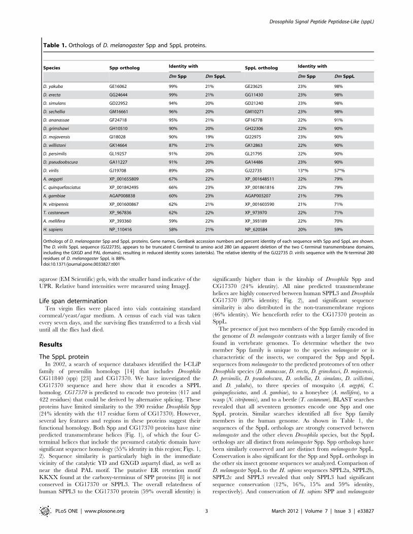
The presence of just two members of the Spp family encoded in
the genome of D. melanogaster contrasts with a larger family of five
found in vertebrate genomes. To determine whether the two
member Spp family is unique to the species melanogaster or is
characteristic of the insects, we compared the Spp and SppL
sequences from melanogaster to the predicted proteomes of ten other
Drosophila species (D. ananassae, D. erecta, D. grimshawi, D. mojavensis,
D. persimilis, D. pseudoobscura, D. sechellia, D. simulans, D. willistoni,
and D. yakuba), to three species of mosquito (A. aegypti, C.
quinquefasciatus, and A. gambiae), to a honeybee (A. mellifera), to a
wasp (N. vitripennis), and to a beetle (T. castaneum). BLAST searches
revealed that all seventeen genomes encode one Spp and one
SppL protein. Similar searches identified all five Spp family
members in the human genome. As shown in Table 1, the
sequences of the SppL orthologs are strongly conserved between
melanogaster and the other eleven Drosophila species, but the SppL
orthologs are all distinct from melanogaster Spp. Spp orthologs have
been similarly conserved and are distinct from melanogaster SppL.
Conservation is also significant for the Spp and SppL orthologs in
the other six insect genome sequences we analyzed. Comparison of
D. melanogaster SppL to the H. sapiens sequences SPPL2a, SPPL2b,
SPPL2c and SPPL3 revealed that only SPPL3 had significant
sequence conservation (12%, 16%, 15% and 59% identity,
respectively). And conservation of H. sapiens SPP and melanogaster
Table 1. Orthologs of D. melanogaster Spp and SppL proteins.
Species Spp ortholog Identity with SppL ortholog Identity with
Dm Spp Dm SppL Dm Spp Dm SppL
D. yakuba GE16062 99% 21% GE23625 23% 98%
D. erecta GG24644 99% 21% GG11430 23% 98%
D. simulans GD22952 94% 20% GD21240 23% 98%
D. sechellia GM16661 96% 20% GM10271 23% 98%
D. ananassae GF24718 95% 21% GF16778 22% 91%
D. grimshawi GH10510 90% 20% GH22306 22% 90%
D. mojavensis GI18028 90% 19% GI22975 23% 90%
D. willistoni GK14664 87% 21% GK12863 22% 90%
D. persimilis GL19257 91% 20% GL21795 22% 90%
D. pseudoobscura GA11227 91% 20% GA14486 23% 90%
D. virilis GJ19708 89% 20% GJ22735 13*% 57*%
A. aegypti XP_001655809 67% 22% XP_001648511 22% 79%
C. quinquefasciatus XP_001842495 66% 23% XP_001861816 22% 79%
A. gambiae AGAP008838 60% 23% AGAP003207 21% 79%
N. vitripennis XP_001600867 62% 21% XP_001603590 21% 71%
T. castaneum XP_967836 62% 22% XP_973970 22% 71%
A. mellifera XP_393360 59% 22% XP_393189 22% 70%
H. sapiens NP_110416 58% 21% NP_620584 20% 59%
Orthologs of D. melanogaster Spp and SppL proteins. Gene names, GenBank accession numbers and percent identity of each sequence with Spp and SppL are shown.The D. virilis SppL sequence (GJ22735), appears to be truncated C-terminal to amino acid 280 (an apparent deletion of the two C-terminal transmembrane domains,including the GXGD and PAL domains), resulting in reduced identity scores (asterisks). The relative identity of the GJ22735 D. virilis sequence with the N-terminal 280residues of D. melanogaster SppL is 88%.doi:10.1371/journal.pone.0033827.t001
Drosophila Signal Peptide Peptidase-Like (sppL)
PLoS ONE | www.plosone.org 3 March 2012 | Volume 7 | Issue 3 | e33827
Spp is highly significant (58%) while conservation of H. sapiens SPP
and melanogaster SppL is less (29%). Note that the sequence
conservation of D. melanogaster SppL and H. sapiens SPPL3 (59%
identity) is almost as great as the similarity of D. melanogaster SppL
to orthologs of non-Drosophila insects (70–79%) and far greater
than the similarity between D. melanogaster SppL and Spp (24%).
The sequence conservation of H. sapiens SPPL3 with SPPL2a/b/c
is similarly low (13%, 16% and 16%, respectively). These data
suggest that insects encode single species of Spp and SppL
proteins, that H. sapiens SPPL3 is an ortholog of the insect SppL
proteins, and that H. sapiens SPP is an ortholog of the insect Spp
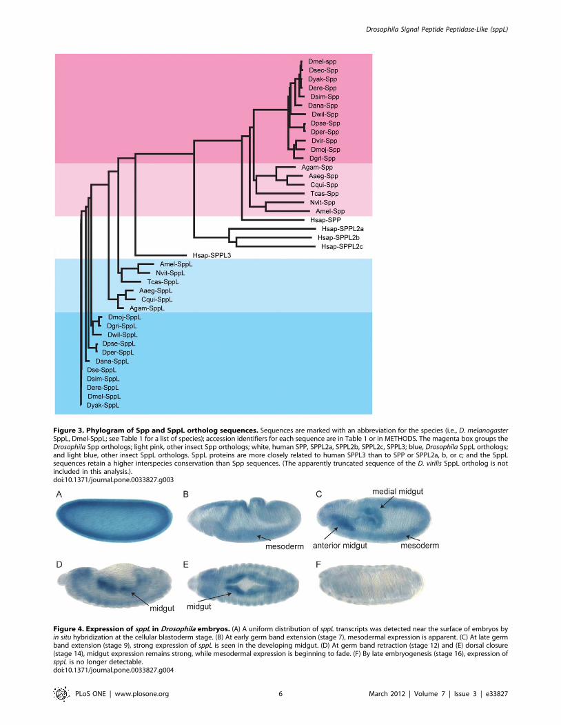
proteins. These relationships are apparent in the phylogram
illustrated in Figure 3.
sppL expression and SppL subcellular localizationTo determine if Drosophila sppL is expressed, we probed embryos
and larvae for transcripts by in situ hybridization. In embryos, we
detected sppL transcripts that were uniformly distributed at cellular
blastoderm (Fig. 4A). During gastrulation, expression in the
mesoderm was prominent during early germ band extension
(Fig. 4B), and was more pronounced at full germ band extension
stages in the anterior and medial portions of the midgut (Fig. 4C).
Expression continued to be strong in the midgut after germ band
retraction, while expression in the mesoderm diminished (Fig. 4D,
E). By late embryogenesis, expression of sppL was no longer
detected (Fig. 4F). Although we did not detect expression in larval
imaginal tissues (data not shown), transcriptional profiling reported
by modENCODE identifies expression at all stages [38]. The
expression program of sppL contrasts with that of spp [23]. spp
expression was not detected in blastoderm stage embryos, but was
detected during later embryo stages and in imaginal discs [23].
Expression in the developing trachea of embryos was consistent
with the presence of incomplete tracheal air filling in spp mutants
[23].
We examined the subcellular localization of SppL and
compared it with that of Spp. Spp protein was found primarily
in a strong perinuclear ring and reticular pattern that is consistent
with the morphology of the ER, and it co-localized with the ER
marker Calreticulin-GFP-KDEL. In order to detect SppL protein,
we engineered a MYC tag at the amino terminus of SppL. When
this protein was expressed in Drosophila S2 cells, we detected a
punctate staining pattern accompanied by a weak perinuclear ring.
This pattern contrasts with the ring of expression and lacy reticular
staining of Spp. While there was some co-localization of SppL and
Calreticulin-GFP-KDEL, the two patterns were distinct (Fig. 5A–
C). SppL did co-localize almost perfectly with KDEL receptor-
GFP, suggesting that SppL resides in both the ER and Golgi
(Fig. 5D–F). The intracellular distribution of SppL is similar to that
of human SPPL3, which localizes predominantly to the Golgi [19].
Possibly relevant are sequences in Spp and SppL that may target
them to the secretory pathway. However, whereas Spp has a C-
terminal KKXX motif that putatively targets it for ER retention,
Figure 1. Sequence similarity of the SppL protein to D. melanogaster Spp and human SPPL3. Three sequences are shown: Drosophila Spp(Dm Spp), Drosophila SppL (Dm SppL), and human SPPL3 (Hs SPPL3). Homologies between Dm Spp and Dm SppL, and between presumptive DmSppL and Hs SPPL3 are indicated: for identity, by a letter; or for similarity, by a colon. Predicted transmembrane domains are highlighted in blueboxes and numbered TM1-TM9. The catalytic regions including the aspartyl diad and PAL motif are shown in red boxes. Dashed lines (—) indicate theextent of the sppL24J and sppL57D deletions.doi:10.1371/journal.pone.0033827.g001
Drosophila Signal Peptide Peptidase-Like (sppL)
PLoS ONE | www.plosone.org 4 March 2012 | Volume 7 | Issue 3 | e33827
intracellular localization of Spp was unchanged when a C-terminal
Myc sequence tag was fused downstream of this sequence [23].
sppL mutationsTo assess sppL function by loss-of-function genetics, we made
sppL deletion alleles in two ways. First, we removed a portion of the
sppL transcription unit by imprecise excision of a P transposon
(P{lacW}sppLSH116). sppL is predicted to produce five transcripts
that are distinguished by alternate use of three non-coding exons
that contribute to the 59UTRs of all of the sppL mRNA species [39;
see Fig. 6]. These five transcripts share a large 59-proximal intron
where P{lacW}sppLSH116 has inserted. P{lacW}sppLSH116 was
isolated as a lethal in a screen for P element mutations [28]. We
determined that recombination of the P{lacW}sppLSH116 chromo-
some yielded viable transposon-bearing chromosomes, indicating
that the lethality of P{lacW}sppLSH116 does not reside with the
insertion. We sequenced PCR products amplified with primers
that flanked the published insertion site and confirmed its
orientation and location 2168 bases downstream of the most 59
sppL start site and within the large sppL intron (Fig. 6).
P{lacW}sppLSH116 flies were engineered to express P element
transposase, and progeny were screened to identify approximately
1000 that lacked the w+ marker of P{lacW}. Lines were created
from these w2 excisions, and genomic DNA from these lines was
then screened in pools of ten using four PCR reactions. The
positions of the proximal primer (c at 1.6 kb) and four distal
primers (b at 2.6, 3.6, 4.6, and 5.7 kb) are indicated in Figure 6.
Deletions resulting from imprecise excision generated PCR
products that were cloned and sequenced. Ten independent
deletions within sppL ranging in size from 0.8 to 2.5 kb were
identified. Deletions 24J and 57D were the largest. Proximal to the
transposon insertion, they eliminate the branch points for the
introns between exons N2-N3 and N2-C1; distally, they remove
the translation start, the first transmembrane (e.g. TM1) domain,
and part of the loop between TM1 and TM2 (Figs. 1, 6).
Second, a deletion (Df(3R)sppL) was created by selecting w2
recombinants between chromosomes carrying FRT elements
PBac{RB}CG17370e00372 and PBac{XP}Lnkd07478 [30] that flank
the sppL protein-coding region [31]. Df(3R)sppL deleted all sppL
sequence from a point 59 of the coding region within the large intron
and extends into the neighboring Lnk gene (Fig. 6). Lnk, which has
been implicated in insulin receptor signaling, is not an essential gene
[40,41,42]. We confirmed the identity of this deletion by PCR
analysis, verifying recombination between the FRT elements
(according to) [31] and the inability to amplify sppL sequences from
deletion homozygotes (not shown).
The Df(3R)sppL, sppL24J and sppL57D alleles are viable, and
Df(3R)sppL could be maintained as a stock without a balancer
chromosome (see Table 2). No morphological abnormalities were
apparent in these flies. Whereas sppL24J and sppL57D were sickly as
homozygotes and were poorly viable, both were viable in trans with
Df(3R)sppL and eclosed with Mendelian frequencies. In addition,
sppL hemizygotes had similar life spans compared to heterozygous
siblings. Female sppL24J/Df(3R)sppL and sppL57D/Df(3R)sppL lived
an average of 13.962.6 and 11.262.4 weeks, respectively, while
males of the same genotype lived 13.262.7 and 9.963.0 weeks.
Heterozygous male and female Df(3R)sppL/TM3 Sb1 lived
12.661.4 and 8.862.0 weeks. All these measured lifespans are
similar to wild type strains [43,44], indicating that sppL is not an
essential gene under the conditions we tested.
To investigate whether sppL function is redundant to other I-
CLiPs, these sppL alleles were crossed with spp and S2P mutants.
Whereas loss of spp was lethal during early larval development,
removal of sppL in the haplo-spp backgrounds spp5/+ or Df(lwr)14,
P(lwr+)/+ heterozygotes had no noticeable effect on viability,
morphology, or fertility. Lethality of spp sppL double mutants
occurred during early larval stages, as it did in spp mutants, and
removal of sppL function did not enhance the spp tracheal air-
filling defect. There is no confounding maternal effect of sppL
expression, since sppL2 females were used to generate the double
mutant larvae. Over-expression studies were similarly unrevealing.
Whereas ectopic expression of spp distorts adult wing morphology,
no morphological phenotypes were observed in the adult flies after
ectopic expression of sppL using a variety of strong GAL4 drivers
(e.g., GMR, ptc, en, T80) at 29uC. Our experiments therefore did
not identify a genetic interaction between spp and sppL. We also
asked if sppL interacts genetically with S2P, since both of these I-
CLiPs are non-essential but might share essential functions. Using
the null mutant S2P1, which can be maintained as a homozygous
stock [6], we created double mutants of S2P1 and either sppL57D/
Df(3R)sppL or sppL24J/Df(3R)sppL. These double mutants were
viable, were normal in size and shape, and fertile.
The accumulation of misfolded proteins in the ER triggers the
unfolded protein response (UPR) [45]. Because the vertebrate SPP
protein was been reported to be associated with the enzymes
responsible for carrying out ER-associated degradation [46], and
because loss of secretory pathway intramembrane proteases might
increase uncleaved proteins or peptides in the ER, we examined
the UPR in spp and sppL mutants. Unexpectedly, our assays of the
UPR-induced alternative splicing of XbpI in control and mutant
larvae revealed a decrease of the UPR in the absence of spp (Fig. 7).
Lack of sppL function had no apparent effect on these assays of the
UPR.
Figure 2. Sequence comparisons of Spp and SppL proteins.Pair-wise comparisons of amino acid identity (%) are plotted for each ofthe nine predicted transmembrane (TM) domains. Comparisonsbetween Drosophila SppL and Drosophila Spp are in blue; comparisonsof Drosophila SppL and human SPPL3 are in red. Whereas strongidentity exists between Drosophila SppL and human SPPL3 in alltransmembrane domains (red), the region of strong identity betweenSpp and SppL (blue) is limited to the C-terminal four transmembranedomains (TM6-TM9) that include the catalytic domains [47].doi:10.1371/journal.pone.0033827.g002
Drosophila Signal Peptide Peptidase-Like (sppL)
PLoS ONE | www.plosone.org 5 March 2012 | Volume 7 | Issue 3 | e33827
Figure 3. Phylogram of Spp and SppL ortholog sequences. Sequences are marked with an abbreviation for the species (i.e., D. melanogasterSppL, Dmel-SppL; see Table 1 for a list of species); accession identifiers for each sequence are in Table 1 or in METHODS. The magenta box groups theDrosophila Spp orthologs; light pink, other insect Spp orthologs; white, human SPP, SPPL2a, SPPL2b, SPPL2c, SPPL3; blue, Drosophila SppL orthologs;and light blue, other insect SppL orthologs. SppL proteins are more closely related to human SPPL3 than to SPP or SPPL2a, b, or c; and the SppLsequences retain a higher interspecies conservation than Spp sequences. (The apparently truncated sequence of the D. virilis SppL ortholog is notincluded in this analysis.).doi:10.1371/journal.pone.0033827.g003
Figure 4. Expression of sppL in Drosophila embryos. (A) A uniform distribution of sppL transcripts was detected near the surface of embryos byin situ hybridization at the cellular blastoderm stage. (B) At early germ band extension (stage 7), mesodermal expression is apparent. (C) At late germband extension (stage 9), strong expression of sppL is seen in the developing midgut. (D) At germ band retraction (stage 12) and (E) dorsal closure(stage 14), midgut expression remains strong, while mesodermal expression is beginning to fade. (F) By late embryogenesis (stage 16), expression ofsppL is no longer detectable.doi:10.1371/journal.pone.0033827.g004
Drosophila Signal Peptide Peptidase-Like (sppL)
PLoS ONE | www.plosone.org 6 March 2012 | Volume 7 | Issue 3 | e33827
Discussion
The presence of sppL in the D. melanogaster genome is not unique
among insects; indeed, BLAST searches of the genome sequences
of sixteen other insect species revealed that all include genes that
can encode one Spp and one SppL protein (Table 1). BLAST
searches identified the five vertebrate Spp family members, but
only two were detected in insect genomes. The genomes wequeried were from ten other Drosophila species (D. ananassae, D.erecta, D. grimshawi, D. mojavensis, D. persimilis, D. pseudoobscura, D.sechellia, D. simulans, D. willistoni, and D. yakuba), three mosquitospecies (A. aegypti and A. gambiae), honeybee (A. mellifera), wasp (N.vitripennis) and beetle (T. castaneum). In each genome, the SPPLprotein retains higher sequence homology to human SPPL3 and
Figure 5. SppL localization to the Golgi and ER. S2 cells were transfected two express either (A–C) MYC-SppL and Crc-GFP-KDEL marking ER, or(D–F) MYC-SppL and KDEL Receptor-GFP marking Golgi and ER. (C, F) In the merged images, MYC-SppL is in red the GFP fusion proteins are in green.Hoechst staining of nuclei is included in blue (note that only two cells in each frame were transfected). While some colocalization of SppL and the ERmarker can be seen in C, extensive colocalization with the ER and Golgi marker is evident in F.doi:10.1371/journal.pone.0033827.g005
Figure 6. The sppL locus. This cartoon of 9.5 kb of chromosome III at cytological band 96F5-6 depicts the sppL gene and the ends of the adjacentTsp96 (pink) and Lnk (blue) genes. Colored boxes indicate the sppL exon structure: coding regions (green) and non-coding 59 and 39 UTRs (yellow).The predicted ‘‘start’’ and ‘‘stop’’ codons of sppL are indicated. Exons N1-N3 are entirely non-coding, while exons C1–C6 contain the sppL openreading frame. The insertion sites of transposons P{lacW}sppLSH116 (also known as P{lacW}l(3)SH116sh116), PBac{XP}Lnkd07478, and PBac{RB}CG17370e00372
are indicated with red triangles. Imprecise excision of P{lacW}sppLSH116 generated the deletion alleles sppL24J and sppL57D. Recombination betweenthe two PBac insertions was used to generate deletion Df(3R)sppL. The extent of these deletions is indicated within parentheses. The sppL57D deletion(not shown) is similar to sppL24J. Black triangles indicate the positions of proximal (c) and distal (b) primers used to screen for excision mutants,denoting the positions of the following oligo sites: osppL-2000s, osppL-4000a, osppL-5000a, osppL-6000a, and osppL-7250a.doi:10.1371/journal.pone.0033827.g006
Drosophila Signal Peptide Peptidase-Like (sppL)
PLoS ONE | www.plosone.org 7 March 2012 | Volume 7 | Issue 3 | e33827
D. melanogaster SppL than it does to SPP or to the vertebrateSPPL2a/b/c proteins (Fig. 3).
Spp and SppL share overall topology and conserved motifs,
including putative aspartyl protease active sites (Figs. 1, 2). Both
Spp and SppL proteins purified from bacterial extracts can cleave
a model prolactin signal sequence, suggesting that their activities
do not depend on protein glycosylation or on other associated
proteins [47]. Despite these similarities, Spp and SppL are distinct.
Their expression patterns are largely non-overlapping during
development, and their subcellular locations differ (Figs. 4, 5).
However, because SPP family proteases are thought to have
substrate specificity in vivo [13], we cannot yet comment on
putative activities of SppL. Over-expression of Spp caused
developmental defects such as wing truncations; in contrast,
ectopic expression of SppL produced no apparent defects.
Most strikingly, spp provides an essential function during
development, while sppL is not required for viability or patterning.
Because knockdown of Xenopus, C. elegans and D. rerio SPPL genes
caused significant developmental abnormalities, we expected to
identify an essential role for Drosophila sppL, but sppL mutants
developed without apparent defects, and spp sppL double mutants
were indistinguishable from spp single mutants (Table 2). This
contrasts with Drosophila spp [23] and with presenilin, for which
functional disruption in a variety of organisms causes develop-
mental defects due to the failure to activate the Notch signaling
pathway (for review, see) [48]. Spp targets type 2 transmembrane
segments, and since the putative catalytic sites of SppL and Spp
have the same orientation in their respective transmembrane
segments 6 and 7, functional redundancy of SppL with either Spp
might be expected. Yet despite the absence of an apparent sppL
mutant phenotype, the strong evolutionary conservation of this
gene suggests that SppL might be redundant with another I-
CLiP(s) or protease(s), precedents being S2P and the caspase Drice
in the Drosophila SREBP pathway [49].
A recent report on the toxicity of over-expressed human
Huntingtin protein in Drosophila indicates that loss-of-function
alleles of spp and sppL reduced Huntingtin-induced motor deficits
in mutant flies [50]. Thus, whereas sppL loss-of-function alleles do
not manifest apparent insufficiency under the standard laboratory
conditions, the ‘‘sensitized’’ genetic background in which Hun-
tingtin is over-expressed unmasked a critical role for sppL function.
Further studies of Huntingtin may be aided by the sppL mutants
and expression patterns we have described, and such studies may
lead to a better understanding of the putative genetic redundancy
of sppL.
Human SPP may be a component of the ER-associated protein
degradation (ERAD) response [46]. Although our assays did not
identify a role for sppL in the ER stress response, we observed that
loss of spp decreased the UPR, a result that suggests that Spp
activity might facilitate the UPR. Our data does not discriminate
between any of the possible mechanisms for this role.
Our findings are reminiscent of genetic studies on the SPP-
related genes of C. elegans. The C. elegans genome encodes a single
SPP (Imp-2) protein, a closely related SPPL (Imp-1), and a
distantly related SPP-like sequence (Imp-1) [14]; the C. briggsae
genome encodes a comparable cadre of SPP relatives. As with
Drosophila spp and sppL mutants, RNAi directed against imp-2
caused developmental defects, while RNAi directed against imp-1
and imp-3 caused no obvious developmental abnormalities [20].
These data for Drosophila and C. elegans contrast with genetic studies
in zebrafish, in which developmental defects were observed after
spp, sppL2a or sppL3 were targeted by morpholinos [21]. We
suggest that in contrast to the invertebrate proteins, the vertebrate
SPP family proteins acquired new essential functions by processes
of gene duplication and diversification.
Table 2. sppL stocks and double mutants.
Genotypes Viability
sppL57D viable
sppL24J viable
Df(3R)sppL viable
sppL57D/Df(3R)sppL viable
sppL24J/Df(3R)sppL viable
spp5/Df(2L)lwr14, p(lwr+) early larval lethal
spp5/Df(2L)lwr14, p(lwr+); sppL24J/Df(3R)sppL early larval lethal
S2P1 viable
S2P1; sppL24J/Df(3R)sppL viable
sppL stocks and double mutants. Terminal phenotypes of sppL and SppL incombination with spp and S2P are listed.doi:10.1371/journal.pone.0033827.t002
Figure 7. The unfolded protein response in spp and sppL mutant larvae. A 77 base pair alternative splice product of the Xbp I cDNA is madeafter induction of the UPR by DTT. Genotypes are w (w1118), spp (w118, Df(2L)lwr14 p(lwr+)), sppL (w118, Df(3R)sppL), and spp sppL (w118, Df(2L)lwr14,p(lwr+), sppLBW1). Samples of RNA were prepared from freshly dissected larvae (0 hour) or from larvae incubated in media for 2 hours with or withoutDTT. Relative intensities of the upper (100 bp) and lower (77 bp) bands in the experimental samples were calculated from the total intensity in eachband measured with ImageJ.doi:10.1371/journal.pone.0033827.g007
Drosophila Signal Peptide Peptidase-Like (sppL)
PLoS ONE | www.plosone.org 8 March 2012 | Volume 7 | Issue 3 | e33827
Acknowledgments
For providing Drosophila lines, we thank Rob Rawson (U. Texas, Southwest
Medical Center), the Bloomington and Szeged Drosophila Stock Centers,
and the Exelixis Collection at Harvard Medical School. We also thank
Hyung Don Ryoo, Susan Younger, Bruno Martoglio, Prashanth Rao,
Sougata Roy, Kevin Hill, and Brenda Ng for helpful discussions, Prashanth
Rao for critically reading the manuscript, and Katja Bruckner, Eric
Rulifson, Bree Grillo-Hill and Helen Wong for sharing equipment and
reagents.
Author Contributions
Conceived and designed the experiments: DJC BB TBK. Performed the
3. Struhl G, Greenwald I (1999) Presenilin is required for activity and nuclearaccess of Notch in Drosophila. Nature 398: 522–525.
4. Ye Y, Lukinova N, Fortini ME (1999) Neurogenic phenotypes and altered Notchprocessing in Drosophila Presenilin mutants. Nature 398: 525–529.
5. Urban S, Lee JR, Freeman M (2002) A family of Rhomboid intramembrane
proteases activates all Drosophila membrane-tethered EGF ligands. EMBO J 21:4277–4286.
6. Matthews KA, Kunte AS, Tambe-Ebot E, Rawson RB (2009) Alternativeprocessing of sterol regulatory element binding protein during larval
development in Drosophila melanogaster. Genetics 181: 119–128.
7. Weihofen A, Lemberg MK, Ploegh HL, Bogyo M, Martoglio B (2000) Releaseof signal peptide fragments into the cytosol requires cleavage in the
transmembrane region by a protease activity that is specifically blocked by anovel cysteine protease inhibitor. J Biol Chem 275: 30951–30956.
8. Weihofen A, Binns K, Lemberg MK, Ashman K, Martoglio B (2002)Identification of signal peptide peptidase, a presenilin-type aspartic protease.
Science 296: 2215–2218.
9. Lemberg MK, Bland FA, Weihofen A, Braud VM, Martoglio B (2001)Intramembrane proteolysis of signal peptides: an essential step in the generation
of HLA-E epitopes. J Immunol 167: 6441–6446.10. McLauchlan J, Lemberg MK, Hope G, Martoglio B (2002) Intramembrane
proteolysis promotes trafficking of hepatitis C virus core protein to lipid droplets.
EMBO J 21: 3980–3988.11. Martoglio B, Graf R, Dobberstein B (1997) Signal peptide fragments of
preprolactin and HIV-1 p-gp160 interact with calmodulin. EMBO J 16:6636–6645.
12. Loureiro J, Lilley BN, Spooner E, Noriega V, Tortorella D, et al. (2006) Signalpeptide peptidase is required for dislocation from the endoplasmic reticulum.
Nature 441: 894–897.
13. Kilic A, Klose S, Dobberstein B, Knust E, Kapp K (2010) The DrosophilaCrumbs signal peptide is unusually long and is a substrate for signal peptide
peptidase. Eur J Cell Biol 89: 449–461.14. Ponting CP, Hutton M, Nyborg A, Baker M, Jansen K, et al. (2002)
Identification of a novel family of presenilin homologues. Hum Mol Genet 11:
1037–1044.15. Friedmann E, Lemberg MK, Weihofen A, Dev KK, Dengler U, et al. (2004)
Consensus analysis of signal peptide peptidase and homologous human asparticproteases reveals opposite topology of catalytic domains compared with
presenilins. J Biol Chem 279: 50790–50798.
16. Fluhrer R, Grammer G, Israel L, Condron MM, Haffner C, et al. (2006) Agamma-secretase-like intramembrane cleavage of TNFalpha by the GxGD
aspartyl protease SPPL2b. Nat Cell Biol 8: 894–896.17. Kirkin V, Cahuzac N, Guardiola-Serrano F, Huault S, Luckerath K, et al.
(2007) The Fas ligand intracellular domain is released by ADAM10 and SPPL2acleavage in T-cells. Cell Death Differ 14: 1678–1687.
18. Martin L, Fluhrer R, Reiss K, Kremmer E, Saftig P, et al. (2008) Regulated
intramembrane proteolysis of Bri2 (Itm2b) by ADAM10 and SPPL2a/SPPL2b.J Biol Chem 283: 1644–1652.
19. Friedmann E, Hauben E, Maylandt K, Schleeger S, Vreugde S, et al. (2006)SPPL2a and SPPL2b promote intramembrane proteolysis of TNFalpha in
20. Grigorenko AP, Moliaka YK, Soto MC, Mello CC, Rogaev EI (2004) TheCaenorhabditis elegans IMPAS gene, imp-2, is essential for development and is
functionally distinct from related presenilins. Proc Natl Acad Sci U S A 101:14955–14960.
21. Krawitz P, Haffner C, Fluhrer R, Steiner H, Schmid B, et al. (2005) Differentiallocalization and identification of a critical aspartate suggest non-redundant
proteolytic functions of the presenilin homologues SPPL2b and SPPL3. J Biol
Chem 280: 39515–39523.22. Han S, Green L, Schnell DJ (2009) The signal peptide peptidase is required for
pollen function in Arabidopsis. Plant Physiol 149: 1289–1301.23. Casso DJ, Tanda S, Biehs B, Martoglio B, Kornberg TB (2005) Drosophila
signal peptide peptidase is an essential protease for larval development. Genetics
170: 139–148.24. Poole SJ, Kauvar LM, Drees B, Kornberg T (1985) The engrailed locus of
Drosophila: structural analysis of an embryonic transcript. Cell 40: 37–43.
25. Brand AH, Perrimon N (1993) Targeted gene expression as a means of alteringcell fates and generating dominant phenotypes. Development 118: 401–415.
26. Ishikawa T, Matsumoto A, Kato T, Jr., Togashi S, Ryo H, et al. (1999) DCRY is
a Drosophila photoreceptor protein implicated in light entrainment of circadianrhythm. Genes Cells 4: 57–65.
27. Biehs B, Sturtevant MA, Bier E (1998) Boundaries in the Drosophila wing
imaginal disc organize vein-specific genetic programs. Development 125:
4245–4257.
28. Oh SW, Kingsley T, Shin HH, Zheng Z, Chen HW, et al. (2003) A P-elementinsertion screen identified mutations in 455 novel essential genes in Drosophila.
Genetics 163: 195–201.
29. Ashburner M (1989) Drosophila: A laboratory manual. Cold Spring Harbor:
Cold Spring Harbor Laboratory Press. pp 44–49.
30. Thibault ST, Singer MA, Miyazaki WY, Milash B, Dompe NA, et al. (2004) Acomplementary transposon tool kit for Drosophila melanogaster using P and
piggyBac. Nat Genet 36: 283–287.
31. Parks AL, Cook KR, Belvin M, Dompe NA, Fawcett R, et al. (2004) Systematic
generation of high-resolution deletion coverage of the Drosophila melanogastergenome. Nat Genet 36: 288–292.
32. Altschul SF, Gish W, Miller W, Myers EW, Lipman DJ (1990) Basic local
alignment search tool. J Mol Biol 215: 403–410.
33. Thompson JD, Higgins DG, Gibson TJ (1994) CLUSTAL W: improving thesensitivity of progressive multiple sequence alignment through sequence
weighting, position-specific gap penalties and weight matrix choice. Nucleic
Acids Res 22: 4673–4680.
34. Chenna R, Sugawara H, Koike T, Lopez R, Gibson TJ, et al. (2003) Multiplesequence alignment with the Clustal series of programs. Nucleic Acids Res 31:
interactive, phylogenetic trees for the web. PLoS One 5: e8934.
36. Krogh A, Larsson B, von Heijne G, Sonnhammer EL (2001) Predictingtransmembrane protein topology with a hidden Markov model: application to
complete genomes. J Mol Biol 305: 567–580.
37. Tusnady GE, Simon I (2001) The HMMTOP transmembrane topology
prediction server. Bioinformatics 17: 849–850.
38. Roy S, Ernst J, Kharchenko PV, Kheradpour P, Negre N, et al. (2010)Identification of functional elements and regulatory circuits by Drosophila
modENCODE. Science 330: 1787–1797.
39. Stapleton M, Liao G, Brokstein P, Hong L, Carninci P, et al. (2002) TheDrosophila gene collection: identification of putative full-length cDNAs for 70%
of D. melanogaster genes. Genome Res 12: 1294–1300.
40. Slack C, Werz C, Wieser D, Alic N, Foley A, et al. (2010) Regulation of lifespan,
metabolism, and stress responses by the Drosophila SH2B protein, Lnk. PLoSGenet 6: e1000881.
41. Song W, Ren D, Li W, Jiang L, Cho KW, et al. (2010) SH2B regulation of
growth, metabolism, and longevity in both insects and mammals. Cell Metab 11:
427–437.
42. Werz C, Kohler K, Hafen E, Stocker H (2009) The Drosophila SH2B familyadaptor Lnk acts in parallel to chico in the insulin signaling pathway. PLoS
Genet 5: e1000596.
43. Paaby AB, Schmidt PS (2009) Dissecting the genetics of longevity in Drosophila
melanogaster. Fly (Austin) 3: 29–38.
44. Spencer CC, Howell CE, Wright AR, Promislow DE (2003) Testing an ‘aginggene’ in long-lived drosophila strains: increased longevity depends on sex and
genetic background. Aging Cell 2: 123–130.
45. Ron D, Walter P (2007) Signal integration in the endoplasmic reticulumunfolded protein response. Nat Rev Mol Cell Biol 8: 519–529.
46. Christianson JC, Olzmann JA, Shaler TA, Sowa ME, Bennett EJ, et al. (2012)Defining human ERAD networks through an integrative mapping strategy. Nat
Cell Biol 14: 93–105.
47. Narayanan S, Sato T, Wolfe MS (2007) A C-terminal region of signal peptidepeptidase defines a functional domain for intramembrane aspartic protease
catalysis. J Biol Chem 282: 20172–20179.
48. Chan YM, Jan YN (1999) Presenilins, processing of beta-amyloid precursor
protein, and notch signaling. Neuron 23: 201–204.
49. Amarneh B, Matthews KA, Rawson RB (2009) Activation of SREBP by thecaspase drice in Drosophila larvae. J Biol Chem.
50. Miller JP, Holcomb J, Al-Ramahi I, de Haro M, Gafni J, et al. (2010) Matrix
metalloproteinases are modifiers of huntingtin proteolysis and toxicity in
Huntington’s disease. Neuron 67: 199–212.
Drosophila Signal Peptide Peptidase-Like (sppL)
PLoS ONE | www.plosone.org 9 March 2012 | Volume 7 | Issue 3 | e33827