Page 1
Expression and Characterization of HgcA and HgcB, Two Proteins Involved in Methylmercury
Biosynthesis
by
Katherine Warfield Rush
A dissertation submitted in partial fulfillment
of the requirements for the degree of
Doctor of Philosophy
(Chemical Biology)
in the University of Michigan
2018
Doctoral Committee:
Professor Stephen W. Ragsdale, Chair
Professor Nicolai Lehnert
Professor Vincent Pecoraro
Professor Janet Smith
Page 2
Katherine W. Rush
[email protected]
ORCID iD: 0000-0001-6842-9055
© Katherine W. Rush 2018
Page 3
ii
Dedication
For my Nana,
Betsy Lee Rush
Page 4
iii
Acknowledgements
The Ragsdale laboratory has been an amazing place to learn. The first of many thank
you’s must go to Prof. Stephen Ragsdale, who has allowed me tremendous independence to
pursue an unestablished project and given freely of his time, guidance, and support. I feel very
lucky to have him as a mentor. I’m extremely grateful for the guidance and friendship of Dr.
Angela Fleischhacker and Dr. Eric Carter, who gave so much of their time to teaching me. I’ve
had wonderful discussions on science and life with Dr. Mehmet Can, Dr. Anjali Patwardhan, Dr.
Johanna Mock, and Marco Hornung, and to Seth Wiley, pizza artisan/EPR master/climbing
buddy/cleric - thank you for making our corner of the lab both thoughtful and hilarious. I also
would like to acknowledge an undergraduate colleague, Mohamad Awada, whose work
contributed to the results in Chapter 3. I’ve been privileged to learn from him and all members of
the Ragsdale lab my time has coincided with: Dr. Andrea Spencer, Dr. Thanyaporn Wongnate,
Heather Aman, Dr. Dariusz Sliwa, Erika Martinez-Nieves, Dr. Panu Pimviriyakul, Liu Liu,
Daniel Esckilsen, Dr. Rodney Burton, and Dr. Anindita Sarkar.
This thesis work was conducted in close collaboration with exceptional colleagues at Oak
Ridge National Laboratory: Dr. Swapneeta Date, Dr. Liyuan Liang, Dr. Jerry Parks, Dr.
Stephen Tomanicek, and Dr. Alexander Johs, whom I am particularly indebted to as my
undergraduate research supervisor and long-time source of encouragement. I’ve gained another
incredible mentor teaching Biochemistry 452 with Prof. Alex Ninfa, and I hope to teach one day
with his enthusiasm for both good science and good people.
At the University of Michigan, I’d like to thank the incredibly smart members of my
cohort within the Program in Chemical Biology, especially my future alpaca farm co-founder,
Dr. Oleta Johnson. My thesis committee members, Prof. Nicolai Lehnert, Prof. Vincent
Pecoraro, and Prof. Janet Smith, have offered many constructive comments over the course of
this work, for which I am grateful. Important Ann Arbor friends I’ve been lucky to make include
Tom Jurkiw, Sarah O’Brien, Dr. Zohar Strinka, James Annand, Sarah Haynes, Paul Campbell,
Dr. Jax Sanders, and John Deisinger.
Page 5
iv
Throughout this process, I’ve been privileged to have the constant and unconditional
support of my family: Dad, Kara, Molly, Big Daniel, Zoë, Little Daniel, Nana, Jim, and Maggie
Shults (who may as well be family). I need to include a few special thank you’s: to Wally and
Derby, my sweet dogs, whose insistence on many long walks around Ann Arbor keeps me happy
and healthy. To my father, Greg Rush, who has never told me I am incapable of anything. To
Jared – I don’t know how to thank you for all the ways you make everything wonderful. Finally,
this work is dedicated to my Nana, Betsy Rush. For these 5 years (and all the ones before), she
has kept up on my weather forecast, provided and packed the moving truck, and set an example
of love and strength for me to follow.
Page 6
v
Table of Contents
Dedication ....................................................................................................................................... ii
Acknowledgements ........................................................................................................................ iii
List of Tables ............................................................................................................................... viii
List of Figures ................................................................................................................................ ix
List of Abbreviations ...................................................................................................................... x
Abstract .......................................................................................................................................... xi
Chapter 1 Introduction to Biological Methylation of Mercury ....................................................... 1
1.1 Mercury and methylmercury in health and the environment .............................................................. 1
1.2 hgcAB-linked mercury methylation .................................................................................................... 2
1.3 Methylcobalamin and mechanisms of Hg methylation ....................................................................... 4
1.4 HgcA and enzymatic corrinoid-dependent methyl transfer ................................................................ 6
1.5 Mercury binding and HgcB, a [4Fe-4S] cluster ferredoxin-like protein ............................................. 8
1.6 Hypothesized mercury methylation process and scope of this dissertation ........................................ 9
1.7 References for Chapter 1 ................................................................................................................... 11
Chapter 2 HgcB............................................................................................................................. 14
2.1 Introduction ....................................................................................................................................... 14
2.2 Materials and Methods ...................................................................................................................... 17
2.2.1 Materials .................................................................................................................................... 17
2.2.2 HgcB fusion tag screen .............................................................................................................. 17
2.2.3 MHB co-expression with pRKISC and affinity purification ..................................................... 18
2.2.4 Construction of MHB cysteine mutants by site-directed mutagenesis ...................................... 19
2.2.5 UV-visible spectroscopy and reductive titrations of MHB ........................................................ 19
2.2.6 EPR spectroscopy of MHB ........................................................................................................ 20
2.2.6 Hg(TNB)2 complex preparation and MHB titration .................................................................. 20
2.2.7 Stopped-flow kinetic studies of MHB variants and Hg(TNB)2 ................................................. 20
Page 7
vi
2.3 Results and Analysis ......................................................................................................................... 21
2.3.1 SDS-PAGE analysis of HgcB fusion tag screen ........................................................................ 21
2.3.2 MHB expression and amylose affinity purification ................................................................... 22
2.3.3 UV-visible spectroscopy and reductive titration of MHB variants ............................................ 23
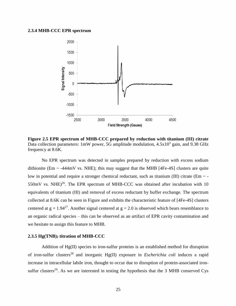
2.3.4 MHB-CCC EPR spectrum ......................................................................................................... 25
2.3.5 Hg(TNB)2 titration of MHB-CCC ............................................................................................. 25
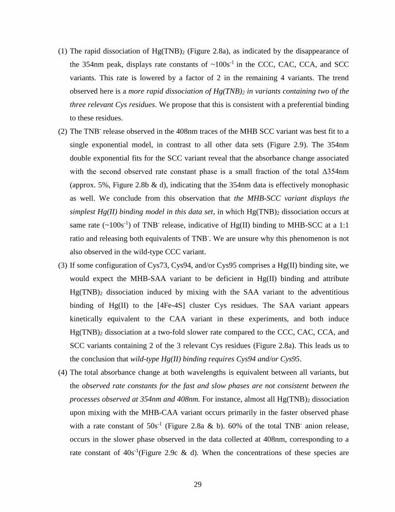
2.3.6 Stopped-flow kinetic analysis of Hg binding to MHB variants ................................................. 27
2.4 Discussion ......................................................................................................................................... 31
2.4.1 Purification of MHB and Cys variants generates an iron-replete [4Fe-4S] protein ................... 31
2.4.2 Hg(TNB)2 addition to MHB results in superstoichiometric release of TNB- ............................ 32
2.4.3 Stopped-flow kinetic comparison between Hg(II) binding rates of MHB variants is consistent
with observed in vivo requirements for conserved Cys residues ........................................................ 33
2.4.4 Conclusions ................................................................................................................................ 35
2.5 References for Chapter 2 ................................................................................................................... 35
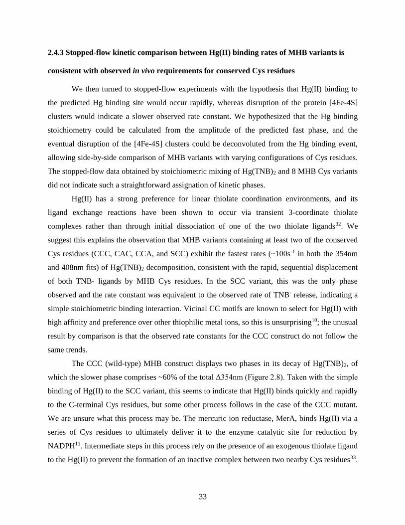
Chapter 3 HgcA ............................................................................................................................ 38
3.1 Introduction ....................................................................................................................................... 38
3.2 Materials and Methods ...................................................................................................................... 41
3.2.1 Materials .................................................................................................................................... 41
3.2.2 HgcA 1-166: EPR spectroscopy ................................................................................................ 42
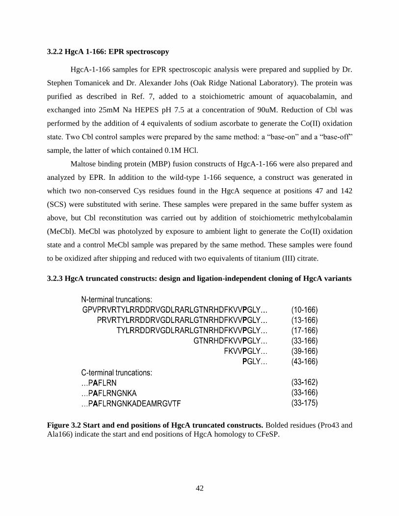
3.2.3 HgcA truncated constructs: design and ligation-independent cloning of HgcA variants .......... 42
3.2.4 HgcA truncated constructs: expression/solubility tests and purification ................................... 43
3.2.5 HgcA truncated constructs: cobalamin reconstitution analysis by UV-visible spectroscopy .... 44
3.2.6 FL-HgcA: restriction digest cloning into pET-Duet-1 ............................................................... 44
3.2.7 FL-HgcA: co-expression with pBAD42-btuCEDFB and Ni-NTA affinity purification ........... 44
3.2.8 FL-HgcA: sample preparation for analysis by UV-visible spectroscopy .................................. 45
3.3 Results and Analysis ......................................................................................................................... 45
3.3.1 HgcA-1-166 constructs: EPR spectra ........................................................................................ 45
3.3.2 HgcA truncated constructs: SDS-PAGE analysis of construct expression and solubility ......... 47
3.3.3 HgcA truncated constructs: UV-visible spectral analysis of cobalamin reconstitution ............. 48
Page 8
vii
3.3.4 FL-HgcA: Analysis of pBAD42-BtuCEDFB co-expression and affinity purification vs. HgcA-
1-175 ................................................................................................................................................... 49
3.3.5 FL-HgcA: UV-visible spectrum ................................................................................................ 51
3.4 Discussion ......................................................................................................................................... 52
3.4.1 HgcA-1-166 constructs do not bind “base-off” cobalamin ........................................................ 52
3.4.2 Screen of HgcA truncated constructs ......................................................................................... 53
3.4.3 FL-HgcA and HgcA-1-175 exhibit different cobalamin spectra after co-expression with pBAD-
BtuCDEFB .......................................................................................................................................... 53
3.4.4 UV-visible spectral features of FL-HgcA .................................................................................. 54
3.4.5 Conclusions ................................................................................................................................ 54
3.5 References for Chapter 3 ................................................................................................................... 55
Chapter 4 Conclusions and Future Directions .............................................................................. 57
4.1 Conclusions ....................................................................................................................................... 57
4.2 Future Directions............................................................................................................................... 59
4.3 References for Chapter 4 ................................................................................................................... 60
Page 9
viii
List of Tables
Table 2.1 Effect of Cys substitution with Ala at Cys 73, Cys94, and Cys95 on in vivo MeHg
production in Desulfovibrio desulfuricans ND132. ...................................................................... 16 Table 2.2 List MHB Cys variant identifiers used in this work ..................................................... 19
Table 2.3 UV-visible absorbance features of MHB variants and cluster occupancy calculation. 24
Page 10
ix
List of Figures
Figure 1.1 Schematic depiction of relevant equilibria in the Hg biogeochemical cycle. ............... 1 Figure 1.2 Structure of the cobalamin cofactor .............................................................................. 5 Figure 1.3 Crystal structures of observed "base-off" cobalamin protein binding modes ............... 7
Figure 1.4 Working model of Hg methylation by HgcA and HgcB ............................................. 10
Figure 2.1 Schematic representation of the HgcB conserved cysteines. ...................................... 16
Figure 2.2 SDS-PAGE analysis of HgcB fusion tag screen ......................................................... 21 Figure 2.3 SDS-PAGE analysis of purified MHB variants. ......................................................... 22 Figure 2.4 Titanium (III) citrate reduction of MHB-CCC. ........................................................... 23 Figure 2.5 EPR spectrum of MHB-CCC prepared by reduction with titanium (III) citrate ......... 25
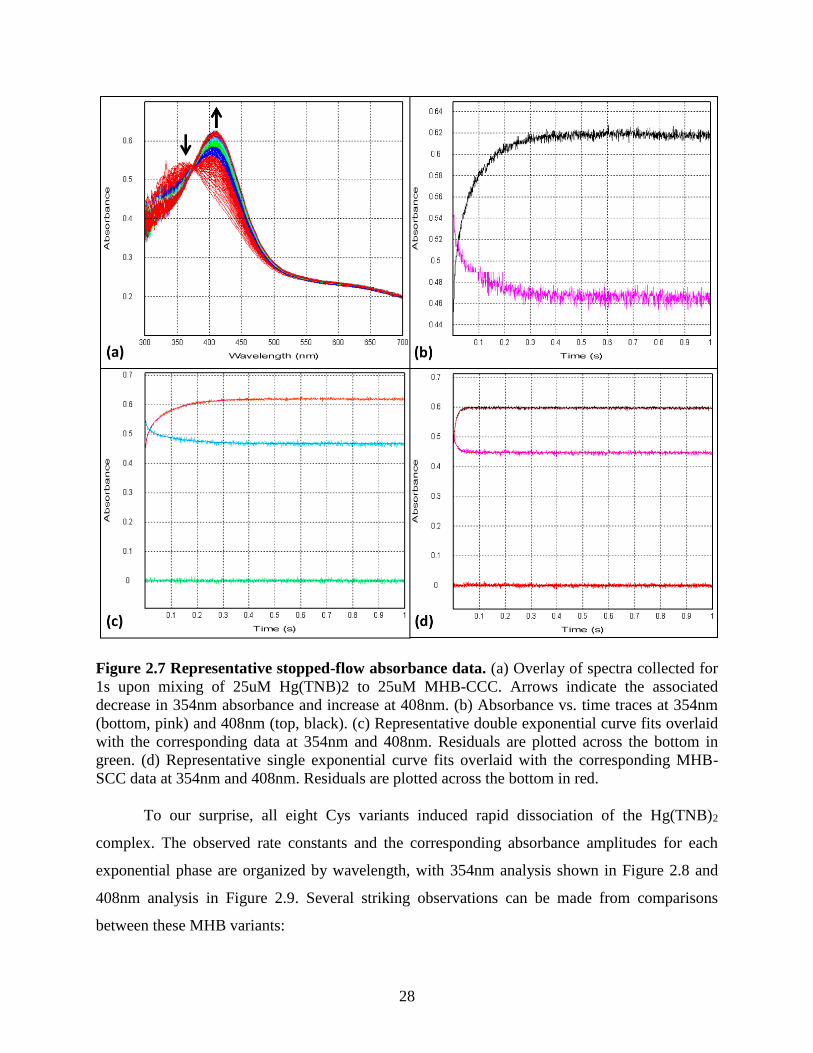
Figure 2.6 Hg(TNB)2 titrations of 6uM MHB-CCC and 20uM MBP .......................................... 26 Figure 2.7 Representative stopped-flow absorbance data. ............................................................ 28
Figure 2.8 Kinetic parameters of 354nm curve fits ...................................................................... 30 Figure 2.9 Kinetic parameters of 408nm curve fits ...................................................................... 31 Figure 3.1 Crystal structures of observed “base-off” cobalamin protein binding modes ............. 39
Figure 3.2 Start and end positions of HgcA truncated constructs................................................. 42
Figure 3.3 EPR spectra of HgcA-1-166 constructs compared to "base-on" and "base-off"
cobalamin ...................................................................................................................................... 46 Figure 3.4 HgcA truncated construct expression test analysis by SDS-PAGE. ........................... 47
Figure 3.5 SDS-PAGE analysis of HgcA truncated construct solubility after cell lysis. ............. 47 Figure 3.6 MBP-HgcA-43-166 amylose affinity purification analysis by SDS-PAGE ............... 48
Figure 3.7 UV-visible spectra of HgcA MBP fusion constructs upon stoichiometric addition of
aquacobalamin .............................................................................................................................. 49 Figure 3.8 SDS-PAGE analysis of FL-HgcA co-expressed with pBAD-BtuCEDFB .................. 50
Figure 3.9 UV-visible spectra of Ni-NTA purified HgcA variants co-expressed with pBAD-
BtuCEDFB. ................................................................................................................................... 51
Figure 3.10 Deconvolution of Cbl species contributing to purified FL-HgcA spectrum ............. 52
Page 11
x
List of Abbreviations
[4Fe-4S] – four-iron four-sulfur cluster
Ala – alanine
Cbl – cobalamin
CFeSP – corrinoid-iron sulfur protein
Co – cobalt
Cys – cysteine
DMB – dimethylbenzimidazole
EPR – electron paramagnetic resonance
Fe – iron
FL – full-length
Hg – mercury
His – histidine
LIC – ligation-independent cloning
MBP – maltose binding protein
MeCbl – methylcobalamin
MeHg – monomethylmercury
MetH – methionine synthase
Methyl-THF – methyltetrahydrofolate
MHB – MBP-HgcB
SAM – S-adenosylmethionine
Ser – serine
SRB – sulfate-reducing bacteria
TM – transmembrane
TNB – 2-nitro-5-thiobenzoate
Page 12
xi
Abstract
Methylmercury biosynthesis is a biologically-mediated process linked to expression of
the recently discovered hgcAB gene products. The environmental conversion of toxic Hg(II) to
MeHg, which exhibits even greater biological toxicity, is of concern to ecological food webs and
piscivorous human populations. The mechanism by which HgcA and HgcB are responsible for
Hg(II) methylation is unknown, but may represent a departure from canonical enzymatic
methylation via carbocation mechanisms. The hgcAB genes are always found in genomic
proximity to one another and predicted to interact within distances amenable to electron transfer
in the proposed mechanism of Hg methylation. HgcA is a 40kDa protein comprised of cytosolic
and transmembrane domains; the cytosolic domain is predicted to bind a cobalamin cofactor,
based on its sequence homology to a known cobalamin-binding protein, which is suggested to
serve as the catalytic center of the mercury methylation reaction. HgcB contains two [4Fe-4S]
clusters and three additional conserved cysteine residues which we hypothesize are responsible
for binding divalent mercury.
No in vitro study of HgcA or HgcB has yet been conducted to our knowledge, and this
dissertation presents the first protocols for the heterologous expression of both HgcA and HgcB.
HgcA contains a C-terminal transmembrane domain and both proteins contain metal cofactors
which present significant barriers to heterologous production; thus, Escherichia coli cell lines
containing co-expression plasmids for cofactor assembly were constructed to circumvent these
challenges. In Chapter 2, the HgcB protein was expressed and purified as a maltose-binding
protein fusion with 2 iron-replete [4Fe-4S] clusters, as determined by UV-visible and EPR
spectroscopic techniques. A series of HgcB variants were constructed containing alanine or
serine substitution of the cysteine residues hypothesized to comprise a mercury binding site. The
mercury binding characteristics of these variants were probed using stopped-flow rapid kinetics
and the results indicated agreement with the requirement for cysteine 73 and either cysteine 94 or
95 observed in vivo. This study (a) indicates that these C-terminal cysteine residues are involved
in the interaction of HgcB and mercury, and (b) establishes a production system to facilitate
Page 13
xii
further exploration of this role. It was found in Chapter 3 that truncated HgcA constructs lacking
the C-terminal binding domain are not competent to bind cobalamin cofactor. The full-length
HgcA protein was expressed in the presence of high intracellular cobalamin and preliminary UV-
visible characterization of the holo-FL-HgcA protein is presented. This expression and
purification method comprises the critical first step toward in vitro validation of the functionality
of HgcA as a mercury methylase, as well as the finding that the C-terminal transmembrane
domain of HgcA is required for cofactor binding. These studies provide the first characterization
of purified HgcA and HgcB, components of a biological system with the puzzling role of
synthesizing highly toxic MeHg.
Page 14
1
Chapter 1 Introduction to Biological Methylation of Mercury
1.1 Mercury and methylmercury in health and the environment
Mercury (Hg) is a trace metal which exhibits high biological toxicity1. It is estimated that
one-third2 to two-thirds3 of yearly Hg inputs into the environment are anthropogenic in origin,
primarily due to coal combustion and industrial process waste; other environmental deposition is
geologic in origin. Elemental Hg(0) is liquid at room temperature, thus its environmental cycling
is controlled by complex atmospheric equilibria at the air-water interface. Hg(0) is deposited into
aquatic systems from the atmosphere, where it is subject to redox processes and
methylation/demethylation equilibria as shown in Figure 1.14. Inorganic Hg(II) is highly toxic
and causes acute kidney failure in vertebrates at low doses1, though its exact mechanisms of
toxicity are poorly understood across kingdoms of life. It is thought that Hg biological trafficking
and toxicity are the result of the high affinity of Hg(II) for thiolate coordination.
Monomethylmercury (MeHg) is a neurotoxin and bioaccumulates within the aquatic food web,
with high MeHg concentrations found in fish, bird, and mammal species at high trophic levels5;
this occurs due to the propensity of MeHg to cross the blood-brain barrier.
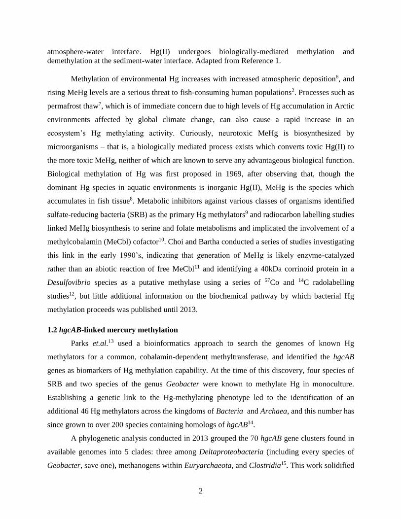
Figure 1.1 Schematic depiction of relevant equilibria in the Hg biogeochemical cycle. Hg(0)
and Hg(II) deposition into the environment results in a complex global cycle across the
Page 15
2
atmosphere-water interface. Hg(II) undergoes biologically-mediated methylation and
demethylation at the sediment-water interface. Adapted from Reference 1.
Methylation of environmental Hg increases with increased atmospheric deposition6, and
rising MeHg levels are a serious threat to fish-consuming human populations2. Processes such as
permafrost thaw7, which is of immediate concern due to high levels of Hg accumulation in Arctic
environments affected by global climate change, can also cause a rapid increase in an
ecosystem’s Hg methylating activity. Curiously, neurotoxic MeHg is biosynthesized by
microorganisms – that is, a biologically mediated process exists which converts toxic Hg(II) to
the more toxic MeHg, neither of which are known to serve any advantageous biological function.
Biological methylation of Hg was first proposed in 1969, after observing that, though the
dominant Hg species in aquatic environments is inorganic Hg(II), MeHg is the species which
accumulates in fish tissue8. Metabolic inhibitors against various classes of organisms identified
sulfate-reducing bacteria (SRB) as the primary Hg methylators9 and radiocarbon labelling studies
linked MeHg biosynthesis to serine and folate metabolisms and implicated the involvement of a
methylcobalamin (MeCbl) cofactor10. Choi and Bartha conducted a series of studies investigating
this link in the early 1990’s, indicating that generation of MeHg is likely enzyme-catalyzed
rather than an abiotic reaction of free MeCbl11 and identifying a 40kDa corrinoid protein in a
Desulfovibrio species as a putative methylase using a series of 57Co and 14C radolabelling
studies12, but little additional information on the biochemical pathway by which bacterial Hg
methylation proceeds was published until 2013.
1.2 hgcAB-linked mercury methylation
Parks et.al.13 used a bioinformatics approach to search the genomes of known Hg
methylators for a common, cobalamin-dependent methyltransferase, and identified the hgcAB
genes as biomarkers of Hg methylation capability. At the time of this discovery, four species of
SRB and two species of the genus Geobacter were known to methylate Hg in monoculture.
Establishing a genetic link to the Hg-methylating phenotype led to the identification of an
additional 46 Hg methylators across the kingdoms of Bacteria and Archaea, and this number has
since grown to over 200 species containing homologs of hgcAB14.
A phylogenetic analysis conducted in 2013 grouped the 70 hgcAB gene clusters found in
available genomes into 5 clades: three among Deltaproteobacteria (including every species of
Geobacter, save one), methanogens within Euryarchaeota, and Clostridia15. This work solidified
Page 16
3
the link between Hg methylation capability and hgcAB by showing that hgcAB- close
evolutionary relatives of a variety of hgcAB+ organisms cannot methylate Hg, whereas all tested
hgcAB+ organisms do methylate Hg; this definitively showed that MeHg biosynthesis requires
the hgcAB genes. The authors of this study emphasize that the vast diversity of newly identified
Hg methylating organisms necessitates an expansion of Hg methylation study to previously
unrecognized environmental niches, including those in which the dominant Hg methylators are
methanogenic Archaea: these include rice paddies16, permafrost and Arctic sediments17, and
freshwater aquatic systems18.
Methanogens have recently been shown to methylate Hg at similar rates to SRB and iron-
reducing bacteria when those rates are adjusted for total cell protein14, in contrast to older reports
that these species were not significant contributors to environmental Hg production9. Syntrophic
effects on Hg methylation are an area of exploration which have also benefited from the
identification of the hgcAB biomarkers; synergistic growth between Hg-methylating SRB and
methanogens was first observed in 199819, and a recent report has shown stimulation of MeHg
production in environmentally relevant combinations of Hg-methylating organisms with
syntrophic metabolic relationships20. One niche in which the hgcAB genes are conspicuously
absent, as determined in a large-scale study of metagenomes17, is the vertebrate digestive
microbiome – this suggests that human inorganic Hg exposure is not exacerbated by conversion
to neurotoxic MeHg within the body.
Two nonexclusive hypotheses exist concerning evolutionary advantages which the hgcAB
genes may provide. One is that these genes confer some resistance to Hg(II) toxicity; this does
not appear to be the case in hgcAB+ organisms, based on cell growth rates in the presence or
absence of Hg in Desulfovibrio desulfuricans ND132 and nearby genetic relatives21. This study,
however, was conducted on a limited number of organisms before the identification of the hgcAB
gene cluster and is not conclusive. It should be noted that methylmetals of arsenic, tin, and
selenium are formed by microbial processes and thought to protect against the toxic effects of
these elements by increasing their volatility and membrane permeability22–24, and this possibility
cannot yet be eliminated for MeHg. The other conceivable function of HgcA and HgcB is that
they catalyze some methyl transfer process associated with cellular carbon metabolism and
methylate Hg(II) adventitiously as a side product. This is supported by two findings from global
proteome studies: addition of Hg(II) to cultures of Desulfovibro desulfuricans ND132 did not
Page 17
4
induce a change in the expression levels of HgcA and HgcB25, and a lower abundance of genes
involved in the folate branch of the acetyl-CoA pathway was observed in a hgcAB deletion
mutant of Geobacter sulfurreducens PCA26. At the time of this writing, no decisive work has
been conducted to directly interrogate other metabolic functions of the hgcAB genes. In
summary, study of HgcA and HgcB is just beginning and no conclusions can yet be drawn as to
their biological role.
As HgcA is a cobalamin-dependent methyltransferase, this introductory chapter will
review the known cobalamin-dependent abiotic and enzymatic methyl transfer processes. The
putative roles of HgcB in both electron transfer through its iron-sulfur clusters and Hg binding
via a cysteine-rich, high-affinity Hg(II) binding site will be also discussed. Finally, the proposed
catalytic cycle for methyl transfer to Hg(II) and open questions will be presented as they relate to
the scope of this dissertation research.
1.3 Methylcobalamin and mechanisms of mercury methylation
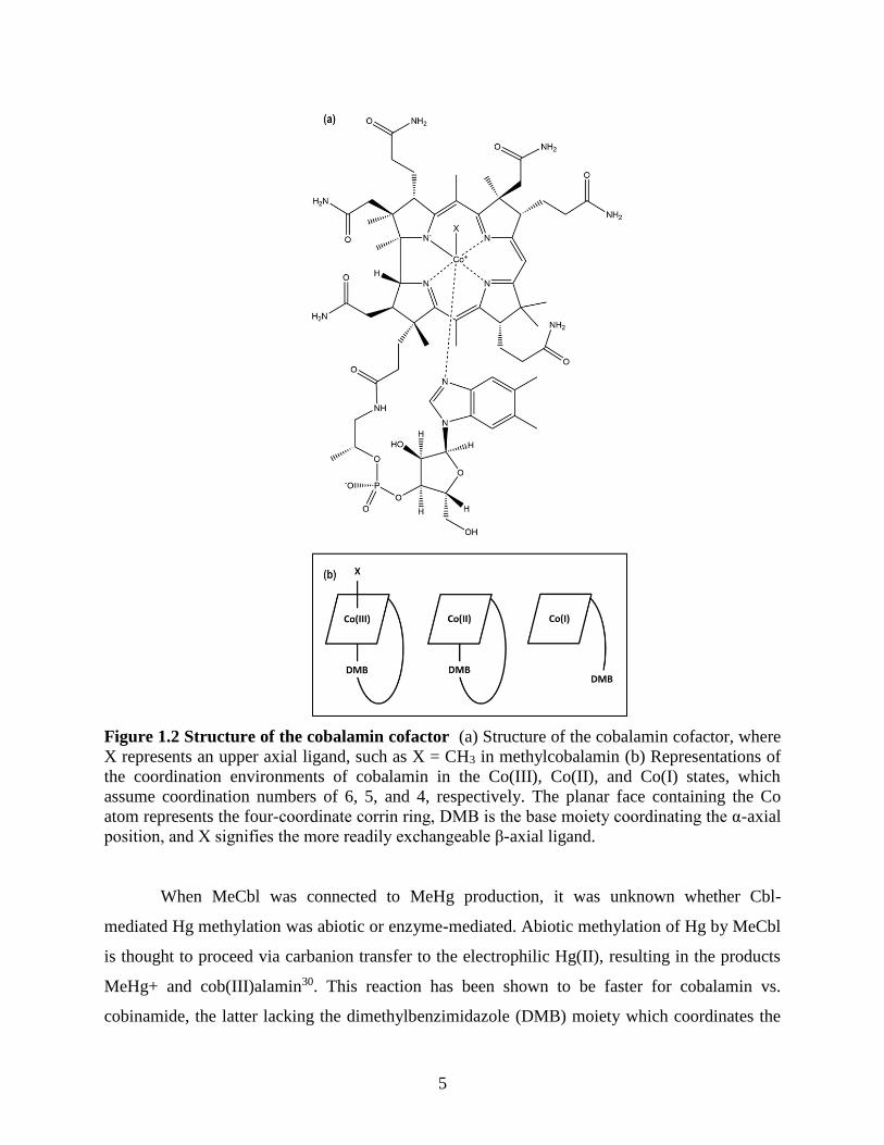
Cobalamin (Cbl), the structure of which is shown in Figure 1.2, is comprised of a cobalt
atom coordinated by the 4 nitrogen atoms of the macrocyclic corrin ligand27. It is unique among
tetrapyrroles in the addition of an intramolecular 5th ligand to the central cobalt which is
covalently attached to the α-face of the macrocycle via a nucleotide linkage28. This axial base
ligand is dimethybenzimidazole (DMB) in Cbl and not present in cobinamide (Cbi); the term
“corrinoid” captures both of these compounds as well as those containing other base moieties.
The Cbl cobalt center can access the +1, +2, and +3 oxidation states, and thus transfer alkyl
cation, radical, and anion species29. In a 1974 review of toxic element cycles in the
environment23, Wood discusses methylation of Hg(II) by methylcobalamin (MeCbl) as an early
mechanistic hypothesis for MeHg biosynthesis, simply because of this capacity to transfer a
methyl anion or radical. Reactions of other known biological methyl transfer donors,
methyltetrahydrofolate (methyl-THF) and S-adenosylmethionine, result in transfer of a methyl
cation; this species would be an unlikely ligand for Hg(II) on the basis of electrostatic repulsion.
Page 18
5
Figure 1.2 Structure of the cobalamin cofactor (a) Structure of the cobalamin cofactor, where
X represents an upper axial ligand, such as X = CH3 in methylcobalamin (b) Representations of
the coordination environments of cobalamin in the Co(III), Co(II), and Co(I) states, which
assume coordination numbers of 6, 5, and 4, respectively. The planar face containing the Co
atom represents the four-coordinate corrin ring, DMB is the base moiety coordinating the α-axial
position, and X signifies the more readily exchangeable β-axial ligand.
When MeCbl was connected to MeHg production, it was unknown whether Cbl-
mediated Hg methylation was abiotic or enzyme-mediated. Abiotic methylation of Hg by MeCbl
is thought to proceed via carbanion transfer to the electrophilic Hg(II), resulting in the products
MeHg+ and cob(III)alamin30. This reaction has been shown to be faster for cobalamin vs.
cobinamide, the latter lacking the dimethylbenzimidazole (DMB) moiety which coordinates the
Page 19
6
central Co atom, but also promoted by low pH, which increases the fraction of cobalamin in the
“base-off” form due to protonation and weakened affinity of the DMB ligand31; this indicates
both that DMB has a positive effect on the dealkylation of Cbl by Hg(II) but that its removability
is also important in the reaction. Though this mechanism is not yet fully resolved, it has been
observed that the abiotic reaction is slow at physiological pH and inhibited when Hg(II) is
supplied as a bis-thiolate complex32. These findings are significant because intracellular
methylation of Hg would presumably occur at near-neutral pH and Hg(II)-thiolate complexes are
understood to be the dominant Hg species in cells and the environment33. Our knowledge of
abiotic Hg methylation by Cbl is therefore consistent with the finding that environmental Hg
methylation is, instead, enzymatically mediated by the corrinoid-dependent HgcA.
1.4 HgcA and enzymatic corrinoid-dependent methyl transfer
Cbl enzyme cofactors catalyze an ever-increasing list of biological reactions34, including
methyl group transfer. Well-characterized examples of this process include methionine synthase
(MetH)35 and the corrinoid iron-sulfur protein (CFeSP)36, which is involved in the Wood-
Ljungdahl pathway of microbial acetogenesis. In both of these examples, the ability of the Cbl
cofactor to access the Co(I) and Co(III) oxidation states is central to the mechanism of methyl
transfer37. The “supernucleophilic” Co(I) state is capable of accepting a methyl group to form a
Me-Co(III) corrinoid. This methyl group can then be transferred to homocysteine or the acetyl
Co-A synthase nickel cluster, in the cases of MetH and CFeSP, respectively. These mechanisms
result in the transfer of a methylcation (CH3+), and the two electrons comprising the Co-C bond
return to the Co to reset the catalytic cycle at the Co(I) state.
Four Cbl protein binding modes for which crystal structures are available are shown in
Figure 1.3. Cofactor binding to these enzymes occurs in the “base-off” binding mode, in which
the lower axial DMB moiety dissociates from the central Co atom and binds via contacts to a
protein Rossmann fold. In the case of MetH, th base is replaced by a protein His ligand35;
CFeSP lacks a lower axial Cbl ligand, and instead binds via a “base-off” mode36. The recently
characterized transcriptional regulator CarH similarly binds a cobalamin cofactor and provides
two protein His ligands in a “base-off, bis-His” configuration reminiscent of common heme
binding motifs38. Homology modelling based on sequence similary to the CFeSP suggests the
cytosolic domain of HgcA, comprising ~18kDa of the 40kDa polypeptide, binds a corrinoid
Page 20
7
cofactor in the “base-off, Cys-on” conformation through a Rossmann fold motif and coordination
to the cobalt through Cys7313. A recent structural study has identified a “base-off, Cys-on” Cbl
binding mode in a structure of a membrane associated corrinoid transport protein, BtuM39. This
Cys residue, however, is suggested to catalyze decyanation of cyano-Cbl, and this enzyme does
not appear to be involved in methyl transfer. Therefore, no system is known which provides an
example of the effect of a lower axial Cys ligand on Co-C bond cleavage.
Figure 1.3 Crystal structures of observed "base-off" cobalamin protein binding modes (a)
“base-off, His-on” methionine synthase (MetH) (b) “base-off” corrinoid iron-sulfur protein
(CFeSP) (c) “base-off, Cys-on” vitamin B12 membrane transporter (BtuM) (d) “base-off, bis-
His” light-dependent transcriptional regulator (CarH). The proteins are represented as colored
cartoons while the cobalamin and relevant protein ligands are represented as sticks and colored
by element. Rendered with Pymol from Protein Data Bank entries (a) 1BMT, (b) 4DJD, (c)
6FFV, and (d) 58CF.
Cys73 is strictly conserved among hgcA genes, has been shown to be critical to Hg
methylation in vivo40, and is hypothesized to have a dramatic effect on corrinoid-dependent
methyl transfer via HgcA. Zhou et.al. constructed density functional theory calculations to model
the trans effect of a thiolate ligand on Co-C bond cleavage41. They determined that a “base-off,
Cys-on” Cbl coordination mode should modulate the bond dissociation energy of the Co-C bond
Page 21
8
such that heterolysis releasing a carbanion (CH3-) would be the energetically preferred mode of
bond dissociation. Though the local protein environment could modulate this and experimental
validation is necessary, this model provides a chemical rationale for the departure of HgcA from
known mechanisms of enzymatic carbocation transfer and supports the mechanistic hypothesis
first proposed in Parks et.al.13 (it should be noted that, though not observed in an enzymatic
system, carbanion transfer is suggested in the abiotic methylation of Hg as discussed in section
1.3, so this is not a corrinoid reaction without chemical precendent). In this mechanism, the
HgcA corrinoid would first assume the Co(I) state to generate Me-Co(III) with an unknown
methyl donor, then methyl transfer to the strongly electrophilic Hg(II) would result in a Co(III)
corrinoid, with a requirement for reduction to Co(I) to reset catalysis. Essential to this process is
the donation of two electrons every cycle to return the Co to the Co(I) state; this is the
hypothesized role of HgcB.
1.5 Mercury binding and HgcB, a [4Fe-4S] cluster ferredoxin-like protein
HgcB is a 10kDa ferredoxin-like protein which contains two four-iron four-sulfur ([4Fe-
4S]) cluster CXXCXXCXXXCP consensus motifs. [4Fe-4S] clusters are an ancient and
ubiquitious biological feature known to store and transfer low potential electrons42, and it is
hypothesized that the HgcB iron-sulfur (Fe-S) clusters are responsible for 2-electron reduction of
the Co(III) HgcA corrinoid after carbanion transfer. In the “base-off” CFeSP, the Co(II)/Co(I)
midpoint redox potential is increased from the “base-on” potential of ~600mV to ~450mV43. We
have no information on the Co(II)/Co(I) redox couple in HgcA, as this will be dependent on its
coordination state, binding mode, and protein environment, but we anticipate that the HgcB
clusters will exhibit sufficiently low midpoint potentials to reduce the corrinoid species. In
addition to the 8 Cys residues involved in the Fe-S clusters, HgcB contains 3 strictly conserved
Cys residues: Cys73, Cys94, and Cys95, with Cys94 and Cys95 occupying the C-terminus.
Hg(II) has an strong preference for thiolate coordination, and it has been proposed that these Cys
residues comprise a high-affinity Hg(II) binding site13. This could facilitate catalysis by
positioning the Hg(II) in a favorable proximity for methylation. Peptide studies have shown that
vicinal Cys residues are highly specific for Hg(II) binding over other thiophilic divalent metal
ions, compared to CAAC and CACA motifs44. Cys-rich motifs, including those with vicinal Cys
Page 22
9
pairs, have been observed and characterized in other Hg(II) binding proteins, notably those found
within the mer operon.
The mer operon encodes genes widespread in aerobic organisms which are responsible
for uptake and degradation of MeHg45. This pathway involves Hg(II) dependent transcriptional
activation by MerR, chaperoned import of Hg(II) or MeHg, cleavage of the Hg-C bond by the
organomercurial lyase MerB, and reduction of the resulting Hg(II) to the relatively inert and
nontoxic Hg(0) by MerA. All of these processes are mediated by Cys-based Hg(II) binding sites:
MerT46 and MerF47, membrane Hg(II) transport proteins, contain Cys-Cys vicinal pairs, as do
MerB48 and MerA49. A variety of Cys motifs are seen in other mer operon binding sites,
including an unusual example in the MerR transcriptional regulator, which has been shown to
bind Hg(II) with high affinity and specificity in a 3-Cys trigonal planar mode, in contrast to the
typically preferred linear geometry50,51. An in vivo mutagenesis study of HgcA and HgcB in the
model Hg methylator Desulfovibrio desulfuricans ND132 investigated the impact of alanine
(Ala) substitution of the three conserved HgcB Cys residues40. These results indicated that Cys73
and either Cys94 or Cys95 are required for in vivo Hg methylation activity, but the loss of one of
the C-terminal pair can be accommodated. This suggests that some configuration of these
residues is required for MeHg production, though in vitro characterization of these mutants
would be required to confirm that the loss of Hg methylating activity is due specifically to Hg
binding deficiency.
1.6 Hypothesized mercury methylation process and scope of this dissertation
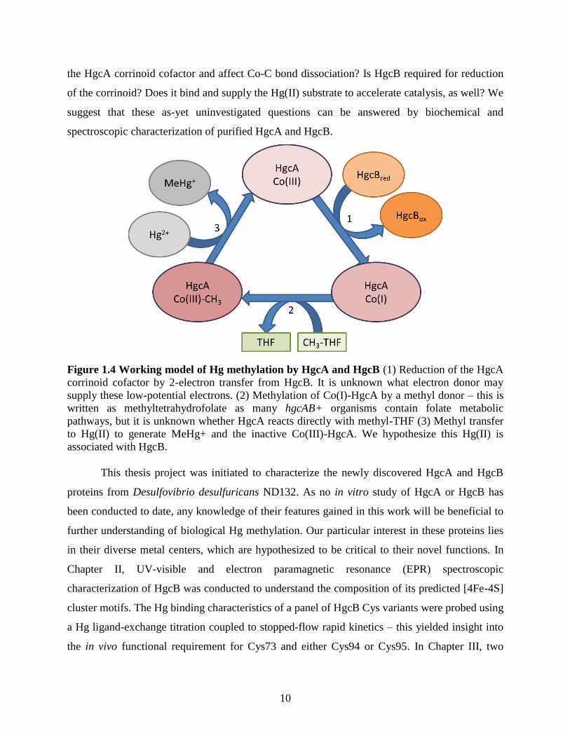
Figure 1.4 depicts the hypothesisized hgcAB-mediated MeHg biosynthesic scheme
presented in Parks et.al. In this model, Co(III)-HgcA is reduced with two electrons transferred
from the HgcB [4Fe-4S] clusters and methylated. The methyl-Co(III) corrinoid then transfers a
methyl anion to a Hg(II) atom coordinated to HgcB via cysteine ligands, resulting in MeHg+ and
a Co(III) species at the end of the catalytic cycle. This is a working model constructed using
insight from homology modelling13, computational prediction41, and in vivo mutagenesis40
studies. Though these have provided some insight into critical features of this process, as
described in previous sections of this chapter, in vitro investigation is required to directly answer
mechanistic questions. Is methylated HgcA capable of methyl transfer to Hg(II)? If so, what is
the electronic nature of the methyl group which is transferred to Hg(II)? Does Cys73 coordinate
Page 23
10
the HgcA corrinoid cofactor and affect Co-C bond dissociation? Is HgcB required for reduction
of the corrinoid? Does it bind and supply the Hg(II) substrate to accelerate catalysis, as well? We
suggest that these as-yet uninvestigated questions can be answered by biochemical and
spectroscopic characterization of purified HgcA and HgcB.
Figure 1.4 Working model of Hg methylation by HgcA and HgcB (1) Reduction of the HgcA
corrinoid cofactor by 2-electron transfer from HgcB. It is unknown what electron donor may
supply these low-potential electrons. (2) Methylation of Co(I)-HgcA by a methyl donor – this is
written as methyltetrahydrofolate as many hgcAB+ organisms contain folate metabolic
pathways, but it is unknown whether HgcA reacts directly with methyl-THF (3) Methyl transfer
to Hg(II) to generate MeHg+ and the inactive Co(III)-HgcA. We hypothesize this Hg(II) is
associated with HgcB.
This thesis project was initiated to characterize the newly discovered HgcA and HgcB
proteins from Desulfovibrio desulfuricans ND132. As no in vitro study of HgcA or HgcB has
been conducted to date, any knowledge of their features gained in this work will be beneficial to
further understanding of biological Hg methylation. Our particular interest in these proteins lies
in their diverse metal centers, which are hypothesized to be critical to their novel functions. In
Chapter II, UV-visible and electron paramagnetic resonance (EPR) spectroscopic
characterization of HgcB was conducted to understand the composition of its predicted [4Fe-4S]
cluster motifs. The Hg binding characteristics of a panel of HgcB Cys variants were probed using
a Hg ligand-exchange titration coupled to stopped-flow rapid kinetics – this yielded insight into
the in vivo functional requirement for Cys73 and either Cys94 or Cys95. In Chapter III, two
Page 24
11
approaches were taken to HgcA production: generation of a truncated HgcA constructs to
promote soluble heterologous expression and co-expression of the full-length transmembrane
protein with a plasmid controlling cellular uptake of cobalamin. The latter technique resulted in
the heterologous expression and purification of full-length HgcA, the UV-visible spectrum of
which will be presented. These results provide the Hg biotransformation field with preliminary
characteristics of these proteins, which will be valuable to further mechanistic understanding of
MeHg biosynthesis.
1.7 References for Chapter 1
(1) Clarkson, T. W. Critical Reviews in Toxicology The Toxicology of Mercury and Its Chemical Compounds.
Crit. Rev. Toxicol. 2006, 36 (8), 609–662.
(2) Fitzgerald, W. F.; Clarkson, T. W. Mercury and Monomethylmercury: Present and Future Concerns.
Environ. Health Perspect. 1991, 96, 159–166.
(3) Nriagu, J. O.; Pacyna, J. M. Quantitative Assessment of Worldwide Contamination of Air, Water and Soils
by Trace Metals. Nature 1988, 333, 134–139.
(4) Mason, R. P.; Abbott, M. L.; Bodaly, R. A.; Bullock, R.; Driscoll, C. T.; Evers, D. C.; Lindberg, S. E.;
Murray, M.; Swain, E. B. Monitoring the Response to Changing Mercury Deposition. Environ. Sci. Technol.
2005, No. 39, 15A–22A.
(5) Evers, D. C.; Clair, T. A. Mercury in Northeastern North America: A Synthesis of Existing Databases.
Ecotoxicology 2005, 14 (1–2), 7–14.
(6) Harris, R. C.; Rudd, J. W. M.; Amyot, M.; Babiarz, C. L.; Beaty, K. G.; Blanchfield, P. J.; Bodaly, R. A.;
Branfireun, B. A.; Gilmour, C. C.; Graydon, J. A.; et al. Whole-Ecosystem Study Shows Rapid Fish-
Mercury Response to Changes in Mercury Deposition. Proc. Natl. Acad. Sci. 2007, 104 (42), 16586–16591.
(7) Yang, Z.; Fang, W.; Lu, X.; Sheng, G. P.; Graham, D. E.; Liang, L.; Wullschleger, S. D.; Gu, B. Warming
Increases Methylmercury Production in an Arctic Soil. Environ. Pollut. 2016, 214, 504–509.
(8) Jensen, S.; Jernelöv, A. Biological Methylation of Mercury in Aquatic Organisms. Nature 1969, 223 (5207),
753–754.
(9) Compeau, G. C.; Bartha, R. Sulfate-Reducing Bacteria: Principal Methylators of Mercury in Anoxic
Estuarine Sediment. Microbiology 1985, 50 (2), 498–502.
(10) Berman, M.; Chase, T.; Bartha, R. Carbon Flow in Mercury Biomethylation by Desulfovibrio-
Desulfuricans. Appl. Environ. Microbiol. 1990, 56 (1), 298–300.
(11) Choi, S. C.; Bartha, R. Cobalamin-Mediated Mercury Methylation by Desulfovibrio Desulfuricans LS. Appl.
Environ. Microbiol. 1993, 59 (1), 290–295.
(12) Choi, S. C.; Chase, T.; Bartha, R. Enzymatic Catalysis of Mercury Methylation by Desulfovibrio
Desulfuricans LS. Appl. Environ. Microbiol. 1994, 60 (4), 1342–1346.
(13) Parks, J. M.; Johs, A.; Podar, M.; Bridou, R.; Hurt Jr., R. A.; Smith, S. D.; Tomanicek, S. J.; Qian, Y.;
Brown, S. D.; Brandt, C. C.; et al. The Genetic Basis for Bacterial Mercury Methylation. Science. 2013, 339,
1332–1336.
(14) Gilmour, C. C.; Bullock, A. L.; McBurney, A.; Podar, M.; Elias, D. A. Robust Mercury Methylation across
Diverse Methanogenic Archaea. MBio 2018, 9 (2), e02403-17.
(15) Gilmour, C. C.; Podar, M.; Bullock, A. L.; Graham, A. M.; Brown, S. D.; Somenahally, A. C.; Johs, A.;
Hurt, R. A.; Bailey, K. L.; Elias, D. A. Mercury Methylation by Novel Microorganisms from New
Environments. Environ. Sci. Technol. 2013, 47 (20), 11810–11820.
(16) Rothenberg, S. E.; Anders, M.; Ajami, N. J.; Petrosino, J. F.; Balogh, E. Water Management Impacts Rice
Methylmercury and the Soil Microbiome. Sci. Total Environ. 2016, 572, 608–617.
(17) Podar, M.; Gilmour, C. C.; Brandt, C. C.; Soren, A.; Brown, S. D.; Crable, B. R.; Palumbo, A. V.;
Somenahally, A. C.; Elias, D. A. Global Prevalence and Distribution of Genes and Microorganisms
Involved in Mercury Methylation. Sci. Adv. 2015, 1 (9), 1–12.
(18) Christensen GA, S. A.; Moberly JG, Miller CM, King AJ, G. C.; Brown SD, Podar M, Brandt CC, B. S.;
Page 25
12
Palumbo AV, Wall JD, E. DA. Carbon Amendments Alter Microbial Community Structure and Net
Mercury Methylation Potential in Sediments. Appl Env. Microbiol 2018, 84 (3), 1–14.
(19) Pak, K. R.; Bartha, R. Mercury Methylation by Interspecies Hydrogen and Acetate Transfer between
Sulfidogens and Methanogens. Appl. Environ. Microbiol. 1998, 64 (6), 1987–1990.
(20) Yu, R. Q.; Reinfelder, J. R.; Hines, M. E.; Barkay, T. Syntrophic Pathways for Microbial Mercury
Methylation. ISME J. 2018, 12 (7), 1826–1835.
(21) Gilmour, C. C.; Elias, D. A.; Kucken, A. M.; Brown, S. D.; Palumbo, A. V.; Schadt, C. W.; Wall, J. D.
Sulfate-Reducing Bacterium Desulfovibrio Desulfuricans ND132 as a Model for Understanding Bacterial
Mercury Methylation. Appl. Environ. Microbiol. 2011, 77 (12), 3938–3951.
(22) Carrillo-González, R.; Šimůnek, J.; Sauvé, S.; Adriano, D. Mechanisms and Pathways of Trace Element
Mobility in Soils. Adv. Agron. 2006, 91 (06), 111–178.
(23) Wood, J. M. Biological Cycles for Toxic Elements in the Environment. Science. 1974, 183 (4129), 1049–
1052.
(24) Nagase, H.; Sato, T.; Yoshioka, Y. Formation, Distribution, and Ecotoxicity of Methylmetals of Tin,
Mercury, and Arsenic in the Environment. Crit. Rev. Environ. Sci. Technol. 1995, 25 (1), 45–91.
(25) Qian, C.; Chen, H.; Johs, A.; Lu, X.; An, J.; Pierce, E. M.; Parks, J. M.; Elias, D. A.; Hettich, R. L.; Gu, B.
Quantitative Proteomic Analysis of Biological Processes and Responses of the Bacterium Desulfovibrio
Desulfuricans ND132 upon Deletion of Its Mercury Methylation Genes. Proteomics 2018, 1700479.
(26) Qian, C.; Johs, A.; Chen, H.; Mann, B. F.; Lu, X.; Abraham, P. E.; Hettich, R. L.; Gu, B. Global Proteome
Response to Deletion of Genes Related to Mercury Methylation and Dissimilatory Metal Reduction Reveals
Changes in Respiratory Metabolism in Geobacter Sulfurreducens PCA. J. Proteome Res. 2016, 15 (10),
3540–3549.
(27) Banerjee, R. V. Chemistry and Biochemistry of B12; John Wiley and Sons, 1999.
(28) Banerjee, R.; Ragsdale, S. W. The Many Faces of Vitamin B 12 : Catalysis by Cobalamin-Dependent
Enzymes. Annu. Rev. Biochem. 2003, 72 (1), 209–247.
(29) Schrauzer, G. N. Mechanisims of Corrin Dependent Enzymatic Reactions. In Fortschritte der Chemie
Organischer Naturstoffe/Progress in the Chemistry of Organic Natural Products; Herz, W., Grisebach, H.,
Kirby, G. W., Eds.; Springer, Vienna, 1974; pp 583–628.
(30) Hill, H. A. O.; Pratt, J. M.; Ridsdale, S.; Williams, F. R.; Williams, R. J. P. Kinetics of Substitution of Co-
Ordinated Carbanions in Cobalt (III) Corrinoids. J. Chem. Soc. D 1970, 124, 341–341.
(31) DeSimone, R. E.; Penley, M. W.; Charbonneau, L.; Smith, S. G.; Wood, J. M.; Hill, H. A. O.; Pratt, J. M.;
Ridsdale, S.; Williams, R. J. P. The Kinetics and Mechanism of Cobalamin-Dependent Methyl and Ethyl
Transfer to Mercuric Ion. Biochim. Biophys. Acta 1973, 304, 851–863.
(32) Bertilsson, L.; Neujahr, M. Y. Methylation of Mercury Compounds by Methylcobalamin. Biochemistry
1971, 10 (14), 2805–2808.
(33) Fretham, S. J. B.; Aschner, M. Mercury. In RSC Metallobiology Series No. 2: Binding, Transport and
Storage of Metal Ions in Biological Cells; Maret, W., Wedd, A., Eds.; Royal Society of Chemistry, 2014; pp
747–767.
(34) Bridwell-Rabb, J.; Drennan, C. L. Vitamin B12 in the Spotlight Again. Curr. Opin. Chem. Biol. 2017, 37,
63–70.
(35) Goulding, C. W.; Postigo, D.; Matthews, R. G. Cobalamin-Dependent Methionine Synthase Is a Modular
Protein with Distinct Regions for Binding Homocysteine, Methyltetrahydrofolate, Cobalamin, and
Adenosylmethionine. Biochemistry 1997, 36 (26), 8082–8091.
(36) Ragsdale, S. W.; Lindahl, P. A.; Munck, E. Mossbauer, EPR, and Optical Studies of the Corrinoid/Iron-
Sulfur Protein Involved in the Synthesis of Acetyl Coenzyme A by Clostridium Thermoaceticum. J. Biol.
Chem. 1987, 262 (29), 14289–14297.
(37) Matthews, R. G.; Koutmos, M.; Datta, S. Cobalamin-Dependent and Cobamide-Dependent
Methyltransferases. Curr. Opin. Struct. Biol. 2008, 18 (6), 658–666.
(38) Jost, M.; Fernandez-Zapata, J.; Polanco, M. C.; Ortiz-Guerrero, J. M.; Chen, P. Y. T.; Kang, G.;
Padmanabhan, S.; Elias-Arnanz, M.; Drennan, C. L. Structural Basis for Gene Regulation by a B12-
Dependent Photoreceptor. Nature 2015, 526 (7574), 536–541.
(39) Rempel, S., Colucci, E., de Gier, J.W., Guskov, A., Slotboom, D. J. Cysteine-Mediated Decyanation of
Vitamin B12 by the Predicted Membrane Transporter BtuM. Nat. Commun. 2018, No. 2018, 1–8.
(40) Smith, S. D.; Bridou, R.; Johs, A.; Parks, J. M.; Elias, D. A.; Hurt, R. A.; Brown, S. D.; Podar, M.; Wall, J.
D. Site-Directed Mutagenesis of HgcA and HgcB Reveals Amino Acid Residues Important for Mercury
Methylation. Appl. Environ. Microbiol. 2015, 81 (9), 3205–3217.
Page 26
13
(41) Zhou, J.; Riccardi, D.; Beste, A.; Smith, J. C.; Parks, J. M. Mercury Methylation by HgcA: Theory Supports
Carbanion Transfer to Hg(II). Inorg. Chem. 2014, 53 (2), 772–777.
(42) Koay, M. S.; Antonkine, M. L.; Gärtner, W.; Lubitz, W. Modelling Low-Potential [Fe4S4] Clusters in
Proteins. Chem. Biodivers. 2008, 5 (8), 1571–1587.
(43) Harder, S. R.; Lu, W. P.; Feinberg, B. A.; Ragsdale, S. W. Spectroelectrochemical Studies of the
Corrinoid/Iron-Sulfur Protein Involved in Acetyl Coenzyme A Synthesis by Clostridium Thermoaceticum.
Biochemistry 1989, 28 (23), 9080–9087.
(44) DeSilva, T. M.; Veglia, G.; Porcelli, F.; Prantner, A. M.; Opella, S. J. Selectivity in Heavy Metal-Binding to
Peptides and Proteins. Biopolymers 2002, 64 (4), 189–197.
(45) Barkay, T.; Miller, S. M.; Summers, A. O. Bacterial Mercury Resistance from Atoms to Ecosystems. FEMS
Microbiol. Rev. 2003, 27 (2–3), 355–384.
(46) Morby, A. P.; Hobman, J. L.; Brown, N. L. The Role of Cysteine Residues in the Transport of Mercuric Ions
by the Tn501 MerT and MerP Mercury‐resistance Proteins. Mol. Microbiol. 1995, 17 (1), 25–35.
(47) Wilson, J. R.; Leang, C.; Morby, A. P.; Hobman, J. L.; Brown, N. L. MerF Is a Mercury Transport Protein:
Different Structures but a Common Mechanism for Mercuric Ion Transporters? FEBS Lett. 2000, 472 (1),
78–82.
(48) Pitts, K. E.; Summers, A. O. The Roles of Thiols in the Bacterial Organomercurial Lyase (MerB).
Biochemistry 2002, 41 (32), 10287–10296.
(49) Brown, N. L.; Ford, S. J.; Pridmore, R. D.; Fritzinger, D. C. Nucleotide Sequence of a Gene from the
Pseudomonas Transposon Tn501 Encoding Mercuric Reductase. Biochemistry 1983, 22 (17), 4089–4095.
(50) Watton, S. P.; Wright, J. G.; MacDonnell, F. M.; Bryson, J. W.; Sabat, M.; O’Halloran, T. V. Trigonal
Mercuric Complex of an Aliphatic Thiolate: A Spectroscopic and Structural Model for the Receptor Site in
the Hg(II) Biosensor MerR. J. Am. Chem. Soc. 1990, 112 (7), 2824–2826.
(51) Wang, D.; Huang, S.; Liu, P.; Liu, X.; He, Y.; Chen, W.; Hu, Q.; Wei, T.; Gan, J.; Ma, J.; et al. Structural
Analysis of the Hg(II)-Regulatory Protein Tn501 MerR from Pseudomonas Aeruginosa. Sci. Rep. 2016, 6,
1–9.
Page 27
14
Chapter 2 HgcB
2.1 Introduction
Methylmercury (MeHg) is a neurotoxin synthesized by microorganisms containing the
hgcAB genes1. Understanding the biosynthesis of MeHg is vital due to the risks posed to aquatic
fish and bird species, as well as fish-consuming human populations, by environmental
conversion of Hg(II) to MeHg2,3. Little is known about the biochemical mechanism by which
this occurs, but it has been shown that deletion of either hgcA or hgcB eliminates the ability of
Desulfovibrio desulfuricans ND132 and Geobacter sulfurreducens PCA to methylate mercury
(Hg) in culture. These genes encode a 40kDa corrinoid-dependent methyltransferase with a C-
terminal transmembrane domain and a 10kDa ferredoxin-like protein with two four-iron four-
sulfur ([4Fe-4S]) consensus motifs, respectively. Based on similarity to characterized corrinoid-
dependent methyltransferase reactions, notably methionine synthase (MetH) and the corrinoid
iron-sulfur protein (CFeSP)4, it is proposed that the HgcA corrinoid is reduced by HgcB to the
Co(I) state, methylated by an as yet unidentified methyl donor, and the methyl group transferred
to Hg(II), resulting in an inactive Co(III) state. HgcB has two proposed roles in this model: to
supply low-potential electrons through its [4Fe-4S] clusters to reduce the HgcA corrinoid
cofactor each catalytic cycle and to bind the Hg(II) substrate via its conserved cysteine (Cys)
residues.
[4Fe-4S] clusters are ancient and ubiquitious cofactors known to store and transfer low
potential electrons5. These are generally encoded by the consensus sequence
CXXCXXCXXXCP and incorporate 4 atoms of divalent iron Fe and 4 equivalents of sulfide.
The Fe atoms are coordinated by the Cys residues and bridged by sulfide ions, resulting in a
cubane structure with an overall charge of +2 in the oxidized state. 1 electron reduction of this
cofactor results in the delocalization of that electron within the cluster and a formal reduced
charge of +1. Low-potential clusters have been observed with midpoint redox potentials ranging
from -280mV to -705mV5. The Co(II) to Co(I) redox couple is low in the range of biological
potentials, and reductions of MetH and CFeSP exhibit midpoint redox potentials of -526mV6 and
Page 28
15
-504mV7, respectively. MetH is reduced by the higher potential flavodoxin protein, but the
protein environment surrounding the corrinoid shifts the effective midpoint potential of the
Co(II)/Co(I) couple by +50mV make the reduction by flavodoxin more favorable8. In the CFeSP,
an internal [4Fe-4S] cluster is responsible for reduction of Co(II) and its 2+/1+ midpoint
potential has been determined as -523mV. Thus it is hypothesized that the HgcB iron-sulfur
clusters are sufficiently low in potential to reduce a similar corrinoid species.
Initial attempts by our collaborators at Oak Ridge National Laboratory to heterologously
produce the hgcB gene from Desulfovibrio desulfuricans ND132 in Escherichia coli BL21 cells
resulted in insoluble protein. Two approaches were combined to obtain soluble, iron-replete
HgcB. The first was the cloning of a series of fusion protein constructs and testing their
expression level and solubility after cell lysis. A maltose binding protein (MBP) tag was chosen
for its high expression, solubility, and ease of purification via affinity resin. The second was the
use of a co-expression plasmid shown to increase [4Fe-4S] cluster loading in overexpression
systems9. The Ragsdale laboratory routinely uses co-expression systems in which cofactor
uptake and/or assembly genes are encoded on a plasmid and co-transformed into the cell line
expressing the protein of interest. The pRKISC plasmid was used in the expression of HgcB for
characterization by UV-visible spectroscopy and EPR to assess its [4Fe-4S] motifs.
Another feature of interest in HgcB is its conserved Cys residues: Cys73, Cys94, and
Cys95. Hg exhibits a high affinity for thiolate coordination and Hg binding proteins must
compete with Hg complexation by intracellular thiolates. Peptide studies of Cys-Cys motifs have
indicated both high affinity and specificity for Hg(II) over other thiophilic metals10. Several
examples of vicinal Cys-Cys motifs are seen in the mer operon11, which encodes MeHg
trafficking and detoxification genes and is highly specialized to bind Hg with high affinity.
Though none of the relevant Cys residues are vicinal in sequence, an unusual 3-coordinate Cys-
Cys-Cys Hg(II) binding site is observed in the MerR transcriptional regulator12, which may also
bear relevance to Hg binding by HgcB. In vivo mutagenic studies of Cys73, Cys94, and Cys95
indicated the effect of alanine (Ala) substitution of these residues on in vivo Hg methylation
activity in Desulfovibrio desulfuricans ND13213. These data are summarized in Table 1 and
suggest that Cys73 and either Cys94 or Cys95 are required for MeHg production.
Page 29
16
HgcB mutation % MeHg produced
C73A None detected
C94A 109%
C95A 120%
C94A/C95A 4%
Table 2.1 Effect of Cys substitution with Ala at Cys 73, Cys94, and Cys95 on in vivo MeHg
production in Desulfovibrio desulfuricans ND132. Percent values of MeHg produced are in
comparison to the wild-type strain (adapted from Ref. 11).
Figure 2.1 Schematic representation of the HgcB conserved cysteines. Cys94 and Cys95 are
found on the C-terminus of the protein and thus likely to be exposed. The structural accessibility
of Cys73 is unknown.
We hypothesize that some configuration of the HgcB conserved Cys residues comprise a
high-affinity Hg binding site. To test this hypothesis, we used stopped-flow kinetics to observe
the dissociation of a colorimetric ligand, 2-nitro-5-thiobenzoate (TNB), from Hg(II) upon mixing
with HgcB. Hg(TNB)2 is a high-affinity, stable complex, and only a high-affinity Hg binding site
could rapidly induce its dissociation This mimics the proposed role of HgcB in binding
intracellular Hg(II), which is typically coordinated to small molecule thiolates; we therefore view
this experiment as a functional assay. We sought to test whether the same analysis of a variety of
Cys73, Cys94, and Cys95 mutants would indicate deficiencies in their ability to bind Hg,
suggesting which Cys residues are required for Hg binding.
Page 30
17
2.2 Materials and Methods
2.2.1 Materials
All chemicals were purchased from Sigma-Aldrich (St. Louis, MO) unless otherwise
stated. All custom synthesized oligonucleotide primers were ordered from Integrated DNA
Technologies (Coralville, Iowa). All restriction enzymes, the Q5 Site-directed mutagenesis kit,
and amylose resin were purchased from New England Biolabs (Ipswich, MA). Pfu Ultra
polymerase was used for all PCR reactions and obtained from Agilent (Santa Clara, CA).
BL21(DE3) and TOP10 chemically competent cell lines were obtained from Invitrogen/Life
Technologies (Carlsbad, CA). P2 resin was purchased from Bio-Rad (Hercules, CA). The codon-
optimized HgcB plasmid was custom synthesized by DNA2.0 and provided to us as a gift from
Dr. Alexander Johs, Oak Ridge National Laboratory. cOmplete EDTA-free protease inhibitor
tablets were acquired from Roche (Indianapolis, IN). Titanium (III) citrate was prepared
similarly to the method of Zehnder and Wurhmann14 in 1M Tris-HCl pH 8.0. Maltose binding
protein (MBP) was purified and provided by Dr. Eric Carter.
2.2.2 HgcB fusion tag screen
Ligation independent cloning was used to generate a series of HgcB constructs with the
N-terminal fusion protein tags GB115, MBP16, Mocr17, and SC-Mocr. The destination vectors are
all derived from the pMCSG7 system and include an N-terminal His6 tag, the fusion protein tag,
and a TEV proteolysis sequence upstream of the protein of interest18. Vectors were transformed
into TOP10 cells and plated on LB agar selection plates containing 100ug/mL ampicillin.
Sequences were verified by Sanger sequencing using the T7 terminator primer (University of
Michigan DNA Sequencing Core) and transformed into BL21(DE3) cells. Cultures grown from
single colonies were stored at -80°C in 25% glycerol and used to inoculate overnight starter
cultures.
To test the expression of each of the four HgcB fusion constructs in BL21(DE3) cells, LB
overnight cultures containing 100ug/mL ampicillin were inoculated from frozen cell stocks. 1mL
of each overnight culture was used to inoculate 1L of LB media containing 100ug/mL ampicillin
and incubated at 37°C with 225rpm shaking. Expression was induced by addition of 1mM IPTG
between OD600 0.6-0.7. After 3 hours, the cells were harvested by centrifugation and frozen at -
Page 31
18
20°C. Whole-cell gel samples were taken before and after induction and analyzed by
nonreducing SDS-PAGE on a 12% polyacrylamide gel.
Each cell pellet was resuspended in 10mL MOPS pH 7, 100mM NaCl, and 5mM DTT
and sonicated at 4°C. 100uL of the lysis mixture was aliquoted into a microcentrifuge tube and
centrifuged at 17,000xg in a tabletop microcentrifuge. The soluble lysate was removed and
added to 100uL of 2X LDS sample loading buffer and 200uL 1X LDS buffer was used to
resuspend the insoluble pellet fraction. These samples were analyzed by nonreducing SDS-
PAGE on a 12% polyacrylamide gel to determine the solubility of each construct after lysis.
2.2.3 MHB co-expression with pRKISC and affinity purification
The protocol for overexpression of MBP-HgcB was modified from existing procedures
for the heterologous expression of Fe-S proteins19,20. The vector containing His6-MBP-HgcB,
abbreviated hereafter as MHB, was co-transformed into BL21(DE3) cells with the pRKISC
vector and plated on LB agar selection plates containing 100ug/mL ampicillin and 5ug/mL (50%
working concentration) tetracycline. A glycerol stock was prepared from a single resulting
colony and used to inoculate LB overnight starter cultures. 20mL of an overnight culture
containing LB medium, 100ug/mL ampicillin, and 10ug/mL tetracycline was used to inoculate
1L of TB medium in a 1L bottle containing the same antibiotic concentrations. This was
incubated at 37°C with 125rpm shaking until an OD600 ~0.6 was reached. At this point, each
bottle was sealed with a septum and screw-cap ring and sparged with nitrogen gas for 1 hour. An
additional 50ug/mL ampicillin and a reducing solution of 1mM sodium sulfide nonahydrate and
1.5mM L-cysteine were added to each culture via syringe and incubated for 10 minutes. 0.5mM
ferrous ammonium sulfate was added and 0.5mM IPTG was used to induce expression of the isc
genes and MHB. Cultures were incubated overnight (18-20hrs) at 15°C with 125rpm shaking.
Cells were harvested by centrifugation and stored at -20°C.
Cell lysis and amylose affinity purification were conducted under anoxic conditions (<
1ppm oxygen) in a Vacuum Atmospheres anaerobic glove box. A cell pellet obtained from 4L of
culture (~12g) was thawed in 50mL of lysis buffer containing 25mM Na HEPES pH 7.5, 5mM
DTT, and 1 cOmplete EDTA-free protease inhibitor with gentle stirring. Cells were sonicated
and the lysate cleared by centrifugation at 100,000xg. The soluble lysate was immediately
applied to an amylose affinity column equilibrated with 5 column volumes (CV) of 25mM Na
Page 32
19
HEPES pH 7.5 and 5mM DTT. 2.5CV of this buffer was used to wash the column and MHB was
eluted with the same buffer containing 20mM maltose. The purified protein was analyzed for
purity by sodium dodecyl sulfate polyacrylamide gel electrophoresis (SDS-PAGE) and the
protein concentration determined by Bradford assay21. Purified MHB was stored in an anaerobic
chamber at 4°C.
2.2.4 Construction of MHB cysteine mutants by site-directed mutagenesis
C94A, C95A, and C94A/C95A mutants were constructed using the QuikChange II site-
directed mutagenesis protocol (Agilent) and transformed into TOP10 chemically competent
cells. Successful transformants were chosen as single colonies from LB agar selection plates
containing 100ug/mL ampicillin and verified by Sanger sequencing using the T7 terminator
primer (University of Michigan DNA Sequencing Core). Cys73 mutants were constructed using
the Q5 site-directed mutagenesis kit, transformed into NEB 5-alpha cells, and verified by Sanger
sequencing as above. The MHB-CCC, MHB-CAC, MHB-CCA, or MHB-CAA vectors
previously assembled were used as templates for the Cys73 mutagenesis reactions, resulting in
the list of MHB variants below (Table 2). All expression of MHB Cys variants was conducted as
described in section 2.2.3.
Identifier Mutations
MHB-CCC none
MHB-CAC C94A
MHB-CCA C95A
MHB-CAA C94A/C95A
MHB-SCC C73S
MHB-SAC C73S, C94A
MHB-SCA C73S, C95A
MHB-SAA C73S, C94A, C95A
Table 2.2 List MHB Cys variant identifiers used in this work
2.2.5 UV-visible spectroscopy and reductive titrations of MHB
Purified MHB was exchanged into 20mM potassium phosphate (KPi) pH 7.4 using a
30kDa MWCO centrifugal concentrator. The UV-visible spectrum was measured using an
Agilent/HP 8453 UV-visible spectrophotometer in a quartz cuvette. A solution of the same
buffer was used as a baseline spectrum. Sequential additions of titanium (III) citrate were used to
generate a reduced MHB spectrum until the 390nm – 420nm region of the spectrum was no
Page 33
20
longer quenched by further addition of reductant. The reduced spectrum was subtracted from the
oxidized spectrum to generate a difference (Δ) spectrum, from which the [4Fe-4S] cluster
concentration could be calculated using established molar extinction coefficients (Abs390 =
4mM-1 iron and Δ415 = 8.2mM-1 for a protein containing two [4Fe-4S] clusters)22. All MHB
variants were titrated with Ti citrate in the same manner.
2.2.6 EPR spectroscopy of MHB
A MHB EPR sample was prepared by buffer exchange into 20mM KPi pH 7.4 and
concentration to 250uM (500uM [4Fe-4S]). 10 equivalents (5mM) of titanium (III) citrate were
added to the concentrated protein solution and allowed to incubate for 1 hour. Unreacted
titanium (III) citrate was removed by buffer exchange in a centrifugal concentrator. The sample
was transferred to an EPR tube and frozen in liquid nitrogen. X-band EPR data were collected at
8.6K in a Bruker EMX spectrometer. The EPR instrument is equipped with a Bruker/ColdEdge
helium cryostat and recirculation system and an Oxford Instruments Mercury iTC temperature
and gas flow controller.
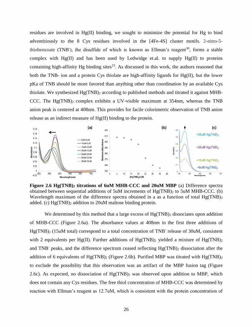
2.2.6 Hg(TNB)2 complex preparation and MHB titration
The Hg(TNB)2 complex was prepared from DTNB and mercuric acetate according to
published methods23 and purified by isocratic gel filtration into 50mM KPi pH 7.4 using a P2
column. Fractions were assessed in the UV-visible range for the presence of a single peak at
354nm and frozen at -20°C until use. Hg(TNB)2 concentration was determined using the
extinction coefficient of 20.8mM-1 at 354nm. The MHB and MBP to be titrated with Hg(TNB)2
were exchanged into 20mM KPi 7.4 by centrifugal ultrafiltration using a 30kDa MWCO. The
protein samples were prepared in a Vacuum Atmospheres anaerobic glove box in quartz cuvettes
and sealed with rubber stoppers. UV-visible measurements were conducted with a Shimadzu
UV-2600 spectrophotometer outside of the anaerobic environment, and Hg(TNB)2 addition to
the sample was made via gas-tight Hamilton syringe through the rubber septum.
2.2.7 Stopped-flow kinetic studies of MHB variants and Hg(TNB)2
MHB samples for stopped flow experiments were prepared anaerobically by buffer
exchange into 20mM KPi pH 7.4. The concentration of all protein samples were adjusted to
25uM, as indicated by a 390nm absorbance of ~0.8. Hg(TNB)2 as prepared in section 2.2.6 was
Page 34
21
diluted to 25uM in the same buffer. This buffer was also used to collect a baseline spectrum prior
to stopped-flow data collection. Rapid kinetic analysis of the MHB-Hg binding interaction was
conducted using an Applied Photophysics stopped-flow instrument housed in a Vacuum
Atmospheres anaerobic glove box with an atmospheric oxygen concentration < 1ppm O2. All
data were collected for 1 second at 20°C in photodiode array (PDA) mode using the 1cm path
length configuration of the instrument. Stopped flow traces were collected in triplicate and fit to
a double exponential model using ProData Viewer software. The deadtime of our instrument has
been characterized as 1.8ms24, so data were fit from 2ms-1s. The MHB-SCC variant was the
exception to this data treatment and could only be fit to a single exponential model. Standard
deviations of triplicate observed rate constant values and amplitude values from the kinetic curve
fits were calculated and plotted as error bars.
2.3 Results and Analysis
2.3.1 SDS-PAGE analysis of HgcB fusion tag screen
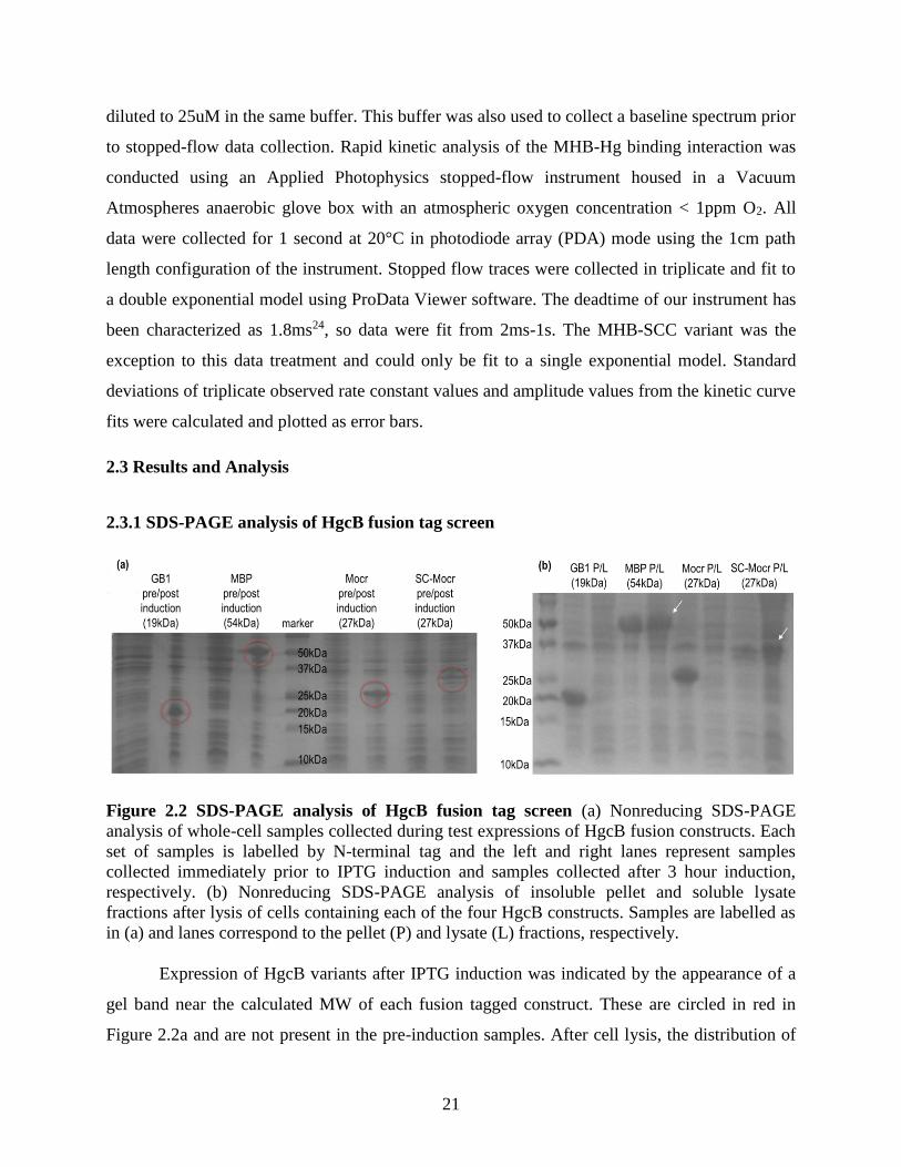
Figure 2.2 SDS-PAGE analysis of HgcB fusion tag screen (a) Nonreducing SDS-PAGE
analysis of whole-cell samples collected during test expressions of HgcB fusion constructs. Each
set of samples is labelled by N-terminal tag and the left and right lanes represent samples
collected immediately prior to IPTG induction and samples collected after 3 hour induction,
respectively. (b) Nonreducing SDS-PAGE analysis of insoluble pellet and soluble lysate
fractions after lysis of cells containing each of the four HgcB constructs. Samples are labelled as
in (a) and lanes correspond to the pellet (P) and lysate (L) fractions, respectively.
Expression of HgcB variants after IPTG induction was indicated by the appearance of a
gel band near the calculated MW of each fusion tagged construct. These are circled in red in
Figure 2.2a and are not present in the pre-induction samples. After cell lysis, the distribution of
Page 35
22
each construct in the insoluble (P) and soluble (L) fractions is shown in Figure 2.2b and
indicated by arrows. From this it can be concluded that GB1-HgcB and Mocr-HgcB are not
soluble after cell lysis under these conditions, and this gel is inconclusive in determining the
fractionation of SC-Mocr-HgcB. MBP-HgcB is soluble after cell lysis, though some remains in
the insoluble fraction.
2.3.2 MHB expression and amylose affinity purification
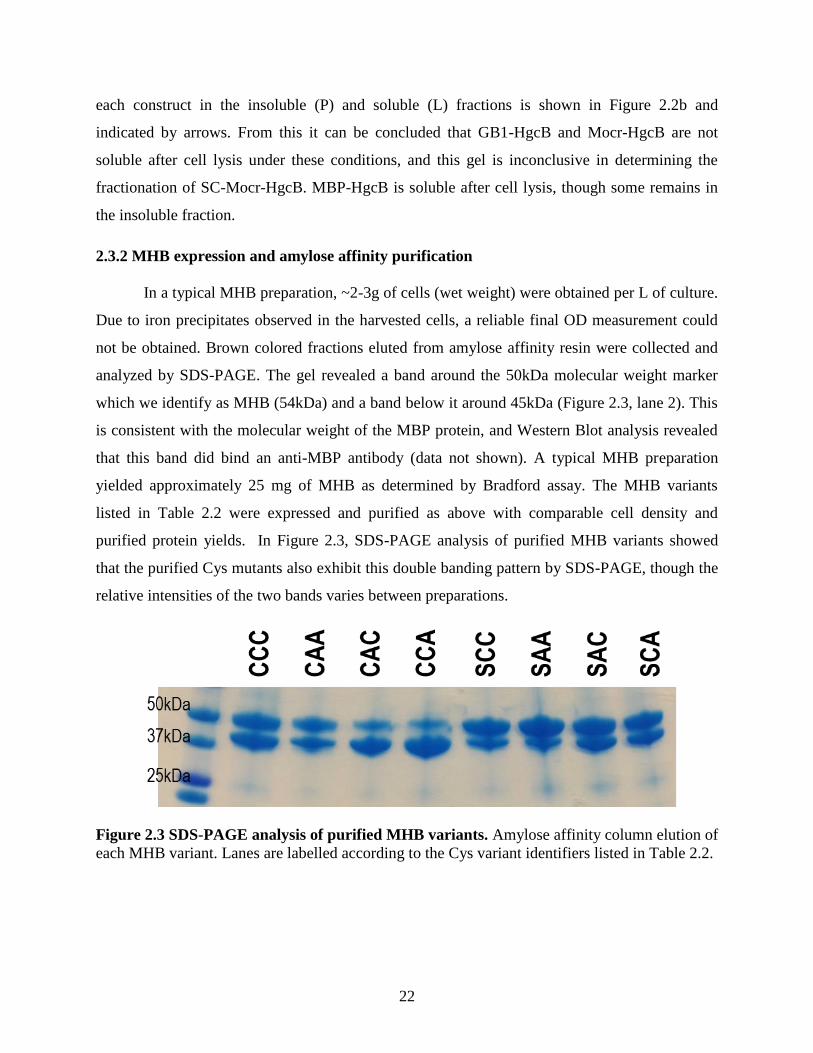
In a typical MHB preparation, ~2-3g of cells (wet weight) were obtained per L of culture.
Due to iron precipitates observed in the harvested cells, a reliable final OD measurement could
not be obtained. Brown colored fractions eluted from amylose affinity resin were collected and
analyzed by SDS-PAGE. The gel revealed a band around the 50kDa molecular weight marker
which we identify as MHB (54kDa) and a band below it around 45kDa (Figure 2.3, lane 2). This
is consistent with the molecular weight of the MBP protein, and Western Blot analysis revealed
that this band did bind an anti-MBP antibody (data not shown). A typical MHB preparation
yielded approximately 25 mg of MHB as determined by Bradford assay. The MHB variants
listed in Table 2.2 were expressed and purified as above with comparable cell density and
purified protein yields. In Figure 2.3, SDS-PAGE analysis of purified MHB variants showed
that the purified Cys mutants also exhibit this double banding pattern by SDS-PAGE, though the
relative intensities of the two bands varies between preparations.
Figure 2.3 SDS-PAGE analysis of purified MHB variants. Amylose affinity column elution of
each MHB variant. Lanes are labelled according to the Cys variant identifiers listed in Table 2.2.
Page 36
23
2.3.3 UV-visible spectroscopy and reductive titration of MHB variants
Reduction of MHB and variants with titanium (III) citrate was monitored by observing
the characteristic decrease in absorbance intensity in the 390nm-420nm region seen in many
[4Fe-4S] proteins25, and is shown in Fig 2.4 below. Sequential additions of titanium (III) citrate
were made until the 415nm absorbance reached a minimum; this required 3-4 equivalents
titanium (III) citrate for all variants. A characteristic reductive titration of the MHB-CCC variant
is shown in Figure 2.4.
Figure 2.4 Titanium (III) citrate reduction of MHB-CCC. 25uM MHB-CCC was titrated with
titanium (III) citrate by sequential additions of 25uM (1 equivalent). These data are
representative of similar titrations of MHB variants with titanium (III) citrate.
As ferredoxins are typically small proteins, the ratio between the 280nm and 390nm
absorbance can generally be used to determine the iron occupancy with good accuracy25. The
280nm aborbance value of MHB, however, is saturated by contributions from the MBP fusion
protein. The iron occupancy of [4Fe-4S] clusters can also be estimated by using the molar
extinction coefficient at of 4mM-1 Fe atoms at 390nm25; these values are calculated in the center
columns of Table 2.3. If the value of Δ415nm is used to calculate protein concentration based on
the value of ε(Δ415nm) = 8.2mM-1 for a protein containing two [4Fe-4S] clusters22, the MHB
concentration values in the leftmost columns of Table 2.3 are in relatively good agreement with
those determined from the 390nm absorbance. Cluster occupancy was calculated by comparing
Page 37
24
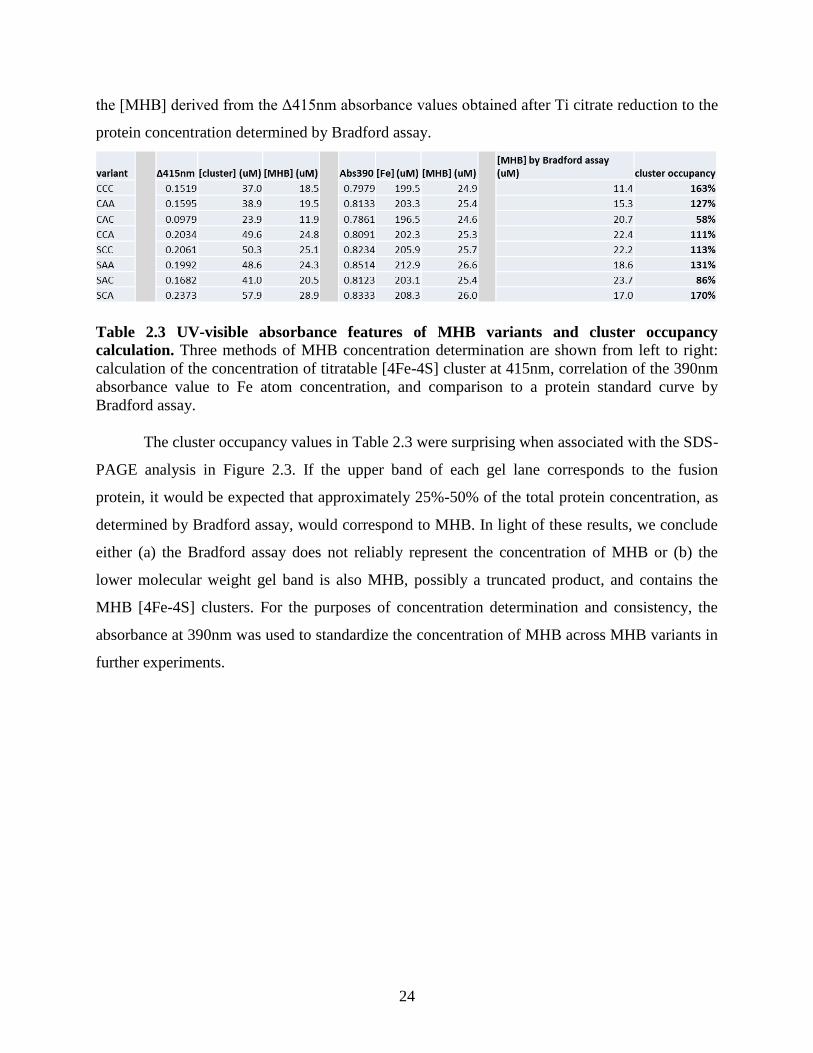
the [MHB] derived from the Δ415nm absorbance values obtained after Ti citrate reduction to the
protein concentration determined by Bradford assay.
Table 2.3 UV-visible absorbance features of MHB variants and cluster occupancy
calculation. Three methods of MHB concentration determination are shown from left to right:
calculation of the concentration of titratable [4Fe-4S] cluster at 415nm, correlation of the 390nm
absorbance value to Fe atom concentration, and comparison to a protein standard curve by
Bradford assay.
The cluster occupancy values in Table 2.3 were surprising when associated with the SDS-
PAGE analysis in Figure 2.3. If the upper band of each gel lane corresponds to the fusion
protein, it would be expected that approximately 25%-50% of the total protein concentration, as
determined by Bradford assay, would correspond to MHB. In light of these results, we conclude
either (a) the Bradford assay does not reliably represent the concentration of MHB or (b) the
lower molecular weight gel band is also MHB, possibly a truncated product, and contains the
MHB [4Fe-4S] clusters. For the purposes of concentration determination and consistency, the
absorbance at 390nm was used to standardize the concentration of MHB across MHB variants in
further experiments.
Page 38
25
2.3.4 MHB-CCC EPR spectrum
Figure 2.5 EPR spectrum of MHB-CCC prepared by reduction with titanium (III) citrate Data collection parameters: 1mW power, 5G amplitude modulation, 4.5x10³ gain, and 9.38 GHz
frequency at 8.6K.
No EPR spectrum was detected in samples prepared by reduction with excess sodium
dithionite (Em ~ -444mV vs. NHE); this may suggest that the MHB [4Fe-4S] clusters are quite
low in potential and require a stronger chemical reductant, such as titanium (III) citrate (Em ~ -
550mV vs. NHE)26. The EPR spectrum of MHB-CCC was obtained after incubation with 10
equivalents of titanium (III) and removal of excess reductant by buffer exchange. The spectrum
collected at 8.6K can be seen in Figure and exhibits the characteristic feature of [4Fe-4S] clusters
centered at g = 1.9427. Another signal centered at g = 2.0 is observed which bears resemblance to
an organic radical species – this can be observed as an artifact of EPR cavity contamination and
we hesitate to assign this feature to MHB.
2.3.5 Hg(TNB)2 titration of MHB-CCC
Addition of Hg(II) species to iron-sulfur proteins is an established method for disruption
of iron-sulfur clusters28 and inorganic Hg(II) exposure in Escherichia coli induces a rapid
increase in intracellular labile iron, thought to occur due to disruption of protein-associated iron-
sulfur clusters29. As we are interested in testing the hypothesis that the 3 MHB conserved Cys
Page 39
26
residues are involved in Hg(II) binding, we sought to minimize the potential for Hg to bind
adventitiously to the 8 Cys residues involved in the [4Fe-4S] cluster motifs. 2-nitro-5-
thiobenzoate (TNB-), the disulfide of which is known as Ellman’s reagent30, forms a stable
complex with Hg(II) and has been used by Ledwidge et.al. to supply Hg(II) to proteins
containing high-affinity Hg binding sites23. As discussed in this work, the authors reasoned that
both the TNB- ion and a protein Cys thiolate are high-affinity ligands for Hg(II), but the lower
pKa of TNB should be more favored than anything other than coordination by an available Cys
thiolate. We synthesized Hg(TNB)2 according to published methods and titrated it against MHB-
CCC. The Hg(TNB)2 complex exhibits a UV-visible maximum at 354nm, whereas the TNB
anion peak is centered at 408nm. This provides for facile colorimetric observation of TNB anion
release as an indirect measure of Hg(II) binding to the protein.
Figure 2.6 Hg(TNB)2 titrations of 6uM MHB-CCC and 20uM MBP (a) Difference spectra
obtained between sequential additions of 5uM increments of Hg(TNB)2 to 5uM MHB-CCC. (b)
Wavelength maximum of the difference spectra obtained in a as a function of total Hg(TNB)2
added. (c) Hg(TNB)2 addition to 20uM maltose binding protein.
We determined by this method that a large excess of Hg(TNB)2 dissociates upon addition
of MHB-CCC (Figure 2.6a). The absorbance values at 408nm in the first three additions of
Hg(TNB)2 (15uM total) correspond to a total concentration of TNB- release of 30uM, consistent
with 2 equivalents per Hg(II). Further additions of Hg(TNB)2 yielded a mixture of Hg(TNB)2
and TNB- peaks, and the difference spectrum ceased reflecting Hg(TNB)2 dissociation after the
addition of 6 equivalents of Hg(TNB)2 (Figure 2.6b). Purified MBP was titrated with Hg(TNB)2
to exclude the possibility that this observation was an artifact of the MBP fusion tag (Figure
2.6c). As expected, no dissociation of Hg(TNB)2 was observed upon addition to MBP, which
does not contain any Cys residues. The free thiol concentration of MHB-CCC was determined by
reaction with Ellman’s reagent as 12.7uM, which is consistent with the protein concentration of
Page 40
27
5uM determined from the [4Fe-4S] cluster absorbance at 390nm. We suggest that the magnitude
of TNB anion release presented here could be explained by the binding of Hg(II) to the MHB
cluster Cys residues and concommittent disruption of the [4Fe-4S] clusters.
2.3.6 Stopped-flow kinetic analysis of Hg binding to MHB variants
Stopped-flow rapid kinetic analysis was used to compare the Hg(TNB)2 dissociation rates
and TNB- release rates of a panel of MHB Cys mutants to determine if differential rates were
observed between the different MHB variants. We hypothesized that Hg(II) binding to an
exposed Cys binding site would occur faster than iron-sulfur cluster displacement and these two
processes could be identified using this technique. The MHB variants in which Cys residues
required for Hg(II) binding are substituted with Ala or Ser would presumably only exhibit the
comparatively slow displacement of the MHB [4Fe-4S] clusters. Data were collected for 1
second upon mixing of 25uM Hg(TNB)2 and 25uM of each MHB variant. Figure 2.7a shows a
representative stopped-flow data set in which the Hg(TNB)2 peak at 354nm disappears with the
evolution of the TNB anion peak at 408nm and panel b overlays the absorbance values at these
wavelengths over a 1 second time scale. These traces fit well to a double exponential kinetic
model and these fits are overlaid with representative traces in Figure 2.7c. The exception to this
data treatment is the MHB-SCC variant, in which the 408nm traces were better fit to a single
exponential kinetic model (though the 354nm trace was still fit to a double exponential), shown
in Figure 2.7d.
Page 41
28
Figure 2.7 Representative stopped-flow absorbance data. (a) Overlay of spectra collected for
1s upon mixing of 25uM Hg(TNB)2 to 25uM MHB-CCC. Arrows indicate the associated
decrease in 354nm absorbance and increase at 408nm. (b) Absorbance vs. time traces at 354nm
(bottom, pink) and 408nm (top, black). (c) Representative double exponential curve fits overlaid
with the corresponding data at 354nm and 408nm. Residuals are plotted across the bottom in
green. (d) Representative single exponential curve fits overlaid with the corresponding MHB-
SCC data at 354nm and 408nm. Residuals are plotted across the bottom in red.
To our surprise, all eight Cys variants induced rapid dissociation of the Hg(TNB)2
complex. The observed rate constants and the corresponding absorbance amplitudes for each
exponential phase are organized by wavelength, with 354nm analysis shown in Figure 2.8 and
408nm analysis in Figure 2.9. Several striking observations can be made from comparisons
between these MHB variants:
Page 42
29
(1) The rapid dissociation of Hg(TNB)2 (Figure 2.8a), as indicated by the disappearance of
the 354nm peak, displays rate constants of ~100s-1 in the CCC, CAC, CCA, and SCC
variants. This rate is lowered by a factor of 2 in the remaining 4 variants. The trend
observed here is a more rapid dissociation of Hg(TNB)2 in variants containing two of the
three relevant Cys residues. We propose that this is consistent with a preferential binding
to these residues.
(2) The TNB- release observed in the 408nm traces of the MHB SCC variant was best fit to a
single exponential model, in contrast to all other data sets (Figure 2.9). The 354nm
double exponential fits for the SCC variant reveal that the absorbance change associated
with the second observed rate constant phase is a small fraction of the total Δ354nm
(approx. 5%, Figure 2.8b & d), indicating that the 354nm data is effectively monophasic
as well. We conclude from this observation that the MHB-SCC variant displays the
simplest Hg(II) binding model in this data set, in which Hg(TNB)2 dissociation occurs at
same rate (~100s-1) of TNB- release, indicative of Hg(II) binding to MHB-SCC at a 1:1
ratio and releasing both equivalents of TNB-. We are unsure why this phenomenon is not
also observed in the wild-type CCC variant.
(3) If some configuration of Cys73, Cys94, and/or Cys95 comprises a Hg(II) binding site, we
would expect the MHB-SAA variant to be deficient in Hg(II) binding and attribute
Hg(TNB)2 dissociation induced by mixing with the SAA variant to the adventitious
binding of Hg(II) to the [4Fe-4S] cluster Cys residues. The SAA variant appears
kinetically equivalent to the CAA variant in these experiments, and both induce
Hg(TNB)2 dissociation at a two-fold slower rate compared to the CCC, CAC, CCA, and
SCC variants containing 2 of the 3 relevant Cys residues (Figure 2.8a). This leads us to
the conclusion that wild-type Hg(II) binding requires Cys94 and/or Cys95.
(4) The total absorbance change at both wavelengths is equivalent between all variants, but
the observed rate constants for the fast and slow phases are not consistent between the
processes observed at 354nm and 408nm. For instance, almost all Hg(TNB)2 dissociation
upon mixing with the MHB-CAA variant occurs primarily in the faster observed phase
with a rate constant of 50s-1 (Figure 2.8a & b). 60% of the total TNB- anion release,
occurs in the slower phase observed in the data collected at 408nm, corresponding to a
rate constant of 40s-1(Figure 2.9c & d). When the concentrations of these species are
Page 43
30
calculated from these absorbance changes using their molar extinction coefficients, there
is good agreement between the 3uM Hg(TNB)2 consumed in this phase and 7uM TNB-
generated. We are unsure, however, of how to assign the more rapid generation of TNB-
at a rate constant of 250s-1 associated with a smaller fraction of Hg(TNB)2 dissociation.
Figure 2.8 Kinetic parameters of 354nm curve fits (a) and (c) show the observed rate
constants for fast (kobs1) and slow (kobs2) exponential phases in the double exponential fit,
respectively. (b) and (d) show the corresponding 354nm absorbance change (amplitude)
represented by each phase.
Page 44
31
Figure 2.9 Kinetic parameters of 408nm curve fits (a) and (c) show the observed rate
constants for fast (kobs1) and slow (kobs2) exponential phases in the double exponential fit,
respectively. (b) and (d) show the corresponding 408nm absorbance change (amplitude)
represented by each phase.
2.4 Discussion
2.4.1 Purification of MHB and Cys variants generates an iron-replete [4Fe-4S] protein
MBP was found to have a positive effect on HgcB expression as a solubilizing affinity
fusion tag and the resulting fusion protein, abbreviated MHB, was expressed with the pRKISC
co-expression system resulting in the purification of iron-replete protein. Expression and
purification of seven MHB variants yielded similar results. Figure 2.3 shows the presence of two
bands in SDS-PAGE analysis of all MHB variants, the ratio of which varies between protein
preparations. The higher molecular weight band is consistent with the weight of the MHB fusion
protein (54kDa) and the lower is consistent with the weight of MBP (43kDa). Both bands were
found to react with a polyclonal MBP antibody (data not shown) and we are unsure whether the
lower band represents MBP not associated with HgcB or a truncated MHB product which retains
the [4Fe-4S] clusters. This complicates the determination of protein concentration by Bradford
assay, so concentrations are instead standardized to the UV-visible features of the [4Fe-4S]
clusters.
Page 45
32
The UV-visible spectra of all MHB variants were similar and used to observe the
reduction of the [4Fe-4S] clusters by titanium (III) citrate. It was found that sodium dithionite
was unable to reduce MHB and 3-4 equivalents of Ti citrate were required to fully reduce both
[4Fe-4S] clusters. The requirement for excess titanium (III) citrate, which has a reported
midpoint redox potential of -550mV at pH 7, as well as the lack of complete reduction observed
in the presence of dithionite may indicate that the average potential of the MHB [4Fe-4S]
clusters may be quite low. The EPR spectrum of MHB-CCC is presented in Figure 2.5 and
exhibits the characteristic S = ½ axial feature of [4Fe-4S] clusters centered at g = 1.9427. We are
currently not assigning the g = 2.0 feature to the MHB protein and working to determine whether
this signal is caused by organic radical contamination. Overall, MHB appears to exhibit standard
characteristics of ferredoxin-like proteins containing 2 [4Fe-4S] motifs.
2.4.2 Hg(TNB)2 addition to MHB results in superstoichiometric release of TNB-
A colorimetric Hg(TNB)2 complex was used to titrate MHB and probe its Hg(II) binding
characteristics. This complex was used because the affinity of Hg(II) for TNB- complexation
should only be bested by Cys thiolate coordination at neutral pH based on their relative pKa
values23, so its colorimetric dissociation should indicate the stoichiometry of Hg(II) binding to
MHB. This experiment indicated a release of TNB- far in excess of the protein concentration –
this is not unsurprising considering that MHB contains 11 Cys residues and Hg(II) addition has
long been used to disrupt iron-sulfur clusters28,31. These data, in the context of relative binding
affinities between MHB mutants lacking different combinations of the Cys73/Cys94/Cys95
residues implicated in in vivo functionality13, indicate that titration of a triple mutant lacking all
three cysteines will still likely induce TNB- release, as it still contains 8 Cys residues involved in
the [4Fe-4S] cluster motifs. This result presented a challenge to determination of the Hg(II)
binding characteristics of MHB. Our goals were to (a) determine the Hg binding stoichiometry of
HgcB and (b) identify the Cys residues required for retain that stoichiometry, but the indirect
quantification of Hg(II) transfer by TNB- release proved too ambiguous to determine this.
Page 46
33
2.4.3 Stopped-flow kinetic comparison between Hg(II) binding rates of MHB variants is
consistent with observed in vivo requirements for conserved Cys residues
We then turned to stopped-flow experiments with the hypothesis that Hg(II) binding to
the predicted Hg binding site would occur rapidly, whereas disruption of the protein [4Fe-4S]
clusters would indicate a slower observed rate constant. We hypothesized that the Hg binding
stoichiometry could be calculated from the amplitude of the predicted fast phase, and the
eventual disruption of the [4Fe-4S] clusters could be deconvoluted from the Hg binding event,
allowing side-by-side comparison of MHB variants with varying configurations of Cys residues.
The stopped-flow data obtained by stoichiometric mixing of Hg(TNB)2 and 8 MHB Cys variants
did not indicate such a straightforward assignation of kinetic phases.
Hg(II) has a strong preference for linear thiolate coordination environments, and its
ligand exchange reactions have been shown to occur via transient 3-coordinate thiolate
complexes rather than through initial dissociation of one of the two thiolate ligands32. We
suggest this explains the observation that MHB variants containing at least two of the conserved
Cys residues (CCC, CAC, CCA, and SCC) exhibit the fastest rates (~100s-1 in both the 354nm
and 408nm fits) of Hg(TNB)2 decomposition, consistent with the rapid, sequential displacement
of both TNB- ligands by MHB Cys residues. In the SCC variant, this was the only phase
observed and the rate constant was equivalent to the observed rate of TNB- release, indicating a
simple stoichiometric binding interaction. Vicinal CC motifs are known to select for Hg(II) with
high affinity and preference over other thiophilic metal ions, so this is unsurprising10; the unusual
result by comparison is that the observed rate constants for the CCC construct do not follow the
same trends.
The CCC (wild-type) MHB construct displays two phases in its decay of Hg(TNB)2, of
which the slower phase comprises ~60% of the total Δ354nm (Figure 2.8). Taken with the simple
binding of Hg(II) to the SCC variant, this seems to indicate that Hg(II) binds quickly and rapidly
to the C-terminal Cys residues, but some other process follows in the case of the CCC mutant.
We are unsure what this process may be. The mercuric ion reductase, MerA, binds Hg(II) via a
series of Cys residues to ultimately deliver it to the enzyme catalytic site for reduction by
NADPH11. Intermediate steps in this process rely on the presence of an exogenous thiolate ligand
to the Hg(II) to prevent the formation of an inactive complex between two nearby Cys residues33.
Page 47
34
It is possible that Cys73 is the ultimate destination of the Hg(II) ion but it is first bound by the C-
terminal Cys residues 94 and 95. The transfer of Hg(II) from Cys94/Cys95 to Cys73 could drive
an equilibrium in which the Hg(II) is not stably bound to two Cys residues, such as is observed
in the SCC variant, and eventually dissociates. This would explain both (1) the second phase
observed in the CCC results compared to the SCC results and (2) the comparatively lower rates
of Hg(TNB)2 dissociation by the CAA and SAA variants. The conserved Cys93 residue near the
corrinoid active site in HgcA is an interesting feature to speculate on in this regard as well, as it
may serve some role in Hg(II) transfer to the active site rather than as a ligand to the cofactor.
The CAA and SAA variants are kinetically equivalent in this experiment, with slow rates
of Hg(TNB)2 dissociation, but comparatively high rates of TNB- release in both observed phases.
We interpret these data as indicative of adventitious Hg binding to the cluster Cys residues.
Displacement of the cluster Fe atoms would be slower than binding to exposed C-terminal Cys
residues, consistent with the slower observed rates of Hg(TNB)2 decay. A chain reaction could
be envisioned, depending on the solvent accessibility of the clusters, in which several Hg atoms
adventitiously exchange one TNB- ligand for a nonspecific protein Cys and lead to an eventual
collapse of the [4Fe-4S] cluster and sudden availability of Cys residues for binding and release
of any TNB- remaining associated with Hg(II). This model is speculative, but such a mechanism
could be reconciled with the observed rapid rates for TNB- release observed in the CAA and
SAA variants, as well as the elevated rates of TNB- release by the SAC and SCA variants.
Though the TNB- and Cys thiolates in this experiment are sufficiently low in formal
redox potential to reduce the Hg(II) ion, we do not believe that this contributes to the observed
reactions under these conditions for two reasons: (1) the association constants for thiolate
coordination of Hg(II) make complexation far more favorable than electron transfer34, and (2)
involvement of the TNB- in thiol/disulfide redox chemistry would be reflected in the UV-visible
spectrum of this experiment. For example, a scenario in which one equivalent of TNB- is
exchanged for one protein Cys residue, followed by reduction of Hg(II) and disulfide bond
formation, would reflect only one equivalent of TNB- released per Hg(TNB)2, rather than the two
equivalents observed in the Δ354nm and Δ408nm values. The low-potential [4Fe-4S] clusters
seem more likely to be involved in Hg(II) reduction – this experiment, however, was conducted
with as-purified oxidized clusters. No reductant is present in the system which could reduce the
clusters for subsequent electron transfer to Hg(II), so this seems an unlikely explanation for the
Page 48
35
kinetic traces observed. It has been proposed that a role for HgcB in electron transfer to Hg(II),
rather than the HgcA corrinoid, could allow a carbocation mechanism to proceed in agreement
with characterized cobalamin-dependent methylases, generating the observed MeHg+. The
Co(III)-C bond, however, is highly stable and only activated for carbocation transfer by
nucleophilic attack of a substrate such as homocysteine35 or the Ni-cluster of acetyl-CoA
synthase36 – Hg(0) is an inert metal and thus unlikely to induce the same chemistry.
2.4.4 Conclusions
This work has established a method for facile expression of HgcB as a MBP fusion
protein in E. coli. UV-visible and EPR features of the purified protein are characteristic of [4Fe-
4S] ferredoxins and the cluster UV-visible absorbance features can be titrated by addition of the
low-potential chemical reductant, titanium (III) citrate. The requirement for titanium (III) citrate
reduction over that of sodium dithionite indicates that the HgcB [4Fe-4S] clusters likely exhibit
low midpoint redox potentials, which agrees with their putative role in 2 electron reduction of a
corrinoid species. The consistency between the CCC (wild-type) protein reductive titration and
the MHB Cys variants indicates that site-directed mutagenesis of these Cys residues does not
adversely affect the incorporation or stability of the [4Fe-4S] clusters.
A bis-2-nitro-5-thiobenzoate Hg(II) complex (Hg(TNB)2) was used as a colorimetric
reporter of Hg(II) binding to MHB. Equilibrium titrations showed TNB- release in great excess
of the available protein thiols hypothesized to bind Hg(II); we interpreted this result as indicative
of adventitious Hg(II) binding to the Cys residues involved in the [4Fe-4S] motifs, thus Hg(II)
binding stoichiometry could not be obtained. The observation of this reaction by stopped-flow
rapid kinetics indicated rapid dissociation of Hg(TNB)2 even in the absence of all three
conserved Cys residues – this was unexpected and likely can also be attributed to nonspecific
binding of Hg(II) to the cluster Cys residues. It can be concluded, however, that MHB variants
containing two of the three conserved Cys residues, Cys73/Cys94/Cys95, induced the fastest
rates of Hg(TNB)2 dissociation, consistent with the Cys requirements for in vivo Hg methylation
activity observed by in vivo mutagenesis experiments13.
2.5 References for Chapter 2
(1) Parks, J. M.; Johs, A.; Podar, M.; Bridou, R.; Hurt Jr., R. A.; Smith, S. D.; Tomanicek, S. J.; Qian, Y.;
Brown, S. D.; Brandt, C. C.; et al. The Genetic Basis for Bacterial Mercury Methylation. Science. 2013, 339
(March), 1332–1336.
Page 49
36
(2) Harris, R. C.; Rudd, J. W. M.; Amyot, M.; Babiarz, C. L.; Beaty, K. G.; Blanchfield, P. J.; Bodaly, R. A.;
Branfireun, B. A.; Gilmour, C. C.; Graydon, J. A.; et al. Whole-Ecosystem Study Shows Rapid Fish-
Mercury Response to Changes in Mercury Deposition. Proc. Natl. Acad. Sci. 2007, 104 (42), 16586–16591.
(3) Clarkson, T. W. Critical Reviews in Toxicology The Toxicology of Mercury and Its Chemical Compounds.
Crit. Rev. Toxicol. 2006, 36 (8), 609–662.
(4) Matthews, R. G.; Koutmos, M.; Datta, S. Cobalamin-Dependent and Cobamide-Dependent
Methyltransferases. Curr. Opin. Struct. Biol. 2008, 18 (6), 658–666.
(5) Koay, M. S.; Antonkine, M. L.; Gärtner, W.; Lubitz, W. Modelling Low-Potential [Fe4S4] Clusters in
Proteins. Chem. Biodivers. 2008, 5 (8), 1571–1587.
(6) Banerjee, R. V.; Matthews, R. G.; Harder, S. R.; Ragsdale, S. W. Mechanism of Reductive Activation of
Cobalamin-Dependent Methionine Synthase: An Electron Paramagnetic Resonance Spectroelectrochemical
Study. Biochemistry 1990, 29 (5), 1129–1135.
(7) Harder, S. R.; Lu, W. P.; Feinberg, B. A.; Ragsdale, S. W. Spectroelectrochemical Studies of the
Corrinoid/Iron-Sulfur Protein Involved in Acetyl Coenzyme A Synthesis by Clostridium Thermoaceticum.
Biochemistry 1989, 28 (23), 9080–9087.
(8) Koutmos, M.; Datta, S.; Pattridge, K. a; Smith, J. L.; Matthews, R. G. Insights into the Reactivation of
Cobalamin-Dependent Methionine Synthase. Proc. Natl. Acad. Sci. U. S. A. 2009, 106 (44), 18527–18532.
(9) Nakamura, M.; Saeki, K.; Takahashi, Y. Hyperproduction of Recombinant Ferredoxins in Escherichia Coli
by Coexpression of the ORF1-ORF2-IscS-IscU-IscA-HscB-HscA-Fdx-ORF3 Gene Cluster. J Biochem
1999, 126, 10–18.
(10) DeSilva, T. M.; Veglia, G.; Porcelli, F.; Prantner, A. M.; Opella, S. J. Selectivity in Heavy Metal-Binding to
Peptides and Proteins. Biopolymers 2002, 64 (4), 189–197.
(11) Barkay, T.; Miller, S. M.; Summers, A. O. Bacterial Mercury Resistance from Atoms to Ecosystems. FEMS
Microbiol. Rev. 2003, 27 (2–3), 355–384.
(12) Wang, D.; Huang, S.; Liu, P.; Liu, X.; He, Y.; Chen, W.; Hu, Q.; Wei, T.; Gan, J.; Ma, J.; et al. Structural
Analysis of the Hg(II)-Regulatory Protein Tn501 MerR from Pseudomonas Aeruginosa. Sci. Rep. 2016, 6
(August), 1–9.
(13) Smith, S. D.; Bridou, R.; Johs, A.; Parks, J. M.; Elias, D. A.; Hurt, R. A.; Brown, S. D.; Podar, M.; Wall, J.
D. Site-Directed Mutagenesis of HgcA and HgcB Reveals Amino Acid Residues Important for Mercury
Methylation. Appl. Environ. Microbiol. 2015, 81 (9), 3205–3217.
(14) Zehnder, A. J. B.; Wuhrmann, K. Titanium (III) Citrate as a Nontoxic Oxidation-Reduction Buffering
System for the Culture of Obligate Anaerobes Author ( s ): Alexander J . B . Zehnder and Karl Wuhrmann
Source : Science , New Series , Vol . 194 , No . 4270 ( Dec . 10 , 1976 ), Pp . 116. Science (80-. ). 1976, 194
(4270), 1165–1166.
(15) Cheng, Y.; Patel, D. J. An Efficient System for Small Protein Expression and Refolding. Biochem. Biophys.
Res. Commun. 2004, 317 (2), 401–405.
(16) Kapust, R. B.; Waugh, D. S. Escherichia Coli Maltose-Binding Protein Is Uncommonly Effective at
Promoting the Solubility of Polypeptides to Which It Is Fused. Protein Sci. 1999, 8 (8), 1668–1674.
(17) DelProposto, J.; Majmudar, C. Y.; Smith, J. L.; Brown, W. C. Mocr: A Novel Fusion Tag for Enhancing
Solubility That Is Compatible with Structural Biology Applications. Protein Expr. Purif. 2009, 63 (1), 40–
49.
(18) Eschenfeldt, W. H.; Stols, L.; Millard, C. S.; Joachimiak, A.; Donnelly, I. A Family of LIC Vectors for
High-Throughput Cloning and Purification of Proteins. 2009, 498, 105–115.
(19) Jeoung, J.-H.; Dobbek, H. Carbon Dioxide Activation at the Ni,Fe-Cluster of Anaerobic Carbon Monoxide
Dehydrogenase. Science. 2007, 317, 490–494.
(20) Lanz, N. D.; Grove, T. L.; Gogonea, C. B.; Lee, K. H.; Krebs, C.; Booker, S. J. RlmN and AtsB as Models
for the Overproduction and Characterization of Radical SAM Proteins, 1st ed.; Elsevier Inc., 2012; Vol.
516.
(21) Bradford, M. M. A Rapid and Sensitive Method for the Quantitation of Microgram Quantities of Protein
Utilizing the Principle of Protein-Dye Binding. Anal. Biochem. 1976, 72 (1–2), 248–254.
(22) Boll, M.; Fuchs, G. Identification and Characterization of the Natural Electron Donor Ferredoxin and of
FAD as a Possible Prosthetic Group of Benzoyl-CoA Reductase (Dearomatizing), a Key Enzyme of
Anaerobic Aromatic Metabolism. Eur. J. Biochem. 1998, 251 (3), 946–954.
(23) Ledwidge, R.; Patel, B.; Dong, A.; Fiedler, D.; Falkowski, M.; Zelikova, J.; Summers, A. O.; Pai, E. F.;
Miller, S. M. NmerA, the Metal Binding Domain of Mercuric Ion Reductase, Removes Hg 2+ from
Proteins, Delivers It to the Catalytic Core, and Protects Cells under Glutathione-Depleted Conditions.
Page 50
37
Biochemistry 2005, 44 (34), 11402–11416.
(24) Carter, E. L.; Ramirez, Y.; Ragsdale, S. W. The Heme-Regulatory Motif of Nuclear Receptor Rev-Erbβ Is a
Key Mediator of Heme and Redox Signaling in Circadian Rhythm Maintenance and Metabolism. J. Biol.
Chem. 2017, 292 (27), 11280–11299.
(25) Sweeney, W. V; Rabinowitz, J. C. PROTEINS CONTAINING 4Fe-4S CLUSTERS: AN OVERVIEW.
Annu. Rev. Biochem. 1980, 49, 139–161.
(26) Seefeldt, L. C.; Ensign, S. A. A Continuous, Spectrophotometric Activity Assay for Nitrogenase Using the
Reductant Titanium(III) Citrate. Anal. Biochem. 1994, 221 (2), 379–386.
(27) Hagen, W. R. EPR SPECTROSCOPY OF IRON-SULFUR PROTEINS. In Advance in Inorganic
Chemistry; 1992; Vol. 38, pp 165–222.
(28) Lovenberg, W.; Buchanon, B. B.; Rabinowitz, J. C. Studies on the Chemical Nature of Clostridial
Ferredoxin. J. Biol. Chem. 1963, 238 (12), 3899–3913.
(29) Lavoie, S. P.; Mapolelo, D. T.; Cowart, D. M.; Polacco, B. J.; Johnson, M. K.; Scott, R. A.; Miller, S. M.;
Summers, A. O. Organic and Inorganic Mercurials Have Distinct Effects on Cellular Thiols, Metal
Homeostasis, and Fe-Binding Proteins in Escherichia Coli. J. Biol. Inorg. Chem. 2015, 20 (8), 1239–1251.
(30) Ellman, G. L. Tissue Sulfhydryl Groups. Arch. Biochem. Biophys. 1959, 82, 70–77.
(31) Ragsdale, S. W.; Lindahl, P. A.; Munck, E. Mossbauer, EPR, and Optical Studies of the Corrinoid/Iron-
Sulfur Protein Involved in the Synthesis of Acetyl Coenzyme A by Clostridium Thermoaceticum. J. Biol.
Chem. 1987, 262 (29), 14289–14297.
(32) Cheesman, B. V.; Arnold, A. P.; Rabenstein, D. L. Nuclear Magnetic Resonance Studies of the Solution
Chemistry of Metal Complexes. XV. Trimethyllead Complexes of Inorganic Ligands. J. Am. Chem. Soc.
1988, 110, 6359–6364.
(33) Miller, S. M.; Ballou, D. P.; Massey, V.; William, C. H. Two-Electron Reduced Mercuric Reductase Binds
Hg(II) to the Active Site Dithiol but Does Not Catalyze Hg(II) Reduction. J. Biol. Chem. 1986, 261 (18),
8081–8084.
(34) Stricks, W.; Kolthoff, I. M. Reactions between Mercuric Mercury and Cysteine and Glutathione. Apparent
Dissociation Constants, Heats and Entropies of Formation of Various Forms of Mercuric Mercapto-Cysteine
and -Glutathione. J. Am. Chem. Soc. 1953, 75 (22), 5673–5681.
(35) Gruber, K.; Kratky, C.; Messerschmidt, A.; Huber, R.; Poulos, T.; Wieghardt, K.; Gruber, K.; Kratky, C.
Cobalamin-Dependent Methionine Synthase. 2001.
(36) Lu, W. P.; Harder, S. R.; Ragsdale, S. W. Controlled Potential Enzymology of Methyl Transfer Reactions
Involved in Acetyl-CoA Synthesis by CO Dehydrogenase and the Corrinoid/Iron-Sulfur Protein from
Clostridium Thermoaceticum. J. Biol. Chem. 1990, 265 (6), 3124–3133.
Page 51
38
Chapter 3 HgcA
3.1 Introduction
As discussed in Chapter 1 of this work, corrinoid-dependent proteins catalyze a variety of
reactions, including methyl transfer1. Their functions and reactivity are tuned by the protein
environment around the cofactor and protein ligand coordation (or lack of). Corrinoid-dependent
methyltransferases typically bind corrinoid cofactors in a “base-off” mode, in which the
dimethylbenzimidazole (DMB), or base, moiety coordinating the alpha axial face of the central
Co atom dissociates and binds within a protein Rossmann fold2. Methionine synthase (MetH)
provides a His ligand to the cofactor3, but the corrinoid iron-sulfur protein (CFeSP) does not4.
BtuM has been recently shown to coordinate one axial position with a Cys residue, the first
example of such a coordination within a protein5. A light-dependent transcriptional regulator,
CarH, binds in a “base-off, bis-His” motif, in which two His ligands coordinate both axial
positions6. The crystal structures of these cobalamin binding modes are depicted in Figure 2.1
below.
Page 52
39
Figure 3.1 Crystal structures of observed “base-off” cobalamin protein binding modes (a)
“base-off, His-on” methionine synthase (MetH) (b) “base-off” corrinoid iron-sulfur protein
(CFeSP) (c) “base-off, Cys-on” vitamin B12 membrane transporter (BtuM) (d) “base-off, bis-
His” light-dependent transcriptional regulator (CarH). The proteins are represented as cartoons
while the cobalamin and relevant protein ligands are represented as sticks and colored by
element. Rendered with Pymol from Protein Data Bank entries (a) 1BMT, (b) 4DJD, (c) 6FFV,
and (d) 58CF.
HgcA is a 40kDa corrinoid protein with a C-terminal transmembrane (TM) domain which
has been implicated as the mercury methylase in methylmercury biosynthesis7. Based on
sequence similarity to the CFeSP, homology modelling suggests that a Cys residue at position 93
could coordinate to the “base-off” cofactor. We hypothesize this coordination would have a
dramatic effect on the electronic environment and the mechanism of Co-C bond cleavage in the
methylation reaction8, potentially rationalizing the proposed transfer mechanism in which a
methyl anion, in a departure from characterized Cbl-dependent methyltransferases, is transferred
to Hg(II)7. Cys93 has been shown to be critical to Hg methylation in Desulfovibrio desulfuricans
ND132 in an in vivo site-directed mutagenesis study9. This work identified several other
requirements for native functional competency of the hgcAB system, including the HgcA TM
Page 53
40
domain – deletion of this region of the hgcA gene eliminated in vivo Hg methylation, indicating
that it is required for MeHg biosynthesis in some capacity. Our current functional understanding
of HgcA, therefore, lacks direct functional assignment of key features which could be supplied
by in vitro study.
HgcA is present at low levels in Desulfovibrio desulfuricans ND13210 and Geobacter
sulfurreducens PCA11, thus it would be impractical to isolate quantities of protein required for in
vitro characterization from these native sources. Heterologous expression of cobalamin-binding
proteins, however, can present a significant technical challenge. Commonly used Escherichia
coli derivative strains (BL21, C41, etc.) do not contain de novo corrinoid biosynthesis pathways,
nor do they have a significant requirement for cobalamin under standard laboratory growth
conditions12. Cobalamin uptake in Escherichia coli is tightly regulated by the binding of
cobalamin to the mRNA encoding the cobalamin uptake gene, btuB, ensuring its transcription is
not initiated when the cell’s need for cobalamin is met13. This presents a barrier to heterologous
expression of enzymes which require corrinoid cofactors for folding, stability, and/or solubility,
as this regulation ensures there is not sufficient intracellular cobalamin available for binding.
In this work, two approaches were taken to heterologous production of cobalamin-replete
HgcA. We first generated a panel of N- and C-terminally truncated variants of the HgcA soluble
domain (lacking the predicted TM helices beginning approximately at position 175) and
attempted cofactor reconstitution. Several variants were found to have enhanced solubility in
comparison to HgcA-1-166, the construct purified as reported in Ref. 7, but none could be
purified containing cobalamin or reconstituted with cofactor. The soluble truncated HgcA
constructs exhibit UV-visible spectral shifts when added to Cbl, but cofactor reconstitution was
assessed by electron paramagnetic resonance (EPR) spectroscopy to more definitively assess the
Cbl coordination environment. “Base-on” cobalamin exhibits characteristic EPR spectral features
arising from the interaction with the DMB nitrogen; the nuclear spin of the coordinating nitrogen
atom causes superhyperfine splitting of EPR spectral features absent in the “base-off”
configuration. EPR analysis of all constructs with which reconstitution was attempted indicated
that the cofactor was still “base-on”.
We reasoned that this inability to bind Cbl could arise from the removal of the TM
domain. TM proteins present their own hurdles to heterologous overexpression, but advances in
TM protein expression and extraction are constantly being made14–16 and we sought to test the
Page 54
41
hypothesis that the full-length (FL) protein is required to bind cofactor. In early 2018, a method
for heterologous expression of cobalamin-binding proteins was published involving co-
expression with a plasmid copy of the endogenous E. coli btu operon17. This operon is
responsible for cobalamin uptake and encodes the outer membrane transporter BtuB, periplasmic
chaperone BtuF, as well as BtuC and BtuD which are responsible for inner membrane trafficking
into the cytosol. The TonB-dependent cobalamin transporter, btuB, has been show to contain a
premature stop codon in BL21-derived laboratory research strains18, rendering it dysfunctional,
so the endogenous btu machinery cannot easily be exploited for the purposes of increases cellular
Cbl uptake. By utilizing an inducible copy of the btu operon, we reasoned that an increase in the
intracellular cobalamin pool could increase the likelihood of holo-FL-HgcA expression. Both
FL-HgcA and HgcA-1-175 were co-expressed and purified from C41(DE3) cells in the presence
of membrane-solubilizing detergents. The full-length and truncated proteins purified were
characterized by SDS-PAGE analysis and UV-visible spectroscopy was used to compare the
preliminary electronic features of the holo protein to known corrinoid proteins.
3.2 Materials and Methods
3.2.1 Materials
All chemicals were purchased from Sigma-Aldrich (St. Louis, MO) unless otherwise
stated. All custom synthesized oligonucleotide primers were ordered from Integrated DNA
Technologies (Coralville, Iowa). The FL-HgcA gene was custom synthesized into a pJ411 vector
by DNA2.0 (Newark, CA). Pfu Ultra polymerase was purchased from Agilent (Santa Clara, CA).
All restriction enzymes, T4 DNA ligase, and amylose affinity resin were procured from New
England Biolabs (Ipswich, MA). TOP10 and BL21(DE3) chemically competent cells were
purchased from Invitrogen/Life Technologies (Carlsbad, CA), and C41(DE3) chemically
competent cells were purchased from Lucigen (Middleton, WI). cOmplete EDTA-free protease
inhibitor tablets were acquired from Roche (Indianapolis, IN). The pET-Duet-1 vector was
purchased from Novagen (Madison, WI) and the pBAD42-BtuCEDFB plasmid was a gift from
the laboratory of Prof. Squire Booker, Pennsylvania State University. Ni-NTA resin and all DNA
purification kits used between cloning steps (QIAquick gel extraction kit, QIAprep spin miniprep
kit, QIAquick PCR purification kit) are from Qiagen (Valencia, CA).
Page 55
42
3.2.2 HgcA 1-166: EPR spectroscopy
HgcA-1-166 samples for EPR spectroscopic analysis were prepared and supplied by Dr.
Stephen Tomanicek and Dr. Alexander Johs (Oak Ridge National Laboratory). The protein was
purified as described in Ref. 7, added to a stoichiometric amount of aquacobalamin, and
exchanged into 25mM Na HEPES pH 7.5 at a concentration of 90uM. Reduction of Cbl was
performed by the addition of 4 equivalents of sodium ascorbate to generate the Co(II) oxidation
state. Two Cbl control samples were prepared by the same method: a “base-on” and a “base-off”
sample, the latter of which contained 0.1M HCl.
Maltose binding protein (MBP) fusion constructs of HgcA-1-166 were also prepared and
analyzed by EPR. In addition to the wild-type 1-166 sequence, a construct was generated in
which two non-conserved Cys residues found in the HgcA sequence at positions 47 and 142
(SCS) were substituted with serine. These samples were prepared in the same buffer system as
above, but Cbl reconstitution was carried out by addition of stoichiometric methylcobalamin
(MeCbl). MeCbl was photolyzed by exposure to ambient light to generate the Co(II) oxidation
state and a control MeCbl sample was prepared by the same method. These samples were found
to be oxidized after shipping and reduced with two equivalents of titanium (III) citrate.
3.2.3 HgcA truncated constructs: design and ligation-independent cloning of HgcA variants
Figure 3.2 Start and end positions of HgcA truncated constructs. Bolded residues (Pro43 and
Ala166) indicate the start and end positions of HgcA homology to CFeSP.
Page 56
43
HgcA constructs were amplified from a commercially custom-synthesized pJ411 plasmid
containing the FL-HgcA gene from Desulfovibrio desulfuricans ND132. This construct was
codon-optimized for expression in Escherichia coli. Ligation-independent cloning (LIC)
procedures were used to amplify HgcA variants with different combinations start and end
positions and insert these constructs into the pMCSG7 vector and a modified pMCSG9 vectors19.
These vectors contain N-terminal sequences encoding His6 and His6-MBP tags, respectively.
Site-directed mutagenesis according to the Agilent QuikChange Site Directed Mutagenesis
protocol was used to remove the His6 tag from pMCSG9 prior to its assembly with the HgcA
constructs, with the resulting vector designated pMCSG9-NH. Sequences were verified by
Sanger sequencing using the T7 terminator primer (University of Michigan DNA Sequencing
Core).
3.2.4 HgcA truncated constructs: expression/solubility tests and purification
His6-HgcA constructs contained in the pMCSG7 vector were transformed into
BL21(DE3) cells. 50mL cultures containing LB medium and 100ug/mL ampicillin were
inoculated from glycerol stocks and grown at 37ºC with 225rpm shaking. HgcA expression was
induced for 3 hours at 37ºC with 1mM IPTG at OD600 ~0.7. Gel samples were prepared from
these cultures before and 3 hours after induction by resuspending a cell pellet prepared by
centrifugation (5 min, 5,000xg) in 100uL 1X LDS loading buffer. These gel samples were
analyzed by SDS-PAGE using 12.5% polyacrylamide gels. The cultures were harvested by
centrifugation at 5,000xg for 15 minutes and stored at -20ºC. To test solubility after cell lysis,
each frozen cell pellet was resuspended in 5mL lysis buffer (50mM Tris pH 7.8, 100mM NaCl,
5mM DTT, 1 Complete EDTA-free protease tablet per 50mL) and sonicated at 4ºC. A 100uL
aliquot of each lysate was centrifuged in a benchtop microcentrifuge at 17,000xg for 10 minutes,
and samples of the soluble and insoluble fractions were prepared for SDS-PAGE analysis.
MBP-HgcA constructs 39-166, 43-166, and 33-175 were expressed as above in 1L
cultures, but induction was carried out overnight (18 hours) at 15ºC. These cell pellets were
harvested and frozen at -20ºC prior to lysis and purification. Cell lysis and amylose affinity
purification were conducted under anoxic conditions (< 1ppm oxygen) in a Vacuum
Atmospheres anaerobic glove box. Pellets were thawed in 50mL lysis buffer containing 50mM
Na HEPES pH 7.5, 100mM NaCl, 5mM DTT, and 1 protease inhibitor tablet. The resuspended
Page 57
44
cells were lysed by sonication at 4ºC and the lysate cleared by centrifugation at 100,000xg. The
soluble lysate was immediately applied to an amylose affinity column equilibrated with 5
column volumes (CV) of 25mM Na HEPES pH 7.5 and 5mM DTT. 2.5CV of this buffer was
used to wash the column and MHB was eluted with the same buffer containing 20mM maltose.
The purified protein was analyzed for homogenaity by SDS-PAGE.
3.2.5 HgcA truncated constructs: cobalamin reconstitution analysis by UV-visible
spectroscopy
All sample handling and data collection was conducted inside an anaerobic glove box.
Purified MBP-HgcA constructs were exchanged into 50mM Na HEPES pH 7.5 in centrifugal
concentrators with a 30kDa molecular weight cut off (MWCO) membrane for cofactor addition.
Aquacobalamin was added to purified MBP-HgcA constructs at a 0.9:1 ratio, to ensure spectral
shifts observed did not arise from unbound cofactor. Protein concentrations for this purpose were
determined by Bradford assay20. UV-visible spectra were collected using an Agilent/HP 8453
UV-visible spectrophotometer.
3.2.6 FL-HgcA: restriction digest cloning into pET-Duet-1
The FL-HgcA coding region and a truncated HgcA-1-175 construct were amplified by
the polymerase chain reaction (PCR) from a custom synthesized, codon-optimized FL-HgcA
vector (pFL-HgcA) with overhangs containing the BamHI and HindIII restriction enzyme sites.
The PCR products and pET-Duet-1 vector were digested with both restriction enzymes, mixed
and ligated using T4 DNA ligase, and transformed into TOP10 cells, generating the Duet-FL-
HgcA and Duet-HgcA-1-175 vectors. Sequences were verified by Sanger sequencing (University
of Michigan DNA Sequencing Core). Each HgcA plasmid was co-transformed into C41(DE3)
cells with pBAD42-BtuCEDFB and plated on LB agar selection plates containing 100ug/mL
ampicillin and 25ug/mL spectinomycin (50% working concentration).
3.2.7 FL-HgcA: co-expression with pBAD42-btuCEDFB and Ni-NTA affinity purification
Overnight starter cultures of C41(DE3) co-expression strains containing pBAD42-
btuCEDFB and either Duet-FL-HgcA (pFL/pBtu) or Duet-HgcA-1-175 (p175/pBtu) were
inoculated from single colonies in LB medium with 100ug/mL ampicillin and 50ug/mL
spectinomycin. 100mL of the M9-ethanolamine media recipe described in Blaszczyk et.al.20 was
Page 58
45
prepared and inoculated with 0.75mL of the LB starter culture. HgcA expression was induced at
OD600 ~0.7 overnight (16-18 hours) at 20ºC with 0.1 mM IPTG. Cells were harvested by
centrifugation at 5,000xg and frozen at -20ºC. All purification steps were conducted in a cold
room at 4ºC. Cell pellets were resuspended in 5mL lysis buffer containing 50mM KPi pH 7.4,
100mM sodium chloride, 10% glycerol, cOmplete EDTA-free protease inhibitor, 2mM β-
mercaptoethanol (BME). The lysis buffer for the FL-HgcA strain also contained 1% N-lauryl
sarcosine. Cells were lysed by sonication and centrifuged at 10,000xg for 30 minutes. 10mM
imidazole was added to each soluble fractions and 2% Triton X-100 was added only to the FL-
HgcA preparation. Each lysate was applied to Ni-NTA resin equilibrated in 50mM KPi pH 7.4,
100mM NaCl, 10% glycerol, 2mM BME and incubated with gentle shaking for 30 minutes. Each
batch of resin was poured into a column and washed with 5CV of the equilibration buffer, and
250mM imidazole was included with 2CV of the same buffer to elute protein bound to the
column. Protein eluted from each column was assessed by UV-visible spectroscopy to determine
if it was purified with bound cofactor.
3.2.8 FL-HgcA: sample preparation for analysis by UV-visible spectroscopy
FL-HgcA for UV-visible and EPR analyses was purified as in section 3.2.7 with the
following exceptions: 3L of cells expressing pFL/pBtu were grown and lysed in 75mL lysis
buffer, and 4mM imidazole, rather than 10mM imidazole, was added to the soluble lysate. The
column elution was concentrated in a centrifugal concentrator with a 10kDa MWCO. The
concentrated FL-HgcA was exchanged three times with the same buffer used for Ni-NTA
column equilibration in section 3.2.7. The flow through the centrifugal concentrator was pink in
color and analyzed by UV-visible spectroscopy along with the concentrated protein. UV-visible
spectra were collected with a Shimadzu UV-2600 spectrophometer.
3.3 Results and Analysis
3.3.1 HgcA-1-166 constructs: EPR spectra
The EPR spectrum of HgcA-1-166 in Figure 3.3a closely resembles that of “base-on” Cbl
(Figure 3.3b), with g values ~2.0 and ~2.3. Hyperfine splitting constants for cobalt and nitrogen
in both of these species were calculated as ACo = 110G and AN = 18G. The acidified “base-off”
Cbl sample (Figure 3.3c), in which protonation and dissociation of the DMB lower axial ligand
Page 59
46
is promoted, exhibits no nitrogen superhyperfine splitting lines and a broadened ACo = 155G,
typical of “base-off” corrinoid species4,22. Thus it was concluded that cobalamin addition to
HgcA-1-166 does not yield a “base-off” reconstituted protein. EPR spectra of the MBP-HgcA
variants in Figure 3.3d-e exhibit virtually identical features to the “base-on” spectra in Figure
3.3a and f.
Figure 3.3 EPR spectra of HgcA-1-166 constructs compared to "base-on" and "base-off"
cobalamin. All EPR data collection parameters: 20mW power, 10G amplitude modulation,
7x10⁴ gain, and 9.28 GHz frequency at 100K. (a) HgcA-1-166 (b) cob(II)alamin (c)
cob(II)alamin in 0.1M HCl. Samples a-c were prepared as described in Section 3.2.2 by
reduction with sodium ascorbate. (d) MBP-HgcA-1-166 (e) MBP-HgcA-1-166 SCS, in which
two nonconserved Cys residues are mutated to serine (f) cob(II)alamin. A cob(II)alamin
spectrum appears in each set of samples as a control comparison (a and f).
Page 60
47
3.3.2 HgcA truncated constructs: SDS-PAGE analysis of construct expression and
solubility
Figure 3.4 HgcA truncated construct expression test analysis by SDS-PAGE. Lanes with
numbered bands contain molecular weight standards. Constructs are labelled as pairs of gel lanes
where the left lane corresponds to a pre-induction gel sample and the right lane corresponds to a
post-induction gel sample of equivalent cell density.
The molecular weights of the HgcA truncated constructs range from 13.8kDa – 17.7kDa.
The SDS-PAGE analysis in Figure 3.4 indicates the presence of band of appropriate molecular
weight band in each of the 7 test expressions, marked by a white arrow, with the exception of 33-
162, which was inconclusive. These constructs include N-terminal His tags but no fusion tag to
promote solubility, so their solubility after lysis was assessed in Figure 3.5. The same cells were
lysed and their corresponding soluble and insoluble fractions were analyzed by SDS-PAGE in
Figure 3.5.
Figure 3.5 SDS-PAGE analysis of HgcA truncated construct solubility after cell lysis. “P”
and “L” lanes designated for each construct correspond to the insoluble pellet and soluble cell
Page 61
48
extract, respectively. White stars to the right of a gel band indicate a band of the appropriate
molecular weight in the soluble lysate fraction.
HgcA-39-166, HgcA-43-166, and HgcA-33-175 were chosen for expression purification
as MBP fusion constructs on the basis of the results in Figure 3.5. Figure 3.6 shows samples
taken throughout the lysis and amylose affinity purification of MBP-HgcA-43-166 and is
representative of all purifications of these constructs. The protein eluted from the amylose
affinity column can be seen in the rightmost gel lanes (Fractions 1-3). The amylose affinity
eluate contains minor contaminating bands, but appears mostly pure and was used for subsequent
cofactor addition experiments.
Figure 3.6 MBP-HgcA-43-166 amylose affinity purification analysis by SDS-PAGE. The
numbered lane contains molecular weight standards. P and L represent the soluble and insoluble
fractions of the cell lysate. The lane labelled “flow-through” contains the column flow collected
as the soluble lysate was loaded on an amylose affinity column, and fractions 1-3 are the
amylose column eluate.
3.3.3 HgcA truncated constructs: UV-visible spectral analysis of cobalamin reconstitution
Substoichiometric (0.9:1) addition of to the purified MBP-HgcA variants yielded changes
to the UV-visible spectrum. The 43-166 and 33-175 variants exhibit a feature at 475nm, which
typically identifies “base-on” cob(II)alamin23. The 500nm region is red-shifted in the 39-166
construct with a peak at 530nm; this feature is generally seen in glutathionylcobalamin, in which
a thiolate ligand coordinates the β-axial position of “base-on” Cbl24. A spectrum of
cob(III)alamin is provided as a reference for the species added to the proteins. These data are
consistent with either (a) reduction of the cobalamin, likely by reduced surface Cys residues of
Page 62
49
HgcA, in the cases of 43-166 and 33-175 or (b) the association of “base-on” Cbl with a protein
Cys residue via β-axial coordination. There is no indication in these spectral changes of a “base-
off” reconstituted HgcA.
Figure 3.7 UV-visible spectra of HgcA MBP fusion constructs upon stoichiometric addition
of aquacobalamin
3.3.4 FL-HgcA: Analysis of pBAD42-BtuCEDFB co-expression and affinity purification vs.
HgcA-1-175
SDS-PAGE analysis of the Duet-FL-HgcA and Duet-HgcA-1-175 purification steps did
not identify distinct bands of corresponding molecular weights (~40kDa and ~20kDa,
respectively) to definitively assign to the two HgcA constructs (data not shown). The same
analysis of the larger scale Duet-FL-HgcA expression (Figure 3.8) appears to indicate
enrichment of an approximately 40kDa species, consistent with the molecular weight of FL-
HgcA.
Page 63
50
Figure 3.8 SDS-PAGE analysis of FL-HgcA co-expressed with pBAD-BtuCEDFB. (1)
soluble cell extract after lysis and centrifugation (2) Ni-NTA column flow-through during
application of lysate (3) Ni-NTA column wash (4) Ni-NTA column elution. The ~40kDa band in
Lane 4 is thought to correspond to FL-HgcA and is marked with a white arrow. pBAD42-
BtuCEDFB is abbreviated ‘pBtu’ for brevity.
The UV-visible spectra of FL-HgcA and HgcA-1-175 co-expressed with the pBAD42-
BtuCEDFB plasmid and purified aerobically by Ni-NTA affinity chromatography are presented
below in Figure 3.9. The UV-visible spectrum of HgcA-1-175 exhibits a peak at 358nm with
shoulders around 402nm and 535nm. This spectrum is not distinctive, but could be consistent
with aquacobalamin, which has a dominant transition at 360nm and a feature composed of two
overlapping peaks at 515nm and 545nm25. A spectrum with peaks at 358nm, 414nm, and a
broader feature which appears to contain peaks at ~538nm and ~562nm is seen in the peach-
colored FL-HgcA elution. This is an unusual cobalamin spectrum – cob(III)alamin compounds
generally exhibit a feature in the mid-300nm range and a broader set of transitions in the 500-
550nm range, with molar extinction coefficients 2.5-3 times higher in the higher energy peaks26.
The ~1:1 ratio between the 358nm peak and 412nm peak is not typical, as there are generally no
major electronic transitions in the 400nm region at all, and cannot be assigned to a well-
characterized cobalamin coordination environment.
Page 64
51
Figure 3.9 UV-visible spectra of Ni-NTA purified HgcA variants co-expressed with pBAD-
BtuCEDFB. The FL-HgcA spectrum indicates maxima at 358nm and 414nm and a broad feature
~538nm and ~562nm. The HgcA-1-175 spectrum is not very distinct and displays a major peak
at 358nm.
3.3.5 FL-HgcA: UV-visible spectrum
The FL-HgcA Ni-NTA column elution was concentrated in a centrifugal ultrafiltration
device with a 10kDa MWCO membrane. The flow through the concentrator membrane was pink
in color and its UV-visible spectrum is presented in Figure 3.10. This spectrum has well defined
features with a maximum at 358nm which is ~3-fold higher in intensity than a shoulder around
540nm. This closely resembles published spectra of aquacobalamin25,26, and likely represents
cofactor nonspecifically associated with the purified FL-HgcA. The FL-HgcA spectrum, on the
other hand, is characterized by a single broad feature with a maximum at 410nm and shoulder
~427nm, with some indication of the lower intensity Co(III) 500-550nm transitions. Reduction
of both Cbl and cobinamide (Cbi), which lacks the lower axial DMB moiety, from Co(III) to
Co(II) is accompanied by a loss of absorbance in the 500-550nm region and emergence of a
feature at 475nm23. Though 412nm is significantly blue-shifted from this characterized state, this
sample was frozen for EPR spectroscopic analysis to determine if it was in the Co(II) oxidation
state. No EPR signal was detected, possibly due to insufficient concentration or an EPR-silent
oxidation state (Co(III)). Though the FL-HgcA sample could not be definitively characterized,
this simple experiment deconvoluted the spectrum observed in Figure 3.9 into two distinct
species: aquacobalamin and some corrinoid coordination environment associated with FL-HgcA.
Page 65
52
Figure 3.10 Deconvolution of Cbl species contributing to purified FL-HgcA spectrum. The
centrifugal concentrator flow-through spectrum exhibits a maximum at 358nm and feature
containing peaks ~515nm and 540nm. The FL-HgcA spectrum is characterized by a broad peak
with a maximum at 410nm.
3.4 Discussion
3.4.1 HgcA-1-166 constructs do not bind “base-off” cobalamin
Parks et.al. reported the purification and cobalamin reconstitution of HgcA-1-166 and a
variant in which Cys73, the residue predicted by homology modelling to coordinate the alpha
axial position of the corrinoid cofactor, is mutated to a threonine (C93T)7. The UV-visible
analysis of these constructs following stoichiometric addition of aqua-Cbl indicated shifts in the
UV-visible spectrum, but the nature of these transitions was unclear. Dr. Stephen Tomanicek and
Dr. Alexander Johs provided us with samples prepared in a similar manner for EPR analysis to
better understand the Cbl coordination environment in these samples. The EPR spectra obtained
in section 3.3.1 overlay with that of cob(II)alamin and the nitrogen superhyperfine triplet
splitting observed indicates the “base-on” configuration. This suggests that Cbl associated with
these samples is not coordinated to the HgcA-1-166 in a “base-off, Cys-on” mode. This clarifies
the shifts observed in the UV-visible spectra as indicative of cobalamin association with the
purified HgcA constructs or reduction by exposed Cys residues, but not cofactor incorporation
Page 66
53
into HgcA in the hypothesized “base-off” mode. Furthermore, the MBP-HgcA-1-166 construct,
which was more highly expressed and readily soluble, also was not reconstituted by addition of
Cbl to purified protein.
3.4.2 Screen of HgcA truncated constructs
Truncation of proteins at the N- and C-termini27 and/or removal of regions with predicted
disorder28 are common structural biology approaches to generate protein constructs suitable for
large-scale expression and purification. The design of such constructs ranges from semi-rational
to random screening, as the effect of seemingly minor alterations can dramatically impact protein
solubility and stability. Here we identify that HgcA constructs lacking the C-terminal
transmembrane domain are more readily soluble after truncation of 30+ residues of the N-
terminus (Figure 3.6). The region of HgcA which exhibits high sequence similarity to the CFeSP
is composed of residues 43-166 – the N-terminal 42 residues are highly positively charged and
no function for this region has been proposed. However, these constructs are not reconstituted by
in vitro addition of Cbl, in a similar result to the HgcA-1-166 construct. It has been observed by
in vivo mutagenesis studies that genomic deletion of the HgcA transmembrane domain
eliminates the host organism’s capacity to synthesize MeHg9. Taken together, the in vivo data are
consistent with the observations in this work that the N-terminal cytosolic domain is not
competent to bind Cbl. However, the mechanism of Cbl incorporation into HgcA in vivo is not
known – many examples of corrinoid proteins exist within the scientific literature which cannot
be purified as soluble proteins from heterologous systems and reconstituted by simple addition of
Cbl17,29. It may be that HgcA must encounter Cbl co-translationally or in the presence of a
chaperone to incorporate “base-off” Cbl, regardless of the presence of the C-terminal
transmembrane domain.
3.4.3 FL-HgcA and HgcA-1-175 exhibit different cobalamin spectra after co-expression
with pBAD-BtuCDEFB
The acquisition of the pBAD42-BtuCEDFB co-expression system made it possible to test
the hypothesis that the HgcA TM domain is required to bind Cbl. FL-HgcA and HgcA-1-175
were purified in the presence of the resulting exaggerated levels of intracellular cobalamin (See
Figure 3.11). Both constructs were purified under the same conditions by Ni-NTA affinity
chromatography and analyzed by UV-visible spectroscopy. The HgcA-1-175 truncated construct
Page 67
54
displayed low intensity spectral features suggestive of aquaCbl in its UV-visible spectrum,
whereas the purified FL-HgcA indicated a higher concentration of Cbl co-purified with the
protein. In light of (a) these results, (b) the suggestion from HgcA in vitro reconstitution
discussed in sections 3.4.1 and 3.4.2, and (c) the in vivo defect in MeHg biosynthesis after
deletion of the HgcA TM domain observed by Smith et.al.9, we conclude that neither addition of
cobalamin to purified HgcA nor an increase in intracellular Cbl during expression result in
purification of holo-HgcA because the HgcA transmembrane domain is structurally or
conformationally required for cofactor binding.
3.4.4 UV-visible spectral features of FL-HgcA
After larger-scale purification of FL-HgcA and buffer exchange, the FL-HgcA spectrum
in Figure 3.9 was deconvoluted into two components. One, with wavelength maxima at 358nm
and 515nm/540nm, corresponds to an an oxidized cob(III)alamin, in which a high intensity peak
near ~350nm and a lower intensity feature in the 500nm-550nm range are observed26. The other
exhibits a single broad feature ~410nm and is not readily assigned to published spectra – the
presence of the reductant BME in purification buffers may be sufficient to reduce the cobalamin
to Co(II), but Co(II) corrinoid spectra are typically characterized by a feature at 475nm23. We are
currently working to optimize the purification and detergent extraction of FL-HgcA to yield
greater amounts of protein toward assigning this coordination environment.
3.4.5 Conclusions
Here we present a variety of attempts to heterologously express and purify HgcA. EPR
analysis of HgcA-1-166 indicated that reported spectral shifts upon cofactor addition were not
indicative of “base-off” incorporation into the protein. A panel of HgcA truncations was
designed and constructs which exhibited robust heterologous expression and solubility after cell
lysis were identified. These constructs were similarly unable to be reconstituted with Cbl in vitro.
Heterologous expression of HgcA-1-175 was compared side-by-side with the full length HgcA
protein in the presence of high intracellular Cbl, promoted by co-expression with an inducible
copy of the btu genes. The truncated variant failed to co-purify with Cbl to the same level as the
FL-HgcA, leading us to the conclusion that the C-terminal TM domain may be structurally or
functionally required for Cbl binding to HgcA. The FL-HgcA exhibits an unusual UV-visible
Page 68
55
spectrum which we hope will lend itself to further characterization and yield insight into the Cbl
binding mode of HgcA.
3.5 References for Chapter 3
(1) Bridwell-Rabb, J.; Drennan, C. L. Vitamin B12 in the Spotlight Again. Curr. Opin. Chem. Biol. 2017, 37,
63–70.
(2) Matthews, R. G.; Koutmos, M.; Datta, S. Cobalamin-Dependent and Cobamide-Dependent
Methyltransferases. Curr. Opin. Struct. Biol. 2008, 18 (6), 658–666.
(3) Drennan, C. L.; Huang, S.; Drummond, J. T.; Matthews, R. G.; Ludwig, M. L. How a Protein Binds B12: A
3.0 Å X-Ray Structure of B12-Binding Domains of Methionine Synthase. Science 1994, 266 (5191), 1669–
1674.
(4) Ragsdale, S. W.; Lindahl, P. A.; Munck, E. Mossbauer, EPR, and Optical Studies of the Corrinoid/Iron-
Sulfur Protein Involved in the Synthesis of Acetyl Coenzyme A by Clostridium Thermoaceticum. J. Biol.
Chem. 1987, 262 (29), 14289–14297.
(5) Rempel, S., Colucci, E., de Gier, J.W., Guskov, A., Slotboom, D. J. Cysteine-Mediated Decyanation of
Vitamin B12 by the Predicted Membrane Transporter BtuM. Nat. Commun. 2018, No. 2018, 1–8.
(6) Jost, M.; Fernandez-Zapata, J.; Polanco, M. C.; Ortiz-Guerrero, J. M.; Chen, P. Y. T.; Kang, G.;
Padmanabhan, S.; Elias-Arnanz, M.; Drennan, C. L. Structural Basis for Gene Regulation by a B12-
Dependent Photoreceptor. Nature 2015, 526 (7574), 536–541.
(7) Parks, J. M.; Johs, A.; Podar, M.; Bridou, R.; Hurt Jr., R. A.; Smith, S. D.; Tomanicek, S. J.; Qian, Y.;
Brown, S. D.; Brandt, C. C.; et al. The Genetic Basis for Bacterial Mercury Methylation. Science. 2013, 339,
1332–1336.
(8) Zhou, J.; Riccardi, D.; Beste, A.; Smith, J. C.; Parks, J. M. Mercury Methylation by HgcA: Theory Supports
Carbanion Transfer to Hg(II). Inorg. Chem. 2014, 53 (2), 772–777.
(9) Smith, S. D.; Bridou, R.; Johs, A.; Parks, J. M.; Elias, D. A.; Hurt, R. A.; Brown, S. D.; Podar, M.; Wall, J.
D. Site-Directed Mutagenesis of HgcA and HgcB Reveals Amino Acid Residues Important for Mercury
Methylation. Appl. Environ. Microbiol. 2015, 81 (9), 3205–3217.
(10) Qian, C.; Chen, H.; Johs, A.; Lu, X.; An, J.; Pierce, E. M.; Parks, J. M.; Elias, D. A.; Hettich, R. L.; Gu, B.
Quantitative Proteomic Analysis of Biological Processes and Responses of the Bacterium Desulfovibrio
Desulfuricans ND132 upon Deletion of Its Mercury Methylation Genes. Proteomics 2018, 1700479.
(11) Qian, C.; Johs, A.; Chen, H.; Mann, B. F.; Lu, X.; Abraham, P. E.; Hettich, R. L.; Gu, B. Global Proteome
Response to Deletion of Genes Related to Mercury Methylation and Dissimilatory Metal Reduction Reveals
Changes in Respiratory Metabolism in Geobacter Sulfurreducens PCA. J. Proteome Res. 2016, 15 (10),
3540–3549.
(12) Lawrence, J. G.; Roth, J. R. Evolution of Coenzyme B12 Synthesis among Enteric Bacteria: Evidence for
Loss and Reacquisition of a Multigene Complex. Genetics 1996, 142 (1), 11–24.
(13) Nou, X.; Kadner, R. J. Adenosylcobalamin Inhibits Ribosome Binding to BtuB RNA. Proc. Natl. Acad. Sci.
2000, 97 (13), 7190–7195.
(14) Wagner, S.; Bader, M. L.; Drew, D.; de Gier, J. W. Rationalizing Membrane Protein Overexpression.
Trends Biotechnol. 2006, 24 (8), 364–371.
(15) Grisshammer, R. Understanding Recombinant Expression of Membrane Proteins. Curr. Opin. Biotechnol.
2006, 17 (4), 337–340.
(16) Denisov, I. G.; Sligar, S. G. Nanodiscs in Membrane Biochemistry and Biophysics. Chem. Rev. 2017, 117
(6), 4669–4713.
(17) Lanz, N. D.; Blaszczyk, A. J.; McCarthy, E. L.; Wang, B.; Wang, R. X.; Jones, B. S.; Booker, S. J.
Enhanced Solubilization of Class B Radical S-Adenosylmethionine Methylases by Improved Cobalamin
Uptake in Escherichia Coli. Biochemistry 2018, 57 (9), 1475–1490.
(18) Studier, F. W.; Daegelen, P.; Lenski, R. E.; Maslov, S.; Kim, J. F. Understanding the Differences between
Genome Sequences of Escherichia Coli B Strains REL606 and BL21(DE3) and Comparison of the E. Coli B
and K-12 Genomes. J. Mol. Biol. 2009, 394 (4), 653–680.
(19) Eschenfeldt, W. H.; Stols, L.; Millard, C. S.; Joachimiak, A.; Donnelly, I. A Family of LIC Vectors for
High-Throughput Cloning and Purification of Proteins. 2009, 498, 105–115.
(20) Bradford, M. M. A Rapid and Sensitive Method for the Quantitation of Microgram Quantities of Protein
Page 69
56
Utilizing the Principle of Protein-Dye Binding. Anal. Biochem. 1976, 72 (1–2), 248–254.
(21) Blaszczyk, A. J.; Wang, R. X.; Booker, S. J. TsrM as a Model for Purifying and Characterizing Cobalamin-
Dependent Radical S-Adenosylmethionine Methylases, 1st ed.; Elsevier Inc., 2017; Vol. 595.
(22) Bayston, J.; Looney, F.; Pilbrow, J.; Winfield, M. Electron Paramagnetic Resonance Studies of
Cob(II)Alamin and Cob(II)Inamides. Biochemistry 1967, 9 (10), 32–41.
(23) Stich, T. A.; Buan, N. R.; Brunold, T. C. Spectroscopic and Computational Studies of Co2+corrinoids:
Spectral and Electronic Properties of the Biologically Relevant Base-on and Base-off Forms of
Co2+cobalamin. J. Am. Chem. Soc. 2004, 126 (31), 9735–9749.
(24) Conrad, K. S.; Brunold, T. C. Spectroscopic and Computational Studies of Glutathionylcobalamin: Nature
of Co-S Bonding and Comparison to Co-C Bonding in Coenzyme B 12. Inorg. Chem. 2011, 50 (18), 8755–
8766.
(25) Hill, J. A.; Pratt, J. M.; Williams, R. J. P. The Chemistry of Vitamin B12. Part I. The Valency and Spectrum
of the Coenzyme. J. Chem. Soc. 1964, 5149–5153.
(26) Stich, T. A.; Brooks, A. J.; Buan, N. R.; Brunold, T. C. Spectroscopic and Computational Studies of Co 3+ -
Corrinoids: Spectral and Electronic Properties of the B 12 Cofactors and Biologically Relevant Precursors.
J. Am. Chem. Soc. 2003, 125 (19), 5897–5914.
(27) Gräslund, S.; Nordlund, P.; Weigelt, J.; Hallberg, B. M.; Bray, J.; Gileadi, O.; Knapp, S.; Oppermann, U.;
Arrowsmith, C.; Hui, R.; et al. Protein Production and Purification. Nat. Methods 2008, 5 (2), 135–146.
(28) Linding, R.; Jensen, L. J.; Diella, F.; Bork, P.; Gibson, T. J.; Russell, R. B. Protein Disorder Prediction:
Implications for Structural Proteomics. Structure 2003, 11 (11), 1453–1459.
(29) Lu, W. P.; Schiau, I.; Cunningham, J. R.; Ragsdale, S. W. Sequence and Expression of the Gene Encoding
the Corrinoid/Iron-Sulfur Protein from Clostridium Thermoaceticum and Reconstitution of the Recombinant
Protein to Full Activity. J. Biol. Chem. 1993, 268 (8), 5605–5614.
Page 70
57
Chapter 4 Conclusions and Future Directions
4.1 Conclusions
Both HgcA and HgcB contain metal cofactors which present challenges to heterologous
production. As these proteins are expressed at low levels in two well-characterized model Hg
methylating organisms, Desulfovibrio desulfuricans ND1321and Geobacter sufurreducens PCA2,
large-scale purification from these hosts appears a difficult and unattractive method. Here we
present protocols for co-expression of these genes in Escherichia coli K12 derived strains with
plasmid-encoded cofactor uptake and/or incorporation systems, developed by the Takahashi3 and
Booker4 laboratories, respectively. While the maltose binding protein (MBP) affinity tag
solubilizes the expressed HgcB and allows for facile affinity purification, our inability to remove
the MBP tag from HgcB after proteolysis is a limitation of this method – this could be
investigated by purification optimization or a different affinity tag could be used. Further
optimization of HgcA purification could include its assembly into nanodiscs to better mimic its
native transmembrane conformation5. Perhaps co-expression of HgcA and HgcB yields a stable
protein complex and would stabilize/solubilize HgcB in the absence of the MBP tag; these
efforts are ongoing in our laboratory. Nonetheless, these are the first successful protocols for
expression and purification of HgcA and HgcB to our knowledge, and the ability to produce
large quantities of these proteins will be critical to future in vitro characterization.
In Chapter 2, we show that heterologously expressed MBP-HgcB (abbreviated MHB)
exhibits UV-visible characteristics typical of [4Fe-4S] cluster ferredoxins. Its UV-visible
spectrum is redox-dependent and can be quenched by chemical reduction. The EPR spectrum of
MHB revealed the feature centered at g = 1.94 indicative of [4Fe-4S] clusters. It is noteworthy
that titanium (III) citrate was required to reduce MHB to the EPR-active +1 state, whereas
dithionite reduced MHB did not exhibit an EPR spectrum, suggesting that the MHB [4Fe-4S]
cluster potentials are quite low; this deserves further study as it could inform the in vivo ability of
HgcB to transfer electrons to a low-potential (~500mV) corrinoid. Furthermore, UV-visible and
EPR spectroscopic studies of a panel of Cys mutants of HgcB exhibit identical characteristics to
Page 71
58
the wild-type construct, suggesting that their overall conformation is not adversely affected by
Ser substitution of Cys73 and/or Ala substitution of Cys94 and Cys95.
A Hg(TNB)2 titration6 was used to probe the Hg(II) binding characteristics of MHB.
Stopped-flow rapid kinetic experiments of the Hg(TNB)2 interaction with MHB and a panel of
variants in which combinations of the conserved Cys 73, Cys94, and/or Cys95 residues were
substituted with Ala or Ser by site-directed mutagenesis. The data obtained indicated differential
rates of Hg(TNB)2 decay and TNB- appearance between various sets of mutants. Briefly, the
SCC variant was found to fit to the simplest model of Hg(II) binding in a single phase – this was
taken as an indication that the Cys94/Cys95 pair bind stoichiometric Hg in the first step of the
MHB:Hg interaction. Curiously, this was not consistent with the wild-type CCC variant, which
exhibited a phase with a similar rate of Hg(TNB)2 decay to the SCC (~100s-1), but an additional
phase was also observed in the fit to a double exponential model. This rate, also observed in the
CAC and CCA mutants, we attribute to bidentate coordination of Hg(II) by HgcB, which appears
to require two of the three relevant Cys residues. The slightly slower, though still rapid, decay of
Hg(TNB)2 induced by CAA and SAA, which contain only Cys73 and no Cys residues,
respectively, we attribute to nonspecific displacement of the iron-sulfur clusters by Hg(II)
binding to the Cys residues involved in their coordination. These data provide insight into the
hypothesis that these Cys residues comprise a Hg(II) binding site and deliver the Hg(II) substrate
to the HgcA corrinoid for methylation, and are consistent with the observed in vivo requirement
for Cys 73 and either Cys94 or Cys95 for Hg methylating activity7.
FL-HgcA was purified in Chapter 3. As a series of HgcA constructs lacking the C-
terminal transmembrane region could not be reconstituted in vitro with Cbl cofactor, a co-
expression system was used to increase the intracellular level of Cbl for incorporation into HgcA
during expression. When compared to a truncated HgcA-1-175 construct expressed in a similar
manner, the truncated variant did not co-purify with a significant amount of Cbl incorporation –
we suggest that this indicates the full-length protein is structurally required for cofactor binding,
though it may or may not directly interact with the bound Cbl. The FL-HgcA UV-visible
spectrum exhibited an unusual and broad feature centered at ~412nm which could not be
correlated to a known Cbl spectrum. No EPR signal was detected in the as-purified, aerobic
protein, but this heterologous expression and detergent extraction can hopefully be optimized to
Page 72
59
generate a robust production method for subsequent studies of HgcA bound to “base-off”
corrinoid cofactor.
4.2 Future Directions
The work presented in this dissertation represents a scratch in the surface of our
understanding of hgcAB-mediated Hg methylation. Though in vivo studies have strongly
indicated the direct involvement of HgcA and HgcB in the Hg methylation reaction7,8, an in vitro
assay showing this functionality would definitively show that HgcA and HgcB comprise the
minimal components necessary for MeHg biosynthesis. Such a functional assay would
necessitate the generation of MeHg, which is highly toxic and dangerously volatile. Additionally,
it is unknown whether an equilibrium HgcAB reaction could utilize MeHg+ as a substrate and
generate dimethylmercury, which exhibits even greater toxicity and membrane permeability. Our
collaborators at Oak Ridge National Laboratory routinely generate and quantify these Hg species
using established and safe methods, so, though care must be taken, this functional assay is within
reach now that methods for HgcA and HgcB expression have been established.
A further hypothesis concerning the in vitro stability and solubility of HgcA and HgcB is
that the two proteins form a complex in vivo – this is based on the fact that they appear “side-by-
side” in genomes in which they are found, are not found encoded without the other, and, in a few
examples, are found as a single fusion gene9. We also observe charge complementarity,
indicating potential for electrostatic interaction, between the theoretical isoelectric points (pI) of
the HgcA and HgcB constructs we study from Desulfovibrio desulfuricans ND132: the soluble
region of HgcA has a theoretical pI of 10.0 whereas that of HgcB is 6.7. Pull-down assays in
which an affinity tag is encoded at the terminus of HgcA or HgcB have been attempted by our
collaborators, but hampered by the low level of HgcAB expression in native organisms.
Heterologous co-expression of these genes could indicate whether they co-purify or improve the
in vitro solubility of either protein – though no results are presented in this work, we have also
constructed plasmids to test the effect of this co-expression on the solubility of HgcA and HgcB
which we hope will provide insight into the HgcA:HgcB interaction. A structural model of HgcA
and HgcB, derived from protein homology and structure prediction, has been computationally
developed by our collaborators and provides a starting point for mutagenesis studies which could
probe specific predicted interactions at the HgcA:HgcB interface.
Page 73
60
The “base-off, Cys-on” HgcA corrinoid coordination environment is hypothesized to
allow catalysis at the Co center which has not been seen in a biological system, namely the
transfer of a methyl anion species10. Spectroscopic determination of the HgcA corrinoid
coordination environment will provide insight into the chemical mechanism and inform the
nature of Co-C bond dissociation. Specifically, the Co oxidation state following single-turnover
methyl transfer to Hg(II) could be easily identified by EPR and would provide insight into the
mechanism of methyl transfer. Of course, structural information concerning the corrinoid
binding site, derived from X-ray crystallographic or extended X-ray absorption fine structure
(EXAFS) data would be extremely valuable to mechanistic study.
More broadly, the biological functions of HgcA and HgcB are yet unknown: is there an
as-yet unidentified benefit that MeHg production confers a host microorganism? Or do these
genes play a primary role in carbon metabolism and adventitiously convert Hg(II) to MeHg?
Proteomic studies have identified some differential expression in hgcAB deletion mutants, but
this has yet to be investigated further1,2. Bioinformatic insight into these differentially expressed
pathways could perhaps yield candidates for in vivo mutagenesis to determine partners of HgcA
and HgcB critical to Hg methylation. We hope that (1) an understanding of the mechanism of
this reaction and its catalytic efficiency and (2) the identification of other pathway components
(electron donor, methyl donor, uptake and export processes, etc.) will provide answers to these
overarching questions about the role of HgcA and HgcB in their microbial hosts.
4.3 References for Chapter 4
(1) Qian, C.; Chen, H.; Johs, A.; Lu, X.; An, J.; Pierce, E. M.; Parks, J. M.; Elias, D. A.; Hettich, R. L.; Gu, B.
Quantitative Proteomic Analysis of Biological Processes and Responses of the Bacterium Desulfovibrio
Desulfuricans ND132 upon Deletion of Its Mercury Methylation Genes. Proteomics 2018, 1700479.
(2) Qian, C.; Johs, A.; Chen, H.; Mann, B. F.; Lu, X.; Abraham, P. E.; Hettich, R. L.; Gu, B. Global Proteome
Response to Deletion of Genes Related to Mercury Methylation and Dissimilatory Metal Reduction Reveals
Changes in Respiratory Metabolism in Geobacter Sulfurreducens PCA. J. Proteome Res. 2016, 15 (10),
3540–3549.
(3) Nakamura, M.; Saeki, K.; Takahashi, Y. Hyperproduction of Recombinant Ferredoxins in Escherichia Coli
by Coexpression of the ORF1-ORF2-IscS-IscU-IscA-HscB-Hs cA-Fdx-ORF3 Gene Cluster. J. Biochem.
1999.
(4) Lanz, N. D.; Blaszczyk, A. J.; McCarthy, E. L.; Wang, B.; Wang, R. X.; Jones, B. S.; Booker, S. J.
Enhanced Solubilization of Class B Radical S-Adenosylmethionine Methylases by Improved Cobalamin
Uptake in Escherichia Coli. Biochemistry 2018, 57 (9), 1475–1490.
(5) Denisov, I. G.; Sligar, S. G. Nanodiscs in Membrane Biochemistry and Biophysics. Chem. Rev. 2017, 117
(6), 4669–4713.
(6) Ledwidge, R.; Patel, B.; Dong, A.; Fiedler, D.; Falkowski, M.; Zelikova, J.; Summers, A. O.; Pai, E. F.;
Miller, S. M. NmerA, the Metal Binding Domain of Mercuric Ion Reductase, Removes Hg 2+ from
Proteins, Delivers It to the Catalytic Core, and Protects Cells under Glutathione-Depleted Conditions.
Page 74
61
Biochemistry 2005, 44 (34), 11402–11416.
(7) Smith, S. D.; Bridou, R.; Johs, A.; Parks, J. M.; Elias, D. A.; Hurt, R. A.; Brown, S. D.; Podar, M.; Wall, J.
D. Site-Directed Mutagenesis of HgcA and HgcB Reveals Amino Acid Residues Important for Mercury
Methylation. Appl. Environ. Microbiol. 2015, 81 (9), 3205–3217.
(8) Parks, J. M.; Johs, A.; Podar, M.; Bridou, R.; Hurt Jr., R. A.; Smith, S. D.; Tomanicek, S. J.; Qian, Y.;
Brown, S. D.; Brandt, C. C.; et al. The Genetic Basis for Bacterial Mercury Methylation. Science (80-. ).
2013, 339 (March), 1332–1336.
(9) Podar, M.; Gilmour, C. C.; Brandt, C. C.; Soren, A.; Brown, S. D.; Crable, B. R.; Palumbo, A. V.;
Somenahally, A. C.; Elias, D. A. Global Prevalence and Distribution of Genes and Microorganisms
Involved in Mercury Methylation. Sci. Adv. 2015, 1 (9), 1–12.
(10) Zhou, J.; Riccardi, D.; Beste, A.; Smith, J. C.; Parks, J. M. Mercury Methylation by HgcA: Theory Supports
Carbanion Transfer to Hg(II). Inorg. Chem. 2014, 53 (2), 772–777.