Instructions for use Title Expression and distribution of inducible nitric oxide synthase in mouse testis Author(s) KON, Yasuhiro; NAMIKI, Yuka; ENDOH, Daiji Citation Japanese Journal of Veterinary Research, 50(2-3): 115-123 Issue Date 2002-11-29 DOI 10.14943/jjvr.50.2-3.115 Doc URL http://hdl.handle.net/2115/2956 Type bulletin (article) File Information KJ00002400481.pdf Hokkaido University Collection of Scholarly and Academic Papers : HUSCAP
Transcript
Instructions for use
Title Expression and distribution of inducible nitric oxide synthase in mouse testis
Expression and distribution of inducible nitric oxide synthase in mouse testis
Yasuhiro Kon1H ,Yuka Namiki)) and Daiji Endoh21
(Accepted for publication: October 3, 2002)
Abstract
Nitric oxide (NO) is a simple and relatively unstable radical under physiological conditions. It is synthesized by three isoforms of NO synthase, that is neuronal, endothelial and inducible (iNOS) isoforms. In the present study, we investigated the distribution of iNOS with immunohistochemical methods in the mouse testis. The iNOS-immunoreactivity was detected on the basal region of the seminiferous tubules, where the cytoplasm of Sertoli cells was selectively immunolabeled. This immunoreactivity was observed by both immunofluorescent and immunoenzyme methods. Weak immunoreactivity was detected on the perinuclear cytoplasm of Sertoli cells throughout the seminiferous stages, whereas in stages I-VIII, it was remarkable on the processes of Sertoli cells surrounding the spermatogonia and early spermatocytes, and elongating into the lumina of seminiferous tubules. By reverse transcriptasepolymerase chain reaction, mRN A for iN OS was found to be expressed in the mouse testis. These results reveal that iNOS is consistently distributed at the front of the testicular environment.
Nitric oxide (NO), produced via the oxidation of a guanidine-nitrogen atom of Larginine, exists as a simple and relatively un-
stable radical under physiological and some pathological conditions. NO is synthesized by at least three isoforms of NO synthase, the neuronal (nNOS) and the endothelial (eNOS) isofoms, which are classified as constitutive
lILaboratory of Experimental Animal Sciences, Department of Disease Control, Graduate School of Veterinary Medicine, Hokkaido University, Sapporo 060-0818, Japan 21Department of Veterinary Radiology, School of Veterinary Medicine, Rakuno Gakuen University, Ebetsu 069-8501, Japan
*Corresponding author: Yasuhiro Kon, Laboratory of Experimental Animal Sciences, Department of Disease Control, Graduate School of Veterinary Medicine, Hokkaido University, Sapporo 060-0818, Japan TEL: +81-11-706-5107 FAX: +81-11-706-5106 E-mail: [email protected]
116 Expression and distribution of inducible nitric oxide synthase in mouse testis
calcium-dependent enzymes, and the inducible isoform (iNOS) , which is neither calcium
nor calmodulin-dependene3J• They have been
characterized as widely distributed, biologically produced molecules that can mediate intracellular and intercellular communication. nNOS is thought to playa role in cyclic-GMPmediated neurotransmission, eNOS is involved in regulation of vascular relaxation, and iNOS is associated with immune defense 13J
• In contrast to the constitutive enzymes, the inducible isoform produces a large amount of NO over a prolonged period of time only after cell activation by cytokines and/or bacterial endotoxinslOl
• It is known that the NO produced as a result of iNOS induction mediates some of the tumoricidal and antimicrobial effects of macrophages, and dues some specific functions of hepatocytes, smooth muscle cells, keratinocytes, pancreatic islets and a variety of other cells9J
•
Spermatogenesis is controled by numerous hormones such as luteinizing hormone and follicular stimulating hormone, and also by various paracrine and autocrine factors, including cytokinessl . Previous reports have demonstrated that cytokines such as interleukin - 1 alpha (IL- 1 a, tumor necrosis factor alpha (TNFa), interferon gamma (IFNy) and interleukin - 6 ( IL - 6) are produced within seminiferous tubules and might play important roles in the regulation of spermatogenesis2,5,6,16l. Interestingly, it was noted that
NO synthesis by cultured Sertoli cells could be highly stimulated under the presence of a combination ofIL- 1 a, IFNa and lipopolysaccharide (LPS) in a time-dependent mannerl,14l.
Additionally, the strong inhibition of NO pro-duction by L-arginine analogs (NG-monometyl -L-arginine and aminoguanidine) indicated that it was dependent upon NO synthase. Although there is growing evidence that Sertoli cells can induce NO in vitro, the precise local-
ization of NO in the seminiferous tubules is still obscure.
In the present study, we examined the localization of iNOS immunohistochemically. It was distributed constitutively on the cytoplasm of Sertoli cells and its expression changed according to the seminiferous stage. Finally, we discuss the function of NO in Sertoli cells, paying attention to the blood-testis barrier.
Materials and Methods
Animals Male C57BL/6 and BALB/c mice aged at
8 weeks were purchased from an animalbreeding company (Sankyo Lab. Co. Ltd, Tokyo, Japan) and maintained under conventional conditions. In the experimental protocols used for animal care and handling, the investigators adhered to the "Guide for the Care and Use of Laboratory Animals, Hok
kaido University, Graduate School of Veterinary Medicine. "
Histological Procedures
The testes from C57BL/6 mice were immediately removed and fixed with Bouin's solution (saturated picric acid: formaldehyde: acetic acid 75: 25: 5) overnight at room temperature. Three-micrometer-thick paraffin sections were routinely prepared and immunostained with the following procedure. For the immunofluorescent method, the deparaffinized sections were incubated with normal goat serum diluted 1 : 100 for 30 min at room temperature and then incubated with rabbit anti-mouse NOS 2 (M -19) antiserum ( sc - 650 ; Santa Cruz Biotech., California, USA) diluted 1 : 400 overnight at 4 °C. Mter washing with phosphate-buffered saline ( PBS) three times, the sections were incu-bated with FITC-Iabeled goat anti-rabbit IgG (Zymed Lab. Inc., San Francisco, USA) for 1
Yasuhiro Kon et al. 117
hr at room temperature. Slides were then mounted with buffered glycerol.
For the immunoenzyme method, the rehydrated sections were incubated with the following sera or solutions : 1) pretreatment with methanol containing 0.1 % H20Z for 30 min,2) normal goat serum diluted 1 : 100 for 1 hr at room temperature, 3) rabbit anti
mouse NOS 2 (M -19) antiserum diluted 1 : 2000 overnight at 4 °C, 4) biotin-conjugated goat anti-rabbit IgG antiserum (Zymed Lab. Inc., San Francisco, USA) diluted 1 : 100 for 1 hr at room temperature, 5) avidin-biotinperoxidase complex diluted 1 : 100 (Vectastain ABC kit, Vector Lab. Inc., Burlingame, USA), and finally 6) 3,3' diaminobenzidine tetrahydrochloride H20 2 solution for about 5 min. After immunohistochemistry, the sections were lightly counterstained with hematoxylin, dehydrated and mounted on cover slips.
Control sections for both immunofluorescent and immunoenzyme methods were incubated with PBS, nonimmunized rabbit serum or preabsorbed antiserum (antigen: Santa Cruz Biotech., sc 650P) substituted for the primary antiserum. The NOS 2 (M -19) antiserum was specific for iNOS and not crossreactive with NOS 1 (nNOS) or NOS 3 (eNOS). NOS 2 expression was detected on whole celllysates prepared from an LPS/IFNy -stimulated RAW 264.7 clone, a macrophage cell line, by Western blot analysis.
Total RNA was isolated from testes ofC57 BL/6 and BALB/c mice with TRIZOL according the manufacturer's protocol (Invitrogen, Tokyo, Japan). Briefly, each testis was removed immediately after death, homogenized with a Mixer Mill (MM 300, Qiagen, Tokyo, Japan) and incubated with 1 ml ofTRIZOL solu-
tion. After extraction with 0.2 ml of chloroform, RNA was precipitated with 0.5 ml of isopropanol, washed with 75 % ethanol and dissolved in diethyl pyrocarbonate-treated distilled water. For preparation of cDNAs, RTPCR was performed using ReverTraAce reverse transcriptase, according the manufacturer's protocol (TOYOBO, Tokyo, Japan) .
The following PCR primer pair was constructed: m-iNOS-F: AATCTTGGAGCGAG TTGTGG; m-iNOS-R: AATGAGGATGCAA GGCTGG (accession no. U43428). In this primer pair, a 583 bp product (2411-2993) could be obtained. PCR was carried out on a BioRad PCR thermal cycler (iCycler, Madison, USA) with the cycling sequence of 94°C for 5 min (one cycle), followed by 35 cycles consisting of denaturation at 94°C for 40 sec, primer annealing at 68 °C for 30 sec, and extension at 72°C for 30 sec. The PCR mixture and enzymes were purchased from Takara (EX Taq, Tokyo, Japan). The amplified samples were electrophoresed with 1 % agarose gel, stained with ethidium bromide, and finally photographed under an ultraviolet lamp. A primer pair for B-actin (accession no. X03765 : forward primer 5' -CATTGTGATGGACTCCGGT GACGG-3' and reverse primer 5' -CATCTCC TGCTCGAAGTCTAGAGC - 3' ) was constructed as a housekeeping standard. The PCR products isolated with GeneClean II (Bio 101 Inc., Vista, USA) were ligated with pGEM -T East vector (Promega, Madison, USA) and then transfected into competent cells, according to the manufacturer's protocol. The clones were picked up and reacted with a cycle sequencing kit containing fluorescent terminators employing standard methods ( Applied Biosystems, Warrington, UK) and finally analyzed on a model 377 automatic sequencer (Perkin-Elmer Ltd., Warrington, UK) .
Results
118 Expression and distribution of inducible nitric oxide synthase in mouse testis
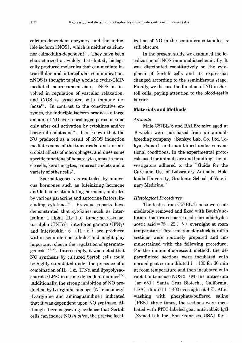
Fig. 1 Immunofluorescence for iNOS. a,b: Immunoreactivity is localized on the processes of Sertoli cells surrounding the spermatogonia and early spermatocytes, and elongating to the lumina of seminiferous tubules. c: With nonimmune serum, the interstitial cells, including Leydig cells and resident macrophages, are false positive. d: The interstitial cells are negative like Sertoli cell processes when using preabsorbed antiserum. bar= 100 11m.
In the present investigation, the iNOS immunoreactivity was detected by both im
munofluorescent and immunoenzyme protocols. A consistent staining pattern was detected mainly on the basal region of the seminiferous tubules, where the cell body of Sertoli cells as well as spermatogonia was located. By the immunofluorescent method, the iNOS-immunoreactivity was found to be remarkably localized on the processes of Sertoli cells surrounding the spermatogonia and
early spermatocytes, and elongating to the lumina of seminiferous tubules (Fig. 1). Some
of the interstitial cells including Leydig cells and resident macrophages were immunostained, and they became negative like Sertoli cells after using preabsorbed antiserum. However, since nonspecific staining was detected on the interstitial cells, but not on Sertoli cells, with nonimmune serum substituted for the primary antiserum, it was clear that the immunoreactivity in the interstitial cells was
Yasuhiro Kon et aL 119
I
XI
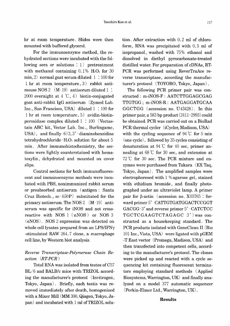
Fig. 2 Immunoenzyme method for iNOS. a: In stage I, iNOS-immunoreactivity is observed on the processes of the Sertoli cells, surrounding the aligned spermatogonia (large arrow), but not on those in the single spermatogonium (small arrow). b: In stage VIII, where the preleptotene and leptotene spermatocytes start to differentiate from spermatogonia, iNOS-immunoreactivity is remarkably detected on the processes of Sertoli cells around their spermatocytes (arrows). c: In stage XI, where zygotene spermatocytes begin to develop, iNOS-immunoreactivity is not detected on the processes around their spermatocytes (arrows). bar= 50 /lm.
120 Expression and distribution of inducible nitric oxide synthase in mouse testis
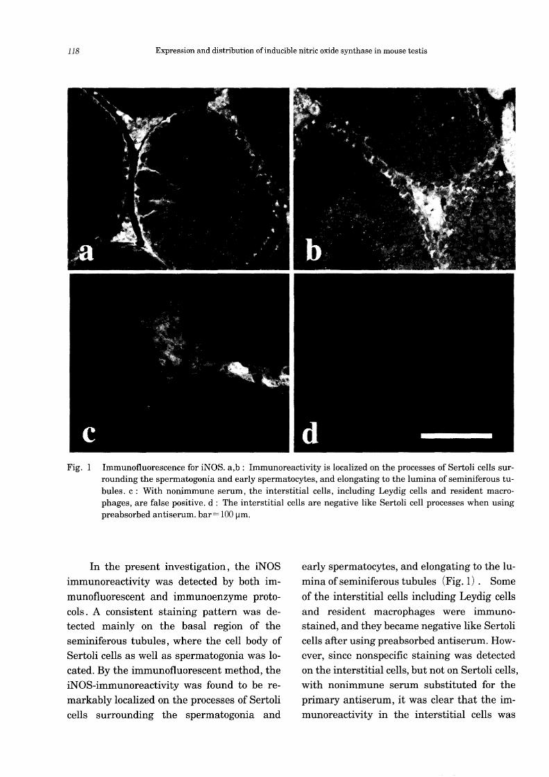
C57BL/6 BALB/c
iNOS ~ 583bp
~-actin ....... 232bp
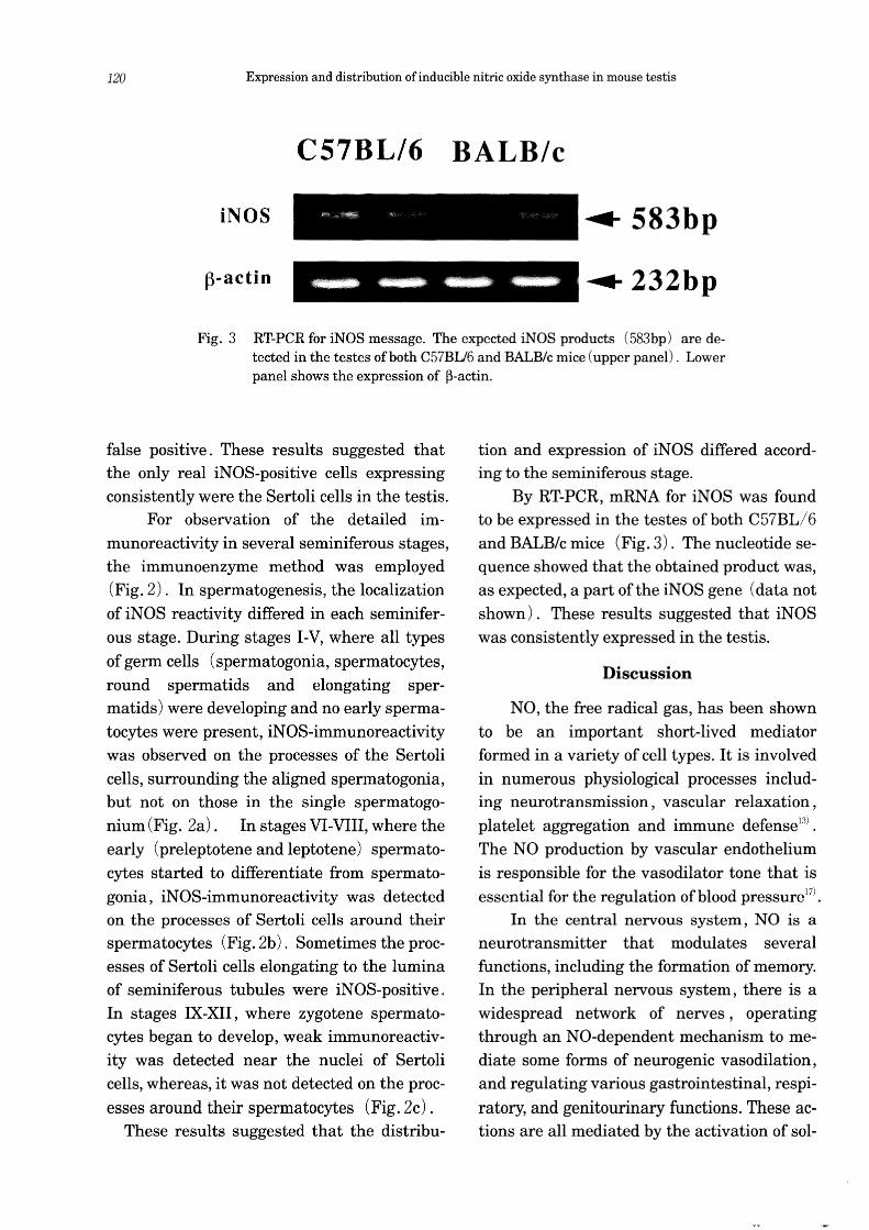
Fig. 3 RT-PCR for iNOS message. The expected iNOS products (583bp) are detected in the testes of both C57BU6 and BALB/c mice (upper panel). Lower panel shows the expression of ~-actin.
false positive. These results suggested that the only real iNOS-positive cells expressing consistently were the Sertoli cells in the testis.
For observation of the detailed immunoreactivity in several seminiferous stages, the immunoenzyme method was employed (Fig. 2). In spermatogenesis, the localization of iNOS reactivity differed in each seminiferous stage. During stages I-V, where all types
of germ cells (spermatogonia, spermatocytes, round spermatids and elongating spermatids) were developing and no early spermatocytes were present, iNOS-immunoreactivity was observed on the processes of the Sertoli cells, surrounding the aligned spermatogonia, but not on those in the single spermatogonium (Fig. 2a). In stages VI-VIII, where the early (preleptotene and leptotene) spermatocytes started to differentiate from spermatogonia, iN OS-immunoreactivity was detected on the processes of Sertoli cells around their spermatocytes (Fig. 2b). Sometimes the processes of Sertoli cells elongating to the lumina of seminiferous tubules were iNOS-positive. In stages IX-XII, where zygotene spermatocytes began to develop, weak immunoreactivity was detected near the nuclei of Sertoli cells, whereas, it was not detected on the proc
esses around their spermatocytes (Fig. 2c) . These results suggested that the distribu-
tion and expression of iN OS differed according to the seminiferous stage.
By RT-PCR, mRNA for iNOS was found to be expressed in the testes of both C57BL/6 and BALB/c mice (Fig. 3). The nucleotide sequence showed that the obtained product was, as expected, a part of the iNOS gene (data not shown). These results suggested that iNOS was consistently expressed in the testis.
Discussion
NO, the free radical gas, has been shown to be an important short-lived mediator formed in a variety of cell types. It is involved in numerous physiological processes including neurotransmission, vascular relaxation, platelet aggregation and immune defense13l
•
The NO production by vascular endothelium is responsible for the vasodilator tone that is essential for the regulation of blood pressureJ7).
In the central nervous system, NO is a neurotransmitter that modulates several functions, including the formation of memory. In the peripheral nervous system, there is a widespread network of nerves, operating through an NO-dependent mechanism to mediate some forms of neurogenic vasodilation, and regulating various gastrointestinal, respi
ratory, and genitourinary functions. These actions are all mediated by the activation of sol-
Yasuhiro Kon et al. 121
uble guanylate cyclase and the consequent increase in the concentration of cyclic GMP in target cells. Although a few reports have hypothesized that iNOS in the testis plays some important roles in spermatogenesis, its real function is not completely known because the investigations were performed in vitro and almost all attention was given to immune defense!' 14). The present study is the first report
about the localization of iNOS in the seminiferous tubules, particularly in Sertoli cells, major cellular elements of the blood-testis barrier.
There is growing evidence that the inducible NOS isoform is expressed constitutively and that expression of the constitutive isoform is also induced!81. For example, it has
been reported that in addition to an increase in the number of endothelial cells expressing eNOS in distal vessels of the hypertensive lung, there is an increase in the number of pulmonary alveolar macrophages expressing both iNOS and eNOS isoforms!51. In both the normal and hyperoxic lung, iNOS expression was detected in bronchial and bronchiolar epithelial cells, in cardiac muscle cells, and in endothelial cells. On the other hand, pulmonary alveolar macrophages were only weakly immunoreactive for iNOS or were immunonegative. Mice lacking iNOS are indistinguishable from wild-type mice in appearance and histology, and exhibit no significant survival advantage over wild-type mice, although upon treat
ment with LPS and IFNI', peritoneal macrophages from the mutant mice do not produce NOIll
. Similarly, disruption of the nNOS gene causes no histological abnormalities in the central nervous system7
). These results suggest that nitric oxides produced by the three isoforms act in compensatory fashion.
In the testis, Sertoli cells and germ cells have been shown in vitro to produce sereval cytokines, some of them being inducible by
the combination of LPS and cytokines2,5,6,!61.
In the present study by RT-PCR, the iNOS message was expressed constitutively in the testis, suggesting that Sertoli cells were consistently stimulated by cytokines. IFN a protein and corresponding mRNA were expressed by peritubular, Sertoli, and germ cells, while IFNI' was found in early spermatids but, in contrast, was not produced by peritubular cells, Sertoli cells, or pachytene spermatocytes3
). TNFa was detected on the pachytene spermatocytes containing a normal 1. 9 kb transcript and the round spermatids containing a 2.8 kb transcript, while RNA from Sertoli cells, but not pachytene spermatocytes or round spermatids, hybridized with a human TNFa receptor p 60 probe in Northern blot analysis21
• These data revealed that TNFa released from germ cells via a paracrine pathway was detected by receptors on Sertoli cells. Thus, it is likely that the intracellular mechanism of TNF-receptor binding locally regulates the transcriptional rate of iNOS mRNA in Sertoli cells.
The timely opening and closing of interSertoli cell tight junctions in the testis are essential for cellular events in the completion of spermatogenesis4
). They permit preleptotene and leptotene spermatocytes to cross the blood-testis barrier from the basal compartment to the adluminal compartment of the seminiferous epithelium so that these cells can continue their further development into
spermatids . However, the mechanism by which these events are regulated remains a mystery in male reproductive physiology. Although several inhibitors of protein kinases and phosphatases can regulate the assembly and maintenance of inter-Sertoli tight junctions in vitro, it is not completely known which molecules participate in these events12
).
Recently, it has been reported that NO can regulate the permeability of water-soluble
122 Expression and distribution of inducible nitric oxide synthase in mouse testis
drugs in the gut, suggesting that a transportenhancing mechanism via NO production may be partly related to the dilation of the tight junction in the epithelium19
). In the present study, iNOS-immunoreactivity was detected on the blood-testis barrier where and when spermatogonia started to be differentiated into spermatocytes. It was hypothesized
that the timely opening and closing of the inter-Sertoli tight junctions during spermatogenesis was likely regulated at least in part, by the NO pathways, which have an autocrine function. Further studies are required to investigate for relationships among NO production, the blood-testis barrier and spermatogenesis.
Acknowledgments
This research was supported in part by a Grant-in-Aid from the Ministry of Education, Science, Sports and Culture of Japan (No. 12460129) .
References
1) Bauche, F., Stephan, J.P., Touzalin, A.M. and Jegou, B. 1998. In vitro regulation of an inducible-type NO synthase in
the rat seminiferous tubule cells. Biol. Reprod. ,58 : 431-438.
2) De, S.K., Chen, H.L., Pace, J.L., Hunt, J. S., Terranova, P.F. and Endes, G.C.1993. Expression of tumor necrosis factor-a in mouse spermatogenic cells. Endocrinology , 133 : 389 -396.
3) Dejucq, N., Dugast, 1., Ruffault, A., van der Meide, P.H. and Jegou, B. 1995. Interferon-a and - 'Y expression in the rat testis. Endocrinology, 136 : 4925-4931.
4) de Kretser, D.M. and Kerr, J.B. 1994. The cytology of the testis. In: The Physiology of Reproduction, 2 nd ed., pp. 837-932, Knobil, E. and Neill, J. eds.,
Raven Press, New York. 5) Gerard, N., Syed, V., Bardin, W., Ge
netet, N. and Jegou, B. 1991. Sertoli cells are the site of interleukin 1 alpha synthesis in rat testis. Mol. Cell. Endocrinol.,82 : R13-R16.
6) Haugen, T.B., Landmark, B.F., Josefsen, G.M., Hansson, V. and Hogset, A. 1994.
The mature form of interleukin 1 alpha is constitutively expressed in immature male germ cells from rat. Mol. Cell. Endocrinol . , 105 : R19- R23.
7) Huang, P.L., Dawson, T.M., Bredt, D.S., Snyder, S.H. and Fishman, M.C. 1993. Targeted disruption of the neuronal nitric oxide synthase gene. Cell, 75 : 1273 -1286.
8) Jegou, B. 1993. The Sertoli-germ cell
communication network in mammals. Int. Rev. Cytol. , 147 : 25-96.
9) Knowles, R.G. and Moncada, A. 1994. Nitric oxide synthesis in mammals. Biochem. J. ,298 : 249-258.
10) Kroncke, K. D., Fehsel, K. and KolbBachofen, V. 1995. Inducible nitric oxide synthase and its product nitric oxide, a small molecule with complex biological activities. Biol. Chem. HoppeSeyer, 376 : 327-343.
11) Laubach, V.E., Shesely, E.G., Smithies, O. and Sherman, P.A. 1995. Mice lacking inducible nitric oxide synthase are not resistant to lipopolysaccharideinduced death. Proc. Natl. Acad. Sci. USA, 92 : 10688-10692.
12) Li, J.C.H., Mruk, D. and Cheng, C. Y. 2001. The inter-Sertoli tight junction permeability barrier is regulated by the interplay of protein phosphatases and kinases: an in vitro study. J. Androl . , 22 : 847-856.
13) Moncada, S. and Higgs, A.1993. The Larginine-nitric oxide pathway. New Eng.
Yasuhiro Kon et al.
J. Med., 329 : 2002-2012. 17) 14) Stephan, J.P., Guillemois, C., Jegou, B.
and Bauche, F. 1995. Nitric oxide pro-duction by Sertoli cells in response to cytokines and lipopolysaccharide. Bio-chem. Biophys. Res. Comm., 213 : 218-224. 18)
15) Steudel, W., Watanabe, M., Dikranian, K., Jacobson, M. and Jones, R.C. 1999. Expression of nitric oxide synthase iso-forms (NOSH and NOSIH) in adult rat lung in hyperoxic pulmonary hyperten- 19) sion. Cell Tissue Res. , 295 : 317 -329.
16) Syed, V., Gerard, N., Kaipia, K., Bardin, C.W., Parvinen, M. and Jegou, B.1993. Identification, ontogeny, and regulation of an interleukin - 6 - like factor in the rat seminiferous tubule. Endocrinology, 132 : 293-299.
123
Waldman, S.A. and Murad, F. 1988. Biochemical mechanisms underlying vascular smooth muscle relaxation: the guanylate cyclase-cyclic GMP system. J. Cardiovasc. Pharmacol ., 12 (SuppI5): SI15-S118. Xue, C. and Johns, R.A. 1996. Upregulation of nitric oxide synthase correlated temporally with onset of pulmonary vascular remodeling in the hypoxic rat. Hypertension, 28 : 743-753. Yamamoto, A., Tatsumi, H., Maruyama, M., Uchiyama, T., Okada, N. and Fujita, T. 2001. Modulation of intestinal permeability by nitric oxide donors: implications in intestinal delivery of poorly absorbable drugs. J. Pharmacol. Exp. Ther. , 296 : 84-90.


![A Comprehensive Toolkit for Inducible, Cell Type-Specific Gene … · A Comprehensive Toolkit for Inducible, Cell Type-Specific Gene Expression in Arabidopsis1 [CC-BY] Ann-Kathrin](https://static.documents.pub/doc/80x56/60b7c27ed2931f72db3927c9/a-comprehensive-toolkit-for-inducible-cell-type-specific-gene-a-comprehensive-toolkit.jpg)


