EXPRESSION OF ALPHA-GLUCOSIDASE GENE IN HYPOPHARYNGEAL GLANDS OF EASTERN HONEYBEE WORKER Apis cerana indica Chanpen Chanchao 1 , Prapaipit Srimawong 2 , Siriwat Wongsiri 1 1 Department of Biology, Faculty of Science, Chulalongkorn University, Bangkok, Thailand. 2 Research center, Ramathibodi Hospital, Bangkok, Thailand. E-mail: [email protected]Received 29 November 2005; accepted 04 October 2006 Summary Duties of worker bees in a hive change according to their age. Temporal and spatial expres- sion of developmentally regulated genes relating to this is interesting. In order to control the age of Apis cerana indica, emerged bees were marked with painting color and counted as 0 day. Bees at desired ages were collected during 0-29 days with the time interval of 3 days. The re- sults showed that hypopharyngeal glands (hpg) change their size from large to shrunken and their colour from creamy to pale. The largest acini were found in worker bees 15 to 18 days old and the smallest in 29 days old. Alpha-glucosidase (ag) activity assay in crude extract was low in emerged bees. Then, the activity increased in workers 18 days old and got the highest in those 24 days old. Activity staining of crude extract by native polyacrylamide gel, ag was low from 18-day and high from 29-day old worker bees. In addition, the expression profile of ag was ob- tained by RT-PCR. It indicates that the expression of ag was high in foragers. The obtained cDNA sequence shows closely relationship (82.81% similarity) to the cDNA in A. mellifera. Keywords: Apis cerana indica, alpha-glucosidase, development, expression, nurse bee, forager, activity, RT-PCR, cDNA, phylogeny. INTRODUCTION Honeybee colonies as highly eusocial insects are composed of 3 castes: a female queen, female workers, and male drones. Most of the population in a colony are workers responsible for all tasks such as brood rearing, wax secreting and comb building, food handling and storing, pollen or nectar foraging, and a community de- fense. A division of labor in honeybee col- onies is based on worker age. The workers change jobs when their age changes. This form of behavioral development is called “age polyethism” (Huang et al. 1991). Hypopharyngeal glands (hpg) located in the heads of worker bees are paired long tu- berous organs connected to many acini (se- cretory glands). In A. mellifera, the acini of emerged bees look plump and creamy. The hpg synthesizes and secretes a substance rich in protein, called bee milk, or royal jelly (rj), which is fed to all larvae (Dade 1994). Later, hpg change to produce diges- tive enzymes, including alpha-glucosidase (ag), invertase, etc (Brouwers 1982). At this stage, worker bees become foragers. The ag is alpha-D-glucoside gluco- hydrolase and belongs to EC 3.2.1.20. The ag catalyzes the hydrolysis of terminal, non–reducing alpha-1, 4-linked glucose residues from aryl-glucosides, disacchari- des or oligosaccharides. Other common names of ag are invertase, sucrase, and maltase. Different proteins from hpg of A. mellifera at different ages were reported. Three major proteins with molecular masses of 50, 56, and 64 kDa were restric- Vol. 50 No. 2 2006 Journal of Apicultural Science 5
Transcript
EXPRESSION OF ALPHA-GLUCOSIDASE GENE IN
HYPOPHARYNGEAL GLANDS OF EASTERN HONEYBEE
WORKER Apis cerana indica
C h a n p e n C h a n c h a o1
, P r a p a i p i t S r i m a w o n g2
,
S i r i w a t W o n g s i r i1
1Department of Biology, Faculty of Science, Chulalongkorn University, Bangkok, Thailand.2Research center, Ramathibodi Hospital, Bangkok, Thailand. E-mail: [email protected]
Received 29 November 2005; accepted 04 October 2006
S u m m a r y
Duties of worker bees in a hive change according to their age. Temporal and spatial expres-
sion of developmentally regulated genes relating to this is interesting. In order to control the age
of Apis cerana indica, emerged bees were marked with painting color and counted as 0 day.
Bees at desired ages were collected during 0-29 days with the time interval of 3 days. The re-
sults showed that hypopharyngeal glands (hpg) change their size from large to shrunken and
their colour from creamy to pale. The largest acini were found in worker bees 15 to 18 days old
and the smallest in 29 days old. Alpha-glucosidase (ag) activity assay in crude extract was low
in emerged bees. Then, the activity increased in workers 18 days old and got the highest in those
24 days old. Activity staining of crude extract by native polyacrylamide gel, ag was low from
18-day and high from 29-day old worker bees. In addition, the expression profile of ag was ob-
tained by RT-PCR. It indicates that the expression of ag was high in foragers. The obtained
cDNA sequence shows closely relationship (82.81% similarity) to the cDNA in A. mellifera.
Vol. 50 No. 2 2006 Journal of Apicultural Science 5
tively found in nurse bees, whereas a major
70-kDa protein was specifically found in
foragers. Immunoblotting analysis against
50, 56, and 64 kDa proteins confirmed that
they were only detected in hpg of nurse
bees and also existent in rj. It suggested
that these proteins were synthesized in hpg
of nurse bees and secreted as constituents
of rj. In addition, a 70 kDa protein was pu-
rified and immunoblotted, this protein was
positively detected in hpg of foragers only.
Subsequently, the 70 kDa protein was char-
acterized and identified as ag (Kubo et al.
1996).
Recently, proteins at molecular masses
of 57 and 85 kDa were also purified from
hpg of A. mellifera foragers and determined
by Sodium Dodecyl Sulfate Polyacryla-
mide Gel Electrophoresis (SDS PAGE).
These proteins were already characterized
as amylase and glucose oxidase, respec-
tively (Ohash i et al. 1999). They are
members of the sequence-related family of
alpha-glycohydrolases (family 13) contain-
ing many important digestive enzymes
(Yao et al., 2003).
Huber and Mathison (1976) purified ag
from whole bodies of honeybees. The
A. mellifera ag, Mr 82,000, is found in the
head and abdomen of honeybees and its
properties are similar to that of ag found in
honey. Thus, this enzyme is probably syn-
thesized in hpg and converts nectar sucrose
into glucose and fructose in the crop. The
pH optimum of each honey bee sucrase
was different from each other according to
the substrates as sucrose (pH 5.5) and
p-nitrophenyl-alpha-glucoside (pH 6.5).
Takewaki et al. (1980 and 1993) re-
ported the purification and some properties
of both agI and agII from adult honey bees.
Two kinds of the enzymes could be
chromatographically separated according
to their solubility in ammonium sulfate.
The molecular mass of agI and agII was
estimated to be approximately 98 and
76 kDa, respectively, by SDS disc electro-
phoresis. Their pH optimum was 5.0. Both
ag readily hydrolysed phenyl-alpha-
glucoside, sucrose and maltose.
Different proteins from worker bees at
different stages indicate the differential
gene expression. That includes the ag gene
also.
In A. mellifera, the mRNA for the ag
was detected, cloned, and characterized.
The deduced amino acid sequence of 650
residues revealed 41.9% identity to maltase
of mosquito (Aedes aegypti) and 42.2%,
46.3% and 46.2% of maltase 1, 2, and 3 of
fruit fly (Drosophila melonogaster), re-
spectively (Ohashi et al. 1996).
According to the data mentioned above,
a lot of researches have been done in
A. mellifera, western bee, but not in
A. cerana indica which is native and eco-
nomic to Thailand. This research is in-
volved in the relationship of size of hpg
and expression of ag in hpg. Specific activ-
ity of ag in crude extract and RT-PCR were
used to analyse the function of ag. Finally,
a phylogenetic tree of ag was constructed
in order to reveal the homology of the gene
in other organisms.
MATERIALS AND METHODS
Sample collection
Colonies of Apis cerana indica were
taken from Nakhon Sri Thammarat provin-
ce and maintained at Nonthaburi province.
Newly emerged workers were marked on
their thorax with paint markers (water-
proof) and introduced into a free-flying
colony. The colony was fed with sugar
candy everyday. Thereafter, marked bees
were collected for 29 days with the time in-
terval of 3 days and were immediately
stored at -20°C until use. For RNA extrac-
tion, nurse bees were collected while they
were feeding brood. Forager bees were col-
lected when they returned to the colony af-
ter foraging for nectar and pollen. Samples
were stored in liquid N2 until used.
6
Measurement of hpg
Dissected hpg were stored in 0.85%
(w/v) NaCl solution. The shortest (a) and
the longest (b) diameters (in µm) of acini
were measured by an ocular scale in light
microscope. The area of hpg was evaluted
as S (µm2) = π × a/2 × b/2 (Sasagawa et al.
1989).
Enzyme assay
The ag activity was determined by mea-
suring glucose liberated from sucrose. The
method was modified from Momose’s
method (Kubo et al. 1996). Twenty hpg
were dissected and stored in 500 µl buffer
insect saline (10 mM Tris, 130 mM NaCl, 5
mM KCl, and 1 mM CaCl2, pH 7.4) con-
taining 1 mM phenylmethylsulfonyl-
fluoride (PMSF), 0.1 µg/ml pepstatin, and
100 µg/ml leupeptin. The glands were ho-
mogenized and centrifuged 2 times at 4°C,
700 × g for 10 min. The supernatant was
stored at -20°C. Activity staining for ag
was modified from Ta n i m u r a et al.
(1979). Briefly, after native PAGE, the gel
was incubated in 10 mM sodium acetate
buffer containing 0.5 M sucrose (pH 5.0) at
45°C for 30 min. After washed, the gel was
boiled in freshly prepared solution of 0.1%
(w/v) triphenyltetrazolium chloride in
0.5 N NaOH for 3 min. The positive band
of ag on the gel appeared in red.
Primer design
Primers of ag in A. cerana for RT-PCR
were designed from cDNA sequences of ag
in A. mellifera (Ohashi et al. 1996, acces-
sion: D79208). Forward primers (FW) are
5’-tcgac ttcta gttgg tagca tgaag g-3’ (FW1),
5’-gctta tcgag gcata cacga-3’ (FW2), and
5’-acgag gaaca aatcg tggat-3’ (FW3), re-
spectively. Also, reverse primers (R) are
5’-ctagt cagtg ctgca catga gaaag g-3’ (R1),
5’-gacgt acatg ccacc aagtg-3’ (R2), and
5’-gtcta ttctt tgaag cggcg-3’ (R3), respec-
tively. In addition, control primers were de-
signed from 28s RNA in A. mellifera (FW:
5’-aaaga tcgaa tgggg atatt c-3’ and R:
5’-caccg ggtcc gtacc tcc-3’) and elongation
factor (ef) genes in A. cerana (FW: 5’-tcgct
tttac tcttg gtgtg a-3’ and R: 5’-aaact cccaa
catat tatct cca-3’).
RT-PCR
Total RNA (200 ng) of emerged bees,
nurse bees 6 days old, and foragers were
isolated from hpg by SV total RNA isola-
tion kit (Promega, catalog# Z3100).
RT-PCR was performed according to ac-
cess RT-PCR system (Promega, catalog#
A1250). The RT-PCR cycling profile of ag
was modified from Ohashi et al. (1996
and 1997). As a control, 28s RNA and ef
primers were used as RNA reference mar-
kers. The PCR amplification was at the
same condition as that of ag primers.
Sequence alignment
Obtained RT-PCR product was purified
by QIAquick PCR purification kit (Qiagen,
catalog# 28104) and directly sequenced by
an automated sequencer (ABI prism, model
377). The nucleotide and deduced amino
acid sequences were aligned by using
Genetyx program. The cDNA sequences
were aligned and compared to the se-
quences of maltase 1 in A. mellifera
(XM_393379), Drosophila melanogaster
CG14934-PA (NM_135678), sucrose spe-
cific enzyme II of the PTS (ScrA) gene in
Lactobacillus sakei (AF401046), and
Culicoides sonorensis clone CsMAL1
maltase (AY603565).
Statistical analysis
ANOVA was applied and multiple range
test was used to detect significant differ-
ences between the means at p < 0.05.
RESULTS
Hypopharyngeal gland size and age
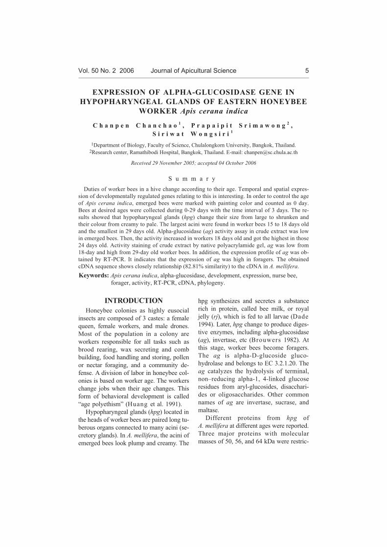
A tendency is visible that the size of
hypopharyngeal glands (hpg) increases
during day 0 to 15 after emerging of
worker bees. The size of hpg of day 0 and
of day 3 is significantly smaller than the
size of hpg of day 15, while the size of hpg
of day 15 to day 18 is not significantly dif-
Vol. 50 No. 2 2006 Journal of Apicultural Science 7
ferent (Fig. 1). After day 18, the size of hpg
is like to decrease. The size of hpg of day
24, 27 and 29 is significantly smaller, than
the size of hpg of day 15. This coincides to
the task of worker bees. Considering the
overall tendency of size changes, while the
increasing size of acini (day 0 to 15) is ob-
served, bees become nurse bees. After the
size of acini decreases (day 15 to 29), bees
become foragers.
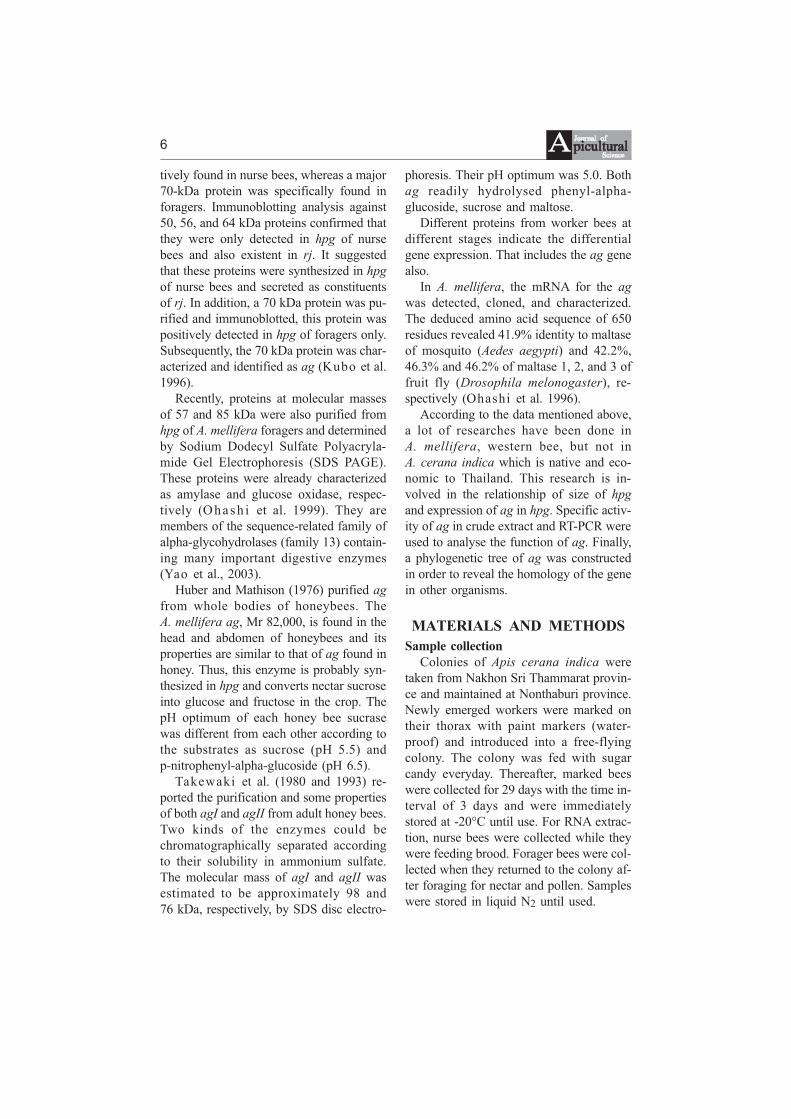
Activity of ag
The ag activity examined from crude
extract of hpg at various ages, from day 0
to day 29, and computed by comparing to
the standard curve of glucose is shown in
Fig. 2. Although specific activity of all
stages is not significantly different by using
One-way ANOVA, the tendency of chang-
ing activity is still visible. It shows that the
specific activity of ag from day 0 (0.0239
unit/mg protein) to day 18 (6.7363
units/mg protein) is lower, than the specific
activity from day 24 (9.0811 units/mg pro-
tein). Although the specific activity of ag
from day 21 (3.4024 units/mg protein) is
low, it is still possible to assume that the
tendency of ag specific activity increased
during the period. After day 24, both de-
crease, on day 27 (5.8187 units/mg protein)
and on day 29 (3.5738 units/mg protein).
By comparing nurse bees (d. 15) and
foragers (d. 24), presented in figures 1 and
2, it is visible that the size of acini de-
creased while the ag activity increased. The
size of acini of bees 24 days old is signifi-
cantly smaller than that size of bees 15
days old. In contrast, the ag activity is still
very low in nurse bees 15 days old. Then, it
is higher in foragers 24 days old.
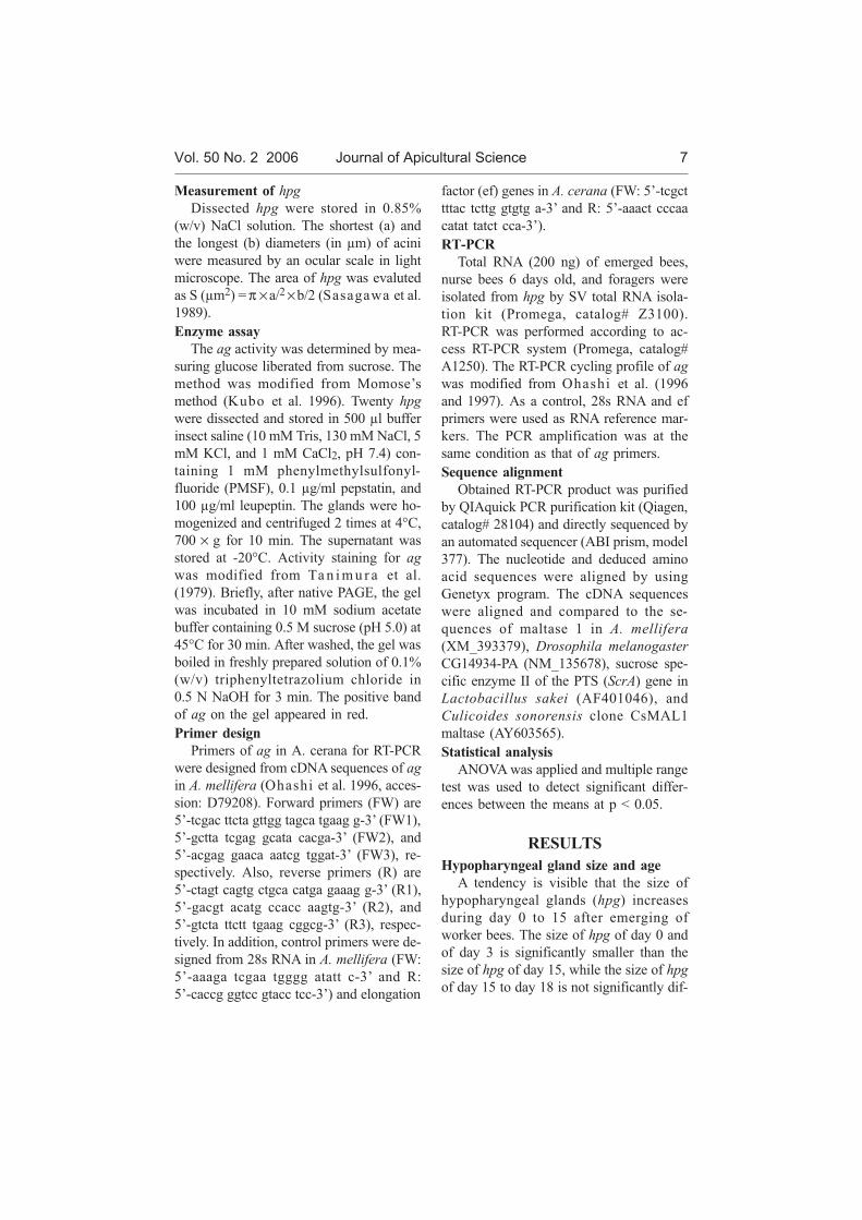
Activity stain of ag
The ag activity on native polyacryla-
mide gel appeared in red. Only one activity
band was visible. Fig. 3 shows that higher
activity of ag could be detected on the lane
containing crude extract of forager bees
(lane 2) as compared to the ag activity of
crude extract of nurse bee (lane 1).
Fig. 3. Native polyacrylamide gel of ag
in crude extract. Detected by activity stain-
ing, a positive band is observed in 6-day
old worker bee (8 µg; lane 1) and forager
bee (8 µg; lane 2).
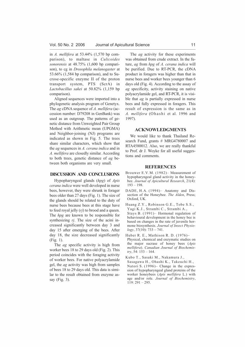
The expression level of ag by RT-PCR
8
Figure 1. The overall size (calculated area) of acini in worker bees at various ages. Ten acini at eachstage were measured. Error bars indicate standard deviation. Different letters indicate significantdifferences between means PMFs<0.05.
Fig. 4 shows that the expression of ag
was not detectable in emerged bees but was
detectable in worker bees (from 6-d.
worker bees to foragers). The expression of
ag was also high in foragers. As control,
the amplified product was obtained from
bees at any stages by using primers of 28S
RNA in A. mellifera (350 bp) and of elon-
gation factor (ef) in A. cerana (100 bp)
(data not shown).
The cDNA sequence of ag and its phylo-
genetic tree
According to RT-PCR, cDNA (1,549
bp) and deduced amino acid (516 residues),
the sequences of ag in A. cerana indica
was obtained (Fig. 5). The ag cDNA of
A. cerana indica was compared to that
from other organisms. The similarity be-
tween cDNA sequences was established us-
ing a program of nucleotide-nucleotide
BLAST (Basic Local Alignment Search
Tool of the National Center for Biotechnol-
ogy Information). The length of cDNA in
base pairs (bp) was from RT-PCR as men-
tioned in Materials and Methods. It is par-
tially similar to the ag in A. mellifera at
82.81% (1,557 bp comparison), to maltase
Vol. 50 No. 2 2006 Journal of Apicultural Science 9
Figure 3. Native polyacrylamide gel of ag incrude extract. Detected by activity staining, apositive band is observed in 6-day old workerbee (8 µg; lane 1) and forager bee (8 µg;lane 2).
Figure 2. The specific activity of ag in worker bees of different age. Error bars indicate standarddeviation. No significant differences between means PMFs<0.05 were determined but the tendencyof activity is visible.
10
Figure . The RT-PCR product (350 bp) of ag from bees at various age. Lane 1, 100 bp laddermarker; lane 2, negative control; lane 3–5, emerged bees; lane 6–8, 6-day old worker bees; lane9–11, nurse bees; and lane 12–14, forager bees.
Figure . Phylogenetic trees of ag by UPGMA (A) and NJ (B). Am malt stands for maltase 1 inA. mellifera. Dm cg stands for Drosophila melanogaster CG14934-PA. Ls scrA stands for sucrosespecific enzyme II of the PTS in Lactobacillus sakei. Cs malt stands for Culicoides sonorensis cloneCsMAL1 maltase. In addition, Ac agand Am agstand for alpha-glucosidase in A. cerana indica andin A. mellifera, respectively. Numbers on branches of both trees indicate genetic distance.
in A. mellifera at 53.44% (1,570 bp com-
parison), to maltase in Culicoides
sonorensis at 48.75% (1,600 bp compari-
son), to cg in Drosophila melanogaster at
53.66% (1,584 bp comparison), and to Su-
crose-specific enzyme II of the proton
transport system, PTS (ScrA) in
Lactobacillus sakei at 50.82% (1,159 bp
comparison).
Aligned sequences were imported into a
phylogenetic analysis program of Genetyx.
The ag cDNA sequence of A. mellifera (ac-
cession number: D79208 in GenBank) was
used as an outgroup. The patterns of ge-
netic distance from Unweighted Pair Group
Method with Arithmetic mean (UPGMA)
and Neighbor-joining (NJ) programs are
indicated as shown in Fig. 5. The trees
share similar characters, which show that
the ag sequences in A. cerana indica and in
A. mellifera are closedly similar. According
to both trees, genetic distance of ag be-
tween both organisms are very small.
DISCUSSION AND CONCLUSIONS
Hypopharyngeal glands (hpg) of Apis
cerana indica were well developed in nurse
bees, however, they were shrunk in forager
bees older than 27 days (Fig. 1). The size of
the glands should be related to the duty of
nurse bees because bees at this stage have
to feed royal jelly (rj) to brood and a queen.
The hpg are known to be responsible for
synthesizing rj. The size of the acini in-
creased significantly between day 3 and
day 15 after emerging of the bees. After
day 18, the size decreased significantly
(Fig. 1).
The ag specific activity is high from
worker bees 18 to 29 days old (Fig. 2). This
period coincides with the foraging activity
of worker bees. For native polyacrylamide
gel, the ag activity was high from samples
of bees 18 to 29 days old. This data is simi-
lar to the result obtained from enzyme as-
say (Fig. 3).
The ag activity for these experiments
was obtained from crude extract. In the fu-
ture, ag from hpg of A. cerana indica will
be purified. Due to RT-PCR, the cDNA
product in foragers was higher than that in
nurse bees and worker bees younger than 6
days old (Fig. 4). According to the assay of
ag specificity, activity staining on native
polyacrylamide gel, and RT-PCR, it is visi-
ble that ag is partially expressed in nurse
bees and fully expressed in foragers. This
result of expression is the same as in
A. mellifera (O h a s h i et al. 1996 and
1997).
ACKNOWLEDGMENTS
We would like to thank Thailand Re-
search Fund, grants # MRG4780007 and
RTA4580012. Also, we are really thankful
to Prof. dr J. Woyke for all useful sugges-
tions and comments.
REFERENCES
Brouwer E.V.M. (1982)– Measurement ofhypopharyngeal gland activity in the honey-bee. Journal of Apicultural Research, 21(4):193 – 198.
DADE, H.A. (1994)– Anatomy and Dis-section of the Honeybee. The Alden, Press;Oxford, UK.
Huang Z.Y., Robinson G.E., Tobe S.S.,
Yagi K.J., Strambi C., Strambi A.,
Stays B. (1991)– Hormonal regulation ofbehavioural development in the honey bee isbased on changes in the rate of juvenile hor-mone biosynthesis. Journal of Insect Physio-logy, 37(10): 733 – 741.
Huber R. E., Mathison R. D. (1976)–Physical, chemical and enzymatic studies onthe major sucrase of honey bees (Apismellifera). Canadian Journal of Biochemis-try, 54: 153 – 164.
Kubo T., Sasaki M., Nakamura J.,
Sasagawa H., Ohashi K., Takeuchi H.,
Natori S. (1996)– Change in the expres-sion of hypopharyngeal gland proteins of theworker honeybees (Apis mellifera L.) withage and/or role. Journal of Biochemistry,119: 291 – 295.
Vol. 50 No. 2 2006 Journal of Apicultural Science 11
Ohashi K., Sawata M., Takeuchi H.,
Natori S., Kubo T. (1996)– Molecularcloning of cDNA and analysis of expressionof the gene for alpha-glucosidase from thehypopharyngeal gland of the honeybee Apismellifera L. Biochemical and BiophysicalResearch Communications, 221: 380 – 385.
Ohashi K., Natori S., Kubo T. (1997)–Change in the mode of gene expression ofthe hypopharynreal gland cells with anage-dependent role change of the workerhoneybee Apis mellifera L. European Jour-nal of Biochemistry, 249: 797-802.
Sasagawa H., Sasaki M., Okada I.
(1989)– Hormonal control of the division oflabor in adult honeybee (Apis mellifera L.) I.Effect of methoprene on corpora allata andhypopharyngeal gland, and it’s alpha -glucosidase activity. Applied Entomologyand Zoology, 24(1): 66 – 77.
Takewaki S., Chiba S., Kimura A.,
Matsui H., Koike Y. (1980)– Purifica-tion and properties of alpha-glucosidase ofthe honey bee Apis mellifera L. Agriculturaland Biological Chemistry, 44(4): 731 – 740.
Takewaki S., Kimura A., Kubota M.,
Chiba S. (1993)– Substrate specificity andsubsite affinites of honeybee alpha--glucosidase II. Bioscience Biotechnologyand Biochemistry, 57(9): 1508 – 1513.
Tanimura T., Kitamura K., Fukuda T.,
Kikuchi T. (1979) – Purification and par-tial characterization of three forms of al-pha-glucosidase from the fruit fly Drosophilamelanogaster. Journal of Biochemistry, 85:123 – 130.
Yao X., Rebecca M., Byers L. (2003)–Multiple sugar binding sites in alpha--glucosidase. Biochemica et Biophysica Acta,1645: 22 – 29..
EKSPRESJA GENU ALFA-GLUKOZYDAZY W GRUCZO£ACH
GARDZIELOWYCH PSZCZO£Y WSCHODNIEJ
Apis cerana indica
C h a n p e n C h a n c h a o , P r a p a i p i t S r i m a w o n g ,
S i r i w a t W o n g s i r i
S t r e s z c z e n i e
Obowi¹zki pszczó³ robotnic w ulu zmieniaj¹ siê wraz z wiekiem. Ciekawym jest zagadnienie
czasowej i przestrzennej ekspresji zwi¹zanych z tym genów, które podlegaj¹ regulacji
œrodowiskowej. W celu kontroli wieku Apis cerana indica pszczo³y po wygryzieniu by³y
zaznaczane farb¹ i zaliczane do grupy 0-dniowych. Pszczo³y w po¿¹danym wieku pobierano od
0 do 29 dnia w odstêpach trzydniowych. Wyniki wskazuj¹, ¿e gruczo³y gardzielowe (hpg)
zmieniaj¹ swoj¹ wielkoœæ i barwê od du¿ych i kremowych do skurczonych i bladych.
Najwiêksze pêcherzyki stwierdzano u robotnic w wieku od 15 do 18 dni, a najmniejsze
u robotnic 29-dniowych. AktywnoϾ glikozydazy (ag) w surowym ekstrakcie oznaczono jako
nisk¹ u wygryzionych pszczó³. Nastêpnie aktywnoœæ wzrasta³a u robotnic 18 dniowych i by³a
najwy¿sza u robotnic 24-dniowych. Aktywnoœæ surowego ekstraktu oznaczana na natywnym
¿elu poliakrylamidowym, ag u 18 dniowych robotnic by³a niska, a u 29-dniowych wysoka.
Ponadto profil aktywnoœci ag uzyskano przy pomocy RT-PCR. Wskazuje on, ¿e ekspresja ag
u pszczó³ zbieraczek by³a wysoka. Uzyskana sekwencja cDNA wskazuje na œcis³y zwi¹zek