Vol. 135, No. 1, 1986 BIOCHEMICAL AND BIOPHYSICAL RESEARCH COMMUNICATIONS February 26, 1986 Pages 105-109 EXPRESSION OF C-MYC ONCOGENE IN HUMAN FIBROBLASTS DURING IN VITRO SENESCENCE Roger Dean, Seong Soo Kim, and Dora Delgado Department of Biochemistry, University of New Mexico School of Medicine Albuquerque, New Mexico Received January 9, 1986 SUMMARY Expression of the c-myc oncogene was determined in pre-confluent early and late passage human (IMR-90) fibroblast by dot blot analysis of cellular mRNA. Significant decreases in c-myc levels were found in late passage when compared to levels found in early passage cells. Cells restimulated with serum after serum restriction also showed reduced levels of c-myc in late passage. Confluent cells expressed levels of c-myc similar to that of pre-confluent cells, when serum stimulation was the same in both cases. Southern blots of Eco RI digested DNA showed 2 fragments of similar size hybridizing to c-myc sequences in both early and late passage cells. 0 1986 Academic Press, Inc. The limited replicative lifespan of diploid human cells in vitro (cellular senescence) has been extensively researched as a cellular model of aging, yet the mechanisms underlying cellular senescence remain unknown. Because cellular senescence is characterized as a loss of growth capacity, a greater understanding of genes whose function is the control of cellular growth should improve our understanding of senescence. Proto-oncogenes are cellular genes whose expression is important in the control of cell growth and differentiation. The human c-myc gene, a proto-oncogene, has been shown to be an important determinant of cell growth and differentiation (1.2). We investigated the possibility that declining growth potential in IMR-90 human fibroblasts may be reflected in c-myc expression changes. Abbreviations used: PD, population doubling. 0006-291X/86 $1.50 105 Copvright 0 1986 by Academic Press, Inc. All righrs of reprohrction in an.v .fbrm reserved.
Transcript
Vol. 135, No. 1, 1986 BIOCHEMICAL AND BIOPHYSICAL RESEARCH COMMUNICATIONS
February 26, 1986 Pages 105-109
EXPRESSION OF C-MYC ONCOGENE IN HUMAN FIBROBLASTS DURING IN VITRO SENESCENCE
Roger Dean, Seong Soo Kim, and Dora Delgado
Department of Biochemistry, University of New Mexico School of Medicine
Albuquerque, New Mexico
Received January 9, 1986
SUMMARY Expression of the c-myc oncogene was determined in pre-confluent early and late passage human (IMR-90) fibroblast by dot blot analysis of cellular mRNA. Significant decreases in c-myc levels were found in late passage when compared to levels found in early passage cells. Cells restimulated with serum after serum restriction also showed reduced levels of c-myc in late passage. Confluent cells expressed levels of c-myc similar to that of pre-confluent cells, when serum stimulation was the same in both cases. Southern blots of Eco RI digested DNA showed 2 fragments of similar size hybridizing to c-myc sequences in both early and late passage cells. 0 1986 Academic Press, Inc.
The limited replicative lifespan of diploid human cells in vitro
(cellular senescence) has been extensively researched as a cellular model
of aging, yet the mechanisms underlying cellular senescence remain
unknown. Because cellular senescence is characterized as a loss of growth
capacity, a greater understanding of genes whose function is the control of
cellular growth should improve our understanding of senescence.
Proto-oncogenes are cellular genes whose expression is important in the
control of cell growth and differentiation. The human c-myc gene, a
proto-oncogene, has been shown to be an important determinant of cell
growth and differentiation (1.2). We investigated the possibility that
declining growth potential in IMR-90 human fibroblasts may be reflected in
c-myc expression changes.
Abbreviations used: PD, population doubling.
0006-291X/86 $1.50
105 Copvright 0 1986 by Academic Press, Inc.
All righrs of reprohrction in an.v .fbrm reserved.
Vol. 135, No. 1, 1986 BIOCHEMICAL AND EIOPHYSICAL RESEARCH COMMUNICATIONS
MATERIALS AND METHODS
Cell growth: IMR-90 fibroblasts were grown in Eagle’s minimal essential medium with Earle’s salts , glutamine (2 mM), and non-essential amino acids. This media was supplemented with lOX4fetal cal$ serum (FCS) and antibiotics. Cells were subcultivated at IX10 cells/cm in fresh media and incubated for 24 hr. These cells, termed pre-confluent, were then used to extract RNA. Alternatively, pre-confluent cells were growth arrested by restricting serum to 0.5% for two days without refeeding. These cells were then restimulated with fresh media containing 10% FCS and incubated 24 hr before RNA was extracted. Confluent cultures of early passage cells were given fresh 10% FCS supplemented media 24 hr before RNA or DNA extraction.
RNA isolation and hybridization: Cells were extracted for RNA according to the method of Chirgwin et a1.(3). Poly A+ RNA was isolated using oligo(dT)-cellulose (Pharmacia) according to the manufacturer’s instructions. For dot blot analysis, RNA in 10X SSC, 2.2M formaldehyde was heated to 65’C, cooled and blotted on nitrocellulose (S&S-BA85) in a 96 well manifold. 8O’C.
After washing twice, blots were baked in vacuum for 3 hr at Human c-myc (third exon) ob@ined from Oncor Inc. was nick
translated with deoxycytidine 5’-[ PI triphosphate (sp. act. 3200 Ci/mM) (Amersham) using a nick translation kit supplied by Bethesda Research Laboratories. Blots were hybridized in 50% formamide-5X SSC containing 0.1% Ficoll, 0.1% polyvinylpyrrolidone, 50 mM sodium phosphate, 0.1% SDS and 250 ug/ml sheared salmon sperm DNA at 43’C for 16 to 24 hr. Blots were washed and exposed to X-ray film for 2-6 days at -7O’C.
DNA isolation and hybridization: Confluent IMR-90 cells were lysed in situ with 50 mM tris, 10 mM EDTA, 10 mM NaCl (pH 8.0) containing 100 ug/z proteinase K and 0.5% SDS. DNA was extracted twice with phenol, once with phenol chloroform and once with chloroform before being precipitated with ethanol and collected on a glass rod. For Southern analysis DNA was digested with Eco RI (1 unit/ug DNA) for 90 min. size fractionated on 0.8% agarose gels and transferred to nitrocellulose using the methods of Maniatis et al. (4). Transferred sequences were hybridized with nick translated c-myc according to manufacturer’s (Oncor inc.) specifications.
RESULTS
To test IMR-90 fibroblasts for age related differences in c-myc
expression, early and late passage cells were grown and subcultivated at
pre-confluent density 24 hr before RNA was extracted. For these
pre-confluent cells (Fig.11 there is a significant decrease in c-myc
sequences. Densitometer comparisons show the decrease to be between 2 and
3 fold. Northern blots of total cellular RNA from pre-confluent early
passage cells showed c-myc sequences as a broad band at about 2.2 -2.4 kb,
corresponding to the proper size and two known promoters found in human
c-myc (5) (data not shown). Density arrested (confluent) and pre-confluent
cells were also compared for differences in c-myc expression (Fig. 11.
Both pre-confluent and density arrested cells received fresh media and
106
Vol. 135, No. 1, 1986 BIOCHEMICAL AND BIOPHYSICAL RESEARCH COMMUNICATIONS
ug mRNA
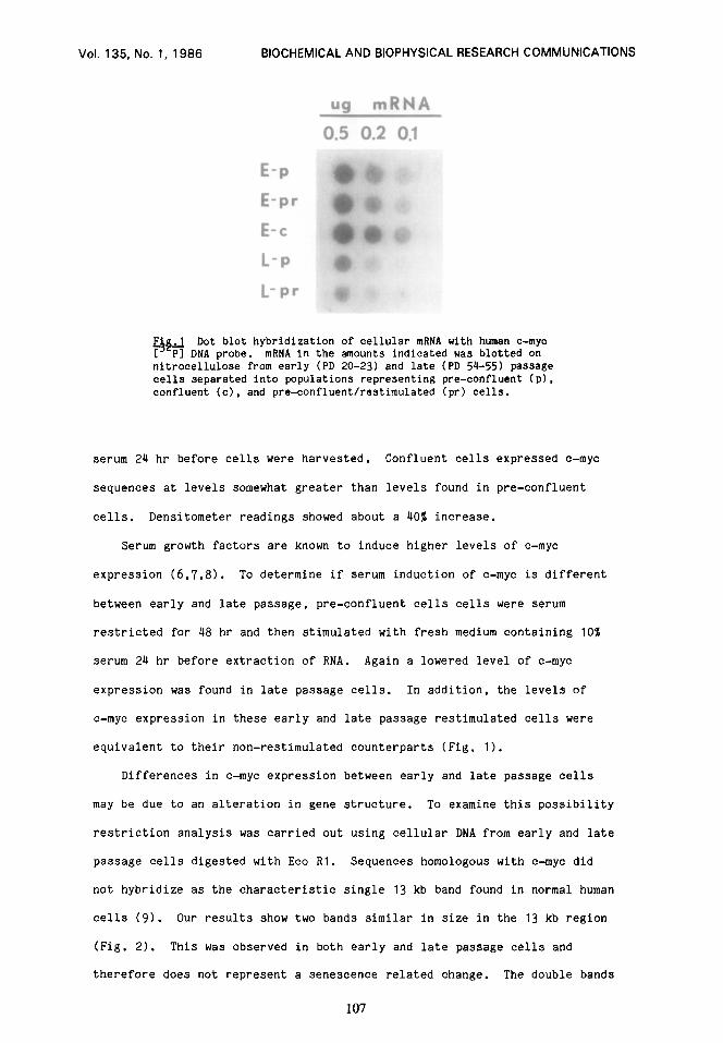
Dot blot hybridization of cellular mRNA with human c-myc ?‘$ DNA probe. mRNA in the amounts indicated was blotted on nitrocelluiose from early (PD ZO-23) and late (PD 54-55) passage cells separated into populations representing pre-confluent (p). confluent (c), and pre-confluent/restimulated (pr) cells.
serum 24 hr before cells were harvested. Confluent cells expressed c-myc
sequences at levels somewhat greater than levels found in pre-confluent
cells. Densitometer readings showed about a 40% increase.
Serum growth factors are known to induce higher levels of c-myc
expression (6.7.8). To determine if serum induction of c-myc is different
between early and late passage, pre-confluent cells cells were serum
restricted for 48 hr and then stimulated with fresh medium containing 10%
serum 24 hr before extraction of RNA. Again a lowered level of c-myc
expression was found in late passage cells. In addition, the levels of
c-myc expression in these early and late passage restimulated cells were
equivalent to their non-restimulated counterparts (Fig. 1).
Differences in c-myc expression between early and late passage cells
may be due to an alteration in gene structure. To examine this possibility
restriction analysis was carried out using cellular DNA from early and late
passage cells digested with Eco Rl. Sequences homologous with c-myc did
not hybridize as the characteristic single 13 kb band found in normal human
cells (9). Our results show two bands similar in size in the 13 kb region
(Fig. 2). This was observed in both early and late passage cells and
therefore does not represent a senescence related change. The double bands
107
Vol. 135, No. 1, 1986 BIOCHEMICAL AND BIOPHYSICAL RESEARCH COMMUNICATIONS
Fig.2 Southern blot hybridization of cellular DNA. 10 ug of DNA from early (PD 20) and late (PD 53) passage cells was digested with Eco Rl, sized on 0.8% agarose gelj2 transferred to nitrocellulose and hybridized with human c-myc [ PI DNA.
are puzzling and further investigations are required to determine if this
is a characteristic of IMR-90 cells alone.
DISCUSSION
Our results demonstrate a significant decrease in the level of c-myc
mRNA sequences found in pre-confluent late passage cells compared to early
passage cells. This result was found in cells examined 24 hr after
subculturing and in cells which had been restimulated with serum after
serum deprivation. The expression of c-myc promotes competence resulting
in cell growth (10). Reduced levels of c-myc expression could reduce entry
of cells into a state of competence resulting in the loss of proliferative
capacity found in late passage cells. The differences in levels of c-myc
expression between early and late passage cells we have found (2 to 3 fold)
are not as dramatic as differences found in 3T3 cells or other cell lines
(5.6). However, the level of c-myc expression in normal human fibroblasts
is very low, some 40 times lower than COLO 320 HSR cells (1). Thus, a
change of 2 to 3 fold in these cells may have a much more dramatic impact
108
Vol. 135, No. 1, 1986 BIOCHEMICAL AND BIOPHYSICAL RESEARCH COMMUNICATIONS
on cellular function than larger differences in less regulated or partially
transformed cell lines.
In contrast to our results with c-myc it has been reported that
c-Ha-ras messenger RNA and protein are elevated in late passage human
fibroblasts (II). Elevated levels of ras expression should promote
increased growth and division which is clearly not the case in senescent
fibroblasts. Thus c-myc expression may be a more important factor in
determining the senescent phenotype and may even determine this phenotype
in the face of elevated ras expression.
It has been reported that confluent arrested 3T3 and human fibroblasts
(7,) show diminished c-myc expression. Alternatively it has been reported
that c-myc is cell cycle invariant and confluent cells express c-myc at
normal levels so long as growth stimulating factors are present in the
media (8). Our results with confluent cells showed a small increase in
c-myc expression when compared to pre-confluent cells as long as both
populations were identically treated with serum growth factors. No
decrease was observed. Thus, our results most closely agree with the
finding that c-myc expression is not highly dependent on cell density.
1.
2.
3.
4.
5.
6.
7.
8.
9.
10.
11.
REFERENCES
Pfeifer-Ohlsson, S., Goustin, A., Rydnert, J., Wahlstrom, T., Bjersing, L., Stehelin, D. and Ohlsson R. (1984) Cell 38,585-596. Nishikura, K., ar-Rushdi, A., Erikson, J., Watt, R., Rovera, G. and Croce, C.M.(1983) Proc. Natl. Acad. Sci. USA 80, 4822-4826. Chirgwin, J.M., Przybyla, A.E., MacDonald, R.I. and Rutter, W.J. (1979). Biochemistry 18, 5294-5299. Maniatis, T., Fritsch, E.F. and Sambrook, J. (1982) Molecular Cloning A Laboratory Manual, pp. 382-387, Cold Spring Harbor Laboratory, Cold Spring Harbor. Battey, J., Moulding, C., Taub, R., Murphy, W., Stewart, T., Potter, H ., Lenoir, G. and Leder, P. (1983) Cell 34, 779-787 Kelly, K., Cochran, B.H., Stiles, C.D. and Leder. P. (1983) Cell 35, 603-610. Campisi, J., Gray, H.E., Pardee. A.B.. Dean, M. and Sonenshein, G.E. (1984) cell 36, 241-246. Thompson, C.B., Challoner, P., Neiman, P. and Groudine, M. (1985) Nature 314, 363-366 Dalla-Favera, R., Martinotti, S., Gallo, R., Erikson, J., and Croce, C. (1983) Science 219, 963-967. Kaczmarek, L., Hyland, J., Watt, R., Rosenberg, M. and Baserga, R. (1985) Science 228, 1313-1314. Srivastava, A., Norris, J.. Smookler Reis, R.J. and Goldstein, S. (1985). J. Biological Chem. 260, 6404-6409.