Expression of immune genes on chromosome 6p21.3–22.1 in schizophrenia Melissa L. Sinkus a,⇑ , Catherine E. Adams b , Judith Logel a , Robert Freedman a , Sherry Leonard a,b a Department of Psychiatry, University of Colorado Denver, Anschutz Medical Campus, Colorado 80045, USA b Veterans Affairs Medical Research Service, Denver, Colorado 80045, USA article info Article history: Received 1 November 2012 Received in revised form 14 January 2013 Accepted 30 January 2013 Available online 8 February 2013 Keywords: Schizophrenia Postmortem Brain Immune genes HLA-A HLA-B Inflammation Smoking Gene expression abstract Schizophrenia is a common mental illness with a large genetic component. Three genome-wide associa- tion studies have implicated the major histocompatibility complex gene region on chromosome 6p21.3– 22.1 in schizophrenia. In addition, nicotine, which is commonly abused in schizophrenia, affects the expression of central nervous system immune genes. Messenger RNA levels for genes in the 6p21.3– 22.1 region were measured in human postmortem hippocampus of 89 subjects. The effects of schizophre- nia diagnosis, smoking and systemic inflammatory illness were compared. Cell-specific expression patterns for the class I major histocompatibility complex gene HLA-A were explored utilizing in situ hybridization. Expression of five genes was altered in schizophrenic subjects. Messenger RNA levels for the class I major histocompatibility complex antigen HLA-B were increased in schizophrenic nonsmokers, while levels for smokers were indistinguishable from those of controls. b2 microglobulin, HLA-A and Notch4 were all expressed in a pattern where inflammatory illness was associated with increased expres- sion in controls but not in subjects with schizophrenia. Schizophrenia was also associated with increased expression of Butyrophilin 2A2. HLA-A was expressed in glutamatergic and GABAergic neurons in the dentate gyrus, hilus, and the stratum pyramidale of the CA1–CA4 regions of the hippocampus, but not in astrocytes. In conclusion, the expression of genes from the major histocompatibility complex region of chromosome 6 with likely roles in synaptic development is altered in schizophrenia. There were also significant interactions between schizophrenia diagnosis and both inflammatory illness and smoking. Ó 2013 Elsevier Inc. All rights reserved. 1. Introduction Genome-wide association studies (GWAS) have demonstrated that the major histocompatibility complex (MHC) gene region on chromosome 6p21.3–22.1 is strongly associated with schizophre- nia (Gejman et al., 2011; Purcell et al., 2009; Shi et al., 2009; Ste- fansson et al., 2009). The MHC region is a gene-rich area with large blocks of genes in high linkage disequilibrium. It is difficult to delineate which genes are responsible for the association with linkage analysis alone. However, information about their patholog- ical affects may be gained by looking at differences in the expres- sion of these genes in schizophrenia. This study investigates the expression of MHC region genes in the human postmortem hippo- campus in subjects with schizophrenia and normal controls. We selected MHC genes with potential brain-specific functions that are also located near SNPs with significant association to schizophrenia in GWAS studies, with the rationale that these genes are likely to exhibit expression changes in schizophrenia. The most studied of these are the class I major histocompatibility complex antigens (MHCI) (Shatz, 2009). In the central nervous system (CNS), MHCI is required for the formation and revision of dendrites during development, as well as for synaptic plasticity in the adult brain (Boulanger, 2009; Corriveau et al., 1998; Huh et al., 2000; Shatz, 2002). MHCI is involved in dendritic pruning, a process of synaptic revision where redundant synaptic connections are elim- inated and useful ones are strengthened. Over-expression of MHCI may induce excessive pruning. Observations of decreased prefron- tal and temporal brain volume (Pantelis et al., 2005; Shenton et al., 2001) and decreased dendritic spine density (Kolluri et al., 2005; Rosoklija et al., 2007) in schizophrenia have led to renewed inter- est in over-pruning as a developmental mechanism in this disor- der. We investigated four MHCI genes (called human leukocyte antigens, HLA, in humans) including HLA-A, -B, -C and -G, as well as two genes involved in MHCI synthesis and assembly (TAP1 and TAPBP)(Fellerhoff and Wank, 2009). We also measured expression of b2 microglobulin (B2M). B2M is not located on chromosome 6 (it is on chromosome 15q21.1–22.2); however, it is a co-subunit of the MHCI protein, and is required for stable cell surface expression of almost all MHCI molecules. Class II major histocompatibility proteins (MHCII) may also play an important role in regulating synapse formation and main- tenance. These proteins are expressed on microglia and their 0889-1591/$ - see front matter Ó 2013 Elsevier Inc. All rights reserved. http://dx.doi.org/10.1016/j.bbi.2013.01.087 ⇑ Corresponding author. Address: Department of Psychiatry, Mailstop 8344, University of Colorado Health Sciences Center, Aurora, Colorado 80045, USA. Tel.: +303 724 4403; fax: +303 724 4425. E-mail address: [email protected](M.L. Sinkus). Brain, Behavior, and Immunity 32 (2013) 51–62 Contents lists available at SciVerse ScienceDirect Brain, Behavior, and Immunity journal homepage: www.elsevier.com/locate/ybrbi
Transcript
Brain, Behavior, and Immunity 32 (2013) 51–62
Contents lists available at SciVerse ScienceDirect
Brain, Behavior, and Immunity
journal homepage: www.elsevier .com/locate /ybrbi
Expression of immune genes on chromosome 6p21.3–22.1 in schizophrenia
Melissa L. Sinkus a,⇑, Catherine E. Adams b, Judith Logel a, Robert Freedman a, Sherry Leonard a,b
a Department of Psychiatry, University of Colorado Denver, Anschutz Medical Campus, Colorado 80045, USAb Veterans Affairs Medical Research Service, Denver, Colorado 80045, USA
a r t i c l e i n f o
Article history:Received 1 November 2012Received in revised form 14 January 2013Accepted 30 January 2013Available online 8 February 2013
Schizophrenia is a common mental illness with a large genetic component. Three genome-wide associa-tion studies have implicated the major histocompatibility complex gene region on chromosome 6p21.3–22.1 in schizophrenia. In addition, nicotine, which is commonly abused in schizophrenia, affects theexpression of central nervous system immune genes. Messenger RNA levels for genes in the 6p21.3–22.1 region were measured in human postmortem hippocampus of 89 subjects. The effects of schizophre-nia diagnosis, smoking and systemic inflammatory illness were compared. Cell-specific expressionpatterns for the class I major histocompatibility complex gene HLA-A were explored utilizing in situhybridization. Expression of five genes was altered in schizophrenic subjects. Messenger RNA levels forthe class I major histocompatibility complex antigen HLA-B were increased in schizophrenic nonsmokers,while levels for smokers were indistinguishable from those of controls. b2 microglobulin, HLA-A andNotch4 were all expressed in a pattern where inflammatory illness was associated with increased expres-sion in controls but not in subjects with schizophrenia. Schizophrenia was also associated with increasedexpression of Butyrophilin 2A2. HLA-A was expressed in glutamatergic and GABAergic neurons in thedentate gyrus, hilus, and the stratum pyramidale of the CA1–CA4 regions of the hippocampus, but notin astrocytes. In conclusion, the expression of genes from the major histocompatibility complex regionof chromosome 6 with likely roles in synaptic development is altered in schizophrenia. There were alsosignificant interactions between schizophrenia diagnosis and both inflammatory illness and smoking.
� 2013 Elsevier Inc. All rights reserved.
1. Introduction
Genome-wide association studies (GWAS) have demonstratedthat the major histocompatibility complex (MHC) gene region onchromosome 6p21.3–22.1 is strongly associated with schizophre-nia (Gejman et al., 2011; Purcell et al., 2009; Shi et al., 2009; Ste-fansson et al., 2009). The MHC region is a gene-rich area withlarge blocks of genes in high linkage disequilibrium. It is difficultto delineate which genes are responsible for the association withlinkage analysis alone. However, information about their patholog-ical affects may be gained by looking at differences in the expres-sion of these genes in schizophrenia. This study investigates theexpression of MHC region genes in the human postmortem hippo-campus in subjects with schizophrenia and normal controls.
We selected MHC genes with potential brain-specific functionsthat are also located near SNPs with significant association toschizophrenia in GWAS studies, with the rationale that these genesare likely to exhibit expression changes in schizophrenia. The moststudied of these are the class I major histocompatibility complex
ll rights reserved.
Psychiatry, Mailstop 8344,a, Colorado 80045, USA. Tel.:
. Sinkus).
antigens (MHCI) (Shatz, 2009). In the central nervous system(CNS), MHCI is required for the formation and revision of dendritesduring development, as well as for synaptic plasticity in the adultbrain (Boulanger, 2009; Corriveau et al., 1998; Huh et al., 2000;Shatz, 2002). MHCI is involved in dendritic pruning, a process ofsynaptic revision where redundant synaptic connections are elim-inated and useful ones are strengthened. Over-expression of MHCImay induce excessive pruning. Observations of decreased prefron-tal and temporal brain volume (Pantelis et al., 2005; Shenton et al.,2001) and decreased dendritic spine density (Kolluri et al., 2005;Rosoklija et al., 2007) in schizophrenia have led to renewed inter-est in over-pruning as a developmental mechanism in this disor-der. We investigated four MHCI genes (called human leukocyteantigens, HLA, in humans) including HLA-A, -B, -C and -G, as wellas two genes involved in MHCI synthesis and assembly (TAP1 andTAPBP) (Fellerhoff and Wank, 2009). We also measured expressionof b2 microglobulin (B2M). B2M is not located on chromosome 6 (itis on chromosome 15q21.1–22.2); however, it is a co-subunit ofthe MHCI protein, and is required for stable cell surface expressionof almost all MHCI molecules.
Class II major histocompatibility proteins (MHCII) may alsoplay an important role in regulating synapse formation and main-tenance. These proteins are expressed on microglia and their
52 M.L. Sinkus et al. / Brain, Behavior, and Immunity 32 (2013) 51–62
expression increases when microglia are activated (Gehrmannet al., 1995). Microglia are a part of the innate immune system inthe brain. They also play a role in synaptic plasticity by alteringthe microenvironment of the synapse via cytokine secretion. Acti-vation is accompanied by an increase in secretion of tumor necro-sis factor a (TNFa), a cytokine that mediates activity-dependentsynaptic scaling (Albensi and Mattson, 2000; Stellwagen andMalenka, 2006). TNFa inhibits long-term potentiation by com-bined activation of TNF receptor 1 and metabotropic glutamatereceptor 5. Microglia also may eliminate dendritic spines byphagocytosis (Blank and Prinz, 2012). Schizophrenia patients haveincreased numbers of activated microglia and fewer dendriticspines (Radewicz et al., 2000; Rosoklija et al., 2007). We thereforemeasured expression for three MHCII genes (HLA-DRA, -DQA1, -DRB5) and the TNF gene.
Two other MHC region genes are of potential interest. TheNotch4 gene is within 7 KB of a SNP with genome-wide significancefor association to schizophrenia in two GWAS (Purcell et al., 2009;Stefansson et al., 2009). Other work suggests a significant decreasein expression in the Notch pathway in schizophrenia (Brennandet al., 2011). Butyrophilin 2A2 is an immune cell-surface protein.Messenger RNA levels of this gene in the brain are higher than inall other organs (Smith et al., 2010), however, the gene is littlestudied and its function in the brain is unknown.
MHC genes in the CNS are regulated by inflammatory factors,including cytokines (Neumann et al., 1997). This fact is relevantin studies of postmortem brain, where many subjects have diedin the presence of infection or other types of systemic inflamma-tion such as autoimmune disease. If not included in the analysis,inflammatory illness could act as a confounding factor that may in-flate estimates of expression levels for these immune genes. Moreimportantly, many studies have demonstrated an association be-tween immune activation and increased risk for schizophrenia(Brown and Derkits, 2009; Brown et al., 2004; Fellerhoff et al.,2006; Khandaker et al., 2012; Patterson, 2007). Interactions be-tween schizophrenia and inflammatory illness may significantlyaffect the expression of these genes. Therefore the presence of sys-temic inflammatory illness was included in our analysis as a factorin MHC gene expression.
Smoking can also act as a confounding factor in studying generegulation in schizophrenia. MHC expression in schizophrenicscould be altered by a high prevalence of smoking, which sup-presses immune function (de Leon and Diaz, 2005; Olincy et al.,1997). More than 80% of schizophrenic patients smoke comparedto approximately 25% of the general population (de Leon and Diaz,2005; Leonard et al., 2001). Nicotine and acetylcholine suppressthe immune response (Gallowitsch-Puerta and Tracey, 2005). Sub-jects who smoke may be suppressing the expression of immunegenes with brain-specific functions. We therefore included smok-ing status as a potential covariate in determining MHC geneexpression.
In recent studies, both MHCI and MHCII have been shown to beinvolved in regulation of synaptic plasticity (Albensi and Mattson,2000; Shatz, 2009). Plasticity is required for successful learningand memory. In the mature adult brain, these processes are mostnotably active in the hippocampus, a key brain region for memoryformation. The hippocampus undergoes functional losses parallelto those found in frontal cortex in schizophrenia, including volumeloss as well as alterations in glutamatergic, GABAergic and neureg-ulin signaling (Heckers and Konradi, 2002; Law et al., 2007; Mexalet al., 2005). Patients with schizophrenia are impaired in multiplecognitive tasks that are mediated though the hippocampus includ-ing sensory gating (Freedman et al., 1996), as well as both spatialand verbal declarative memory (Stone and Hsi, 2011; Stoneand Seidman, 2008). In schizophrenia, this cognitive dysfunctionis both chronic and disabling. For these reasons we chose to
look for differences in MHC gene expression in thehippocampus.
We measured gene expression using quantitative real-time PCR(QRT-PCR) in postmortem hippocampus in 89 subjects. We evalu-ated changes in gene expression as a function of schizophrenia diag-nosis, smoking status, and the presence of systemic inflammatoryillness at the time of death. MHCI and MHCII promote processes insynaptic development that decrease dendritic spine density andsynapse number (Blank and Prinz, 2012; Shatz, 2002); schizophreniais associated with both decreased dendritic spine density and re-duced synaptic function (Meador-Woodruff and Healy, 2000; Ros-oklija et al., 2007). Our hypothesis is that MHCI and MHCIIexpression is increased in schizophrenia, while Notch4 expressionmay be decreased. The presence of inflammatory illness would in-crease expression of these genes, while smoking may suppress it.
Prior to the current study, cell specific expression patterns ofMHCI genes have not been explored in the human brain. Since virtu-ally nothing is known about how these genes are expressed in thehuman brain, and animal models do not fully explain gene functionin human brain, it is important to understand which cells expressMHC genes in the human brain as a beginning to understandinghow these genes are associated with neuropsychiatric disease. Wetherefore initiated studies to define expression patterns in the hu-man hippocampus. The MHCI gene HLA-A was the most highly ex-pressed MHCI gene in our hippocampal tissue samples andtherefore a good candidate for exploring expression patterns. Weutilized in situ hybridization and immunofluorescent double label-ing to localize hippocampal expression of HLA-A to specific cell types.
2. Methods
2.1. Subjects
The Schizophrenia Research Center Brain Bank at the Universityof Colorado Medical Campus provided all of the postmortem braintissue, which was donated through the Colorado Anatomical GiftAct. The Brain Bank contains brain tissue from subjects with a diag-nosis of schizophrenia as well as from unaffected individuals. Con-trol subjects were evaluated by the same procedures asschizophrenic subjects as follows: Pre- and postmortem parame-ters were recorded for each subject from an extensive review ofhospital, autopsy, and neuropathology reports, as well as struc-tured interviews with physicians and family, as described previ-ously (Mexal et al., 2006; Mexal et al., 2005). Based on thisinformation, all subjects were evaluated by two independent psy-chiatrists and A DSM-IV diagnosis of was confirmed in the affectedsubjects. Controls were determined not to have any neuropsychiat-ric disorder by the same procedures. Utilizing these data, group dif-ferences in variables that may affect gene expression wereevaluated by analysis of variance (ANOVA), to insure that nogroups differed on any of the key variables. These variables in-cluded brain pH, subject age, tissue storage time, gender, RNAquality as indicated by RNA integrity number (RIN), and postmor-tem interval (PMI) (Groups are summarized in Table 1; for a de-tailed table of all subjects, see Table S1 in the supplementalmaterial). Tissue samples from ninety eight subjects were initiallyevaluated for the study. Seven samples were excluded due to poortissue quality (RIN 6 5 and/or brain pH < 6). Two additional sub-jects were excluded due to the use of immunosuppressant drugs(chemotherapy agents and corticosteroids). The remaining 89 sam-ples were included in the analysis. After excluding these nine sam-ples, there were no significant differences among groups for RIN,brain pH, storage time, subject age, or gender distribution. How-ever, schizophrenic subjects had a significantly longer PMI com-pared to controls.
Mean (Standard Deviation). RIN: RNA integrity number; PMI: postmortem interval in hours; Storage: brain storage time in days; age: subject age at death. Inflam: number inthe group with an inflammatory condition. There are no significant differences among the groups in RIN, pH, storage time, age, or sex ratio.
a Schizophrenic subjects had longer postmortem intervals compared to controls (p < 0.001).
Fig. 1. Relative expression levels of B2M and three HLA genes in control subjectswithout an inflammatory condition. Mean expression compared to GAPDH,expressed as a percentage. Bars are one S.E.M. HLA-G does not have a bar becausethere was no detectable expression of this gene.
M.L. Sinkus et al. / Brain, Behavior, and Immunity 32 (2013) 51–62 53
In postmortem brain studies, PMI is defined as the time elapsedbetween the time of death and the time the brain is removed fromthe skull. In many brain banks, affected individuals have longerPMIs compared to controls because they are relatively rare, there-fore extra time and effort is invested to locate the next of kin andseek consent for tissue donation. Such is the case at the Schizo-phrenia Research Center Brain Bank at the University of Colorado.Although the groups were matched for all other variables, theschizophrenic subjects had a significantly longer PMI comparedto controls (p < 0.001). PMI has been shown to have a limited effecton mRNA and protein assays in postmortem brain. No relationshipbetween PMI and total amounts of RNA has been found for PMIsbetween 0 and 48 h (Harrison et al., 1995). The subjects in thisstudy all had PMIs less than 48 h (range: 2–45 h) therefore a longerPMI in the affected subjects is unlikely to have an effect on our re-sults. However, to avoid any possible confounding effect we evalu-ated the effect of PMI on mRNA levels for each gene in the study. IfPMI was found to have a significant effect on expression levels forany gene, PMI was included as a covariate in the analyses for thatgene. In our samples PMI was only associated with mRNA levels forthe TAPBP gene.
Subjects were classified by diagnosis (schizophrenia vs. con-trol). In this brain collection, no information about the level ofsmoking or severity of infection/inflammation was available forthe subjects in the study; therefore smoking status was classifiedcategorically (smoking vs. nonsmoking). Information about inflam-matory illness was also classified categorically as the presence orabsence of a major infection (e.g. sepsis, pneumonia, encephalitis)or an inflammatory disease (Stevens-Johnson syndrome, rheuma-toid arthritis, scleroderma) at the time of death. Diagnoses andcauses of death are in Table S1.
2.2. Brain tissue
Human brain was collected at autopsy from local autopsy ser-vices following family consent. At autopsy, the brain was weighed,examined for gross pathology, and divided sagittally. One hemi-sphere was preserved for neuropathological analysis at the macro-scopic and microscopic level. Microscopic evaluations includedBielchowsky silver stain on multiple cerebral areas to rule outabnormal neuropathology, such as plaques and tangles associatedwith Alzheimer’s and other conditions. Patients with neuritic find-ings or ambiguous neuropathology reports were excluded. Theother hemisphere was sliced coronally into 1 cm slices. Hippocam-pal sections of approximately 1 g were dissected from these sec-tions, frozen on dry ice, and stored at �80 �C. Hippocampal tissuein each sample for this study was taken from the anterior-mostsection of the hippocampus that included CA4, CA3, CA2, CA1, den-tate gyrus, and subiculum.
For in situ hybridization, 25 hippocampal slices from each offour randomly chosen brains were used to visualize HLA-A expres-sion patterns and localize them to specific cell types (see Table S2).
2.3. RNA isolation and evaluation
RNA was isolated from human hippocampus with Trizol (LifeTechnologies, Gaithersburg, MD) following the manufacturer’s pro-tocol, and purified with an RNeasy mini kit (Qiagen, Valencia, CA).RNA integrity numbers (RIN) were derived from 300 ng of totalRNA using an Agilent 2100 Bioanalyzer and an Agilent 6000 Nanomicrofluidic chip kit (Agilent Technologies, Santa Clara, CA).The most reliable QRT-PCR gene expression results are obtainedwith RIN > 5 and amplicon length < 200 bp (Fleige and Pfaffl,2006; Fleige et al., 2006). Specimens in this study all had RINscores > 5.1 (mean RIN score = 7.3 ± 1.1) and all amplicons assayedwere < 200 bp.
2.4. Quantitative RT-PCR
cDNA was synthesized from total RNA using the SuperscriptFirst Strand Synthesis System for RT-PCR (Invitrogen, Carlsbad,CA). Gene expression was evaluated utilizing Taqman� geneexpression assays (Life Technologies, Carlsbad, California; for a fulllist of assays, amplicon lengths and target sequences, see Table S3.PCR was performed in a 7900HT thermal cycler (Life Technologies,Carlsbad, California). Cycling conditions were: 50 �C for 2 min,95 �C for 15 min, and 40 cycles of 94 �C for 15 s, 58 �C for 30 s,and 72 �C for 30 s. All assays were done in triplicate. The coefficientof variation for triplicates was <1%. Selection of a gene for normal-ization of mRNA levels was done by comparison of a panel ofhousekeeping genes (HIST1H2AG, HIST1H2BJ, GAPDH, B2M and POL-R2A) using NormFinder (Andersen et al., 2004). The gene GAPDHwas chosen by the NormFinder algorithm. It showed the higheststability and smallest intergroup variation. In order to eliminatebetween-run variability between the test and normalization genes,GAPDH was run as an endogenous internal control for each sample
54 M.L. Sinkus et al. / Brain, Behavior, and Immunity 32 (2013) 51–62
by utilizing duplex Taqman assays. Messenger RNA levels werethen normalized using GAPDH. Duplex assays were screened priorto use and no significant differences in PCR efficiency or thresholdcycle were found in duplex vs. singleplex reactions for GAPDH orany of the test genes. For each experiment, a sample without theaddition of reverse transcriptase enzyme or without the additionof cDNA was included to assess amplification from genomic DNAand nonspecific product formation, respectively.
2.5. In situ hybridization tissue preparation
Frozen blocks of hippocampal tissue were embedded in Tissue-Tek O.C.T. compound (Sakura Finetek, Torrance, CA). Coronal tissuesections 12 lm thick were cut at �18 �C from the embedded blockson a microtome cryostat (Bright Instrument Company, Hunting-don, England) and thaw-mounted on 3-aminopropyltriethoxysi-lane coated slides. The mounted sections were quick frozen ondry ice and stored at -80 �C until use.
2.6. Probe synthesis
A full length HLA-A clone (I.M.A.G.E. Clone I.D.: 3510139) waspurchased from American Type Culture Collection (Manassas,VA). A 360 bp fragment containing bp 959–1319 was amplifiedwith Taq polymerase. This region corresponds to exons 5–8, cross-ing intron/exon borders. These exons correspond to the transmem-brane portion of the HLA-A protein. BLAST searches were done toconfirm sequence specificity. Probe primers and sequence are inFigure S1.
The fragment was cloned into the vector pDrive and used totransform EZ competent cells with a Qiagen PCR Cloning Kit (Qia-gen, Valencia, CA). Plasmid DNA was isolated with a Qiagen Plas-mid Mini Kit. A sample of plasmid DNA was digested with EcoRIand run on a 1% agarose gel with ethidium bromide, revealing asingle insert band of approximately 360 bp. The clone was se-quenced using a 3100 Avant Genetic Analyzer (Applied Biosystems,Foster City, CA). Digoxigenin (DIG) labeled sense and antisenseRNA probes were synthesized with a DIG RNA labeling kit (RocheApplied Science, Indianapolis, IN). Labeling efficiency was deter-mined by northern dot blot.
2.7. In situ hybridization procedure
Tissue sections were thawed and dried at room temperature for30 min. Sections were rinsed in phosphate-buffered saline (PBS)then fixed with 4% paraformaldehyde in PBS, pH 7.2, for 20 minat room temperature. Then they were rinsed in dimethylpyrocar-bonate treated water. In situ hybridization was performed withan ISHyb in situ hybridization kit (BioChain, Hayward, CA). Foreach experiment a DIG labeled sense mRNA probe control was alsoincluded as a specificity control.
The slides were rinsed with room temperature PBS, pH 7.4.Nonspecific binding was blocked by incubating in PBS containing0.3% Triton X-100 (PBS-T) plus 5% normal mouse serum for onehour at room temperature. The probe was visualized with a Dy-Light-549 conjugated mouse anti-DIG antibody (Jackson Immuno-Research, West Grove, PA), diluted 1:200 with PBS-T. Nuclei werelabeled in 10 nm 40,6-diamidino-2-phenylindole (DAPI nuclearstain). After all labeling procedures were complete, lipofuscin in-duced autofluorescence was blocked by incubating for 10 min atroom temperature in 0.1% Sudan Black B dissolved in 70% ethanolas previously described (Newton et al., 2002; Schnell et al., 1999).The tissue was allowed to dry at room temperature, covered withProlong anti-fade solution (Invitrogen, Carlsbad, CA), coverslipped,allowed to cure at room temperature in the dark for 24 h, andstored at �20 �C. Slides were viewed under a Nikon Microphot
FXA fluorescence microscope and imaged using a Spot RT Sliderdigital camera (Diagnostic Instruments, Sterling Heights, MI).
2.8. Double fluorescent labeling
HLA-A mRNA expression was visualized by in situ hybridizationcombined with immunofluorescent counterstaining to localize celltype specific markers. A primary antibody against the neurotrans-mitter glutamate was used to label glutamatergic cells; an anti-body for GFAP was used to label astrocytes. An antibody againstboth the 65 and 67 kDa isoforms of glutamate decarboxylase(GAD65/GAD67) was used to label GABAergic interneurons. Forall experiments, a deletion control without primary antibody wasalso performed as a specificity control.
Nonspecific binding was blocked by incubating in PBS-T plus 5%normal donkey serum. Slides were rinsed with PBS, placed in ahumidity box and covered with 100–200 ul of PBS-T plus a primaryantibody (rabbit anti-glutamate (1:500), anti-GFAP (1:500), (Milli-pore, Billerica, MA) or anti-GAD65/GAD67 (1:200) (Abcam, Cam-bridge, MA). A deletion control without primary antibody wasincluded. The humidity box was sealed and incubated overnightat 4 �C.
Slides were rinsed in room temperature PBS, placed in a humid-ity box with 100–200 ul fluorescein isothiocyanate (FITC) conju-gated donkey anti-rabbit antibody (Jackson ImmunoResearch,West Grove, PA) diluted 1:100 in PBS-T, and incubated at 37 �Cfor 3 h, minimizing light exposure. Slides were rinsed four timesfor 10 min in 37 �C PBS.
2.9. Statistical analysis
Ninety eight brain tissue samples were initially evaluated forthe study. Seven samples were excluded due to poor tissue quality(RIN 6 5 and/or brain pH < 6). Two additional subjects were ex-cluded due to the use of immunosuppressant drugs (chemotherapyagents and corticosteroids). The remaining 89 samples were in-cluded in the analysis.
Subjects were classified by diagnosis (schizophrenia vs. con-trol), smoking status (smoking vs. nonsmoking) and presence ofinflammatory disease. Information about the level of smokingwas not available for the subjects. Individuals with a major infec-tion at the time of their death according to chart review (e.g. sepsis,pneumonia, encephalitis) or who had an inflammatory disease(Stevens-Johnson syndrome, rheumatoid arthritis, scleroderma)were given a group designation (inflammatory condition vs. nosuch condition). This inflammatory factor was used in the analysisas a potential variable in determining gene expression.
To determine whether the proportion of males to females wasdifferent across the experimental groups, a chi-squared test wasperformed. Group means were compared for PMI, brain pH, subjectage, storage time, and RIN score using an ANOVA.
For each subject, mean normalized expression values for thegene of interest were calculated using GAPDH mRNA levels. Expres-sion values were log2 transformed to approximate statistical testassumptions. To investigate the effects of schizophrenia, smokingand inflammation on gene expression, a multivariate linear regres-sion was used that included main and two-way interaction termsfor the three categorical variables of diagnosis, smoking status,and the presence of systemic inflammation at the time of death.Since PMI had a significant effect on expression levels for TAPBP,this factor was also included as a covariate in the model for thatgene. Backwards variable selection was used to eliminate non-sig-nificant effects and identify a reduced model for each gene. Thesubject groupings for each gene were therefore determined bythese variables and interactions. These groupings are presentedin the results graphs for each gene (Figs. 2–7). Main effects were
Fig. 2. Interactive effects of schizophrenia and inflammatory illness on HLA-A levels. Least squares means of log transformed expression relative to GAPDH. Means areadjusted for all other effects in the model. (A). Controls with inflammatory illness had significantly increased HLA-A expression compared to those with no inflammatoryillness [F(1,87) = 12.77; p = 0.002; fold D = 1.6]. This difference in expression did not occur in the schizophrenic subjects. (B). Smokers had significantly lower HLA-Aexpression [F(1,87) = 5.26; p = 0.038; fold D = 1.2]. Bars are one S.E.M.
Fig. 3. Interactive effects of schizophrenia, smoking and inflammatory illness on HLA-B expression. Least squares means of log transformed expression relative to GAPDH.Means are adjusted for all other effects in the model. (A). Schizophrenic nonsmokers had significantly increased HLA-B expression compared Control nonsmokers[F(1,87) = 10.69; p = 0.005; fold D = 2.3], and compared to schizophrenic smokers [F(1,87) = 22.99; p < 0.001; fold D = 2.7]. (B). Nonsmokers with an inflammatory illness hadsignificantly increased HLA-B expression compared to those with no inflammatory illness [F(1,87) = 18.15; p = 0.001; fold D = 2.4]. This difference in expression did not occurin the smokers. Bars are one S.E.M.
M.L. Sinkus et al. / Brain, Behavior, and Immunity 32 (2013) 51–62 55
constrained to remain in the model if an interaction was selected.Least-squares means estimates were calculated for parameterswith significant effects and used to illustrate effect sizes. Theseestimates were adjusted for all other parameters in the model.Hypotheses about significant differences between groups weretested using linear contrasts performed with the groups of interest.To adjust for multiple testing, false discovery rate controlling (FDR)adjustments (Benjamini and Hochberg, 1995) were globally per-
formed. All p-values presented are therefore empirical p-valuesgenerated by FDR adjustment.
The HLA-C gene was expressed in a binary, ‘‘on–off’’ fashion.Binary logistic regression was used to analyze expression of thisgene, with main and interaction terms for diagnosis, smoking sta-tus, and the presence of systemic inflammation at the time ofdeath. All analyses were performed using SAS 9.3 statistical soft-ware (SAS, Cary, NC).
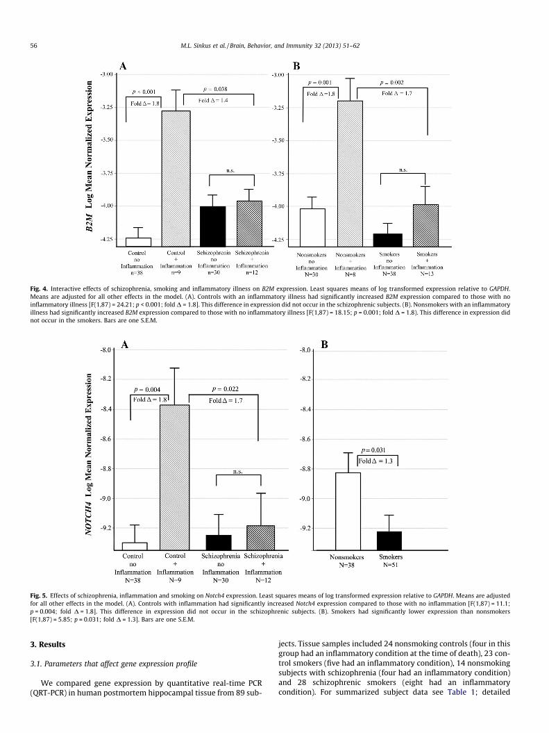
Fig. 4. Interactive effects of schizophrenia, smoking and inflammatory illness on B2M expression. Least squares means of log transformed expression relative to GAPDH.Means are adjusted for all other effects in the model. (A). Controls with an inflammatory illness had significantly increased B2M expression compared to those with noinflammatory illness [F(1,87) = 24.21; p < 0.001; fold D = 1.8]. This difference in expression did not occur in the schizophrenic subjects. (B). Nonsmokers with an inflammatoryillness had significantly increased B2M expression compared to those with no inflammatory illness [F(1,87) = 18.15; p = 0.001; fold D = 1.8). This difference in expression didnot occur in the smokers. Bars are one S.E.M.
Fig. 5. Effects of schizophrenia, inflammation and smoking on Notch4 expression. Least squares means of log transformed expression relative to GAPDH. Means are adjustedfor all other effects in the model. (A). Controls with inflammation had significantly increased Notch4 expression compared to those with no inflammation [F(1,87) = 11.1;p = 0.004; fold D = 1.8]. This difference in expression did not occur in the schizophrenic subjects. (B). Smokers had significantly lower expression than nonsmokers[F(1,87) = 5.85; p = 0.031; fold D = 1.3]. Bars are one S.E.M.
56 M.L. Sinkus et al. / Brain, Behavior, and Immunity 32 (2013) 51–62
3. Results
3.1. Parameters that affect gene expression profile
We compared gene expression by quantitative real-time PCR(QRT-PCR) in human postmortem hippocampal tissue from 89 sub-
jects. Tissue samples included 24 nonsmoking controls (four in thisgroup had an inflammatory condition at the time of death), 23 con-trol smokers (five had an inflammatory condition), 14 nonsmokingsubjects with schizophrenia (four had an inflammatory condition)and 28 schizophrenic smokers (eight had an inflammatorycondition). For summarized subject data see Table 1; detailed
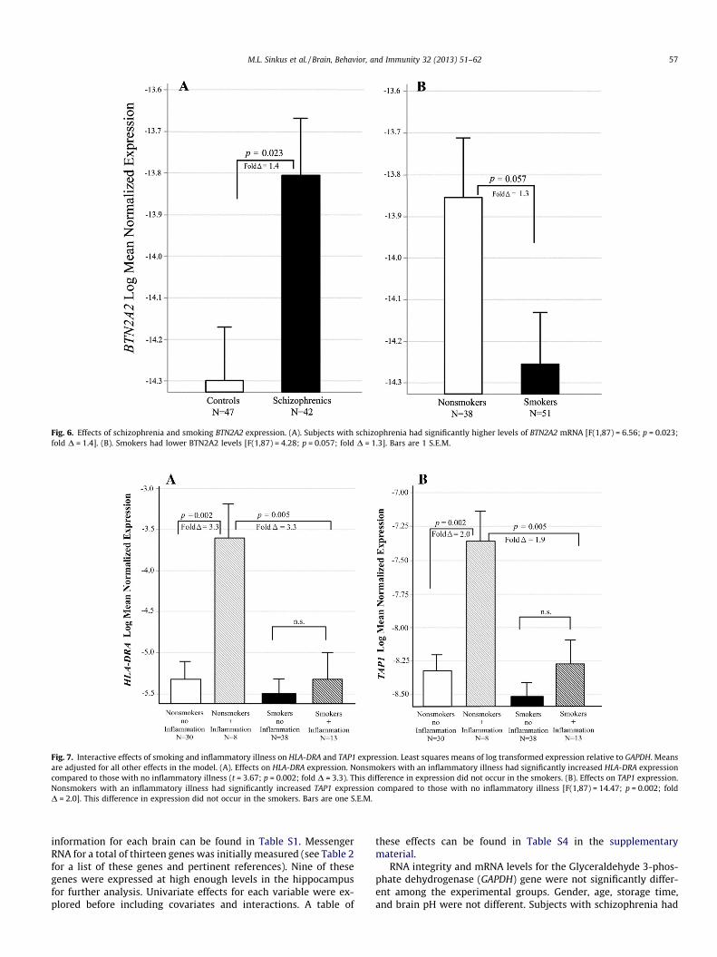
Fig. 6. Effects of schizophrenia and smoking BTN2A2 expression. (A). Subjects with schizophrenia had significantly higher levels of BTN2A2 mRNA [F(1,87) = 6.56; p = 0.023;fold D = 1.4]. (B). Smokers had lower BTN2A2 levels [F(1,87) = 4.28; p = 0.057; fold D = 1.3]. Bars are 1 S.E.M.
Fig. 7. Interactive effects of smoking and inflammatory illness on HLA-DRA and TAP1 expression. Least squares means of log transformed expression relative to GAPDH. Meansare adjusted for all other effects in the model. (A). Effects on HLA-DRA expression. Nonsmokers with an inflammatory illness had significantly increased HLA-DRA expressioncompared to those with no inflammatory illness (t = 3.67; p = 0.002; fold D = 3.3). This difference in expression did not occur in the smokers. (B). Effects on TAP1 expression.Nonsmokers with an inflammatory illness had significantly increased TAP1 expression compared to those with no inflammatory illness [F(1,87) = 14.47; p = 0.002; foldD = 2.0]. This difference in expression did not occur in the smokers. Bars are one S.E.M.
M.L. Sinkus et al. / Brain, Behavior, and Immunity 32 (2013) 51–62 57
information for each brain can be found in Table S1. MessengerRNA for a total of thirteen genes was initially measured (see Table 2for a list of these genes and pertinent references). Nine of thesegenes were expressed at high enough levels in the hippocampusfor further analysis. Univariate effects for each variable were ex-plored before including covariates and interactions. A table of
these effects can be found in Table S4 in the supplementarymaterial.
RNA integrity and mRNA levels for the Glyceraldehyde 3-phos-phate dehydrogenase (GAPDH) gene were not significantly differ-ent among the experimental groups. Gender, age, storage time,and brain pH were not different. Subjects with schizophrenia had
Fig. 8. HLA-A mRNA in the Human Hippocampus. This slice was taken from a control nonsmoking subject (subject SL283, see Table S1) but the qualitative pattern in allsubjects tested was the same. DG = dentate gyrus; gc = granule cells; CA1–4 = cornu ammonis areas 1–4. SI = subiculum. This image is a composite of 85 photomicrographstaken at 4X magnification. Bar = 1 mm.
58 M.L. Sinkus et al. / Brain, Behavior, and Immunity 32 (2013) 51–62
a longer mean post mortem interval (PMI) [mean PMI =20.1 ± 9.0 (SD) h] compared to controls (mean = 13.0 ± 7.1 h;p < 0.001). However, PMI only affected mRNA levels of theTAPBP gene, with increasing PMI associated with decreasedtranscript.
Table 2Genes evaluated in this study.
Gene symbola Official gene name Location
HLA-A Major histocompatibility complex, class I, A 6p21.3-MHCI regio
HLA-B Major histocompatibility complex, class I, B
HLA-C Major histocompatibility complex, class I, CHLA-G Major histocompatibility complex, class I, GB2M Beta-2 microglobulin (co-subunit of MHCI) 15q21-q22.2
TAP1 Transporter 1, ATP-binding cassette, sub-family B (MHCI synthesis)
6p21.3-MHCII regi
TAPBP TAP binding protein; AKA tapasin, (MHCIchaperone protein)
6p21.3-extendedMHCII region
HLA-DRA Major histocompatibility complex, class II, DRalpha
6p21.3-MHCII regi
HLA-DRB5 Major histocompatibility complex, class II, DRbeta 5
HLA-DQA1 Major histocompatibility complex, class II, DQalpha 1
NOTCH4 Notch 4 6p21.3-MHCII regi
TNF Tumor necrosis factor alpha 6p21.3
BTN2A2 Butyrophilin, subfamily 2, member A2 6p22.1
Gene symbol = official gene symbol designated by the HUGO Gene Nomenclature ComReferences = previous studies that have demonstrated genetic association or differential
a Human MHCI and MHCII genes were initially called human leukocyte antigens (HLAretained in the official gene symbols, but not in the HGNC full length names of the gen
3.2. Relative expression levels of MHCI genes
HLA-A was the most highly expressed MHCI gene assayed. Incontrols it was expressed in essentially a 1:1 ratio with b2-micro-globulin (see Fig. 1). HLA-B and HLA-C were expressed 3.5 and 5-
References
n Purcell et al. (2009)- alleles associated with rs3130375, a SNP significant at3.66 � 10�7
Mexal et al. (2005)-HLA-A expression increased in postmortemhippocampus of schizophrenic nonsmokersShatz et al. (2002)-MHCI essential for axono-dendritic remodeling
Mexal et al. (2005)-expression increased in post-mortem hippocampus ofschizophrenic nonsmokers
on Fellerhoff et al. (2009)-A1/B1 genotype associated with schizophrenia withodds ratio 14.1Purcell et al. (2009)-SNP within 20 KB associated at 4.49 � 10�5
Fellerhoff et al. (2009)-01/01 genotype associated with schizophreniaon Purcell et al. (2009) - DRB and DQA alleles associated with rs3130375
A SNP significant at 3.66 � 10�7; DRA within 20 KB of a SNP associated at8.34 � 10�6
Shi et al. (2009) HLA-DQA1-upstream and intronic SNPs significant at6.88 � 10�8 and 8.87 � 10�8 respectively
on Purcell et al. (2009), Stefansson et al. (2009)-genome wide association of aSNP 7 KB from the geneBrennand et al. (2011)-significant decrease in expression in the NOTCHpathway in schizophreniaAlbensi and Mattson (2000)-Evidence for the involvement of TNF andNF-kappaB in hippocampal synaptic plasticityStellwagen and Malenka (2006)-Synaptic scaling mediated by glialTNF-alphaPurcell et al. (2009) SNP within 20 KB associated at 1.18 � 10�5
Smith et al. (2010)-Brain has the highest mRNA levels of BTN2A2 comparedto all other organs
mittee (HGNC). Official gene name = official HGNC full length name of the gene.expression in schizophrenia.) prior to understanding the full scope of their function. This designation has been
es.
M.L. Sinkus et al. / Brain, Behavior, and Immunity 32 (2013) 51–62 59
fold less respectively compared to HLA-A. There was no detectableexpression of HLA-G in any of the subjects.
3.3. Hippocampal MHC gene expression in subjects with schizophrenia
To investigate the effects of schizophrenia, smoking and inflam-matory illness on gene expression, multivariate linear regressionmodeling was used that included main and interaction terms fordiagnosis, smoking status, the presence of a systemic inflammatoryillness at the time of death. A reduced model containing all signif-icant effects was produced for each gene analyzed. Global adjust-ment for multiple testing was performed utilizing false discoveryrate (FDR) methods, therefore all p-values given are empirical p-values generated by FDR adjustment (adj p).
For HLA-A, mRNA levels differed across schizophrenia diagnosisand the presence of inflammatory disease at the time of death (adjp = 0.045 for the interaction). Controls with an inflammatory ill-ness had significantly increased HLA-A expression compared tocontrols with none (adj p = 0.002; fold D = 1.6; see Fig. 2A). Thisdifference in expression did not occur in the schizophrenic sub-jects. Controls with an inflammatory illness also had significantlyhigher HLA-A expression compared to schizophrenics with such ill-ness (adj p = 0.023; fold D = 1.5). Smoking also had an effect onHLA-A levels. Smokers had decreased expression in all subjects(adj p = 0.038, fold D = 1.2; Fig. 2B) after adjusting for the schizo-phrenia and inflammatory illness groups.
For the gene HLA-B, mRNA levels differed across groups definedby diagnosis and smoking status (adj p = 0.017 for the interaction).Fig. 3A illustrates the interaction between these two variables.Schizophrenic nonsmokers had significantly increased HLA-Bexpression compared control nonsmokers (adj p = 0.005; foldD = 2.3), and compared to schizophrenic smokers (adj p < 0.001;fold D = 2.7; Fig. 3A). There was also an interaction between the ef-fects of smoking and inflammatory illness (adj p = 0.022 for theinteraction). Nonsmokers with an inflammatory illness at the timeof death had significantly increased HLA-B expression compared tothose with no inflammatory illness (adj p = 0.001; fold D = 2.4).This difference in expression did not occur in the smokers (Fig. 3B).
HLA-C was expressed in a binary pattern. Forty-two out of the89 subjects expressed no detectable HLA-C, while the remainderdisplayed moderate expression (threshold cycle �28). Schizo-phrenic subjects tended to be more likely to be non-expressers,however this was not statistically significant after adjustment formultiple testing (v2 = 3.94, adj p = 0.063). Among subjects who ex-pressed HLA-C, there was no difference in expression between con-trols and schizophrenics.
B2M mRNA levels differed across groups defined by schizophre-nia diagnosis and the presence of inflammatory illness (adjp = 0.011 for the interaction). Controls with an inflammatory ill-ness at the time of death had significantly increased B2M expres-sion compared to those with no inflammatory illness (adjp < 0.001; fold D = 1.8; see Fig. 4A). This difference in expressiondid not occur in the schizophrenic subjects. A similar interactionwas found between smoking and inflammatory illness (adjp = 0.032 for the interaction). Nonsmokers with inflammatory ill-ness at the time of death had significantly increased B2M expres-sion compared to those with no inflammatory illness (adjp = 0.001; fold D = 1.8). This difference in expression did not occurin the smokers (Fig. 4B)
Notch4 expression differed across groups defined by schizo-phrenia and inflammatory illness in a pattern similar to thosefound in B2M and HLA-A (adj p = 0.022 for the interaction; seeFig. 5). Controls with an inflammatory illness had significantly in-creased Notch4 expression compared to those with no inflamma-tory illness (adj p = 0.004; fold D = 1.8, Fig. 5A). This difference inexpression did not occur in the schizophrenic subjects. Smoking
decreased expression of this gene in all subjects (adj p = 0.031, foldD = 1.3, Fig. 5B) after adjusting for the schizophrenia and inflam-matory illness groups.
For the butyrophilin 2A2 gene, Expression was increased inschizophrenia (adj p = 0.023, fold D = 1.4; Fig. 6A). Expression wasdecreased in smokers, but this was only marginally significant aftercorrection for multiple testing (adj p = 0.057, fold D = 1.3; Fig. 6B).
3.4. Genes with no expression changes in schizophrenia
HLA-DRA levels differed only across the combined effects ofsmoking and inflammatory disease at the time of death (adjp = 0.023 for the interaction). Nonsmokers with an inflammatoryillness had significantly increased HLA-DRA expression comparedto those with no inflammatory illness (adj p = 0.002; fold D = 3.3;see Fig. 7A). This increase in expression did not occur in thesmokers.
Similarly, TAP1 levels differed only across the combined effects ofsmoking and inflammatory illness at the time of death (adj p = 0.045for the interaction). Nonsmokers with an inflammatory illness hadsignificantly increased TAP1 expression compared to those with noinflammatory illness (adj p = 0.002; fold D = 2.0; see Fig. 7B). Thisdifference in expression did not occur in the smokers for this gene.
Expression of the TAPBP gene was associated only with PMI: in-creased PMI was associated with decreased transcript (b = �0.01;adj p = 0.023).
3.5. Genes with poor expression in the hippocampus
For four genes, mRNA levels detected by the Taqman assay weretoo low for accurate analysis (mean threshold cycle >34). These in-cluded HLA-G, HLA-DQA1, HLA-DRB5 and TNF.
3.6. In Situ hybridization
The in situ procedure was performed with sense riboprobes as anegative control. The antisense probe labeled cells bright red (Fig-ure S2-A in the supplemental material). The sense probe produceda much weaker signal (Figure S2, B).
HLA-A mRNA was very well expressed, allowing visualization ofthe architecture of the whole hippocampus. Labeled HLA-A mRNAwas visible in cells of the granule cell layer of the dentate gyrus,the hilus, and the stratum pyramidale of the CA1, CA2, CA3, andCA4 regions of the hippocampus (Fig. 8).
To localize HLA-A expression to specific cell types, in situ hybrid-ization was combined with immunofluorescent labeling for cell-type specific proteins. For astroglia, FITC (green) immunofluores-cent labeling for glial fibrillary acidic protein (GFAP) was combinedwith in situ hybridization for HLA-A mRNA (DyLight 549, red). Cellsthat labeled for HLA-A mRNA were not observed to contain fila-ments labeled for GFAP and cells that labeled for GFAP did not labelfor HLA-A (Fig. 9A–C). The two labels appeared to be mutuallyexclusive.
When cells were dually labeled for HLA-A mRNA and glutamate,the two fluorescent tags followed a similar distribution in the cellbodies and processes, producing a yellow color (Fig. 9D–F).
A FITC labeled antibody against both the 65 and 67 kDa iso-forms of glutamate decarboxylase (GAD65/GAD67) was used to la-bel GABAergic interneurons. All observed cells that were positivefor GAD also labeled for HLA-A mRNA. The HLA-A label (red) wasprimarily visible in the cell body, but also in cell processes at lowintensity (Fig. 9G). GAD labeling (green) was present in cell bodiesbut was more intense in cell processes (Fig. 9H). This staining pat-tern produced bi-colored cells with yellow/orange cell bodies andgreen processes (Fig. 9I). These labeling patterns were identicalin all four subjects chosen for in situ hybridization, regardless of
Fig. 9. Double Labeling for HLA-A and Cell Specific Markers in the Hippocampus. (A–C) Double Labeling for HLA-A and GFAP in a group of cells in the CA3 region. HLA-A mRNAwas detected by in situ hybridization and labeling with a DyLight 549 conjugated antibody (red). GFAP was detected by FITC labeled antibodies (green). Nuclei were labeledwith DAPI nuclear stain (blue). (A). Fluorescence microscopy image showing HLA-A positive cells. (B). Fluorescence microscopy showing intermediate filaments labeled forGFAP. (C). Merged image with all three wavelengths. HLA-A labeled cells do not appear to contain filaments labeled for GFAP. (D–F) Double Labeling for HLA-A and Glutamatein a group of cells is in the CA3 region. HLA-A mRNA was detected by in situ hybridization and labeling with a DyLight 549 conjugated antibody (red).Glutamate was detectedwith a FITC labeled antibody (green). Nuclei were labeled with DAPI nuclear stain (blue). (D). Fluorescence microscopy image showing HLA-A positive cells. (E). Fluorescencemicroscopy showing glutamate labeled cells. (F). Merged image with all three wavelengths. (G–I) Double labeling for HLA-A and GAD65/GAD67 in the dentate gyruspolymorphic layer. HLA-A mRNA was detected by in situ hybridization and labeling with a DyLight 549 conjugated antibody (red). GAD65 and GAD67 were detected by a FITClabeled antibody (green). Nuclei were labeled with DAPI nuclear stain (blue). (G). Fluorescence microscopy image showing HLA-A positive cells. (H). Fluorescence microscopyshowing GAD65/GAD67 labeled cells. (I). Merged image with all three wavelengths. Bar = 25 microns.
60 M.L. Sinkus et al. / Brain, Behavior, and Immunity 32 (2013) 51–62
diagnosis, smoking status, or the presence of systemic inflamma-tion at the time of death.
4. Discussion
QRT-PCR was used to measure changes in immune gene expres-sion in the post-mortem hippocampus and to evaluate the effectsof schizophrenia diagnosis, smoking, and the presence of systemicinflammatory illness at the time of death. In situ hybridization wasused to locate and characterize cells expressing HLA-A, the mosthighly expressed MHC gene.
HLA-A mRNA was found in the granular cells of the dentategyrus, in the hilus, and in the stratum pyramidale of the CA1–CA4 regions of the hippocampus. HLA-A mRNA was observed incells that express glutamate and GAD65/67, but not those express-ing GFAP. This suggests that HLA-A is expressed in glutamatergicneurons and GABAergic interneurons but not in astrocytes. Thisis the first study to look at HLA-A expression in the human brainand to localize it to specific cell types. Future studies might furtherdefine expression in specific neuronal subtypes (for example par-valbumin expressing cells) and look at the expression of HLA-Band -C.
We identified two genes that were over-expressed in schizo-phrenia after adjustment for all other variables: HLA-B and BTN2A2.The butyrophilins are a little-studied group of MHC-associatedmembrane proteins. In T-cells, binding of BTN2A2 receptors de-creases secretion of interferon gamma and interleukin-2 (Smithet al., 2010). The role of butyrophilins in the brain has not beenstudied. In immune function they transduce signals from cell tocell that inhibit activation of transcription factors such as NF-jB.NF-jB signaling is critically involved in synapse formation andspine maturation. NF-jB blockade in the forebrain is associatedwith reduced levels of mature spines (Schmeisser et al., 2012), afinding that has been observed in schizophrenia (Rosoklija et al.,2007). This is consistent with previous studies that have foundabnormal immune function in schizophrenia.
HLA-B is a member of the MHCI gene family. Its expression wasincreased in schizophrenia, but only in the nonsmokers. Levels forsmokers were indistinguishable from those of controls. This pat-tern could indicate that HLA-B is over-expressed in schizophrenia,but smoking returns expression levels to within normal limits. Inaddition, over the entire group (including both schizophrenicsand controls), inflammation significantly increased HLA-B expres-sion in nonsmoking subjects, but not in smokers. This suggests that
M.L. Sinkus et al. / Brain, Behavior, and Immunity 32 (2013) 51–62 61
inflammatory illness may further increase the high levels of anHLA-B found in the nonsmoking schizophrenic subjects.
HLA-A, b2 microglobulin, and Notch4 expression displayed asimilar expression pattern with respect to schizophrenia. Expres-sion of these genes was increased significantly in control subjectswho had systemic inflammatory illness at the time of their death,but this difference in expression was not seen in the schizophrenicsubjects. This pattern could indicate that up-regulation of thesegenes normally occurs with such inflammation, but in schizophre-nia, this up-regulation fails to occur.
Taken together these expression data suggest a scenario ofaberrant MHCI expression in schizophrenia which is exacerbatedduring infection and other systemic inflammation. B2M is requiredfor stable expression of almost all MHCIs; its expression may rep-resent the maximum quantity of MHCI proteins on the cell surface.Both the HLA-A and HLA-B proteins require dimerization with b2microglobulin for stable expression. HLA-B expression was in-creased 2.3-fold in nonsmoking schizophrenic subjects. These datasuggest that in these subjects, increased quantities of HLA-B arecompeting for dimerization with a limited quantity of b2 micro-globulin. This may cause a shift from HLA-A to HLA-B expressionon the cell surface. Additionally, in the presence of inflammation,HLA-A failed to up-regulate in schizophrenic subjects (smokersand nonsmokers included). This could cause a further shift towardHLA-B expression in schizophrenia during inflammatory illness.The significance of these shifts is unclear because the roles of dif-ferent MHCI genes in the human brain have not been determined.In animal models, different MHCI genes are expressed in uniquesubsets of neurons throughout the CNS (Boulanger, 2009). The dif-ferent expression patterns seen in HLA-A and HLA-B may reflectheterogeneous roles in neuronal function. Individuals with thisaberrant expression pattern may be more susceptible to destruc-tion of synapses during infectious illness. Further, infection orinflammation during important periods in brain developmentmay increase risk for schizophrenia. Several different lines of studyhave previously demonstrated a link between prenatal or earlychildhood exposure to infection and schizophrenia (Brown andDerkits, 2009; Khandaker et al., 2012; Patterson, 2007).
There is now an extensive body of research linking inflamma-tion to other psychiatric disorders, especially major depressive dis-order (Krishnadas and Cavanagh, 2012; Miller et al., 2009).Inflammatory molecules appear to disrupt normal neural functionand connectedness. Research has yet to uncover what determineswhy one individual experiences a psychotic disorder while anotherdevelops an affective one, given similar psychological or physiolog-ical stressors. Individual genetic background likely plays a role. Inaddition, the timing of the exposure to inflammation may also bean important factor. Earlier exposures, especially prenatal or earlychildhood ones, may be more disruptive to neurodevelopment,resulting in greater cognitive impairment and psychosis.
In this collection of brain samples, which contains both subjectswith schizophrenia and unaffected controls, smoking had a signif-icant effect on expression of most of the genes that were analyzed.Smoking decreased expression of both HLA-A and Notch4. Smokingalso appears to interact with inflammation so that it inhibits theup-regulation that occurs during inflammatory illness in nonsmok-ers. This pattern was found in HLA-B, B2M, TAP1 and HLA-DRA.These patterns are meaningful for future studies of immune genesin schizophrenia because schizophrenia patients tend to smokemore than the general public (de Leon and Diaz, 2005). Measuringexpression of these genes without accounting for smoking mayproduce misleading results. Stimulation of nicotinic receptorsmight be a mechanism for these effects. The a7nAchR is a likelycandidate as a regulator due to its role in the inhibition of cytokinerelease, which is unique among the nicotinic receptors (Wanget al., 2003). Since there have been recent clinical trials of a7nAchR
agonists to improve cognition in schizophrenia (Freedman et al.,2008), these medications may also become a factor in this research.
Previous studies have reported conflicting results for the effectof schizophrenia on HLA-A expression (Kano et al., 2011; Mexalet al., 2005); however, these studies did not account for inflamma-tory illness in the subjects. This could have spuriously affected theoutcomes of these studies. One factor that may act as a confounderin this and other studies is the use of psychotropic medications. Allof the schizophrenic subjects in this study were prescribed one ormore medications at the time of their death. These included vari-ous antipsychotics, mood stabilizers, and other adjunctive medica-tions. Noncompliance is also common in this population and isdifficult to document. Several studies have suggested that antipsy-chotic drugs can regulate the immune response by altering cyto-kine secretion; however the pattern of changes is complex andconflicts from one study to the next (Cazzullo et al., 2002; Chenet al., 2012; Kim et al., 2001; Zhang et al., 2004).
The MHC gene region on chromosome 6p21.3–22.1 is a largeblock of over 100 genes which is consistently associated withschizophrenia in GWAS studies. These genes are so tightly linkedthat is difficult to narrow the association with current techniques.We evaluated nine of these genes for expression changes in schizo-phrenia. Expression of five was altered in subjects with schizophre-nia. These are primarily MHCI genes with putative roles in themodification of dendritic connections and in synaptic plasticity.
Acknowledgments
The authors would like to acknowledge Dr. Brandie Wagner allof her advice and consultation on the statistical analysis. Dr. Sinkuswould like to acknowledge the following funding sources: T32MH15442, the Developmental Psychobiology Endowment Fund,and the Ebaugh Foundation. Funding for Dr. Leonard: MH81177and the Veterans Affairs Medical Research Service.
Appendix A. Supplementary data
Supplementary data associated with this article can be found, inthe online version, at http://dx.doi.org/10.1016/j.bbi.2013.01.087.
References
Albensi, B.C., Mattson, M.P., 2000. Evidence for the involvement of TNF and NF-jB inhippocampal synaptic plasticity. Synapse 35, 151–159.
Andersen, C.L., Jensen, J.L., Ørntoft, T.F., 2004. Normalization of real-timequantitative reverse transcription-PCR data: a model-based varianceestimation approach to identify genes suited for normalization, applied tobladder and colon cancer data sets. Cancer Res. 64, 5245–5250.
Benjamini, Y., Hochberg, Y., 1995. Controlling the false discovery rate-a practicaland powerful approach to multiple testing. J. Royal Statistical Soc. Ser. B-Method. 57, 289–300.
Blank, T., Prinz, M., 2012. Microglia as modulators of cognition and neuropsychiatricdisorders. Glia (ePub ahead of print).
Boulanger, L.M., 2009. Immune proteins in brain development and synapticplasticity. Neuron 64, 93–109.
Brennand, K.J., Simone, A., Jou, J., Gelboin-Burkhart, C., Tran, N., Sangar, S., Li, Y., Mu,Y., Chen, G., Yu, D., McCarthy, S., Sebat, J., Gage, F.H., 2011. Modellingschizophrenia using human induced pluripotent stem cells. Nature 473, 221–225.
Brown, A.S., Derkits, E.J., 2009. Prenatal infection and schizophrenia: a review ofepidemiologic and translational studies. Am. J. Psychiatry 167, 261–280.
Brown, A.S., Hooton, J., Schaefer, C.A., Zhang, H., Petkova, E., Babulas, V., Perrin, M.,Gorman, J.M., Susser, E.S., 2004. Elevated maternal interleukin-8 levels and riskof schizophrenia in adult offspring. Am. J. Psychiatry 161, 889–895.
Cazzullo, C.L., Sacchetti, E., Galluzzo, A., Panariello, A., Adorni, A., Pegoraro, M., Bosis,S., Colombo, F., Trabattoni, D., Zagliani, A., Clerici, M., 2002. Cytokine profiles inschizophrenic patients treated with risperidone: a 3-month follow-up study.Prog. Neuropsychopharmacol. Biol. Psychiatry 26, 33–39.
Chen, M.L., Tsai, T.C., Wang, L.K., Lin, Y.Y., Tsai, Y.M., Lee, M.C., Tsai, F.M., 2012.Risperidone modulates the cytokine and chemokine release of dendritic cellsand induces TNF-directed cell apoptosis in neutrophils. Int. Immunopharmacol.12, 197–204.
62 M.L. Sinkus et al. / Brain, Behavior, and Immunity 32 (2013) 51–62
Corriveau, R.A., Huh, G.S., Shatz, C.J., 1998. Regulation of class I MHC geneexpression in the developing and mature CNS by neural activity. Neuron 21,505–520.
de Leon, J., Diaz, F.J., 2005. A meta-analysis of worldwide studies demonstrates anassociation between schizophrenia and tobacco smoking behaviors.Schizophrenia Res. 76, 135–157.
Fellerhoff, B., Laumbacher, B., Mueller, N., Gu, S., Wank, R., 2006. Associationsbetween Chlamydophila infections, schizophrenia and risk of HLA-A10. Mol.Psychiatry 12, 264–272.
Fellerhoff, B., Wank, R., 2009. Transporter associated with antigen processing andthe chaperone tapasin: are non-classical HLA genes keys to the pathogenesis ofschizophrenia? Med. Hypotheses 72, 535–538.
Fleige, S., Pfaffl, M.W., 2006. RNA integrity and the effect on the real-time qRT-PCRperformance. Mol. Aspects Med. 27, 126–139.
Fleige, S., Walf, V., Huch, S., Prgomet, C., Sehm, J., Pfaffl, M., 2006. Comparison ofrelative mRNA quantification models and the impact of RNA integrity inquantitative real-time RT-PCR. Biotechnol. Lett. 28, 1601–1613.
Freedman, R., Adler, L.E., MylesWorsley, M., Nagamoto, H.T., Miller, C., Kisley, M.,McRae, K., Cawthra, E., Waldo, M., 1996. Inhibitory gating of an evoked responseto repeated auditory stimuli in schizophrenic and normal subjects-humanrecordings, computer simulation, and an animal model. Arch. Gen. Psychiatry53, 1114–1121.
Gallowitsch-Puerta, M., Tracey, K.J., 2005. Immunologic role of the cholinergic anti-inflammatory pathway and the nicotinic acetylcholine alpha 7 receptor. HumanImmunol.: Patient-Based Res. 1062, 209–219.
Gehrmann, J., Matsumoto, Y., Kreutzberg, G.W., 1995. Microglia: intrinsicimmuneffector cell of the brain. Brain Res. Rev. 20, 269–287.
Gejman, P.V., Sanders, A.R., Kendler, K.S., 2011. Genetics of Schizophrenia: NewFindings and Challenges. Annu. Rev. Genomics Hum. Genet. 12, 121–144.
Harrison, P.J., Heath, P.R., Eastwood, S.L., Burnet, P.W.J., McDonald, B., Pearson,R.C.A., 1995. The relative importance of premortem acidosis and postmorteminterval for human brain gene expression studies: Selective mRNA vulnerabilityand comparison with their encoded proteins. Neurosci. Lett. 200, 151–154.
Heckers, S., Konradi, C., 2002. Hippocampal neurons in schizophrenia. J. NeuralTransmission 109, 891–905.
Huh, G.S., Boulanger, L.M., Du, H.P., Riquelme, P.A., Brotz, T.M., Shatz, C.J., 2000.Functional requirement for class I MHC in CNS development and plasticity.Science 290, 2155–2159.
Kano, S.i., Nwulia, E., Niwa, M., Chen, Y., Sawa, A., Cascella, N., 2011. Altered MHCclass I expression in dorsolateral prefrontal cortex of nonsmoker patients withschizophrenia. Neurosci. Res. 71, 289–293.
Khandaker, G.M., Zimbron, J., Dalman, C., Lewis, G., Jones, P.B., 2012. Childhoodinfection and adult schizophrenia: a meta-analysis of population-based studies.Schizophr. Res. 139, 161–168.
Kim, D.J., Kim, W.Y., Yoon, S.J., Go, H.J., Choi, B.M., Jun, T.Y., Kim, Y.K., 2001. Effect ofrisperidone on serum cytokines. Int. J.Neurosci. 111, 11.
Kolluri, N., Sun, Z.X., Sampson, A.R., Lewis, D.A., 2005. Lamina-specific reductions indendritic spine density in the prefrontal cortex of subjects with schizophrenia.Am. J. Psychiatry 162, 1200–1202.
Krishnadas, R., Cavanagh, J., 2012. Depression: an inflammatory illness? J. Neurol.Neurosurg. Psychiatry 83, 495–502.
Law, A.J., Kleinman, J.E., Weinberger, D.R., Weickert, C.S., 2007. Disease-associatedintronic variants in the ErbB4 gene are related to altered ErbB4 splice-variantexpression in the brain in schizophrenia. Hum. Mol. Genet. 16, 129–141.
Leonard, S., Adler, L.E., Benhammou, K., Berger, R., Breese, C.R., Drebing, C., Gault, J.,Lee, M.J., Logel, J., Olincy, A., Ross, R.G., Stevens, K., Sullivan, B., Vianzon, R.,Vernich, D.E., Waldo, M., Walton, K., Freedman, R., 2001. Smoking and mentalillness. Pharmacol. Biochem. Behav. 70, 561–570.
Mexal, S., Berger, R., Adams, C.E., Ross, R.G., Freedman, R., Leonard, S., 2006. Brain pHhas a significant impact on human postmortem hippocampal gene expressionprofiles. Brain Res. 1106, 1–11.
Mexal, S., Frank, M., Berger, R., Adams, C.E., Ross, R.G., Freedman, R., Leonard, S.,2005. Differential modulation of gene expression in the NMDA postsynapticdensity of schizophrenic and control smokers. Mol. Brain Res. 139, 317–332.
Miller, A.H., Maletic, V., Raison, C.L., 2009. Inflammation and its discontents: therole of cytokines in the pathophysiology of major depression. Biol. Psychiatry65, 732–741.
Neumann, H., Schmidt, H., Cavalie, A., Jenne, D., Wekerle, H., 1997. Majorhistocompatibility complex (MHC) class I gene expression in single neurons
of the central nervous system: differential regulation by interferon (IFN)-gamma and tumor necrosis factor (TNF)-alpha. J. Exp. Med. 185, 305–316.
Newton, S.S., Dow, A., Terwilliger, R., Duman, R., 2002. A simplified method forcombined immunohistochemistry and in-situ hybridization in fresh-frozen,cryocut mouse brain sections. Brain Res. protocols 9, 214–219.
Olincy, A., Young, D.A., Freedman, R., 1997. Increased levels of the nicotinemetabolite cotinine in schizophrenic smokers compared to other smokers. Biol.Psychiatry 42, 1–5.
Pantelis, C., Yucel, M., Wood, S.J., Velakoulis, D., Sun, D., Berger, G., Stuart, G.W.,Yung, A., Phillips, L., McGorry, P.D., 2005. Structural brain imaging evidence formultiple pathological processes at different stages of brain development inschizophrenia. Schizophr. Bull. 31, 672–696.
Purcell, S.M., Wray, N.R., Stone, J.L., Visscher, P.M., O’Donovan, M.C., Sullivan, P.F.,Sklar, P., 2009. Common polygenic variation contributes to risk of schizophreniaand bipolar disorder. Nature 460, 748–752.
Radewicz, K., Garey, L.J., Gentleman, S.M., Reynolds, R., 2000. Increase in HLA-DRimmunoreactive microglia in frontal and temporal cortex of chronicschizophrenics. J. Neuropathol. Exp. Neurol. 59, 137–150.
Rosoklija, G., Derkits, E., Serafimova, T., Dika, A., Mancevski, B., Stankov, A., Davceva,N., Jakovski, Z., Pavlovski, G., Todorova, L., Todorov, B., Duma, A., Dwork, A.J.,2007. Post mortem studies of dendritic abnormalities in schizophrenia andmood disorders. Schizophr. Bull. 33, 271–272.
Schmeisser, M.J., Baumann, B., Johannsen, S., Vindedal, G.F., Jensen, V., Hvalby, Ø.C.,Sprengel, R., Seither, J., Maqbool, A., Magnutzki, A., Lattke, M., Oswald, F.,Boeckers, T.M., Wirth, T., 2012. IjB kinase/nuclear factor jB-dependent insulin-like growth factor 2 (Igf2) expression regulates synapse formation and spinematuration via Igf2 receptor signaling. J. Neurosci. 32, 5688–5703.
Schnell, S.A., Staines, W.A., Wessendorf, M.W., 1999. Reduction of lipofuscin-likeautofluorescence in fluorescently labeled tissue. J. Histochem. Cytochem. 47,719–730.
Shatz, C.J., 2002. Neural activity, immune genes and synaptic remodeling in braindevelopment. FASEB J. 16, A378–A379.
Shatz, C.J., 2009. MHC class I: an unexpected role in neuronal plasticity. Neuron 64,40–45.
Shenton, M.E., Dickey, C.C., Frumin, M., McCarley, R.W., 2001. A review of MRIfindings in schizophrenia. Schizophr. Res. 49, 1–52.
Shi, J., Levinson, D.F., Duan, J., Sanders, A.R., Zheng, Y., Pe’er, I., Dudbridge, F., Holmans,P.A., Whittemore, A.S., Mowry, B.J., Olincy, A., Amin, F., Cloninger, C.R., Silverman,J.M., Buccola, N.G., Byerley, W.F., Black, D.W., Crowe, R.R., Oksenberg, J.R., Mirel,D.B., Kendler, K.S., Freedman, R., Gejman, P.V., 2009. Common variants onchromosome 6p22.1 are associated with schizophrenia. Nature 460, 753–757.
Smith, I.A., Knezevic, B.R., Ammann, J.U., Rhodes, D.A., Aw, D., Palmer, D.B., Mather,I.H., Trowsdale, J., 2010. BTN1A1, the mammary gland butyrophilin, andBTN2A2 are both inhibitors of T cell activation. J. Immunol. 184, 3514–3525.
Stefansson, H., Ophoff, R.A., Steinberg, S., Andreassen, O.A., Cichon, S., Rujescu, D.,Werge, T., Pietilainen, O.P., Mors, O., Mortensen, P.B., Sigurdsson, E., Gustafsson,O., Nyegaard, M., Tuulio-Henriksson, A., Ingason, A., Hansen, T., Suvisaari, J.,Lonnqvist, J., Paunio, T., Borglum, A.D., Hartmann, A., Fink-Jensen, A.,Nordentoft, M., Hougaard, D., Norgaard-Pedersen, B., Bottcher, Y., Olesen, J.,Breuer, R., Moller, H.J., Giegling, I., Rasmussen, H.B., Timm, S., Mattheisen, M.,Bitter, I., Rethelyi, J.M., Magnusdottir, B.B., Sigmundsson, T., Olason, P., Masson,G., Gulcher, J.R., Haraldsson, M., Fossdal, R., Thorgeirsson, T.E., Thorsteinsdottir,U., Ruggeri, M., Tosato, S., Franke, B., Strengman, E., Kiemeney, L.A., Melle, I.,Djurovic, S., Abramova, L., Kaleda, V., Sanjuan, J., de, F.R., Bramon, E., Vassos, E.,Fraser, G., Ettinger, U., Picchioni, M., Walker, N., Toulopoulou, T., Need, A.C., Ge,D., Yoon, J.L., Shianna, K.V., Freimer, N.B., Cantor, R.M., Murray, R., Kong, A.,Golimbet, V., Carracedo, A., Arango, C., Costas, J., Jonsson, E.G., Terenius, L.,Agartz, I., Petursson, H., Nothen, M.M., Rietschel, M., Matthews, P.M., Muglia, P.,Peltonen, L., St, C.D., Goldstein, D.B., Stefansson, K., Collier, D.A., 2009. Commonvariants conferring risk of schizophrenia. Nature 460, 744–747.
Stellwagen, D., Malenka, R.C., 2006. Synaptic scaling mediated by glial TNF-alpha.Nature 440, 1054–1059.
Stone, W.S., Seidman, L.J., 2008. Toward a model of memory enhancement inschizophrenia: glucose administration and hippocampal function. Schizophr.Bull. 34, 93–108.
Wang, H., Yu, M., Ochani, M., Amella, C.A., Tanovic, M., Susarla, S., Li, J.H., Wang, H.C.,Yang, H., Ulloa, L., Al-Abed, Y., Czura, C.J., Tracey, K.J., 2003. Nicotinicacetylcholine receptor alpha 7 subunit is an essential regulator ofinflammation. Nature 421, 384–388.
Zhang, X.Y., Zhou, D.F., Cao, L.Y., Zhang, P.Y., Wu, G.Y., Shen, Y.C., 2004. Changes inserum interleukin-2, -6, and -8 levels before and during treatment withrisperidone and haloperidol: relationship to outcome in schizophrenia. J. clin.psychiatry 65, 940–947.