BMC Neuroscience (2001) 2:7 http://www.biomedcentral.com/1471-2202/2/7 BMC Neuroscience (2001) 2:7 Research article Expression of ionotropic glutamate receptors in the retina of the rdta transgenic mouse Ling O Liu 1 , Aicha Laabich 2 , Andrea Hardison 3 and Nigel GF Cooper* 1,2 Address: 1 Department of Ophthalmology and Visual Science, University of Louisville School of Medicine, Louisville, Kentucky USA, 2 Department of Anatomical Sciences and Neurobiology, University of Louisville School of Medicine, Louisville, Kentucky USA and 3 Kentucky Wesleyan College, Owensboro, Kentucky USA E-mail: Ling O Liu - [email protected]; Aicha Laabich - [email protected]; Andrea Hardison - [email protected]; Nigel GF Cooper* - [email protected]*Corresponding author Abstract Background: The expression of retinal CaMKII is up-regulated in the retina of the rdta mouse in which rod photoreceptors are genetically ablated. As ionotropic glutamate receptors are known substrates of CAMKII, this study set out to determine if the protein levels of ionotropic glutamate receptors in the rdta mouse retina are also affected. Results: The NMDA receptor subunits (NR1, NR2A/B) and the GluR1; AMPA receptor subunit (GluR1) were examined in immunolabeled western blots. The results demonstrate that the amounts of NR1 and NR2A/B receptor subunits are significantly increased in crude synaptic membrane fractions isolated from retinae of the rdta mice when compared to their normal, littermate controls. The GluR1 receptor subunit and its phosphorylation are simultaneously increased in retinae of the rdta mice. Conclusions: These data indicate that the NMDA receptors and AMPA (GluR1) receptors are altered in the retinae of rdta mice that lack rod photoreceptors. Because the rods are lost at an early stage in development, it is likely that these results are indicative of synaptic reorganization in the retina. Background Glutamate is believed to be the major excitatory neuro- transmitter in the retina [1,2], as it is in the rest of the central nervous system. Glutamate receptors are charac- terized by their sensitivity to specific glutamate ana- logues and by specific features of the glutamate-elicited currents. Ionotropic glutamate receptors mediate fast synaptic transmission between neurons because the re- ceptors and the ion channel form one complex. Two types of ionotropic glutamate receptors have been classi- fied: (1) NMDA receptors, which bind glutamate and the glutamate analogue N-methyl-D-aspartate (NMDA); (2) non-NMDA receptors, which are stimulated by kainate, AA-amino-3-hydroxy-5-methyl-4-isoxazolepropionic acid (AMPA), and quisqualate, but not NMDA. Glutama- te binding to non-NMDA receptors opens ion channels more permeable to sodium (Na + ) and potassium (K + ) than calcium (Ca 2+ ). In contrast to the non-NMDA re- ceptors, the high conductance channel associated with the NMDA receptors is permeable to Ca 2+ as well as to Na + and K + . Also NMDA-gated currents typically have slower kinetics than kainate- and AMPA-gated channels [3]. Published: 23 May 2001 BMC Neuroscience 2001, 2:7 This article is available from: http://www.biomedcentral.com/1471-2202/2/7 (c) 2001 Liu et al, licensee BioMed Central Ltd. Received: 30 March 2001 Accepted: 23 May 2001
BMC Neuroscience (2001) 2:7Research articleExpression of ionotropic glutamate receptors in the retina of the rdta transgenic mouseLing O Liu1, Aicha Laabich2, Andrea Hardison3 and Nigel GF Cooper*1,2
Address: 1Department of Ophthalmology and Visual Science, University of Louisville School of Medicine, Louisville, Kentucky USA, 2Department of Anatomical Sciences and Neurobiology, University of Louisville School of Medicine, Louisville, Kentucky USA and 3Kentucky
AbstractBackground: The expression of retinal CaMKII is up-regulated in the retina of the rdta mouse inwhich rod photoreceptors are genetically ablated. As ionotropic glutamate receptors are knownsubstrates of CAMKII, this study set out to determine if the protein levels of ionotropic glutamatereceptors in the rdta mouse retina are also affected.
Results: The NMDA receptor subunits (NR1, NR2A/B) and the GluR1; AMPA receptor subunit(GluR1) were examined in immunolabeled western blots. The results demonstrate that theamounts of NR1 and NR2A/B receptor subunits are significantly increased in crude synapticmembrane fractions isolated from retinae of the rdta mice when compared to their normal,littermate controls. The GluR1 receptor subunit and its phosphorylation are simultaneouslyincreased in retinae of the rdta mice.
Conclusions: These data indicate that the NMDA receptors and AMPA (GluR1) receptors arealtered in the retinae of rdta mice that lack rod photoreceptors. Because the rods are lost at anearly stage in development, it is likely that these results are indicative of synaptic reorganization inthe retina.
BackgroundGlutamate is believed to be the major excitatory neuro-
transmitter in the retina [1,2], as it is in the rest of the
central nervous system. Glutamate receptors are charac-terized by their sensitivity to specific glutamate ana-
logues and by specific features of the glutamate-elicited
currents. Ionotropic glutamate receptors mediate fast
synaptic transmission between neurons because the re-
ceptors and the ion channel form one complex. Two
types of ionotropic glutamate receptors have been classi-
fied: (1) NMDA receptors, which bind glutamate and the
units are present in the neonatal forebrain, and over the
course of development, they are replaced or supplement-
ed with NR2A-containing receptors [13,20]. Dynamic
and environmentally driven changes in the NMDA re-
ceptor expression have been shown in the visual cortex
[21,22] and LGN [23] where the expression is altered by
visual activity. For example, dark rearing from birth at-
tenuates the developmental increase in NR2A in the vis-
ual cortex [24,25].
In contrast to previous observations that the AMPA re-
ceptor expression is relatively constant in glutamate sen-sitive neurons [26], recent studies have shown that the
expression of AMPA receptor subunits, especially GluR1,
the major functional subunit of the AMPA receptor [11]
is regulated developmentally at regional, cellular, and
synaptic levels [27,28,29,30]. It has been shown that the
AMPA receptors can move in and out of synapses as syn-
aptic connections are strengthened and weakened
[31,32,33]. These dynamic changes were blocked by
NMDA receptor antagonists [33]. However, the molecu-
lar basis for this activity-induced change in AMPA recep-
tors remains unknown.
In the present study, the prospect of a visual-regulated
expression and composition of NMDA receptor subunits,
NR1 and NR2A/B in the retina is tested by using the rdta
transgenic mouse in which the rod photoreceptors have
been genetically ablated. The expression of GluR1 is also
examined because it is the major functional subunit of
the AMPA receptors. This receptor subunit is a substrate
of calcium/calmodulin-dependent protein kinase II
(CaMKII) which is up-regulated in the retina of the rdta
mice [34].
ResultsExpression of β-actin is increased in the retinal synaptic membrane fraction isolated from the rdta transgenic mouseNo differences could be detected in the levels of β-actin
in retinal homogenates isolated from rdta mice and their
littermate controls, and therefore, this molecule was pre-
viously used as an internal reference [34]. In this study,
β-actin was assessed in the synaptic membrane fraction
(SPM) (Figure 1A). A 90% increase of the antibody bind-
ing to β-actin was observed in western blots of the SPM
fraction of the retinae from the rdta mice relative to their
littermate controls (Figure 1B). Therefore the significant
increase in the membrane-associated pool seen here
must represent an insignificant fraction of the homoge-
nate-associated pool observed in the previous study. Ex-
perimentally induced changes in β-actin levels
associated with forebrain synapses have been reported
recently [35], and the increases observed here are most
likely due to changes in the cytoskeletal structure of syn-
apses in the retinae of the rdta mice. This observation re-
quires further investigation.
NR1 expression and NR2A/B composition are altered in the rdta retinaTo determine if the expression of the NMDA receptor
and/or its composition was altered as a result of rod pho-
toreceptor ablation, immunoblots were analyzed for the
NR1 (Figure 2A), the NR2A (Figure 2B) and the NR2B(Figure 2C) subunits. The immunolabeled NR1, NR2A
and NR2B subunits were increased by approximately
123%, 62% and 100% (Figure 2D) respectively; in the
membrane fractions isolated from the retinae of the rdta
mice.
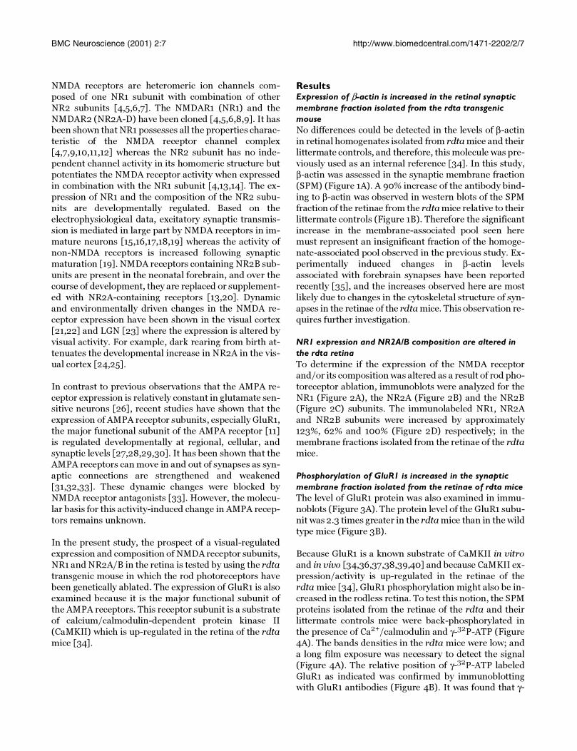
Phosphorylation of GluR1 is increased in the synaptic membrane fraction isolated from the retinae of rdta miceThe level of GluR1 protein was also examined in immu-
noblots (Figure 3A). The protein level of the GluR1 subu-
nit was 2.3 times greater in the rdta mice than in the wild
type mice (Figure 3B).
Because GluR1 is a known substrate of CaMKII in vitro
and in vivo [34,36,37,38,39,40] and because CaMKII ex-
pression/activity is up-regulated in the retinae of the
rdta mice [34], GluR1 phosphorylation might also be in-
creased in the rodless retina. To test this notion, the SPM
proteins isolated from the retinae of the rdta and their
littermate controls mice were back-phosphorylated in
the presence of Ca2+/calmodulin and γ-32P-ATP (Figure
4A). The bands densities in the rdta mice were low; and
a long film exposure was necessary to detect the signal
(Figure 4A). The relative position of γ-32P-ATP labeled
GluR1 as indicated was confirmed by immunoblottingwith GluR1 antibodies (Figure 4B). It was found that γ-
Figure 1Western blot analysis of the immunolabeling of β-actin in the SPM fraction of the mouse retina. (A) Immunob-lots of 6 µg of retinal SPM protein from control animals and the rdta mice were incubated with anti-β-actin antibody. Theimmunolabeling densities were calculated as the integrated density values (IDV) and plotted in B. (B) The graph presents thechanges in the antibody binding to β-actin in three littermate control and three rdta mice. The average density calculated asIDV for the littermate controls was taken as 100%. The immunolabeling of β-actin in the SPM fraction is significantly increasedin the rdta retina. Values are means ± S.E.M (n = 3), P < 0.05, one way ANOVA with Bonferroni correction.
32P-ATP labeling of GluR1 by this procedure was greater
in the retinae of control mice than in the retina of the
rdta mice (Figure 4A), whereas the expression of GluR1
was increased in the rdta mice relative to their littermate
controls (Figure 4B). These data indicate that GluR1
phosphorylation in vivo was enhanced in the rdta reti-
nae relative to their littermate controls. This result was
confirmed using an additional technique. GluR1 was im-
munoprecipitated with anti-GluR1 antibody and then
subjected to in vitro phosphorylation (Figure 4C). To-gether, these data indicate that in vivo, the amount of
GluR1 and the phosphorylation of GluR1 are simultane-
ously enhanced in retinal synaptic fractions isolated
from the retinae of rdta mice.
DiscussionTo our knowledge, this is the first study to demonstrate
that the protein levels of NMDA and AMPA receptors are
altered in the retina when the rod photoreceptors are ab-
sent. Western blots analyses demonstrate that the
amounts of NR1, NR2A/B and β-actin are significantlyincreased in the retinae of rdta mice compared to their
Figure 2Western blots analyses of NMDA receptor subunits in the mouse retina. (A),(B) and (C) Immunoblots of 6 µg ofretinal SPM fraction proteins from controls and the rdta mice were first incubated with anti-NR1 antibody (A), and subse-quently reprobed with anti-NR2A (B) and NR2B (C) antibodies, respectively. The immunolabeling densities were calculated asthe integrated density values (IDV), and plotted in D. (D) The graph presents the changes in the antibody binding to NR1,NR2A and NR2B in littermate controls and the rdta mice. The average density calculated as IDV for the littermate controlswas taken as 100. Values are means ± S.E.M (n = 3), P < 0.05, one way ANOVA with Bonferroni correction.
littermate controls. Furthermore, the amount of GluR1
expression and its phosphorylation are simultaneously
increased in the retinae of the rdta mice. As the retinalthickness of the rdta animals is reduced by as much as
30% relative to littermate control animals, the estimated
increases in synaptic relevant molecules as a percentage
of total retinal protein could be over-estimated by at leastthis amount. Even accounting for such a correction the
Figure 3Western blot analysis of GluR1 in the SPM fraction of the mouse retina. (A) Immunoblots of 6 µg of retinal SPMprotein from control animals and the rdta mice were incubated with anti-GluR1 antibody. The immunolabeling densities werecalculated as the integrated density values (IDV) and plotted in B. (B) The graph presents the changes in the antibody bindingto GluR1 in the littermate control and rdta mice. The average density calculated as IDV for the littermate controls was taken as100%. The immunolabeling of GluR1 in the SPM fraction is significantly increased in the rdta retina. Values are means ± S.E.M (n= 3), P < 0.05, one way ANOVA with Bonferroni correction.
changes reported here remain significant. A previous
study using similar western blot demonstrations of an el-
evation in calcium/calmodulin kinase II protein, a mole-
cule intimately related to those studied here, was shown
to be at least qualitatively correct using an independent
immunohistochemical assessment [34].
The results presented here are consistent with studies
demonstrating activity-based changes in receptors as re-
ported for other parts of the nervous system
[21,23,24,25,41]. NR1 expression is not altered in the
dark-reared visual cortex [24,25] but contrarily, an in-
traocular injection of tetrodotoxin (TTX) does increaseNR1-antibody binding in layer IV of the cortical column
driven by the blocked eye [41]. The latter study supports
the notion that neural activity is a regulating factor for
NR1 expression. It should not be surprising to find simi-
larities and differences between the experimental para-
digms of dark-rearing/visual cortex on the one hand, and
photoreceptor-ablation/retina on the other.
Electrophysiological and pharmacological studies have
shown that different subunit configurations comprising
the receptor confer different functional properties and
selectivity to the NMDA receptors [7,11,13,42,43]. For
example, the kinetics of the NMDA receptors are regulat-
ed by the combinatorial associations of the NR1 and NR2subunits [44]. Thus, mature NMDA receptors, which
Figure 4Back -phosphorylation of GluR1 in the retina of the rdta mice and their littermate controls. (A) Ten microgramsof SPM protein from the retinae of the rdta mice and their littermate controls were back-phosphorylated by endogenous CaM-KII in the presence of Ca2+/CaM. The samples were then electrophoresed by SDS-PAGE and subsequently transferred tonitrocellular membrane. The phosphorylated bands were visualized by autoradiography. The in vitro phosphorylation washigher in the littermate controls than in the rdta mice. (B) The same blots were then incubated with anti-GluR1 antibody andvisualized by alkaline phosphatase conjugated secondary antibody and NBI/BICP substrate. The molecular weight of GluR1 isindicated. The density for GluR1 was elevated in the rdta mice relative to the littermate controls. (C) GluR1s were immuno-precipitated after the back-phosphorylation and subjected to SDS-PAGE. The gel was dried and the image was visualized byautoradiography. These data indicate that the phosphorylation of GluR1 is increased in vivo in the retina of the rdta mice rela-tive to their littermate controls.
AcknowledgmentsThe authors thank Dr. Maureen A McCall for providing the rdta transgenic mouse line. This work was supported by NSF-EPS-9874764, Kentucky Li-ons Eye Research Foundation and an unrestricted grant from Research to Prevent Blindness, Inc.
References1. Barnstable CJ: Monoclonal antibodies which recognize differ-
ent cell types in the retina. Nature 1980, 286:231-2352. Marc RE, Murry RF, Basinger SF: Pattern recognition of amino
acid signatures in retinal neurons. J Neurosci 1995, 15:5106-51293. Mayer ML, Westbrook GL: Permeation and block of N-methyl-
D-aspartic acid receptor channels by divalent cations inmouse cultured central neurones. J Physiol 1987, 394:501-527
4. Ishii T, Moriyoshi K, Sugihara H, Sakurada K, Kadotani H, Yokoi M,Akazawa C, Shigemoto R, Mizuno N, Masu M, Nakanishi S: Molecu-lar characterization of the family of N-methyl-D-aspartatereceptor subunits. J Biol Chem 1993, 268:2836-2843
5. Katsuwada T, Kashiwabuchi N, Mori H, Sakimura K, Kushiya E, ArakiK, Megure H, Masaki H, Kumanishi T, Arakawa M, Mishina M: Molec-ular diversity of the NMDA receptor channel. Nature 1992,358:36-41
6. Meguro H, Mori H, Araki K, Kushiya E, Kutsuwada T, Yamazaki M,Kumanishi T, Arakawa M, Sakimura K, Mishina M: Functional char-acterization of a heteromeric NMDA receptor channel ex-pressed from cloned cDNAs. Nature 1992, 357:70-74
7. Monyer H, Sprengel R, Schoepfer R, Herb A, Higuchi M, Lomeli H,Burnashev N, Sakmann B, Seeburg PH: Heteromeric NMDA re-ceptors: molecular and functional distinction of subtypes. Sci-ence 1992, 256:1217-1221
8. Ikeda K, Nagasawa M, Mori H, Araki K, Sakimura K, Watanabe M, In-oue Y, Mishina M: Cloning and expression of the 4 subunits ofthe NMDA receptor channel. FEBS Lett 1992, 313:34-38
9. Moriyoshi K, Masu M, Ishii T, Shigemoto R, Mizuno N, Nakanishi S:Molecular cloning and characterization of the rat NMDA re-ceptor. Nature 1991, 354:31-37
10. Karp SJ, Masu M, Eki T, Ozawa K, Nakanishi S: Molecular cloningand chromosomal localization of the key subunit of the hu-man N-methyl-D-aspartate receptor. J Biol Chem 1993,268:3728-3733
11. Nakanishi S: Molecular diversity of glutamate receptors andimplications for brain function. Science 1992, 258:597-603
12. Sugihara H, Moriyoshi K, Ishii T, Masu M, Nakanishi S: Structuresand properties of seven isoforms of the NMDA receptor gen-erated by alternative splicing. Biochem Biophys Res Commun 1992,185:826-832
13. Monyer H, Burnashev N, Laurie DJ, Sakmann B, Seeburg PH: Devel-opmental and regional expression in the rat brain and func-tional properties of four NMDA receptors. Neuron 1994,12:529-540
14. Sheng M, Cummings J, Rolden LA, Jan YN, Jan LY: Changing subunitcomposition of heteromeric NMDA receptors during devel-opment of rat cortex. Nature 1994, 368:144-147
15. Durand GM, Kovalchuk Y, Konnerth A: Long-term potentiationand functional synapse induction in developing hippocam-pus. Nature 1996, 381:71-75
16. Isaac JT, Nicoll RA, Malenka RC: Evidence for silent synapses: im-plications for the expression of LTP. Neuron 1995, 15:427-34
17. Kullmann DM: Amplitude fluctuations of dual-component EP-SCs in hippocampal pyramidal cells: implications for long-term potentiation. Neuron 1994, 12:1111-1120
18. Liao D, Hessler N, Malinow R: Activation of postsynaptically si-lent synapses during pairing-induced LTP in CA1 region ofhippocampal slice. Nature 1995, 375:400-404
19. Wu GY, Malinow R, Cline HT: Maturation of a central glutama-tergic synapse. Science 1996, 274:972-976
NR2B) in the cat visual cortex and the effects of dark rearing.Mol Brain Res 2000, 78:196-200
25. Quinlan EM, Philpot BD, Huganir RL, Bear M: Rapid, experience-dependent expression of synaptic NMDA receptors in visualcortex in vivo. Nature Neurosci 1999, 2:352-357
26. Barinaga M: New clues to how neurons strengthen their con-nections. Science 1999, 284:1755-1757
27. Archibald K, Molnar E, Henley JM: Differential changes in the sub-cellular distribution of alpha-amono-3-hydroxy-5-methyl-4-isoxazole propionate and N-methyl-D-asparate receptors inneonate and adult rat cortex. Neurosci Lett 1999, 270:49-52
28. Jakowec MW, Jackson-Lewis V, Chen X, Langston JW, Przedborski S:The postnatal development of AMPA receptor subunits inthe basal ganglia of the rat. Dev Neurosci 1998, 20:19-33
29. Martin LJ, Furuta A, Blackstone CD: AMPA receptor protein indeveloping rat brain: glutamate receptor-1 expression andlocalization change at regional, cellular, and subcellular lev-els with maturation. Neuroscience 1998, 83:917-928
30. Standley S, Wagle N, Baudry M: Developmental changes in sub-cellular AMPA/GluR receptor populations in rat forebrain.Dev Brain Res 1998, 107:277-283
31. Carroll RC, Beattie EC, Xia H, Luscher C, Altschuler Y, Nicoll RA,Malenka RC, von Zastrow M: Dynamin-dependent endocytosisof ionotropic glutamate receptors. Proc Natl Acad Sci USA 1999,96:14112-14117
32. Carroll RC, Lissin DV, von Zastrow M, Nicoll , Malenka RC: Rapidredistribution of glutamate receptors contributes to long-term depression in hippocampal cultures. Nat Neurosci 1999,2:454-460
33. Shi SH, Hayashi Y, Petralia RS, Zaman SH, Wenthold RJ, Svoboda Kand, Malinow R: Rapid spine delivery and redistribution ofAMPA receptors after synaptic NMDA receptor activation.Science 1999, 284:1811-1816
34. Liu OL, Li G, McCall AM, Cooper NGF: Photoreceptor regulatedexpression of Ca2+/calmodulin-dependent protein kinase IIin the mouse retina. Mol Brain Res 2000, 82:150-166
35. Wyneken U, Smalla KH, Marengo JJ, Soto D, de la Cerda A, Tischmey-er W, Grimm R, Boeckers TM, Wolf G. Orrego F, Gundelfinger ED:Kainate-induced seizures alter protein composition and N-methyl-D-aspartate receptor function of rat forebrain post-synaptic densities. Neuroscience 2001, 102:65-74
36. Barria A, Derkach V, Soderling T: Identification of the Ca2+/cal-modulin-dependent protein kinase II regulatory phosphor-ylation site in the alpha-amino-3-hydroxyl-5-methyl-4-isoxazole-propionate-type glutamate receptor J Biol Chem1997, 272:32727-32730
37. Barria A, Muller D, Derkach V, Griffith LC, Soderling TR: Regulatoryphosphorylation of AMPA-type glutamate receptors byCaM-KII during long-term potentiation. Science 1997,276:2042-2045
38. Hayashi Y, Ishida A, Katagiri H, Mishina M, Fujisawa H, Manabe T,Takahashi T: Calcium and calmodulin-dependent phosphor-ylation of AMPA type glutamate receptor subunits by en-dogenous protein kinases in the post-synaptic density. MolBrain Res 1997, 46:338-342
39. McGlade-McCulloh E, Yamamoto H, Tan SE, Brickey DA, SoderlingTR: Phosphorylation and regulation of glutamate receptorsby calcium/calmodulin-dependent protein kinase II. Nature1993, 362:640-642
40. Tan SE, Wenthold RJ, Soderling TR: Phosphorylation of AMPA-type glutamate receptors by calcium/calmodulin-dependentprotein kinase II and protein kinase C in cultured hoppocam-pal neurons. J Neurosci 1994, 14:1123-1129
41. Catalano S, Chang CK, Shatz CJ: Activity-dependent regulationof NMDAR1 immunoreactivity in the developing visual cor-tex. J Neurosci 1997, 17:8376-8390
43. McBain CJ, Mayer ML: N-methyl-D-aspartic acid receptor struc-ture and function. Physiol Res 1994, 74:723-760
44. Williams K, Russell SL, Shen YM, Molinoff PB: Developmentalswitch in the expression of NMDA receptors occurs in vivoand in vitro. Neuron 1993, 10:267-278
45. Ehinger B, Ottersen OP, Storm-Mathisen J, Dowling JE: Bipolar cellsin the turtle retina are strongly immunoreactive for gluta-mate. Proc Batl Acad Sci. USA 1988, 85:8321-8325
46. Jojich L, Pourcho RG: Glutamate immunoreactivity in the catretina: a quantitative study. Vis Neurosci. 1996, 13:117-133
47. Kalloniatis M, Fletcher EL: Immunocytochemical localization ofthe amino acid neurotransmitters in the chicken retina. JComp Neurol 1993, 336:174-193
48. Van Haesendonck E, Missotten L: Glutamate-like immunoreac-tivity in the retina of a marine teleost, the dragonet. NeurosciLett 1990, 111:281-286
49. Yang CY, Yazulla S: Glutamate- GABA-, and GAD-immunore-activities co-localize in bipolar cells of tiger salamander reti-na. Vis Neurosci 1994, 11:1193-1203
50. Matus A, Brinkhaus H, Wagner U: Actin dynamics in dendriticspines: a form of regulated plasticity at excitatory synapses.Hippocampus 2000, 10:555-560
51. Ter-Margarian A, Djamgoz MB: Cytochalasin inhibits light-de-pendent synaptic plasticity of horizontal cells in teleost reti-na. Neurosci Lett 1992, 147:131-135
52. Fletcher EL, Hack I, Brandstatter JH, Wassle H: Synaptic localiza-tion of NMDA receptor subunits in the rat retina. J Comp Neu-rol 2000, 420:98-112
53. Suen PC, Wu K, Xu JL, Lin SY, Levine ES, Black IB: NMDA receptorsubunits in the postsynaptic density of rat brain: expressionand phosphorylation by endogenous protein kinase. Mol BrainRes 1998, 59:215-228
54. Hall RA, Soderling TR: Quantification of AMPA receptor sur-face expression in cultured hippocampal neurons. Neurosc1997, 78:361-371
55. Leonard AS, Hell JW: Cyclic AMP-dependent protein kinaseand protein kinase C phosphorylate N-methyl-D-aspartatereceptors at different sites. J Biol Chem 1997, 272:12107-12115
56. Gardoni F, Bellone C, Cattabeni F, Di Luca M: Protein kinace C ac-tivation modulates α-calmodulin kinase II binding to NR2Asubunit of N-Methyl-D-Aspartate receptor complex J biolchem 2001, 276:7609-7613
57. Raymond LA, Blackstone CD, Huganir RL: Phosphorylation ofamino acid neurotransmitter receptors in synaptic plastici-ty. Trends Neurosci 1993, 16:147-153
59. Swope SL, Moss SJ, Blackstone CD, Huganir RL: Phosphorylation ofligand-gated ion channels: a possible mode of synaptic plas-ticity. FASEB J 1992, 6:2514-2523
60. Swope SL, Moss SJ, Raymond LA, Huganir RL: Regulation of ligand-gated ion channels by protein phosphorylation. Adv SecondMessenger Phosphoprotein Res 1999, 33:49-78
61. Lee HK, Barbarosie M, Kameyama K, Bear MF, Huganir RL: Regula-tion of distinct AMPA receptor phosphorylation sites duringbi-directional synaptic plasticity. Nature 2000, 405:955-999
62. Vinade L, Dosemeci A: Regulation of the phosphorylation stateof the AMPA receptor GluR1 subunit in the postsynapticdensity. Cell Mol Neurobiol 2000, 20:451-463
63. Bliss TV, Collingridge GL: A synaptic model of memory: long-term potentiation in the hippocampus. Nature 1993, 361:31-39
64. Pires RS, Ferro ES, Britto LRG: Expression of the AMPA-typeglutamate receptor subunits in the chick optic tectumchanges biphasically after retinal deafferentation. Brain Res1998, 810:283-287
65. Pires RS, Reboucas NA, Duroisin RM, Britto LR: Retinal lesions in-duce differential changes in the expression of flip and flop iso-forms of the glutamate receptor subunit GluR1 in the chickoptic tectum. Mol Brain Res 2000, 76:341-346
66. Bowes C, Li T, Frankel WN, Danciger M, Coffin JM, Applebury ML,Farber DB: Localization of a retroviral element within the rdgene coding for the beta subunit of cGMP phosphodieste-rase. Proc Natl Acad Sci USA 1993, 90:2955-2959
67. Duvoisin RM, Zhang C, Hamassaki-Britto DE, Britto LRG: Changesin expression of glutamate receptor subunits following pho-toreceptor degeneration in the rd retina. Neurosci Lett 1995,183:83-86
68. McCall MA, Gregg RG. Kerriman K, Goto Y, Peachey NS, StanfordLR: Morphological and physiological consequences of the se-lective elimination of rod photoreceptors in transgenic mice.Exp Eye Res 1996, 63:35-50
69. Laabich A, Cooper NGF: Regulation of calcium/calmodulin-de-pendent protein kinase II in the adult rat retina is mediated
by ionotropic glutamate receptors. Exp Eye Res 1999, 68:703-713
70. De Robertis E: Ultrastructure and cytochemistry of the synap-tic region. The macromolecular components involved innerve transmission are being studied. Science 1967, 156:907-914
71. Caputi A, Gardoni F, Cimino M, Pastorino L, Cattabeni F, Luca MD:CaMKII-dependent phosphorylation of NR2A and NR2B isdecreased in animals characterized by hippocampal damageand impaired LTP. Eur J Neurosci 1999, 11:141-148
Publish with BioMedcentral and every scientist can read your work free of charge
"BioMedcentral will be the most significant development for disseminating the results of biomedical research in our lifetime."
Paul Nurse, Director-General, Imperial Cancer Research Fund
Publish with BMc and your research papers will be:
available free of charge to the entire biomedical community
peer reviewed and published immediately upon acceptance
cited in PubMed and archived on PubMed Central
yours - you keep the copyright
[email protected] your manuscript here:http://www.biomedcentral.com/manuscript/


![[AICHA LAKHSSASS] - soa.utexas.edu · [AICHA LAKHSSASS] Phone 737-781-1185 Email aicha.lakhssass@gmail.com LinkedIN linkedin.com/in/aichalakhssass 2018 University of Texas at Austin](https://static.documents.pub/doc/80x56/5b98c97309d3f2ef798cb6c8/aicha-lakhssass-soa-aicha-lakhssass-phone-737-781-1185-email-aichalakhssassgmailcom.jpg)


